Embed Size (px)
Citation preview
TATTOO PIGMENTS IN SKIN:
Determination and Quantitative Extraction of Red Tattoo Pigments
Dissertation
Zur Erlangung des Doktorgrades der Naturwissenschaften
(Dr. rer. nat.)
an der Fakultät für Chemie und Pharmazie
der Universität Regensburg
vorgelegt von
Eva Engel aus Wittislingen
2007
The experimental part of this work was carried out between August 2004 and
July 2007 at the Institute for Organic Chemistry at the University of Regensburg
under the supervision of Prof. Dr. B. König.
The PhD - thesis was submitted on: 03. August 2007
The colloquium took place on: 14. September 2007
Board of Examiners: Prof. Dr. A. Buschauer (Chairman)
Prof. Dr. B. König (1st Referee)
PD Dr. W. Bäumler (2nd Referee)
Prof. Dr. A. Göpferich (Examiner)
Danksagung
Mein besonderer Dank gilt Herrn Prof. Dr. B. König für die Überlassung des
interessanten und vielseitgen Themas, die ausgezeichneten
Arbeitsbedingungen, seine Unterstüzung und das stets mit Anregungen und
Diskussionen verbundene Interesse an dieser Arbeit.
Herrn Prof. Dr. M. Landthaler danke ich für die Föderung und Unterstützung des
Forschungsprojektes.
Für die Möglichkeit eines dreimonatigen Aufenthaltes am National Center for
Toxicological Research (NCTR) der US Food and Drug Administration (FDA) in
Jefferson, Arkansas, bedanke ich mich bei Dr. Paul C. Howard.
Besonders bedanken möchte ich mich vor allem bei PD Dr. Wolfgang Bäumler
für das spannende und aufregende Thema, die vielseitige und unermüdliche
Untersützung und Betreuung, sowie sein unendliches Engagement an meiner
Arbeit. – Es leben die Gallier!
Herrn Dr. Rudolf Vasold danke ich für die hervorragende Betreuung meiner
gesamten Arbeit, die Hilfestellungen bei praktische Arbeiten, das Übermitteln
der unzähligen HPLC- und GC-Fertigkeiten, die Fahrten und Flüge zu den
gemeinsamen Fortbildungen sowie seine vielen Süßigkeiten und seinen
unvergesslichen Kaffee. – I appreciate it!
Für die finanzielle Unterstützung gilt mein Dank der Deutschen
Forschungsgemeinschaft (DFG), für die zweijährige Förderung des Tattoo-
Projektes, dem Oak Ridge Insitute for Sciene and Education (ORISE) des US
Department of Engery für die Föderung des dreimonatigen
Forschungsaufenthalts am National Center for Toxicological Research
(NCTR) der FDA in Jefferson, Arkansas, und der Klinik und Poliklinik für
Dermatologie für die Finanzierung vieler Tagungsreisen.
Den Mitarbeitern der Zentralen Analytik der Fakultät für Chemie und Pharmazie
danke ich für die stets schnelle und gewissenhafte Durchführung der
analytischen Messungen. Insbesonderen Herrn Josef Kiermeier für Messung
der Massenspektren und unzähligen LC/MS-Kupplungen, sowie die vielen
fachlichen Ratschläge.
Frau E. Liebl, Herrn Dr. W. Braig, Frau Dr. C. Braig, Frau H. Leffler-Schuster,
Frau S. Graetz, Frau B. Bazidura, Frau R. Hoheisel und allen übrigen
Festangestellten des Lehrstuhls König danke ich für ihre Unterstützung.
Den Mitarbeitern der HPLC-Abteilung, Frau Simone Strauß und Herrn Ernst
Lauterschlager, danke ich für ihre stete Hilfsbereitschaft.
Großer Dank geht an Francesco Santarelli für die Probenvorbereitungen, seine
Fähigkeit in allen praktischen Dingen und die schöne gemeinsame Zeit sowie
die italienischen Momente im Labor. – Danke Franz!
Frau Helga Leffler-Schuster danke ich für ihre Ratschläge und ihre Fürsorge
sowie ihre erlebnisreichen Berichte aus aller Welt.
Für ihr großes Engagement während ihrer Zulassungsarbeiten danke ich
Rüdiger Schraml, Matthias Gottschalk, Andrea Spannberger und Katharina
Gastl.
Auch bei Karin Lehner und Christina Högner bedanke ich mich für die schöne
Zeit im und außerhalb des Labors.
Für die sehr gute Zusammenarbeit im Rahmen der Forschungsprojekte danke
ich Herrn Dr. Tim Maisch und Frau Dr. Heidi Ulrich (Klinik und Poliklinik für
Dermatologie, Universität Regensburg) sowie allen Mitarbeitern des
Forschungsbaus.
Herrn Dr. Jürgen Odermatt vom Zentrum für Holzwirtschaft der Universität
Hamburg danke ich für die Einladung zum Fachmeeting „Pyrolyse“ sowie seine
Hilfsbereitschaft und fachlichen Ratschläge im Bereich der Pyrolyse-GC.
Allen aktuellen wie ehemaligen Mitarbeitern des Lehrstuhls danke ich für die
gute Zusammenarbeit und das sehr angenehme Arbeitsklima – vor und nach
Feierabend. Besonderer Dank gilt dabei:
Dr. Giovanni Imperato für unvergessene Toskana-Urlaube, fantastische
Abendessen, die Steigerung der allgemeinen Heiterkeit und die vielen
gemeinsamen Erlebnisse.
Dr. Stefan Ritter für alle kulinarischen Abende sowie die vielen sonstigen
gemeinsamen Unternehmungen und schwäbischen Momente.
Dr. Stefan Miltschitzky für alle gemeinsamen kulinarischen Unternehmungen,
das Erlebnis die USA-Botschaft in Frankfurt besuchen zu dürfen und die
gemeinsame Zeit am Ende unsere USA-Aufenthalts. – It’s awesome!
Dr. Christoph Bonauer für seine Unterstützung bei Bewerbungsangelegenheiten
und seine Gastfreundschaft.
Michael Egger, Harald Schmaderer, Stefan Stadlbauer für ihre große
Hilfsbereitschaft und Unterstützung bei Problemen aller Art und für die vielen
lustigen Abende.
Dr. Noemi Colombo und Maria Elena Silva danke ich für die gemeinsamen
Sportkurse und die wuderschönen italienischen Abende.
Herzlicher Dank geht an Dr. Rudolf Vasold, Katharina Gastl, Daniel Vomasta
und Michael Egger für das Korrekturlesen dieser Arbeit.
Meinen Studienkolleginnen Saskia Wällisch, Melanie Grosser, Corinna Gerstl
danke ich für ihre Freundschaft und alle gemeinsamen Unternehmungen und
Erlebnisse während der gesamten Studienzeit in Regensburg.
Meiner Schwester Sabine danke dafür, dass sie immer für mich da ist.
Mein persönlicher Dank gilt meinem lieben Daniel für seine Liebe, seine
Unterstützung und sein Verständnis zu jeder Zeit sowohl zuhause wie auch im
Labor. Ebenso danke ich seiner Familie, die mir ein zweites Zuhause bot.
Zuletzt, aber vor allem, danke ich meiner Familie für ihre großartige
Unterstützung, ihre Aufmunterungen und den großen Rückhalt während meines
gesamten Studiums.

W ie Neugier steht immer an erster Stelle eines Problems,
das gelöst werden will.
- Galileo Galilei -

Für Daniel &
meine Familie
Table of Contents
1. Establishment of an Extraction Method for the Recovery of Tattoo Pigments from Human Skin using HPLC Diode Array Technology..................................................................... 1
1.1. Introduction.................................................................. 1
1.2. Materials and Methods ................................................ 4
1.3. Results and Discussion ............................................. 10
1.4. Conclusions............................................................... 18
1.5. References................................................................ 19
2. Modern Tattoos Cause High Concentrations of Hazardous Pigments in Human Skin.................................... 22
2.1. Introduction................................................................ 22
2.2. Materials and Methods .............................................. 24
2.3. Results and Discussion ............................................. 27
2.4. Potential Health Problems......................................... 31
2.5. References................................................................ 33
3. Photochemical Cleavage of a Tattoo Pigment by UVB Radiation or Natural Sunlight................................................ 35
3.1. Introduction................................................................ 35
3.2. Materials and Methods .............................................. 38
3.3. Results and Discussions ........................................... 40
3.4. Conclusions............................................................... 50
3.5. References................................................................ 51
4. Tattoo Pigments in Skin: Concentration, Transportation and Light Induced Decomposition of an Azo Pigment using SKH-1 Mouse Model .................................................... 53
4.1. Introduction................................................................ 53
4.2. Materials and Methods .............................................. 54
4.3. Results ...................................................................... 58
4.4. Discussion ................................................................. 62
4.5. Conclusion................................................................. 69
4.6. References................................................................ 70
5. Modern Tattoos Contain Azo Pigments: an in-vivo Proof of Pigment Red 22 and Pigment Red 170............................. 72
5.1. Introduction................................................................ 72
5.2. Materials and Methods .............................................. 73
5.3. Results and Discussion ............................................. 75
5.5. Conclusion................................................................. 80
5.5. References................................................................ 81
6. Azo Pigments and a Basall Cell Carcinoma at the Thumb ..................................................................................... 82
6.1. Introduction................................................................ 82
6.2. Case Report .............................................................. 82
6.3. Materials and Methods .............................................. 83
6.4. Results ...................................................................... 85
6.5. Discussion ................................................................. 88
6.6. References................................................................ 90

7. Abbreviations ......................................................................... 91
8. Summary................................................................................. 93
9. Zusammenfassung................................................................. 97
10. Appendix............................................................................... 102
1. Establishment of an Extraction Method for the Recovery of Tattoo Pigments from Human Skin using HPLC Diode
Array Technology∗
1.1. Introduction
The number of tattooed individuals increased significantly,1-3 especially among
youth.3-8 In the United States, ~16% of the population is tattooed, whereas in
Europe it is ~10%. In the past, people used inorganic metal salts, containing
chromium, manganese, mercury, and cobalt. Due to their brilliance and their
great insolubility, today many azo pigments are used for tattooing. A significant
number of these azo pigments are organic pigments manufactured primarily for
other uses such as printing, painting cars, and coloring other consumer
products. Tattoo inks contain many components. Frequently, tattoo inks are
pigment mixtures and may contain components such as titanium dioxide for
lightening the ink shade. Precursors and byproducts of pigment synthesis may
also be present. In addition, diluents used to suspend pigments may be
complex mixtures.9,10
Tattoo colorants are also used as permanent make-up make up for application
on the eyelid, eyebrow, and lip.11 Many tattoo pigments are manufactured for
other intended uses and may not have an established history for safe use in
tattooing.9,10 Currently, there is no legal requirement for listing ingredients,
including pigments, on the labeling of tattoo inks. The U.S. Food and Drug
administration considers the pigments used in tattoo inks to be color additives,
which require pre-market approval. Currently, no pigments have been approved
for use in tattoo inks.12
In the process of tattooing, the pigment suspension is initially deposited on the
skin and then implanted by needle punctures. Some of the deposited pigment
may be recognized by macrophages as foreign bodies and carried from the site
of the tattoo via the lymphatic or circulatory system. As a result, tattoo pigments
∗ Results of this chapter have been published: Engel, E.; Santarelli, F.; Vasold, R.; Ulrich, H.; Maisch, T.; König,B.; Landthaler, M.; Gopee, N.V.; Howard, P.C.; Bäumler, W. Anal. Chem. 2006, 78, 6440.
1
may frequently be found in lymph nodes.13 Pigment remaining at the site of the
tattoo is usually found in the dermis and may be found intracellularly. Deposition
of the pigment into the dermis results in the permanence usually associated
with tattoos. Due to their insolubility, tattoo pigments are resistant to enzymatic
degradation in the skin. Frequently, pigments in a tattoo are aggregated into
crystals with a size ranging from about 0.1 to 10 μm.14
Because of an improved self-image or social stigmatization many tattooed
individuals undergo a therapy of tattoo removal by using predominantly Q-
switch lasers.15
In fact, high intensities and short pulse durations of a laser are necessary to
destroy selectively the pigments and not the surrounding skin.16 After being
absorbed in the pigment molecule, the energy of the laser light is converted to
heat or breaks chemical bonds inside the molecule. Additionally, the ultrashort
heating may lead to the disruption of the pigment particle. The laser irradiation
changes the shape and the size of the tattoo particles abruptly as proved by
histology.17
It is well known that an increase of temperature in a number of azo dyes above
280 °C forms 3,3’-dichlorobenzidine,18 a proven genotoxin toward human
lymphocytes.19 Laser irradiation of the two widely used azo compounds,
Pigment Red 22 (PR 22) and Pigment Red 9 (PR 9) resulted in the
photodecomposition products 2-methyl-5-nitroaniline (MNA), 4-nitrotoluene
(NT), 2,5-dichloraniline (DCA), and 1,4-dichlorobenzene (DCB)15 (Figure 1). NT
is toxic as shown with human lymphocytes.20 5-Nitro-o-toluidine, which is also
designated to MNA, may cause liver dysfunction as shown with workers from a
hair dye factory.21 Additionally, MNA and other di-nitro-toluenes showed
greatest mutagenic activity toward Salmonella typhimurium YG as
demonstrated by Sayama et al.22 DCB has been reported to cause tumors in
kidney of male rats and in liver of male and female mice,23 whereas DCA was
capable of inducing nephrotoxicity in rats.24
However, toxicity and mutagenic activity of molecules is correlated to the
respective concentration taken up by a human. Presently, little is known about
the concentrations of pigments or decomposition products in tattooed sites of
the skin. In light of the millions of people with tattoos, scientific investigations on
2
cutaneous concentrations of pigments and byproducts are critically needed to
evaluate risks associated with tattooing.
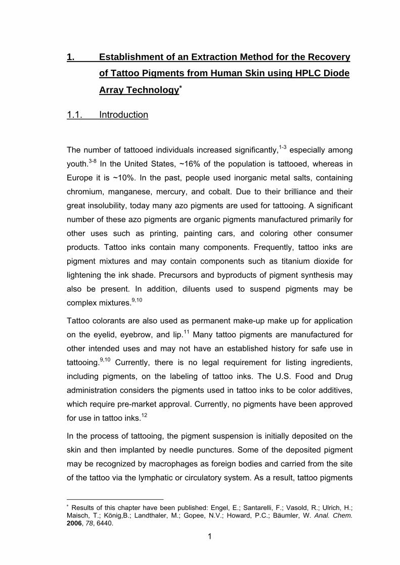
R1
NN
OH NHO
R3
R2
R6
R4 R5
R1
R3R2
+ N2 +
OH NHO
R6
R5R4
R1NH2
R3R2
+
OH NHO
NH2
R6
R5R4
R1 R2 R3 R4 R5 R6 name
Cl H Cl H H OCH3 PR 9
CH3 H NO2 H H H PR 22
Figure 1: Chemical structure of PR 22 and PR 9 used as coloring pigments in Cardinal Red
(CR) and I8, respectively. For both pigments, the possible decomposition pattern is shown.
Additional decomposition products may occur (chlorine, oxidation). The substituents of the
pigment molecules are listed in the table portion.
As a first step, we established a method for the quantitative extraction of tattoo
pigments and their respective decomposition products from different media. The
extraction step was validated through recovery experiments. That is, pigments
such as PR 9 and PR 22 synthesized in high pure quality or decomposition
products were added to aqueous suspensions or homogenized skin at a known
concentration, extracted, and quantified by using high-performance liquid
chromatography diode array (HPLC-DAD) technology.
3
1.2. Materials and Methods
Tattoo Pigments.∗ PR 22 (C.I. 12315, CAS 6448-95-9) and PR 9 (C.I. 12460,
CAS 6410-38-4) were synthesized via azo coupling according to Cook et al.25
The starting material for the synthesis of PR 22 was naphthol AS (NAS; 99%,
Sigma-Aldrich, Steinheim, Germany) and MNA (99%, Aldrich Chemical Co.,
Inc., Milwaukee, WI). PR 9 was synthesized using methoxynaphthol AS (m-
NAS; 98%, Aldrich Chemical Co., Inc.) and DCA (99%, Acros Organics). Both
raw products were purified by slurrying in acetonitrile (LiChroSolv, Merck,
Darmstadt Germany).
Stock Solutions. For the stock solutions of the non-volatile compounds MNA,
NAS, PR 22, m-NAS, and PR 9 with the following concentrations were
dissolved in chloroform: MNA (0.5 mg/mL), NAS (0.25 mg/mL), PR 22
(0.3 mg/mL), m-NAS (0.2 mg/mL), and PR 9 (0.3 mg/mL). The volatile
compounds were also dissolved in chloroform at the following concentrations:
NT (5.0 mg/mL) (>98%, Fluka, Buchs, Switzerland), DCA (10.0 mg/mL), and
DCB (4.0 mg/mL) (>99%, Fluka). The ISTD stock solution was 9,10-
diphenylanthracene (1.0 mg/mL acetonitrile/methylene chloride (1:1) (Oekanal,
Sigma-Aldrich, Seelze, Germany). All used solvents were of gradient grade
quality for liquid chromatography (LiChroSolv, Merck, Darmstadt, Germany).
Extraction from Solvents. One milliliter of the stock solution of the non-volatile
compounds was added to a 15-mL PP test tube (Cellstar, Greiner Bio-one,
Frickenhausen, Germany). The solvent was removed by blowing nitrogen
(2 bar, 20 min, 60 °C) (nitrogen 5.0, Linde Gas, Höllriegelskreuth, Germany).
One hundred microliters of the undiluted stock solution of the volatile
compounds was added. The tube was filled with either 5 mL of water, produced
by a Milli-Q Ultrapure water purification system (Millipore, Schwalbach,
Germany) or 5 mL of phosphate-buffered saline (PBS; Biochrom, Berlin,
Germany). Afterwards, 1 mL of methanol (LiChroSolv, Merck) was added. The
compounds were extracted with 3 mL of methylene chloride four times.
∗ PR 22 and PR 9 were synthesized by Matthias Gottschalk in his Zulassungsarbeit.
4
The total volume of the four extraction steps (each with 3 mL of methylene
chloride) was collected in a modified Kuderna-Danish concentrator (with
attached Snyder column). Then 200 µL of diethylene glycol dimethyl ether
(Diglyme) (Fluka, Deisenhofen, Germany), a low volatile liquid, was added. This
compound works as keeper; it enables the solvents to evaporate and
momentarily prevents vaporization of the volatile compounds (NT, DCA, DCB).
The solution was concentrated in the 200 µL of Diglyme under stirring and
heating to 60 °C for 20 min. Finally, the remaining solvent mixture of methylene
chloride and methanol was completely evaporated under continuing stirring,
elevated temperature (60 °C), and a gentle stream of nitrogen (2 bar, 20 min).
For the HPLC analysis, each sample, consisting of the extracted compounds
concentrated in 200 µL of keeper, was reconstituted in 1.7 mL of chloroform and
a 100-mL solution of internal standard. After the final step, the total volume of
each samples was 2 mL.
Enzymes. A total of 7500 units of collagenase (type VII, Sigma-Aldrich,
Taufkirchen, Germany) were dissolved in 1 mL of PBS (Biochrom). Proteinase
K was used as a ready-to-use solution (Dako Corp., Carpinteria, CA). ATL
buffer and proteinase K (> 600 AU/mL) were purchased from Qiagen (Hilden,
Germany).
Skin Preparation. Human skin was obtained from surgical excisions
(Department of Dermatology, University of Regensburg, Germany) and stored
at - 80 °C. For further treatment, the tissue was chopped up to slices with size
of ~1 cm2 and adipose tissue was removed by a scalpel. Each sample was
powdered in liquid nitrogen and added to Eppendorf cups (Eppendorf,
Wesseling-Berzdorf, Germany), and 400 µL of PBS was added. Proteins were
denatured by heating at 95 °C for 5 min according to Gaber et al.26 After cooling
to room temperature, 240 µL of collagenase solution (7500 units/mL), a 250-µL
aliquot of proteinase K ready-to-use solution (10 units/mL) and 250 µmL of PBS
were added. The suspension was stirred at 37 °C for 12 h. Subsequently, the
digested skin was centrifuged (10 min, 13000 rpm, 20 °C). A total of 180 µL of
buffer ATL and 20 µL of proteinase K were added to the pellet, mixed by
vortexing, and incubated at 55 °C for 2.5 hrs until the tissue was completely
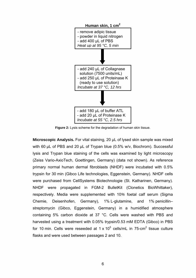
lysed (flow scheme, see Figure 2).
5
Human skin, 1 cm2
- add 240 µL of Collagnase solution (7500 units/mL) - add 250 µL of Proteinase K (ready to use solution) Incubate at 37 °C, 12 hrs
- remove adipic tissue - powder in liquid nitrogen - add 400 µL of PBS Heat up at 95 °C, 5 min
- add 180 µL of buffer ATL - add 20 µL of Proteinase K Incubate at 55 °C, 2.5 hrs
Figure 2: Lysis scheme for the degradation of human skin tissue.
Microscopic Analysis. For vital staining, 20 µL of lysed skin sample was mixed
with 60 µL of PBS and 20 µL of Trypan blue (0.5% w/v, Biochrom). Successful
lysis and Trypan blue staining of the cells was examined by light microscopy
(Zeiss Vario-AxioTech, Goettingen, Germany) (data not shown). As reference
primary normal human dermal fibroblasts (NHDF) were incubated with 0.5%
trypsin for 30 min (Gibco Life technologies, Eggenstein, Germany). NHDF cells
were purchased from CellSystems Biotechnologie (St. Katharinen, Germany).
NHDF were propagated in FGM-2 BulletKit (Clonetics BioWhittaker),
respectively. Media were supplemented with 10% foetal calf serum (Sigma
Chemie, Deisenhofen, Germany), 1% L-glutamine, and 1% penicillin–
streptomycin (Gibco, Eggenstein, Germany) in a humidified atmosphere
containing 5% carbon dioxide at 37 °C. Cells were washed with PBS and
harvested using a treatment with 0.05% trypsin/0.53 mM EDTA (Gibco) in PBS
for 10 min. Cells were reseeded at 1 x 10⁵ cells/mL in 75-cm2 tissue culture
flasks and were used between passages 2 and 10.
6
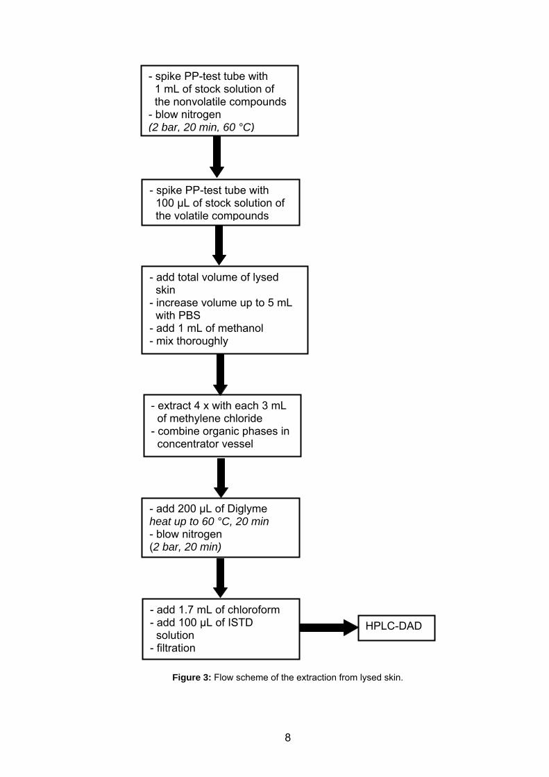
Extraction from Lysed Skin. For extracting skin, 1 mL of the stock solution of
the nonvolatile compounds was added to a PP test tube, and the solvent was
removed under a stream of nitrogen. A 100-µL sample of the stock solution
containing volatile compounds was added. Then, the solution resulting from
digestion of 1 cm2 of human skin was added to the test tube. The volume was
increased up to 5 mL with PBS. The aqueous skin phase was stabilized by
adding 1 mL of methanol. The resulting mixture was extracted 4 times with 3 mL
of methylene chloride.
The total volume of the four extraction steps (each with 3 mL of methylene
chloride) was collected in a modified Kuderna-Danish concentrator (with
attached Snyder column). Then 200 µL of Diglyme was added. The solution
was concentrated in the 200 µL of Diglyme under stirring and heating to 60 °C
for 20 min. Finally, the remaining solvent mixture of methylene chloride and
methanol was completely evaporated under continuing stirring, elevated
temperature (60 °C) and a gentle stream of nitrogen (2 bar, 20 min). For the
HPLC analysis, each sample, consisting of the extracted compounds
concentrated in the 200 µL keeper, was reconstituted in 1.7 mL of chloroform
and 100 µL of a solution of internal standard. After the final step, the total
volume of each sample was 2 mL (flow scheme, Figure 3).
7
- spike PP-test tube with 1 mL of stock solution of the nonvolatile compounds - blow nitrogen (2 bar, 20 min, 60 °C)
- spike PP-test tube with 100 µL of stock solution of the volatile compounds
- add total volume of lysed skin - increase volume up to 5 mL with PBS - add 1 mL of methanol - mix thoroughly
- extract 4 x with each 3 mL of methylene chloride - combine organic phases in concentrator vessel
- add 200 µL of Diglyme heat up to 60 °C, 20 min - blow nitrogen (2 bar, 20 min)
- add 1.7 mL of chloroform - add 100 µL of ISTD solution - filtration
HPLC-DAD
Figure 3: Flow scheme of the extraction from lysed skin.
8
HPLC Analysis. The samples were filtered using a PTFE filter (Chromafil, O-
20/15, organic, pore size 0.2 mm; Machery-Nagel, Düren, Germany). A 10-µL
sample was analyzed using a model 1100 HPLC (Agilent
Technologies,Waldbronn, Germany) fitted with a C18 analytical column
(Phenomenex Luna, particle size 3 µm, 150 x 4.60 mm, Aschaffenburg,
Germany) and DAD. The data were analyzed using a HPLC-3D ChemStation
Rev. B.01.01. Gradient elution was done with water (0.0059 w/v% trifluoroacetic
acid) (solvent A) and acetonitrile (solvent B) at a constant flow rate of
1.0 mL/min. A gradient profile with the following proportions of solvent B was
applied [t (min), % B]: (0, 10), (20, 95), (50, 95). The chromatograms were
monitored with wavelength switching [t (min), λ (nm)]: (0, 258), (17, 220), (19,
258).
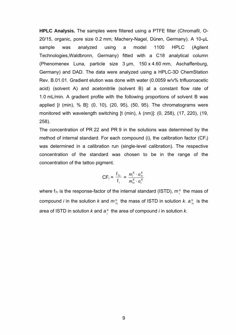
The concentration of PR 22 and PR 9 in the solutions was determined by the
method of internal standard. For each compound (i), the calibration factor (CFi)
was determined in a calibration run (single-level calibration). The respective
concentration of the standard was chosen to be in the range of the
concentration of the tattoo pigment.
CFi = ffTr
i = K
iKTr
KTr
Ki
amam⋅⋅
where fTr is the response-factor of the internal standard (ISTD), m i the mass of
compound i in the solution k and m the mass of ISTD in solution k. a is the
area of ISTD in solution k and a i the area of compound i in solution k.
K
TrK
TrK
K
9
1.3. Results and Discussion
Tattoos are popular because they are adornments; on the other hand, they can
also show a variety of adverse reactions.27–31 Besides adverse reactions, there
is another risk factor regarding the colorants used for tattooing. Since these
compounds are predominantly not produced for tattooing but are also
ingredients of paints and varnishes, there are no specific declarations on the
ingredients. The colorants consist of starting material and byproducts of the
synthesis, titanium dioxide for the lightening of the colorant, and other
unspecified compounds in different concentrations. On one hand there are
regulations that relate to ingredients in paints and varnishes, but these
regulations are different from those regulating cosmetics, foods, and drugs.
In Europe, many of the azo pigments used in tattoos such as PR 22 are not
allowed in cosmetics since they can be cleaved, yielding carcinogenic amines.32
In the United States, the FDA considers the inks used in intradermal tattoos,
including permanent make-up, to be cosmetics and considers the pigments
used in the inks to be color additives requiring premarket approval under the
Federal Food, Drug, and Cosmetic Act. However, because of other public
health priorities and a previous lack of evidence of safety concerns, FDA has
not traditionally regulated tattoo inks or the pigments used in them. In addition,
concerns raised by the scientific community regarding the pigments used in
these inks have prompted FDA to investigate the safe use of tattoo inks. FDA
continues to evaluate the extent and severity of adverse events associated with
tattooing and is conducting research on inks.33
A major obstacle for a risk assessment of tattoo pigments is the fact that the
amount of tattoo colorants in the skin is unknown. Therefore, the major goal of
the present investigations was the development of a procedure that allows the
determination of the concentration of tattoo pigments in the skin. First, an
extraction method was established to separate the pigment molecule from skin
constituents. To quantify the pigment concentration, the method has to be
verified using recovery experiments. That is, a certain amount of pigments was
added to water, PBS, or skin and the respective recovery rates were
determined by HPLC.
10
Two widespread tattoo pigments (PR 22, PR 9) were used for the present
investigations. However, tattoo colorants from tattoo studios exhibit a purity of
usually less than 80% (area %, HPLC analysis, data not shown), which is
useless for precise recovery experiments. Therefore, both pigments were
synthesized, yielding a high purity (> 98%, area %, HPLC, data not shown) that
is comparable to pharmaceutical grade.
In laser removal of tattoos, the pigments in the skin are irradiated with very high
laser intensities leading to temperatures in the pigments higher than 400 °C.
Previous investigations of our group showed that the two pigments, PR 22 and
PR 9, are decomposed by laser light and the products could be identified as NT,
MNA, NAS, DCB, DCA, and m-NAS.15 During laser irradiation, the
concentration of these products increased up to 70-fold. Therefore, the
extraction method of pigments was extended to the respective laser-induced
decomposition compounds.
Establishment of the Workup Scheme. The workup scheme was established
representatively for one pigment (PR 22) and one decomposition product (NT).
A 1-mL sample of PR 22 stock solution was diluted with 1 mL of methanol and
10 mL of methylene chloride to obtain the volume of extraction solution.
Nitrogen was blown into the flask until PR 22 was dried (2 bar, 35 °C) (for
conditions see Table 1, study 1).
Study 1 shows a high recovery rate for PR 22, but the value for NT was below
60% (Table 1). Due to the high volatility of the three compounds NT, DCA, and
DCB, a special treatment during the workup of the extraction solution was
necessary. High volatility of these substances is shown by their high vapor
pressure: NT (0.4 hPa, 20 °C), DCA (0.057 hPa, 20 °C), and DCB (0.8 hPa,
20 °C). Thus, the keeper Diglyme was added to the extraction volume prior to
solvent removal. A keeper is a low-volatile liquid and retains the volatile
compound while the solvent can evaporate. Nevertheless, evaporation under a
gentle stream of nitrogen and elevated temperature is necessary.
The conditions in study 2 with addition of Diglyme as keeper increased the
recovery rate of NT up to almost 100%, with a good yield for PR 22. However,
some PR 22 was adhered to the flask wall. In study 3, the temperature was
increased to 60 °C and the flask wall was rinsed additionally with methylene
11
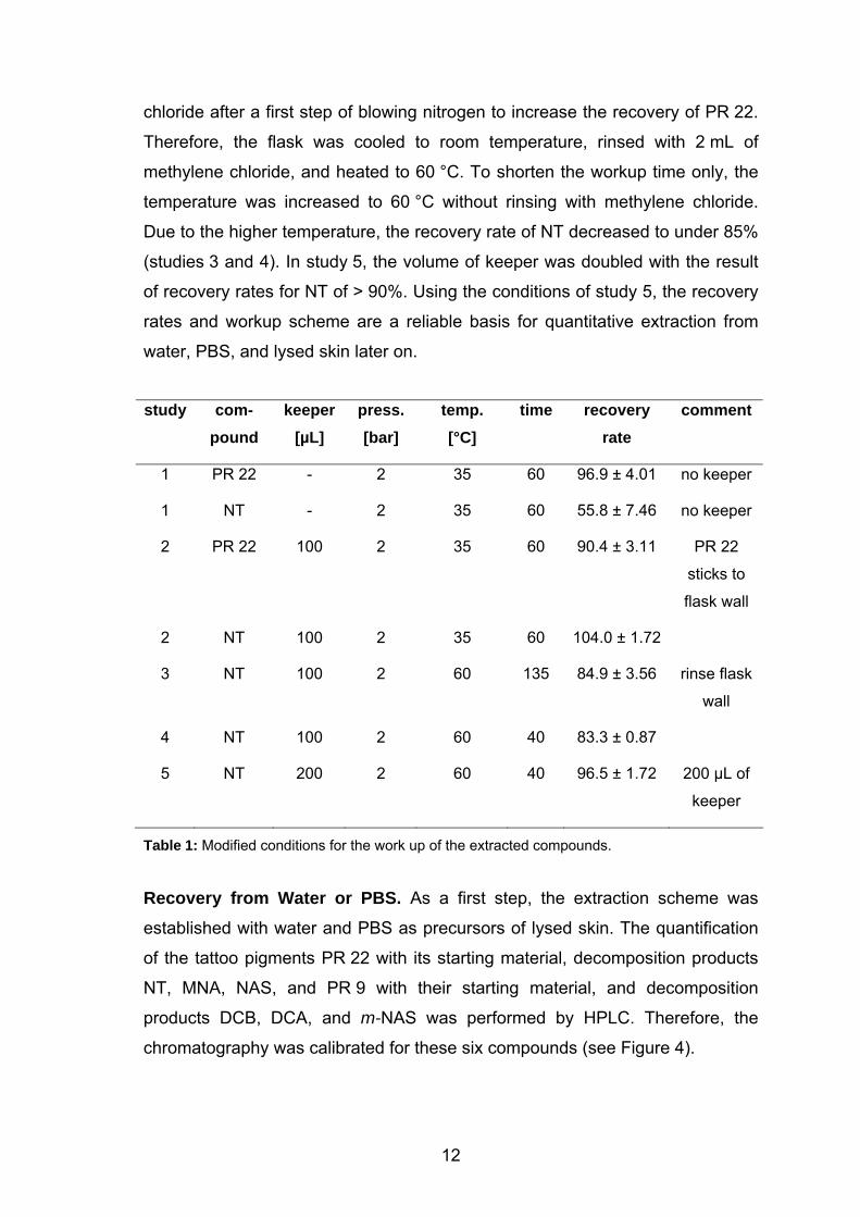
chloride after a first step of blowing nitrogen to increase the recovery of PR 22.
Therefore, the flask was cooled to room temperature, rinsed with 2 mL of
methylene chloride, and heated to 60 °C. To shorten the workup time only, the
temperature was increased to 60 °C without rinsing with methylene chloride.
Due to the higher temperature, the recovery rate of NT decreased to under 85%
(studies 3 and 4). In study 5, the volume of keeper was doubled with the result
of recovery rates for NT of > 90%. Using the conditions of study 5, the recovery
rates and workup scheme are a reliable basis for quantitative extraction from
water, PBS, and lysed skin later on.
study com-
pound keeper
[µL] press. [bar]
temp. [°C]
time recovery rate
comment
1 PR 22 - 2 35 60 96.9 ± 4.01 no keeper
1 NT - 2 35 60 55.8 ± 7.46 no keeper
2 PR 22 100 2 35 60 90.4 ± 3.11 PR 22
sticks to
flask wall
2 NT 100 2 35 60 104.0 ± 1.72
3 NT 100 2 60 135 84.9 ± 3.56 rinse flask
wall
4 NT 100 2 60 40 83.3 ± 0.87
5 NT 200 2 60 40 96.5 ± 1.72 200 µL of
keeper
Table 1: Modified conditions for the work up of the extracted compounds.
Recovery from Water or PBS. As a first step, the extraction scheme was
established with water and PBS as precursors of lysed skin. The quantification
of the tattoo pigments PR 22 with its starting material, decomposition products
NT, MNA, NAS, and PR 9 with their starting material, and decomposition
products DCB, DCA, and m-NAS was performed by HPLC. Therefore, the
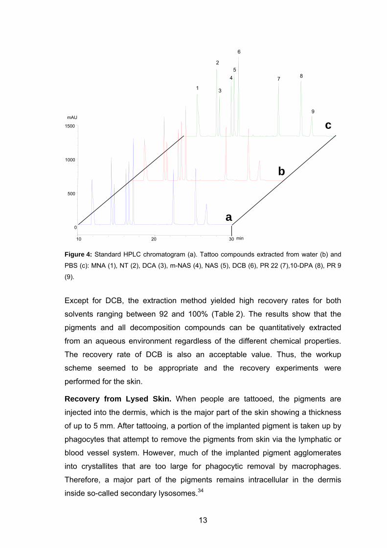
chromatography was calibrated for these six compounds (see Figure 4).
12
6
2 5
Figure 4: Standard HPLC chromatogram (a). Tattoo compounds extracted from water (b) and
PBS (c): MNA (1), NT (2), DCA (3), m-NAS (4), NAS (5), DCB (6), PR 22 (7),10-DPA (8), PR 9
(9).
Except for DCB, the extraction method yielded high recovery rates for both
solvents ranging between 92 and 100% (Table 2). The results show that the
pigments and all decomposition compounds can be quantitatively extracted
from an aqueous environment regardless of the different chemical properties.
The recovery rate of DCB is also an acceptable value. Thus, the workup
scheme seemed to be appropriate and the recovery experiments were
performed for the skin.
Recovery from Lysed Skin. When people are tattooed, the pigments are
injected into the dermis, which is the major part of the skin showing a thickness
of up to 5 mm. After tattooing, a portion of the implanted pigment is taken up by
phagocytes that attempt to remove the pigments from skin via the lymphatic or
blood vessel system. However, much of the implanted pigment agglomerates
into crystallites that are too large for phagocytic removal by macrophages.
Therefore, a major part of the pigments remains intracellular in the dermis
inside so-called secondary lysosomes.34
7
1
4
3
min10 20 30
1000
mAU
0
500
1500 c
b
a
8
9
13
water PBS lysed skin
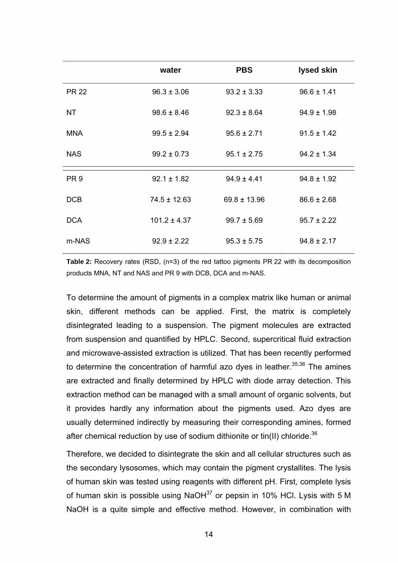
PR 22 96.3 ± 3.06 93.2 ± 3.33 96.6 ± 1.41
NT 98.6 ± 8.46 92.3 ± 8.64 94.9 ± 1.98
MNA 99.5 ± 2.94 95.6 ± 2.71 91.5 ± 1.42
NAS 99.2 ± 0.73 95.1 ± 2.75 94.2 ± 1.34
PR 9 92.1 ± 1.82 94.9 ± 4.41 94.8 ± 1.92
DCB 74.5 ± 12.63 69.8 ± 13.96 86.6 ± 2.68
DCA 101.2 ± 4.37 99.7 ± 5.69 95.7 ± 2.22
m-NAS 92.9 ± 2.22 95.3 ± 5.75 94.8 ± 2.17
Table 2: Recovery rates (RSD, (n=3) of the red tattoo pigments PR 22 with its decomposition
products MNA, NT and NAS and PR 9 with DCB, DCA and m-NAS.
To determine the amount of pigments in a complex matrix like human or animal
skin, different methods can be applied. First, the matrix is completely
disintegrated leading to a suspension. The pigment molecules are extracted
from suspension and quantified by HPLC. Second, supercritical fluid extraction
and microwave-assisted extraction is utilized. That has been recently performed
to determine the concentration of harmful azo dyes in leather.35,36 The amines
are extracted and finally determined by HPLC with diode array detection. This
extraction method can be managed with a small amount of organic solvents, but
it provides hardly any information about the pigments used. Azo dyes are
usually determined indirectly by measuring their corresponding amines, formed
after chemical reduction by use of sodium dithionite or tin(II) chloride.36
Therefore, we decided to disintegrate the skin and all cellular structures such as
the secondary lysosomes, which may contain the pigment crystallites. The lysis
of human skin was tested using reagents with different pH. First, complete lysis
of human skin is possible using NaOH37 or pepsin in 10% HCl. Lysis with 5 M
NaOH is a quite simple and effective method. However, in combination with
14
high temperature (50 °C), the tattoo pigments are cleaved and the structural
information is lost. In addition, the treatment of skin with pepsin in 10% HCl
leads to a satisfying tissue degradation, but the low pH makes the extraction of
pigments impossible. Therefore, the disintegration of skin at neutral pH was
applied.
Our extraction scheme provides a degreasing and extraction method of human
skin for determining the azo compound, corresponding amines, and other
decomposition products. The advantage of our established degradation is the
sensitivity; skin is dissolved without destroying the chemical substances. The
degradation is divided in several steps. High temperature is used for denaturing
the proteins. Collagenase breaks down collagen and elastin and sets cells free
from the extracellular matrix. Proteinase K, an endolytic protease, cleaves
peptide bonds at the carboxylic sides of aliphatic, aromatic, or hydrophobic
amino acids. The smallest peptide to be hydrolyzed is a tetrapeptide. Thus,
proteinase K destroys cell proteins resulting in damaged cell membranes.
Finally, the ATL buffer also contributes toward tissue lysis. These four steps
guarantee a complete and gentle lysis of human skin without destroying the
structure of the molecules that should be extracted quantitatively.
As mentioned above, the pigments of a real tattoo are intracellularly localized.
Complete disintegration of the cells in the skin is proven by Trypan blue
staining. Trypan blue penetrates through damaged cell membranes into the
cytoplasm of dead cells. Its anions bind to cell proteins and stain the cells blue.
Living cells exclude the dye and appear in the microscope transparent. The
results of lysed skin were verified and compared with living NHDF cells (bright
under the microscope) and NHDF cells incubated with trypsin (blue under the
microscope). On the microscope slide with the lysed skin, no bright and no blue-
stained spots (<5%) could be seen. That evidenced the total lysis of human skin
tissue. No individual cells showing a typical cellular shape were detected. In
contrast, 98 ± 3% blue stained cells were detected upon trypsin treatment of
NHDF cells, revealing nonviable cells containing a damaged cell membrane but
retaining their cellular shape.
15
For the extraction of chemical substances from human or animal tissue, several
methods are described in the literature. The recovery for pontamine sky blue
and evans blue from skin has been performed for the measurement of capillary
permeability. Nitta et al. recovered pontamine sky blue from the skin of adult
male albino rabbits with recovery rates from 72.5 to 93.5% depending on the
injected concentration.38 Ankier et al. extracted pontamine sky blue from mouse
pinna with over 96% (96.1 ± 4.7%).39 Harada et al. recovered azovan blue
(evans blue) with recoveries of 96% and trypan blue with > 90% from the skin of
rats.40 These three groups based their calculations on photometric
measurements of the supernatant of extracted skin. Liquid-liquid extraction and
photometric measurements was the chosen method of the following groups for
the recovery of evans blue and pontamine sky blue. Suzuki and Arai extracted
evans blue from skin of adult albino rabbits with 96.84% recovery.41 Katayama
et al. also used evans blue for recovery experiments from skin of guinea pigs
and rats (95%).42 Humphrey recovered evans blue from rat skin in almost
complete recovery.43 The recovery of pontamine sky blue could be optimized to
almost 96% and of evans blue to almost quantitiative recovery rates.
However, the advantages of pontamine sky blue and evans blue are their
known chemical and physical properties and their high solubility in aqueous
solutions. In contrast to that, the pigments PR 22 and PR 9 are declared as
insoluble, showing a poor solubility in organic solvents such as dichloromethane
and chloroform. At the same time, the recovery must operate for the respective
decomposition products, which are partially volatile.
Yeganeh and McLachlan recovered terbinafine from skin at ~ 60%, from liver,
adipose, and muscle at more than 76%.37 Villain et al.44 established a
procedure to screen for benzodiazepines and hypnotics in human hair. The
extraction recovery of these compounds ranged from 32 to 76%. Kim et al.
demonstrated an analysis of hair samples of cannabis abusers. They
established an extraction method for the detection of cannabidiol, cannabinol,
and Δ-9-tetrahydrocannabinol with recovery rates from 37.9 to 94.5%.45
Gratacos-Cubarsi et al. detected residues of sulfamethazine, a sulphonamide
chemotherapeutic agent, in cattle and pig hair. Recovery rates varied from 70 to
85%.46
16
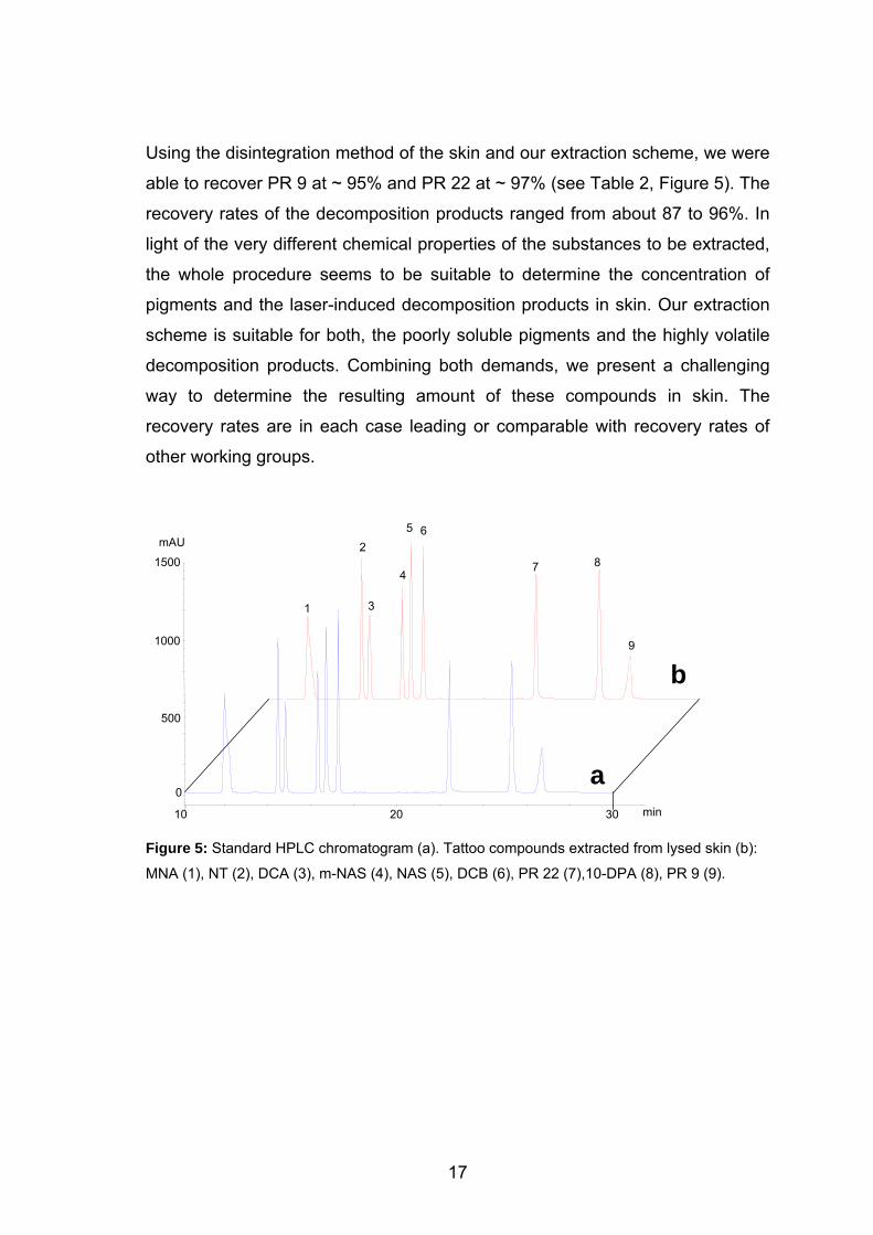
Using the disintegration method of the skin and our extraction scheme, we were
able to recover PR 9 at ~ 95% and PR 22 at ~ 97% (see Table 2, Figure 5). The
recovery rates of the decomposition products ranged from about 87 to 96%. In
light of the very different chemical properties of the substances to be extracted,
the whole procedure seems to be suitable to determine the concentration of
pigments and the laser-induced decomposition products in skin. Our extraction
scheme is suitable for both, the poorly soluble pigments and the highly volatile
decomposition products. Combining both demands, we present a challenging
way to determine the resulting amount of these compounds in skin. The
recovery rates are in each case leading or comparable with recovery rates of
other working groups.
6
Figure 5: Standard HPLC chromatogram (a). Tattoo compounds extracted from lysed skin (b):
MNA (1), NT (2), DCA (3), m-NAS (4), NAS (5), DCB (6), PR 22 (7),10-DPA (8), PR 9 (9).
9
8 7 4
3
2
1
a0
500
1000
min 10 20 30
mAU
1500
b
5
17
1.4. Conclusions
Despite the poor solubility of the azo pigments and the high volatility of some of
the decomposition products, our method provides an effective scheme for the
extraction of tattoo pigments from human skin. Our extraction results in the
recovery of all nonvolatile and volatile compounds, except for DCB, from water,
and PBS shows recovery rates of more than 91%. The RSD (n = 3) for
extraction of the nonvolatile compounds from all matrixes was in the range of
1.34 - 5.75%. These values demonstrate the reliability of the extraction and
workup method. The volatile compounds NT, DCB, and DCA show higher RSD
(n = 3), because of their high vapor pressure. Extraction of DCB from water and
PBS could be obtained with RSD (n = 3) 12.63 and 13.96%. Nevertheless,
extraction from lysed skin resulted in 86.5% with RSD (n = 3) of 2.68%. For the
extraction from lysed skin, the RDS (n = 3) of each compound does not exceed
3 %. Thus, we have established a reliable extraction scheme of tattoo pigments
and their decomposition products from water, PBS, and lysed skin as the basis
for the extraction of pigments from real tattoos.
18
1.5. References
1 Brown, K.M.; Perlmutter, P.; McDermott, R.J. J. Sch. Health 2000, 70, 355. 2 Drews, D.R.; Allison, C.K.; Probst, J.R. Psychol. Rep. 2000, 86, 475. 3 Marcoux, D. Dermatol. Clin. 2000, 18, 667. 4 Ceniceros, S. J. Nerv. Ment. Dis. 1998, 186, 503. 5 Greif, J.; Hewitt, W. Adv. Nurse Pract. 1998, 6, 26. 6 Armstrong, M.L.; Masten, Y.; Martin, R. MCN Am. J. Matern. Child Nurs.
2000, 25, 258. 7 Armstrong, M.L.; Murphy, K.P.; Sallee, A.; Watson, G. Mil. Med. 2000,165,
135. 8 Anderson, R.R. Arch. Dermatol. 2001, 137, 210. 9 Bäumler, W.; Eibler, E.T.; Sens, B.; Hohenleutner, U.; Landthaler, M. Lasers
Surg. Med. 2000, 26, 13. 10 Papameletiou, D.; Zenie, A.; Schwela, D.; Bäumler, W. 2003;
http://europe.eu.int/comm/consumers/cons_safe/news/eis_tattoo_proc_0520
03_en.pdf. 11 Kilmer, S.L.; Anderson, R.R. J. Dermatol. Surg. Oncol. 1993, 19, 330. 12 homepage FDA;
http://www.fda.gov/bbs/topics/answers/2004/ANS01295.html. 13 Gopee, N.V.; Cui, Y.; Olson, G.; Warbritton, A.R.; Miller, B.J.; Couch, L.H.;
Wamer, W.G.; Howard, P.C. Toxicol. Appl. Pharmacol. 2005, 209, 145. 14 Ferguson J.E.; Andrew S.M.; Jones C.J.; August P.J. Br. J. Dermatol. 1997,
137, 405. 15 Vasold, R.; Naarmann, N.; Ulrich, H.; Fischer, D.; König, B.; Landthaler, M.;
Bäumler, W. Photochem. Photobiol. 2004, 80, 185. 16 Anderson, R.R.; Parrish, J.A. Science 1983, 220, 524. 17 Zelickson, B.D.; Mehregan, D.A.; Zarrin, A.A.; Coles, C.; Hartwig, P.; Olson,
S.; Leaf-Davis, J. Lasers Surg. Med. 1994, 15, 364. 18 Az, R.; Dewald, B.; Schnaittmann, D. Dyes pigment. 1991, 15, 1. 19 Chen, S.C.; Kao, C.M.; Huang, M.H.; Shih, M.K.; Chen, Y.L.; Huang, S.P.;
Liu, T.Z. Toxicol. Sci. 2003, 72, 283.
19
20 Huang, Q.G.; Kong, L.R.; Liu, Y.B.; Wang, L.S. Bull. Environ. Contam.
Toxicol. 1996, 57, 349. 21 Shimizu, H.; Kumada, T.; Nakano, S.; Kiriyama, S.; Sone, Y.; Honda, T.;
Watanabe, K.; Nakano, I.; Fukuda, Y.;. Hayakawa, T. Gut 2002, 50, 266. 22 Sayama, M.; Mori, M.; Shoji, M.; Uda, S.; Kakikawa, M.; Kondo, T.; Kodaira
K.I. Mutat. Res. 1998, 420, 27. 23 NTP Toxicology and Carcinogenesis Studies of 1,4-Dichlorobenzene (CAS
No. 106-46-7) in F344/N Rats and B6C3F1 Mice, TR 319 (Gavage Studies)
(1987). 24 Lo, H.H.; Brown, P.I.; Rankin, G.O. Toxicology 1990, 63, 215. 25 Cook, W.L.; Gebler, D.P.; Pratt, N.E. Production of organic pigments and
printing inks containing them; PCT Int. Appl. 2001. 26 Gaber, Y.; Tiedemann K.; Reinhard D.P.; Brinckmann J. Phlebologie 2004,
33, 8. 27 Goldberg, H.M. Plast. Reconstr. Surg. 1998, 98, 1315. 28 Blumental, G.; Okun, M.R.; J.A. Ponitch, J.A. J. Am. Acad. Dermatol. 1982,
6, 485. 29 Papameletiou, D.; Zenie, A.; Schwela, D.; Bäumler, W. 2003;
http://europa.eu.int/comm/consumers/cons_safe/news/eis_tattoo_risk_0520
03_en.pdf. 30 Nilles, M.; Eckert, F. Hautarzt 1998, 41, 236. 31 Zinberg, M.; Heilman, E.; Glickman, F. J. Dermatol. Surg. Oncol. 1982, 8,
955. 32 homepage leffingwell http://www.leffingwell.com/cosmetics/vol_1en.pdf. 33 homepage FDA http://www.cfsan.fda.gov/~dms/cos-204.html. 34 Ferguson, J.E.; Andrew, S.M.; Jones C.J.; August P.J. Br. J. Dermatol.
1997, 137, 405. 35 Sparr Eskilsson, C.; Davidsson, R.; Mathiasson, L. J. Chromatogr. A 2002,
955, 215. 36 Ahlström, L.H.; Raab, J.; Mathiasson, L. Anal. Chim. Acta 2005, 552, 76. 37 Yeganeh, M.H.; McLachlan, A.J. Biomed. Chromatogr. 2000, 14, 261. 38 Nitta, R.; Hayashi, H.; Norimatsu, K. Exp. Biol. Med. 1963, 113, 185. 39 Ankier, S.I.; Whiteside, M.L. Biochem. Pharmacol. 1969, 18, 2197.
20
21
40 Harada, M.; Takeuchi, M.; Fukao, T.; Katagiri, K. J. Pharm. Pharmacol.
1971, 23, 218. 41 Suzuki, M.; Arai, H. Jpn. J. Pharmacol. 1966, 16, 25. 42 Katayama, S.; Shionoya, H.; Ohtake, S. Microbiol. Immunobiol. 1978, 22,
89. 43 Humphrey, D.M. Biotech. Histochem. 1993, 68, 342. 44 Villain M.; Concheiro, M.; Cirimele, V.; Kintz, P. J. Chromatorgr. B Analyt.
Technol. Biomed. Life Sci. 2005, 825, 72. 45 Kim, J.Y.; Suh, S.I., In, M.K.; Paeng K.J.; Chung, B.C. Arch. Pharm. Res.
2005, 28, 1086. 46 Gratacos-Cubarsi, M.; Castellari, M.; Garcia-Regueiro, J.A. J. Chromatorgr.
B Analyt. Technol. Biomed. Life Sci. 2006, 832, 121.
2. Modern Tattoos Cause High Concentrations of
Hazardous Pigments in Human Skin∗
2.1. Introduction
In recent years, the number of tattooed individuals has increased significantly,1,2
especially among young people.3 In the United States, up to 24% of the
population are tattooed,2 whereas in European countries like Germany
approximately 9% of the population and about 12% in the United Kingdom have
tattoos.4,5 Nowadays, azo pigments are frequently used for tattooing because of
their colour intensity and their longevity. However, azo pigments are primarily
manufactured for other purposes such as printing, the painting of cars, and the
staining of various consumer products. Tattoo colorants are mixtures of
pigments (colour) and multiple other ingredients. These colorants usually
contain titanium dioxide for lightening the shade,6 precursors and by-products of
pigment synthesis, as well as diluents that are used for pigment suspension.7,8
Tattoo colorants are also applied for permanent make-up on eyelids, eyebrows,
and lips.9
Despite the high incidence of tattoos worldwide, no common legal requirement
for listing ingredients has been introduced so far. In Europe, many azo pigments
employed in tattoos (e.g. Pigment Red 22) are not allowed for use in cosmetics
because they may be decomposed yielding carcinogenic amines.10
In the process of tattooing, pigment suspension is deposited in the dermis by
piercing the skin with tiny solid needles that are moistened with tattoo colorant.
On closer examination, tattooing is a complex procedure that includes various
risks for the skin and maybe even for the human body. Pigments and impurities
may cause adverse skin reactions at the site of the tattoo.11-22 In addition, part
of the colorants are transported to other anatomical locations such as lymph
nodes.23,24 Laser light could cleave pigments in the skin during tattoo removal25
or pigment decomposition may be caused by ultraviolet radiation during solar
∗ Results of this chapter are submitted: Engel, E.; Santarelli, F.; Vasold, R.; Maisch, T.; Howard, P.C.; Ulrich, H.; Prantl, L.; König, B.; Landthaler, M.; Bäumler, W. Br. J. Dermatol. 2007 ∗ Sample preparation was done by F. Santarelli.
22

light exposure; both procedures have been known to cause hazardous
compounds such as carcinogenic amines.26
To estimate the risk of any health problems that tattooing might involve for the
skin, the pigment concentration in tissue should be determined – a procedure
that has not been attempted so far.
23
2.2. Materials and Methods
Pigments. The red tattoo pigment PR 22 (C.I. 12315, CAS 6448-95-9) was
either synthesized in pure quality (> 98%)27 or purchased as original tattoo
pigment (purity ~ 80%, data not shown).7 The pigments were suspended in
concentrations of 10% (w/v) and 25% (w/v) in a vehicle of 10% of glycerol (87%,
Merck) in water (Milli-Q® Ultra-pure Water-Purification System, Millipore) with
the addition of 100 µL of isopropanol as solubility enhancer.
Skin. Pigskin was purchased from a local butchery. Human skin was obtained
from skin excisions for other reasons. Excision sites were either abdomen or
upper arms. The fatty tissue was removed; skin thickness measured
approximately 3 to 4 mm depending on the excision site. Researchers used the
tattoo machine, type “new lightning” (Deep Colours GmbH, Germany), and
typical tattooing needles (‘liners, shaders’) to inject the colorant into the skin. On
round needles (‘liners’), tips are arranged in a circle, whereas flat needles
(‘shaders’) have linear tips. All needles were solid needles with either four flat
(4F), four round (4R), eight flat (8F), or eight round (8R) tips. Tattoo artists tend
to use round needles with nine tips (9R). Both needles and the tattoo machine
are frequently used for tattooing worldwide.
Tattooing. We applied different methods for skin tattooing. In method (A),
researchers tattooed pigskin either with synthesized PR 22 or with commercial
PR 22 (method B). In method (C), professional tattoo artists tattooed pigskin
with synthesized PR 22. In method (D), researchers tattooed human skin either
with commercial PR 22 or with synthesized PR 22 (method E).
A rectangular skin area of about 1 by 3 cm was tattooed (Figure 1a). We made
three punch biopsies measuring 5 mm in diameter, extracted the pigment of
these samples separately and calculated the mean value for the resulting three
values.27 The concentration of skin pigment was calculated as follows: Using a
5 mm circular punch biopsy, the skin volume is a cylinder with a skin area of
19.63 mm2 times the height of the cylinder. However, only skin material stained
with tattoo pigment may be used for analysis. Hence, we histologically
determined the cylinder height in order to be able to calculate the pigmented
material for each sample. For this height determination, we performed an
24
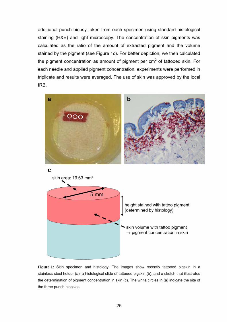
additional punch biopsy taken from each specimen using standard histological
staining (H&E) and light microscopy. The concentration of skin pigments was
calculated as the ratio of the amount of extracted pigment and the volume
stained by the pigment (see Figure 1c). For better depiction, we then calculated
the pigment concentration as amount of pigment per cm2 of tattooed skin. For
each needle and applied pigment concentration, experiments were performed in
triplicate and results were averaged. The use of skin was approved by the local
IRB.
a b
c
5 mm
skin area: 19.63 mm²
height stained with tattoo pigment(determined by histology)
skin volume with tattoo pigment→ pigment concentration in skin
Figure 1: Skin specimen and histology. The images show recently tattooed pigskin in a
stainless steel holder (a), a histological slide of tattooed pigskin (b), and a sketch that illustrates
the determination of pigment concentration in skin (c). The white circles in (a) indicate the site of
the three punch biopsies.
25
Disintegration and Extraction. In contrast to our previous report27and
according to Gaber et al.28, we inserted each sample in 400 µL of PBS
(Phosphate Buffered Saline, Biochrom) at 95 °C for 20 min. After the samples
had cooled to room temperature, we added 180 µL of tissue lysis buffer (buffer
ATL) and 15 µL of Proteinase K (QIAGEN). Samples were stirred at 55 °C for
30 min until complete lysis of the tissue. Each process was carried out as
previously described.27 The concentrated residue was reconstituted in
methylene chloride (LiChroSolv®, Merck). We used transmission electron
microscopy (TEM) for evaluating pigment size and shape.
26
2.3. Results and Discussion
Based upon a very recently established procedure,27 we quantitatively extracted
pigments from tissue after tattooing and, for the first time, determined their
respective concentration in the skin. However, the investigation of tattooing on a
scientific level represents a challenge owing to the large variety of tattooing
procedures available. Therefore, we used different pigment suspensions and
different needles, and both researchers and tattoo artists performed human and
pigskin tattooing. This laborious procedure should help to avoid the generation
of random values for pigment concentration in skin. Since red tattoo pigments
frequently cause allergic skin reactions,11 we used the widespread red pigment
PR 22 in our experiments.
Usually, vertically vibrating needles are used for tattooing that inject pigments
into the skin with an initial penetration depth of up to 2 mm. Special machines
produce this vibration at a frequency of about 30 Hz. Needles exhibit different
shapes and number of tips. For tattooing we initially used original tattoo
colorants from the tattoo market.7 However, these colorants usually exhibit a
purity of less than 80%. Since these impurities may affect the precise recovery
experiments, we additionally synthesized PR 22 in a high purity of about 98%.
To determine the pigment concentration in skin, we first used pigskin that is
available in a standardized manner at all times. After performing the same
experiments with human skin, we compared extraction results to pigskin
experiments. To consider different concentrations of pigments as applied in
routine tattoo practice, we used PR 22 at concentrations of 10% (w/v) or
25% (w/v).
Synthesized PR 22 in Pigskin. For synthesized PR 22, values for pigment
concentration are shown in Table 1 (method A) as amount of pigment per 1 cm2
of tattooed skin. Values range from 0.63 mg/cm2 to 5.19 mg/cm2 depending
upon the different concentration applied to the skin as well as the type and
shape of needles used. Histology showed the depth of tattoo pigments to
depend upon skin properties like surface tension. In accordance with our
previous report,27 relative standard deviation (RSD) does not depend upon the
27
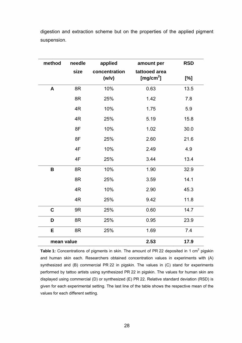
digestion and extraction scheme but on the properties of the applied pigment
suspension.
method needle size
applied concentration
amount per tattooed area
RSD
(w/v) [mg/cm2] [%]
A 8R 10% 0.63 13.5
8R 25% 1.42 7.8
4R 10% 1.75 5.9
4R 25% 5.19 15.8
8F 10% 1.02 30.0
8F 25% 2.60 21.6
4F 10% 2.49 4.9
4F 25% 3.44 13.4
B 8R 10% 1.90 32.9
8R 25% 3.59 14.1
4R 10% 2.90 45.3
4R 25% 9.42 11.8
C 9R 25% 0.60 14.7
D 8R 25% 0.95 23.9
E 8R 25% 1.69 7.4
mean value 2.53 17.9
Table 1: Concentrations of pigments in skin. The amount of PR 22 deposited in 1 cm2 pigskin
and human skin each. Researchers obtained concentration values in experiments with (A)
synthesized and (B) commercial PR 22 in pigskin. The values in (C) stand for experiments
performed by tattoo artists using synthesized PR 22 in pigskin. The values for human skin are
displayed using commercial (D) or synthesized (E) PR 22. Relative standard deviation (RSD) is
given for each experimental setting. The last line of the table shows the respective mean of the
values for each different setting.
28
A needle in a group of four tips (4R) results in higher values because the area
covered by one puncture of 4R is smaller than the area covered by 8R, i.e.
more needle injections are necessary for tattooing a certain area of skin when
using 4R. Flat needles with 8 tips (8F) result in slightly higher values than 8R.
Flat needles show the same correlation between the number of tips and the
amount of tattoo pigment injected into the skin.
Comparison of Synthesized and Commercial PR 22. The injection of
commercial PR 22 resulted in higher amounts of pigment in the skin as
compared to synthesized PR 22 (Table 1, conditions A and B), although the
commercial colorant contained not only pigment but also impurities up to 20%.
Azo pigments tend to agglomerate requiring additional procedures after
synthesis. Thus, chemical companies optimise their manufacturing processes
that leads to a lower aggregation susceptibility.29
The pigment synthesized in our laboratory30 was highly pure PR 22 and did not
receive any further treatment. This could explain the different agglomeration
and aggregation of primary crystallites. These differences are shown in the TEM
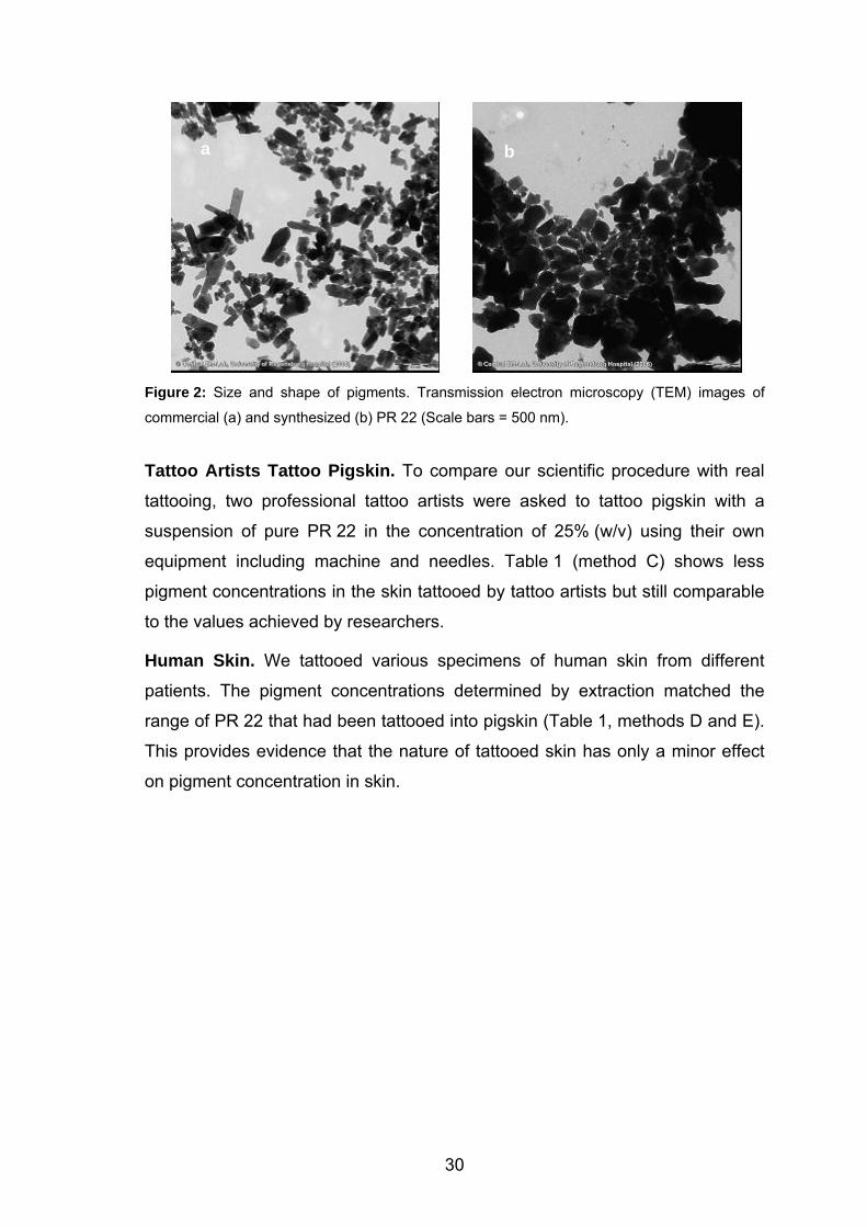
pictures (Figure 2) of commercial or synthesized PR 22 with different mean
particle diameters of about 154 nm and 202 nm respectively. Hence, the
commercial and our synthesized pigments showed a different sedimentation
behaviour in suspension. We measured a decrease in pigment concentration by
30% in the supernatant of suspension for the synthesized sample, whereas, in
the commercial sample, the concentration remained unchanged.
This difference suggests a different amount of pigment attached to the needle
when dipped into such suspensions. This clearly affects the concentration of
pigments injected into the skin but should reflect the various conditions in
routine tattooing. The mixture of ingredients in tattoo colorants is neither
regulated nor standardized. Despite these facts, the resulting concentrations of
pigments in skin are in a confined range regardless the methods used for
tattooing (see Table 1).
29
a b
Figure 2: Size and shape of pigments. Transmission electron microscopy (TEM) images of
commercial (a) and synthesized (b) PR 22 (Scale bars = 500 nm).
Tattoo Artists Tattoo Pigskin. To compare our scientific procedure with real
tattooing, two professional tattoo artists were asked to tattoo pigskin with a
suspension of pure PR 22 in the concentration of 25% (w/v) using their own
equipment including machine and needles. Table 1 (method C) shows less
pigment concentrations in the skin tattooed by tattoo artists but still comparable
to the values achieved by researchers.
Human Skin. We tattooed various specimens of human skin from different
patients. The pigment concentrations determined by extraction matched the
range of PR 22 that had been tattooed into pigskin (Table 1, methods D and E).
This provides evidence that the nature of tattooed skin has only a minor effect
on pigment concentration in skin.
30
2.4. Potential Health Problems
For the first time, the concentration of tattoo pigment in skin has been
determined ranging from 0.60 to 9.42 mg/cm2. Values and their RSD depend
upon the different methods used in our experiments, which should reflect the
different conditions in the daily practice of tattooing.
Regardless the different methods used, these values yield an overall mean
concentration of 2.53 mg of pigment in 1 cm2 of tattooed skin (Table 1). Thus,
about 253 mg of azo pigment PR 22 are deposited in the dermis for a typical
tattoo covering a skin area of 100 cm2. This is an alarming fact because, in
Europe, many azo pigments such as Pigment Red 22 are forbidden to be used
in cosmetics, which are only applied to the skin surface.8 This prohibition is
based upon the assumption that such pigments are to some extent absorbed by
skin. This absorption may cause health problems, in particular when
carcinogenic amines are formed owing to the decomposition of azo pigments.
However, in tattooing, hundreds of milligrams are directly injected into the skin.
The medical literature contains multiple case reports16 on adverse skin
reactions after tattooing such as cutaneous pseudolymphoma,15 granulomatous
tattoo reactions,12,17 allergic reactions,19,21,22 pseudoepitheliomatous epidermal
hyperplasia,13 and even non-melanoma skin cancer14,17 or malignant
melanoma18,20 - albeit skin cancer is assumed to occur only occasionally. The
extent to which these adverse reactions are caused by pigments, by other
ingredients, or by impurities including bacteria or viruses remains unclear.
Unfortunately, physicians are not obliged to report health problems caused by
tattoo colorants. As a first step, dermatologists could report on the incidence
and the possible reasons of skin problems after tattooing.
Long-term health problems in either skin or other organs could be evaluated by
epidemiological studies that are lacking so far. More detailed information on this
topic is important since, according to our calculation, the frequent use of azo
pigments has only started 10 to 15 years ago. Such studies are definitely
necessary to assess whether health problems caused by tattoo colorants are
only individual cases without any relevance for public health. However, if
tattooing involves any major health risk, it could affect not only the skin but also
31
other organs because of the transportation of colorants inside the human body.
The exact mechanisms of transportation and the extent of pigment
transportation are yet unknown. Thus, pigments are injected into the skin and
are transported inside the body similar to medical drugs, which necessitate
years of clinical trials with regard to possible side effects and health risks.
However, unlike medical drugs, these colorants do not have an established
history of safety use.7,8
In our investigation, we aimed to determine the concentration of a typical tattoo
pigment in the skin immediately after tattooing. This is an important step
towards risk assessments with regard to potential health problems caused by
tattoo pigments, in particular for the skin. Since approximately 20% of colorants
represent impurities, these substances can be included in risk assessment.
Other risks may result from the possible light-induced decomposition of tattoo
pigments in skin. The ultraviolet part of the solar light spectrum may decompose
tattoo pigments as shown for Pigment Yellow 74.26
Many tattooed individuals change their mind and request the removal of their
tattoo after some time. A widespread method of tattoo removal is the application
of short laser pulses at high intensities.31 Laser light penetrates the skin and is
selectively absorbed in pigments. The high absorption coefficient of pigments
and the high laser intensities lead to temperatures well above 400 °C.25 In
previous investigations, we showed pigments PR 22 and PR 9 to be
decomposed by laser light. The products of this decomposition were identified
as 4-nitrotoluene, 2-methyl-5-nitroaniline, naphthol AS, 1,4-dichlorobenzene,
2,5-dichloroaniline, or methoxy-naphthol AS25, which are proven to be toxic or
even carcinogenic. In the worst case, each pigment deposited in the skin is
decomposed into carcinogenic amines during laser light exposure. Thus, more
research is necessary in order to find out if this decomposition causes any
major health problems for tattooed individuals.
32
2.5. References
1 Drews, D.R.; Allison, C.K.; Probst, J.R. Psychol. Rep. 2000, 86, 475. 2 Laumann, A.E.; Derick, A.J. J. Am. Acad. Dermatol. 2006, 55, 413. 3 Marcoux, D. Dermatol. Clin. 2000, 18, 667. 4 Long, G.E.; Rickman, L.S. Clin. Infect. Dis. 1994, 18, 610. 5 Stirn, A.; Brahler, E.; Hinz, A. Psychother. Psychosom. Med. Psychol. 2006,
56, 445. 6 Timko, A.L.; Miller, C.H.; Johnson, F.B.; Ross E. Arch. Dermatol. 2001, 137,
143. 7 Baumler, W.; Eibler, E.T.; Sens, B.; Hohenleutner, U.; Landthaler, M.
Lasers. Surg. Med. 2000, 26, 13. 8 Papameletiou, D.; Zenie, A.; Schwela, D.; Bäumler, W. 2003
http://ec.europa.eu/consumers/cons_safe/news/eis_tattoo_risk_052003_en.
pdf. 9 Prinz, B.M.; Vavricka, S.R.; Graf, P.; Burg, G.; Dammer R. Br. J. Dermatol.
2004, 150, 245. 10 homepage leffingwell http://www.leffingwell.com/cosmetics/vol_1en.pdf. 11 Antony, F.C.; Harland, C.C. Br. J. Dermatol. 2003, 149, 94-98. 12 Bachmeyer, C.; Blum, L.; Petitjean, B.; Kemiche, F.; Pertuiset E. J. Eur.
Acad. Dermatol. Venereol. 2007, 21, 550. 13 Balfour, E.; Olhoffer, I.; Leffell, D.; Handerson T. Am. J. Dermatopathol.
2003, 25, 338. 14 Doumat, F.; Kaise, W.; Barbaud, A., Schmutz J.L. Dermatology 2004, 208,
181. 15 Gutermuth, J.; Hein, R.; Fend, F.; Ring, J.; Jakob, T. J. Eur. Acad. Dermatol.
Venereol. 2007, 21, 566. 16 Jacob, C.I. Dermatol. Surg. 2002, 28, 962. 17 Morales-Callaghan, A.M. Jr.; Aguilar-Bernier, M. Jr.; Martinez-Garcia, G.;
Miranda-Romero, A. J. Am. Acad. Dermatol. 2006, 55, 71. 18 Paradisi, A.; Capizzi, R.; De Simone, C.; Fossati, B.; Proietti, L.; Amiero,
P.L. Melanoma Res. 2006, 16, 375. 19 Steinbrecher, I.; Hemmer, W.; Jarisch, R. J. Dtsch. Dermatol. Ges. 2004, 2,
1007.
33
34
20 Stinco, G.; De Francesco, V.; Frattasio, A.; Quinkenstein, E.; Patrone, P.
Dermatology 2003, 206, 345. 21 Tsuruta, D.; Sowa, J.; Higashi, N.; Kobayashi H.; Ishii, M. Lancet 2004, 364,
730. 22 Zwad, J.; Jakob, A.; Gross, C.; Rompel R. J. Dtsch. Dermatol. Ges. 2007, 5,
8. 23 Friedman, T.; Westreich, M.; Mozes, S.N.; Dorenbaum, A.; Herman, O.
Plast. Reconstr. Surg. 2003, 111, 2120. 24 Moehrle, M.; Blaheta, H.J.; Ruck, P. Dermatology 2001, 203, 342. 25 Vasold, R.; Naarmann, N.; Ulrich, H.; Fischer, D.; Landthaler, M.; Baumler,
W. Photochem. Photobiol. 2004, 80, 185. 26 Cui, Y.; Spann, A.P.; Couch, L.H.; Gopee, N.V.; Evans, F.E.; Churchwell,
M.I.; Williams, L.D.; Doerge, D.R.; Howard, P.C. Photochem. Photobiol.
2004, 80, 175. 27 Engel, E.; Santarelli, F.; Vasold, R.; Ulrich, H.; Maisch, T.; König, B.;
Landthaler, M.; Gopee, N.V.; Howard, P.C.; Bäumler, W. Anal. Chem. 2006,
78, 6440. 28 Gaber, Y.; Tiedemann, K.; Reinhard, D.P.; Brinkmann J. Phlebologie 2004,
33, 8. 29 Zollinger, H. Color Chemistry. Wiley-VCH 2003. 30 Cook, W.L.; Gebler, D.P.; Pratt, N.E. Production of organic pigments and
printing inks containing them; PCT Int. Appl. 2001. 31 Kuperman-Beade, M.; Levine, V.J.; Ashinoff, R. Am. J. Clin. Dermatol. 2001,
2, 21.
3. Photochemical Cleavage of a Tattoo Pigment by UVB
Radiation or Natural Sunlight∗
3.1. Introduction
UVB radiation (280 - 320 nm) is absorbed well by many biological
macromolecules such as proteins, lipids and DNA. The transformation of radiant
energy into photochemical energy can be damaging to the cell.1 When UVA
radiation is absorbed by tissue, reactive oxygen species (ROS) such as oxygen
radicals or singlet oxygen are produced. These, too, can damage cellular
components leading to premature aging of the skin or skin cancer.2
In addition to the endogenous substances in the skin, exogenous materials can
absorb UV radiation. These can include pigments applied into the skin as a
tattoo or permanent make-up (PMU). On the one hand, tattoos can serve to
willingly isolate an individual from society; on the other hand, in recent times
decorative tattoos and permanent make-up (tattooed eyeliner, eye shadow and
lip contours) have become enormously popular. In the USA 16% of the
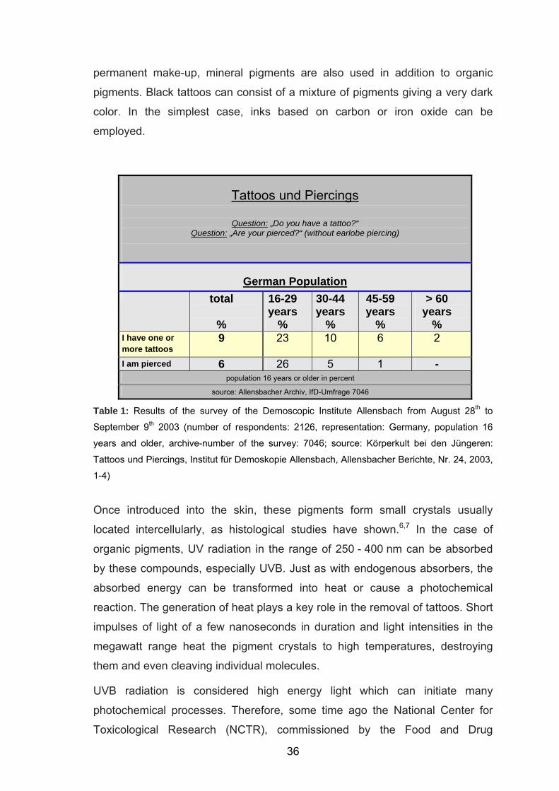
population possess tattoos; the numbers are similar in Europe. According to a
survey by the Demoscopic Institute of Allensbach in 2003, about 9% of the
population in Germany have at least one tattoo, among younger people (age
16 – 29 years) 23% (Table 1).3 In recent years the number of people with
tattoos has further risen.
In the past, inorganic pigments such as titanium dioxide (white), cadmium
sulfide (yellow), chromium oxide (green), cadmium selenide (red) and iron
oxides (black) were employed.4 Today, mostly dye-based pigments are used for
colored tattoos. Chemical analyses have shown that these include industrial
organic pigments such as azo dyes or polycyclic compounds.5 These pigments
are usually used to dye or paint consumer goods (for example, car paints). The
tattoo artist enjoys using these pigments, because they are very durable and
almost insoluble and thus provide for a brilliant, permanent tattoo. For
∗ Results of this chapter have been published: Engel, E.; Spannberger, A.; Vasold, R.; König, B.; Landthaler, M.; Bäumler, W. J. Dtsch. Dermatol. Ges. 2007, 5, 583. ∗ HPLC analysis was performed by A. Spannberger in her Zulassungsarbeit.
35
permanent make-up, mineral pigments are also used in addition to organic
pigments. Black tattoos can consist of a mixture of pigments giving a very dark
color. In the simplest case, inks based on carbon or iron oxide can be
employed.
Tattoos und Piercings
Question: „Do you have a tattoo?“ Question: „Are your pierced?“ (without earlobe piercing)
German Population
total
%
16-29 years
%
30-44 years
%
45-59 years
%
> 60 years
% I have one or more tattoos
9 23 10 6 2
I am pierced 6 26 5 1 - population 16 years or older in percent
source: Allensbacher Archiv, IfD-Umfrage 7046
Table 1: Results of the survey of the Demoscopic Institute Allensbach from August 28th to
September 9th 2003 (number of respondents: 2126, representation: Germany, population 16
years and older, archive-number of the survey: 7046; source: Körperkult bei den Jüngeren:
Tattoos und Piercings, Institut für Demoskopie Allensbach, Allensbacher Berichte, Nr. 24, 2003,
1-4)
Once introduced into the skin, these pigments form small crystals usually
located intercellularly, as histological studies have shown.6,7 In the case of
organic pigments, UV radiation in the range of 250 - 400 nm can be absorbed
by these compounds, especially UVB. Just as with endogenous absorbers, the
absorbed energy can be transformed into heat or cause a photochemical
reaction. The generation of heat plays a key role in the removal of tattoos. Short
impulses of light of a few nanoseconds in duration and light intensities in the
megawatt range heat the pigment crystals to high temperatures, destroying
them and even cleaving individual molecules.
UVB radiation is considered high energy light which can initiate many
photochemical processes. Therefore, some time ago the National Center for
Toxicological Research (NCTR), commissioned by the Food and Drug
36
Administration (FDA), examined cleavage of tattoo pigments by UV radiation.
UVB-induced cleavage of a popular yellow pigment (Pigment Yellow 74: PY 74)
in vitro was found.8 Toxic decomposition products were identified with likely
involvement of reactive oxygen species. Red tattoo pigments can be involved in
toxic allergic or granulomatous skin reactions.9–12 One of the most common red
pigments is Pigment Red 22 (PR 22) which we have already examined with
regard to laser-induced cleavage.13 In this study, possible photochemical
cleavage of this red pigment, which has appropriate absorption in the UVB
range, was examined using chromatography (HPLC) and mass spectrometry.
Using the same methods in a long-term experiment, the effects of sunlight on
this pigment were also studied.
37
3.2. Materials and Methods
Pigments and Chemicals. Pigment Red 22 (PR 22, CAS 6448-95-9,
C.I. 12315) is a widely used azo dye belonging to the group of naphthol-AS
dyes. It is synthesized by azo coupling according to Cook et al.14 and after
purification displays a purity of over 98% (area %, data not shown15). This purity
is comparable to pharmaceutical purity.
Preparation of the Solutions. Highly purified PR 22 was dissolved in
tetrahydrofuran (0.19 mg/mL [THF, p.a., Merck, Darmstadt, Germany]), dioxane
(0.06 mg/mL [p.a., Merck, Darmstadt, Germany]), dichloromethane (0.2 mg/mL
[HPLC Gradient Grade, Mallinckrodt Baker, Deventer, The Netherlands]) and
chloroform (0.20 mg/mL [LiChrosolv, Merck, Darmstadt, Germany]).
Reference Substances. Reference substances for PR 22 were dissolved in
acetonitrile in the following concentrations: 2-methyl-5-nitroaniline (0.25 mg/mL
[99%, Aldrich Chemical Company, Inc., Milwaukee, USA]), 4-nitrotoluene
(0.25 mg/mL [> 99%, Fluka, Buchs Switzerland]) and naphthol-AS (0.1 mg/mL
[99%, Sigma- Aldrich, Steinheim, Germany]).
UVB Exposure. For UVB exposure precision test tubes of quartz glass
(SUPRASIL, Hellma, 110-QS, thickness 2 mm, Müllheim, Germany) were filled
with 600 μL pigment solution. To reduce oxygen partial pressure, the tubes
were rinsed before and after filling with argon gas for 10 minutes. The tubes
were closed securely and exposed to a broad band UV lamp (280 - 320 nm,
intensity 0.0015 W/cm2, Type UV 800, Waldmann Lichttechnik, Villingen-
Schwenningen, Germany) at a distance of 25 cm. The samples were irradiated
for at least 4 hours or to a color change from orange to yellow. For each
irradiated sample, a reference was incubated in the dark at 4 °C.
Sunlight Exposure. A 1.0 mL portion of the pigment solution was filtered using
a PTFE filter (Chromalfil, O-20/15, organic, pore size 0.2 μm, Machery-Nagel,
Düren, Germany) into an injection flask (DAB- 10 quality,
Chromatographiehandel Müller, Fridolfing, Germany). To reduce oxygen partial
pressure, the injection flasks were also rinsed with argon gas for 10 minutes
before and after being filled. The injection flasks were securely sealed and
38
placed on the inside of a window on the west side of the building in natural
sunlight for 110 days (May - August 2005). For each sample a reference was
kept at 4 °C in the dark.
Preparation and HPLC Analysis. For HPLC analysis all samples were filtered
using a PTFE filter and injected into a modular HPLC system (Hewlett- Packard
Ltd., Waldbronn, Germany). The system consists of an HP 1050 quaternary
pump (Mod. Nr. 79852AX), an HP 1050 autosampler (Mod. Nr. 79855A), an HP
1050 4-channel online degasser (Mod. Nr. G1303AX), an Agilent 1100 column
thermostat (Mod. Nr. 61316a) and an Agilent 1100 photo diode array detector
(Mod. G1315b, Agilent Technologies Ltd., Waldbronn, Germany). Evaluation
was done by the ChemStation Version HPLC-3DChemStation Rev. A.08.04
(1008). The mixture to be analyzed was separated by a C18 column
(Phenomenex luna, particle size 3 μm, 150 x 4.60 mm, Aschaffenburg,
Germany). A binary eluent mixture consisting of water (0.0059% [w/v]
trifluoroacetic acid) (eluent A) and acetonitrile (eluent B) was pumped with a
constant flow of 1.0 mL/min. The following gradient profile was used (t [min], %
B: [0, 10], [20, 95], [50, 95]). The chromatograms were registered at 258 nm.
The injected volume was 10 μL.
LC/MS online Coupling. Using a triple stage mass spectrometer (TSQ 7000,
Thermoquest Finnigan, Toronto, Canada) the masses of the compounds,
especially the UVB- and sunlight-induced decomposition products, were
determined.
39
3.3. Results and Discussions
A research team of the National Center for Toxicological Research (NCTR) of
the FDA recently showed that a pigment (PY 74) in solvent is cleaved into toxic
products photochemically when exposed to UV radiation in a sunlight
simulator.8 These authors concluded form their in vitro study that these
substances can be generated in tattooed skin when this skin is exposed for a
longer period of time to sunlight or other sources of UV radiation. This could
also be the case for medical UV radiation sources.
Many organic pigments used in tattooing or for PMU do indeed absorb light
from the UV spectrum. In order to estimate a possible risk of photochemical
cleavage of these pigments in the skin, certain factors must be known. One is
the dose of UV radiation on the tattooed skin. Further, how deep the radiation
penetrates the skin must be known. Considering the absorption coefficient of
the pigments in this spectral range, the energy available for photochemical
cleavage can be estimated. Knowledge of the pigment concentration in the skin
and thus the amount of possible decomposition products would be a further
basis for estimating the possible health risk.
In this study, we sought to determine if the widely used Pigment Red 22 can be
photochemically cleaved by UVB radiation or even sunlight. At present, it is not
possible to perform this experiment in skin. Therefore, a quantitative study in
solutions is a first and indicative step. Most pigments are poorly soluble and, in
the skin, exist as small crystals. Such chemical analyses are best done using
solvents capable of adequately dissolving these pigments. The solvents should
not themselves influence photochemical cleavage of the pigments, i.e. they
should be inert. Pigment Red 22 is an azo dye out of the group of naphthol-AS
pigments, used in the chemical industry as a dye, for example, in paints. The
use of this pigment on the skin surface is not allowed by the cosmetic laws
(Annex IV of the Cosmetics Directive of the EU).16 Due to lack of legal
regulation, such pigments can be used for tattooing or PMU (trade name e.g.
Devil’s Red). As such pigments are produced for industrial purposes, they can
display impurities of 10% or more. These impurities can disturb chemical
analytic tests. For this reason, PR 22 used in this study was synthesized in
40
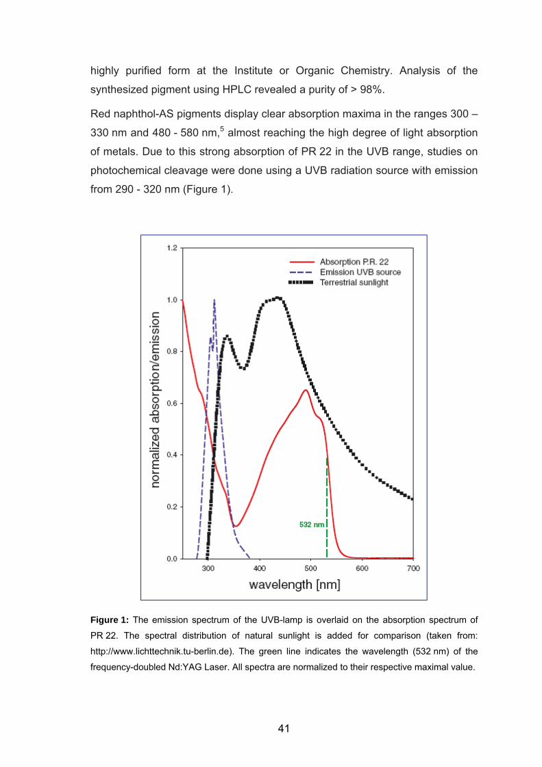
highly purified form at the Institute or Organic Chemistry. Analysis of the
synthesized pigment using HPLC revealed a purity of > 98%.
Red naphthol-AS pigments display clear absorption maxima in the ranges 300 –
330 nm and 480 - 580 nm,5 almost reaching the high degree of light absorption
of metals. Due to this strong absorption of PR 22 in the UVB range, studies on
photochemical cleavage were done using a UVB radiation source with emission
from 290 - 320 nm (Figure 1).
Figure 1: The emission spectrum of the UVB-lamp is overlaid on the absorption spectrum of
PR 22. The spectral distribution of natural sunlight is added for comparison (taken from:
http://www.lichttechnik.tu-berlin.de). The green line indicates the wavelength (532 nm) of the
frequency-doubled Nd:YAG Laser. All spectra are normalized to their respective maximal value.
41
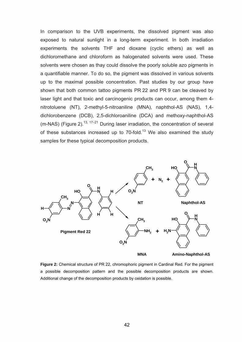
In comparison to the UVB experiments, the dissolved pigment was also
exposed to natural sunlight in a long-term experiment. In both irradiation
experiments the solvents THF and dioxane (cyclic ethers) as well as
dichloromethane and chloroform as halogenated solvents were used. These
solvents were chosen as thay could dissolve the poorly soluble azo pigments in
a quantifiable manner. To do so, the pigment was dissolved in various solvents
up to the maximal possible concentration. Past studies by our group have
shown that both common tattoo pigments PR 22 and PR 9 can be cleaved by
laser light and that toxic and carcinogenic products can occur, among them 4-
nitrotoluene (NT), 2-methyl-5-nitroaniline (MNA), naphthol-AS (NAS), 1,4-
dichlorobenzene (DCB), 2,5-dichloroaniline (DCA) and methoxy-naphthol-AS
(m-NAS) (Figure 2).13, 17–21 During laser irradiation, the concentration of several
of these substances increased up to 70-fold.13 We also examined the study
samples for these typical decomposition products.
OH NHO
CH3
O2N
CH3
O2N
NH2
CH3
NHN
OH NHO
H
HHO2N OH N
HO
NH2
Naphthol-ASNT
MNA
Pigment Red 22
Amino-Naphthol-AS
N2+ +
+
Figure 2: Chemical structure of PR 22, chromophoric pigment in Cardinal Red. For the pigment
a possible decomposition pattern and the possible decomposition products are shown.
Additional change of the decomposition products by oxidation is possible.
42
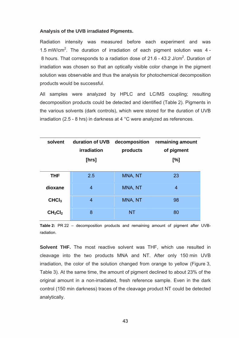
Analysis of the UVB irradiated Pigments.
Radiation intensity was measured before each experiment and was
1.5 mW/cm2. The duration of irradiation of each pigment solution was 4 -
8 hours. That corresponds to a radiation dose of 21.6 - 43.2 J/cm2. Duration of
irradiation was chosen so that an optically visible color change in the pigment
solution was observable and thus the analysis for photochemical decomposition
products would be successful.
All samples were analyzed by HPLC and LC/MS coupling; resulting
decomposition products could be detected and identified (Table 2). Pigments in
the various solvents (dark controls), which were stored for the duration of UVB
irradiation (2.5 - 8 hrs) in darkness at 4 °C were analyzed as references.
solvent duration of UVB irradiation
[hrs]
decomposition products
remaining amount of pigment
[%]
THF 2.5 MNA, NT 23
dioxane 4 MNA, NT 4
CHCl3 4 MNA, NT 98
CH2Cl2 8 NT 80
Table 2: PR 22 – decomposition products and remaining amount of pigment after UVB-
radiation.
Solvent THF. The most reactive solvent was THF, which use resulted in
cleavage into the two products MNA and NT. After only 150 min UVB
irradiation, the color of the solution changed from orange to yellow (Figure 3,
Table 3). At the same time, the amount of pigment declined to about 23% of the
original amount in a non-irradiated, fresh reference sample. Even in the dark
control (150 min darkness) traces of the cleavage product NT could be detected
analytically.
43
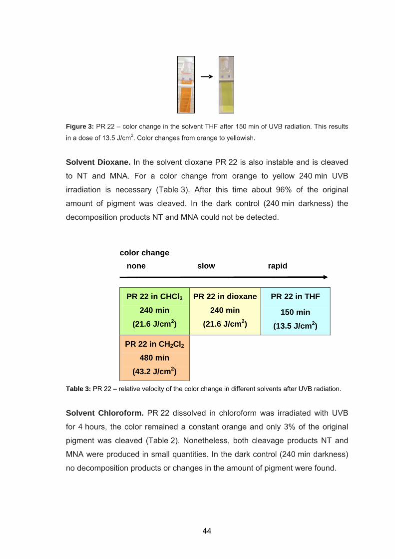
Figure 3: PR 22 – color change in the solvent THF after 150 min of UVB radiation. This results
in a dose of 13.5 J/cm2. Color changes from orange to yellowish.
Solvent Dioxane. In the solvent dioxane PR 22 is also instable and is cleaved
to NT and MNA. For a color change from orange to yellow 240 min UVB
irradiation is necessary (Table 3). After this time about 96% of the original
amount of pigment was cleaved. In the dark control (240 min darkness) the
decomposition products NT and MNA could not be detected.
color change none slow rapid
PR 22 in CHCl3 240 min
(21.6 J/cm2)
PR 22 in dioxane240 min
(21.6 J/cm2)
PR 22 in THF
150 min (13.5 J/cm2)
PR 22 in CH2Cl2 480 min
(43.2 J/cm2)
Table 3: PR 22 – relative velocity of the color change in different solvents after UVB radiation.
Solvent Chloroform. PR 22 dissolved in chloroform was irradiated with UVB
for 4 hours, the color remained a constant orange and only 3% of the original
pigment was cleaved (Table 2). Nonetheless, both cleavage products NT and
MNA were produced in small quantities. In the dark control (240 min darkness)
no decomposition products or changes in the amount of pigment were found.
44
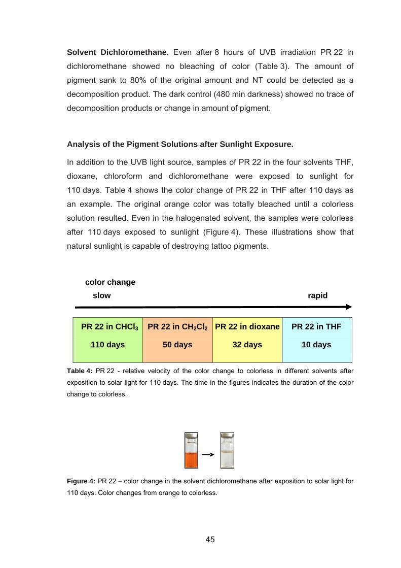
Solvent Dichloromethane. Even after 8 hours of UVB irradiation PR 22 in
dichloromethane showed no bleaching of color (Table 3). The amount of
pigment sank to 80% of the original amount and NT could be detected as a
decomposition product. The dark control (480 min darkness) showed no trace of
decomposition products or change in amount of pigment.
Analysis of the Pigment Solutions after Sunlight Exposure.
In addition to the UVB light source, samples of PR 22 in the four solvents THF,
dioxane, chloroform and dichloromethane were exposed to sunlight for
110 days. Table 4 shows the color change of PR 22 in THF after 110 days as
an example. The original orange color was totally bleached until a colorless
solution resulted. Even in the halogenated solvent, the samples were colorless
after 110 days exposed to sunlight (Figure 4). These illustrations show that
natural sunlight is capable of destroying tattoo pigments.
color change slow rapid
PR 22 in CHCl3
110 days
PR 22 in CH2Cl2
50 days
PR 22 in dioxane
32 days
PR 22 in THF
10 days
Table 4: PR 22 - relative velocity of the color change to colorless in different solvents after
exposition to solar light for 110 days. The time in the figures indicates the duration of the color
change to colorless.
Figure 4: PR 22 – color change in the solvent dichloromethane after exposition to solar light for
110 days. Color changes from orange to colorless.
45
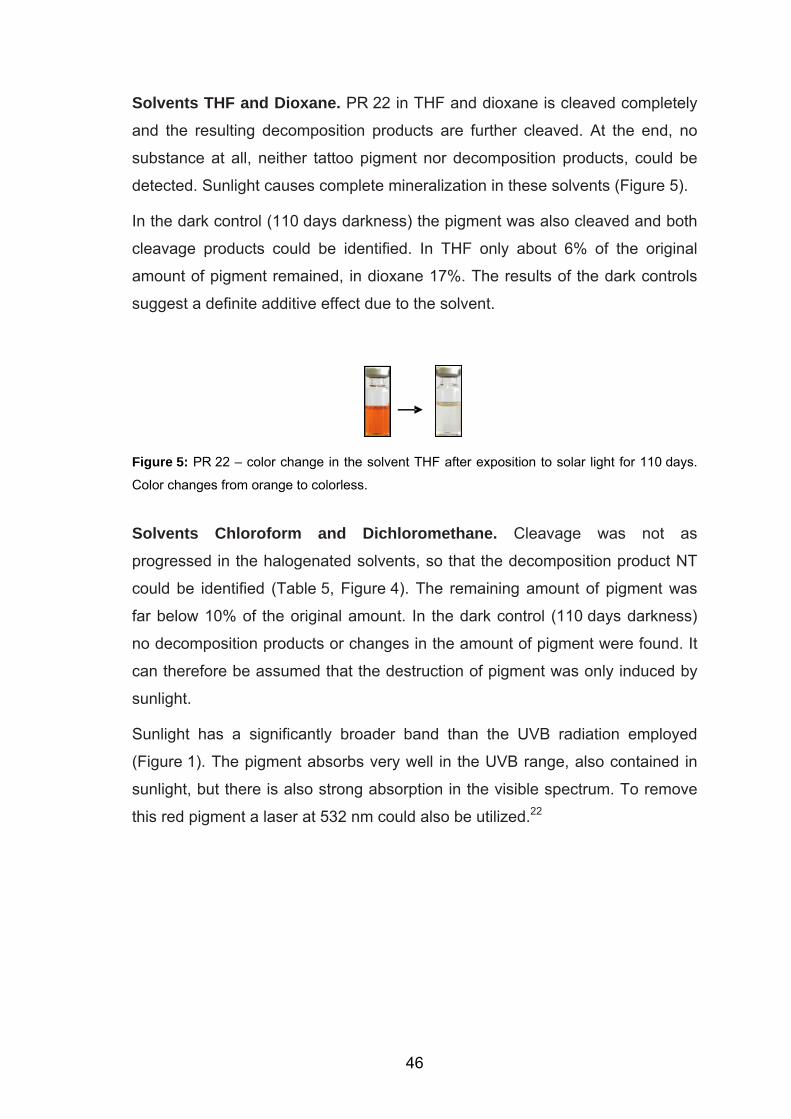
Solvents THF and Dioxane. PR 22 in THF and dioxane is cleaved completely
and the resulting decomposition products are further cleaved. At the end, no
substance at all, neither tattoo pigment nor decomposition products, could be
detected. Sunlight causes complete mineralization in these solvents (Figure 5).
In the dark control (110 days darkness) the pigment was also cleaved and both
cleavage products could be identified. In THF only about 6% of the original
amount of pigment remained, in dioxane 17%. The results of the dark controls
suggest a definite additive effect due to the solvent.
Figure 5: PR 22 – color change in the solvent THF after exposition to solar light for 110 days.
Color changes from orange to colorless.
Solvents Chloroform and Dichloromethane. Cleavage was not as
progressed in the halogenated solvents, so that the decomposition product NT
could be identified (Table 5, Figure 4). The remaining amount of pigment was
far below 10% of the original amount. In the dark control (110 days darkness)
no decomposition products or changes in the amount of pigment were found. It
can therefore be assumed that the destruction of pigment was only induced by
sunlight.
Sunlight has a significantly broader band than the UVB radiation employed
(Figure 1). The pigment absorbs very well in the UVB range, also contained in
sunlight, but there is also strong absorption in the visible spectrum. To remove
this red pigment a laser at 532 nm could also be utilized.22
46
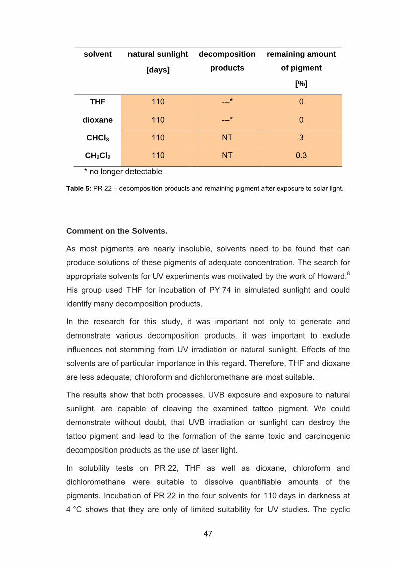
solvent natural sunlight
[days]
decomposition products
remaining amount of pigment
[%]
THF 110 ---* 0
dioxane 110 ---* 0
CHCl3 110 NT 3
CH2Cl2 110 NT 0.3
* no longer detectable
Table 5: PR 22 – decomposition products and remaining pigment after exposure to solar light.
Comment on the Solvents.
As most pigments are nearly insoluble, solvents need to be found that can
produce solutions of these pigments of adequate concentration. The search for
appropriate solvents for UV experiments was motivated by the work of Howard.8
His group used THF for incubation of PY 74 in simulated sunlight and could
identify many decomposition products.
In the research for this study, it was important not only to generate and
demonstrate various decomposition products, it was important to exclude
influences not stemming from UV irradiation or natural sunlight. Effects of the
solvents are of particular importance in this regard. Therefore, THF and dioxane
are less adequate; chloroform and dichloromethane are most suitable.
The results show that both processes, UVB exposure and exposure to natural
sunlight, are capable of cleaving the examined tattoo pigment. We could
demonstrate without doubt, that UVB irradiation or sunlight can destroy the
tattoo pigment and lead to the formation of the same toxic and carcinogenic
decomposition products as the use of laser light.
In solubility tests on PR 22, THF as well as dioxane, chloroform and
dichloromethane were suitable to dissolve quantifiable amounts of the
pigments. Incubation of PR 22 in the four solvents for 110 days in darkness at
4 °C shows that they are only of limited suitability for UV studies. The cyclic
47
ethers THF and dioxane were so reactive, that even without UV irradiation a
portion of the pigment was destroyed. THF and dioxane are thus not suitable for
photochemical studies of the pigment, as it cannot be established which
reactions are mediated by the solvent and which are purely induced by UV
radiation.
The halogenated solvents chloroform and dichloromethane, in contrast, have no
influence on the stability of the pigment and the amount of dye remained
unchanged during 110 days in darkness. Both solvents are well suited for
photochemical studies of pigments in vitro.
Comment on the Radiation Doses.
Unfortunately, as far as we know, no scientific reports on the chemical stability
of tattoo pigments in the skin after light exposure exist. Patients do report
repeatedly of fading of tattoos or even almost total disappearance, especially in
cases of PMU. These reports are, unfortunately, not subject to scientific
analysis.
The light doses employed may at first appear somewhat high. They therefore
have to be correlated to natural UVB radiation on earth or to medical-
therapeutic light doses (Table 6). Depending on the angle of incidence of the
sun and geographic latitude, highly variable UVB light intensities reach the skin
and the tattoo pigments therein. The cumulative UVB dose in Germany is about
330 J/cm2.23 In the treatment of psoriasis, artificial UVB light sources are
employed that can apply an average cumulative dose of 20 J/cm2 in the
tattooed area in a matter of a few weeks.24
In comparing the dose of natural or medically applied UVB light with the dose
used in our in vitro experiments, the following must be kept in mind: histology
shows that the pigment occurs in crystalline from usually intracellularly in the
dermis. These pigment crystals are found at a depth of about 0.25 - 1.7 mm in
the papillary as well as the reticular dermis.6 The intensity of UVB radiation
decreases dramatically at these depths. The effect of UVB light is probably
limited to pigment in the papillary dermis. Due to the very high absorption
coefficient of the pigments, the little amount of UVB which reaches the pigment
48
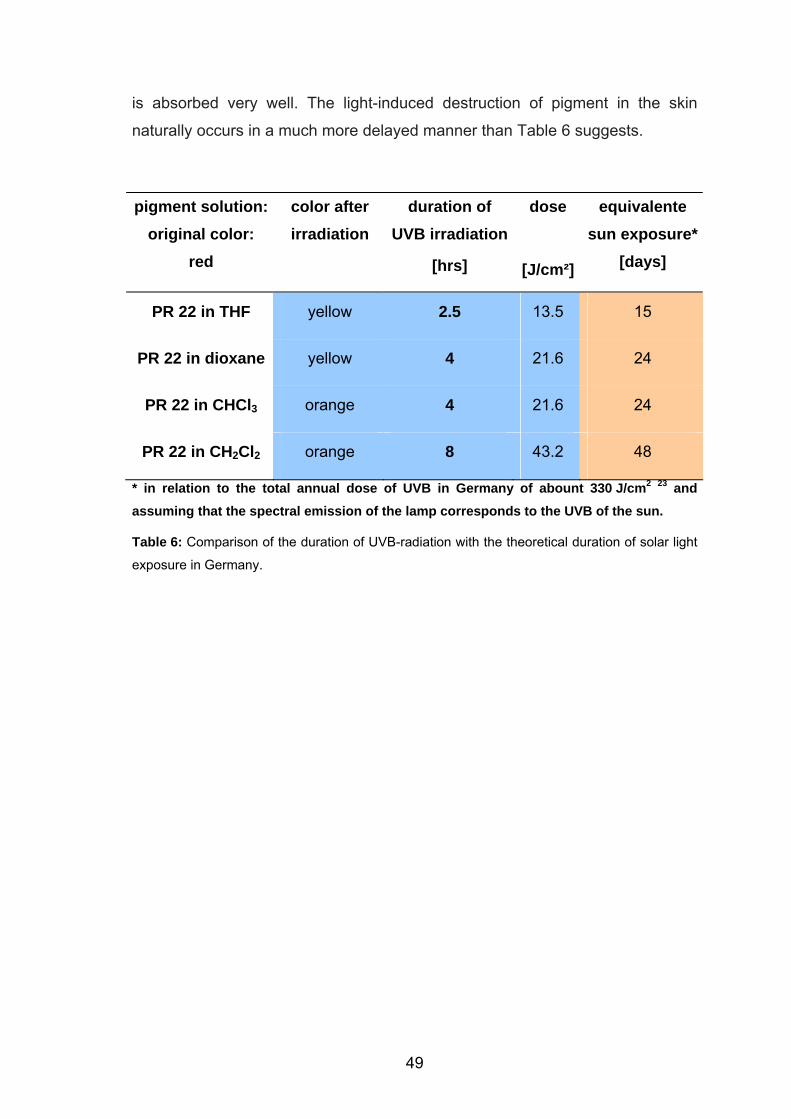
is absorbed very well. The light-induced destruction of pigment in the skin
naturally occurs in a much more delayed manner than Table 6 suggests.
pigment solution: original color:
red
color after irradiation
duration of UVB irradiation
[hrs]
dose
[J/cm²]
equivalente sun exposure*
[days]
PR 22 in THF yellow 2.5 13.5 15
PR 22 in dioxane yellow 4 21.6 24
PR 22 in CHCl3 orange 4 21.6 24
PR 22 in CH2Cl2 orange 8 43.2 48
* in relation to the total annual dose of UVB in Germany of abount 330 J/cm2 23 and assuming that the spectral emission of the lamp corresponds to the UVB of the sun.
Table 6: Comparison of the duration of UVB-radiation with the theoretical duration of solar light
exposure in Germany.
49
3.4. Conclusions
Our results show for the first time that the tattoo pigment PR 22 is chemically
altered when exposed to sunlight or broad band UVB radiation. For in vitro
studies, the solvents chloroform and dichloromethane appear most suitable, as
they adequately dissolve the pigment and hardly affect the light-induced
cleavage process. For PR 22, the decomposition products MNA and NT
postulated from laser treatment could be detected. Further degradation of the
detected decomposition products cannot be ruled out. The toxicological
assessment based on available literature shows that both MNA and NT can
have cumulative effects on the organism and can be toxic on inspiration,
swallowing or contact with the skin. MNA is also a mutagen and appears in
category 2 of cancer-causing substances. NT also possesses genotoxic
potential. To which extent these results apply to the in vivo situation must be
clarified by further studies.
An assessment of the health hazard cannot be made on the basis of the current
data. Toxicity and carcinogenicity of chemical compounds as well as their light-
induced decomposition products depends, among other factors, on their
concentration in the skin. Considering the large number of people with tattoos
that spend time in the sun or receive medically indicated UVB therapy, it is
important to determine the concentration of tattoo pigments in the skin in order
to evaluate the associated risk. Further studies on tattooed skin are needed.
50
3.5. References
1 Fisher, G.J.; Datta, S.C.; Talwar, H.S.; Wang, Z.Q.; Varani, J.; Kang, S.;
Voorhees, J.J. Nature 1996, 379, 335. 2 Hanson, K.M.; Simon, J.D. Proc. Natl. Acad. Sci. U.S.A. 1998, 95, 10576. 3 Allensbach IfD. Körperkult bei den Jüngeren: Tattoos und piercings. 2003;
Nr. 24, IfD-Umfrage 7046. 4 Lehmann, G.; Pierchalla, P. Derm. Beruf Umwelt 1988, 36, 152. 5 Baumler, W.; Eibler, E.T.; Hohenleutner, U.; Sens, B.; Sauer, J.; Landthaler,
M. Lasers Surg. Med. 2000, 26, 13. 6 Ferguson, J.E.; Andrew, S.M.; Jones, C.J.; August P.J. Br. J. Dermatol.
1997, 137, 405. 7 Ross, V.; Naseef, G.; Lin, G.; Kelly, M.; Michaud, N.; Flotte, T.J.; Raythen,
J.; Anderson, R,R. Arch. Dermatol. 1998, 134, 167. 8 Cui, Y.; Spann, A.P.; Couch, L.H.; Gopee, N.V.; Evans, F.E.; Churchwell,
M.I.; Williams, L.D.; Doerge, D.R.; Howard, P.C. Photochem. Photobiol.
2004, 80, 175. 9 Steinbrecher, I.; Hemmer, W.; Jarisch, R. J. Dtsch. Dermatol. Ges. 2004, 2,
1007. 10 Greve, B.; Chytry, R.; Raulin, C. Contact Derm. 2003, 49, 265. 11 Balfour, E.; Olhoffer, I.; Leffell, D.; Handerson, T. Am. J. Dermatopathol.
2003, 25, 338. 12 Mortimer, N.J.; Chave, T.A.; Johnston, G.A. Clin. Exp. Dermatol. 2003, 28,
508. 13 Vasold, R.; Naarmann, N.; Ulrich, H.; Fischer, D.; Konig, B.; Landthaler, M.;
Baumler, W. Photochem. Photobiol. 2004, 80, 180. 14 Cook, W.L.; Gebler, D.P.; Pratt, N.E. Production of organic pigments and
printing inks containing them; PCT Int. Appl. 2001. 15 Engel, E.; Santarelli, F.; Vasold, R.; Ulrich, H.; Maisch, T.; Konig, B.;
Landthaler, M.; Gopee, N.V.; Howard, P.C.; Baumler, W. Anal. Chem. 2006,
78, 6440. 16 homepage leffingwell http://www.leffingwell.com/cosmetics/vol_1en.pdf.
51
52
17 Huang, Q.G.; Kong, L.R.; Liu, Y.B.; Wang, L.S. Bull. Environ. Contam.
Toxicol. 1996, 57, 349. 18 Shimizu, H.; Kumada, T.; Nakano, S.; Kiriyama, S.; Sone, Y.; Honda, T.;
Watanabe, K.; Nakano, I.; Fukuda, Y.; Hayakawa, T. Gut 2002, 50, 266. 19 Sayama, M.; Mori, M.; Shoji, M.; Uda, S.; Kakikawa, M.; Kondo, T.; Kodaira,
K.I. Mutat. Res. 1998, 420, 27. 20 NTP Toxicology and Carcinogenesis Studies of 1,4-Dichlorobenzene (CAS
No. 106-46-7) in F344/N Rats and B6C3F1 Mice (Gavage Studies). Natl.
Toxicol. Program Tech. Rep. Ser. 1987, 319, 1. 21 Lo, H.H.; Brown, P.I.; Rankin, G.O. Toxicology 1990, 63, 215. 22 Kuperman-Beade, M.; Levine, V.J.; Ashinoff, R. Am. J. Clin. Dermatol. 2001,
2, 21. 23 Feister, U.; Jakel, E.; Gericke, K. Photochem. Photobiol. 2002, 76, 281. 24 Gerber, W.; Arheilger, B.; Ha, T.A.; Hermann, J.; Ockenfels, H.M. Br. J.
Dermatol. 2003, 149, 1250.
4. Tattoo Pigments in Skin: Concentration, Transportation and Light Induced Decomposition of an Azo Pigment
using SKH-1 Mouse Model∗
4.1. Introduction
Puncturing tattoo pigments into the skin can be compared with an injury of the
upper skin layers. As soon as the pigments are injected into the dermis they are
recognized by the body as foreign particles. By means of phagocytosis, the
tattoo pigments are removed from the site of tattooed skin and are transported
by the lymphatic system to other anatomical locations. As described in literature
lymph nodes located next to the tattoo show frequently black pigmentation
mimicking metastatic malignant melanoma or positive sentinel lymph node in
melanoma.1,2
Based upon sentinel node biopsy, these pigmented lymph nodes are removed
and analyzed by histopathology. However, histopathological examination of the
lymph nodes does not detect metastases. In such cases, the pigmentation of
the lymph nodes is caused by decorative tattoos of the skin area next to these
lymph nodes. In addition, exposure of pigments in the skin to solar light or laser
radiation during tattoo removal can cause decomposition of the pigments
yielding hazardous compounds such as carcinogenic amines.3-5
To investigate transportation of tattoo pigments after tattooing, we established
an animal model using SKH-1 hairless mice. The mice received tattoos with
Pigment Red 22 (PR 22) on their back. The extraction of pigments immediately
after tattooing yielded the concentration of pigments that is placed in the body.
In contrast to that, the extraction of pigments from skin six weeks (42 days) after
tattooing should elucidate the extent of pigment transportation in the mice. To
investigate the decomposition of pigments by laser or solar light, laser irradiates
tattooed skin (ex vivo) or living mice are exposed to solar light for 31 days.
∗ A manuscript is in preparation: Engel, E.; Santarelli, F.; Vasold, R.; Maisch, T.; Howard, P.C.; Ulrich, H.; König, B.; Landthaler, M.; Bäumler, W. Toxicol. Appl. Pharmacol. 2007. ∗ Tattooing of the mice was performed by P.C. Howard. ∗ Samle preparation was done by F. Santarelli.
53
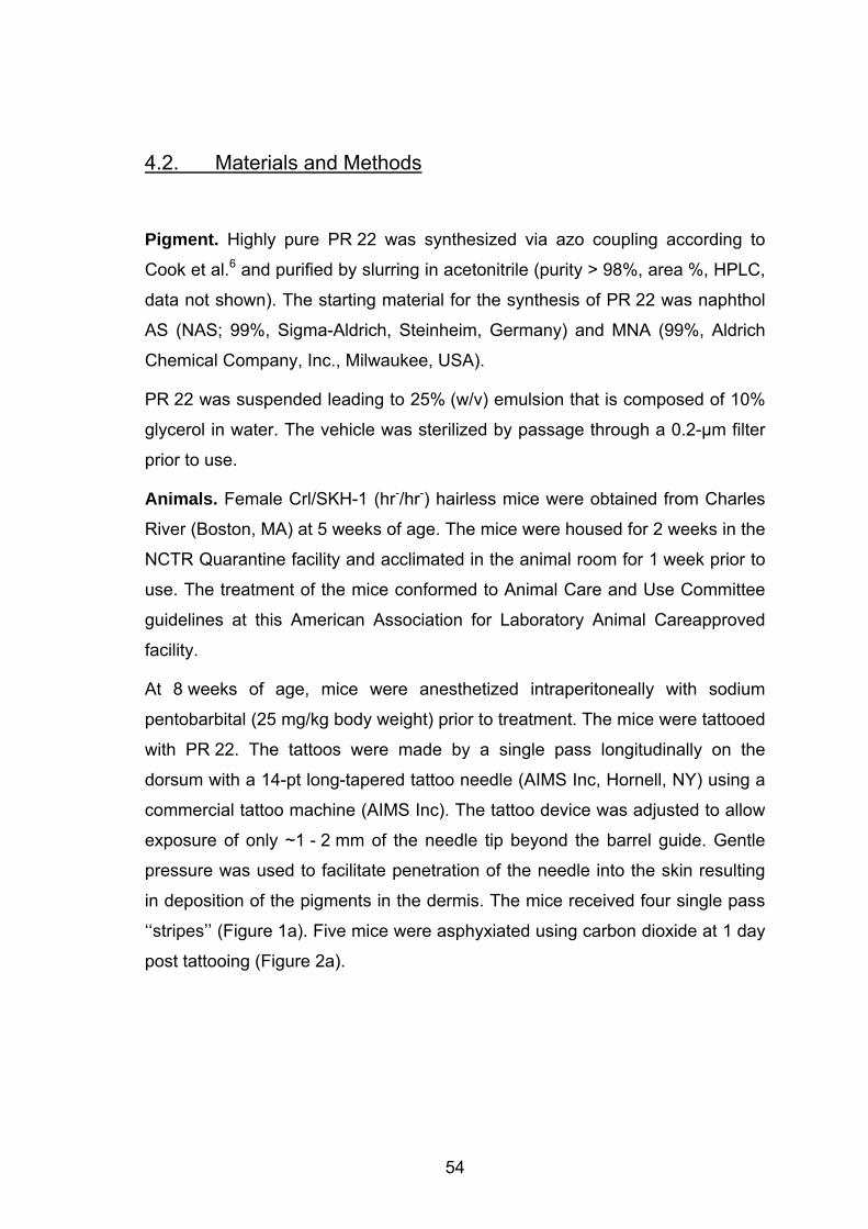
4.2. Materials and Methods
Pigment. Highly pure PR 22 was synthesized via azo coupling according to
Cook et al.6 and purified by slurring in acetonitrile (purity > 98%, area %, HPLC,
data not shown). The starting material for the synthesis of PR 22 was naphthol
AS (NAS; 99%, Sigma-Aldrich, Steinheim, Germany) and MNA (99%, Aldrich
Chemical Company, Inc., Milwaukee, USA).
PR 22 was suspended leading to 25% (w/v) emulsion that is composed of 10%
glycerol in water. The vehicle was sterilized by passage through a 0.2-µm filter
prior to use.
Animals. Female Crl/SKH-1 (hr-/hr-) hairless mice were obtained from Charles
River (Boston, MA) at 5 weeks of age. The mice were housed for 2 weeks in the
NCTR Quarantine facility and acclimated in the animal room for 1 week prior to
use. The treatment of the mice conformed to Animal Care and Use Committee
guidelines at this American Association for Laboratory Animal Careapproved
facility.
At 8 weeks of age, mice were anesthetized intraperitoneally with sodium
pentobarbital (25 mg/kg body weight) prior to treatment. The mice were tattooed
with PR 22. The tattoos were made by a single pass longitudinally on the
dorsum with a 14-pt long-tapered tattoo needle (AIMS Inc, Hornell, NY) using a
commercial tattoo machine (AIMS Inc). The tattoo device was adjusted to allow
exposure of only ~1 - 2 mm of the needle tip beyond the barrel guide. Gentle
pressure was used to facilitate penetration of the needle into the skin resulting
in deposition of the pigments in the dermis. The mice received four single pass
‘‘stripes’’ (Figure 1a). Five mice were asphyxiated using carbon dioxide at 1 day
post tattooing (Figure 2a).
54
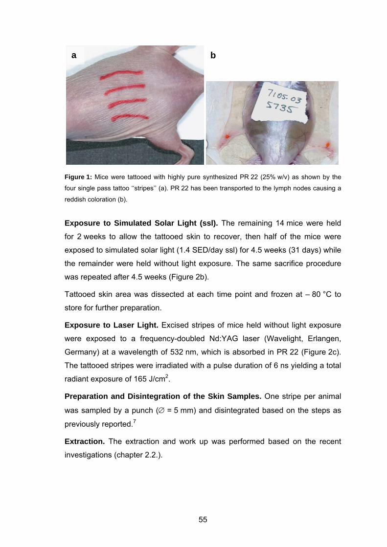
b
b a
Figure 1: Mice were tattooed with highly pure synthesized PR 22 (25% w/v) as shown by the
four single pass tattoo ‘‘stripes’’ (a). PR 22 has been transported to the lymph nodes causing a
reddish coloration (b).
Exposure to Simulated Solar Light (ssl). The remaining 14 mice were held
for 2 weeks to allow the tattooed skin to recover, then half of the mice were
exposed to simulated solar light (1.4 SED/day ssl) for 4.5 weeks (31 days) while
the remainder were held without light exposure. The same sacrifice procedure
was repeated after 4.5 weeks (Figure 2b).
Tattooed skin area was dissected at each time point and frozen at – 80 °C to
store for further preparation.
Exposure to Laser Light. Excised stripes of mice held without light exposure
were exposed to a frequency-doubled Nd:YAG laser (Wavelight, Erlangen,
Germany) at a wavelength of 532 nm, which is absorbed in PR 22 (Figure 2c).
The tattooed stripes were irradiated with a pulse duration of 6 ns yielding a total
radiant exposure of 165 J/cm2.
Preparation and Disintegration of the Skin Samples. One stripe per animal
was sampled by a punch (∅ = 5 mm) and disintegrated based on the steps as
previously reported.7
Extraction. The extraction and work up was performed based on the recent
investigations (chapter 2.2.).
55
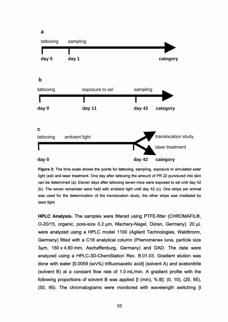
exposure to ssl tattooing sampling
day 0 day 42 category
tattooing ambient light translocation study laser treatment
c
Figure 2: The time scale shows the points for tattooing, sampling, exposure to simulated solar
light (ssl) and laser treatment. One day after tattooing the amount of PR 22 punctured into skin
can be determined (a). Eleven days after tattooing seven mice were exposed to ssl until day 42
(b). The seven remainder were held with ambient light until day 42 (c). One stripe per animal
was used for the determination of the translocation study, the other stripe was irradiated by
laser light.
HPLC Analysis. The samples were filtered using PTFE-filter (CHROMAFIL®,
O-20/15, organic, pore-size 0.2 µm, Machery-Nagel, Düren, Germany). 20 µL
were analyzed using a HPLC model 1100 (Agilent Technologies, Waldbronn,
Germany) fitted with a C18 analytical column (Phenomenex luna, particle size
3µm, 150 x 4.60 mm, Aschaffenburg, Germany) and DAD. The data were
analyzed using a HPLC-3D-ChemStation Rev. B.01.03. Gradient elution was
done with water [0.0059 (w/v%) trifluoroacetic acid] (solvent A) and acetonitrile
(solvent B) at a constant flow rate of 1.0 mL/min. A gradient profile with the
following proportions of solvent B was applied [t (min), % B]: (0, 10), (20, 95),
(50, 95). The chromatograms were monitored with wavelength switching [t
a
tattooing sampling
day 0 day 1 category
b
day 0 day 11 day 42 category
56
(min), λ nm]: (0, 258), (11.85, 228), (12.70, 280), (15.77, 228), (19.60, 280),
(24.50, 266).
The concentration of PR 22, 2-methyl-5-nitroaniline (MNA), 4-nitrotoluene (NT)
and Naphthol AS in the solutions was determined by the method of internal
standard. For each compound (i), the calibration factor (CFi) was determined in
a calibration run (single level calibration). The respective concentration of the
standard was chosen to be in the range of the concentration of the tattoo
pigment.
CFi = ffTr
i = K
iKTr
Tri
amam⋅⋅ KK
K
K K
K
Where fTr is the response-factor of the internal standard (ISTD), m i the mass of
compound i in the solution k and m the mass of ISTD in solution k. a is the
area of ISTD in solution k and a i the area of compound i in solution k.
Tr Tr
57
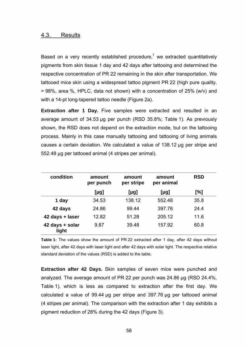
4.3. Results
Based on a very recently established procedure,7 we extracted quantitatively
pigments from skin tissue 1 day and 42 days after tattooing and determined the
respective concentration of PR 22 remaining in the skin after transportation. We
tattooed mice skin using a widespread tattoo pigment PR 22 (high pure quality,
> 98%, area %, HPLC, data not shown) with a concentration of 25% (w/v) and
with a 14-pt long-tapered tattoo needle (Figure 2a).
Extraction after 1 Day. Five samples were extracted and resulted in an
average amount of 34.53 µg per punch (RSD 35.8%; Table 1). As previously
shown, the RSD does not depend on the extraction mode, but on the tattooing
process. Mainly in this case manually tattooing and tattooing of living animals
causes a certain deviation. We calculated a value of 138.12 µg per stripe and
552.48 µg per tattooed animal (4 stripes per animal).
condition amount per punch
amount per stripe
amount per animal
RSD
[µg] [µg] [µg] [%] 1 day 34.53 138.12 552.48 35.8
42 days 24.86 99.44 397.76 24.4 42 days + laser 12.82 51.28 205.12 11.6 42 days + solar
light 9.87 39.48 157.92 60.8
Table 1: The values show the amount of PR 22 extracted after 1 day, after 42 days without
laser light, after 42 days with laser light and after 42 days with solar light. The respective relative
standard deviation of the values (RSD) is added to the table.
Extraction after 42 Days. Skin samples of seven mice were punched and
analyzed. The average amount of PR 22 per punch was 24.86 µg (RSD 24.4%,
Table 1), which is less as compared to extraction after the first day. We
calculated a value of 99.44 µg per stripe and 397.76 µg per tattooed animal
(4 stripes per animal). The comparison with the extraction after 1 day exhibits a
pigment reduction of 28% during the 42 days (Figure 3).
58
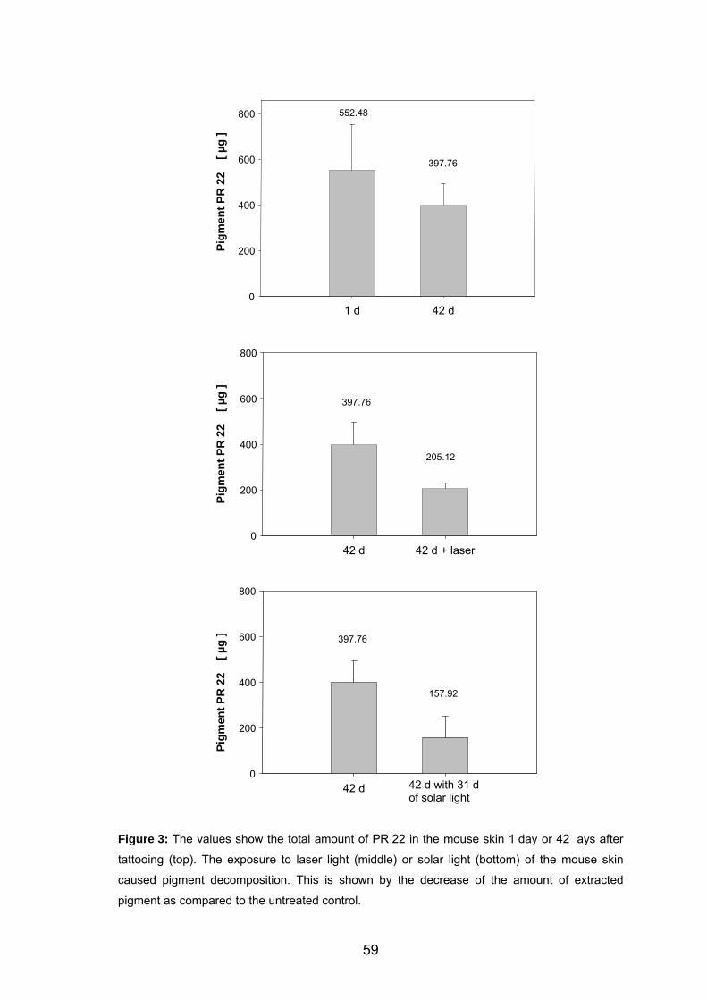
1 d 42 d
Pigm
ent P
R 2
2
[ µg
]
0
200
400
600
800
42 d 42 d + laser
Pigm
ent P
R 2
2
[ µg
]
0
200
400
600
800
42 d 42 d with 31 dof solar light
800
600
400
200
0
Pigm
ent P
R 2
2
[ µg
] 397.76
157.92
205.12
397.76
397.76
552.48
Figure 3: The values show the total amount of PR 22 in the mouse skin 1 day or 42 ays after
tattooing (top). The exposure to laser light (middle) or solar light (bottom) of the mouse skin
caused pigment decomposition. This is shown by the decrease of the amount of extracted
pigment as compared to the untreated control.
59
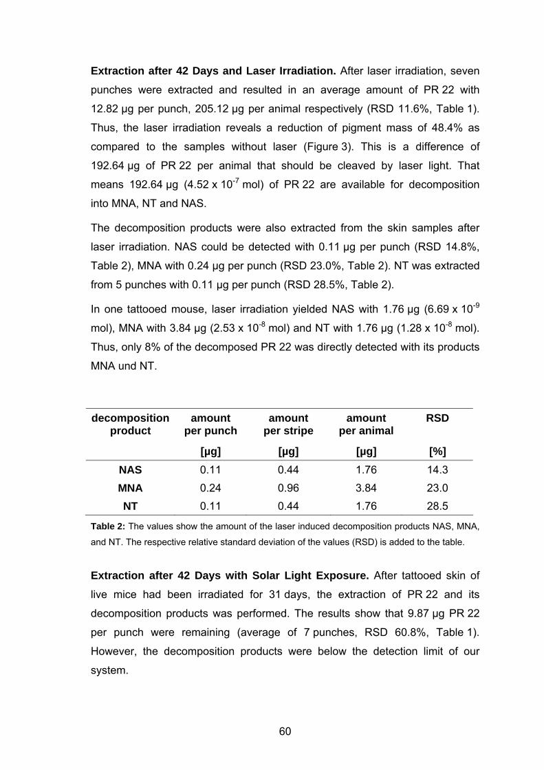
Extraction after 42 Days and Laser Irradiation. After laser irradiation, seven
punches were extracted and resulted in an average amount of PR 22 with
12.82 µg per punch, 205.12 µg per animal respectively (RSD 11.6%, Table 1).
Thus, the laser irradiation reveals a reduction of pigment mass of 48.4% as
compared to the samples without laser (Figure 3). This is a difference of
192.64 µg of PR 22 per animal that should be cleaved by laser light. That
means 192.64 µg (4.52 x 10⁻⁷ mol) of PR 22 are available for decomposition
into MNA, NT and NAS.
The decomposition products were also extracted from the skin samples after
laser irradiation. NAS could be detected with 0.11 µg per punch (RSD 14.8%,
Table 2), MNA with 0.24 µg per punch (RSD 23.0%, Table 2). NT was extracted
from 5 punches with 0.11 µg per punch (RSD 28.5%, Table 2).
In one tattooed mouse, laser irradiation yielded NAS with 1.76 µg (6.69 x 10⁻⁹
mol), MNA with 3.84 µg (2.53 x 10⁻⁸ mol) and NT with 1.76 µg (1.28 x 10⁻⁸ mol).
Thus, only 8% of the decomposed PR 22 was directly detected with its products
MNA und NT.
decomposition product
amount per punch
amount per stripe
amount per animal
RSD
[µg] [µg] [µg] [%] NAS 0.11 0.44 1.76 14.3 MNA 0.24 0.96 3.84 23.0 NT 0.11 0.44 1.76 28.5
Table 2: The values show the amount of the laser induced decomposition products NAS, MNA,
and NT. The respective relative standard deviation of the values (RSD) is added to the table.
Extraction after 42 Days with Solar Light Exposure. After tattooed skin of
live mice had been irradiated for 31 days, the extraction of PR 22 and its
decomposition products was performed. The results show that 9.87 µg PR 22
per punch were remaining (average of 7 punches, RSD 60.8%, Table 1).
However, the decomposition products were below the detection limit of our
system.
60
After 42 days of exposure to simulated solar light, up to 157.92 µg of PR 22
stay inside the dermis of an animal. That is, up to 60% of the pigment is cleaved
by the simulated solar light (Figure 3) during irradiation.
61
4.4. Discussion
A major prerequisite for a health risk assessment is the knowledge of the
pigment concentration in the human body. Tattooing is an archaic procedure
and there is no information about the amount of pigments punctured in the skin,
which is currently a major obstacle for such a risk assessment. Since the
concentration of pigments in tissue is unknown, there is a critical need for
scientific investigations. Since it is difficult to study tattooing and pigment
transportation in humans, we applied the SKH-1 mouse model for these
investigations.
Transportation of Pigments. In the process of tattooing, the pigment
suspension is simply punctured in the skin using tiny needles. Deposition of the
pigment in the dermis results in the permanence usually associated with tattoos.
Tattoo colorants are injected into the skin and are partly transported to other
anatomical locations, whereas the exact mechanisms of transportation and
distribution remain unknown. Among others, the local lymph nodes show the
same color as the tattoos in the skin (Figure 2b).8 Thus, tattoo colorants are
administered like medical drugs, but without any medical reason.
The lack of information might be due to the complex pigment chemistry, the
high insolubility of pigments and the difficulty to extract pigments quantitatively
from tissue. Based on a simple consideration, the amount of pigment placed in
the body must be equal to the amount in the skin directly after tattooing. After
that, one part of the pigments is transport away, in particular via lymphatic
system. The other part stays in the skin and causes the respective tattoo color.
Our results for the SKH-1 mice show that immediately after tattooing an
average amount of 34.53 µg of PR 22 was extracted from one punch. This
corresponds to 552.48 µg of tattoo pigment per tattooed animal (4 stripes per
animal).
The used tattoo needle showed a size of 14 pt (4.94 mm), that defines the width
of one single pass stripe. Based on this consideration each mouse received a
concentration of PR 22 of 176 mg/ 100 cm2. This value matches very well the
62
previously reported amount of 253 mg/ 100 cm2, which is a mean value of
different methods used (chapter 2.3.).
The reduction of the amount of PR 22 in the mouse skin is demonstrated by the
decrease of PR 22 from 34.53 µg to 24.86 µg within 42 days. With regard to the
size and number of stripes, 397.76 µg PR 22 per animal remained in the skin.
This leads to a concentration of 127 mg/ 100 cm2 considering the needle size of
14 pt. The comparison with the extraction after 1 day exhibits a pigment
reduction of 28% during the 42 days (Figure 3).
Laser Treatment. After several years people often decide to remove their
tattoos. Beside adverse reactions9-13 of the pigments itself, the main reasons for
removing tattoos are improved self-image or social stigmatization. Traditional
modalities are the removal of the pigment-containing skin using salabrasion,14
cryosurgery,15 surgical excision16 or CO2-laser application.17 However, these
methods induce permanent scarring.
Using Q-switch lasers, tattoo removal is associated with significantly lower risk
of scarring.18 The majority of tattoo pigment is found within cells, and not free,
within the dermis. While many pigment particles measure “a few microns”,
others are significantly larger19 or when accumulated within cells may act as
larger aggregate bodies. According to the principles of selective
photothermolysis20 the laser impulses show a high intensity and ultra short
pulse durations of a few nano seconds (Q-switched lasers).
After being absorbed in the pigment molecule, the energy of the laser light is
converted to heat or breaks chemical bonds inside the molecule. Additionally,
the ultrashort heating may lead to the disruption of the pigment particle. The
laser irradiation changes the shape and the size of the tattoo particles abruptly
as proved by histology.19
It is well known that an increase of temperature in a number of azo dyes above
280 °C forms 3,3’-dichlorobenzidine,21 a proven genotoxin towards human
lymphocytes.22 Laser irradiation of the widely used azo compound Pigment
Red 22 (PR 22) and resulted in the photodecomposition products 2-methyl-5-
nitroaniline (MNA), and 4-nitro-toluene (NT) (Figure 4).5 NT is toxic as shown
with human lymphocytes.23 5-nitro-o-toluidine, which is also designated to MNA,
may cause liver dysfunction as shown with workers from a hair dye factory.24
63
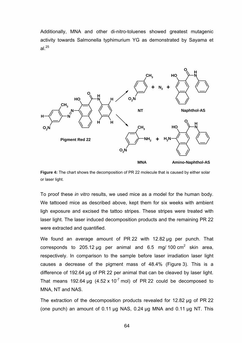
Additionally, MNA and other di-nitro-toluenes showed greatest mutagenic
activity towards Salmonella typhimurium YG as demonstrated by Sayama et
al.25
OH NHO
CH3
O2N
CH3
O2N
NH2
CH3
NHN
OH NHO
H
HHO2N OH N
HO
NH2
Naphthol-ASNT
MNA
Pigment Red 22
Amino-Naphthol-AS
N2+ +
+
Figure 4: The chart shows the decomposition of PR 22 molecule that is caused by either solar
or laser light.
To proof these in vitro results, we used mice as a model for the human body.
We tattooed mice as described above, kept them for six weeks with ambient
ligh exposure and excised the tattoo stripes. These stripes were treated with
laser light. The laser induced decomposition products and the remaining PR 22
were extracted and quantified.
We found an average amount of PR 22 with 12.82 µg per punch. That
corresponds to 205.12 µg per animal and 6.5 mg/ 100 cm2 skin area,
respectively. In comparison to the sample before laser irradiation laser light
causes a decrease of the pigment mass of 48.4% (Figure 3). This is a
difference of 192.64 µg of PR 22 per animal that can be cleaved by laser light.
That means 192.64 µg (4.52 x 10⁻⁷ mol) of PR 22 could be decomposed to
MNA, NT and NAS.
The extraction of the decomposition products revealed for 12.82 µg of PR 22
(one punch) an amount of 0.11 µg NAS, 0.24 µg MNA and 0.11 µg NT. This
64
yields in one tattooed mouse (192.64 µg of PR 22; 4.52 x 10⁻⁷ mol) NAS with
1.76 µg (6.69 x 10⁻⁹ mol), MNA with 3.84 µg (2.53 x 10⁻⁸ mol) and NT with
1.76 µg (1.28 x 10⁻⁸ mol). Thus, only 8% of the decomposed PR 22 was directly
identified by its products such as MNA und NT. The major part 12.82 µg of
PR 22 could have been decomposed by other pathways. However, it is more
likely that these major decomposition products has been metabolized to other
products and could have been therefore not detected by our analysis.
Irradiation with Solar Light. Recently a research team of the National Center
for Toxicological Research (NCTR) of the FDA showed that a yellow tattoo
pigment (PY 74) in solution is cleaved into toxic products photochemically when
exposed to UV radiation in a sunlight simulator.4 These authors concluded form
their in vitro study that these substances can be generated in tattooed skin
when this skin is exposed for a longer period of time to sunlight or other sources
of UV radiation.
For the first time we could show that solutions of PR 22 are chemically altered
when exposed to natural sunlight or broadband UVB radiation. The
decomposition products MNA and NT postulated from laser treatment could be
detected.3
To verify these in vitro results, the mouse model was used again. In contrast to
experiments with laser irradiation, the skin of the mice could heal from tattooing
for 10 days. Then the mice were exposed to simulated solar light (1.4 SED/day
ssl) for 32 days. After solar light exposure, the tattooed stripes were excised
and analyzed.
An average amount of 9.87 µg of PR 22 was extracted from one punch
(5.02 mg/ 100cm2). That is, up to 157.92 µg of PR 22 stay in side the dermis.
When comparing this value with the initial amount of pigment in the skin, up to
60% of the pigment is cleaved by the simulated solar light (Figure 3) during
irradiation. The concentrations of the decomposition products were below the
detection limit of our system. Obviously, PR 22 decomposed and the products
disappeared during the period of irradiation (32 days) either by metabolization
or by lymphatic transportation.
Time Scale. With the results of this study we can show now a time scale
regarding the concentration of tattoo pigment in skin and its changes.
65
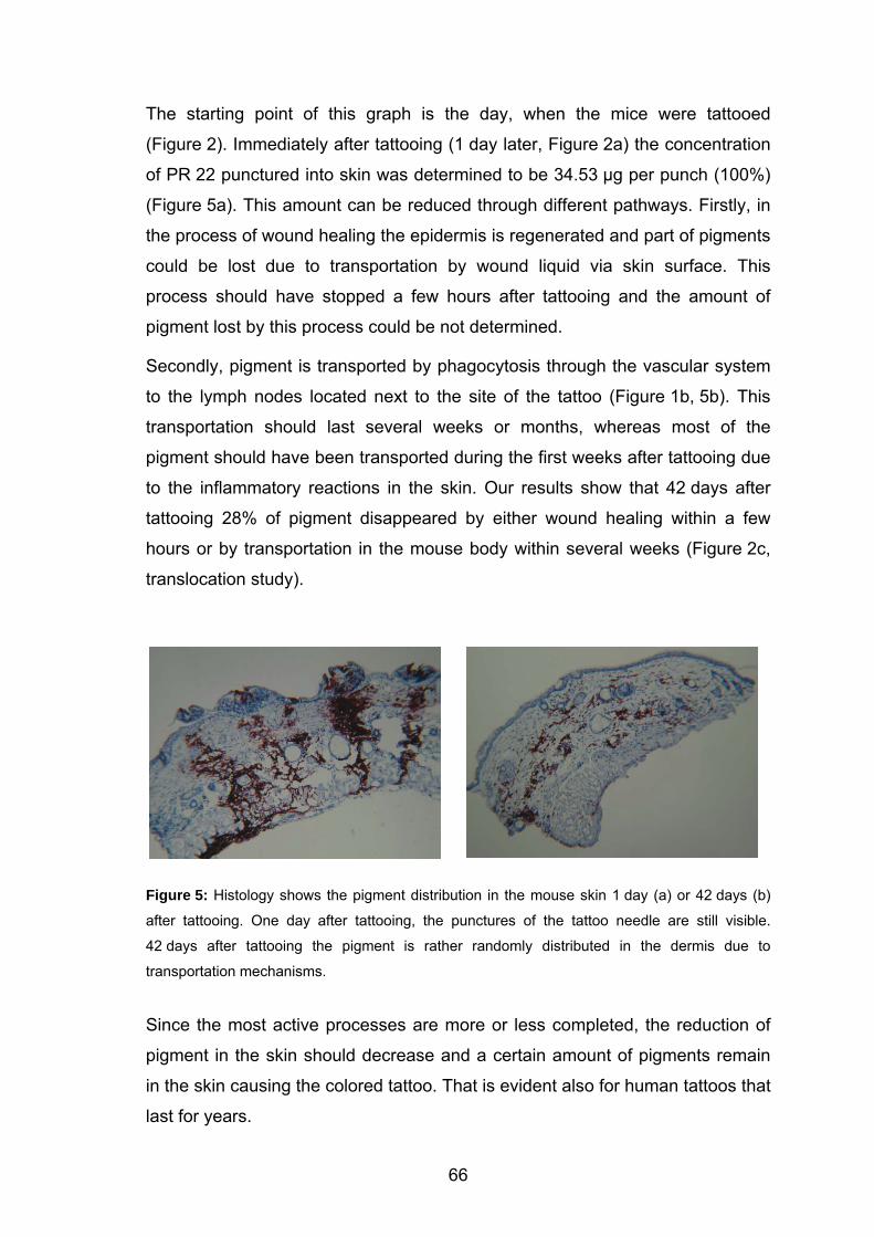
The starting point of this graph is the day, when the mice were tattooed
(Figure 2). Immediately after tattooing (1 day later, Figure 2a) the concentration
of PR 22 punctured into skin was determined to be 34.53 µg per punch (100%)
(Figure 5a). This amount can be reduced through different pathways. Firstly, in
the process of wound healing the epidermis is regenerated and part of pigments
could be lost due to transportation by wound liquid via skin surface. This
process should have stopped a few hours after tattooing and the amount of
pigment lost by this process could be not determined.
Secondly, pigment is transported by phagocytosis through the vascular system
to the lymph nodes located next to the site of the tattoo (Figure 1b, 5b). This
transportation should last several weeks or months, whereas most of the
pigment should have been transported during the first weeks after tattooing due
to the inflammatory reactions in the skin. Our results show that 42 days after
tattooing 28% of pigment disappeared by either wound healing within a few
hours or by transportation in the mouse body within several weeks (Figure 2c,
translocation study).
Figure 5: Histology shows the pigment distribution in the mouse skin 1 day (a) or 42 days (b)
after tattooing. One day after tattooing, the punctures of the tattoo needle are still visible.
42 days after tattooing the pigment is rather randomly distributed in the dermis due to
transportation mechanisms.
Since the most active processes are more or less completed, the reduction of
pigment in the skin should decrease and a certain amount of pigments remain
in the skin causing the colored tattoo. That is evident also for human tattoos that
last for years.
66
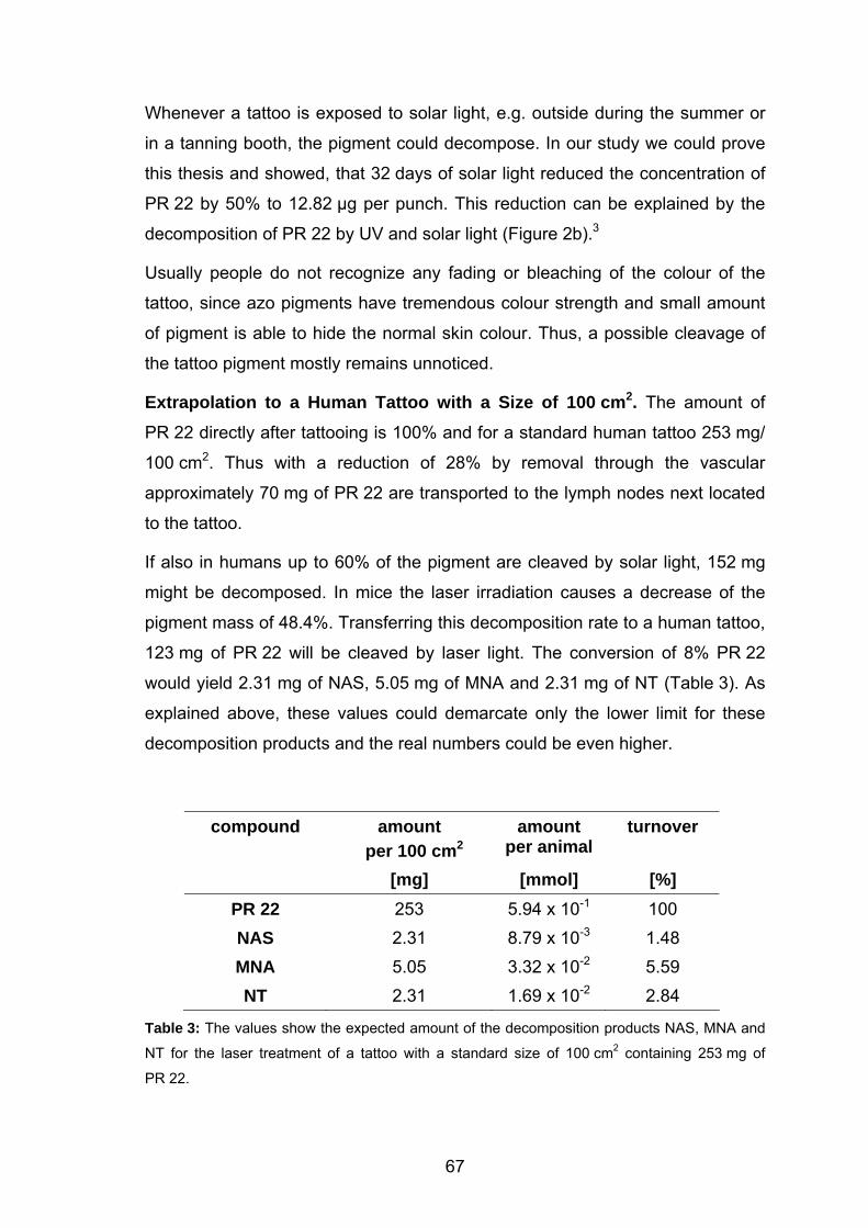
Whenever a tattoo is exposed to solar light, e.g. outside during the summer or
in a tanning booth, the pigment could decompose. In our study we could prove
this thesis and showed, that 32 days of solar light reduced the concentration of
PR 22 by 50% to 12.82 µg per punch. This reduction can be explained by the
decomposition of PR 22 by UV and solar light (Figure 2b).3
Usually people do not recognize any fading or bleaching of the colour of the
tattoo, since azo pigments have tremendous colour strength and small amount
of pigment is able to hide the normal skin colour. Thus, a possible cleavage of
the tattoo pigment mostly remains unnoticed.
Extrapolation to a Human Tattoo with a Size of 100 cm2. The amount of
PR 22 directly after tattooing is 100% and for a standard human tattoo 253 mg/
100 cm2. Thus with a reduction of 28% by removal through the vascular
approximately 70 mg of PR 22 are transported to the lymph nodes next located
to the tattoo.
If also in humans up to 60% of the pigment are cleaved by solar light, 152 mg
might be decomposed. In mice the laser irradiation causes a decrease of the
pigment mass of 48.4%. Transferring this decomposition rate to a human tattoo,
123 mg of PR 22 will be cleaved by laser light. The conversion of 8% PR 22
would yield 2.31 mg of NAS, 5.05 mg of MNA and 2.31 mg of NT (Table 3). As
explained above, these values could demarcate only the lower limit for these
decomposition products and the real numbers could be even higher.
compound amount per 100 cm2
amount per animal
turnover
[mg] [mmol] [%] PR 22 253 5.94 x 10⁻¹ 100 NAS 2.31 8.79 x 10⁻³ 1.48 MNA 5.05 3.32 x 10⁻² 5.59 NT 2.31 1.69 x 10⁻² 2.84
Table 3: The values show the expected amount of the decomposition products NAS, MNA and
NT for the laser treatment of a tattoo with a standard size of 100 cm2 containing 253 mg of
PR 22.
67

Comment on Radiant Exposure. The radiant exposure used in the SKH-1
mouse model differs from that radiant exposure applied in clinical practice. Here
tattoo removal is done in several sessions witch each 3 - 5 J/cm2 yielding a
lower total radiant exposure. Nevertheless our results are more than a proof of
evidence and might have an impact on clinical practice.
68
4.5. Conclusion
We could show for the first time that after tattooing and healing of the wound
28.0% of the injected pigment is removed from the site of the tattoo. This
corresponds to approximately 150 µg of tattoo pigment.
Up to 50% of PR 22 located inside the mouse skin can be cleaved by laser light.
Fortunately we could show, that only 8% of PR 22 are decomposed into the
hazardous compounds MNA and NT.
Regarding the exposure of tattoos to solar light we found that up to 60% of the
pigment is cleaved. The ex vivo postulated decomposition products (NAS, MNA,
NT) could not be detected. The reason might be, that the decomposition
products are removed by the lymphatic system from the site of the tattoo as
soon as they are generated.
69
4.6. References
1 Moehrle, M.; Blaheta, H. J.; Ruck, P. Dermatology 2001, 203, 342. 2 Friedman, T.; Westreich, M.; Mozes, S. N.; Dorenbaum, A.; Herman, O.
Plast. Reconstr. Surg. 2003, 111, 2120. 3 Engel, E.; Spannberger, A.; Vasold, R.; Konig, B.; Landthaler, M.; Baumler,
W. J. Dtsch. Dermatol. Ges. 2007, 5, 583. 4 Cui, Y.; Spann, A. P.; Couch, L. H.; Gopee, N. V.; Evans, F. E.; Churchwell,
M. I.; Williams, L. D.; Doerge, D. R.; Howard, P. C. Photochem. Photobiol.
2004, 80, 175. 5 Vasold, R.; N.Naarmann; Ulirch, H.; Fischer, D.; König, B.; Landthaler, M.;
Baeumler, W. Photochem. Photobiol. 2004, 80, 185. 6 Cook, W.L.; Gebler, D.P.; Pratt, N.E. Production of organic pigments and
printing inks containing them; PCT Int. Appl. 2001. 7 Engel, E.; Santarelli, F.; Vasold, R.; Ulrich, H.; Maisch, T.; König, B.;
Landthaler, M.; Gopee, N. V.; Howard, P. C.; Bäumler, W. Anal. Chem.
2006, 78, 6440. 8 Gopee, N. V.; Cui, Y.; Olson, G.; Warbritton, A. R.; Miller, B. J.; Couch, L.
H.; Warmer, W. G.; Howard, P. C. Tox. Appl. Pharm. 2005, 209, 145. 9 Blumental, G.; Okun, M. R.; Ponitch, J. A. J. Am. Acad. Dermatol. 1982, 6,
485. 10 Goldberg, H. M. Plast. Reconstr. Surg. 1998, 98, 1315. 11 Nilles, M.; Eckert, F. Hautarzt 1990, 41, 283. 12 Zinberg, M.; Heilman, E.; Glickmann, F. J. Dermatol. Surg. Oncol. 1982, 8,
955. 13 Papameletiou, D.; Zenie, A.; Schwela, D.; Bäumler, W. 2003;
http://europe.eu.int/comm/consumers/cons_safe/news/eis_tattoo_proc_0520
03_en.pdf. 14 Velden, E. M. v. d.; Walle, H. B. v. d.; Groote, A. D. Int. J. Dermatol. 1993,
32, 376. 15 Colver, G. B.; Dawber, R. P. Clin. Exp. Dermatol. 1984, 9, 364. 16 O'Donnell, B. P.; Mulvaney, M. J.; James, W. D.; McMarlin, S. L. Dermatol.
Surg. 1995, 21, 601.
70
71
17 Arellano, C. R.; Leopold, D. A.; Shafiroff, B. B. Plast. Reconst. Surg. 1982,
70, 699. 18 Taylor, C. R.; Gange, R. W.; Dover, J. S.; Flotte, T. J.; Gonzales, E.;
Michaud, N.; Anderson, R. R. Arch. Dermatol. 1990, 126, 893. 19 Zelickson, B. D.; Mehregan, D. A.; Zarrin, A. A.; Coles, C.; Hartwig, P.;
Olson, S.; Leaf-Davis, J. Lasers Surg. Med. 1994, 15, 364. 20 Anderson, R. R.; Parrish, J. A. Sience 1983, 220, 524. 21 Az, R.; Dewald, B.; Schnaitmann, D. Dyes Pigment. 1991, 15, 1. 22 Chen, S. C.; Kao, C. M.; Huang, M. H.; Shih, M. K.; Chen, Y. L.; Huang, S.
P.; T.Z.Liu Toxicol. Sci. 2003, 72, 283. 23 Huang, Q.-G.; Kong, L.-R.; Liu, Y.-B.; Wang, L.-S. Bull. Environ. Contam.
Toxicol. 1996, 57, 349. 24 Shimizu, H.; Kumada, T.; Nakano, S.; Kirijama, S.; Sone, Y.; Honda, T.;
Watanabe, K.; Nakano, I.; Fukuda, Y.; Hayakawa, T. Gut 2002, 50, 266. 25 Sayama, M.; Mori, M.; Shoji, M.; Uda, S.; Kakikawa, M.; Kondo, T.; Kodaira,
K.-I. Mutat. Res. 1998, 420, 27.
5. Modern Tattoos Contain Azo Pigments: an in-vivo Proof
of Pigment Red 22 and Pigment Red 170∗
5.1. Introduction
The determination of the amount of tattoo pigment inside the skin is a first and
important step towards risk assessment of tattooing. In the past years, we have
analyzed tattoo pigments in vitro,1 in animals (chapter 4.) and in ex vivo skin
specimen (chapter 2.). We detected decomposition of pigments caused by
exposure to laser or solar light and analyzed the respective decomposition
products.2-4
With regard to risk assessment, a major step is the analysis of tattoos in human
skin, in particular the quantification of pigment concentration in real tattoos.
Since it is difficult to perform studies with humans, we analyzed tattooed skin of
humans that are provided by forensic medicine. The tattoos of these individuals
have existed for a long time and all transportation processes in the skin should
have been more or less finished at the time of excision. That is, the extraction
should yield the concentration of pigments that is present in a typical tattoo.
A major obstacle is the lack of information about the pigment used for tattooing.
Usually, neither the tattooist nor the tattooed individual knows anything about
the chemicals that are punctured in the skin. Therefore, we focused on red
pigments (red tattoos) and established a chemical database for those pigments
that are worldwide in use. After extraction, the identity of the pigment is initially
unknown. However, if the pigment is in our database, we should be able to
identify the pigment and to quantify its concentration in the skin specimen.
∗ This chapter is part of a manuscript, in preparation: Engel, E.; Gastl, K.; Santarelli, F.; Vasold, R.; Maisch, T.; Penning, R.; Ulrich, H.; König, B.; Landthaler, M.; Bäumler, W. Toxicol. Appl. Pharmacol. 2007. ∗ The database was established by K. Gastl as described in her Zulassungsarbeit.
72
5.2. Materials and Methods
Pigments. The red tattoo pigment Pigment Red 22 (PR 22, C.I. 12315,
CAS 6448-95-9) was synthesized in pure quality (> 98%).5 Pigment Red 170
(PR 170, C.I. 12475, CAS 2786-76-7) was purchased from Simon-und-Werner
(Clariant Products GmbH (Germany) in standard organic pigment quality.
Tattooed Skin. Tattooed skin was obtained from skin excisions for other
reasons (Dept. of Forensic Medicine, Munich) and stored at - 80 °C. Excision
site was the left forearm. The fatty tissue was removed; skin thickness
measured approximately 2 to 3 mm. We made three punch biopsies measuring
5 mm in diameter and extracted the pigments as previously reported (chapter
2.2.).
HPLC Analysis. The samples were filtered using a PTFE filter (Chromafil, O-
20/15, organic, pore size 0.2 mm; Machery-Nagel, Düren, Germany). A 10-µL
sample was analyzed using a HPLC model 1100 (Agilent
Technologies,Waldbronn, Germany) fitted with a C18 analytical column
(Phenomenex Luna, particle size 3 µm, 150 x 4.60 mm, Aschaffenburg,
Germany) and DAD. The data were analyzed using a HPLC-3D ChemStation
Rev. B.01.01. Gradient elution was done with water (0.0059 w/v% trifluoroacetic
acid) (solvent A) and acetonitrile (solvent B) at a constant flow rate of
1.0 mL/min. A gradient profile with the following proportions of solvent B was
applied [t (min), % B]: (0, 10), (20, 95), (50, 95). The chromatograms were
monitored at 258 nm.
The concentration of PR 22 in the solution was determined by the method of
internal standard. For each compound (i), the calibration factor (CFi) was
determined in a calibration run (single level calibration). The respective
concentration of the standard was chosen to be in the range of the
concentration of the tattoo pigment.
73

CFi = ffTr
i = K
iKTr
Tri
amam⋅⋅ KK
K
K K
K
Where fTr is the response-factor of the internal standard (ISTD), m i the mass of
compound i in the solution k and m the mass of ISTD in solution k. a is the
area of ISTD in solution k and a i the area of compound i in solution k.
Tr Tr
74
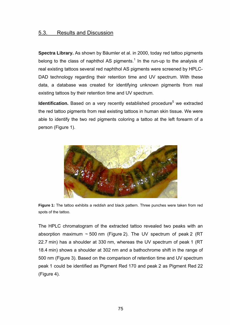
5.3. Results and Discussion
Spectra Library. As shown by Bäumler et al. in 2000, today red tattoo pigments
belong to the class of naphthol AS pigments.1 In the run-up to the analysis of
real existing tattoos several red naphthol AS pigments were screened by HPLC-
DAD technology regarding their retention time and UV spectrum. With these
data, a database was created for identifying unknown pigments from real
existing tattoos by their retention time and UV spectrum.
Identification. Based on a very recently established procedure5 we extracted
the red tattoo pigments from real existing tattoos in human skin tissue. We were
able to identify the two red pigments coloring a tattoo at the left forearm of a
person (Figure 1).
Figure 1: The tattoo exhibits a reddish and black pattern. Three punches were taken from red
spots of the tattoo.
The HPLC chromatogram of the extracted tattoo revealed two peaks with an
absorption maximum ~ 500 nm (Figure 2). The UV spectrum of peak 2 (RT
22.7 min) has a shoulder at 330 nm, whereas the UV spectrum of peak 1 (RT
18.4 min) shows a shoulder at 302 nm and a bathochrome shift in the range of
500 nm (Figure 3). Based on the comparison of retention time and UV spectrum
peak 1 could be identified as Pigment Red 170 and peak 2 as Pigment Red 22
(Figure 4).
75
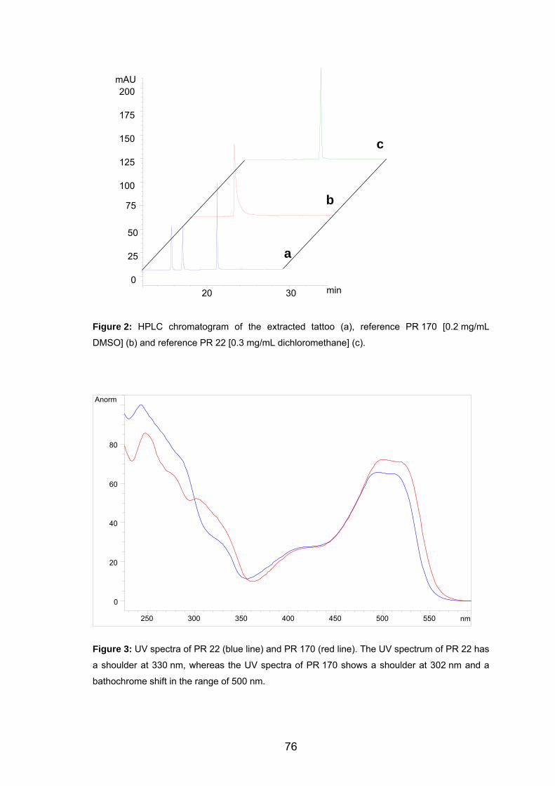
Figure 2: HPLC chromatogram of the extracted tattoo (a), reference PR 170 [0.2 mg/mL
DMSO] (b) and reference PR 22 [0.3 mg/mL dichloromethane] (c).
Figure 3: UV spectra of PR 22 (blue line) and PR 170 (red line). The UV spectrum of PR 22 has
a shoulder at 330 nm, whereas the UV spectra of PR 170 shows a shoulder at 302 nm and a
bathochrome shift in the range of 500 nm.
a
b
c
20 30 min
mAU
0
25
175
200
50
75
100
125
150
norm A
nm250 300 350 400 450 500 550
0
20
40
60
80
76
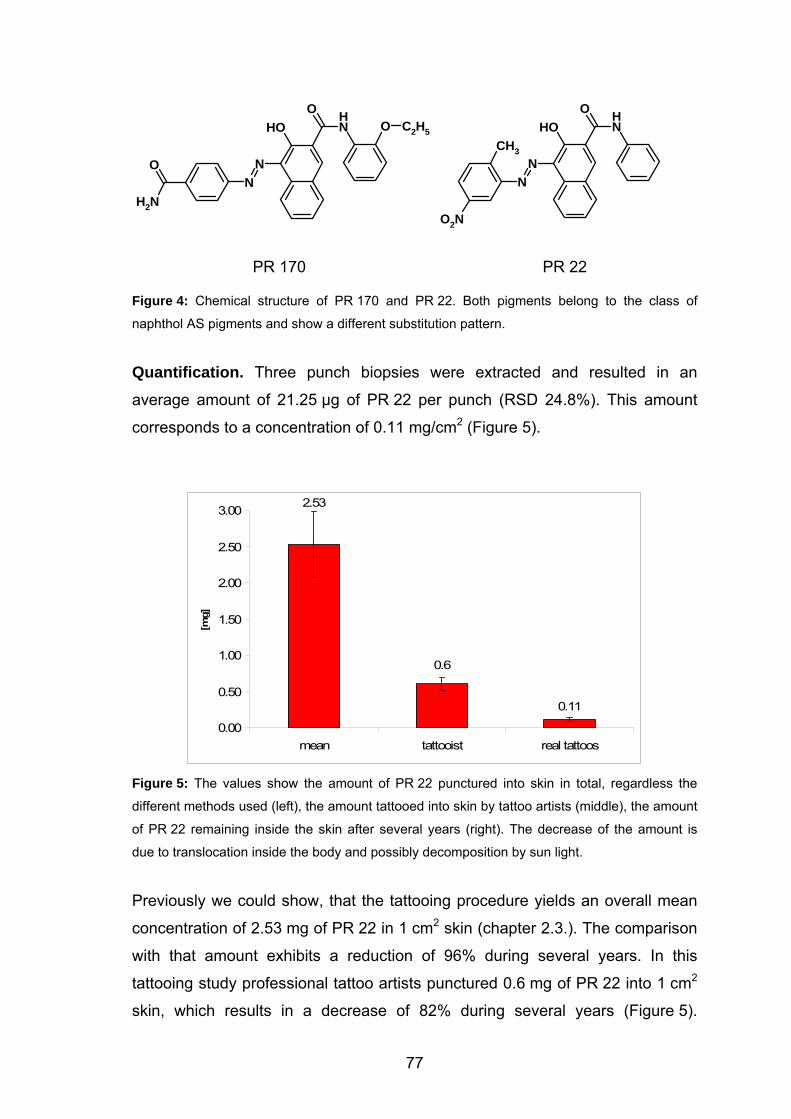
OH
N
NHO
N
O C2H5
O
NH2
OH
N
NHO
N
CH3
O2N
PR 170 PR 22
Figure 4: Chemical structure of PR 170 and PR 22. Both pigments belong to the class of
naphthol AS pigments and show a different substitution pattern.
Quantification. Three punch biopsies were extracted and resulted in an
average amount of 21.25 µg of PR 22 per punch (RSD 24.8%). This amount
corresponds to a concentration of 0.11 mg/cm2 (Figure 5).
2.53
0.6
0.11
0.00
0.50
1.00
1.50
2.00
2.50
3.00
mean tattooist real tattoos
[mg]
Figure 5: The values show the amount of PR 22 punctured into skin in total, regardless the
different methods used (left), the amount tattooed into skin by tattoo artists (middle), the amount
of PR 22 remaining inside the skin after several years (right). The decrease of the amount is
due to translocation inside the body and possibly decomposition by sun light.
Previously we could show, that the tattooing procedure yields an overall mean
concentration of 2.53 mg of PR 22 in 1 cm2 skin (chapter 2.3.). The comparison
with that amount exhibits a reduction of 96% during several years. In this
tattooing study professional tattoo artists punctured 0.6 mg of PR 22 into 1 cm2
skin, which results in a decrease of 82% during several years (Figure 5).
77
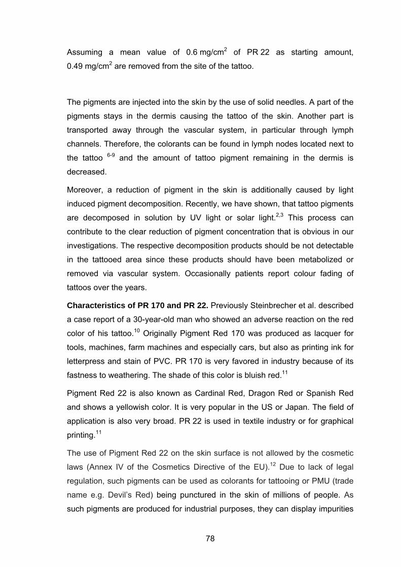
Assuming a mean value of 0.6 mg/cm2 of PR 22 as starting amount,
0.49 mg/cm2 are removed from the site of the tattoo.
The pigments are injected into the skin by the use of solid needles. A part of the
pigments stays in the dermis causing the tattoo of the skin. Another part is
transported away through the vascular system, in particular through lymph
channels. Therefore, the colorants can be found in lymph nodes located next to
the tattoo 6-9 and the amount of tattoo pigment remaining in the dermis is
decreased.
Moreover, a reduction of pigment in the skin is additionally caused by light
induced pigment decomposition. Recently, we have shown, that tattoo pigments
are decomposed in solution by UV light or solar light.2,3 This process can
contribute to the clear reduction of pigment concentration that is obvious in our
investigations. The respective decomposition products should be not detectable
in the tattooed area since these products should have been metabolized or
removed via vascular system. Occasionally patients report colour fading of
tattoos over the years.
Characteristics of PR 170 and PR 22. Previously Steinbrecher et al. described
a case report of a 30-year-old man who showed an adverse reaction on the red
color of his tattoo.10 Originally Pigment Red 170 was produced as lacquer for
tools, machines, farm machines and especially cars, but also as printing ink for
letterpress and stain of PVC. PR 170 is very favored in industry because of its
fastness to weathering. The shade of this color is bluish red.11
Pigment Red 22 is also known as Cardinal Red, Dragon Red or Spanish Red
and shows a yellowish color. It is very popular in the US or Japan. The field of
application is also very broad. PR 22 is used in textile industry or for graphical
printing.11
The use of Pigment Red 22 on the skin surface is not allowed by the cosmetic
laws (Annex IV of the Cosmetics Directive of the EU).12 Due to lack of legal
regulation, such pigments can be used as colorants for tattooing or PMU (trade
name e.g. Devil’s Red) being punctured in the skin of millions of people. As
such pigments are produced for industrial purposes, they can display impurities
78

of 10% or more. These impurities may be components such as titanium dioxide
for lightening the ink shade, precursors and byproducts of pigment synthesis.
The medical literature reports on malignant skin lesions such as basal cell
carcinoma13,14 or melanoma15,16 in tattoos, but without providing evidence for
causality.
79
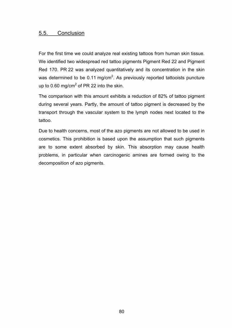
5.5. Conclusion
For the first time we could analyze real existing tattoos from human skin tissue.
We identified two widespread red tattoo pigments Pigment Red 22 and Pigment
Red 170. PR 22 was analyzed quantitatively and its concentration in the skin
was determined to be 0.11 mg/cm2. As previously reported tattooists puncture
up to 0.60 mg/cm2 of PR 22 into the skin.
The comparison with this amount exhibits a reduction of 82% of tattoo pigment
during several years. Partly, the amount of tattoo pigment is decreased by the
transport through the vascular system to the lymph nodes next located to the
tattoo.
Due to health concerns, most of the azo pigments are not allowed to be used in
cosmetics. This prohibition is based upon the assumption that such pigments
are to some extent absorbed by skin. This absorption may cause health
problems, in particular when carcinogenic amines are formed owing to the
decomposition of azo pigments.
80
81
5.5. References
1 Baumler, W.; Eibler, E. T.; Hohenleutner, U.; Sens, B.; Sauer, J.; Landthaler,
M. Lasers Surg. Med. 2000, 26, 13. 2 Cui, Y.; Spann, A. P.; Couch, L. H.; Gopee, N. V.; Evans, F. E.; Churchwell,
M. I.; Williams, L. D.; Doerge, D. R.; Howard, P. C. Photochem. Photobiol.
2004, 80, 175. 3 Engel, E.; Spannberger, A.; Vasold, R.; Konig, B.; Landthaler, M.; Baumler,
W. J. Dtsch. Dermatol. Ges. 2007, 5, 583. 4 Vasold, R.; Naarmann, N.; Ulrich, H.; Fischer, D.; Konig, B.; Landthaler, M.;
Baumler, W. Photochem. Photobiol. 2004, 80, 185. 5 Engel, E.; Santarelli, F.; Vasold, R.; Ulrich, H.; Maisch, T.; König, B.;
Landthaler, M.; Gopee, N. V.; Howard, P. C.; Bäumler, W. Anal. Chem.
2006, 78, 6440. 6 Friedman, T.; Westreich, M.; Mozes, S. N.; Dorenbaum, A.; Herman, O.
Plast. Reconstr. Surg. 2003, 111, 2120. 7 Moehrle, M.; Blaheta, H. J.; Ruck, P. Dermatology 2001, 203, 342. 8 Gopee, N. V.; Cui, Y.; Olson, G.; Warbritton, A. R.; Miller, B. J.; Couch, L.
H.; Warmer, W. G.; Howard, P. C. Tox. Appl. Pharm. 2005, 209, 145. 9 Mangas, C.; Fernandez-Figueras, M. T.; Carrascosa, J. M.; Soria, X.;
Paradelo, C.; Ferrandiz, C.; Just, M. Dermatol. Surg. 2007, 33, 766. 10 Steinbrecher, I.; Hemmer, W.; Jarisch, R. J. Dtsch. Dermatol. Ges. 2004, 2,
1007. 11 Herbst, W.; Hunger, K. New York: VCH publishers 1995. 12 homepage leffingwell http://www.leffingwell.com/cosmetics/vol_1en.pdf. 13 Birnie, A. J.; Kulkarni, K.; Varma, S. Clin. Exp. Dermatol. 2006, 31, 820. 14 Doumat, F.; Kaise, W.; Barbaud, A.; Schmutz, J. L. Dermatology 2004, 208,
181. 15 Paradisi, A.; Capizzi, R.; De Simone, C.; Fossati, B.; Proietti, I.; Amerio, P.
L. Melanoma Res. 2006, 16, 375. 16 Stinco, G.; De Francesco, V.; Frattasio, A.; Quinkenstein, E.; Patrone, P.
Dermatology 2003, 206, 345.
6. Azo Pigments and a Basall Cell Carcinoma at the
Thumb∗
6.1. Introduction
Basal cell carcinoma is the most common malignancy in humans. Skin cancer,
predominantly basal cell carcinoma and squamous cell carcinoma, have
accounted for about 40% of all cancers in the United States and their frequency
has been increasing.1 However, the nail unit is a very uncommon site for basal
cell carcinoma to develop.2-4 In 2006, Martinelli et al. reviewed the English
literature and found only 17 patients with a basal cell carcinoma at this
location.2
The cause of basal cell carcinoma located at the fingers remains unclear.
Comparable to other locations, ultraviolet radiation, chronic actinic skin damage,
suppression of the immune system or chemical compounds like arsenic may
play a role.
6.2. Case Report
A 58-year-old male patient presented an aching lesion at the left thumb that
persists since three years. The clinical inspection of the lesion showed an
erosive, erythematic lesion (1 x 0.5 cm) at the left thumb including the lateral
nail fold.
In addition, the patient reported that he is an enthusiastic angler. He has
recognized that successful fishing is improved when the fishing baits are
colored. Therefore, he dips the baits into a colorant prior to fishing and each
time the color gets into contact with the left thumb. To elucidate the role of the
colorant, chemical analysis was performed to identify the unknown chemical
sample.
∗ Results of this chapter have been accepted for publication: Engel, E.; Ulrich, H.; Vasold, R.; König, B.; Landthaler, M.; Süttinger, R.; Bäumler, W. Dermatology 2007, in press.
82
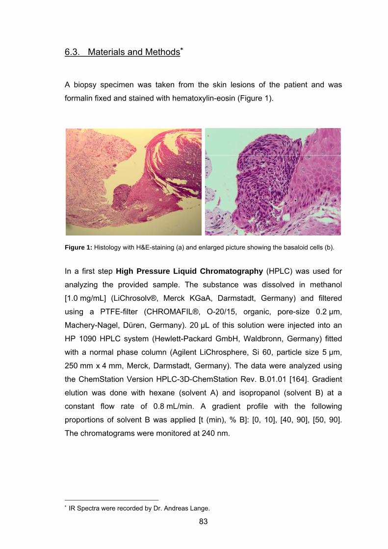
6.3. Materials and Methods∗
A biopsy specimen was taken from the skin lesions of the patient and was
formalin fixed and stained with hematoxylin-eosin (Figure 1).
Figure 1: Histology with H&E-staining (a) and enlarged picture showing the basaloid cells (b).
In a first step High Pressure Liquid Chromatography (HPLC) was used for
analyzing the provided sample. The substance was dissolved in methanol
[1.0 mg/mL] (LiChrosolv®, Merck KGaA, Darmstadt, Germany) and filtered
using a PTFE-filter (CHROMAFIL®, O-20/15, organic, pore-size 0.2 µm,
Machery-Nagel, Düren, Germany). 20 µL of this solution were injected into an
HP 1090 HPLC system (Hewlett-Packard GmbH, Waldbronn, Germany) fitted
with a normal phase column (Agilent LiChrosphere, Si 60, particle size 5 µm,
250 mm x 4 mm, Merck, Darmstadt, Germany). The data were analyzed using
the ChemStation Version HPLC-3D-ChemStation Rev. B.01.01 [164]. Gradient
elution was done with hexane (solvent A) and isopropanol (solvent B) at a
constant flow rate of 0.8 mL/min. A gradient profile with the following
proportions of solvent B was applied [t (min), % B]: [0, 10], [40, 90], [50, 90].
The chromatograms were monitored at 240 nm.
∗ IR Spectra were recorded by Dr. Andreas Lange.
83
Electrospray Mass Spectrometry (ESI-MS) was applied collecting further
information. Using a triple stage mass spectrometer (TSQ 7000, Thermoquest
Finnigan, Toronto, Canada) the mass of the respective compound was
determined.
In a next step the substance was analyzed by Infrared Spectroscopy (IR). The
identification was done by comparing the fingerprint of the compound with an IR
spectra database. FTIR spectra (3200 – 400 cm-1) of KBr pellets were recorded
via a FTS 800 spectrometer (Scimitar Series, DigiLab, Varian, Darmstadt,
Germany) and background correction.
84
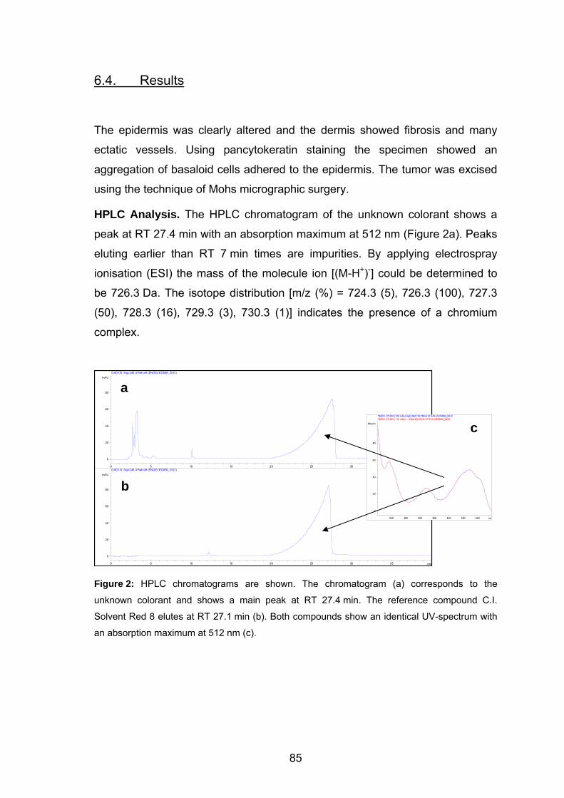
6.4. Results
The epidermis was clearly altered and the dermis showed fibrosis and many
ectatic vessels. Using pancytokeratin staining the specimen showed an
aggregation of basaloid cells adhered to the epidermis. The tumor was excised
using the technique of Mohs micrographic surgery.
HPLC Analysis. The HPLC chromatogram of the unknown colorant shows a
peak at RT 27.4 min with an absorption maximum at 512 nm (Figure 2a). Peaks
eluting earlier than RT 7 min times are impurities. By applying electrospray
ionisation (ESI) the mass of the molecule ion [(M-H+)-] could be determined to
be 726.3 Da. The isotope distribution [m/z (%) = 724.3 (5), 726.3 (100), 727.3
(50), 728.3 (16), 729.3 (3), 730.3 (1)] indicates the presence of a chromium
complex.
DAD1 B, Sig=240,4 Ref=off (ENGEL\ED048_02.D)
mAU
Figure 2: HPLC chromatograms are shown. The chromatogram (a) corresponds to the
unknown colorant and shows a main peak at RT 27.4 min. The reference compound C.I.
Solvent Red 8 elutes at RT 27.1 min (b). Both compounds show an identical UV-spectrum with
an absorption maximum at 512 nm (c).
min0 5 10 15 20 25 30 35
0
20
40
60
80
min0 5 10 15 20 25 30 35
mAU
0
20
40
60
80
DAD1 B, Sig=240,4 Ref=off (ENGEL\ED056_03.D)
nm250 300 350 400 450 500 550
Mnorm
0
20
40
60
80
*DAD1, 27.040 (120 mAU,Up2) Ref=19.755 & 31.445 of ED056_03.D*DAD1, 27.465 (112 mAU, - ) Ref=20.262 & 31.874 of ED048_02.D
a
c
b
85
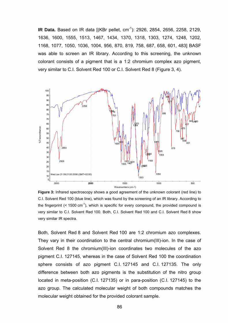
IR Data. Based on IR data [(KBr pellet, cm-1): 2926, 2854, 2656, 2258, 2129,
1636, 1600, 1555, 1513, 1467, 1434, 1370, 1318, 1303, 1274, 1248, 1202,
1168, 1077, 1050, 1036, 1004, 956, 870, 819, 758, 687, 658, 601, 483] BASF
was able to screen an IR library. According to this screening, the unknown
colorant consists of a pigment that is a 1:2 chromium complex azo pigment,
very similar to C.I. Solvent Red 100 or C.I. Solvent Red 8 (Figure 3, 4).
Wed Jun 21 09:21:00 2006 (GMT+02:00)
41
483512563
601
617
658
675
687
731
758
819
871
906
956
10041026
1050
1077
1168
11841202
12481274
1303
13701384
143414671513
1555
16001654
170117191735
2258
2854
2926
C.I. Solvent Red 100Anglerfarbe v. Dr.Bäumler
5 10
15
20
25
30
35
40
45
50
55
60
65
70
75
80
85
90
95
100
%Tr
ansm
ittan
ce
500 1000 2000 2000 1500 3000 Wavenumbers (cm-1)
Figure 3: Infrared spectroscopy shows a good agreement of the unknown colorant (red line) to
C.I. Solvent Red 100 (blue line), which was found by the screening of an IR library. According to
the fingerprint (< 1500 cm-1), which is specific for every compound, the provided compound is
very similar to C.I. Solvent Red 100. Both, C.I. Solvent Red 100 and C.I. Solvent Red 8 show
very similar IR spectra.
Both, Solvent Red 8 and Solvent Red 100 are 1:2 chromium azo complexes.
They vary in their coordination to the central chromium(III)-ion. In the case of
Solvent Red 8 the chromium(III)-ion coordinates two molecules of the azo
pigment C.I. 127145, whereas in the case of Solvent Red 100 the coordination
sphere consists of azo pigment C.I. 127145 and C.I. 127135. The only
difference between both azo pigments is the substitution of the nitro group
located in meta-position (C.I. 127135) or in para-position (C.I. 127145) to the
azo group. The calculated molecular weight of both compounds matches the
molecular weight obtained for the provided colorant sample.
86
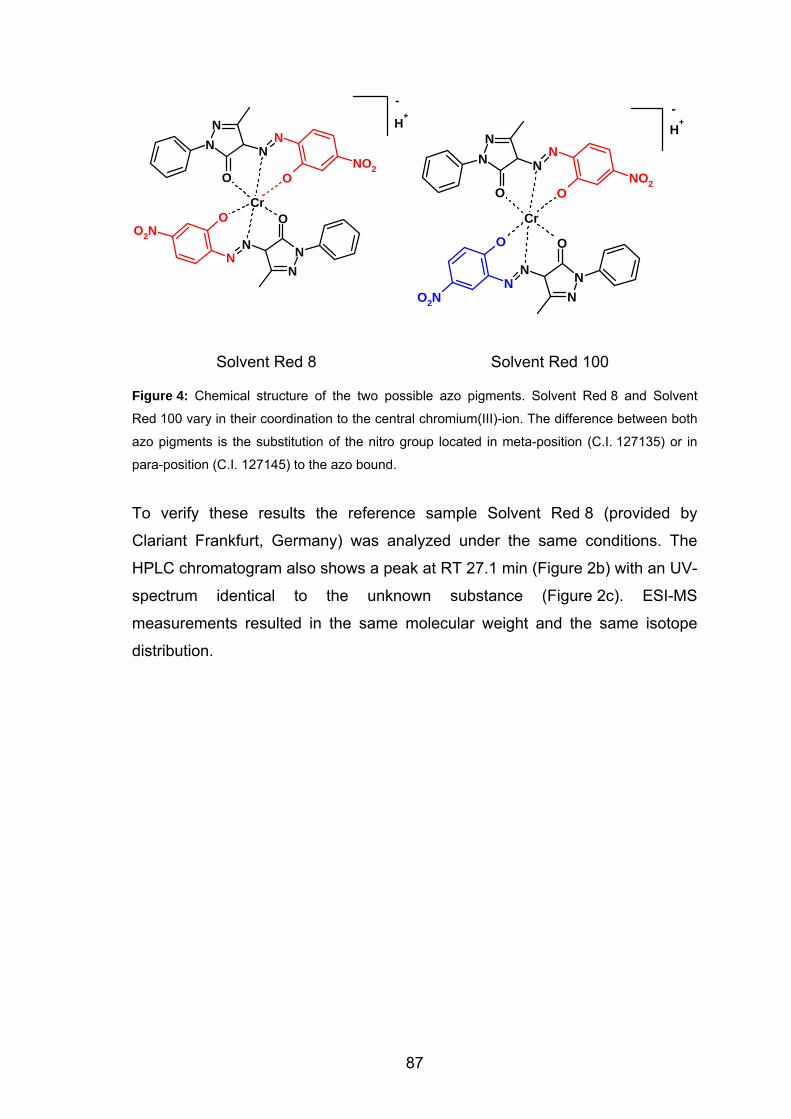
-
H+
NNN
N
O2N
Cr
NN N
N
NO2O
O
O
O
-H
+
Cr
NN N
N
NO2OO
NNN
N
O O
O2N
Solvent Red 8 Solvent Red 100
Figure 4: Chemical structure of the two possible azo pigments. Solvent Red 8 and Solvent
Red 100 vary in their coordination to the central chromium(III)-ion. The difference between both
azo pigments is the substitution of the nitro group located in meta-position (C.I. 127135) or in
para-position (C.I. 127145) to the azo bound.
To verify these results the reference sample Solvent Red 8 (provided by
Clariant Frankfurt, Germany) was analyzed under the same conditions. The
HPLC chromatogram also shows a peak at RT 27.1 min (Figure 2b) with an UV-
spectrum identical to the unknown substance (Figure 2c). ESI-MS
measurements resulted in the same molecular weight and the same isotope
distribution.
87
6.5. Discussion
Basal cell carcinoma located at the fingers (Figure 4) is rare and the cause for
the tumor at this site remains usually unclear. Our patient reported a frequent
contact of his finger to the colorant he is using to stain the fishing baits. The
origin of the colorant was unclear and a list of ingredients was not available.
Therefore, no warning notices were on the bottle containing this colorant.
Due to this fact, the colorant was suspected to be associated with the basal cell
carcinoma at the thumb. Therefore, we prompted the patient to provide a bottle
with the colorant for further chemical analysis. Since there was no indication for
the chemical nature of the colorant, the liquid, red colorant was dried and
investigated by the means of three different analytical methods.
The results of three methods in all likelihood suggest Solvent Red 8 being the
red color in the colorant. Azo pigments such as Solvent Red 8 are usually
applied to stain consumer goods. Due to health concerns, most of the azo
pigments are not allowed to be used in cosmetics, that is, to get into contact
with skin.5
In contrast to cosmetics, tattooing is not regulated and therefore such azo
pigments are used as tattoo colorants being punctured in the skin of millions of
people.6-8 The medical literature reports on malignant skin lesions such as basal
cell carcinoma9,10 or melanoma11,12 in tattoos, but without providing evidence for
causality. Part of the pigments and their impurities are transported from the skin
into the lymph nodes staining them for years.13
Solvent Red 8 is a wide spread azo pigment used in paints for cars and wood,
whereas Solvent Red 100 is used for textiles such as leather, silk or wool. The
chemical synthesis of Solvent Red 8 requires the aromatic amine 2-amino-5-
nitrophenol. The azo pigments of tattoo colorants also contain such amines.7
The pigments are simply manufactured to stain consumer goods but not to be
used as pharmaceutical compounds in humans. Therefore, the azo pigments
contain a high percentage of impurities like the aromatic amines, which are
88
frequently toxic or carcinogenic. Additionally, the pigments can be cleaved by
light and can release these amines.
2-amino-5-nitrophenol has been examined by US National Cancer Institute
(NTP-studies). The compound was mutagenic in Salmonella typhimurium
strains. 2-Amino-5-nitrophenol induced forward mutations in mouse L5178Y
lymphoma cells. An increase in chromosomal aberrations and sister chromatid
exchanges was observed in cultured Chinese hamster ovary (CHO) cells
following incubation with 2-amino-5-nitrophenol. Rats that received 2-amino-5-
nitrophenol responded with an increased incidence of acinar cell adenomas of
the pancreas.14 Comparable findings are reported for 2-amino-4-nitrophenol that
is used fort he synthesis of Solvent Red 100.15
Based on these facts and the results of our chemical analysis, there might be a
certain probability that the carcinogenic amines in the colorant Solvent Red 8
have induced the basal cell carcinoma in our patient. Consequently, the role of
azo pigments that are punctured in the skin for tattooing should be also
highlighted in the near future.
89
90
6.6. References
1 Parker S.L.; Tong T.; Bolden S.; Wingo P.A. CA Cancer J. Clin. 1997, 47, 5. 2 Martinelli P.T.; Cohen P.R.; Schulze K.E.; Dorsey K.E.; Nelson B.R.;
Dermatol. Surg. 2006, 32, 320.
3 Galeano M.; Lentini M.; Stagno D'Alcontres F.; Colonna M. Hand. Surg.
2002, 7, 295.
4 Matsushita K.; Kawada A.; Aragane Y.; Tezuka T. J. Dermatol. 2003, 30,
250.
5 European-Commission: Commission directive 2004/93/ec. Official Journal of
the European Union 2004;L300/13. 6 Baumler W.; Eibler E.T.; Hohenleutner U.; Sens B.; Sauer J.; Landthaler M.
Lasers Surg. Med. 2000, 26, 13. 7 Vasold R.; Naarmann N.; Ulrich H.; Fischer D.; Konig B.; Landthaler M.;
Baumler W. Photochem. Photobiol. 2004, 80, 185. 8 Engel E.; Santarelli F.; Vasold R.; Ulrich H.; Maisch T.; Konig B.; Landthaler
M.; Gopee N.V.; Howard P.C.; Baumler W. Anal. Chem. 2006, 78, 6440. 9 Birnie A.J.; Kulkarni K.; Varma S. Clin. Exp. Dermatol. 2006, 31, 820. 10 Doumat F.; Kaise W.; Barbaud A.; Schmutz J.L. Dermatology 2004, 208,
181. 11 Paradisi A.; Capizzi R.; De Simone C.; Fossati B.; Proietti I.; Amerio P.L.
Melanoma. Res. 2006, 16, 375. 12 Stinco G.; De Francesco V.; Frattasio A.; Quinkenstein E.; Patrone P.
Dermatology 2003, 206, 345. 13 Jack C.; Adwani A.; Krishnan H. Int. Semin. Surg. Oncol. 2005, 2, 28. 14 Ntp toxicology and carcinogenesis studies of 2-amino-5-nitrophenol (cas no.
121-88-0) in f344/n rats and b6c3f1 mice (gavage studies). Natl. Toxicol.
Program Tech. Rep. Ser. 1988, 334, 1. 15 Ntp toxicology and carcinogenesis studies of 2-amino-4-nitrophenol (cas no.
99-57-0) in f344/n rats and b6c3f1 mice (gavage studies). Natl. Toxicol.
Program Tech. Rep. Ser. 1988, 339, 1.
7. Abbreviations
ACN acetonitrile
ATL buffer tissue lysis buffer
C.I. colour index
CAS-No. chemical abstract service number
CF i calibration factor of the compound i
Crl/SKH-1 Charles River / Skin and Cancer Hospital
DAD diode array technology
DCA 2,5-dichloroaniline
DCB 1,4-dichlorobenzene
Diglyme diethylene glycol dimethyl ether
DNA deoxyribonucleic acid
DPA 9,10-diphenyl-anthracene
ESI electrospray ionisation
FDA Food and Drug Administration
FTIR Fourier transformed infrared spectroscopy
H&E haematoxylin and eosin staining
HPLC high performance liquid chromatography
hr-/hr- hairless strain
hrs hours
Hz
hertz
IR infrared spectroscopy
ISTD internal standard
M molecule
min minute
mL milliliter
µL microliter
MNA 2-methyl-5-nitroaniline
m-NAS methoxy-NAS
MS mass spectroscopy
naphthol-AS arylide of the 2-hydroxy-3-naphthoic acid
NAS naphthol-AS
NCTR National Center for Toxicological Research
Nd:YAG neodymium-doped yttrium-aluminium-garnet
NHDF normal human dermal fibroblasts
NT 4-nitrotoluene
NTP national toxicology program
PBS phosphate buffered saline
PMU permanent make-up
PP polypropylene
PR 22 Pigment Red 22
91

92
PR 9 Pigment Red 9
press. pressure
pt points
PTFE polytetrafluorethylene
PY 74 Pigment Yellow 74
ROS reactive oxygen species
RSD relative standard deviation
RT retention time
SED standard erythema dose
ssl simulated solar light
TEM transmission electron microscopy
temp. temperature
TFA trifluoroacetic acid
THF tetrahydrofuran
UV ultraviolet
w/v weight/volume
8. Summary
The number of tattooed individuals increased significantly, especially among
youth. In the United States, up to 24% of the population has tattoos, whereas in
European countries like Germany about 9% and the United Kingdom about 12%
are tattooed. Today frequently azo pigments are used for tattooing since they
are brilliant and provide a long-lasting tattoo in the skin. These azo pigments
are manufactured primarily for other purposes like printing, painting cars and
coloring various consumer products. Tattoo colorants are mixtures of pigments
(color) and multiple other ingredients. These colorants may contain titanium
dioxide for lightening the shade, precursors and byproducts of pigment
synthesis, as well as diluents that are used to suspend the pigments. Tattoo
colorants are also used for permanent make-up at the eyelid, eyebrow or lip.
Despite the millions of people with tattoos, there is currently no common legal
requirement for listing ingredients, including the pigments. That is, for tattooing
non-FDA-approved pigment is introduced into skin to produce indelible designs.
In Europe, many of azo pigments used in tattoos (e.g. Pigment Red 22) are not
allowed in cosmetics since they can be decomposed yielding carcinogenic
amines. The FDA continues to evaluate the extent and severity of adverse
events associated with tattooing and is conducting research on colorants.
On closer examination, tattooing is a complex procedure that includes different
risks for the skin and even for the human body. The pigments and the impurities
could cause adverse reactions of the skin at the site of tattooing. In addition,
part of the colorants are transported away to other anatomical locations like the
lymph nodes.
Moreover, tattoos may be exposed to solar light or - in case of tattoo removal -
to laser light. Both procedures have been known to result in the decomposition
of such pigments in vitro causing hazardous compounds such as carcinogenic
amines.
93
To estimate the risk of any health problems of tattooing, the colorant
concentration in the skin and the human body after tattooing of the colorants
must be determined. This has not been performed so far and was now firstly
investigated by our research group.
Therefore, an extraction method was established to determine the concentration
of tattoo pigments and decomposition products quantitatively (chapter 1). The
extraction of two widely used azo compounds, Pigment Red 22 (PR 22) and
Rigment Red 9 (PR 9) and their laser induced decomposition products 2-
methyl-5-nitroaniline (MNA), 4-nitro-toluene (NT), 2,5-dichloroaniline (DCA) and
1,4-dichlorobenzene (DCB) were accomplished using recovery experiments and
HPLC-DAD technology. Despite the poor solubility of the pigments, a nearly
complete recovery from aqueous suspension (> 92%) or lysed skin (> 94%) was
achieved. The decomposition products were extracted from aqueous
suspension or skin showing a recovery of up to 100%, except for the very
volatile DCB.
Based on this extraction procedure we could determine the amount of tattoo
pigment punctured into skin (chapter 2). We tattooed excised pigskin and
human skin with Pigment Red 22 under various conditions. After tattooing, we
quantitatively extracted the pigment in order to determine the pigment
concentration in skin. The concentration of pigments ranged from about 0.60 to
9.42 mg per cm2 of tattooed skin (mean value 2.53 mg/cm²) depending upon
the size of the pigment crystals, the pigment concentration applied to the skin
surface, and the respective procedure of tattooing.
It is known from literature, that a yellow tattoo pigment (Pigment Yellow 74) is
cleaved by simulated solar light into toxic compounds. Since PR 22 is cleaved
by laser light into toxic or even carcinogenic compounds we investigated the
influence of UVB radiation and natural sun light on PR 22 (chapter 3). PR 22
was dissolved in different solvents (tetrahydrofuran, dioxane, chloroform and
dichloromethane). The solutions were irradiated with either UVB-radiation or
with natural sunlight. An evident cleavage of the pigment was detected in all
94
solvents, when exposed to UVB radiation or natural sunlight. The primary
decomposition products (NAS, MNA, NT) were identified. In tetrahydrofuran and
dioxane the pigment concentration decreased significantly during UVB
irradiation, whereas the pigment was completely destroyed during sunlight
exposure. In chloroform and dichloromethane the concentration of PR 22
decreased only slightly during UVB irradiation, whereas during sunlight
exposure the pigment was almost completely destroyed. We found that PR 22
is cleaved in tetrahydrofuran and dioxane without any influence of radiation.
Since chloroform and dichloromethane do not affect the cleavage process,
these solvent are optimal for such in vitro experiments. Last but not least we
demonstrated that PR 22 is cleaved by natural sunlight and broad band UVB
radiation into toxic and carcinogenic compounds.
Puncturing tattoo pigments into the skin can be compared with an injury of the
upper skin layers. As soon as the pigments are injected into the dermis they are
recognized by the body as foreign particles. By means of phagocytosis, the
tattoo pigments are removed from the site of tattooed skin and are transported
by the lymphatic system to other anatomical locations like lymph nodes located
next to the tattoo.
To investigate transportation of tattoo pigments after tattooing, we established
an animal model using SKH-1 hairless mice (chapter 4). The mice received
tattoos with PR 22 on their back. The extraction of pigments immediately after
tattooing yielded the concentration of PR 22 of 34.53 µg per punch
(1.76 mg/cm2) that is placed in the body. In contrast to that, the extraction of
PR 22 from skin six weeks (42 days) after tattooing elucidates the extent of
pigment transportation in the mice. Only 24.86 µg of PR 22 per punch remain
inside the dermis (1.27 mg/cm2). That corresponds to a removal of 28% of
tattoo pigment.
To investigate the decomposition of pigments by solar light, tattooed living mice
were exposed to solar light for 31 days. We found that up to 60% of the pigment
is cleaved (PR 22 remaining: 0.50 mg/cm2). The ex vivo postulated
decomposition products (NAS, MNA, NT) could not be detected. The reason
95
might be, that the decomposition products are removed by the lymphatic system
from the site of the tattoo as soon as they are generated.
Ex vivo laser treatment of tattooed mouse skin showed that up to 50% of PR 22
located inside the mouse skin can be cleaved by laser light (PR 22 remaining:
0.65 mg/cm2). Fortunately we could show, that only 8% of PR 22 are
decomposed into the hazardous compounds MNA and NT.
For the first time we could quantitatively analyze real existing tattoos in human
skin tissue (chapter 5). The extraction of a tattoo revealed PR 22 and PR 170 to
be the red tattoo pigments used by the artist. Several years after tattooing the
concentration of PR 22 inside the skin is 0.11 mg/cm2. Concerning the
concentration of 0.6 mg/cm2 punctured into skin by tattooist 82% of tattoo
pigment are transported from the site of the tattoo during several years.
The last chapter (chapter 6) deals with an interesting additional problem of azo
dyes. We describe a case report of a 58-year-old patient with a periungual basal
cell carcinoma at the thumb. Basal cell carcinoma is the most common
malignant neoplasm of the skin, whereas the localization at the nail unit is very
rare. The specific feature of the reported case is the frequent exposure to
fishing baits that he had stained with an unknown colorant. The use of
chromatography, mass spectrometry and infrared spectroscopy revealed the
colorant as the 1:2 chromium complex azo pigment Solvent Red 8. Solvent
Red 8 is a widespread synthetic azo pigment that is applied to stain consumer
products. Compounds such as Solvent Red 8 can be cleaved to carcinogenic
amines under e.g. light exposure, in particular after incorporation into the
human body. As a result of the frequent skin contact to this azo pigment, this
hazard compound might have induced the basal cell carcinoma in our patient.
96
9. Zusammenfassung
Die Popularität von Tätowierungen hat in die letzten Jahren, vor allem unter den
Jugendlichen, stark zugenommen. In den Vereinigten Staaten sind bis zu 24 %
der Bevölkerung tätowiert, die Zahlen in Europa sind ähnlich. In Deutschland
tragen rund 9 % der Bevölkerung eine Tätowierung, in England sind es schon
12 %. Für die farbigen Tätowierungen werden inzwischen mehrheitlich Farben
auf der Basis von industriellen Pigmenten eingesetzt. Chemische Analysen
haben ergeben, dass es sich häufig um organische Pigmente wie
Azoverbindungen oder polyzyklische Verbindungen handelt. Diese Pigmente
werden eigentlich zum Färben oder Lackieren von Konsumgütern produziert
(z.B. Autolacke). Die Tätowierer setzen diese Pigmente gerne ein, weil sie sehr
beständig und nahezu unlöslich sind. Dies sind genau die Eigenschaften, die für
ein brillantes, beständiges Tattoo in der Haut sorgen. Wie eigene Recherchen
ergeben haben, werden die Farbstoffe in Pulverform oder als Emulsionen von
einigen Großhändlern in USA oder Europa an Tätowierer ohne Angaben zu den
Inhaltsstoffen verkauft. Diese Substanzgemische enthalten in wechselnder
Zusammensetzung neben der eigentlichen farbgebenden Komponente, Vor-
und Zwischenprodukte aus dem Syntheseprozess des Pigments sowie große
Mengen an Titandioxid (Farbaufhellung) und weitere nicht spezifizierte
Zusatzstoffe. Tätowierungsfarbstoffe kommen in großem Umfang auch bei
Permanent Make-up im Bereich der Lippenkonturen, des Augenlids oder der
Augenbrauen zum Einsatz.
Trotz der großen Anzahl von mehreren Millionen Menschen, die eine
Tätowierung tragen, gibt es derzeit keine einheitlichen Regelung zur
Deklarierung der Pigmente und Inhaltsstoffe. Im Rahmen von Tätowierungen
werden also Substanzen in die Haut eingebracht, die von der US FDA zu
diesem Zweck nicht zugelassen sind. Auch in Deutschland unterliegt die
Verwendung von Tätowierungspigmenten derzeit keiner gesetzlichen Kontrolle,
da es sich weder um Kosmetika noch um Arzneimittel handelt.
Interessanterweise ist der Einsatz vieler Pigmente (z.B. Pigment Red 22) auf
der Hautoberfläche durch die Kosmetikverordnung verboten (Annex IV of the
97
Cosmetics Directive of the EU), da sie in karzinogene Amine gespalten werden
könnten. Werden sie aber in die Haut eingestochen, gibt es keine
Einschränkungen. Mit Blick auf die Millionen von Tätowierten in Deutschland ist
der Gesetzgeber hier dringend gefragt, entsprechende Regelungen zu
erlassen.
Bei näherer Betrachtung ist Tätowieren ein komplexer Prozess, der
verschiedene Risiken für die Haut, bzw. für den menschlichen Körper birgt. Die
Pigmente und die darin enthaltenen Verunreinigungen können zu
unerwünschten Hautreaktionen führen. Ein Teil des eingebrachten
Substanzgemisches, vor allem Verbindungen mit kleinem Molekulargewicht,
wird innerhalb kurzer Zeit über das Gefäßsystem der Haut abtransportiert. Ein
deutlicher Hinweis darauf ist häufig die Färbung der lokoregionären
Lymphknoten.
Von zunehmendem Interesse ist auch der Einfluss von Sonnenstrahlung, oder
im Falle einer Entfernung, des Laserlichts auf Tätowierfarbstoffe. In beiden
Fällen kann es in vitro zur Spaltung der Tätowierungspigmente in karzinogene
Amine kommen.
Die Toxizität und Karzinogenität von chemischen Verbindungen, wie auch
deren Licht induzierten Spaltprodukten, hängen unter anderem von deren
Konzentration in der Haut ab. Bedenkt man, dass sich viele Menschen mit
ihrem Tattoo in der Sonne aufhalten bzw. sich einer medizinisch indizierten
Therapie mit UVB-Licht unterziehen etc., ist es wichtig, die Konzentration von
Tätowierungspigmenten in der Haut zu bestimmen, um damit eventuell
verbundene Risiken einschätzen zu können. Bisher war die Menge an Pigment,
die beim Tätowieren in die Haut eingebracht wird, völlig unklar.
Zur Bestimmung des Gehalts an Tätowierungspigmenten in der Haut wurde
erstmals ein Extraktionsschema etabliert, mit dem sowohl die Pigmente als
auch deren Spaltprodukte quantitativ extrahiert werden können (Kapitel 1). Die
Extraktion zweier weit verbreiteter Azopigmente, Pigment Red 22 (PR 22) und
Pigment Red 9 (PR 9), und deren Laser-induzierten Spaltprodukte 2-Methyl-5-
nitroanilin (MNA), 4-Nitrotoluol (NT), 2,5-Dichloranilin (DCA) und 1,4-
98
Dichlorbenzol (DCB) wurde mittels Recovery-Experimenten und quantitativer
HPLC (DAD-Technologie) etabliert. Trotz der nur geringe Löslichkeit der
Pigmente konnten diese fast vollständig aus wässriger Suspension (> 92 %)
oder lysierter Haut (> 94 %) extrahiert werden. Bis auf das sehr flüchtige DCB
wurden die Spaltprodukte nahezu vollständig (~ 100 %) extrahiert.
Dieses Extraktionsschema war die Basis für die anschließenden
Tätowierungsstudien (Kapitel 2). Durch fachgerechtes Tätowieren wurde
exzidierte Schweinehaut und Menschenhaut mit PR 22 in verschiedenen
Konzentrationen und verschiedenen Nadelformen und Nadelgrößen tätowiert.
Somit konnte erstmals die Menge an Farbstoff quantitativ bestimmt werden, die
durch Tätowieren in die Haut eingebracht wird. Der Gehalt an Pigment belief
sich, je nach Größe der Pigmentkristalle, der verwendeten Konzentration der
Suspension und des jeweiligen Tätowierungsprozesses, auf Werte zwischen
0.60 und 9.92 mg/cm2 (Durchschnitt 2.53 mg/cm2).
Aus der Literatur ist bekannt, dass ein gelbes Tätowierungspigment (Pigment
Yellow 74) durch Sonnenlicht in toxische Produkte gespalten werden kann. Da
PR 22 durch Laserlicht in toxische, z.T. sogar karzinogene Substanzen
gespalten werden kann, wurde der Einfluß von UVB-Strahlung und natürlichem
Sonnenlicht auf PR 22 untersucht (Kapitel 3). Dazu wurde PR 22 in
verschiedenen Lösungsmitteln (Tetrahydrofuran, Dioxan, Chloroform und
Dichlormethan) gelöst und die Lösungen entweder mit UVB-Strahlung oder mit
natürlichem Sonnenlicht bestrahlt. In allen Lösungsmitteln konnte zweifelsfrei
die Licht induzierte Spaltung des Pigments nachgewiesen und die primären
Spaltprodukte (NAS, MNA, NT) identifiziert werden. In den Lösungsmitteln
Tetrahydrofuran und Dioxan nimmt die Konzentration des Pigments durch die
UV-B Strahlung deutlich ab, während es durch das Sonnenlicht völlig zerstört
wird. In Chloroform und Dichlormethan nimmt die Konzentration von PR 22
durch die UV-B Strahlung nur etwas ab, Sonnenlicht zerstört es jedoch nahezu
vollständig. Wir konnten zeigen, dass PR 22 in Tetrahydrofuran und Dioxan
schon ohne den Einfluss von Licht zerstört wird. Da sich die Lösungsmittel
Chloroform und Dichlormethan selbst gegenüber den ablaufenden chemischen
99
Spaltprozessen neutral verhalten, sind sie für in vitro Untersuchungen am
besten geeignet. Es konnte gezeigt werden, das Pigment Red 22 unter dem
Einfluss natürlichen Sonnenlichts und breitbandiger UV-B-Strahlung in toxische
oder kanzerogene Substanzen gespalten wird.
Diese Umverteilung der Tätowierungspigmente wurde im Tiermodel an
haarlosen SKH-1 Mäuse studiert (Kapitel 4). Jedem Tier wurden PR 22 in
4 Streifen auf den Rücken tätowiert. Einen Tag nach dem Tätowieren wurde die
Menge an Farbstoff bestimmt, die in den Körper eingebracht werden kann
(34.53 µg an PR 22 pro Stanze (1.76 mg/cm2). Nach 6 Wochen (42 Tage)
wurde das Ausmaß der Umverteilung im Körper ersichtlich. Nur noch 24.86 µg
pro Stanze (1.27 mg/cm2) verblieben in der Haut. Demnach wurden 28 % der
eingebrachten Pigmentmenge umverteilt.
Zur Untersuchung, welchen Einfluss Sonnenlicht auf bestehende
Tätowierungen hat, wurden tätowierte Mäuse 31 Tage lang im simulierten
Sonnenlicht ausgesetzt. Es zeigte sich, dass bis zu 60% der vorhandenen
Pigmentmenge gespalten wurde und die Konzentration nur noch 0.5 mg/cm2
betrug. Die ex vivo postulierten Spaltprodukte konnten nicht nachgewiesen
werden. Womöglich wurden sie sofort nach der Entstehung durch das
lymphatische System abtransportiert.
Im Rahmen einer ex vivo Laserbehandlung tätowierter Mäusehaut wurde ~
50 % der vorhandenen Pigmentmenge gespalten. Davon wurden wiederum nur
8 % in die postulierten Spaltprodukte MNA und NT gespalten. Die
Konzentration an PR 22 betrug nach der Laserbehandlung nur noch
0.65 mg/cm2.
Im Rahmen dieser Dissertation ist es erstmals gelungen, den Gehalt an
Tätowierungspigment in einer seit mehreren Jahren am Menschen
bestehenden Tätowierung zu bestimmen (Kapitel 5). Aus einer roten
Tätowierung konnten PR 22 und PR 170 eindeutig als die beiden farbgebende
Pigmente extrahiert werden. Der Gehalt an PR 22 in dieser Tätowierung betrug
0.11 mg/cm2. Geht man davon aus, dass Tätowierer im Mittel 0.6 mg/cm2 an
100
Tätowierungsfarbstoff in die Haut einbringen, müßten in diesem Fall 82 % des
Pigments abtransportiert worden sein.
Im letzten Kapitel (Kapitel 6) wird ein zusätzliches Problem von Azofarbstoffen
beschrieben. In der Klinik und Poliklinik für Dermatologie stellte sich ein 58-
jähriger Patient mit einem periungualen Basalzellkarzinom an seinem linken
Daumen vor. Basalzellkarzinome sind die häufigsten malignen Neoplasien der
Haut. Die Lokalisation im Bereich des Fingernagels ist jedoch sehr
ungewöhnlich. Die Besonderheit dieses Falles ist, dass der Patient häufige
Kontakt zu Anglerködern hatte, die er mit einem unbekannten Farbstoff
einfärbte. Mittels HPLC, Massenspektroskopie und Infrarotspektroskopie konnte
die Substanz als 1:2-Chromkomplex-Azofarbstoff Solvent Red 8 identifiziert
werden. Solvent Red 8 ist ein weitverbreiteter synthetischer Azofarbstoff, der
zum Färben von Konsumgütern eingesetzt wird. Substanzen wie Solvent Red 8
können z.B. durch Licht in karzinogene Substanzen gespalten werden, u.a.
auch nachdem sie in den Körper eingebracht wurden. Durch den häufigen
Hautkontakt mit diesem Azofarbstoff, kann das Basalzellkarzinom bei unserem
Patienten entstanden sein.
101

10. Appendix
PUBLICATIONS
• Modern tattoos contain azo pigments: an in vivo proof of Pigment Red 22 and Pigment Red 170, Engel. E.; Gastl, K.; Santarelli, F.; Vasold, R.; Maisch, T.; Penning, R.; Ulrich, H.; König, B.; Landthaler, M.; Bäumler, W. Toxicol. Appl. Pharmacol., 2007, in preparation.
• Tattoo pigments in skin: concentration, transportation and light induced decomposition of an azo pigment using SKH-1 mouse model, Engel. E.; Santarelli, F.; Vasold, R.; Maisch, T.; Howard, P.C.; Ulrich, H.; König, B.; Landthaler, M.; Bäumler, W. Toxicol. Appl. Pharmacol., 2007, in preparation.
• Tätowierungspigmente im Fokus der aktuellen Forschung, Engel. E.; Bäumler, W.; Vasold, R. Nachrichten aus der Chemie, 2007, in press.
• Investigations on the light induced decomposition of Indocyanine Green (ICG), Engel. E.; Schraml, R.; Maisch, T.; Kobuch, K.; König, B.; Szeimies, RM.; Hillenkamp, J.; Bäumler, W.; Vasold, R. Invest. Ophthalmol. Vis. Sci., 2007, under review.
• Modern tattooing leads to high concentrations of hazardous pigments in the human skin, Engel, E.; Santarelli, F.; Vasold, R.; Maisch, T.; Howard, P.C.; Ulrich, H.; Prantl, L.; König, B.; Landthaler, M.; Bäumler, W. Br. J. Dermatol., 2007, under review.
• Azo pigments and a basal cell carcinoma at the thumb, Engel E.; Ulrich, H.; Vasold, R.; König, B.; Landthaler, M.; Süttinger, R.; Bäumler, W. Dermatology, 2007, in press.
• Photochemische Spaltung eines Tätowierungspigments durch UV-Strahlung oder Sonnenlicht, Engel, E.; Spannberger, A.; Vasold, R.; König, B.; Landthaler, M.; Bäumler, W. J. Dtsch. Dermatol. Ges., 2007, 5, 583.
• Establishment of an extraction method for the recovery of tattoo pigments from human skin using HPLC-DAD technology, Engel E.; Santarelli, F.; Vasold, R.; Ulrich, H.; Maisch, T.; König, B.; Landthaler, M.; Gopee, N.V.; Howard, P.C.; Bäumler, W. Anal. Chem., 2006, 78, 6440.
102

• Singlet oxygen generation by UVA light exposure of endogenous photosensitizers, Baier J.; Maisch, T.; Maier, M.; Engel, E.; Bäumler, W. Biophys. J., 2006, 91, 1452.
• Mol4D – Moleküle in der 4. Dimension, Engel, E.; Kruppa, M.; König, B. Angew. Chem. 2004, 116, 6744.
• Mol4D – Molecules in the 4th Dimension, Engel, E.; Kruppa, M.; König, B. Angew. Chem. Int. Ed. 2004, 43, 6582.
103
Conferences and Presentations
ORAL PRESENTATIONS
• Engel, E. “Photochemical cleavage of a widely used tattoo pigment by UVB radiation and natural sunlight”, 2. World Congress on Work-related and Environmental Allergy, Weimar, 06/2007
• Engel, E. “The archaic procedure tattooing – a first indication of the amount of tattoo pigment in skin“, 2. World Congress on Work-related and Environmental Allergy, Weimar, 06/2007
• Engel, E. “Analyse von Tätowierungspigmenten in Hautgewebe mittels Pyrolyse-GC“, 1.Pyrolyse-Workshop, SIM, Oberhausen, 05/2007
• Engel, E. “Quantifizierung von Tätowierungspigmenten in Hautgewebe“, 44. Tagung der Deutschen Dermatologischen Gesellschaft, Dresden, 04/2007
• Engel, E. “Entwicklung einer Methode zur Extrahierung von Pigmenten aus der Haut“, 44. Tagung der Deutschen Dermatologischen Gesellschaft, Dresden, 04/2007
• Engel, E. “Tätowierungspigmente: Quantitative Extraktion aus der Haut”, Dermatologisches Fortbildungskolloquium am Klinikum der Universität Regensburg, 12/2006
• Engel, E. “The neverending story: tattoos in human skin”, Weihnachtskolloquium des Institut für Organische Chemie, Universität Regensburg, 12/2005
• Engel, E. “Tätowierungspigmente, UV-Licht und Laser”, Dermatologisches Fortbildungskolloquium am Klinikum der Universität Regensburg, 06/2005
104
POSTER PRESENTATIONS
• Engel, E.; Santarelli, F.; Vasold, R.; Maisch, T.; Ulrich, H.; König, B.; Landthaler, M; Bäumler, W. „Photochemische Spaltung von Tätowierungspigmenten durch UV-B-Strahlung oder Sonnenlicht“, 44. Tagung der Deutschen Dermatologischen Gesellschaft, Dresden, 04/2007
• Engel, E.; Santarelli, F.; Vasold, R.; Maisch, T.; Ulrich, H.; König, B.; Landthaler, M; Bäumler, W. „Entwicklung einer Methode zur Extrahierung von Pigmenten aus der Haut“, 44. Tagung der Deutschen Dermatologischen Gesellschaft, Dresden, 04/2007
• Engel, E.; Santarelli, F.; Maisch, T.; Ulrich, H.; König, B.; Landthaler, M., Vasold, R.; Bäumler, W. „Concentration of tattoo pigment in pig skin after artificial and manually tattooing“”, 58th Pittsburgh Conference on Analytical Chemistry and Applied Spectroscopy (Pittcon), Chicago, USA, 02/2007
• Engel, E.; Santarelli, F.; Vasold, R.; Maisch, T.; Ulrich, H.; Bäumler, W.; König, B.; Landthaler, M. „Tattoos in skin: tattooing and quantitative extraction of a widely used tattoo pigment“, Skin and Forrmulation 2nd Symposium, Versailles, France, 09/2006
• Engel, E.; Santarelli, F.; Vasold, R.; Bäumler, W.; Maisch, T.; Ulrich, H.; König, B.; Landthaler, M. “Determination of the amount of tattoo pigments in skin“, 1st European Chemistry Congress, Budapest, Hungary, 08/2006
• Engel, E.; Bäumler, W.; Maisch, T.; Ulrich, H.; König, B.; Landthaler, M., Gopee, N.V.; Howard, P., Vasold, R. “Extraction procedure of tattoo pigments from skin”, 57th Pittsburgh Conference on Analytical Chemistry and Applied Spectroscopy (Pittcon), Orlando, USA, 03/2006
• Engel, E.; Bäumler, W.; Maisch, T.; Ulrich, H.; König, B.; Landthaler, M., Gopee, N.V.; Howard, P., Vasold, R. “Extraction procedure of tattoo pigments from skin”, 45th Annual Meeting of the Society of Toxicology (SOT), San Diego, USA, 03/2006
• Engel, E.; Vasold R., Bäumler W.; König B. “Chemische Analyse organischer Tätowierungspigmente in zellulärer Matrix“, Tag der Naturwissenschaften der Universität Regenburg, 06/2005
105

• Bäumler W.; Engel, E.; Vasold R., Ulrich H.; Landthaler M. “Zerstörung von Tätowierungspigmenten durch UV-Licht oder Laser“, 43. DDG-Tagung, Dresden, 04/2005
106

ADDITIONAL CONFERENCES
• Seminar “Probenvorbereitung – ein entscheidender Schritt in der Analytik”, Supelco, Munich, Germany, 04/2007
• GC/SPME-Seminar, Supelco, Munich, Germany, 11/2005
• 44th Annual Meeting of the Society of Toxicology (SOT), New Orleans, USA, 03/2005
• HPLC/GS-Seminar, Agilent Technologies, Little Rock, USA, 02/2005
107
Curriculum Vitae
Personal Details
Name: Eva Engel
Date of birth: 08.04.1980
Place of Birth: Ulm/ Donau
Education
08/2004 – 09/2007 PhD at the section of analytics (Dr. Rudolf
Vasold) under Prof. Dr. Burkhard König
(Dpartment of Organic Chemistry at the
University of Regensburg)
09/2002 – 03/2003 Certification in Chemistry (Zulassungsarbeit)
at the section of analytics (Dr. Rudolf
Vasold) under Prof. Dr. Burkhard König
(Dpartment of Organic Chemistry at the
University of Regensburg), thesis on the
“chemical analysis of tattoo pigments in
human skin”.
11/1999 – 06/2004 study, Chemistry and Biology, University of
Regensburg
School Education
06/1999 Abitur (A levels), majors in Biology and Latin.
09/1990 – 06/1998 Gymnasium (grammar school) in Dillingen/
Donau.
09/1986 – 07/1990 Volksschule (primary school) in Wittislingen.
108
109
Stay Abroad
01/2005 – 03/2005 Research at the National Center for
Toxicological Research (NCTR) of the US
Food and Drug Administration in Jefferson,
Arkansas, in the working group of Dr. Paul C.
Howard.
Honors
05/2007 Poster award for the best poster, 44. Tagung
der Deutschen Dermatologischen
Gesellschaft, Dresden, Germany, 04/2007
08/2006 Fellowship of the GDCh for the 1st European
Chemistry Congress, Budapest, Hungary,
08/2006
01/2005 – 03/2005 Fellowship of postgraduate research
programs, Oak Ridge Institute for Science
and Education (ORISE), US Department of
Energy
09/2002 – 03/2003 Scholarship for lectureship candidates
founded by the German Association of
Chemical Industry (VCI)