Embed Size (px)
Citation preview

RESEARCH Open Access
Tridimensional model structure and patterns ofmolecular evolution of Pepino mosaic virus TGBp3proteinBeata Hasiów-Jaroszewska1*, Anna Czerwoniec2, Henryk Pospieszny1 and Santiago F Elena3,4
Abstract
Background: Pepino mosaic virus (PepMV) is considered one of the most dangerous pathogens infecting tomatoesworldwide. The virus is highly diverse and four distinct genotypes, as well as inter-strain recombinants, havealready been described. The isolates display a wide range on symptoms on infected plant species, ranging frommild mosaic to severe necrosis. However, little is known about the mechanisms and pattern of PepMV molecularevolution and about the role of individual proteins in host-pathogen interactions.
Methods: The nucleotide sequences of the triple gene block 3 (TGB3) from PepMV isolates varying insymptomatology and geographic origin have been analyzed. The modes and patterns of molecular evolution ofthe TGBp3 protein were investigated by evaluating the selective constraints to which particular amino acid residueshave been subjected during the course of diversification. The tridimensional structure of TGBp3 protein has beenmodeled de novo using the Rosetta algorithm. The correlation between symptoms development and location ofspecific amino acids residues was analyzed.
Results: The results have shown that TGBp3 has been evolving mainly under the action of purifying selectionoperating on several amino acid sites, thus highlighting its functional role during PepMV infection. Interestingly,amino acid 67, which has been previously shown to be a necrosis determinant, was found to be under positiveselection.
Conclusions: Identification of diverse selection events in TGB3p3 will help unraveling its biological functions and isessential to an understanding of the evolutionary constraints exerted on the Potexvirus genome. The estimatedtridimensional structure of TGBp3 will serve as a platform for further sequence, structural and function analysis andwill stimulate new experimental advances.
Keywords: molecular evolution, PepMV, protein modeling, selective constraints, TGBp3, virus evolution
BackgroundPepino mosaic virus (PepMV) belongs to Potexvirus genuswithin the Flexiviridae family and it is a well-knownpathogen infecting tomato plants worldwide. PepMV pos-sesses a single-stranded, positive-sense RNA genome ofapproximately 6.4 kb, flanked by 5’ and 3’ untranslatedregions (UTRs) with a 5’ cap and a 3’poly(A) tail. PepMVcontains five conserved open reading frames (ORFs).ORF1 encodes a 164 kDa replication-related protein with
three functional domains: an N-terminal mRNA cappingenzyme, a central RNA helicase, and a C-terminal RNA-dependent RNA polymerase (RdRp). ORF2 through ORF4encode movement proteins (TGBp1, TGBp2 and TGBp3)of 26, 14, and 9 kDa, respectively. These three ORFs over-lap in the genome and, hence, are called the triple geneblock. The 25 kDa CP is encoded by ORF5 [1-3]. Thevirus is highly diverse; four different genotypes (CH2, LP,EU, and LP) and a large number of inter-strain recombi-nants have been described [4,5]. PepMV isolates differboth in host range as well in the symptoms induced onsusceptible plants species. Recently, it was proved that onesingle mutation in TGBp3 protein converts a mild
* Correspondence: [email protected] of Plant Protection-National Research Institute, ul. Wł. Węgorka 20,60-318 Poznań, PolandFull list of author information is available at the end of the article
Hasiów-Jaroszewska et al. Virology Journal 2011, 8:318http://www.virologyj.com/content/8/1/318
© 2011 Hasiów-Jaroszewska et al; licensee BioMed Central Ltd. This is an Open Access article distributed under the terms of theCreative Commons Attribution License (http://creativecommons.org/licenses/by/2.0), which permits unrestricted use, distribution, andreproduction in any medium, provided the original work is properly cited.

pathotype of CH2 genotype into a necrotic one. This is theonly association reported between particular amino acidsites and symptomatology [6]. However, our preliminaryresults with isolates from the EU genotype showed that weare dealing with a universal mechanism of variability. Byperforming site-directed mutagenesis of infectious clonesof the EU genotype, we confirmed that amino acid 67 isresponsible for inducing necrosis on tomato, irrespectiveof the strain (unpublished results). This finding suggeststhat TGBp3 structure is important for virulence, sincesubstitution of certain amino acids results in changes inthe infectivity of the virus. As the tridimensional structureof TGBp3 is not yet available, de novo modeling representsa suitable and convenient alternative for analyzing therelationship between amino acids present at certain posi-tions, structure and virulence. This is the approach under-taken in this study.A second aim of this study is to verify the nature of the
selective pressures operating on TGBp3. Nucleotidessubstitutions can be classified into two categories on thebasis of their effects on the protein amino acid composi-tion: synonymous and nonsynonymous replacements. Ingeneral synonymous substitutions accumulate at rate dSper synonymous site. It is generally accepted that synon-ymous substitutions accumulate neutrally because theyhave no effect on the amino acids composition and thusshall not affect the folding and function of proteins. Incontrast, nonsynonymous substitutions accumulate atrate dN per synonymous site. Since these substitutionsinvolve amino acid replacements, they must be subjectedto selection. The average ratio of these two substitutionsrates ω = dN/dS across a gene, provides information onwhether it has been fixing amino acid replacements in aneutral fashion (ω = 1), amino acids changes have beenremoved by the action of purifying selection (ω < 1), orchanges have been fixed by positive evolution (ω > 1) [7].However, averaging ω across the entire coding sequenceof a gene is a gross approximation since highly conservedand hypervariable sites may coexist. It is more sensitiveto allow the ω rates ratio to vary among sites and to takethe presence of individual sites at which ω > 1 as an evi-dence for positive selection [7]. In this study, sequenceanalysis of the PepMV TGB3 was conducted and ω ratesratio were calculated. Additionally, a computational ana-lysis of TGB3 sequences to investigate the extent ofrecombination events was performed. Taken together, inthis study we present results of computational analysesevaluating the patterns of molecular evolution and theireffect on protein structure and virulence.
MethodsVirus isolates and phylogenetic analysisEighteen sequences of PepMV TGB3 representing iso-lates from different genotypes and geographic regions
were retrieved from GenBank and used for further ana-lysis (Table 1). For some isolates detailed biologicalinformation was also available. Sequences were trans-lated into amino acids using BioEdit [8]. Multiple pro-tein sequence alignments were obtained using MUSCLE[9] and then protein-coding nucleotide sequence align-ments were constructed based on their correspondingprotein sequence alignments using PAL2NAL[10].It is well known that recombination affects the estima-
tion of ω and interferes with phylogenetic reconstruc-tion [11]. Therefore the PepMV sequences were initiallyanalyzed for evidence of recombination events using theRDP package [12]. The sequences were analyzed usingthe following methods: 3 Seq, Chimaera, Lard, Bootscan,RDP, Genecovn, and MaxChi. Only recombinationbreakpoints supported by more than three methodswere considered as valid. With this stringent criterion,no significant recombination signals were identified inthe analyzed sequences. The best-fitting model ofnucleotide substitution was investigated using theMODELTEST implemented in MEGA5; Kimura 2-para-meters model (K2P) was chosen. Maximum-likelihoodphylogenetic tree was constructed using MEGA5 andK2P model of nucleotide substitution [13]. Reliability ofthe obtained tree was evaluated using the bootstrapmethod based on 1000 pseudoreplicates. Both nucleo-tide and amino acids sequences were scrutinized to findparticular positions distinguishing mild and severeisolates.
Table 1 Isolates used in study
Isolate Origin Genotype Symptoms on S.lycopersicum
AccessionNumbers
1806 Belgium EU mild FJ457098
DB1 Netherlands EU necrosis FJ940224
SW Poland EU mild EF408822
UK UK EU - AF340024
SP13 Spain EU - AF484251
PMUO618 Spain EU FJ263318
H Hungary EU mild (yellowmosaic)
AM491606
No Poland EU mild FJ612602
LP2001 Peru LP asymptomatic AJ606361
US3 USA EU - AY508411.1
Ch1 Chile CH1 - DQ000984.1
P22 Poland CH2 mild HQ650560
Cl308 Spain CH2 - GU130097
PMU0848 Spain CH2 - FJ263365
P19 Poland CH2 necrosis HQ650559
Ch2 Chile CH2 - DQ000985
Amo7 Spain CH2 - GU130090
US2 USA recombinantCH1/CH2
- AY509927
Hasiów-Jaroszewska et al. Virology Journal 2011, 8:318http://www.virologyj.com/content/8/1/318
Page 2 of 8
Analysis of selective pressureOur analysis of selective pressures was based on evaluatingω for each codon in the nucleotide sequence alignment.The single-likelihood ancestor counting (SLAC), fixed-effects likelihood (FEL) and internal branches fixed-effectslikelihood (IFEL) methods were used to evaluate ω percodon [14]. Both SLAC and FEL methods used using thedefault significance level of p = 0.1; a Bayes factor of 40was used as selection threshold for REL. These analyseswere performed using the HyPhy package [15] as imple-mented in the DATAMONKEY webserver [16]. Nucleo-tide substitutions were modeled according to the K2Pscheme. If a gene evolves under purifying selection mostof the time but is occasionally subject to episodes of posi-tive selection, a comparison between two distant relatedsequences is unlikely to yield a ω ratio significantly > 1.To specifically test whether phylogenetic branches withnecrotic isolates underwent positive selection events, weperformed two independent analyses of branch-specificcodon models. First, the M2 branch site model allowing ωto vary among sites in the protein and across branches onthe tree and aim to detect positive selection affecting a fewsites along particular lineages [17] was evaluated using theprogram CODEML from the PAML version 4.4 package[18]. Second, we applied the sliding-window analysisimplemented in SWAPSC [19]. SWAPSC uses a statisti-cally optimized window size to detect selective constraintsin specific codon regions of the alignment at a particularbranch of phylogenetic tree. The method estimates thenull distribution of dS and dN from simulated sequencealignments. A statistically optimal window size is then esti-mated that makes the detection of adaptive evolutionindependent of the window size. Four codons was the esti-mated optimal window size. Simulated sequences weregenerated with the program EVOLVER from the PAMLpackage [18] with parameters estimated from the truesequence alignments after running M0 codon-basedmodel in CODEML.
Identification of functionally important amino acidresiduesTo identify potentially functional and structural aminoacids residues we used the Bayesian methods implemen-ted in the ConSurf server [20]. Functionally importantresidues, e.g. involved in ligand binding and protein-pro-tein interactions, are often evolutionarily conserved andare most likely to be solvent-accessible, whereas con-served residues within the protein core most probablyhave an important structural role in maintaining theprotein properly folded [20].
Secondary structure prediction and mutation mappingSecondary structure prediction and tertiary fold-recogni-tion (FR) were performed using the GENESILICO meta-
server gateway [21]. Secondary structure was predictedusing PSIPRED [22], PROFSEC [23], PROF [24], SABLE[25], and JNET [26]. FR methods did not report anygood template structure for homology modeling proce-dure. Hence, de novo methods for structure modelingwere applied.
De novo structural modeling of TGBp3To obtain a tridimensional structural model of theTGB3 protein we used TGBp3 sequences of P19, P22and DB1 isolates. P19 and P22 represent necrotic andmild pathotypes of the CH2 genotype, respectively. DB1isolates were described as necrotic isolates belonging tothe EU genotype. The ROSETTA [27] algorithm wasused for de novo modeling of TGBp3. Hundreds ofthousands of decoys were generated and clustered toidentify the best low-energy conformations. Selection ofmodels based on the average energy clusters, size, den-sity and visual inspection of final structures was per-formed. The quality of the final models was evaluatedusing the neural network based algorithm implementedin the PROQ server [28,29] and the MetaMQAPII meta-server [30]. In the first case, the quality is quantified bythe location in the plane formed by the two indexesLGscore (i.e., the -log of a p-value) and MaxSub (ranging0 - 1). Depending on the specific values of theseindexes, the model can be qualified as: correct ifLGscore > 1.5 and MaxSub > 0.1, as good if LGscore > 3and MaxSub > 0.5, and as very good if LGscore > 5 andMaxSub > 0.8). Mapping of the electrostatic potentialon protein surfaces was calculated with APBS (AdaptivePoisson-Boltzmann Solver) [31]. This procedure wasperformed to reveal potential electrostatic changes onthe surface on the protein that may influence interactionbetween other proteins and elements in local cellenvironment.
ResultsAnalysis of TGBp3The analysis of TGBp3 sequences reveled striking differ-ences between particular isolates. TGBp3 consists of246-258 nt and 82-86 amino acids, respectively, depend-ing on the isolate considered. The protein of the Peru-vian isolate LP-2001 from Lycopersicon peruvianum hastwo extra amino acids in the C-terminal region (86)than the characteristic 82-84 of other isolates. It isunclear whether this polymorphism in TGBp3 lengthhas any affect on symptoms. The nucleotide identitybetween the 18 sequences studied ranged from 82% to98.7% and from 77.3% to 98.7% at the amino acid level.The average transition/transversion rates ratio was high,3.9, as observed for other plant RNA viruses [32].The phylogenetic tree shown in Figure 1 illustrates the
presence of two major groups containing isolates from
Hasiów-Jaroszewska et al. Virology Journal 2011, 8:318http://www.virologyj.com/content/8/1/318
Page 3 of 8
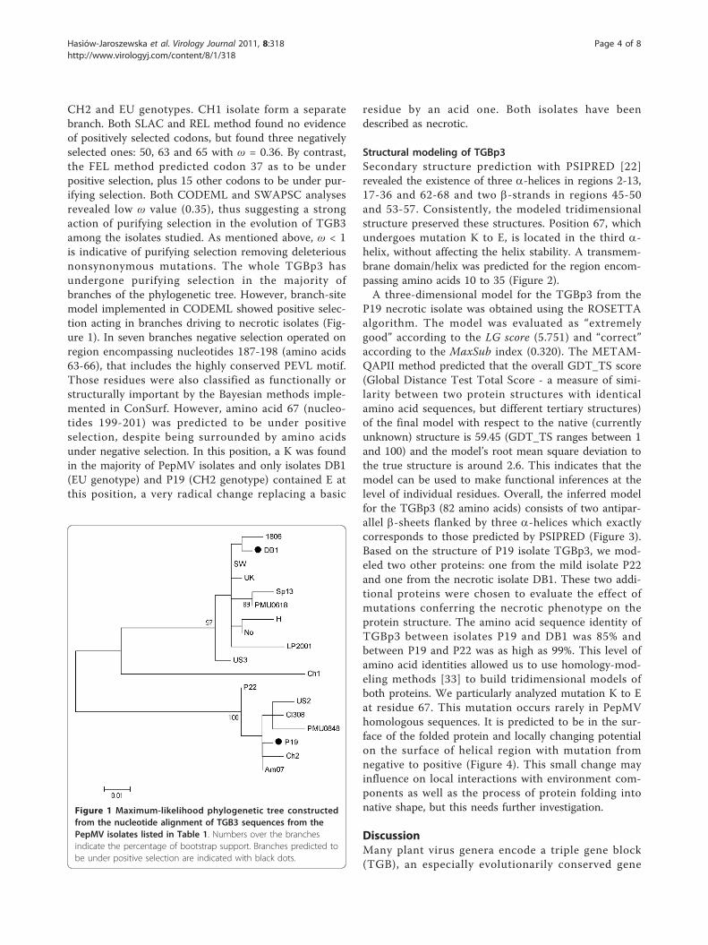
CH2 and EU genotypes. CH1 isolate form a separatebranch. Both SLAC and REL method found no evidenceof positively selected codons, but found three negativelyselected ones: 50, 63 and 65 with ω = 0.36. By contrast,the FEL method predicted codon 37 as to be underpositive selection, plus 15 other codons to be under pur-ifying selection. Both CODEML and SWAPSC analysesrevealed low ω value (0.35), thus suggesting a strongaction of purifying selection in the evolution of TGB3among the isolates studied. As mentioned above, ω < 1is indicative of purifying selection removing deleteriousnonsynonymous mutations. The whole TGBp3 hasundergone purifying selection in the majority ofbranches of the phylogenetic tree. However, branch-sitemodel implemented in CODEML showed positive selec-tion acting in branches driving to necrotic isolates (Fig-ure 1). In seven branches negative selection operated onregion encompassing nucleotides 187-198 (amino acids63-66), that includes the highly conserved PEVL motif.Those residues were also classified as functionally orstructurally important by the Bayesian methods imple-mented in ConSurf. However, amino acid 67 (nucleo-tides 199-201) was predicted to be under positiveselection, despite being surrounded by amino acidsunder negative selection. In this position, a K was foundin the majority of PepMV isolates and only isolates DB1(EU genotype) and P19 (CH2 genotype) contained E atthis position, a very radical change replacing a basic
residue by an acid one. Both isolates have beendescribed as necrotic.
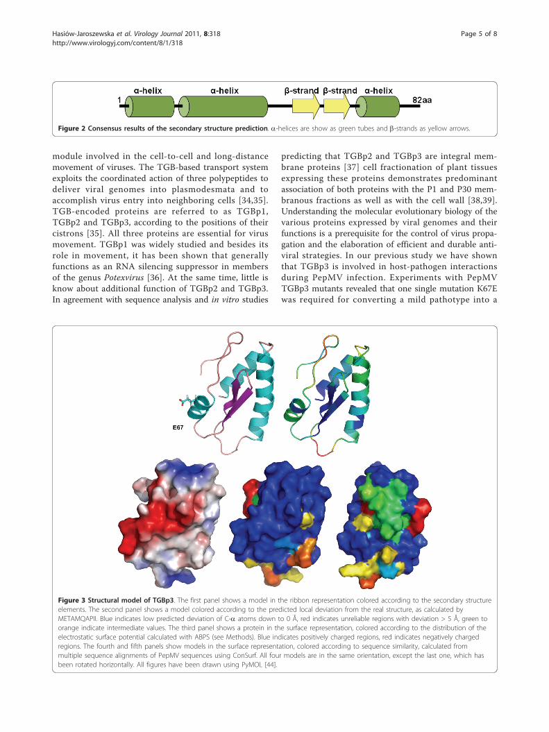
Structural modeling of TGBp3Secondary structure prediction with PSIPRED [22]revealed the existence of three a-helices in regions 2-13,17-36 and 62-68 and two b-strands in regions 45-50and 53-57. Consistently, the modeled tridimensionalstructure preserved these structures. Position 67, whichundergoes mutation K to E, is located in the third a-helix, without affecting the helix stability. A transmem-brane domain/helix was predicted for the region encom-passing amino acids 10 to 35 (Figure 2).A three-dimensional model for the TGBp3 from the
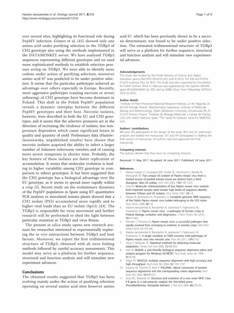
P19 necrotic isolate was obtained using the ROSETTAalgorithm. The model was evaluated as “extremelygood” according to the LG score (5.751) and “correct”according to the MaxSub index (0.320). The METAM-QAPII method predicted that the overall GDT_TS score(Global Distance Test Total Score - a measure of simi-larity between two protein structures with identicalamino acid sequences, but different tertiary structures)of the final model with respect to the native (currentlyunknown) structure is 59.45 (GDT_TS ranges between 1and 100) and the model’s root mean square deviation tothe true structure is around 2.6. This indicates that themodel can be used to make functional inferences at thelevel of individual residues. Overall, the inferred modelfor the TGBp3 (82 amino acids) consists of two antipar-allel b-sheets flanked by three a-helices which exactlycorresponds to those predicted by PSIPRED (Figure 3).Based on the structure of P19 isolate TGBp3, we mod-eled two other proteins: one from the mild isolate P22and one from the necrotic isolate DB1. These two addi-tional proteins were chosen to evaluate the effect ofmutations conferring the necrotic phenotype on theprotein structure. The amino acid sequence identity ofTGBp3 between isolates P19 and DB1 was 85% andbetween P19 and P22 was as high as 99%. This level ofamino acid identities allowed us to use homology-mod-eling methods [33] to build tridimensional models ofboth proteins. We particularly analyzed mutation K to Eat residue 67. This mutation occurs rarely in PepMVhomologous sequences. It is predicted to be in the sur-face of the folded protein and locally changing potentialon the surface of helical region with mutation fromnegative to positive (Figure 4). This small change mayinfluence on local interactions with environment com-ponents as well as the process of protein folding intonative shape, but this needs further investigation.
DiscussionMany plant virus genera encode a triple gene block(TGB), an especially evolutionarily conserved gene
Figure 1 Maximum-likelihood phylogenetic tree constructedfrom the nucleotide alignment of TGB3 sequences from thePepMV isolates listed in Table 1. Numbers over the branchesindicate the percentage of bootstrap support. Branches predicted tobe under positive selection are indicated with black dots.
Hasiów-Jaroszewska et al. Virology Journal 2011, 8:318http://www.virologyj.com/content/8/1/318
Page 4 of 8
module involved in the cell-to-cell and long-distancemovement of viruses. The TGB-based transport systemexploits the coordinated action of three polypeptides todeliver viral genomes into plasmodesmata and toaccomplish virus entry into neighboring cells [34,35].TGB-encoded proteins are referred to as TGBp1,TGBp2 and TGBp3, according to the positions of theircistrons [35]. All three proteins are essential for virusmovement. TGBp1 was widely studied and besides itsrole in movement, it has been shown that generallyfunctions as an RNA silencing suppressor in membersof the genus Potexvirus [36]. At the same time, little isknow about additional function of TGBp2 and TGBp3.In agreement with sequence analysis and in vitro studies
predicting that TGBp2 and TGBp3 are integral mem-brane proteins [37] cell fractionation of plant tissuesexpressing these proteins demonstrates predominantassociation of both proteins with the P1 and P30 mem-branous fractions as well as with the cell wall [38,39].Understanding the molecular evolutionary biology of thevarious proteins expressed by viral genomes and theirfunctions is a prerequisite for the control of virus propa-gation and the elaboration of efficient and durable anti-viral strategies. In our previous study we have shownthat TGBp3 is involved in host-pathogen interactionsduring PepMV infection. Experiments with PepMVTGBp3 mutants revealed that one single mutation K67Ewas required for converting a mild pathotype into a
Figure 2 Consensus results of the secondary structure prediction. a-helices are show as green tubes and b-strands as yellow arrows.
Figure 3 Structural model of TGBp3. The first panel shows a model in the ribbon representation colored according to the secondary structureelements. The second panel shows a model colored according to the predicted local deviation from the real structure, as calculated byMETAMQAPII. Blue indicates low predicted deviation of C-a atoms down to 0 Å, red indicates unreliable regions with deviation > 5 Å, green toorange indicate intermediate values. The third panel shows a protein in the surface representation, colored according to the distribution of theelectrostatic surface potential calculated with ABPS (see Methods). Blue indicates positively charged regions, red indicates negatively chargedregions. The fourth and fifth panels show models in the surface representation, colored according to sequence similarity, calculated frommultiple sequence alignments of PepMV sequences using ConSurf. All four models are in the same orientation, except the last one, which hasbeen rotated horizontally. All figures have been drawn using PyMOL [44].
Hasiów-Jaroszewska et al. Virology Journal 2011, 8:318http://www.virologyj.com/content/8/1/318
Page 5 of 8

necrotic one [6]. Mutant viruses of mild PepMV straininduced necrosis on Datura inoxia and Solanum lyco-persicum. Symptoms of viral infection strongly dependedon the inoculated host plant and result from species-specific host-pathogen interactions.Secondary and tertiary TGBp3 structure predictions
showed that the protein consists of three a-helices andtwo b-strands. The secondary structure predictionplaced amino acid 67 in one of the a-helices. Our tridi-mensional structural predictions revealed that the regionencompassing amino acid 67 in isolates P19, P22 andDB1 is located on the surface of the protein and thusmutations in this region, specially when the physicalproperties dramatically change as it is the case of muta-tion K67E, must have a strong impact on the ability ofTGBp3 to interact with other protein. It is well knownthat in many cases amino acids can be replaced withoutimpairing protein function, even if these are of quite dif-ferent physico-chemical characteristics. However, thechange of a positively charged K by a negatively chargedE may change the local property of the protein surface,jeopardizing its ability to establish the correct interac-tions with other viral proteins or cell components. Sup-porting the existence of such functionality in thissurface region, we have identified nucleotide sites 187-198, spanning amino acids 63-66 that were under theaction of negative selection in seven branches of thephylogenetic tree describing the evolutionary history ofPepMV. In particular, the PEVL motif is highly con-served in all isolates analyzed.In general, functionally essential protein parts are
negatively selected for (conserved), while other partscan be positively selected for. The ω mean valueobtained for the TGBp3 cistron strongly suggests a
predominant action of purifying selection. In goodagreement with this average value, three differentapproaches detected strong signal of purifying selectionfor particular codons. Nevertheless, most codons areevolutionarily neutral. However, perhaps the most inter-esting result from the analyses of selective constraints ifthat amino acid K67 has been identified as under posi-tive selection in the branch leading to necrotic isolates,suggesting that necrosis may be an adaptive trait. Sinceit is close to the region of amino acids 63-66, which is(i) under negative selection (and hence likely has animportant function) and (ii) predicted to be in the sur-face of the folded protein, we can speculate that it maybe involved in the formation of protein-protein complexthat determine the development of symptoms. Theresults obtained with ConSurf indicated that aminoacids 61-66 played functional and structural roles in theprotein. A protein function is however, the results ofthe functional and structural communication betweensites and, therefore, the ability of a given site to changedepends on the interactions it must establish with otherresidues of the molecule. Mutations at either nearbysites (like K67E), or functionally related distant sites inthe structure, will change the selective constraints [40].Functional sites, like binding domains, are less prone toamino acid changes than less important protein regions.Furthermore, some classes of proteins evolve faster thanothers [41].The analysis of the TGB1 gene in PepMV populations
clearly provides a mechanism for its rapid evolution andadaptation to the ever-changing environments [42]. Inthe light of what is so far known about PepMV evolu-tionary dynamics [43] it seems that TGBp3 evolvesmostly by the action of purifying selection operating
Figure 4 Differences in mutated region for the TGBp3 for isolates P19, P22 and DB1. First three proteins are in the surface representation,colored according to the distribution of the electrostatic surface potential calculated with ABPS (see Methods). Blue indicates positively chargedregions, red indicates negatively charged regions. The fourth picture shows three superimposed regions - LEA for P19 (blue) and DB1 (yellow)isolates, LKA for P22 isolate (green). All figures have been drawn using PyMOL [44].
Hasiów-Jaroszewska et al. Virology Journal 2011, 8:318http://www.virologyj.com/content/8/1/318
Page 6 of 8
over several sites, highlighting its functional role duringPepMV infection. Gómez et al. [43] showed only oneamino acid under purifying selection in the TGBp3 ofCH2 genotype also using the methods implemented inthe DATAMONKEY server. We have analyzed TGBp3sequences representing different genotypes and we usedmore sophisticated methods to establish selection pres-sure acting on TGBp3. We were able to identify morecodons under action of purifying selection, moreoveramino acid 67 was predicted to be under positive selec-tion. It seems that the particular pathotypes achieved anadvantage over others especially in Europe. Recently,more aggressive pathotypes (causing necrosis or severeyellowing) of CH2 genotype have become dominant inPoland. This shift in the Polish PepMV populationreveals a dynamic interplay between the differentPepMV genotypes and their host. Necrotic isolates,however, were described in both the EU and CH2 geno-types, and it seems that the selective pressures act in thedirection of increasing the virulence of isolates, less tem-perature dependent which cause significant losses inquality and quantity of yield. Preliminary data (Hasiów-Jaroszewska, unpublished results) have shown thatnecrotic isolates acquired the ability to infect a largernumber of Solanum tuberosum varieties and of causingmore severe symptoms in shorter time. Probably, thekey feature of these isolates are faster replication oraccumulation. It seems that molecular evolution is lead-ing to higher variability among CH2 genotype in com-parison to others genotypes. It has been suggested thatthe CH2 genotype has a biological advantage over theEU genotype, as it seems to spread more rapidly withina crop [5]. Recent study on the evolutionary dynamicsof the PepMV population in Spain using RT-quantitativePCR analyses in inoculated tomato plants showed that aCH2 isolate (PS5) accumulated more rapidly and tohigher viral loads than an EU isolate (Sp13) [43]. TheTGBp3 is responsible for virus movement and furtherresearch will be performed to shed the light betweenparticular mutation in TGBp3 and virus fitness.The present in silico study opens new research ave-
nues for researches interested in experimentally explor-ing the in vivo interactions between TGBp3 and hostfactors. Moreover, we report the first tridimensionalstructure of TGBp3, obtained with de novo foldingmethods followed by careful accuracy assessments. Thismodel may serve as a platform for further sequence,structural and function analysis and will stimulate newexperiment advances.
ConclusionsThe obtained results suggested that TGBp3 has beenevolving mainly under the action of purifying selectionoperating on several amino acid sites however amino
acid 67, which has been previously shown to be a necro-sis determinant, was found to be under positive selec-tion. The estimated tridimensional structure of TGBp3will serve as a platform for further sequence, structuraland function analysis and will stimulate new experimen-tal advances.
AcknowledgementsThis study was funded by the Polish Ministry of Science and HigherEducation grants 0067/P01/2010/70 (AC) and N N310 163 438 and IP2010012470 Iuventus Plus (to BHJ). The study was also supported by Foundationfor Polish Science. Work in Valencia was supported by the Spanish MICINNgrant BFU2009-06993 (to SFE) and by EMBO Short Term Fellowship ASTF424-2010 (to BHJ).
Author details1Institute of Plant Protection-National Research Institute, ul. Wł. Węgorka 20,60-318 Poznań, Poland. 2Bioinformatics Laboratory, Institute of MolecularBiology and Biotechnology, Adam Mickiewicz University, Umultowska 89, PL-61-614 Poznan, Poland. 3Instituto de Biología Molecular y Celular de Plantas,CSIC-UPV, 46022 València, Spain. 4The Santa Fe Institute, Santa Fe, NM87501,USA.
Authors’ contributionsBHJ and SFE participated in the design of the study. BHJ and AC performedanalyses and drafted the manuscript. SFE and HP participated in drafting thefinal version of manuscript. All authors read and approved the finalmanuscript.
Competing interestsThe authors declare that they have no competing interests.
Received: 11 May 2011 Accepted: 24 June 2011 Published: 24 June 2011
References1. Maroon-Lango CJ, Guaragna MA, Jordan RL, Hammond J, Bandla M,
Marquardt SK: Two unique US isolates of Pepino mosaic virus from alimited source of pooled tomato tissue are distinct from a third(European -like) US isolate. Arch Virol 2005, 150:1187-1201.
2. Ling KS: Molecular characterization of two Pepino mosaic virus variantsfrom imported tomato seed reveals high levels of sequence identitybetween Chilean and US isolates. Virus Genes 2007, 34:1-8.
3. Hasiów B, Borodynko N, Pospieszny H: Complete genomic RNA sequenceof the Polish Pepino mosaic virus isolate belonging to the US2 strain.Virus Genes 2008, 36:1-8.
4. Hasiów-Jaroszewska B, Borodynko N, Jackowiak P, Figlerowicz M,Pospieszny H: Pepino mosaic virus - a pathogen of tomato crops inPoland: biology, evolution and diagnostics. J Plant Protect Res 2010,50:477-483.
5. Hanssen I, Thomma B: Pepino mosaic virus: a successful pathogen thatrapidly evolved from emerging to endemic in tomato crops. Mol PlantPathol 2010, 11:179-189.
6. Hasiów-Jaroszewska B, Borodynko N, Jackowiak P, Figlerowicz M,Pospieszny H: A single mutation in TGB3 converts mild pathotype ofPepino mosaic virus into necrotic one. Virus Res 2011, 159:57-61.
7. Yang Z, Bielawski JP: Statistical methods for detecting molecularadaptation. Trends Ecol Evol 2000, 15:496-503.
8. Hall TA: BioEdit: a user-friendly biological sequence alignment editor andanalysis program for Windows 95/98/NT. Nucl Acids Symp Ser 1999,41:95-98.
9. Edgar RC: MUSCLE: multiple sequence alignment with high accuracy andhigh throughput. Nucl Acids Res 2004, 32:1792-1797.
10. Suyama M, Torrents D, Bork P: PAL2NAL: robust conversion of proteinsequence alignemnts into the corresponding codon alignments. NuclAcids Res 2006, 34:609-612.
11. Silva MC, Edwards SV: Structure and evolution of a new avian MHC CIassII B gene in a sub-antarctic seabird, the thin-billed prion(Procellariiformes: Pachyptila belcheri). J Mol Evol 2009, 68:279-291.
Hasiów-Jaroszewska et al. Virology Journal 2011, 8:318http://www.virologyj.com/content/8/1/318
Page 7 of 8
12. Martin DP, Lemey P, Lott M, Moulton V, Posada D, Lefeuvre P: RDP3: aflexible and fast computer program for analyzing recombination.Bioinformatics 2010, 26:2462-2463.
13. Tamura K, Peterson D, Peterson N, Stecher G, Nei M, Kumar S: MEGA5:molecular evolutionary genetics using maximum likelihood, evolutionarydistance, and maximum parsimony methods. Mol Biol Evol 2011.
14. Kosakovsky Pond SL, Frost SDW: Not so different after all: A comparisonof methods for detecting amino acid sites under selection. Mol Biol Evol2005, 22:1208-1222.
15. Kosakovsky Pond S, Frost SDW, Muse SV: HyPhy: hypothesis testing usingphylogenies. Bioinformatics 2005, 21:676-679.
16. Kosakovsky Pond SL, Frost SDW: DATAMONKEY: rapid detection ofselective pressure on individual sites of codon alignments. Bioinformatics2005, 21:2531-2533.
17. Yang Z, Nielsen R, Goldman N, Pedersen AMK: Codon-substitution modelsfor heterogeneous selection pressure at amino acid sites. Genetics 2000,155:431-449.
18. Yang Z: PAML: a program package for phylogenetic analysis bymaximum likelihood. Comput Appl Biosci 2000, 13:555-556.
19. Fares MA: SWAPSC: sliding-window analysis procedure to detectselective constraints. Bioinformatics 2004, 20:2867-2868.
20. Ashkenazy H, Erez E, Martz E, Pupko T, Ben-T N: ConSurf 2010: calculatingevolutionary conservation in sequence and structure of proteins andnucleic acids. Nucl Acids Res 2010, 38:529-533.
21. Kurowski MA, Bujnicki JM: GeneSilico protein structure prediction meta-server. Nucl Acids Res 2003, 31:3305-3307.
22. McGuffin LJ, Bryson K, Jones DT: The PSIPRED protein structure predictionserver. Bioinformatics 2000, 16:404-405.
23. Rost B, Yachdav G, Liu J: The PredictProtein server. Nucleic Acids Res 2004,32:321-326.
24. Ouali M, King RD: Cascaded multiple classifiers for secondary structureprediction. Protein Sci 2000, 9:1162-1176.
25. Adamczak R, Porollo A, Meller J: Accurate prediction of solventaccessibility using neural networks-based regression. Proteins 2004,56:753-767.
26. Cuff JA, Barton GJ: Application of multiple sequence alignment profilesto improve protein secondary structure prediction. Proteins 2000,40:502-511.
27. Simons KT, Kooperberg C, Huang E, Baker D: Assembly of protein tertiarystructure from fragments with similar local sequences using simulatedannealing and bayesian scoring function. J Mol Biol 1997, 268:209-225.
28. Siew N, Elofsson A, Rychlewski L, Fischer D: MaxSub: An automatedmeasure to assess the quality of protein structure predictions.Bioinformatics 2000, 16:776-785.
29. Wallner B, Elofsson A: Can correct protein models be identified. Protein Sci2003, 12:1073-1086.
30. Pawlowski M, Gajda MJ, Matlak R, Bujnicki JM: MetaMQAP: a meta-serverfor the quality assessment of protein models. BMC Bioinformatics 2008,9:403.
31. Baker NA, Sept D, Holst MJ, McCammon JA: Electrostatics of cellularcomponents: Application to microtubules and the ribosome. Proc NatlAcad Sci USA 2001, 98:10037-10041.
32. Tromas N, Elena SF: The rate and spectrum of spontaneous mutations ina plant RNA virus. Genetics 2010, 185:983-989.
33. Kosinski J, Cymerman IA, Feder M, Kurowski MA, Sasin JM, Bujnicki JM: A‘Frankenstein’s monster’ approach to comparative modeling: mergingthe finest fragments of fold-recognition models and iterative modelrefinement aided by 3D structure evaluation. Proteins 2003, 53:369-79.
34. Morozov SY, Solovyev AG: Triple gene block: modular design of amultifunctional machine for plant virus movement. J Gen Virol 2003,84:1351-1366.
35. Beck DL, Guilford PJ, Voot DM, Andersen MT, Forster RLS: Triple gene blockproteins of White clover mosaic potexvirus are required for transport.Virology 1991, 83:695-702.
36. Senshu H, Ozeki J, Komatsu K, Hashimoto M, Hatada K, Aoyama M,Kagiwada S, Yamaji Y, Namba S: Variability in the level of RNA silencingsuppression caused by triple gene block protein 1 (TGBp1) from variouspotexviruses during infection. J Gen Virol 2009, 90:1014-24.
37. Morozov SY, Miroshnichenko NA, Solovyev AG, Zelenina DA, Fedorkin ON,Lukasheva LI, Grachev SA, Chernov BK: In vitro membrane binding of the
translation products of the carlavirus 7-kDa protein genes. Virology 1991,183:782-785.
38. Cowan GH, Lioliopoulou F, Ziegler A, Torrance L: Subcellular localisation,protein interactions, and RNA binding of potato mop-top virus triplegene block proteins. Virology 2002, 298:106-115.
39. Gorshkova EN, Erokhina TN, Stroganova TA, Yelina NE, Zamyatnin AA Jr,Kalinina NO, Schiemann J, Solovyev AG, Morozov SYu: Immunodetectionand fluorescent microscopy of transgenically expressed hordeivirusTGBp3 movement protein reveals its association with endoplasmicreticulum elements in close proximity to plasmodesmata. J Gen Virol2003, 84:985-994.
40. Tourasse NJ, Li WH: Selective constraints, amino acid composition, andthe rate of protein evolution. Mol Biol Evol 2000, 17:656-664.
41. Farfan M, Minana-Galbis D, Carmenn Fuste M, Loren G: Divergentevolution and purifying selection of the flaA gene sequences inAeromas. Biol Direct 2009, 4:23-39.
42. Hasiów-Jaroszewska B, Jackowiak P, Borodynko N, Figlerowicz M,Pospieszny H: Quasispecies nature of Pepino mosaic virus and itsevolutionary dynamics. Virus Genes 2010, 41:260-267.
43. Gómez P, Sempere RN, Elena SF, Aranda MA: Mixed infections of Pepinomosaic virus strains modulate the evolutionary dynamics of thisemergent virus. J Virol 2009, 83:12378-12387.
44. DeLano WL: The PyMOL User’s Manual. DeLano Scientific, San Carlos, CA,USA; 2002.
doi:10.1186/1743-422X-8-318Cite this article as: Hasiów-Jaroszewska et al.: Tridimensional modelstructure and patterns of molecular evolution of Pepino mosaic virusTGBp3 protein. Virology Journal 2011 8:318.
Submit your next manuscript to BioMed Centraland take full advantage of:
• Convenient online submission
• Thorough peer review
• No space constraints or color figure charges
• Immediate publication on acceptance
• Inclusion in PubMed, CAS, Scopus and Google Scholar
• Research which is freely available for redistribution
Submit your manuscript at www.biomedcentral.com/submit
Hasiów-Jaroszewska et al. Virology Journal 2011, 8:318http://www.virologyj.com/content/8/1/318
Page 8 of 8