Embed Size (px)
Citation preview
Journal of Plastic, Reconstructive & Aesthetic Surgery (2016) 69, 524e532
Treatment of compound tibia fracture withmicrovascular latissimus dorsi flap and theIlizarov technique: A cross-sectional study oflong-term outcomes*
J.P. Repo a,*, I. Barner-Rasmussen a, R.P. Roine b,c,H. Sintonen d, E.J. Tukiainen a
a Department of Plastic Surgery, University of Helsinki and Helsinki University Hospital, HUS, Helsinki,Finlandb Group Administration, University of Helsinki and Helsinki University Hospital, Helsinki, Finlandc Department of Health and Social Management, Research Centre for Comparative Effectiveness andPatient Safety, University of Eastern Finland, Kuopio, Finlandd Department of Public Health, University of Helsinki, Helsinki, Finland
Received 2 October 2015; accepted 22 December 2015
KEYWORDSTibia fracture;Latissimus dorsi;Muscle flap;Lower extremityreconstruction;Ilizarov;Distractionosteogenesis
* This work has been presented as a* Corresponding author. DepartmentE-mail address: mrjussirepo@gmai
http://dx.doi.org/10.1016/j.bjps.2015.11748-6815/ª 2016 British Association of
Summary Background: Extensive compound tibial fractures present reconstructive chal-lenges. The present study aimed to assess the outcomes of microvascular latissimus dorsi(LD) flap combined with the Ilizarov technique for extensive compound tibial fractures withbone loss and bone healing complications.Methods: Patient records were reviewed retrospectively. The Lower Extremity FunctionalScale (LEFS), the Disabilities of the Arm, Hand and Shoulder (DASH), and the 15D health-related quality of life (HRQoL) instrument were applied.Results: Between 1989 and 2014, 16 patients underwent reconstruction with a microvascularLD flap and bone transport (11/16) or late bone lengthening (5/16). The mean clinicalfollow-up time was 6.6 (standard deviation (SD): 6.5) years. Three patients had minor compli-cations requiring reoperation. Partial necrosis of one flap required late flap reconstruction inone case. Late bone grafting was used to enhance union in eight of 16 cases. The mean newbone gain was 3.8 cm (SD: 2.5).
Overall, 11 patients completed the questionnaires in a mean of 22.3 years (SD: 2.4) after sur-gery. The main findings revealed a relatively good function of the reconstructed limb and goodshoulder function. The mean HRQoL was comparable to that of an age-standardized sample ofthe general population.
poster in the 8th Congress of the Baltic Association of Surgeons 10/9/2015.of Plastic Surgery, Helsinki University Hospital, P.O. Box 266, 00029 HUS, Finland.l.com (J.P. Repo).
2.011Plastic, Reconstructive and Aesthetic Surgeons. Published by Elsevier Ltd. All rights reserved.
Compound tibia fracture treatment with free muscle flap and Ilizarov technique 525
Conclusion: Segmental tibia transport and lengthening to correct limb length discrepancy donot compromise the microvascular muscle flap. Combined microvascular LD flap reconstructionand the Ilizarov technique can be used in treating acute compound tibial defects, pseudoar-throsis, and osteitis, all associated with significant amputation risk. Fair long-term functionaloutcomes and HRQoL are achieved when these combined techniques are used.ª 2016 British Association of Plastic, Reconstructive and Aesthetic Surgeons. Published byElsevier Ltd. All rights reserved.
Introduction
Compound tibia fracture with significant zone of injury orsequelae of bone healing complications can be managedwith complex methods including the Ilizarov technique ofdistraction osteogenesis,1,2 the Masquelet technique,3 orvascular bone transfers, such as the iliac crest and fibula.4
In cases of extensive soft-tissue loss, local or pedicledmuscle flaps or free flaps may be indicated.5 Tibia length-ening by distraction osteogenesis may also be used to cor-rect posttraumatic limb length discrepancy.6,7
In 1989, Gavril Ilizarov introduced his technique ofdistraction osteogenesis.8,9 In this technique, the bone isstabilized with an external fixator and corticotomy is per-formed outside the fracture site, thereby enabling forma-tion of new bone through distraction. Free muscle flaptransfer combined with the Ilizarov technique to recon-struct lower-extremity compound defects has been previ-ously described.1,10
There are several reports concerning assessment of thelong-term outcomes of these combined techniques; how-ever, only a few have focused on the long-term outcomesassessed by patient-reported outcome measures. In thepresent study, the microvascular LD flap and distractionosteogenesis was used in limb salvage of acute tibial de-fects with large zones of injury due to combined absolutebone defect and soft-tissue loss. This technique has alsoproven reliable in treating prolonged sequelae of compli-cations including osteomyelitis and pseudoarthrosis withimpaired bone blood circulation. An additional indicationfor external tibia distraction has been correction of latetraumatic limb length discrepancy after microvascular flapreconstruction.
The present study aimed to assess the reliability of thiscombined method and to report the long-term outcomes ofall patients with traumatic acute or chronic compound tibiadefect treated with microvascular LD flap reconstructionand the Ilizarov distraction osteogenesis (either bonetransport or lengthening) in the authors’ institution be-tween 1989 and 2014.
Patients and methods
The study was approved by the Ethics Committee of theHelsinki University Hospital. Patients were identified fromthe hospital records, and their patient records wereretrospectively reviewed. Patients with femoral recon-struction, intramedullary distraction osteogenesis, fracture
stabilization with external fixation only, and soft-tissuereconstruction other than LD were excluded. The resultswere reported following the STROBE11 guidelines for cross-sectional studies.
Outcome measures
Shoulder function was examined by the main section of theFinnish version12 of the Disabilities of the Arm, Shoulderand Hand13 (DASH) questionnaire. It comprises 30 questions(physical activities, 23 questions; symptoms, seven ques-tions). The DASH rewards a total score between 0 and 100points.13
The function of the reconstructed limb was assessed bythe Finnish version14 of the Lower Extremity FunctionalScale15 (LEFS). It contains 20 function-related questions.The total score ranges between 0 and 80, with higher scoresrepresenting better functional ability.
HRQoL was measured by the 15D16 questionnaire. It is acomprehensive, 15-dimensional HRQoL instrument thatcompares positively with other analogous, generic HRQoLinstruments.16e19 Incorporating population-based prefer-ence weights into the dimensions yields a single index scorethat ranges from 0 (equivalent to being dead) to 1 (bestpossible HRQoL). A difference �0.015 in the 15D score isestimated to be clinically important.20 The authors hy-pothesized that the patients enjoy a HRQoL comparable tothat of an age-standardized general population.
The level of physical activity was assessed by the fre-quency intensity time (FIT) index.21 The index is obtainedby multiplying the scores of each question together,yielding a score between 0 and 100 (the higher the score,the greater the physical activity).
Finally, a questionnaire designed for the study chartedcomorbidities and the use of analgesics. A written informedconsent was obtained from the patients participating in thecross-sectional assessment with patient-reported outcomemeasures.
Statistical analysis
Results are obtained as means with SD, medians, or ranges.HRQoL results of the patients were compared with those ofan age-standardized sample of the general Finnish popu-lation (n Z 2413) obtained from the Health 2011 Survey.22
The statistical significance of the differences between pa-tients and the general population was compared using theindependent samples t-test. A significance level was set at
526 J.P. Repo et al.
p-value <0.05. Analyses were performed with SPSS 20 (SPSSInc., Chicago, IL, USA).
Results
Sixteen patients with traumatic compound tibial defecttreated with a microvascular LD flap and the Ilizarovdistraction osteogenesis (segmental bone transport,n Z 11; lengthening of the tibia, n Z 5) between the years1989 and 2014 were identified. Their mean age was 33 years(SD 13.2). The trauma mechanism was automobile (n Z 6),motorcycle (n Z 3), moped (n Z 1), or tractor (n Z 1)collision, lift bar hit (n Z 1), train accident (n Z 1), fall(n Z 2), or a blast (previous war injury) (n Z 1). Thetrauma was of high energy in 14 of the 16 cases, leading toan open fracture in 13. The patient characteristics togetherwith the GustiloeAnderson open fracture classification23
are presented in Table 1. The defect location was meta-physeal (n Z 8), meta-epiphyseal (n Z 6), epiphyseal withintra-articular involvement (n Z 1), or diaphyseal (n Z 1).
The accompanying injuries included an ipsilateral femurfracture in three patients, metatarsal fracture in one, and abilateral tibial pilon fracture in one patient. Five patientswere smokers. One patient had type II diabetes. Theremaining patients had no significant comorbidities.
The mean number of operations before microvasculartransfer was 1.9 (SD: 1.3). In 15 of the 16 patients, revisionsand external fixation were applied in the primary treatmentfacility. One patient had undergone previous reconstructionwith a LD flap and three patients with local muscle flapsbefore admission. In one closed fracture patient, a leg casthad been used to stabilize the fracture. One patient pre-sented with bone transport that began earlier (9 cm ofdistracted bone) along with imminent extrusion of thebone. The size of the soft-tissue defect ranged from 4 � 6to 10 � 30 cm. Loss of functional units was observed in 15 ofthe 16 patients (Table 1).
Soft-tissue reconstruction
Patients underwent soft-tissue reconstruction with micro-vascular LD muscle (n Z 10) or musculocutaneous (n Z 6)flaps either early (<30 days; n Z 9) (Figures. 1e6) or late(>30 days) (n Z 7). The median time from trauma to flapreconstruction was 18 days (range: 1 daye26.9 years),depending on when the patient was referred to the author’sclinic. An additional microvascular iliac crest transfer (size:4 � 12 cm) was performed in one patient with a veryextensive injury, moving it to a more proximal area.Moreover, 8/16 of patients underwent cancellous bonegrafting to enhance bony union simultaneously with the flapreconstruction. The sizes of the microvascular flaps rangedbetween 3 � 15 and 15 � 30 cm.
The recipient arteries were the posterior (n Z 10) oranterior tibial (n Z 3), or popliteal vessels (n Z 2). Eitherend-to-side (n Z 9) or end-to-end (n Z 7) anastomoseswere used. Vein grafts from the great saphenous were usedin two cases, and those from the cephalic vein were used inone case. The recipient veins were concomitant (n Z 13),popliteal (n Z 2), and posterior tibial (n Z 1) vessels.Additional skin grafting was used in 15 of the 16 cases.
Ilizarov technique
Indications for bone reconstruction were large primarydefect (n Z 10), tibial length discrepancy (n Z 6), long-standing pseudoarthrosis (nZ 4), or nonunion (nZ 2). Fourpatients had two or more of these indications. In the bonetransport cases (n Z 11), corticotomy was performedeither simultaneously with (n Z 4) or 1 daye2.8 years(median: 23 days) after microvascular flap reconstruction.The tibial bone was unifocally osteotomized above (n Z 10)or beneath the defect (n Z 1). A deep infection was pre-sent in two of the 11 patients when the transport processbegan.
Five of the 16 patients underwent tibia bone length-ening. The timing of corticotomy from the soft-tissuereconstruction in this group ranged between 67 days and5.6 years (median: 2.1 years). One additional patient un-derwent lengthening for limb length discrepancy aftercompletion of bone transport. In five of the six cases, thebone was osteotomized proximal to the defect.
The osteotomized bone ends were stabilized by externalfixation with an Orthofix (Orthofix SRL, Verona, Italy;n Z 10), Fixel (AMP INC, Seattle, WA, USA; n Z 3), AO(DePuy Synthes, West Palm Beach, FL, USA; n Z 2), or anIlizarov device (Smith & Nephew Orthopedics, Memphis,TN, USA; n Z 1).
Clinical follow-up
The mean follow-up time was 6.6 years (SD: 6.5). Compli-cations were encountered in 13 of the 16 patients (Table 2).Postoperatively, one patient had temporary peroneal palsy.Overall, 14 of the 16 patients underwent reoperations(Table 3). The mean time of external fixation was 178 days(SD: 99), thus resulting in 54 days/cm fixation index.
In the bone transport group, the mean time to fullweight bearing and complete radiological bone union fromthe beginning of transport was 14.3 (SD: 12.7) and 16.0 (SD:22.1) months, respectively. These data were unavailablefor one patient. In this group, the median amount of newbone was 4 cm (range: 2.0e12.0 cm). In the bone length-ening group (6/16), full weight bearing and radiologicalbone union was achieved in a median of 10.8 (SD: 16.6) and32.2 (SD: 18.0) months, respectively. The median amount ofnew bone gained was 3.2 cm (range: 2.0e4.0 cm).
Two patients experienced severe pain in the recon-structed limb requiring long-term analgesia with opioids.One patient had a permanent antecurvatum of 10� andtibial varus of 5�. In the remaining patients, there were norotational deformities and axial malalignment was <5�. Theoverall mean tibial length discrepancy after treatment was2.0 cm (SD: 1.1). In addition, ipsilateral femoral shorteningof 2 and 3 cm was observed in two patients, respectively.
One patient in the lengthening group and seven of the 11patients in the transport groups required special insoles.Furthermore, five of the 16 patients had clinically impairedankle motion (dorsiflexion). One patient had limited kneejoint mobility. However, all the patients were able toambulate independently.
The working status was available for 13 of the 16 pa-tients. Twelve of the 13 patients returned to work.
Table 1 Patient demographic and clinical data.
No./age/sex
Traumamechanism
Gustilograde
Soft tissuedefect (cm)
Loss of functional units Timeto flap(d)
Bonedefect(cm)
From flapto bonedistraction
Fixationtime
Time forunion (d)
Newbone(cm)
1/50/W Fall (3 m) IIIC 2 � 5 Partial injury of the tibialnerve
25 5 428 208 1334 2
2/42/M A IIIC 10 � 20 Partial loss of posteriortibial m., 10-cm tendonloss
13 7 0 369 1623 6
3/31/M Grenade IIIC 10 � 10 Partial loss of tibialisanterior
361 12 0 116 N/A 12
4/47/M A IIIB 5 � 5 Partial loss ofgastrognemius and ofanterior tibial m.
8526 4.5 0 151 197 3
5/15/M Train IIIB 5 � 10 Partial loss of tibialisanterior and of EDC
5 7 1 153 836 7
6/47/M A IIIC 7 � 20 Partial loss of soleus andgastrocnemius
1 7 45 105 676 7
7/22/M Motorcycleaccident
IIIC 10 � 15 Partial loss ofgastrocnemius
14 8 80 116 447 5.5
8/44/M Motorcycleaccident
e 7 � 1 Anterior tibial m. 9440 5 0 167 244 3.5
9/23/M A (rally) IIIC 15 � 20 Anterior compartment,partial loss ofgastrocnemius
9 12 63 126 393 4
10/24/M A IIIB 15 � 8 Anterior compartment 10 5 0 160 720 311/15/M Moped
accidentIIIA 10 � 10 Peroneus, flexor
digitorum longus19 3 1036 105 269 3
12/44/M A IIIB 5 � 8 Anterior compartment 15 3 2055 158 (L) 347 313/49/M Lift bar hit e 1 � 1 Anterior compartment 971 3 347 137 (L) 1444 414/33/W Fall e 8 � 8 Anterior compartment 548 5 67 396 (L) 1794 215/21/M A IIIB 6 � 8 Partial loss of
gastrognemius7 3e4 1098 54 (L) 880 1.5
16/18/M Motorcycleaccident
IIIC 10 � 20 Anterior compartment 31 5 436 244 (L) 676 4
d, days; L, bone lengthening; A, automobile accident; y, years; M, man; W, woman; EDC, extensor digitorum communis; N/A, notavailable.
Compound tibia fracture treatment with free muscle flap and Ilizarov technique 527
However, four patients had to shift to lighter work. Theyoungest patient in the study finished school and chose anoccupation requiring physical activity.
Long-term measurement outcomes
Two patients were excluded from the mailing list due tounknown addresses. The response rate was 11/14. Themean time from soft-tissue reconstruction to questionnairefollow-up was 22.5 years (SD: 2.4).
The mean DASH score, measuring the donor-site func-tion, was 8.9 (SD: 6.7). The most frequent limitations wereassociated with heavy household chores and recreationalactivities during which force or impact is applied throughthe arm.
Concerning the reconstructed site, the LEFS revealed amean overall score of 59 points (SD: 8.6) in the bonetransport group, whereas the lengthening group received amean of 62 points (SD: 5.3). The mobility of patients wasmost limited when running on uneven ground or hopping.
The 15D instrument revealed that the mean HRQoL scoreof patients was 0.907 and that of the age-standardizedsample of the general population 0.931 (Figure 7). Thedifference is clinically important but not statistically sig-nificant. The patients were statistically significantly worseoff on the dimensions of “moving” (p < 0.01), “usual ac-tivities” (p < 0.01), and “discomfort and symptoms”(p < 0.001; Figure 7).
The mean FIT index was 36 (SD: 22). All patients werephysically active with seven of the 11 patients cycling,swimming, or performing gym exercises, and four of the 11patients making walking rounds. Finally, the questionnairedesigned for the study revealed that one patient requiredmild analgesics.
Discussion
Only a few articles have assessed the long-term outcomesof extensive compound tibial defects treated with com-bined free LD flap coverage and Ilizarov distraction
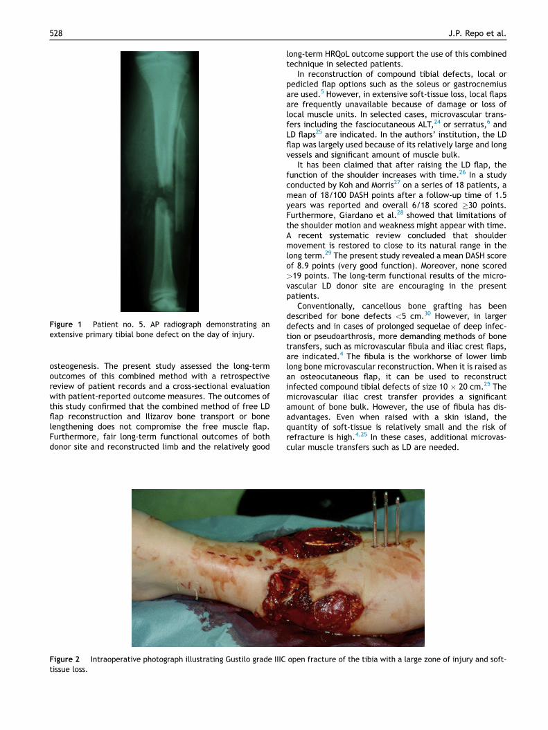
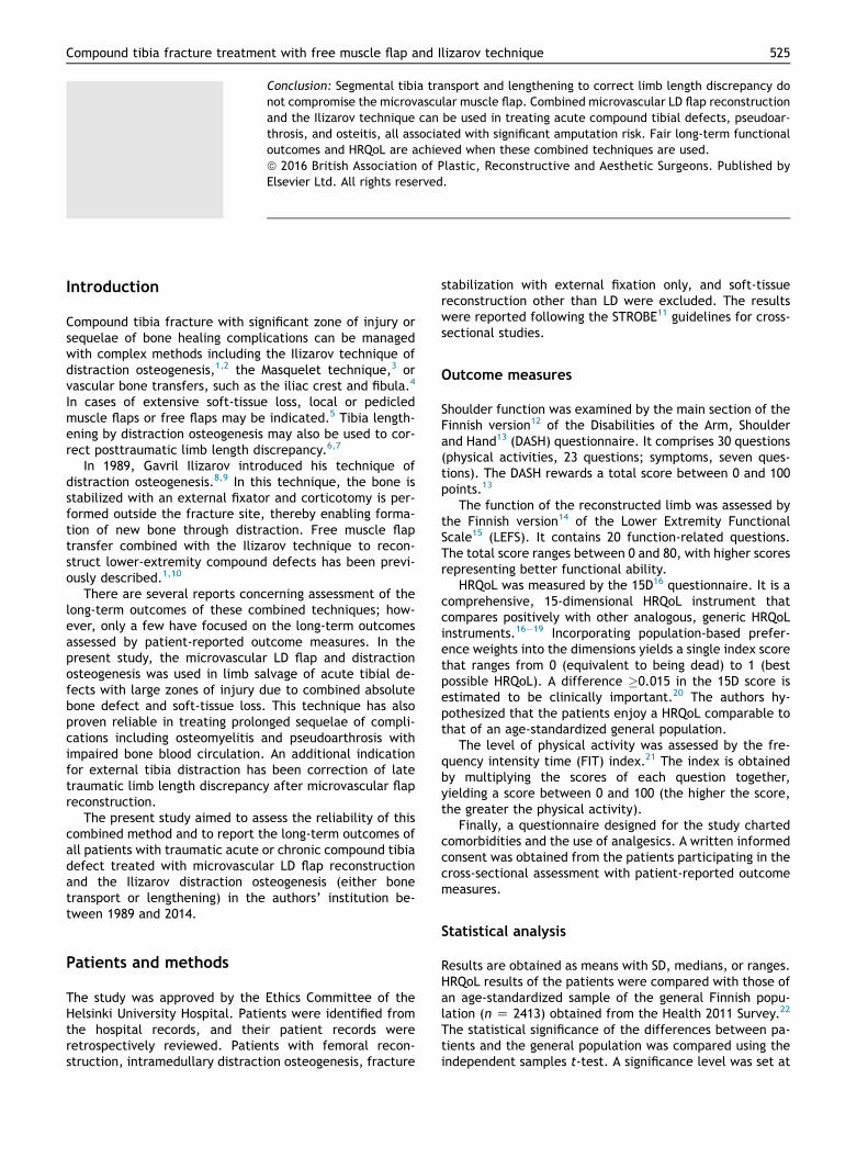
Figure 1 Patient no. 5. AP radiograph demonstrating anextensive primary tibial bone defect on the day of injury.
528 J.P. Repo et al.
osteogenesis. The present study assessed the long-termoutcomes of this combined method with a retrospectivereview of patient records and a cross-sectional evaluationwith patient-reported outcome measures. The outcomes ofthis study confirmed that the combined method of free LDflap reconstruction and Ilizarov bone transport or bonelengthening does not compromise the free muscle flap.Furthermore, fair long-term functional outcomes of bothdonor site and reconstructed limb and the relatively good
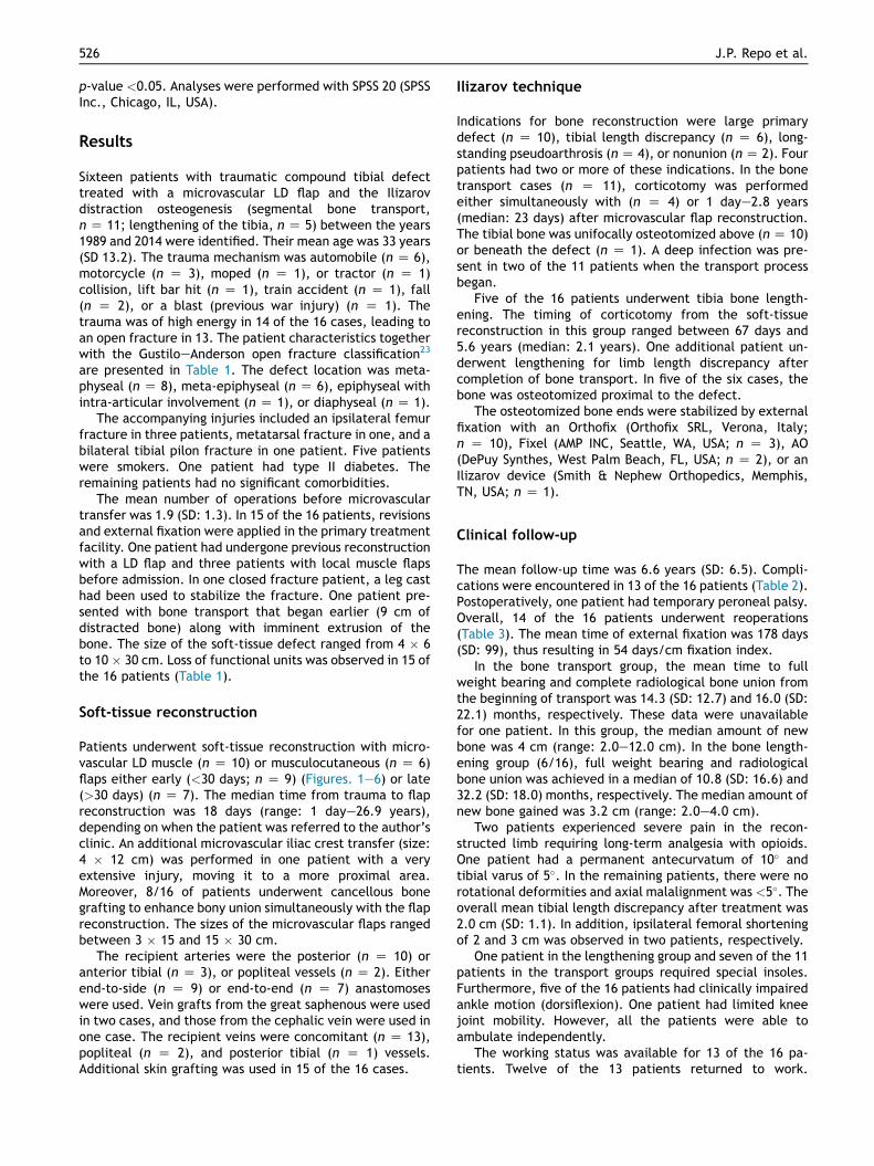
Figure 2 Intraoperative photograph illustrating Gustilo grade IIICtissue loss.
long-term HRQoL outcome support the use of this combinedtechnique in selected patients.
In reconstruction of compound tibial defects, local orpedicled flap options such as the soleus or gastrocnemiusare used.5 However, in extensive soft-tissue loss, local flapsare frequently unavailable because of damage or loss oflocal muscle units. In selected cases, microvascular trans-fers including the fasciocutaneous ALT,24 or serratus,6 andLD flaps25 are indicated. In the authors’ institution, the LDflap was largely used because of its relatively large and longvessels and significant amount of muscle bulk.
It has been claimed that after raising the LD flap, thefunction of the shoulder increases with time.26 In a studyconducted by Koh and Morris27 on a series of 18 patients, amean of 18/100 DASH points after a follow-up time of 1.5years was reported and overall 6/18 scored �30 points.Furthermore, Giardano et al.28 showed that limitations ofthe shoulder motion and weakness might appear with time.A recent systematic review concluded that shouldermovement is restored to close to its natural range in thelong term.29 The present study revealed a mean DASH scoreof 8.9 points (very good function). Moreover, none scored>19 points. The long-term functional results of the micro-vascular LD donor site are encouraging in the presentpatients.
Conventionally, cancellous bone grafting has beendescribed for bone defects <5 cm.30 However, in largerdefects and in cases of prolonged sequelae of deep infec-tion or pseudoarthrosis, more demanding methods of bonetransfers, such as microvascular fibula and iliac crest flaps,are indicated.4 The fibula is the workhorse of lower limblong bone microvascular reconstruction. When it is raised asan osteocutaneous flap, it can be used to reconstructinfected compound tibial defects of size 10 � 20 cm.25 Themicrovascular iliac crest transfer provides a significantamount of bone bulk. However, the use of fibula has dis-advantages. Even when raised with a skin island, thequantity of soft-tissue is relatively small and the risk ofrefracture is high.4,25 In these cases, additional microvas-cular muscle transfers such as LD are needed.
open fracture of the tibia with a large zone of injury and soft-
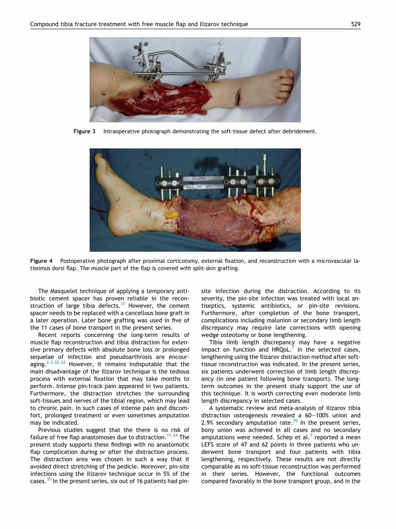
Figure 4 Postoperative photograph after proximal corticotomy, external fixation, and reconstruction with a microvascular la-tissimus dorsi flap. The muscle part of the flap is covered with split-skin grafting.
Figure 3 Intraoperative photograph demonstrating the soft-tissue defect after debridement.
Compound tibia fracture treatment with free muscle flap and Ilizarov technique 529
The Masquelet technique of applying a temporary anti-biotic cement spacer has proven reliable in the recon-struction of large tibia defects.31 However, the cementspacer needs to be replaced with a cancellous bone graft ina later operation. Later bone grafting was used in five ofthe 11 cases of bone transport in the present series.
Recent reports concerning the long-term results ofmuscle flap reconstruction and tibia distraction for exten-sive primary defects with absolute bone loss or prolongedsequelae of infection and pseudoarthrosis are encour-aging.2,3,32,33 However, it remains indisputable that themain disadvantage of the Ilizarov technique is the tediousprocess with external fixation that may take months toperform. Intense pin-track pain appeared in two patients.Furthermore, the distraction stretches the surroundingsoft-tissues and nerves of the tibial region, which may leadto chronic pain. In such cases of intense pain and discom-fort, prolonged treatment or even sometimes amputationmay be indicated.
Previous studies suggest that the there is no risk offailure of free flap anastomoses due to distraction.11,34 Thepresent study supports these findings with no anastomoticflap complication during or after the distraction process.The distraction area was chosen in such a way that itavoided direct stretching of the pedicle. Moreover, pin-siteinfections using the Ilizarov technique occur in 5% of thecases.35 In the present series, six out of 16 patients had pin-
site infection during the distraction. According to itsseverity, the pin-site infection was treated with local an-tiseptics, systemic antibiotics, or pin-site revisions.Furthermore, after completion of the bone transport,complications including malunion or secondary limb lengthdiscrepancy may require late corrections with openingwedge osteotomy or bone lengthening.
Tibia limb length discrepancy may have a negativeimpact on function and HRQoL.7 In the selected cases,lengthening using the Ilizarov distraction method after soft-tissue reconstruction was indicated. In the present series,six patients underwent correction of limb length discrep-ancy (in one patient following bone transport). The long-term outcomes in the present study support the use ofthis technique. It is worth correcting even moderate limblength discrepancy in selected cases.
A systematic review and meta-analysis of Ilizarov tibiadistraction osteogenesis revealed a 60e100% union and2.9% secondary amputation rate.35 In the present series,bony union was achieved in all cases and no secondaryamputations were needed. Schep et al.7 reported a meanLEFS score of 47 and 62 points in three patients who un-derwent bone transport and four patients with tibialengthening, respectively. These results are not directlycomparable as no soft-tissue reconstruction was performedin their series. However, the functional outcomescompared favorably in the bone transport group, and in the
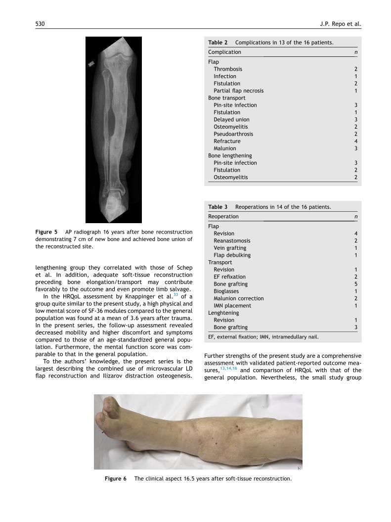
Figure 5 AP radiograph 16 years after bone reconstructiondemonstrating 7 cm of new bone and achieved bone union ofthe reconstructed site.
Table 2 Complications in 13 of the 16 patients.
Complication n
FlapThrombosis 2Infection 1Fistulation 2Partial flap necrosis 1
Bone transportPin-site infection 3Fistulation 1Delayed union 3Osteomyelitis 2Pseudoarthrosis 2Refracture 4Malunion 3
Bone lengtheningPin-site infection 3Fistulation 2Osteomyelitis 2
Table 3 Reoperations in 14 of the 16 patients.
Reoperation n
FlapRevision 4Reanastomosis 2Vein grafting 1Flap debulking 1
TransportRevision 1EF refixation 2Bone grafting 5Bioglasses 1Malunion correction 2IMN placement 1
LenghteningRevision 1Bone grafting 3
EF, external fixation; IMN, intramedullary nail.
530 J.P. Repo et al.
lengthening group they correlated with those of Schepet al. In addition, adequate soft-tissue reconstructionpreceding bone elongation/transport may contributefavorably to the outcome and even promote limb salvage.
In the HRQoL assessment by Knappinger et al.33 of agroup quite similar to the present study, a high physical andlow mental score of SF-36 modules compared to the generalpopulation was found at a mean of 3.6 years after trauma.In the present series, the follow-up assessment revealeddecreased mobility and higher discomfort and symptomscompared to those of an age-standardized general popu-lation. Furthermore, the mental function score was com-parable to that in the general population.
To the authors’ knowledge, the present series is thelargest describing the combined use of microvascular LDflap reconstruction and Ilizarov distraction osteogenesis.
Figure 6 The clinical aspect 16.5 yea
Further strengths of the present study are a comprehensiveassessment with validated patient-reported outcome mea-sures,13,14,16 and comparison of HRQoL with that of thegeneral population. Nevertheless, the small study group
rs after soft-tissue reconstruction.
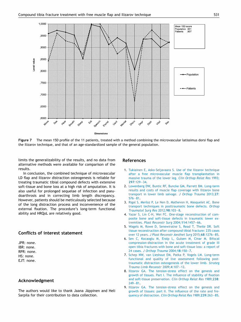
Figure 7 The mean 15D profile of the 11 patients, treated with a method combining the microvascular latissimus dorsi flap andthe Ilizarov technique, and that of an age-standardized sample of the general population.
Compound tibia fracture treatment with free muscle flap and Ilizarov technique 531
limits the generalizability of the results, and no data fromalternative methods were available for comparison of theresults.
In conclusion, the combined technique of microvascularLD flap and Ilizarov distraction osteogenesis is reliable fortreating traumatic tibial compound defects with extensivesoft-tissue and bone loss at a high risk of amputation. It isalso useful for prolonged sequelae of infection and pseu-doarthrosis and in correcting limb length discrepancy.However, patients should be meticulously selected becauseof the long distraction process and inconvenience of theexternal fixation. The procedure’s long-term functionalability and HRQoL are relatively good.
Conflicts of interest statement
JPR: none.IBR: none.RPR: none.HS: none.EJT: none.
Acknowledgment
The authors would like to thank Jaana Jappinen and HeliSarpila for their contribution to data collection.
References
1. Tukiainen E, Asko-Seljavaara S. Use of the Ilizarov techniqueafter a free microvascular muscle flap transplantation inmassive trauma of the lower leg. Clin Orthop Relat Res 1993;297:129e34.
2. Lowenberg DW, Buntic RF, Buncke GM, Parrett BM. Long-termresults and costs of muscle flap coverage with Ilizarov bonetransport in lower limb salvage. J Orthop Trauma 2013;27:576e81.
3. Rigal S, Merloz P, Le Nen D, Mathevon H, Masquelet AC. Bonetransport techniques in posttraumatic bone defects. OrthopTraumatol Surg Res 2012;98:103e8.
4. Yazar S, Lin C-H, Wei FC. One-stage reconstruction of com-posite bone and soft-tissue defects in traumatic lower ex-tremities. Plast Reconstr Surg 2004;114:1457e66.
5. Wagels M, Rowe D, Senewiratne S, Read T, Theile DR. Softtissue reconstruction after compound tibial fracture: 235 casesover 12 years. J Plast Reconstr Aesthet Surg 2015;68:1276e85.
6. Sen C, Kocaoglu M, Eralp L, Gulsen M, Cinar M. Bifocalcompression-distraction in the acute treatment of grade IIIopen tibia fractures with bone and soft-tissue loss: a report of24 cases. J Orthop Trauma 2004;18:150e7.
7. Schep NW, van Lieshout EM, Patka P, Vogels LM. Long-termfunctional and quality of live assessment following post-traumatic distraction osteogenesis of the lower limb. StrategTrauma Limb Reconstr 2009;4:107e12.
8. Ilizarov GA. The tension-stress effect on the genesis andgrowth of tissues. Part I. The influence of stability of fixationand soft-tissue preservation. Clin Orthop Relat Res 1989;238:249e81.
9. Ilizarov GA. The tension-stress effect on the genesis andgrowth of tissues: part II. The influence of the rate and fre-quency of distraction. Clin Orthop Relat Res 1989;239:263e85.
532 J.P. Repo et al.
10. Jupiter JB, Kour AK, Palumbo MD, Yaremchuck MJ. Limbreconstruction by free-tissue transfer combined with the Ili-zarov method. Plast Reconstr Surg 1991;88:943e51.
11. Vandenbroucke JP, von Elm E, Altman DG, et al. Strengtheningthe reporting of observational studies in epidemiology(STROBE): explanation and elaboration. Int J Surg 2014;12:1500e24.
12. Aro H, Hacklin E, Madanat R, Stranberg N. DASH-kyselykaa-vakkeen suomentaminen ja kulttuuriadaptaatio. Suom Ortopja Traumatol 2009;32:252e4.
13. Hudak PL, Amadio PC, Bombardier C, The Upper ExtremityCollaborative Group (UECG). Development of an upper ex-tremity outcome measure: the DASH (disabilities of the arm,shoulder and hand). Am J Ind Med 1996;29:602e8.
14. Repo JP, Tukiainen EJ, Roine RP, Ilves O, Jarvenpaa S,Hakkinen A. Lower extremity functional scale (LEFS)ekyselylomakkeen suomen kielisen version luotettavuus javaliditeetti. Suom Ortop ja Traumatol 2015;38:205.
15. Binkley JM, Stratford PW, Lott SA, Riddle DL. The lower ex-tremity functional scale (LEFS): scale development, measure-ment properties, and clinical application. North Americanorthopaedic rehabilitation research network. Phys Ther 1999;79:371e83.
16. Sintonen H. The 15D instrument of health-related quality oflife: properties and applications. Ann Med 2001;33:328e36.
17. Stavem K. Reliability, validity and responsiveness of two mul-tiattribute utility measures in patients with chronic obstructivepulmonary disease. Qual Life Res 1999;8:45e54.
18. Hawthorne G, Richardson J, Day NA. A comparison of theassessment of quality of life (AQoL) with four other genericutility instruments. Ann Med 2001;33:358e70.
19. Moock J, Kohlmann T. Comparing preference-based quality-of-life measures: results from rehabilitation patients withmusculoskeletal, cardiovascular, or psychosomatic disorders.Qual Life Res 2008;17:485e95.
20. Alanne S, Roine RP, Rasanen P, Vainiola T, Sintonen H. Esti-mating the minimum important change in the 15D scores. QualLife Res 2015;24:599e606.
21. Kasari DS. The effects of exercise on serum lipid levels incollege women. Unpublished Master’s Thesis. Missoula: Uni-versity of Montana; 1976. p. 46.
22. Report 68/2012. Helsinki. In: Koskinen S, Lundqvist A,Ristiluoma N, editors. Health, functional capacity and welfarein Finland in 2011. National Institute for Health and Welfare(THL); 2012.
23. Gustilo RB, Mendoza RM, Williams DN. Problems in the man-agement of type III (severe) open fractures: a new classifica-tion of type III open fractures. J Trauma 1984;24:742e6.
24. Yang YF, Xu ZH, Zhang GM, et al. Modified classification andsingle-stage microsurgical repair of posttraumatic infectedmassive bone defects in lower extremities. J Reconstr Micro-surg 2013;29:593e600.
25. Knobloch K, Herold C, Vogt PM. Free latissimus dorsi flaptransfer for reconstruction of soft tissue defects of the lowerextremity. Oper Orthop Traumatol 2012;24:122e30.
26. Spears SL, Hess CL. A review of the biomechanical and func-tional changes in the shoulder following transfer of the latis-simus dorsi muscles. Plast Reconstr Surg 2005;115:2070e3.
27. Koh CE, Morrison WA. Functional impairment after latissimusdorsi flap. ANZ J Surg 2009;117:1387e94.
28. Giordano S, Kaariainen K, Alavaikko J, Kaistila T, Kuokkanen H.Latissimus dorsi free flap harvesting may affect the shoulderjoint in long run. Scand J Surg 2011;100:202e7.
29. Lee KT, Mun GH. A systematic review of functional donor-sitemorbidity after latissimus dorsi muscle transfer. PlastReconstr Surg 2014;134:303e14.
30. Myeroff C, Archdeacon M. Autogenous bone graft: donor sitesand techniques. J Bone Jt Surg Am 2011;93:2227e36.
31. Karger C, Kishi T, Schneider L, Fitoussi F, Masquelet AC.Treatment of posttraumatic bone defects by the inducedmembrane technique. Orthop Traumatol Surg Res 2012;98:97e102.
32. Chim H, Sontich JK, Kaufman BR. Free tissue transfer withdistraction osteogenesis is effective for limb salvage of theinfected traumatized lower extremity. Plast Reconstr Surg2011;127:2364e72.
33. Krappinger D, Irenberger A, Zegg M, Huber B. Treatment oflarge posttraumatic tibial bone defects using the Ilizarovmethod: a subjective outcome assessment. Arch OrthopTrauma Surg 2013;133:789e95.
34. Hollenbeck ST, Woo S, Ong S, Fitch RD, Erdmann D, Levin LS.The combined use of the Ilizarov method and microsurgicaltechniques for limb salvage. Ann Plast Surg 2009 May;62(5):486e91.
35. Papakostidis C, Bhandari M, Giannoudis PV. Distractionosteogenesis in the treatment of long bone defects of thelower limbs: effectiveness, complications and clinical results;a systematic review and meta-analysis. Bone Jt J 2013;95:1673e80.