Embed Size (px)
Citation preview
Central Annals of Orthopedics & Rheumatology
Cite this article: Mcmurtry J, Mounasamy V (2015) Segmental Tibia Fractures. Ann Orthop Rheumatol 3(3): 1051.
*Corresponding authorJohn McMurtry, Department of Orthopedic Surgery, Virginia Commonwealth University, P.O Box 980153, Richmond, VA, 23298. Fax: 804-828-1572, Email: John.
Submitted: 04 August 2015
Accepted: 12 August 2015
Published: 14 August 2015
Copyright© 2015 Mcmurtry et al.
OPEN ACCESS
Review Article
Segmental Tibia FracturesJohn Mcmurtry*, Varatharaj MounasamyDepartment of Orthopedic Surgery, Virginia Commonwealth University Health System, USA
ABBREVIATIONSIMN: Intramedullary Nail
INTRODUCTIONFractures of the tibia account for approximately 17-21%
of all lower extremity fractures and approximately 2% of all combined fractures [1,2]. Within this number, 3% to 13% of tibia fractures are classified as segmental with most literature on this injury consisting of case series or retrospective reviews [3,4]. Segmental fractures of the tibia present a challenge to treating orthopedic surgeons due to their infrequent presentation, wide zone of tissue injury, and increased rate of complications. Segmental tibia fractures (AO 42-C2) are defined by two or more distinct fracture lines isolating an interposed cortical segment which excludes butterfly fragmentation [5]. Severe soft tissue defects are common due to their association with high energy trauma mechanisms of injury [6]. As a result of the trauma to the surrounding soft tissues approximately 15% of tibial shaft fractures represent open injuries with reports of segmental tibia fractures having an open injury 53% to 80% of presentations [7-9]. The perfusion of the intermediate segment is provided through endosteal and periosteal blood supply, which sustains increased damage leading to impaired fracture healing with the already precarious blood supply of the intermediary cortical segment [6,10]. With this in mind Woll et al. classified segmental tibial fractures as “an extremely high risk injury” with postoperative complications noted more frequently than in any other category of tibia fracture [6,8].
Classification
The OTA fracture classification defines segmental tibia fractures as complex fractures with an intermediate segmental fragment and thus without contact between the proximal and distal fragments. This classification can be further subdivided into 42-C2.1 with a simple segmental pattern, 42-C2.2 with a segmental pattern as well as wedge fragments, 42-C2.3 with two intermediate segmental fragments, and finally 42-C3.1 with two to three intermediate segmental fragments [11]. A second classification proposed by Melis et al. [12] in 1981 divides segmental tibia fractures into four distinct categories based on fragment fixation with an intramedullary tibial nail construct. Type I identifies a segmental fragment between the proximal and middle third of the tibial diaphysis with Type II identifying a segmental fragment between the middle and distal third of the tibial diaphysis. Type III represents a long segmental fragment between the proximal and distal third of the tibial diaphysis with Type IV segmental fragment which is entirely contained in the middle third of the tibial diaphysis.
Complications
Segmental tibia fractures inherently lead to greater risk of complications which include delayed union, nonunion, infection, and compartment syndrome. At present no standard criteria exist which can define delayed union and the occasional resultant nonunion. Although no standard criteria exist for delayed union, an often used time frame is 6 months after the initial trauma, whereas nonunion is diagnosed 9 months after the initial trauma
Keywords•Segmental tibia fracture•Tibia fracture•Tibial intramedullary nail•Externalfixationfortibiafracture
Abstract
Segmental fractures of the tibia present a challenge to treating orthopedic surgeons due to their infrequent presentation, wide zone of tissue injury, and increased rate of complications. The average union times range from 15 weeks to greater than 40 weeks with fractures demonstrating more delayed unions and nonunion in open injuries. Management primarily using a cast or brace, although infrequently used, is only indicated for low risk patients with a closed fracture, minimal shortening, and minimal angulation. Uni/multi planar external fixation, ringed external fixation, plate osteosynthesis, and Ender Nail placement are useful in selected clinical situations, but intramedullary nail placement represents the most common treatment strategy. Contention over the use of reamed versus unreamed locked intramedullary nails exists but the recommended treatment of closed segmentaltibial shaft fractures is with reamed locked intramedullary nailing. The recommended treatment of open segmental tibial shaft fractures is with unreamed locked intramedullary nailing to maximize fracture biology and to minimize risk of devascularization of the intercalary segment. Although treatment of segmental tibia fractures can be daunting excellent outcomes can be achieved with adherence to meticulous soft tissue management, optimalimplant choice, and close clinical follow up to minimize known complications.
Central
Mcmurtry et al. (2015)Email:
Ann Orthop Rheumatol 3(3): 1051 (2015) 2/6
[13]. In addition to time from injury, radiographic evidence of healing all four cortices on orthogonal radiographs as well as full and painless weight bearing assist the clinician in the determination of bony union [4]. With this in mind, the median time to union for segmental versus matched non segmental tibia fractures was significantly longer at 34 weeks and 24 weeks respectively [4]. There is a wide variation between reported time for union in segmental tibia fractures with average union times ranging from 15 weeks to greater than 40 weeks [5,14] . Tibialdiaphyseal fractures demonstrate more delayed unions and nonunions in open injuries which proportionally increase in number as the grade of injury increases [5,15]. This holds true for segmental tibia fractures, within the limitation of small case numbers, with a greater percentage of open fractures demonstrate an increased time to union [4].
As there exist at least two fracture lines for segmental tibia fractures, the criteria for union are applied to all fracture sites within the criteria outlines above. Much attention is paid to whether the proximal or distal fracture site demonstrates a greater propensity for delayed or nonunion, though most authors agree that the distal fracture site is more often problematic for achieving union [5,8-10]. Less soft tissue coverage, decreased surrounding muscle mass, more often in communication with an open injury, and increased chance of fragment displacement is implicated in slower healing at the distal segmental fracture site [5,8]. These risk factors for altered union also hold true for proximal fractures if they have severe soft tissue damage or compartment syndrome causing muscle loss and/or fasciotomy wounds [16]. Compartment syndrome as a result of injuries causing a segmental tibial fracture has been reported to occur in approximately 7-50% of patients [5,9]. There is no different in objective or subjective criteria used for the diagnosis of compartment syndrome in this patient population. Segmental tibia fractures more often present with compartment syndrome
in open fractures (14-35%) as compared to closed injuries (4-26%) [8,16].
TREATMENT
Casting/Bracing
Charnley noted that a “double fracture of the tibia” should never be initially treated with an open operation as the danger of converting the central fragment into a tubular sequestrum was not worth the risk [17]. Langard and Bo stated that initial “non-operative treatment was considered essential” in many
Figure 1 Anterior-Posterior and Lateral initial radiographs of a 49 year-old male who sustained bilateral segmental tibia fractures as a pedestrian stuck by a motor vehicle. The left tibia fracture was classified as a Grade I openfracture.
Figure 2 Anterior-Posterior and Lateral initial radiographs of a 49 year-old male who sustained bilateral segmental tibia fractures as a pedestrian stuck by a motor vehicle, which underwent placement of a Tibial intramedullary nail.
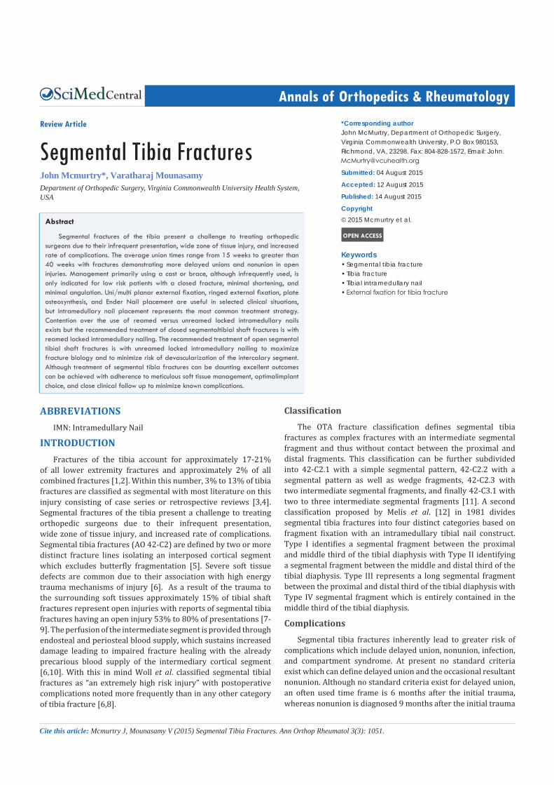
Figure 3 Anterior-Posterior and Lateral initial radiographs of a 49 year-old male who sustained bilateral segmental tibia fractures as a pedestrian stuck by a motor vehicle, which underwent placement of a Tibial intramedullary nail. Repeat radiographs at 13 months s/p injury demonstrate abundant callous and bony union of the fracture sites.

Central
Mcmurtry et al. (2015)Email:
Ann Orthop Rheumatol 3(3): 1051 (2015) 3/6
patients with segmental tibia fractures due to the high incidence of concomitant injuries. They used this algorithm even in the face of open fractures with non-operative treatment consisting of skeletal traction and plaster casting, going on to achieve union in 13 open and 7 closed fractures in 21 patients [3]. Melis et al. tempered the desire for conservative treatment when he noted poor outcomes with non-operative treatment of segmental fractures described in the French and Italian literature between the years 1956 and 1965[10]. The desire for a more rigid construct to add stability to the segmental fracture and the increasingly frequent use of intramedullary nails led to the decrease in conservative management. Several series of segmental tibia fractures published in 1992 and 2003 have treated 3 fractures non-operatively with unacceptable results related to malunion [5,8]. Sarmiento noted uneventful union with minimal residual deformity in non-segmental tibia fractures treated with casting and eventual functional bracing, but his results are without detailed descriptions of outcome measures [18,19,20]. More recently Sarmiento reported on 48 closed segmental tibia fractures treated with casting and functional bracing with all fractures going on to union [14]. Conservative management should be reserved for low risk patients with a closed fracture, minimal shortening, and minimal angulation after a thorough discussion of risks and complications. Overall, surgery is the preferred treatment for segmental tibia fractures given the difficulty in maintaining an acceptable reduction in a functional brace or cast.
External Fixation
External fixation provides a viable treatment option for many segmental tibia fractures as it provides stability to a grossly unstable injury while avoiding hardware near the site of the often associated soft tissue injury. This method is thought to “leave a small footprint” and maintain the biology of the fracture in a comparable manner to conservative treatment [21]. Initial management by external fixator due to immediate stabilization, decreased operative time, reduced blood loss, and improved blood supply at fracture sites has been well established in non-segmental tibia fractures [22-24]. This use of external fixation for the acute initial management of segmental tibia fractures with extensive soft tissue compromise represents a viable option for the treating surgeon. This allows for access to the injured tissue and provides initial stability which is unable to be obtained by casting or splinting. Unfortunately, the frequency of pin tract infection and malunion lead to removal and conversion to altered methods of definitive fixation when external fixation is used as the definitive treatment strategy [25]. Rommens et al reported on 18 fractures treated with external fixation of which 50% were complicated by “bone-healing disturbances” to include pseudarthrosis, refracture, delayed union, and malunion [6]. This increased rate of bony union complications was thought to be related to the lack of stability in bi-dimensional planes [6]. Woll and Duwelius reported a complication rate of 55% for infection, malunion, and nonunion in the care of 20 segmental tibia fractures treated with external fixation [8]. Giannoudis et al achieved a similar deep infection and malunion rate of 50% when treating 8 segmental tibia fractures with external fixation [5]. Ilizarov external fixators were not used frequently in the treatment of these injuries, but Öztürkmen et al demonstrated successful
treatment of 24 adult patients all of whom went on to union with good to excellent function results. The largest complication with this treatment method is patient intolerance and pin tract infection, but an overwhelming majority of these infections are readily treated with a short course of oral antibiotics [26]. Giotakis et al used circular external fixation to include Ilizarov (Smith and Nephew, Memphis, Tennessee), Sheffield Ring Fixator (Orthofix, Verona, Italy), and Taylor Spatial Frame (Smith and Nephew, Memphis, Tennessee) in the treatment of 20 segmental tibia fractures. Of the 20 patients there were 2 nonunions treated with either continued external fixation or revision with bone grafting, 3 malunions detected which were not clinically relevant, and one deep pin tract infection requiring antibiotics and local debridement. Circular external fixation has the advantage of circumferential control, post surgical correction outside of the operating room, capture all fracture segments, provides minimal disruption of fracture biology, and possibly allows almost immediate partial weight-bearing [21]. The use of non-circular external fixation for segmental tibia fractures should be limited to the most dire of circumstances, but circular external fixation presents a viable alternative to internal fixation in the hands of an experienced surgeon.
Plate Osteosynthesis
The key to treatment of tibia fractures is to preserve the soft tissue envelope, minimize interventions at the site of fracture, reproduce anatomic length, alignment, and rotation while optimizing the chances of obtaining union. Segmental fractures of the tibia challenge these aforementioned principals and the ability to provide stability with the use of most standard implants. In 1976 Langàrd and Bø treated 23 segmental tibia fractures with plate osteosynthesis and found a complication rate of 26% and 57% for closed and open fractures, respectively [3]. In a series of 22 patients treated with plate osteosynthesis published in 1989, Rommens et al found a 60% complication rate with greater than 25% chance of wound complication and infection. Not surprisingly, approximately 20% of tibias went on to develop pseudarthrosis with some progressing to implant failure [6]. Several additional series of segmental tibia fractures use plate osteosynthesis sparingly with infection and wound complications being almost universally described [4,5,8]. As a greater understanding of fracture biology and healing has developed, so has the use of plate osteosynthesisdecreased. With the current selection of internal, external and non-operative interventions there is very limited indications for primary plate osteosynthesis in the treatment of segmental tibia fractures.
Intramedullary Implant
Most surgeons advocate treatment of segmental tibia fractures with intramedullary nail placement in light of the significant difficulty and complications rates of other currently available treatment modalities. Intramedullary nail placement avoids many of the concerns related to other modalities such as control of length, rotation, alignment, dissection of the fracture site, disruption of the fracture vascularity, early weight-bearing, and incision site away from traumatic open wounds [16]. Duan et al, in a Cochrane Review on intramedullary nailing for adult diaphysealtibial fractures, was unable to draw a definitive conclusion on the best technique for intramedullary

Central
Mcmurtry et al. (2015)Email:
Ann Orthop Rheumatol 3(3): 1051 (2015) 4/6
nail placement although moderate evidence suggests no clear difference in complications between reamed and unreamed nailing. They also noted that reamed nailing demonstrated a decreased incidence of implant failure, less re-operation related to nonunion, but this was only true in closed tibia fractures [27]. In a review of open tibialdiaphyseal fractures Mundi et al echoed these findings of superiority of reamed nailing in closed tibia fractures, but no significant difference between methods was detected in open fractures [28].
Initially tibial nails were without interlocking screws, which limited control of the proximal and distal fragments. In 1969 Zucman and Maurer published their treatment of 36 segmental tibial fractures with unreamedKuntscher-type nails in a blind fashion. Of their 36 patients, 92% went on to union, but with 16% rate of septic union all present in patient who sustained open fractures [29]. In 1972 Pantazopoulos et al reported on their results of blind unreamedKuntscher nailing of 13 segmental tibial fractures with one nonunion, no cases of infection, and no cases of malunion [30]. In 1981, Melis et al detailed their treatment of 38 segmental tibia fractures with tight-fitting reamed Kuntscher-Herzog intramedullary nails and supplementation with immobilization in a long leg cast. With strict adherence to this algorithm, in 22 closed and 16 open fractures, they reported one malunion, one non-union, and one infection [10]. Woll and Duwelius reported on their treatment of 31 segmental tibia fractures with seven fractures being treated with unreamedLottes’ nails and the remaining fractures were treated with External fixation, plate osteosynthesis, and nonoperative treatment. Of the four treatment modalities unreamed unlocked Lottes’ nails demonstrated the lowest complication rate of 40% which included malunion and nonunion [8]. The authors were convinced that the high rate of nonunion and malunion was related to the lack of distal rotational control and hypothesized that distally locked intramedullary nails would provide a much lower rate of complications. Merianos et al evaluated the use of Ender nails for the treatment of 22 segmental tibia fractures with all patients achieving union. The limitations of rotation and length control of Ender nails were illustrated as close to one third of fractures were shortened and/or malunited [31].
In 1985 Klemm and Borner detailed one of the early reports of tibial fractures treated between 1976 and 1983 with reamed locked intramedullary nailing. Forty-one of the 401 tibia fractures were segmental with an overall delayed union of 0.8%, infection rate of 2.2%, and an excellent or good outcome in 94% of patients [32]. With the results of Klemm and Birner in mind, Wu and Shih treated 38 segmental tibial shaft fractures with reamed interlocking intramedullary tibial nailing and achieved a union rate of 97% without any deep infections, clinically significant malalignment, or implant failures. Of note, all fractures were not immediately treated with intramedullary nailing as a wait time of approximately 1 week was used to allow decreased calf swelling, open wound healing, and stabilization of systemic conditions [33]. With the positive results of reamed locked intramedullary nailing of segment tibia fractures, Huang et al treated 33 segmental tibia fractures with this technique. This series detailed rates of 3% malunion, 9% delayed unions, no cases of nonunion, and 6% deep infection rate which occurred in two of the reported open fractures. Giannoudis et al treated 27 cases of segmental
tibia fractures with 14 unreamed locking intramedullary nails, 2 reamed locking intramedullary nails, and the remaining 11 cases were treated with other various modalities. There were a large number of complications with 8 of 14 unreamedtibial nails undergoing a secondary procedure to achieve union, eradicate infection, or to undergo amputation. This is in contrast to the 2 reamed locked tibial nails did not undergo secondary procedures or develop complications which could be related to their treatment of only closed fractures [5].
The decision of unreamed versus reamed tibial nails is much less certain in segmental tibia fractures as reports in the literature are exceedingly less common than standard fractures of the tibial diaphysis. One of the main advantages of treatment with an intramedullary nail is the ability to preserve the blood flow to the osseous pathology by minimizing disruption of blood flow from surrounding tissue [26]. The effect of reaming and nail size was evaluated in a canine tibia model with surprising results [34, 35]. Nutrient artery flow for intact tibias in reamed and unreamed nail placement was evaluated in a mongrel canine model over 14 days post operatively. Blood flow after unreamed nailing went from 44% of baseline immediately after nailing, but approached baseline with 99% blood flow at day 14. This is in stark contrast to the reamed tibial nail with no flow rate at day zero and a resumption of 26% of baseline at day 14 [34]. Hupel et al used a segmental tibia fracture canine model to evaluate the effect of tibial nail size on osseous cortical blood flow. The canine group with an unreamed “tight” locked intramedullary tibial nail demonstrated a nearly 75% decrease in blood flow which was still decreased by 50% at 11 weeks post intervention. Compared to the canine group with an unreamed “loose” locked intramedullary tibial nail which demonstrated an approximately 50% decrease in blood flow which returned to greater than pre-surgical flow at 11 weeks post intervention [35]. One concern with reamed intramedullary nailing of segmental tibia fractures is rotational displacement of the fragments which can strip the surrounding soft tissues and cause an increased rate of avascular complications [36]. When using a reamed intramedullary nail it was suggested to reduce and stabilize the fracture with a pointed reduction clamp to avoid this potential stripping of soft tissues from the fracture fragment [5,10]. With these principals in mind, Kakar and Tornetta followed 51 patients to union with segmental tibia fractures treated with unreamed locked intramedullary nail placement with only a 9% revision rate [16]. Most recently Terra et al compared healing in matched controls of 30 segmental and non segmental tibia fractures treated with 18 unreamed locked intramedullary nails, 4 reamed locked intramedullary nails, 3 plate osteosynthesis, and 5 external fixation. The preferred treatment was unreamed locked intramedullary nailing, but the authors report a greater than 55% rate of reoperation to obtain union [4]. The preferred treatment of closed segmental tibial shaft fractures is reamed locked intramedullary nailing to maximize biomechanical stability of the construct. The preferred treatment of open segmental tibial shaft fractures is unreamed locked intramedullary nailing to maximize fracture biology and to minimize the risk of devascularization of the intercalary segment.

Central
Mcmurtry et al. (2015)Email:
Ann Orthop Rheumatol 3(3): 1051 (2015) 5/6
DISCUSSION & CONCLUSION
Summary
Segmental fractures of the tibia, defined by two or more distinct fracture lines isolating an interposed cortical segment, present a challenge to treating orthopedic surgeons due to their infrequent presentation, wide zone of tissue injury, and increased rate of complications [5]. Severe soft tissue defects are common with reports of segmental tibia fractures having an open injury 53% to 80% of presentations [7-9]. The average union times range from 15 weeks to greater than 40 weeks with fractures demonstrating more delayed unions and nonunions in open injuries, which increase in number as the grade of injury increases [5,14,15]. Managementprimarily using a cast or brace, although not frequently used, is only indicated for low risk patients with a closed fracture, minimal shortening, and minimal angulation. Uni/multi planar external fixation, ringed external fixation, plate osteosynthesis, and Ender Nail placement are useful in selected clinical situations, but Intramedullary nail placement represents the most common treatment strategy. Contention over the use of reamed versus unreamed locked intramedullary nails exists but we recommend treatment of closed segmental tibial shaft fractures with reamed locked intramedullary nailing. We also recommend treatment of open segmental tibial shaft fractures with unreamed locked intramedullary nailing to maximize fracture biology and to minimize the risk of devascularization of the intercalary segment. Although treatment of segmental tibia fractures can be daunting, excellent outcomes can be achieved with adherence to meticulous soft tissue management, optimal implant choice, and close clinical follow up to minimize known complications.
REFERENCES1. Kaye JA, Jick H. Epidemiology of lower limb fractures in general
practice in the United Kingdom. Inj Prev. 2004; 10: 368-374.
2. Court-Brown CM, Caesar B. Epidemiology of adult fractures: A review. Injury. 2006; 37: 691-697.
3. Langård O, Bo O. Segmental tibial shaft fractures. Acta Orthop Scand. 1976; 47: 351-357.
4. Teraa M, Blokhuis TJ, Tang L, Leenen LP. Segmental tibial fractures: an infrequent but demanding injury. Clin Orthop Relat Res. 2013; 471: 2790-2796.
5. Giannoudis PV, Hinsche AF, Cohen A, Macdonald DA, Matthews SJ, Smith RM. Segmental tibial fractures: an assessment of procedures in 27 cases. Injury. 2003; 34: 756-762.
6. Rommens PM, Coosemans W, Broos PL. The difficult healing of segmental fractures of the tibial shaft. Arch Orthop Trauma Surg. 1989; 108: 238-242.
7. Weiss RJ, Montgomery SM, Ehlin A, Al Dabbagh Z, Stark A, Jansson KA. Decreasing incidence of tibial shaft fractures between 1998 and 2004: information based on 10,627 Swedish inpatients. Acta Orthop. 2008; 79: 526-533.
8. Woll TS, Duwelius PJ. The segmental tibial fracture. Clin Orthop Relat Res. 1992; 204-207.
9. Zucman J, Maurer P. Two-level fractures of the tibia. Results in thirty-six cases treated by blind nailing. J Bone Joint Surg Br. 1969; 51: 686-693.
10. Melis GC, Sotgiu F, Lepori M, Guido P. Intramedullary nailing in segmental tibial fractures. J Bone Joint Surg Am. 1981; 63: 1310-1318.
11. Marsh JL, Slongo TF, Agel J, Broderick JS, Creevey W, DeCoster TA, et al. Fracture and dislocation classification compendium-2007: Orthopaedic Trauma Association classification, database and outcomes committee. J Orthop Trauma. 2007; 21: S1-133.
12. Melis GC, Sotgiu F, Lepori M, Guido P. Intramedullary nailing in segmental tibial fractures. J Bone Joint Surg Am. 1981; 63: 1310-1318.
13. Bishop JA, Palanca AA, Bellino MJ, Lowenberg DW. Assessment of compromised fracture healing. J Am Acad Orthop Surg. 2012; 20: 273-282.
14. Sarmiento A, Latta LL. Functional treatment of closed segmental fractures of the tibia. Acta Chir Orthop Traumatol Cech. 2008; 75: 325-331.
15. Papakostidis C, Kanakaris NK, Pretel J, Faour O, Morell DJ, Giannoudis PV. Prevalence of complications of open tibial shaft fractures stratified as per the Gustilo-Anderson classification. Injury. 2011; 42: 1408-1415.
16. Kakar S, Tornetta P 3rd. Segmental tibia fractures: a prospective evaluation. Clin Orthop Relat Res. 2007; 460: 196-201.
17. CHARNLEY, JOHN: The Closed Treatment of Common Fractures. Ed. 3. London, Churchill Livingstone, 1972.
18. Sarmiento A. A functional below-the-knee cast for tibial fractures. J Bone Joint Surg Am. 1967; 49: 855-875.
19. Sarmiento A. Functional bracing of tibial and femoral shaft fractures. Clin Orthop Relat Res. 1972; 82: 2-13.
20. Sarmiento A. Functional bracing of tibial fractures. Clin Orthop Relat Res. 1974: 202-219.
21. Giotakis N, Panchani SK, Narayan B, Larkin JJ, Al Maskari S, Nayagam S. Segmental fractures of the tibia treated by circular external fixation. J Bone Joint Surg Br. 2010; 92: 687-692.
22. El-Sayed M, Atef A. Management of simple (types A and B) closed tibial shaft fractures using percutaneous lag-screw fixation and Ilizarov external fixation in adults. International orthopaedics. 2012; 36: 2133-2138.
23. Helland P, Bøe A, Mølster AO, Solheim E, Hordvik M. Open tibial fractures treated with the Ex-fi-re external fixation system. Clin Orthop Relat Res. 1996; : 209-220.
24. Bråten M, Helland P, Grøntvedt T, Aamodt A, Benum P, Mølster A. External fixation versus locked intramedullary nailing in tibial shaft fractures: a prospective, randomised study of 78 patients. Arch Orthop Trauma Surg. 2005; 125: 21-26.
25. Xu X, Li X, Liu L, Wu W. A meta-analysis of external fixator versus intramedullary nails for open tibial fracture fixation. J Orthop Surg Res. 2014; 9:75.
26. Oztürkmen Y, Karamehmetoğlu M, Karadeniz H, Azboy I, Caniklioğlu M. Acute treatment of segmental tibial fractures with the Ilizarov method. Injury. 2009; 40: 321-326.
27. Duan X, Al-Qwbani M, Zeng Y, Zhang W, Xiang Z. Intramedullary nailing for tibial shaft fractures in adults. Cochrane Database Syst Rev. 2012.
28. Mundi R, Chaudhry H, Niroopan G, Petrisor B, Bhandari, M. Open Tibial Fractures: Updated Guidelines for Management. JBJS Reviews, 2015; 3.
29. Zucman J, Maurer P. Two-level fractures of the tibia. Results in thirty-six cases treated by blind nailing. J Bone Joint Surg Br. 1969; 51: 686-693.
Central
Mcmurtry et al. (2015)Email:
Ann Orthop Rheumatol 3(3): 1051 (2015) 6/6
Mcmurtry J, Mounasamy V (2015) Segmental Tibia Fractures. Ann Orthop Rheumatol 3(3): 1051.
Cite this article
30. Pantazopoulos T, Galanos P, Agoropoulos Z, Hartofilakidis-Garofalidi. Treatment of double tibial fractures by blind intramedullary nailing. Clin Orthop Relat Res. 1972; 84: 137-143.
31. Merianos P, Papagiannakos K, Scretas E, Smyrnis P. Ender nails for segmental tibial fracture. Early weight bearing in 22 cases. Acta Orthop Scand. 1988; 59: 297-301.
32. Klemm KW, Börner M. Interlocking nailing of complex fractures of the femur and tibia. Clin Orthop Relat Res. 1986: 89-100.
33. Wu CC, Shih CH. Segmental tibial shaft fractures treated with interlocking nailing. J Orthop Trauma. 1993; 7: 468-472.
34. Brinker MR, Cook SD, Dunlap JN, Christakis P, Elliott MN. Early changes in nutrient artery blood flow following tibial nailing with and without reaming: a preliminary study. J Orthop Trauma. 1999; 13: 129-133.
35. Hupel TM, Aksenov SA, Schemitsch EH. Cortical bone blood flow in loose and tight fitting locked unreamed intramedullary nailing: a canine segmental tibia fracture model. J Orthop Trauma. 1998; 12: 127-135.
36. Arastu MH, Sheehan B, Paolucci EO, Buckley RE. Does it really spin? Intra-medullary nailing of segmental tibial fractures--a cadaveric study. Injury. 2015; 46: 643-648.