Embed Size (px)
Citation preview

N e o n ata l R a d i o l o g y Ba s i c s Respiratory Disorders Presenting in the Newborn Period 2 B - 1
Chapter2BRespiratory Disorders Presenting
in the Newborn Period
Content and Objectives
Transient Tachypnea of the Newborn 2B-3
Meconium Aspiration Syndrome 2B-7
Neonatal Pneumonia 2B-10
Neonatal Chylothorax 2B-15
Continuing Nursing Education Test CNE-1
Objectives: 1. List four typical x-ray findings for the infant with transient tachypnea of the newborn (TTN). 2. Describe the appearance of the chest x-ray of the infant with meconium aspiration sydrome (MAS). 3. Identify the most common organisms that cause neonatal pneumonia. 4. Outline the common radiographic findings in neonatal pneumonia. 5. Identify the most common x-ray findings for pleural effusion. 6. Describe the chest x-ray of an infant with chylothorax.

2 B - 2 Respiratory Disorders Presenting in the Newborn Period N e o n ata l R a d i o l o g y Ba s i c s

N e o n ata l R a d i o l o g y Ba s i c s Respiratory Disorders Presenting in the Newborn Period 2 B - 3
Transient Tachypnea of the Newborn
Transient tachypnea of the newborn (TTN), a condition also known as retained lung fluid in the newborn, was
first described as a syndrome by Mary Ellen Avery in 1966.1 Other names that have been applied to this problem are wet lung syndrome and respiratory distress syndrome Type II. Swischuk describes this as retained fluid syndrome.2 It is a diagnosis of exclusion and is generally a benign, self-limiting condition of the term or near-term neonate lasting 24 to 48 hours and occasionally up to 72 hours. It is not a true disease, but a condition caused by retained fetal pulmonary fluid.2 The most prominent findings are tachypnea with respiratory rates as high as 120 per minute, grunting, nasal flaring, minimal to no retractions, minimal cyanosis, and mild hypoxemia. The onset of clinical findings appears soon after birth or at birth. Both the clinical and x-ray findings resolve within 72 hours.
LUNG LIQUID CLEARANCE MECHANISMSIt has been proposed that the cause of this syndrome is
a disturbance in normal pulmonary adaptation resulting in delay in clearance of lung liquid. Despite the common occur-rence of retained lung fluid, the exact mechanisms and patho-physiology of absorption of lung liquid have not been fully explained. Abnormalities of or lack of labor, cesarean deliv-ery, abnormal circulatory adaptation after delivery, pulmo-nary artery hypertension, and poor left ventricular function are thought to contribute to the occurrence of retained lung fluid. It was previously thought that the chest compression produced by vaginal delivery and the subsequent evacuation of fluid from the trachea and mouth were a major factor in
fetal lung liquid clearance. Now, these are thought to play a minor or negligible role in lung liquid clearance. The actual process is much more complex.3
The fetal lung produces a large volume of fluid, 3–5 mL/kg/hour. This fluid is distinct in composition from amniotic fluid. It is produced, eliminated through the trachea, swallowed by the fetus, and adds to the amniotic fluid pool. One of the primary driving forces for this liquid production is active chloride secretion. Active transport of the chloride ion across pulmonary epithelium generates an electrical potential difference, causing a shift of liquid from the microcircula-tion to the interstitium and into the alveoli. Near the time of birth and prior to labor, lung liquid secretion slows, and absorption begins as the pulmonary epithelium changes from a chloride-secreting to a sodium-absorbing barrier. This tran-sition is influenced by neurohumoral factors, catecholamines, steroids, and vasopressin. Labor increases lung liquid absorp-tion. In studies using fetal lambs, lung liquid was reduced from 21 mL/kg at the beginning of labor to 6 mL/kg at the time of delivery.3 The process of normal lung liquid clearance continues for several hours after birth, with the majority of fluid entering the microcirculation. Air inflation at birth shifts fluid from the alveoli to the interstitium and the perivascular spaces around large blood vessels and airways. With sponta-neous respiration, intrathoracic pressure drops and lymphatic drainage increases. The flow of lung liquid out of the alveoli is facilitated by the higher protein content and osmotic pressure of blood and lymph fluid. The potential pathways of lung liquid clearance after birth include the blood vessels, lym-phatics, pleural space, mediastinum, and upper airway.
In term fetal lambs, lymphatic flow doubles after breath-ing is initiated. The concentration of protein in lymph also drops, suggesting a shift of low-protein lung liquid to the interstitium and lymphatics. As low-protein lung fluid enters
Chapter 2BRespiratory Disorders Presenting
in the Newborn Period
Editor
Carol Trotter, PhD, RN, NNP-BC
2 B - 4 Respiratory Disorders Presenting in the Newborn Period N e o n ata l R a d i o l o g y Ba s i c s
the lymphatics, the concentration of the lymph protein is diluted. When the lung fluid leaves the lymphatics and enters the bloodstream, the protein content of the lymph fluid returns to normal. About 10 percent of lung liquid is esti-mated to leave the lungs via the lymphatics by way of the thoracic duct and drainage into the superior vena cava. Most lung liquid enters the pulmonary microcirculation or seeps into the mediastinum, with subsequent absorption into the bloodstream.
As noted earlier, factors that delay resorption of lung liquid are cesarean delivery, lack of labor or prolonged labor, increased lung microvascular pressure from hypoxemia, increased left ventricular pressure, and low plasma protein concentration. Krantz studied 4,659 neonates and found an increase in retained lung fluid in neonates delivered by cesar-ean section.4 Rupture of membranes or uterine contractions prior to cesarean section decreased the incidence of retained lung fluid in full-term neonates. Neonates with low Apgar scores also had higher incidence rates.5 Halliday examined neonates with retained lung fluid by serial echocardiography and found that mild left ventricular failure and myocardial dysfunction were present in this group.5
Gowen evaluated the electrical potential difference and ion transport across the nasal epithelium in term neonates as an index of ion transport across the pulmonary epithelium.6 This study supported the role of epithelial ion transport dysfunc-tion in retained lung fluid and the delay in transition of the epithelium from a chloride-secreting to a sodium-absorbing
barrier as a pathophysiologic component of retained lung fluid. It also supported the role of labor in the normal transi-tion process.
Lung function in this disorder has been studied. Compared with normal healthy neonates, those with retained lung fluid were found to have high total ventilation, high breathing frequency, low tidal volume, high dead space, prolonged
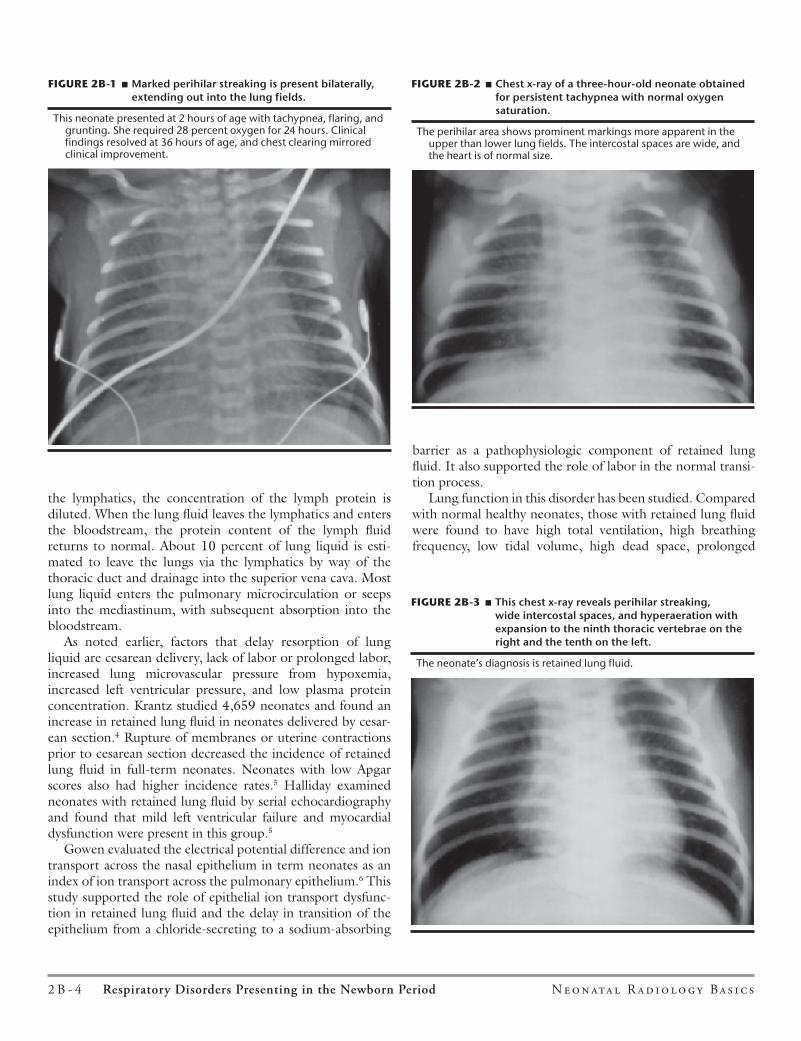
FIGURE 2B-1 n Marked perihilar streaking is present bilaterally, extending out into the lung fields.
This neonate presented at 2 hours of age with tachypnea, flaring, and grunting. She required 28 percent oxygen for 24 hours. Clinical findings resolved at 36 hours of age, and chest clearing mirrored clinical improvement.
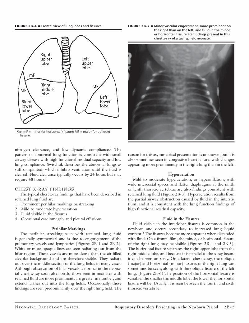
FIGURE 2B-2 n Chest x-ray of a three-hour-old neonate obtained for persistent tachypnea with normal oxygen saturation.
The perihilar area shows prominent markings more apparent in the upper than lower lung fields. The intercostal spaces are wide, and the heart is of normal size.
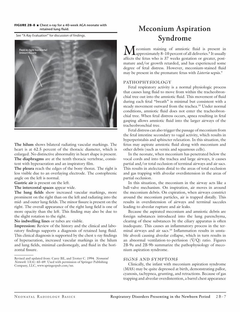
FIGURE 2B-3 n This chest x-ray reveals perihilar streaking, wide intercostal spaces, and hyperaeration with expansion to the ninth thoracic vertebrae on the right and the tenth on the left.
The neonate’s diagnosis is retained lung fluid.
N e o n ata l R a d i o l o g y Ba s i c s Respiratory Disorders Presenting in the Newborn Period 2 B - 5
nitrogen clearance, and low dynamic compliance.7 The pattern of abnormal lung function is consistent with small airway disease with high functional residual capacity and low lung compliance. Swischuk describes the abnormal lungs as stiff or splinted, which inhibits ventilation until the fluid is cleared. Fluid clearance typically occurs by 24 hours but may require 48 hours.2
CHEST X-RAY FINDINGSThe typical chest x-ray findings that have been described in
retained lung fluid are:1. Prominent perihilar markings or streaking2. Mild to moderate hyperaeration3. Fluid visible in the fissures4. Occasional cardiomegaly and pleural effusions
Perihilar MarkingsThe perihilar streaking seen with retained lung fluid
is generally symmetrical and is due to engorgement of the pulmonary vessels and lymphatics (Figures 2B-1 and 2B-2). White or more opaque lines are seen radiating out from the hilar region. These vessels are more dense than the air-filled alveolar background and are therefore visible. They radiate out over the middle section of the lung fields in many cases. Although observation of hilar vessels is normal in the neona-tal chest x-ray soon after birth, those seen in neonates with retained fluid are more prominent, are greater in number, and extend farther out into the lung fields. Occasionally, these findings are seen predominantly over the right lung field. The
reason for this asymmetrical presentation is unknown, but it is also sometimes seen in congestive heart failure, with changes appearing more prominently in the right lung than in the left.
HyperaerationMild to moderate hyperaeration, or hyperinflation, with
wide intercostal spaces and flatter diaphragms at the ninth or tenth thoracic vertebrae are also findings consistent with retained lung fluid (Figure 2B-3). Hyperaeration results from the partial airway obstruction caused by fluid in the intersti-tium, and it is consistent with the lung function findings of high functional residual capacity.
Fluid in the FissuresFluid visible in the interlobar fissures is common in the
newborn and occurs secondary to increased lung liquid content.2 The fissures become more apparent when distended with fluid. On a frontal film, the minor, or horizontal, fissure of the right lung may be visible (Figures 2B-4 and 2B-5). The horizontal fissure separates the right upper lobe from the right middle lobe, and because it is parallel to the x-ray beam, it can be seen on x-ray. On a lateral chest x-ray, the oblique (major) and horizontal (minor) fissures of the right lung can sometimes be seen, along with the oblique fissure of the left lung. (Figure 2B-6) The position of the horizontal fissure is variable; the smaller the middle lobe, the lower the horizontal fissure will be. Usually, it is seen between the fourth and sixth thoracic vertebrae.
FIGURE 2B-4 n Frontal view of lung lobes and fissures.
Key: mF = minor (or horizontal) fissure; MF = major (or oblique) fissure.
FIGURE 2B-5 n Minor vascular engorgment, more prominent on the right than on the left, and fluid in the minor, or horizontal, fissure are findings present in this chest x-ray of a tachypneic neonate.

2 B - 6 Respiratory Disorders Presenting in the Newborn Period N e o n ata l R a d i o l o g y Ba s i c s
CardiomegalyThe etiology of the cardiomegaly seen in retained lung
fluid is unclear. It may be a result of myocardial insuffi-ciency due to perinatal asphyxia, hypoxemia, or abnormal pulmonary vascular transition. Infiltrates or pleural effusions occur at times and represent a pattern of alveolar fluid collec tion that is more consolidated in certain areas of the lung (Figure 2B-7).
Other FindingsIf the initial chest x-ray is taken very early in the neona-
tal course, it may show a pattern of reticulogranularity not unlike that seen in respiratory distress syndrome. This is due to the retained fluid still within the alveoli. The x-ray, however, shows normal to increased lung expansion rather than the decreased lung expansion typical of respiratory dis-tress syndrome.
As was mentioned earlier, retained lung fluid is a diagno-sis of exclusion. Pneumonia may mimic the x-ray findings of retained lung fluid. The vascular engorgement seen with congestive heart failure secondary to congenital heart defects may also be confused with the perihilar streaking of retained lung fluid. It is essential to consider the history and the clini-cal, laboratory, and chest x-ray findings before arriving at the diagnostic impression of retained lung fluid.
CASE STUDYA 40-week-gestation, 3.5 kg AGA male neonate was deliv-
ered by repeat cesarean section to a 29-year-old, gravida 3, para 2 mother. Prenatal care was initiated in the first trimester, and no problems of pregnancy occurred. The cesarean section was a scheduled repeat without labor. The neonate’s Apgar scores were 8 and 9 at one and five minutes. The delivery room examination was normal. On admission to the nursery, a respiratory rate of 70 was recorded with clear breath sounds, equal bilaterally. Nursery admission vital signs were tempera-ture 36.5°C (97.7°F), heart rate 140 bpm, respiratory rate 70 breaths per minute, and capillary refill time two seconds.
At one hour of age, the neonate’s respiratory rate had risen to 80 to 90 per minute, and nasal flaring was present without retractions or grunting. Re-examination at this time revealed clear breath sounds; slight duskiness of the mucous mem-branes; normal pulses; no murmur; a normal S1 and S2; and normal reflexes, tone, and alertness. Pulse oximetry revealed an oxygen saturation of 87–88 percent, and the neonate was placed in 27 percent oxygen, at which time the oxygen satu-ration rose to 95 percent. A radial artery gas was obtained after oxygen therapy had been initiated, and revealed a pH of 7.39, a PCO2 of 38, a PaO2 of 65, and a bicarbonate level of 24. A complete blood count was obtained and revealed a white count of 14,000/mm3 with 47 percent PMNs, 7 percent bands, 2 percent eosinophils, 1 percent basophils, 7 percent monocytes, 36 percent lymphocytes, a platelet count of 180,000/mm3, and a hemoglobin of 18 gm/dL with a hematocrit of 53 percent. The smear was normal.
X-Ray Evaluation (Figure 2B-8)Indication for the initial x-ray was tachypnea and mild
hypoxemia in a term neonate at one hour of age.Penetration: appears to be normally exposed.Rotation is slight, to the right.The soft tissues of the neck, chest, and extremities are of normal thickness and without emphysema. Skin folds are noted at the left shoulder.The bony framework is intact, with 12 ribs bilaterally. Clavicles are intact; humeri and vertebral bodies are without abnormality.The trachea shows a straight air column.
FIGURE 2B-7 n This neonate’s clinical presentation, course, and history were all consistent with retained lung fluid.
The chest xray shows hilar densities that are not symmetrical and have the appearance of infiltrates.
FIGURE 2B-6 n Lateral view of the major, or oblique, fissures of the right and left lung and the minor, or horizontal, fissure of the right lung.
N e o n ata l R a d i o l o g y Ba s i c s Respiratory Disorders Presenting in the Newborn Period 2 B - 7
The hilum shows bilateral radiating vascular markings. The heart is at 62.5 percent of the thoracic diameter, which is enlarged. No distinctive abnormality in heart shape is present.The diaphragms are at the tenth thoracic vertebrae, consis-tent with hyperaeration and an inspiratory film.The pleura reach the edges of the bony thorax. The right is less visible due to an overlaying electrode. The costophrenic angle on the left is normal.Gastric air is present on the left.The intercostal spaces appear wide.The lung fields show increased vascular markings, more prominent on the right than on the left and radiating into the mid- and outer lung fields. The minor fissure is present on the right. The overall appearance of the right lung field is one of more opacity than the left. This finding may also be due to the slight rotation to the right.No indwelling lines or tubes are visible.Impression: Review of the history and the clinical and labo-ratory findings supports a diagnosis of retained lung fluid. This clinical diagnosis is supported by the chest x-ray findings of hyperaeration, increased vascular markings in the hilum and lung fields, minimal cardiomegaly, and fluid in the hori-zontal fissure.
Revised and updated from: Carey BE, and Trotter C. 1994. Neonatal Network: 13(4): 65–69. Used with permission of Springer Publishing Company, LLC, www.springerpub.com/nn.
Meconium Aspiration Syndrome
Meconium staining of amniotic fluid is present in approximately 8–10 percent of all deliveries.8 It usually
affects the fetus who is 37 weeks gestation or greater, post-mature and/or growth retarded, and has experienced some degree of fetal distress. However, meconium-stained fluid may be present in the premature fetus with Listeria sepsis.9
PATHOPHYSIOLOGYFetal respiratory activity is a normal physiologic process
that causes lung fluid to move from within the tracheobron-chial tree out into the amniotic fluid. This movement of fluid during each fetal “breath” is minimal but consistent with a steady movement outward from the trachea.10 Under normal conditions, amniotic fluid does not enter the tracheobron-chial tree. When fetal distress occurs, apnea resulting in fetal gasping allows amniotic fluid into the larger airways of the tracheobronchial tree.
Fetal distress can also trigger the passage of meconium from the fetal intestine secondary to vagal activity, which results in hyperperistalsis and sphincter relaxation. In this situation, the fetus may aspirate amniotic fluid along with meconium and other debris (such as vernix and squamous cells).
In the neonate, when meconium has penetrated below the vocal cords and into the trachea and large airways, it causes partial and/or total occlusion of terminal airways and air sacs. This results in atelectasis distal to the areas of total occlusion and gas trapping with alveolar overdistension in the areas of partial occlusion.
In this situation, the meconium in the airway acts like a ball-valve mechanism. On inspiration, air moves in around the meconium debris. On expiration, when airways constrict around the meconium particles, air is trapped distally. This results in overdistension of airways and terminal saccules leading to alveolar rupture and air leaks.
Because the aspirated meconium and amniotic debris are foreign substances introduced into the lung parenchyma, clearing of these substances by the ciliary apparatus is often inadequate. This causes an inflammatory process in the ter-minal airways and air sacs.10 Inflammation results in unsta-ble alveoli causing alveolar collapse, which in turn results in an abnormal ventilation-to-perfusion (V̇/Q̇) ratio. Figures 2B-9a and 2B-9b summarize the pathophysiology of meco-nium aspiration syndrome.
SIGNS AND SYMPTOMSClinically, the infant with meconium aspiration syndrome
(MAS) may be quite depressed at birth, demonstrating pallor, cyanosis, tachypnea, grunting, and retractions. Because of gas trapping and alveolar overdistention, a barrel chest appearance
FIGURE 2B-8 n Chest x-ray for a 40-week AGA neonate with retained lung fluid.
See “X-Ray Evaluation” for discussion of findings.
2 B - 8 Respiratory Disorders Presenting in the Newborn Period N e o n ata l R a d i o l o g y Ba s i c s
may be observed. Rales and rhonchi may be auscultated on physical exam.
Depending on the severity of meconium aspiration, both respiratory and metabolic acidosis can develop due to hypox-emia and hypercarbia. The hypoxemia and hypercarbia are secondary to the V̇/Q̇ mismatching described above. Acidosis of any origin can increase the risk for and/or potentiate per-sistent pulmonary hypertension (PPHN).
CHEST X-RAY FINDINGSInfiltrates
Complete occlusion of the airway results in atelectasis. Atelectatic areas on x-ray film appear more gray/white than the air-filled normal alveoli. This is because atelectatic areas have a greater density than air-filled alveoli and therefore block the beam from reaching the x-ray cassette. This pro-duces an x-ray film with bilateral, diffuse, patchy, fluffy, or nodular (more dense, gray/white areas) infiltrates and asym-metric areas of opacity. These infiltrates may present in a focal or generalized manner (Figure 2B-10).
FIGURE 2B-9�a n Pathophysiology of meconium aspiration syndrome.
From: Miller MJ, Fanaroff AA and Martin RJ. 2006. The respiratory system, Part 5: Respiratory disorders in preterm and term infants. In Neonatal–Perinatal Medicine: Diseases of the Fetus and Infant, 8th ed., Martin RJ, Fanaroff AA, and Walsh MC, eds. St. Louis: Mosby, 1123. Reprinted by permission.
FIGURE 2B-9�b n Pathophysiology of meconium aspiration syndrome.
FIGURE 2B-9b � Alveolar function in meconium aspiration.
Normal function
Complete obstruction
(atelectasis and pulmonary shunting)
Partial obstruction
•Inspiration
•Expiration
ball valve effect�airway
collapse on expiration traps
air, distends alveolus, can
affect pulmonary circulation
•Overdistention and rupture
•Atelectasis results due to
loss of surfactant
FIGURE 2B-10 n Term neonate with meconium aspiration syndrome.
Note the diffuse, bilateral, dense infiltrates as well as the right pleural effusion. The heart size is within normal limits. The tip of the endotracheal tube is properly located between the clavicles and the carina.
3 2N E O N A T A L N E T W O R K
R A D I O L O G Y B A S I C S
In the neonate, when meconium has penetrated below thevocal cords and into the trachea and large airways, it causespartial and/or total occlusion of terminal airways and air sacs.This results in atelectasis distal to the areas of total occlusionand gas trapping with alveolar overdistension in the areas ofpartial occlusion.
In this situation, the meconium in the airway acts like aball-valve mechanism. On inspiration, air moves in around themeconium debris. On expiration, when airways constrictaround the meconium particles, air is trapped distally. Thisresults in overdistension of airways and terminal saccules lead-ing to alveolar rupture and air leaks (Figure 9).
Because the aspirated meconium and amniotic debris areforeign substances introduced into the lung parenchyma,clearing of these substances by the ciliary apparatus is ofteninadequate. This causes an inflammatory process in the termi-nal airways and air sacs.15 Inflammation results in unstablealveoli causing alveolar collapse, which in turn results in anabnormal ventilation-to-perfusion (V̇/Q̇) ratio.
SIGNS AND SYMPTOMSClinically, the infant with meconium aspiration syndrome
may be quite depressed at birth, demonstrating pallor,cyanosis, tachypnea, grunting, and retractions. Because of gastrapping and alveolar overdistension, a barrel chest appear-ance may be observed. Rales and rhonchi may be auscultatedon physical exam.
Depending on the severity of meconium aspiration, bothrespiratory and metabolic acidosis can develop due to hypox-emia and hypercarbia. The hypoxemia and hypercarbia aresecondary to the V̇/Q̇ mismatching described above. Acidosisof any origin can increase the risk for and/or potentiate per-sistent pulmonary hypertension (PPHN).
CHEST X-RAY FINDINGS
InfiltratesComplete occlusion of the airway results in atelectasis.
Atelectatic areas on x-ray film appear more gray/white thanthe air-filled normal alveoli. This is because atelectatic areashave a greater density than air-filled alveoli and thereforeblock the beam from reaching the x-ray cassette. This
FIGURE 9 � Effects of meconium on pulmonary function: (A) normalalveolar function and gas exchange, (B) completeobstruction with atelectasis and intrapulmonary shunt-ing, (C and D) ball valve effect, (C) partial obstruction oninspiration and air entry, (D) airway collapse on expira-tion and air trapping plus distention and impingementon vasculature, (E) overdistention and rupture with airleak, (F) surfactant displacement and atelectasis.
From: Turnage CS. 1989. Meconium aspiration syndrome. Journal ofPerninatal and Neonatal Nursing 3(2): 72. Reprinted by permission ofAspen Publishers, Inc.
FIGURE 10 � Term neonate with meconium aspiration syndrome.Note the diffuse, bilateral, dense infiltrates as well asthe right pleural effusion. The heart size is within nor-mal limits. The tip of the endotracheal tube is properlylocated between the clavicles and the carina.
Endotrachealtube tip
Pleuraleffusion
UVC TipL1-T12
UAC tip:L4
N e o n ata l R a d i o l o g y Ba s i c s Respiratory Disorders Presenting in the Newborn Period 2 B - 9
Infiltrates may also occur as a result of inflammation of the airways and air sacs. With inflammation, the linings of the air sacs become injured, predisposing them to cellular necrosis. Cellular necrosis causes fluid accumulation within the alveoli, resulting in atelectasis. Cellular damage to capillary walls also results from inflammation, leading to pulmonary edema and pleural effusions secondary to leaky capillary beds.
Hyperinflation and Air LeaksPartial occlusion of the airway and air sacs by meconium
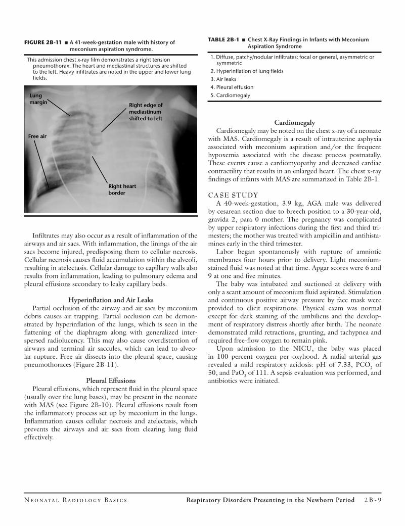
debris causes air trapping. Partial occlusion can be demon-strated by hyperinflation of the lungs, which is seen in the flattening of the diaphragm along with generalized inter-spersed radiolucency. This may also cause overdistention of airways and terminal air saccules, which can lead to alveo-lar rupture. Free air dissects into the pleural space, causing pneumothoraces (Figure 2B-11).
Pleural EffusionsPleural effusions, which represent fluid in the pleural space
(usually over the lung bases), may be present in the neonate with MAS (see Figure 2B-10). Pleural effusions result from the inflammatory process set up by meconium in the lungs. Inflammation causes cellular necrosis and atelectasis, which prevents the airways and air sacs from clearing lung fluid effectively.
CardiomegalyCardiomegaly may be noted on the chest x-ray of a neonate
with MAS. Cardiomegaly is a result of intrauterine asphyxia associated with meconium aspiration and/or the frequent hypoxemia associated with the disease process postnatally. These events cause a cardiomyopathy and decreased cardiac contractility that results in an enlarged heart. The chest x-ray findings of infants with MAS are summarized in Table 2B-1.
CASE STUDYA 40-week-gestation, 3.9 kg, AGA male was delivered
by cesarean section due to breech position to a 30-year-old, gravida 2, para 0 mother. The pregnancy was complicated by upper respiratory infections during the first and third tri-mesters; the mother was treated with ampicillin and antihista-mines early in the third trimester.
Labor began spontaneously with rupture of amniotic membranes four hours prior to delivery. Light meconium-stained fluid was noted at that time. Apgar scores were 6 and 9 at one and five minutes.
The baby was intubated and suctioned at delivery with only a scant amount of meconium fluid aspirated. Stimulation and continuous positive airway pressure by face mask were provided to elicit respirations. Physical exam was normal except for dark staining of the umbilicus and the develop-ment of respiratory distress shortly after birth. The neonate demonstrated mild retractions, grunting, and tachypnea and required free-flow oxygen to remain pink.
Upon admission to the NICU, the baby was placed in 100 percent oxygen per oxyhood. A radial arterial gas revealed a mild respiratory acidosis: pH of 7.33, PCO2 of 50, and PaO2 of 111. A sepsis evaluation was performed, and antibiotics were initiated.
FIGURE 2B-11 n A 41-week-gestation male with history of meconium aspiration syndrome.
This admission chest x-ray film demonstrates a right tension pneumothorax. The heart and mediastinal structures are shifted to the left. Heavy infiltrates are noted in the upper and lower lung fields.
3 3N E O N A T A L N E T W O R K
R A D I O L O G Y B A S I C S
produces an x-ray film with bilateral, diffuse, patchy, fluffy, ornodular (more dense, gray/white areas) infiltrates and asym-metric areas of opacity. These infiltrates may present in a focalor generalized manner (Figure 10).
Infiltrates may also occur as a result of inflammation of theairways and air sacs. With inflammation, the linings of the airsacs become injured, predisposing them to cellular necrosis.Cellular necrosis causes fluid accumulation within the alveoli,resulting in atelectasis. Cellular damage to capillary walls alsoresults from inflammation, leading to pulmonary edema andpleural effusions secondary to leaky capillary beds.
Hyperinflation and Air LeaksPartial occlusion of the airway and air sacs by meconium
debris causes air trapping. Partial occlusion can be demon-strated by hyperinflation of the lungs, which is seen in theflattening of the diaphragm along with generalized inter-spersed radiolucency. This may also cause overdistension ofairways and terminal air saccules, which can lead to alveolarrupture. Free air dissects into the pleural space, causingpneumothoraces (Figure 11).
Pleural EffusionsPleural effusions, which represent fluid in the pleural space
(usually over the lung bases), may be present in the neonatewith meconium aspiration syndrome (MAS) (see Figure 10).Pleural effusions result from the inflammatory process set upby meconium in the lungs. Inflammation causes cellularnecrosis and atelectasis, which prevents the airways and airsacs from clearing lung fluid effectively.
CardiomegalyCardiomegaly may be noted on the chest x-ray of a
neonate with meconium aspiration syndrome. Cardiomegalyis a result of intrauterine asphyxia associated with meconiumaspiration and/or the frequent hypoxemia associated with thedisease process postnatally. These events cause a cardiomyopa-thy and decreased cardiac contractility that results in anenlarged heart. The chest x-ray findings of infants with MASare summarized in Table 1.
CASE STUDYA 40-week-gestation, 3.9 kg, AGA male was delivered by
cesarean section due to breech position to a 30-year-old,gravida 2, para 0 mother. The pregnancy was complicated byupper respiratory infections during the first and thirdtrimesters; the mother was treated with ampicillin and antihis-tamines early in the third trimester.
Labor began spontaneously with rupture of amnioticmembranes four hours prior to delivery. Light meconium-stained fluid was noted at that time. Apgar scores were 6 and9 at one and five minutes.
The baby was intubated and suctioned at delivery withonly a scant amount of meconium fluid aspirated. Stimulationand continuous positive airway pressure by face mask were
FIGURE 11 � A 41-week-gestation male with history of meconiumaspiration syndrome. This admission chest x-ray filmdemonstrates a right tension pneumothorax. The heartand mediastinal structures are shifted to the left. Heavyinfiltrates are noted in the upper and lower lung fields.
Lungmargin
Free air
Right edge ofmediastinumshifted to left
Right heartborder
FIGURE 12 � A 40-week-gestation male with history of meconiumaspiration syndrome. This admission chest film has avery characteristic pattern of diffuse, patchy infiltrates.The lucencies on the right mediastinal and right sub-pulmonic areas most likely represent free air.
Areas oflucency
TABLE 1 � Chest X-Ray Findings in Infants with MeconiumAspiration Syndrome
1. Diffuse, patchy/nodular infiltrates: focal or general, asymmetric or symmetric
2. Hyperinflation of lung fields
3. Air leaks
4. Pleural effusion
5. Cardiomegaly
TABLE 2B-1 n Chest X-Ray Findings in Infants with Meconium Aspiration Syndrome
1. Diffuse, patchy/nodular infiltrates: focal or general, asymmetric or symmetric
2. Hyperinflation of lung fields3. Air leaks4. Pleural effusion5. Cardiomegaly

2 B - 1 0 Respiratory Disorders Presenting in the Newborn Period N e o n ata l R a d i o l o g y Ba s i c s
X-Ray EvaluationAn admission x-ray examination (Figure 2B-12) was indi-
cated because of respiratory distress consisting of grunting, retracting, tachypnea, and an oxygen requirement. Findings were as follows:Penetration: appears to be normally exposed.No significant rotation is noted.The soft tissues of the neck, chest, and extremities are of normal thickness and without emphysema.The bony framework is intact, with 12 ribs bilaterally and normal vertebrae. Clavicles are intact.The trachea shows a straight air column.The hilar area on the left is difficult to distinguish because of patchy infiltrates.The heart is of normal configuration, location, and size.The thymus is present.The diaphragm is between the ninth and tenth ribs bilaterally.Gastric air is present on the left.The intercostal spaces are normal in size.The lung fields demonstrate bilateral, diffuse, patchy infil-trates. The areas of lucency on the right represent a pneumo-thorax in the right subpulmonic area and the right lower lobe. There may also be free air located at the right mediastinal area, although a lateral film may be required to distinguish it.Impression: The history, clinical, laboratory, and x-ray data support the diagnosis of meconium aspiration with a small right pneumothorax.
Revised and updated from: Moore CS. 1994. Meconium aspiration syndrome. Neonatal Network 13(7): 57–60. Used with permission of Springer Publishing Company, LLC, www.springerpub.com/nn.
Neonatal Pneumonia
Neonatal pneumonia occurs in approximately one percent of term neonates and ten percent of preterm
neonates. The mortality rate for perinatally acquired pneu-monia varies and has been estimated to be 20 percent, with a higher mortality rate of 50 percent for postnatally acquired pneumonia.11 In one recent review of perinatally acquired infection, the overall mortality was 10 percent.12 This decline was attributed to increased use of perinatal antibiotics. The most common bacterial cause of neonatal pneumonia is Group B Streptococcus, mainly serotypes I and II, followed by Gram-negative enteric bacilli. Neonates requiring prolonged hospitalization in the neonatal intensive care unit are at risk for developing pneumonia from hospital-acquired organisms. These organisms are mainly Gram-negative—Pseudomonas, Klebsiella, Escherichia coli, and Serratia marcescens—but may also include Staphylococcus aureus, Staphylococcus epidermidis, and Candida. Less commonly, prenatally acquired viral infec-tions such as herpes, cytomegalovirus, varicella-zoster, as well as syphilis may present with neonatal pneumonia.
Perinatally acquired infectious agents can cause pneumo-nia later in the neonatal intensive care setting, presenting in the neonate at 4–11 weeks of age.13 Organisms causing this later onset of pneumonia following perinatal acquisition include Chlamydia trachomatis, Ureaplasma urealyticum, Mycoplasma hominis, and Pneumocystis carinii.13 Community acquired viral infections may also be seen in the neonatal intensive care setting with viral spread from caregivers or family members. Respiratory syncytial virus (RSV), entero-virus, adenovirus, and parainfluenza viruses are examples of these. Table 2B-2 summarizes by type organisms that may cause neonatal pneumonia.
PATHOPHYSIOLOGYPredisposing factors for the development of infection and
pneumonia include immune system immaturity, colonization of the maternal genital tract with potential pathogens, mater-nal urinary tract infection, amnionitis, prolonged rupture of membranes, prematurity, and the need for neonatal intensive care. Bacterial infection can occur secondary to hematoge-nous spread from the mother to the fetus in utero. Infection may occur by the ascending route from the maternal vaginal tract during labor, from aspiration of organisms during deliv-ery, and postnatally from inhalation of infected respiratory droplets. Neonates requiring intensive care are at high risk of colonization of the upper respiratory tract with potentially pathogenic organisms and the passage of pathogens from caregivers or contaminated equipment.
In bacterial pneumonia, alveolar involvement is often more intense than in viral infection. Proteinaceous fluid may partly or completely fill alveoli. This is followed by an influx of polymorphonuclear leukocytes and red blood cells.
FIGURE 2B-12 n A 40-week-gestation male with history of meconium aspiration syndrome.
This admission chest film has a very characteristic pattern of diffuse, patchy infiltrates. The lucencies on the right mediastinal and right subpulmonic areas most likely represent free air.
3 3N E O N A T A L N E T W O R K
R A D I O L O G Y B A S I C S
produces an x-ray film with bilateral, diffuse, patchy, fluffy, ornodular (more dense, gray/white areas) infiltrates and asym-metric areas of opacity. These infiltrates may present in a focalor generalized manner (Figure 10).
Infiltrates may also occur as a result of inflammation of theairways and air sacs. With inflammation, the linings of the airsacs become injured, predisposing them to cellular necrosis.Cellular necrosis causes fluid accumulation within the alveoli,resulting in atelectasis. Cellular damage to capillary walls alsoresults from inflammation, leading to pulmonary edema andpleural effusions secondary to leaky capillary beds.
Hyperinflation and Air LeaksPartial occlusion of the airway and air sacs by meconium
debris causes air trapping. Partial occlusion can be demon-strated by hyperinflation of the lungs, which is seen in theflattening of the diaphragm along with generalized inter-spersed radiolucency. This may also cause overdistension ofairways and terminal air saccules, which can lead to alveolarrupture. Free air dissects into the pleural space, causingpneumothoraces (Figure 11).
Pleural EffusionsPleural effusions, which represent fluid in the pleural space
(usually over the lung bases), may be present in the neonatewith meconium aspiration syndrome (MAS) (see Figure 10).Pleural effusions result from the inflammatory process set upby meconium in the lungs. Inflammation causes cellularnecrosis and atelectasis, which prevents the airways and airsacs from clearing lung fluid effectively.
CardiomegalyCardiomegaly may be noted on the chest x-ray of a
neonate with meconium aspiration syndrome. Cardiomegalyis a result of intrauterine asphyxia associated with meconiumaspiration and/or the frequent hypoxemia associated with thedisease process postnatally. These events cause a cardiomyopa-thy and decreased cardiac contractility that results in anenlarged heart. The chest x-ray findings of infants with MASare summarized in Table 1.
CASE STUDYA 40-week-gestation, 3.9 kg, AGA male was delivered by
cesarean section due to breech position to a 30-year-old,gravida 2, para 0 mother. The pregnancy was complicated byupper respiratory infections during the first and thirdtrimesters; the mother was treated with ampicillin and antihis-tamines early in the third trimester.
Labor began spontaneously with rupture of amnioticmembranes four hours prior to delivery. Light meconium-stained fluid was noted at that time. Apgar scores were 6 and9 at one and five minutes.
The baby was intubated and suctioned at delivery withonly a scant amount of meconium fluid aspirated. Stimulationand continuous positive airway pressure by face mask were
FIGURE 11 � A 41-week-gestation male with history of meconiumaspiration syndrome. This admission chest x-ray filmdemonstrates a right tension pneumothorax. The heartand mediastinal structures are shifted to the left. Heavyinfiltrates are noted in the upper and lower lung fields.
Lungmargin
Free air
Right edge ofmediastinumshifted to left
Right heartborder
FIGURE 12 � A 40-week-gestation male with history of meconiumaspiration syndrome. This admission chest film has avery characteristic pattern of diffuse, patchy infiltrates.The lucencies on the right mediastinal and right sub-pulmonic areas most likely represent free air.
Areas oflucency
TABLE 1 � Chest X-Ray Findings in Infants with MeconiumAspiration Syndrome
1. Diffuse, patchy/nodular infiltrates: focal or general, asymmetric or symmetric
2. Hyperinflation of lung fields
3. Air leaks
4. Pleural effusion
5. Cardiomegaly
N e o n ata l R a d i o l o g y Ba s i c s Respiratory Disorders Presenting in the Newborn Period 2 B - 1 1
During resolution, alveolar macrophages enter the alveoli and remove intra-alveolar debris, restoring normal lung func-tioning. Staphylococcus aureus and Klebsiella organisms cause more severe damage to alveoli and can destroy lung tissue by causing necrosis of the septum between alveoli. In some cases, abscess formation occurs.
Viruses and Mycoplasma may also be acquired transplacen-tally, during the labor and delivery process, or postnatally. Viral and mycoplasmal pneumonias involve the bronchi and peri-bronchial interstitium more than the alveoli. These organisms can cause loss of epithelial ciliary appendages and sloughing into the airways. The end result is stasis of mucus and mucus secretion, as well as bronchial obstruction with atelectasis, usually around the hilum. A secondary inflammatory reaction is characterized by mononuclear infiltration into the submu-cosa and perivascular areas. This further narrows the airway lumen. Smooth muscle constriction, a response to the inflam-matory process, leads to increasing airway obstruction and bronchospasm. In severe cases of viral and mycoplasmal infec-tion, inflammatory exudate extends into the alveoli.14
Fungal infection, of which Candida is the most common, may be acquired in utero, during the birth process, or post-natally. Congenitally acquired pneumonia can be diffuse as a result of an established inflammatory process at birth. Candida causes tissue invasion in the pharynx and larynx. Fungi proliferate and may form a thick layer of hyphae, which line the upper and lower respiratory tract. Ulceration of the pharynx, larynx, and lower respiratory tract can be seen. As the invasive process extends, secondary inflammation occurs, and the lungs become consolidated and hemorrhagic. Fungal thrombi from infected central lines are another cause of pneu-monia. Emboli from the fungal thrombus shower the lungs, and the result may be a diffuse pneumonia.15
CLINICAL PRESENTATIONThe clinical findings in neonatal pneumonia vary, depend-
ing on the infecting organism and the timing of its acqui-sition. Acute respiratory distress is frequently present with intrauterine and intrapartum-acquired ascending infection. The neonate may also have poor Apgar scores, temperature instability, and poor tone and activity. Overall, the clini-cal signs of pneumonia in the early neonatal period may be indistinguishable from those of respiratory distress resulting from hyaline membrane disease, TTN/retained lung fluid, or sepsis. Late-acquired pneumonia may have a gradual or an abrupt onset, depending on the causative agent. Respiratory deterioration, apnea, and temperature instability may be the presenting findings.
CHEST X-RAY FINDINGSChest x-rays are required to support the diagnosis of pneu-
monia and distinguish the infection from other causes of respi-ratory distress. The appearance of pneumonia on x-ray varies, depending upon the duration of infection at the time the x-ray
is obtained; the etiology of the pneumonia; and the presence of other respiratory disease, such as respiratory distress syndrome or bronchopulmonary dysplasia. Pneumonia acquired in utero is likely to be better established than pneumonia acquired during delivery. In some neonates, minimal or no abnormal-ity may be apparent on the first x-ray if it is taken soon after initial clinical signs, but follow-up x-rays 24 hours later may demonstrate the pneumonia. Serial x-rays are more valuable in making the diagnosis than is one isolated x-ray, and they are helpful in following the course of the disease.
Lung disease has been divided radiologically into two cate gories: air space, or alveolar, disease and interstitial disease. The two may be seen separately, but they may also coexist. Additionally, interstitial disease frequently progresses to involve the alveoli, often making it difficult to separate the two categories of lung disease. Air space disease involves the alveoli, which fill with fluid or exudate that displaces air. This may occur in small, discrete areas or in multiple areas, which can coalesce and appear as white or dense lung fields. In inter-stitial disease, the disease pattern is distributed throughout the lung tissue, usually bilaterally, and produces linear strands of density and granularity in the hilar and peribronchial areas.16
Pulmonary patterns described in neonatal pneumonia include (1) infiltrative patterns, (2) changes in lung volume, and (3) pleural effusions. Table 2B-3 lists x-ray findings in pneumonia resulting from various pathogens.
Infiltrative PatternsInfiltrative patterns are summarized in Table 2B-4.
Consolidation results when alveoli fill with inflammatory fluid or exudate and may involve the lobe of a lung or a segment of a lobe (Figure 2B-13). Initially, consolidation begins in the periphery and then progresses to the entire lobe of a lung. As
TABLE 2B-2 n Pathogenic Organisms That May Produce Neonatal Pneumonia (partial listing)
Bacteria: Gram-positiveGroup B beta-hemolytic
StreptococcusStreptococcus (other types)Staphylococcus aureusStaphylococcus epidermidisListeria monocytogenes
Bacteria: Gram-negativeEscherichia coliKlebsiellaEnterobacterPseudomonas aeruginosaSerratia marcescensHaemophilus influenzae
FungiCandida albicans
MycoplasmaUreaplasma urealyticumMycoplasma hominis
VirusesHerpes simplexCytomegalovirusRubellaVaricellaRespiratory syncytialInfluenzaAdenovirusEnterovirus
OtherTreponema pallidum (syphilis)Chlamydia trachomatisPneumocystis carinii

2 B - 1 2 Respiratory Disorders Presenting in the Newborn Period N e o n ata l R a d i o l o g y Ba s i c s
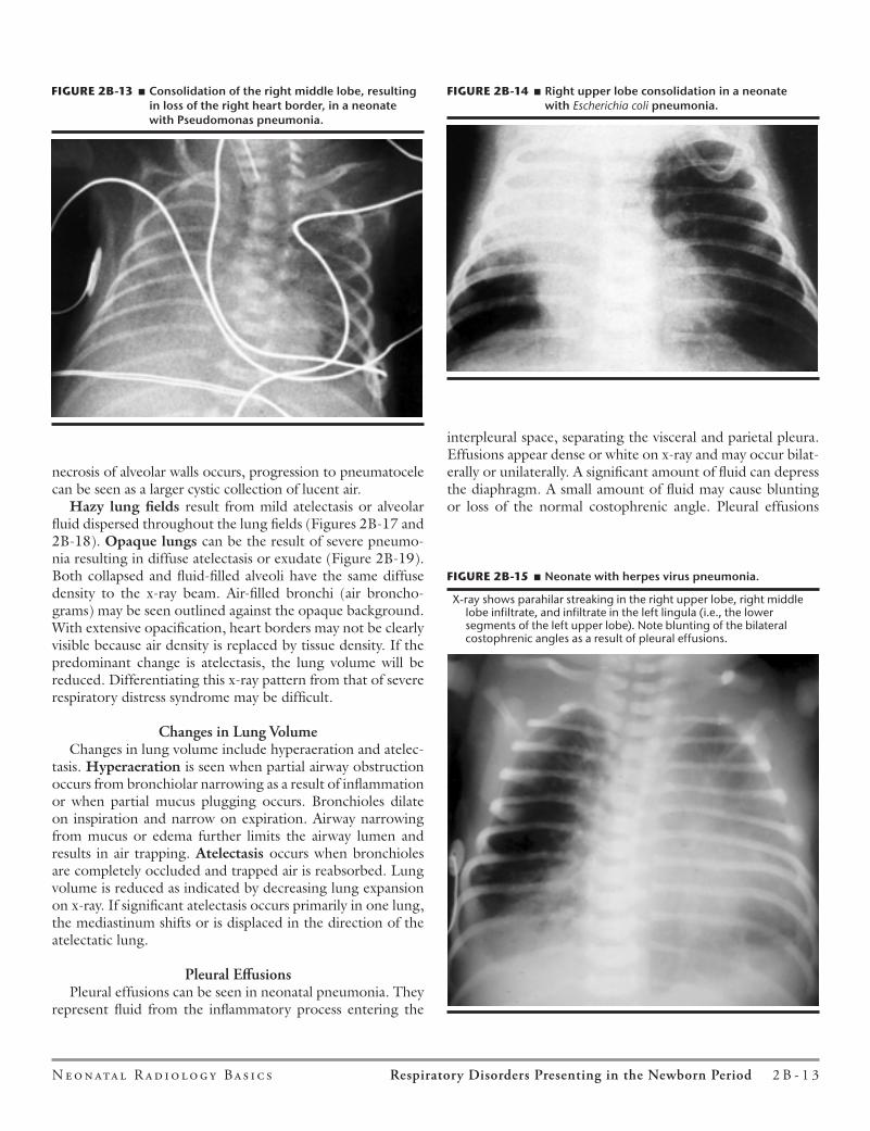
consolidation occurs, air bronchograms (air in the bronchi) can be seen. Most lobar consolidations are associated with bacterial infection. There is usually no change in the volume of the involved lobe because the alveoli contain fluid or exudate. The affected lobe can be determined by attempt-ing to visualize the heart border. Because the anterior segments of the right upper lobe and the right middle lobe are in contact with the right heart border, consolidation in these areas will result in loss of visualiza-tion of the heart border (Figure 2B-14). If right lower lobe consolida-tion has occurred, the area will appear more dense or opaque than other areas of the lung, but the heart border will remain visible. The upper lobe of the left lung has two lower segments, called the lingula, that are in
contact with the left heart border. Disease in this portion of the lung results in loss of visualization of the heart border.
Patchy infiltrates can be scattered through-out both lung fields or localized to one lobe. They have poorly defined borders and reflect alveolar disease. When localized to one lung, they are usually the result of bacterial or myco-plasmal infection. They may also be seen sec-ondary to segmental atelectasis occurring with viral and mycoplasmal infection.
Hilar and peribronchial infiltrates occur as the inflammatory process spreads from the hilum outward, thickening the bronchial tissue and causing partial atelectasis. The hilar area appears smudged or ragged, and opaque streaks radiate outward following the bronchi (Figure 2B-15).
A reticular or reticulogranular pattern of infiltrates occurs when the alveoli and termi-nal bronchioles are air-filled and seen against a background of alveoli that are partially filled with fluid or exudate or are atelectatic. This is a fine, bubbly pattern similar to that of respiratory distress syndrome and often seen with Group B beta-hemolytic streptococcal pneumonia (Figure 2B-16). Nodular or miliary infiltrates show up as a larger bubbly pattern than that of reticular infiltrates. Nodules of 2–4 mm are seen against a background of black, hyperaerated lungs and represent alveolar and interstitial disease with air trapping. This pattern has been reported in staph-ylococcal pneumonia and some Gram-negative bacillary pneumonias, such as Pseudomonas.17 If
TABLE 2B-4 n Radiologic Findings That May Be Present in Neonatal Pneumonia
Lung field infiltratesConsolidationPatchy alveolarHilar, peribronchialReticular, reticulogranularMiliary, nodularOpacificationDistribution: Can be bilateral, unilateral, diffuse,
localized, lobar, patchy, scattered, basalar, apicalLoss of heart border (silhouette sign); consolidation;atelectasis
Lung volume changesHyperaerationAtelectasisShift of the mediastinumPleural effusions
TABLE 2B-3 n Etiology of Pneumonia and Radiologic Picture
Bacteria Radiologic Picture
Group B ß-hemolytic Streptococcus
Diffuse reticulogranularity or opacification; patchy infiltrate; pleural effusion
Streptococcus pneumoniae Patchy lobar consolidation; pleural effusion
Staphylococcus aureus Diffuse infiltrate; nodular or miliary pneumatocele
Staphylococcus epidermidis Hazy lung fields or infiltrates
Listeria monocytogenes Coarse bilateral patchy infiltrates
Escherichia coli Consolidation in one or more lobes; pneumatocele
Klebsiella Bilateral consolidation; lung abscess; pneumatocele
Pseudomonas and Serratia Parenchymal consolidation (patchy area or over lung bases) pneumatocele
Haemophilus influenzae Nonspecific; may mimic RDS or Group B ß-hemolytic Streptococcus
Viruses
Herpes Perihilar infiltrates; streaky, lobar consolidation; later pleural effusion
Cytomegalovirus Nonspecific, perihilar streaking; hazy lung fields; infiltrates; opacification
Rubella Interstitial infiltrates; hazy lung fields
Respiratory syncytial Hyperexpansion; patchy consolidation
Adenovirus, enterovirus Hyperexpansion; patchy consolidation
Fungi
Candida albicans Diffuse granularity; opacification; coarse infiltrates
Mycoplasma
Ureaplasma urealyticum Fine reticular pattern progressing to air space opacification and consolidation
Mycoplasma hominis Diffuse reticular pattern; opacification; pleural effusion
Other
Treponema pallidum (syphilis)
Diffuse opacification; consolidation
Chlamydia trachomatis Hyperinflation, with streaky infiltrates
Pneumocystis carinii Diffuse haziness; granularity; opacity
Adapted from: Christensen RD, Thibeault DW, and Hall RT. 1986. Neonatal bacterial and fungal pneumonia. In Neonatal Pulmonary Care, Thibeault DW, and Gregory GA, eds. Norwalk, Connecticut: Appleton-Century-Crofts, 593–608; and Murphy T. 1986. Respiratory syncytial virus and miscellaneous agents. In Neonatal Pulmonary Care, Thibeault DW, and Gregory GA, eds. Norwalk, Connecticut: Appleton-Century-Crofts, 624–638.
N e o n ata l R a d i o l o g y Ba s i c s Respiratory Disorders Presenting in the Newborn Period 2 B - 1 3
necrosis of alveolar walls occurs, progression to pneumatocele can be seen as a larger cystic collection of lucent air.
Hazy lung fields result from mild atelectasis or alveolar fluid dispersed throughout the lung fields (Figures 2B-17 and 2B-18). Opaque lungs can be the result of severe pneumo-nia resulting in diffuse atelectasis or exudate (Figure 2B-19). Both collapsed and fluid-filled alveoli have the same diffuse density to the x-ray beam. Air-filled bronchi (air broncho-grams) may be seen outlined against the opaque background. With extensive opacification, heart borders may not be clearly visible because air density is replaced by tissue density. If the predominant change is atelectasis, the lung volume will be reduced. Differentiating this x-ray pattern from that of severe respiratory distress syndrome may be difficult.
Changes in Lung VolumeChanges in lung volume include hyperaeration and atelec-
tasis. Hyperaeration is seen when partial airway obstruction occurs from bronchiolar narrowing as a result of inflammation or when partial mucus plugging occurs. Bronchioles dilate on inspiration and narrow on expiration. Airway narrowing from mucus or edema further limits the airway lumen and results in air trapping. Atelectasis occurs when bronchioles are completely occluded and trapped air is reabsorbed. Lung volume is reduced as indicated by decreasing lung expansion on x-ray. If significant atelectasis occurs primarily in one lung, the mediastinum shifts or is displaced in the direction of the atelectatic lung.
Pleural EffusionsPleural effusions can be seen in neonatal pneumonia. They
represent fluid from the inflammatory process entering the
interpleural space, separating the visceral and parietal pleura. Effusions appear dense or white on x-ray and may occur bilat-erally or unilaterally. A significant amount of fluid can depress the diaphragm. A small amount of fluid may cause blunting or loss of the normal costophrenic angle. Pleural effusions
FIGURE 2B-13 n Consolidation of the right middle lobe, resulting in loss of the right heart border, in a neonate with Pseudomonas pneumonia.
3 7N E O N A T A L N E T W O R K
R A D I O L O G Y B A S I C S
alveoli and terminal bronchioles are air-filled and seen againsta background of alveoli that are partially filled with fluid orexudate or are atelectatic. This is a fine, bubbly pattern similarto that of respiratory distress syndrome and often seen withGroup B β-hemolytic streptococcal pneumonia (Figure 16).Nodular or miliary infiltrates show up as a larger bubblypattern than that of reticular infiltrates. Nodules of 2–4 mmare seen against a background of black, hyperaerated lungsand represent alveolar and interstitial disease with air trapping.This pattern has been reported in staphylococcal pneumoniaand some Gram-negative bacillary pneumonias, such asPseudomonas.26 If necrosis of alveolar walls occurs, progres-sion to pneumatocele can be seen as a larger cystic collectionof lucent air.
Hazy lung fields result from mild atelectasis or alveolarfluid dispersed throughout the lung fields (Figures 17 and18).25 Opaque lungs can be the result of severe pneumonia
FIGURE 13 � Consolidation of the right middle lobe, resulting in lossof the right heart border, in a neonate withPseudomonas pneumonia.
FIGURE 14 � Right upper lobe consolidation in a neonate withEscherichia coli pneumonia.
FIGURE 15 � Neonate with herpes virus pneumonia.
X-ray shows parahilar streaking in the right upper lobe, right middle lobeinfiltrate, and infiltrate in the left lingula (i.e., the lower segments of theleft upper lobe). Note blunting of the bilateral costophrenic angles as aresult of pleural effusions.
FIGURE 16 � Bilateral lung fields show reticulogranularity and airbronchograms in a neonate with Group B β-hemolyticstreptococcal pneumonia.
FIGURE 2B-14 n Right upper lobe consolidation in a neonate with Escherichia coli pneumonia.
3 7N E O N A T A L N E T W O R K
R A D I O L O G Y B A S I C S
alveoli and terminal bronchioles are air-filled and seen againsta background of alveoli that are partially filled with fluid orexudate or are atelectatic. This is a fine, bubbly pattern similarto that of respiratory distress syndrome and often seen withGroup B β-hemolytic streptococcal pneumonia (Figure 16).Nodular or miliary infiltrates show up as a larger bubblypattern than that of reticular infiltrates. Nodules of 2–4 mmare seen against a background of black, hyperaerated lungsand represent alveolar and interstitial disease with air trapping.This pattern has been reported in staphylococcal pneumoniaand some Gram-negative bacillary pneumonias, such asPseudomonas.26 If necrosis of alveolar walls occurs, progres-sion to pneumatocele can be seen as a larger cystic collectionof lucent air.
Hazy lung fields result from mild atelectasis or alveolarfluid dispersed throughout the lung fields (Figures 17 and18).25 Opaque lungs can be the result of severe pneumonia
FIGURE 13 � Consolidation of the right middle lobe, resulting in lossof the right heart border, in a neonate withPseudomonas pneumonia.
FIGURE 14 � Right upper lobe consolidation in a neonate withEscherichia coli pneumonia.
FIGURE 15 � Neonate with herpes virus pneumonia.
X-ray shows parahilar streaking in the right upper lobe, right middle lobeinfiltrate, and infiltrate in the left lingula (i.e., the lower segments of theleft upper lobe). Note blunting of the bilateral costophrenic angles as aresult of pleural effusions.
FIGURE 16 � Bilateral lung fields show reticulogranularity and airbronchograms in a neonate with Group B β-hemolyticstreptococcal pneumonia.
FIGURE 2B-15 n Neonate with herpes virus pneumonia.
X-ray shows parahilar streaking in the right upper lobe, right middle lobe infiltrate, and infiltrate in the left lingula (i.e., the lower segments of the left upper lobe). Note blunting of the bilateral costophrenic angles as a result of pleural effusions.
3 7N E O N A T A L N E T W O R K
R A D I O L O G Y B A S I C S
alveoli and terminal bronchioles are air-filled and seen againsta background of alveoli that are partially filled with fluid orexudate or are atelectatic. This is a fine, bubbly pattern similarto that of respiratory distress syndrome and often seen withGroup B β-hemolytic streptococcal pneumonia (Figure 16).Nodular or miliary infiltrates show up as a larger bubblypattern than that of reticular infiltrates. Nodules of 2–4 mmare seen against a background of black, hyperaerated lungsand represent alveolar and interstitial disease with air trapping.This pattern has been reported in staphylococcal pneumoniaand some Gram-negative bacillary pneumonias, such asPseudomonas.26 If necrosis of alveolar walls occurs, progres-sion to pneumatocele can be seen as a larger cystic collectionof lucent air.
Hazy lung fields result from mild atelectasis or alveolarfluid dispersed throughout the lung fields (Figures 17 and18).25 Opaque lungs can be the result of severe pneumonia
FIGURE 13 � Consolidation of the right middle lobe, resulting in lossof the right heart border, in a neonate withPseudomonas pneumonia.
FIGURE 14 � Right upper lobe consolidation in a neonate withEscherichia coli pneumonia.
FIGURE 15 � Neonate with herpes virus pneumonia.
X-ray shows parahilar streaking in the right upper lobe, right middle lobeinfiltrate, and infiltrate in the left lingula (i.e., the lower segments of theleft upper lobe). Note blunting of the bilateral costophrenic angles as aresult of pleural effusions.
FIGURE 16 � Bilateral lung fields show reticulogranularity and airbronchograms in a neonate with Group B β-hemolyticstreptococcal pneumonia.

2 B - 1 4 Respiratory Disorders Presenting in the Newborn Period N e o n ata l R a d i o l o g y Ba s i c s
may be seen with bacterial, fungal, and, more rarely, viral or mycoplasmal pneumonia.
DIFFERENTIAL DIAGNOSISThe chest x-ray must be reviewed with the patient’s history,
clinical information, and laboratory data in mind. The varied chest x-ray presentations of neonatal pneumonia may mimic the reticulogranularity and opacification of respiratory distress syndrome and the patchy infiltrates and perihilar streaking of retained lung fluid and meconium aspiration syndrome. Mucus plugging in the airways of neonates with bronchopulmonary
dysplasia can result in patchy atelectasis, which must be differ-entiated from atelectasis resulting from infection.
CASE STUDYA 36-week-gestation, 3 kg, AGA female was delivered vag-
inally to a 34-year-old gravida 4, para 3 mother. The mother had received intermittent prenatal care for a total of three visits. Rupture of membranes occurred 36 hours prior to
FIGURE 2B-16 n Bilateral lung fields show reticulogranularity and air bronchograms in a neonate with Group B ß-hemolytic streptococcal pneumonia.
3 7N E O N A T A L N E T W O R K
R A D I O L O G Y B A S I C S
alveoli and terminal bronchioles are air-filled and seen againsta background of alveoli that are partially filled with fluid orexudate or are atelectatic. This is a fine, bubbly pattern similarto that of respiratory distress syndrome and often seen withGroup B β-hemolytic streptococcal pneumonia (Figure 16).Nodular or miliary infiltrates show up as a larger bubblypattern than that of reticular infiltrates. Nodules of 2–4 mmare seen against a background of black, hyperaerated lungsand represent alveolar and interstitial disease with air trapping.This pattern has been reported in staphylococcal pneumoniaand some Gram-negative bacillary pneumonias, such asPseudomonas.26 If necrosis of alveolar walls occurs, progres-sion to pneumatocele can be seen as a larger cystic collectionof lucent air.
Hazy lung fields result from mild atelectasis or alveolarfluid dispersed throughout the lung fields (Figures 17 and18).25 Opaque lungs can be the result of severe pneumonia
FIGURE 13 � Consolidation of the right middle lobe, resulting in lossof the right heart border, in a neonate withPseudomonas pneumonia.
FIGURE 14 � Right upper lobe consolidation in a neonate withEscherichia coli pneumonia.
FIGURE 15 � Neonate with herpes virus pneumonia.
X-ray shows parahilar streaking in the right upper lobe, right middle lobeinfiltrate, and infiltrate in the left lingula (i.e., the lower segments of theleft upper lobe). Note blunting of the bilateral costophrenic angles as aresult of pleural effusions.
FIGURE 16 � Bilateral lung fields show reticulogranularity and airbronchograms in a neonate with Group B β-hemolyticstreptococcal pneumonia.
FIGURE 2B-17 n Hazy lung fields in a neonate with cytomegalovirus pneumonia.
3 8N E O N A T A L N E T W O R K
R A D I O L O G Y B A S I C S
resulting in diffuse atelectasis or exudate (Figure 19). Both col-lapsed and fluid-filled alveoli have the same diffuse density tothe x-ray beam. Air-filled bronchi (air bronchograms) may beseen outlined against the opaque background. With extensiveopacification, heart borders may not be clearly visible becauseair density is replaced by tissue density. If the predominantchange is atelectasis, the lung volume will be reduced. If exu-date or fluid from inflammation fills the alveoli, lung expansionmay initially appear near normal.24 Group B β-hemolyticStreptococcus, Haemophilus influenzae, Candida, andmycoplasmal pneumonias may present with this chest x-ray pat-tern. Differentiating this x-ray pattern from that of severe respi-ratory distress syndrome may be difficult.
Changes in Lung VolumeChanges in lung volume include hyperaeration and atelec-
tasis. Hyperaeration is seen when partial airway obstructionoccurs from bronchiolar narrowing as a result of inflamma-tion or when partial mucus plugging occurs. Bronchiolesdilate on inspiration and narrow on expiration. Airway nar-rowing from mucus or edema further limits the airway lumenand results in air trapping. Atelectasis occurs when bronchi-oles are completely occluded and trapped air is reabsorbed.Lung volume is reduced as indicated by decreasing lungexpansion on x-ray. If atelectasis occurs primarily in one lung,the mediastinum shifts or is displaced in the direction of theatelectatic lung.17
Pleural EffusionsPleural effusions can be seen in neonatal pneumonia. They
represent fluid from the inflammatory process entering theinterpleural space, separating the visceral and parietal pleura.Effusions appear dense or white on x-ray and may occur
bilaterally or unilaterally. A significant amount of fluid candepress the diaphragm. A small amount of fluid may causeblunting or loss of the normal costophrenic angle. Pleuraleffusions may be seen with bacterial, fungal, and, more rarely,viral or mycoplasmal pneumonia.
DIFFERENTIAL DIAGNOSISThe chest x-ray must be reviewed with the patient’s histo-
ry, clinical information, and laboratory data in mind. The
FIGURE 18 � Hazy lung fields in a neonate with Staphylococcusepidermidis pneumonia.
FIGURE 17 � Hazy lung fields in a neonate with cytomegaloviruspneumonia.
FIGURE 19 � Bilateral opacification in a 30-day-old prematureneonate with Candida albicans sepsis and pneumonia.
The left hemithorax is more opacified than the right. Air bronchogramsare visible on the right.
FIGURE 2B-18 n Hazy lung fields in a neonate with Staphylococcus epidermidis pneumonia.
3 8N E O N A T A L N E T W O R K
R A D I O L O G Y B A S I C S
resulting in diffuse atelectasis or exudate (Figure 19). Both col-lapsed and fluid-filled alveoli have the same diffuse density tothe x-ray beam. Air-filled bronchi (air bronchograms) may beseen outlined against the opaque background. With extensiveopacification, heart borders may not be clearly visible becauseair density is replaced by tissue density. If the predominantchange is atelectasis, the lung volume will be reduced. If exu-date or fluid from inflammation fills the alveoli, lung expansionmay initially appear near normal.24 Group B β-hemolyticStreptococcus, Haemophilus influenzae, Candida, andmycoplasmal pneumonias may present with this chest x-ray pat-tern. Differentiating this x-ray pattern from that of severe respi-ratory distress syndrome may be difficult.
Changes in Lung VolumeChanges in lung volume include hyperaeration and atelec-
tasis. Hyperaeration is seen when partial airway obstructionoccurs from bronchiolar narrowing as a result of inflamma-tion or when partial mucus plugging occurs. Bronchiolesdilate on inspiration and narrow on expiration. Airway nar-rowing from mucus or edema further limits the airway lumenand results in air trapping. Atelectasis occurs when bronchi-oles are completely occluded and trapped air is reabsorbed.Lung volume is reduced as indicated by decreasing lungexpansion on x-ray. If atelectasis occurs primarily in one lung,the mediastinum shifts or is displaced in the direction of theatelectatic lung.17
Pleural EffusionsPleural effusions can be seen in neonatal pneumonia. They
represent fluid from the inflammatory process entering theinterpleural space, separating the visceral and parietal pleura.Effusions appear dense or white on x-ray and may occur
bilaterally or unilaterally. A significant amount of fluid candepress the diaphragm. A small amount of fluid may causeblunting or loss of the normal costophrenic angle. Pleuraleffusions may be seen with bacterial, fungal, and, more rarely,viral or mycoplasmal pneumonia.
DIFFERENTIAL DIAGNOSISThe chest x-ray must be reviewed with the patient’s histo-
ry, clinical information, and laboratory data in mind. The
FIGURE 18 � Hazy lung fields in a neonate with Staphylococcusepidermidis pneumonia.
FIGURE 17 � Hazy lung fields in a neonate with cytomegaloviruspneumonia.
FIGURE 19 � Bilateral opacification in a 30-day-old prematureneonate with Candida albicans sepsis and pneumonia.
The left hemithorax is more opacified than the right. Air bronchogramsare visible on the right.
FIGURE 2B-19� n Bilateral opacification in a 30-day-old premature neonate with Candida albicans sepsis and pneumonia.
The left hemithorax is more opacified than the right. Air bronchograms are visible on the right.
3 8N E O N A T A L N E T W O R K
R A D I O L O G Y B A S I C S
resulting in diffuse atelectasis or exudate (Figure 19). Both col-lapsed and fluid-filled alveoli have the same diffuse density tothe x-ray beam. Air-filled bronchi (air bronchograms) may beseen outlined against the opaque background. With extensiveopacification, heart borders may not be clearly visible becauseair density is replaced by tissue density. If the predominantchange is atelectasis, the lung volume will be reduced. If exu-date or fluid from inflammation fills the alveoli, lung expansionmay initially appear near normal.24 Group B β-hemolyticStreptococcus, Haemophilus influenzae, Candida, andmycoplasmal pneumonias may present with this chest x-ray pat-tern. Differentiating this x-ray pattern from that of severe respi-ratory distress syndrome may be difficult.
Changes in Lung VolumeChanges in lung volume include hyperaeration and atelec-
tasis. Hyperaeration is seen when partial airway obstructionoccurs from bronchiolar narrowing as a result of inflamma-tion or when partial mucus plugging occurs. Bronchiolesdilate on inspiration and narrow on expiration. Airway nar-rowing from mucus or edema further limits the airway lumenand results in air trapping. Atelectasis occurs when bronchi-oles are completely occluded and trapped air is reabsorbed.Lung volume is reduced as indicated by decreasing lungexpansion on x-ray. If atelectasis occurs primarily in one lung,the mediastinum shifts or is displaced in the direction of theatelectatic lung.17
Pleural EffusionsPleural effusions can be seen in neonatal pneumonia. They
represent fluid from the inflammatory process entering theinterpleural space, separating the visceral and parietal pleura.Effusions appear dense or white on x-ray and may occur
bilaterally or unilaterally. A significant amount of fluid candepress the diaphragm. A small amount of fluid may causeblunting or loss of the normal costophrenic angle. Pleuraleffusions may be seen with bacterial, fungal, and, more rarely,viral or mycoplasmal pneumonia.
DIFFERENTIAL DIAGNOSISThe chest x-ray must be reviewed with the patient’s histo-
ry, clinical information, and laboratory data in mind. The
FIGURE 18 � Hazy lung fields in a neonate with Staphylococcusepidermidis pneumonia.
FIGURE 17 � Hazy lung fields in a neonate with cytomegaloviruspneumonia.
FIGURE 19 � Bilateral opacification in a 30-day-old prematureneonate with Candida albicans sepsis and pneumonia.
The left hemithorax is more opacified than the right. Air bronchogramsare visible on the right.

N e o n ata l R a d i o l o g y Ba s i c s Respiratory Disorders Presenting in the Newborn Period 2 B - 1 5
delivery, and the mother delivered 40 minutes after hospital admission. The neonate’s Apgar scores were 5 and 7 at one and five minutes. She required oxygen for cyanosis and was retracting and grunting in the delivery room. She was trans-ferred to the NICU. An arterial blood gas following admis-sion revealed respiratory acidosis and mild metabolic acidosis. Mild hypotension was present and corrected. The neonate was intubated and placed on mechanical ventilation. A sepsis workup was initiated and antibiotics begun. The admission x-ray is shown in Figure 2B-16.
X-Ray EvaluationThe indication for this film was admission to the NICU with respiratory distress and the admission diagnosis of “rule out respiratory distress syndrome; rule out sepsis.”Penetration: appears to be normal.Rotation is present to the right.The soft tissues appear normal.The bony framework is intact. Twelve ribs are present bilat-erally. The vertebrae are normal, and the clavicles are intact.The trachea is deviated to the right because of rotation. The tracheal air column is straight, indicating an inspiratory film. The tip of the endotracheal tube is located just below the first thoracic vertebra (T1). The tracheal bifurcation is visible at the fourth thoracic vertebra (T4), and the right mainstem bronchus is visible.The hilar area appears normal in size.The diaphragm is at the ninth thoracic vertebra (T9) bilaterally.The pleura reach the edges of the bony thorax, and the cos-tophrenic angles are visible bilaterally.The gastric bubble is visible on the left.The intercostal spaces appear more narrowed on the right than on the left because of rotation. They appear normal in size.The lung fields reveal a diffuse bilateral reticulogranular pattern, with air bronchograms visible bilaterally. The right lung appears more opaque than the left because of rotation.The endotracheal tube sits just below the first thoracic ver-tebra (T1). An umbilical artery catheter is positioned at the 12th thoracic vertebra (T12) and was subsequently reposi-tioned between the third and fourth lumbar vertebrae.
Impression: The history, clinical presentation, and x-ray are consistent with neonatal pneumonia. However, because this neonate is 36 weeks gestational age and prenatal care and pregnancy dating are poor, respiratory distress syndrome is also a possibility. Tracheal cultures and blood cultures taken following admission to the NICU were positive for Group B beta hemolytic Streptococcus.
Revised and updated from: Carey BE, and Trotter C. 1997. Neonatal pneumonia. Neonatal Network 16(5): 65–71. Used with permission of Springer Publishing Company, LLC, www.springerpub.com/nn.
Neonatal Chylothorax
Barbara E. Carey, MN, RN, NNP-BC
A chylothorax is an accumulation of lymphatic fluid that collects in the pleural space. It is the most common
cause of a large pleural effusion in the newborn.18,19 Estimated incidences vary from 1 in 10,000 deliveries to 1 in 2,000 admissions to the NICU.19,20 Chylothorax may be unilateral or, infrequently, bilateral and can occur spontaneously or be acquired secondary to trauma or surgical procedures. The right lung is more commonly affected than the left.
ETIOLOGYSpontaneous chylothorax is thought to occur secondary to
overdistention, rupture, or tear in the thoracic duct during the birth process as a result of transient elevated central venous pressure. Other possible contributory factors may include birth trauma leading to tearing of the duct and congenital abnormality of the duct predisposing it to rupture. Another etiology that has been suggested is the occurrence of major fistulas, which form because of the failure of some channels to connect with the lymphatic network, allowing free move-ment of chyle into the pleural space. Cases have been found to be associated with congenital defects such as extralobar sequestration and Noonan, Turner’s, and Down syndromes.
It has been estimated that 70 percent of fats are absorbed into the lymphatic system via the gastrointestinal system and are transported into the venous blood via the thoracic duct. The major thoracic duct crosses from the right to the left posterior mediastinum at the fifth thoracic vertebra (T5) and ascends to drain into the junction of the left subclavian and the internal jugular veins. This is why a right-sided chy-lothorax generally occurs when an injury or abnormality is below the level of T5 and a left chylothorax when an injury or abnormality is above T5.20
Thoracic and cardiovascular surgeries are the most common predisposing iatrogenic causes for chylothorax. Surgery requiring open repair and manipulation of the aortic arch, such as coarctation, patent ductus arteriosus, or subclavian-pulmonary artery shunt procedures, have a higher incidence of chylothorax than cardiac surgeries not involving the aortic arch. In one study, thoracotomy and ligation of a patent ductus arteriosus was found to be the single most common antecedent to chylothorax, complicating 1 percent of cases.21 Superior vena caval (SVC) obstruction has been reported to predispose to chylothorax because of the high central venous pressure that it produces. SVC obstruction usually occurs sec-ondary to thrombosis from indwelling catheters.2
2 B - 1 6 Respiratory Disorders Presenting in the Newborn Period N e o n ata l R a d i o l o g y Ba s i c s
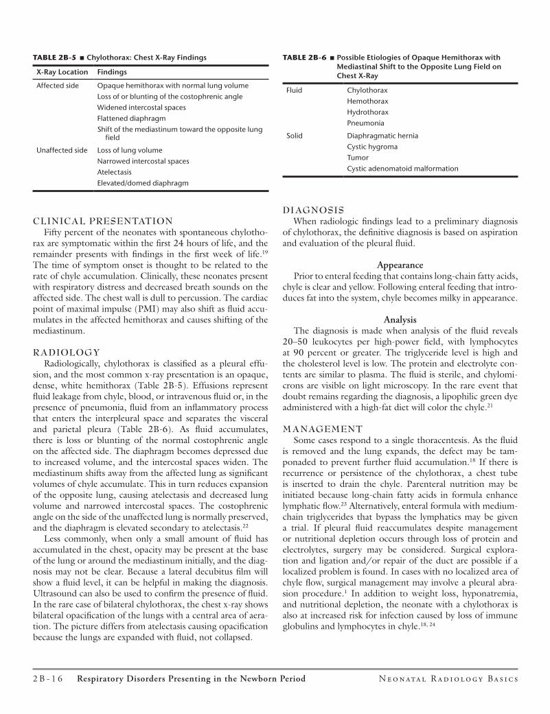
CLINICAL PRESENTATIONFifty percent of the neonates with spontaneous chylotho-
rax are symptomatic within the first 24 hours of life, and the remainder presents with findings in the first week of life.19 The time of symptom onset is thought to be related to the rate of chyle accumulation. Clinically, these neonates present with respiratory distress and decreased breath sounds on the affected side. The chest wall is dull to percussion. The cardiac point of maximal impulse (PMI) may also shift as fluid accu-mulates in the affected hemithorax and causes shifting of the mediastinum.
RADIOLOGYRadiologically, chylothorax is classified as a pleural effu-
sion, and the most common x-ray presentation is an opaque, dense, white hemithorax (Table 2B-5). Effusions represent fluid leakage from chyle, blood, or intravenous fluid or, in the presence of pneumonia, fluid from an inflammatory process that enters the interpleural space and separates the visceral and parietal pleura (Table 2B-6). As fluid accumulates, there is loss or blunting of the normal costophrenic angle on the affected side. The diaphragm becomes depressed due to increased volume, and the intercostal spaces widen. The mediastinum shifts away from the affected lung as significant volumes of chyle accumulate. This in turn reduces expansion of the opposite lung, causing atelectasis and decreased lung volume and narrowed intercostal spaces. The costophrenic angle on the side of the unaffected lung is normally preserved, and the diaphragm is elevated secondary to atelectasis.22
Less commonly, when only a small amount of fluid has accumulated in the chest, opacity may be present at the base of the lung or around the mediastinum initially, and the diag-nosis may not be clear. Because a lateral decubitus film will show a fluid level, it can be helpful in making the diagnosis. Ultrasound can also be used to confirm the presence of fluid. In the rare case of bilateral chylothorax, the chest x-ray shows bilateral opacification of the lungs with a central area of aera-tion. The picture differs from atelectasis causing opacification because the lungs are expanded with fluid, not collapsed.
DIAGNOSISWhen radiologic findings lead to a preliminary diagnosis
of chylothorax, the definitive diagnosis is based on aspiration and evaluation of the pleural fluid.
Appearance Prior to enteral feeding that contains long-chain fatty acids,
chyle is clear and yellow. Following enteral feeding that intro-duces fat into the system, chyle becomes milky in appearance.
Analysis The diagnosis is made when analysis of the fluid reveals
20–50 leukocytes per high-power field, with lymphocytes at 90 percent or greater. The triglyceride level is high and the cholesterol level is low. The protein and electrolyte con-tents are similar to plasma. The fluid is sterile, and chylomi-crons are visible on light microscopy. In the rare event that doubt remains regarding the diagnosis, a lipophilic green dye administered with a high-fat diet will color the chyle.21
MANAGEMENTSome cases respond to a single thoracentesis. As the fluid
is removed and the lung expands, the defect may be tam-ponaded to prevent further fluid accumulation.18 If there is recurrence or persistence of the chylothorax, a chest tube is inserted to drain the chyle. Parenteral nutrition may be initiated because long-chain fatty acids in formula enhance lymphatic flow.23 Alternatively, enteral formula with medium-chain triglycerides that bypass the lymphatics may be given a trial. If pleural fluid reaccumulates despite management or nutritional depletion occurs through loss of protein and electrolytes, surgery may be considered. Surgical explora-tion and ligation and/or repair of the duct are possible if a localized problem is found. In cases with no localized area of chyle flow, surgical management may involve a pleural abra-sion procedure.1 In addition to weight loss, hyponatremia, and nutritional depletion, the neonate with a chylothorax is also at increased risk for infection caused by loss of immune globulins and lymphocytes in chyle.18, 24
TABLE 2B-6 n Possible Etiologies of Opaque Hemithorax with Mediastinal Shift to the Opposite Lung Field on Chest X-Ray
Fluid ChylothoraxHemothoraxHydrothoraxPneumonia
Solid Diaphragmatic herniaCystic hygromaTumorCystic adenomatoid malformation
TABLE 2B-5 n Chylothorax: Chest X-Ray Findings
X-Ray Location Findings
Affected side Opaque hemithorax with normal lung volumeLoss of or blunting of the costophrenic angleWidened intercostal spacesFlattened diaphragmShift of the mediastinum toward the opposite lung
field
Unaffected side Loss of lung volumeNarrowed intercostal spacesAtelectasisElevated/domed diaphragm

N e o n ata l R a d i o l o g y Ba s i c s Respiratory Disorders Presenting in the Newborn Period 2 B - 1 7
CASE STUDYA 41-week gestation, 3.3 kg, AGA male was born by vaginal
delivery to a 39 year old, gravida 1, para 0, ab 0, VDRL test negative, rubella-immune mother after 27 hours of labor. The mother sought prenatal care at 20 weeks gestation and had only four prenatal visits. She informed her obstetrician that she was healthy and wanted a natural pregnancy without excessive testing or ultrasound. Counseling the mother about the usual monitoring and testing procedures met with resis-tance, and despite documented telephone follow-up, she remained adamant in refusing normal testing for age and stage of pregnancy. Her labor was complicated by shoulder dystocia, and the delivery required forceps. Apgar scores were 6 and 7 at one and five minutes: minus 2 for color and minus 1 each for heart rate and respiratory effort at one minute and minus 2 for color and minus 1 for respiratory effort at five minutes. The neonate was cyanotic and required bag and mask ventilation and oxygen. Breath sounds were decreased on the right. The infant was transferred to the NICU for further evaluation. Clinical exam revealed poor breath sounds bilaterally, more decreased on the right, and PMI shifted to the left. Transillumination was performed to rule out a pneu-mothorax and was found to be negative. An initial arterial blood gas revealed respiratory acidosis and hypoxemia. The infant was intubated, an umbilical arterial line was placed, and mechanical ventilation was begun. A sepsis workup was done and antibiotics were administered. The admission diagnosis was term neonate, respiratory distress, rule out sepsis, rule out pneumonia. The admission chest x-ray is shown in Figure 2B-20.
X-Ray EvaluationThe indication for this film was admission to the NICU with respiratory distress requiring intubation and mechanical ventilation.Penetration appears normal.Rotation is not present. The spine lies in the center of the film, and the clavicles are straight.The soft tissues are normal in appearance.The bony framework is intact with 12 ribs present bilater-ally. Ribs, vertebrae, and clavicles are intact. The ribs in the right lung field are horizontal in appearance, and the ribs in the left lung field are downward sloping.The trachea is slightly deviated to the left. The tracheal air column is straight, indicating an inspiratory film. The tip of the endotracheal tube is located at the third thoracic verte-bra. The tracheal bifurcation is visible at the fourth thoracic vertebra, and the right and left mainstem bronchi are visible.The hilum. The right hilum shows minimal aeration and air bronchograms in the area of the sixth through ninth thoracic vertebrae. The left hilum shows air bronchograms in the area of the fourth through the eleventh vertebrae. The mediasti-num is shifted to the left. The cardiac silhouette is difficult to
visualize because of atelectasis. The apex appears to be shifted to the left rib margin at intercostal spaces 7 through 9.The diaphragm is not visible on the right and is visible at the tenth intercostal space on the left.The pleura reaches the edge of the thorax on the left, and the costophrenic angle is present. The right pleural edge cannot be seen clearly, but may be visible at the midclavicular line, compressed by a pleural effusion.Gastric air is present on the left.The right lung field shows linear opacity to approximately the midclavicular line. A line of delineation, possibly repre-senting the compressed pleural edge, is seen from the fifth through the ninth intercostal space, with minimal aeration. The mediastinum is shifted to the left. The left lung field shows atelectasis and air bronchograms.Tubes and lines. The endotracheal tube is visible at the third thoracic vertebra, and the umbilical artery catheter tip is visible between the seventh and eighth thoracic vertebral bodies.Impression. The changes seen in the right lung are consis-tent with a right pleural effusion with mediastinal shift, and the clinical presentation is consistent with the diagnosis. Analysis of fluid aspirated from the right chest was consistent with chyle.
Originally published: Carey BE. 2001. Neonatal chylothorax. Neonatal Network 20(2): 53–55. Used with permission of Springer Publishing Company, LLC, www.springerpub.com/nn.
FIGURE 2B-20 n Initial chest x-ray following intubation and umbilical artery line placement.

2 B - 1 8 Respiratory Disorders Presenting in the Newborn Period N e o n ata l R a d i o l o g y Ba s i c s
REFERENCES 1. Avery ME, and Fletcher BD. 1974. The Lung and Its Disorders in the
Newborn Infant. Philadelphia: Saunders, 268. 2. Swischuk LE. 2004. Respiratory system. In Imaging of the Newborn
Infant and Young Child, 5th ed. Philidelphia: Lippincott Williams & Wilkins, 1–170.
3. Bland R. 1988. Lung liquid clearance before and after birth. Seminars in Perinatology 12(2): 124–133.
4. Krantz ME, et al. 1986. Epidemiological analysis of the increased risk of disturbed neonatal adaptation after cesarean section. Acta Paediatrica Scandinavica 75(5): 832–839.
5. Halliday HL, McClure G, and Reid MM. 1981. Transient tachypnea of the newborn: Two distinct clinical entities. Archives of Disease in Childhood 56: 322.
6. Gowen CW, et al. 1988. Electrical potential difference and ion transport across the nasal epithelium of term neonates: Correlation with mode of delivery, transient tachypnea of the newborn. Journal of Pediatrics 113(1 part 1): 121–127.
7. Sandberg K, et al. 1987. Lung function in newborn infants with tachypnea of unknown cause. Pediatric Research 22(5): 581–585.
8. Gregory GA, et al. 1974. Meconium aspiration in infants—a prospective study. Journal of Pediatrics 85(6): 848–852.
9. Brady J, and Goldman S. 1986. Management of meconium aspiration syndrome. In Neonatal Pulmonary Care, 2nd ed., Thibeault DW, and Gregory GA, eds. Norwalk, Connecticut: Appleton-Century-Crofts, 483–498.
10. Miller MJ, Fanaroff AA, and Martin RJ. 2006. The respiratory system, Part 5: Respiratory disorders in preterm and term infant. In Neonatal–Perinatal Medicine: Diseases of the Fetus and Infant, 8th ed., Martin RJ, Fanaroff AA, and Walsh MC, eds. St. Louis: Mosby, 1122–1146.
11. Dennehy P. 1987. Respiratory infections in the newborn. Clinics in Perinatology 14(3): 667–682.
12. Philip AG. 1994. The changing face of neonatal infection: Experience at a regional medical center. Pediatric Infectious Disease Journal 13(12): 1098–1102.
13. Campbell PW, and Stokes DC. 1994. Pneumonia. In Respiratory Disease in Children: Diagnosis and Management, Loughlin GM, and Eigen H, eds. Philadelphia: Lippincott Williams & Wilkins, 351–353.
14. Cassel GH, Waites KB, and Crouse DT. 1991. Perinatal mycoplasmal infections. Clinics in Perinatology 18(2): 263–271.
15. Baley JE. 1991. Neonatal candidiasis: The current challenge. Clinics in Perinatology 18(2): 263–271.
16. Novelline RA. 2004. Squire’s Fundamentals of Radiology. Cambridge, Massachusetts: Harvard University Press, 104–105.
17. Flores JAM. 1988. Miliary pattern in neonatal pneumonia. Pediatric Radiology 18(4): 355–356.
18. Greenbough A, and Robertson NR. 1999. Acute respiratory disease in the newborn. In Textbook of Neonatology, Reenie JM, and Robertson NR, eds. New York: Churchill Livingstone, 558–560.
19. Kirks DR. 1991. Respiratory system. In Practical Pediatric Imaging, Kirks DR, ed. Baltimore: Lippincott Williams & Wilkins, 580–582.
20. Van Aerde J, et al. 1984. Spontaneous chylothorax in newborns. American Journal of Disease of Children 138(10): 961–964.
21. Puntis JW, Roberts KD, and Handy D. 1987. How should chylothorax be managed? Archives of Disease in Childhood 62(5): 593–596.
22. Gordon I, Matthew D, and Dinwiddie R. 1987. Respiratory system. In Diagnostic Imaging in Pediatrics, Gordon I, ed. London: Chapman and Hall, 40–46.
23. Brodman K, Zavelson T, and Schiebler G. 1974. Treatment of congenital chylothorax. Journal of Pediatrics 85(4): 516–517.
24. McWilliams BC, Fan LL, and Murphy SA. 1981. Transient T-cell depression in post-operative chylothorax. Journal of Pediatrics 99(4): 595–597.