Embed Size (px)
Citation preview
Newborn Adaptation to
Extrauterine Life
and Newborn Assessment
Self–Learning Module
Developed by the
Interprofessional Education and Research Committee of the
Champlain Maternal Newborn Regional Program (CMNRP)
2013
Newborn Adaptation to Extrauterine Life: A Self-Learning Module
© CMNRP 2013 Page 2 of 21
Introduction…………………………………………………………………………………….……………………………………………...3
Objectives………………………………………………………………………………………………………………………………………..3
1. Newborn Adaptation to Extrauterine Life …………………….……………………………………………...…….4
1.1 Fetal circulation
1.2 Neonatal Circulation
1.3 Respiratory Adaptation
1.4 The Newborn Transitional Period
1.5 Cardiovascular Changes
2. Initial Assessment of the Newborn …………………………………….…………………………………….…….….…10
2.1 Apgar Score
2.2 Vital Signs
a) RESPIRATIONS
b) HEART RATE
c) TEMPERATURE
2.3 Medication Administration
a) VITAMIN K
b) EYE PROPHYLAXIS
2.4 Newborn Measurements
a) WEIGHT
b) LENGTH
c) HEAD CIRCUMFERENCE
d) CHEST CIRCUMFERENCE
e) ABDOMINAL GIRTH
3. Complete Physical Examination of the Newborn.….……..…………………………………………………15
References…………………………………………………………………………………………………………..…………..……….…19
Additional Resources …………………………………………………………………………………………………..……………...20
Acknowledgements …………………………………………………………………………………………………….…………….…21
Disclaimer: This self-learning module is intended for health care providers caring for term,
low-risk newborns. Please refer to institutional policies and procedures.
TABLE OF CONTENTS
Newborn Adaptation to Extrauterine Life: A Self-Learning Module
© CMNRP 2013 Page 3 of 21
Upon completion of this self-learning module, the nurse will be able to:
1. Describe the newborn’s physiological adaptation to extrauterine life.
2. Demonstrate a complete physical assessment of the newborn outlining the usual findings,
normal variations and abnormalities.
3. Identify skills requiring further enhancement to meet the above objectives and outline a
learning plan to meet these needs.
Introduction
Objectives
Newborn Adaptation to Extrauterine Life: A Self-Learning Module
© CMNRP 2013 Page 4 of 21
The immediate postpartum period is a time of significant physiological adaptation for both the
mother and baby. The newborn must adapt from being completely dependent on another for
life sustaining oxygen and nutrients to an independent being, a task accomplished over a period
of hours to days. Successful transition from fetal to neonatal life requires a complex interaction
between the following systems:
• Respiratory
• Cardiovascular
• Thermoregulatory
• Immunologic
Establishing respirations is critical to the newborn’s transition, as lungs become the organ of gas
exchange after separation from maternal uteroplacental circulation. Over 90% of newborns
make the transition from intrauterine life to extrauterine life without difficulty, requiring little
to no assistance (NRP, 2010). However, for the 10% of newborns who do require assistance,
about 1% require extensive resuscitative measures to survive. All personnel who care for
newborns immediately after birth should have skills in neonatal resuscitation and maintain
their Neonatal Resuscitation Program (NRP) status.
1.1 Fetal circulation
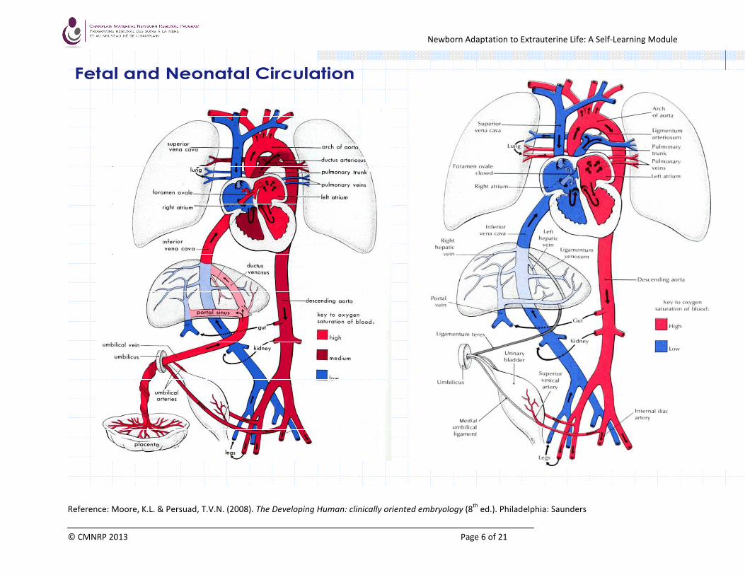
In utero, oxygenated blood flows to the fetus from the placenta through the umbilical vein.
Although a small amount of oxygenated blood is delivered to the liver, most blood diverts the
hepatic system through the ductus venosus, which forms a connection between the umbilical
vein and the inferior vena cava. Oxygenated blood from the inferior vena cava enters the right
atrium and most of it is directed through the foramen ovale to the left atrium, then to the left
ventricle, and onto the ascending aorta, where it is primarily directed to the fetal heart and
brain (Askin, 2009).
Deoxygenated blood from the head and upper extremities comes back to the right atrium by
the superior vena cava, where it blends with oxygenated blood from the placenta. This blood
1. Newborn Adaptation to Extrauterine Life
Newborn Adaptation to Extrauterine Life: A Self-Learning Module
© CMNRP 2013 Page 5 of 21
enters the right ventricle and pulmonary artery, where 90% of it is shunted across the ductus
arteriosus and into the descending aorta, providing oxygen to the lower half of the fetal body
and eventually draining back to the placenta through the two umbilical arteries. The remaining
10% of the blood coming from the right ventricle perfuses lung tissue to meet metabolic needs
(Askin, 2009).
1.2 Neonatal Circulation
With the infant’s first breath and exposure to increased oxygen levels, there is an increased
blood flow to the lungs causing the closure of the foramen ovale. Constriction of the ductus
arteriosus is a gradual process that results from a reduction of pulmonary vascular resistance
(PVR), increasing systemic vascular resistance (SVR) and sensitivity to a rise in arterial PaO2
levels. The removal of the placenta decreases prostaglandin levels (which helped to maintain
ductal patency) further influencing closure (Alvaro & Rigatto, 2005; Kenner, 2003).
At birth, the clamping of the umbilical cord eliminates the placenta as a reservoir for blood,
triggering an increase in systemic vascular resistance (SVR), an increase in blood pressure, and
increased pressures in the left side of the heart. The removal of the placenta also eliminates
the need for blood flow through the ductus venosus, causing functional elimination of this fetal
shunt. Systemic venous blood flow is then directed through the portal system for hepatic
circulation. Umbilical vessels constrict, with functional closure occurring immediately. Fibrous
infiltration leads to anatomic closure in the first week of life (Alvaro & Rigatto, 2005).
Successful transition and closure of fetal shunts creates a neonatal circulation where
deoxygenated blood returns to the heart through the inferior and superior vena cava. Blood
then enters the right atrium to the right ventricle and travels through the pulmonary artery to
the pulmonary vascular bed. Oxygenated blood returns through pulmonary veins to the left
atrium, the left ventricle, and through the aorta to systemic circulation. Hypoxia, acidosis and
congenital heart defects are conditions that lead to a sustained high PVR and may interfere
with the normal sequence of events (Askin, 2008).
The graphics on the next page illustrate fetal and neonatal circulation.
Newborn Adaptation to Extrauterine Life: A Self-Learning Module
© CMNRP 2013 Page 6 of 21
Fetal and Neonatal Circulation
Reference: Moore, K.L. & Persuad, T.V.N. (2008). The Developing Human: clinically oriented embryology (8th
ed.). Philadelphia: Saunders
Newborn Adaptation to Extrauterine Life: A Self-Learning Module
© CMNRP 2013 Page 7 of 21
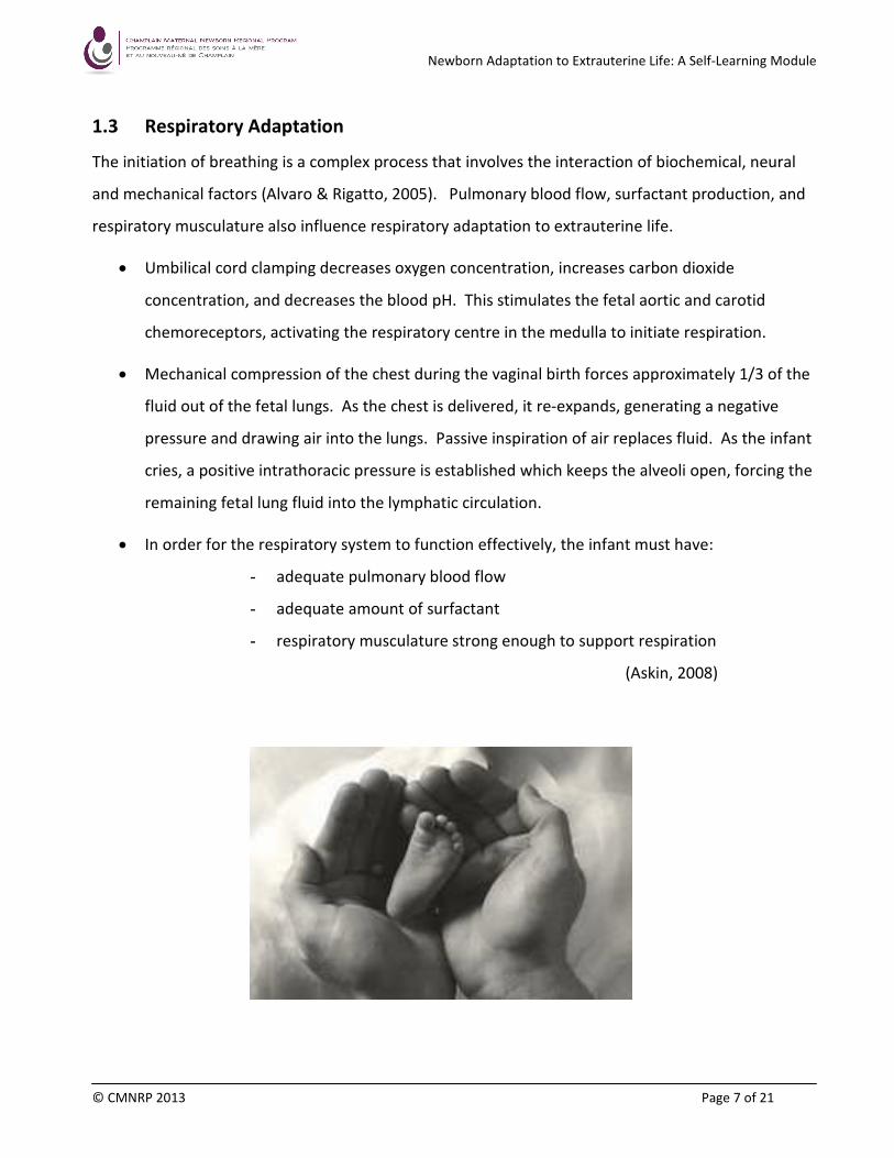
1.3 Respiratory Adaptation
The initiation of breathing is a complex process that involves the interaction of biochemical, neural
and mechanical factors (Alvaro & Rigatto, 2005). Pulmonary blood flow, surfactant production, and
respiratory musculature also influence respiratory adaptation to extrauterine life.
• Umbilical cord clamping decreases oxygen concentration, increases carbon dioxide
concentration, and decreases the blood pH. This stimulates the fetal aortic and carotid
chemoreceptors, activating the respiratory centre in the medulla to initiate respiration.
• Mechanical compression of the chest during the vaginal birth forces approximately 1/3 of the
fluid out of the fetal lungs. As the chest is delivered, it re-expands, generating a negative
pressure and drawing air into the lungs. Passive inspiration of air replaces fluid. As the infant
cries, a positive intrathoracic pressure is established which keeps the alveoli open, forcing the
remaining fetal lung fluid into the lymphatic circulation.
• In order for the respiratory system to function effectively, the infant must have:
- adequate pulmonary blood flow
- adequate amount of surfactant
- respiratory musculature strong enough to support respiration
(Askin, 2008)
Newborn Adaptation to Extrauterine Life: A Self-Learning Module
© CMNRP 2013 Page 8 of 21
1.4 The Newborn Transitional Period
Healthy full-term newborns show a predictable pattern of behavioural changes, behavioural states
and cues, sensory abilities, and physiologic adaptations during the first 6- 8 hours following delivery.
This transitional period is divided into an initial period of reactivity and inactivity and a second period
of reactivity (Askin, 2008).
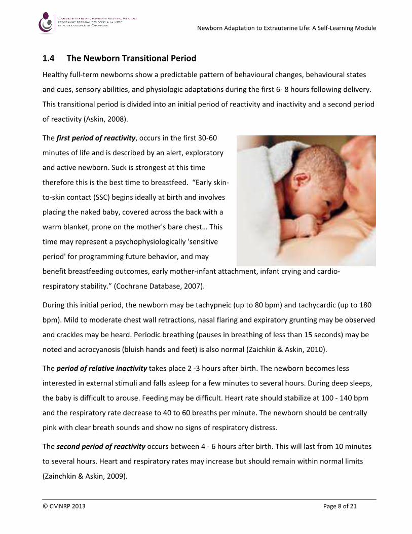
The first period of reactivity, occurs in the first 30-60
minutes of life and is described by an alert, exploratory
and active newborn. Suck is strongest at this time
therefore this is the best time to breastfeed. “Early skin-
to-skin contact (SSC) begins ideally at birth and involves
placing the naked baby, covered across the back with a
warm blanket, prone on the mother's bare chest… This
time may represent a psychophysiologically 'sensitive
period' for programming future behavior, and may
benefit breastfeeding outcomes, early mother-infant attachment, infant crying and cardio-
respiratory stability.” (Cochrane Database, 2007).
During this initial period, the newborn may be tachypneic (up to 80 bpm) and tachycardic (up to 180
bpm). Mild to moderate chest wall retractions, nasal flaring and expiratory grunting may be observed
and crackles may be heard. Periodic breathing (pauses in breathing of less than 15 seconds) may be
noted and acrocyanosis (bluish hands and feet) is also normal (Zaichkin & Askin, 2010).
The period of relative inactivity takes place 2 -3 hours after birth. The newborn becomes less
interested in external stimuli and falls asleep for a few minutes to several hours. During deep sleeps,
the baby is difficult to arouse. Feeding may be difficult. Heart rate should stabilize at 100 - 140 bpm
and the respiratory rate decrease to 40 to 60 breaths per minute. The newborn should be centrally
pink with clear breath sounds and show no signs of respiratory distress.
The second period of reactivity occurs between 4 - 6 hours after birth. This will last from 10 minutes
to several hours. Heart and respiratory rates may increase but should remain within normal limits
(Zainchkin & Askin, 2009).
Newborn Adaptation to Extrauterine Life: A Self-Learning Module
© CMNRP 2013 Page 9 of 21
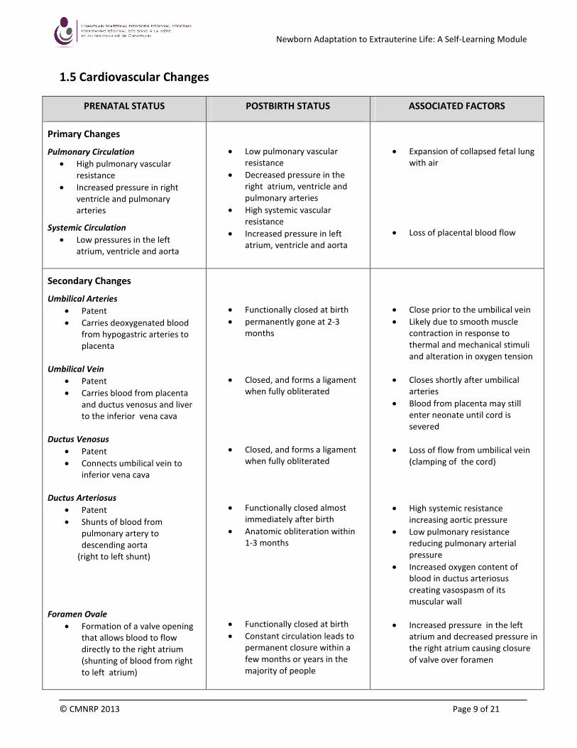
1.5 Cardiovascular Changes
PRENATAL STATUS POSTBIRTH STATUS ASSOCIATED FACTORS
Primary Changes
Pulmonary Circulation
• High pulmonary vascular
resistance
• Increased pressure in right
ventricle and pulmonary
arteries
Systemic Circulation
• Low pressures in the left
atrium, ventricle and aorta
• Low pulmonary vascular
resistance
• Decreased pressure in the
right atrium, ventricle and
pulmonary arteries
• High systemic vascular
resistance
• Increased pressure in left
atrium, ventricle and aorta
• Expansion of collapsed fetal lung
with air
• Loss of placental blood flow
Secondary Changes
Umbilical Arteries
• Patent
• Carries deoxygenated blood
from hypogastric arteries to
placenta
Umbilical Vein
• Patent
• Carries blood from placenta
and ductus venosus and liver
to the inferior vena cava
Ductus Venosus
• Patent
• Connects umbilical vein to
inferior vena cava
Ductus Arteriosus
• Patent
• Shunts of blood from
pulmonary artery to
descending aorta
(right to left shunt)
Foramen Ovale
• Formation of a valve opening
that allows blood to flow
directly to the right atrium
(shunting of blood from right
to left atrium)
• Functionally closed at birth
• permanently gone at 2-3
months
• Closed, and forms a ligament
when fully obliterated
• Closed, and forms a ligament
when fully obliterated
• Functionally closed almost
immediately after birth
• Anatomic obliteration within
1-3 months
• Functionally closed at birth
• Constant circulation leads to
permanent closure within a
few months or years in the
majority of people
• Close prior to the umbilical vein
• Likely due to smooth muscle
contraction in response to
thermal and mechanical stimuli
and alteration in oxygen tension
• Closes shortly after umbilical
arteries
• Blood from placenta may still
enter neonate until cord is
severed
• Loss of flow from umbilical vein
(clamping of the cord)
• High systemic resistance
increasing aortic pressure
• Low pulmonary resistance
reducing pulmonary arterial
pressure
• Increased oxygen content of
blood in ductus arteriosus
creating vasospasm of its
muscular wall
• Increased pressure in the left
atrium and decreased pressure in
the right atrium causing closure
of valve over foramen
Newborn Adaptation to Extrauterine Life: A Self-Learning Module
© CMNRP 2013 Page 10 of 21
The physical examination of the newborn begins at birth and continues throughout the hospital stay.
Newborn assessment includes observation, auscultation, and palpation, proceeding in a systematic
head-to-toe fashion, although it can be adapted to the particular infant and situation. In order to
obtain quality data, the assessment is organized to minimize stress for the infant. It is best to begin
by observing the symmetry, respirations, movement, and behaviour of the baby. The infant’s
respiratory rate, heart rate, colour and axillary temperature should be measured and recorded.
At birth, a healthy newborn should be immediately put skin-to-skin on his/her mother, dried and
covered by dry warm blankets. The newborn’s Apgar scoring and initial assessments should be done
while remaining skin-to-skin (unless resuscitation is required). Administration of medications,
measurements and weight should be delayed until a period of uninterrupted skin-to-skin contact on
mother for at least one hour, or until after the newborn’s first attempt to breastfeed.
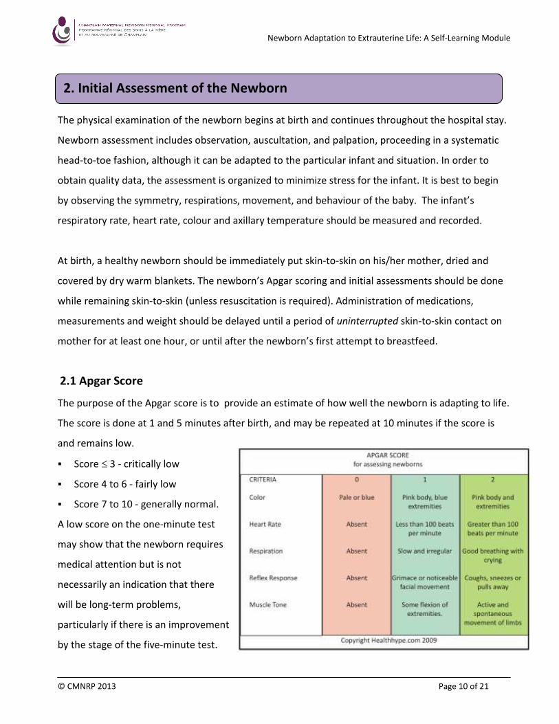
2.1 Apgar Score
The purpose of the Apgar score is to provide an estimate of how well the newborn is adapting to life.
The score is done at 1 and 5 minutes after birth, and may be repeated at 10 minutes if the score is
and remains low.
� Score ≤ 3 - critically low
� Score 4 to 6 - fairly low
� Score 7 to 10 - generally normal.
A low score on the one-minute test
may show that the newborn requires
medical attention but is not
necessarily an indication that there
will be long-term problems,
particularly if there is an improvement
by the stage of the five-minute test.
2. Initial Assessment of the Newborn
Newborn Adaptation to Extrauterine Life: A Self-Learning Module
© CMNRP 2013 Page 11 of 21
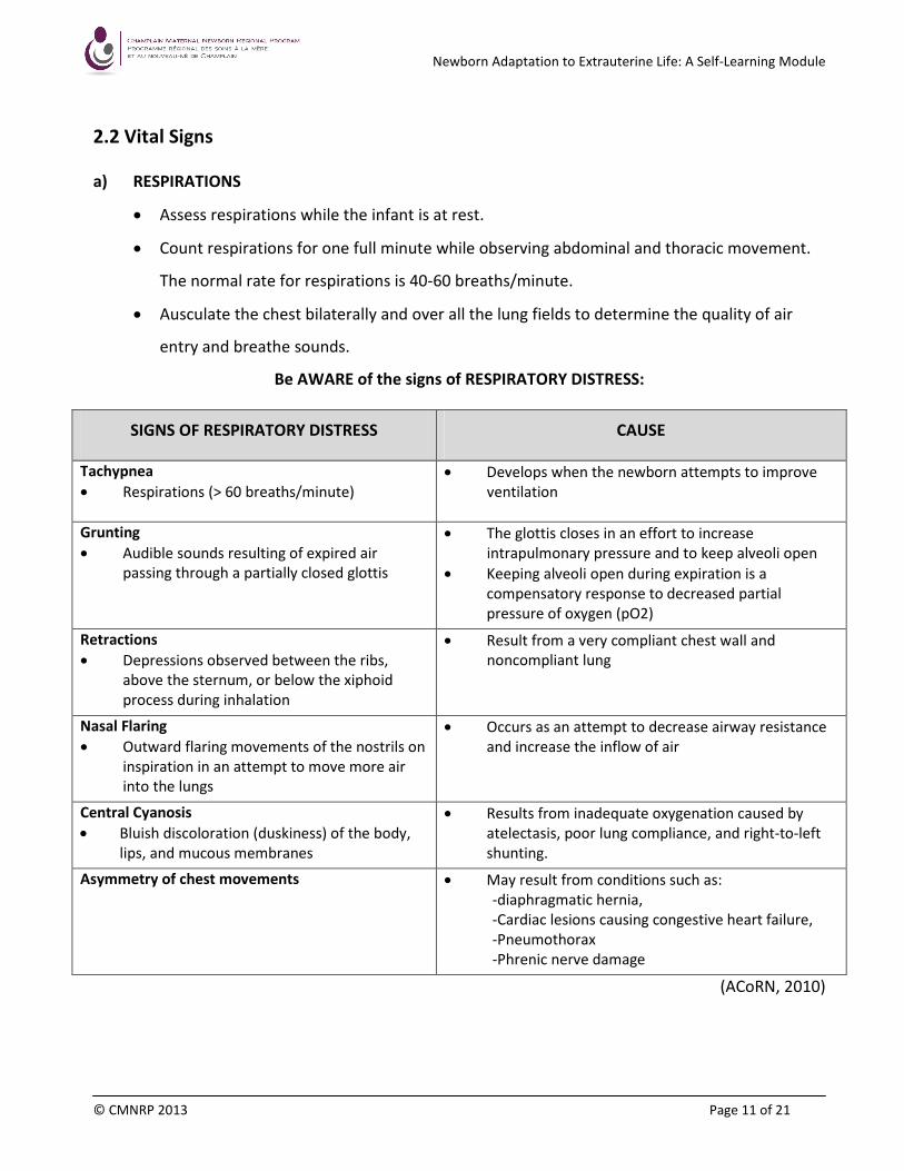
2.2 Vital Signs
a) RESPIRATIONS
• Assess respirations while the infant is at rest.
• Count respirations for one full minute while observing abdominal and thoracic movement.
The normal rate for respirations is 40-60 breaths/minute.
• Ausculate the chest bilaterally and over all the lung fields to determine the quality of air
entry and breathe sounds.
Be AWARE of the signs of RESPIRATORY DISTRESS:
SIGNS OF RESPIRATORY DISTRESS CAUSE
Tachypnea
• Respirations (> 60 breaths/minute)
• Develops when the newborn attempts to improve
ventilation
Grunting
• Audible sounds resulting of expired air
passing through a partially closed glottis
• The glottis closes in an effort to increase
intrapulmonary pressure and to keep alveoli open
• Keeping alveoli open during expiration is a
compensatory response to decreased partial
pressure of oxygen (pO2)
Retractions
• Depressions observed between the ribs,
above the sternum, or below the xiphoid
process during inhalation
• Result from a very compliant chest wall and
noncompliant lung
Nasal Flaring
• Outward flaring movements of the nostrils on
inspiration in an attempt to move more air
into the lungs
• Occurs as an attempt to decrease airway resistance
and increase the inflow of air
Central Cyanosis
• Bluish discoloration (duskiness) of the body,
lips, and mucous membranes
• Results from inadequate oxygenation caused by
atelectasis, poor lung compliance, and right-to-left
shunting.
Asymmetry of chest movements
• May result from conditions such as:
-diaphragmatic hernia,
-Cardiac lesions causing congestive heart failure,
-Pneumothorax
-Phrenic nerve damage
(ACoRN, 2010)
Newborn Adaptation to Extrauterine Life: A Self-Learning Module
© CMNRP 2013 Page 12 of 21
b) HEART RATE
• Place the warm stethoscope on the left side of the infant’s chest, near the edge of the
sternum and slightly lower than the nipple line. The heart sounds can be clearly heard at
the point of maximal intensity (PMI) which is located at the third to fourth intercostal space
just lateral to the mid-clavicular line.
• Assess the heart rate and the rhythm for one full minute, noting heart sounds (i.e.:
murmurs).
• The normal heart rate ranges from 100-160 beats/minute, but some term babies may have
a resting heart rate as low as 80 bpm (ACoRN, 2010).
c) TEMPERATURE
• Assess the temperature via the axillary route. This is the safest and most convenient route
to measure temperature in the neonate. The risk of trauma is increased with a temperature
taken rectally. The use of tympanic thermometers is not recommended as readings may not
be accurate in children under 2 years of age (ACoRN, 2010).
• The normal axilla temperature ranges from 36.3 – 37.2°C (ACoRN, 2010).
NOTE: A cold stressed infant may exhibit normal to elevated axilla temperatures in response
to metabolism of brown fat.
• If using an electronic thermometer, apply a clean probe cover to temperature probe and
ensure that it is firmly in place.
• Place the probe gently against the axilla (in alignment with the length of the baby’s body)
and hold the infant’s arm pressed firmly against the side and hold the thermometer in place
until a reading is obtained.
2.3 Medication Administration
Newborns in Canada routinely receive two medications at birth: an intramuscular injection of Vitamin
K and an antibiotic agent for eye prophylaxis. Parents may refuse Vitamin K; however, it is important
to document the reason for refusal as well as information given to parents about the risks of
disregarding the recommended treatments. Some institutions have specific forms for parents to sign
should they refuse standard procedures recommended/required for newborn care.
Newborn Adaptation to Extrauterine Life: A Self-Learning Module
© CMNRP 2013 Page 13 of 21
a) VITAMIN K
Intramuscular administration of Vitamin K (phytonadione) is the most effective method of preventing
hemorrhagic disease of the newborn. It is administered to the newborn in order to facilitate normal
clotting until the newborn’s intestinal tract produces the bacteria necessary to synthesize Vitamin K.
Vitamin K should be given as a single intramuscular dose to all newborns within 6 hours of birth
(Canadian Paediatric Society and College of Family Physicians of Canada, 2004). For infants weighing
less than or equal to 1500 grams, the dose is 0.5 mg IM; for infants weighing more than 1500 grams,
the dose is 1.0 mg IM.
NOTE: If parents refuse Vitamin K IM, an oral dose of 2 mg can be given at the first feeding, with
follow-up doses given at 2 to 4 weeks of age and 6 to 8 weeks of age. This treatment is not
recommended because it is less effective in preventing late hemorrhagic disease of the
newborn. Parents should be advised of the importance of the baby receiving the follow-up
doses and be cautioned that their infants remain at increased risk of late hemorrhagic disease
(CPW & CFPC’ 2004; Health Canada, 2000).
b) EYE PROPHYLAXIS
All newborns should receive a prophylactic agent against ophthalmia neonatorum, from gonorrhea
or chlamydia, except for very premature newborns whose lids are fused at the time of birth. It is
recommended that each eye be treated with a 1-cm ribbon of 0.5% erythromycin ointment. The
eyes should not be irrigated with sterile water or saline. The administration may be delayed for up to
two hours after birth to enable parent–infant contact and initial stabilization of the baby (Health
Canada, 2000).
According to the Health Protection and Promotion Act, R.S.O. 1990, Regulation 557, section 33, the
administration of ophthalmic eye prophylaxis to newborns is a fundamental part of healthcare in
Ontario and healthcare providers are required to administer an effective ophthalmic agent into the
eyes of newborn newborns. The Health Protection and Promotion Act (HPPA) supersedes the Health
Care Consent Act, 1996 and does not allow for exemptions based on informed choice in the
application of eye prophylaxis for newborns. The expectation is that healthcare professionals will
Newborn Adaptation to Extrauterine Life: A Self-Learning Module
© CMNRP 2013 Page 14 of 21
discuss this with parents and healthcare professionals who do not comply with the requirements of
the Act may be fined and reported to their regulatory body.
The HPPA states that parents who refuse the administration of eye prophylaxis must be reported to
the Medical Officer of Health serving the area where the care and treatment was provided. If a
parent refuses prophylaxis treatment to be given, the Ottawa Public Health Communicable Disease
Program (or similar) must be notified immediately at (613) 580-6744 ext 24224 and the information
documented in the chart.
2.4 Newborn Measurements
a) WEIGHT
• Place a paper or warm blanket on the scale basket and zero scale.
• Remove the infant’s clothing/blanket (no diaper).
• Place the infant on the scale, keeping one hand over the infant without touching.
• If this is the first weight following birth, encourage support person to take a picture of the
baby on the scale so the weight can be seen (useful if transcription error occurs).
• Read and note weight.
b) LENGTH
• Lay the infant on a flat surface in a recumbent position.
• Place a hand over the knees so that the infant’s legs are extended.
• With the foot flexed, draw a line marking the bottom of the heel.
• Continue to immobilize the infant and draw a line at the infant’s head.
• Remove the infant and measure the distance between the two points.
c) HEAD CIRCUMFERENCE
• Wrap measuring tape around the largest area of the infant’s head, over the occipital, parietal,
and frontal prominences. Begin above the eyebrows and ears, and continue around the back
of the head. Take the largest of several measurements.
• NOTE: Cranial molding or scalp edema may affect the measurements.
• Measure head circumference (HC) daily or as ordered if abnormal results are obtained.
Newborn Adaptation to Extrauterine Life: A Self-Learning Module
© CMNRP 2013 Page 15 of 21
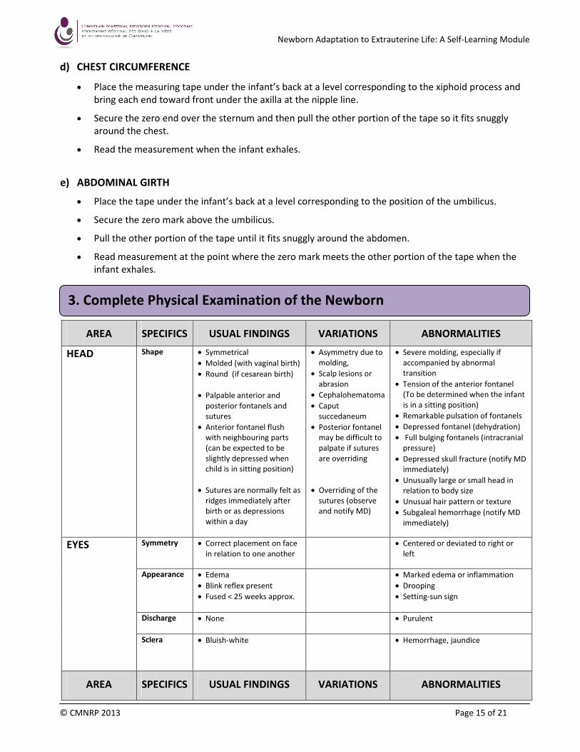
d) CHEST CIRCUMFERENCE
• Place the measuring tape under the infant’s back at a level corresponding to the xiphoid process and
bring each end toward front under the axilla at the nipple line.
• Secure the zero end over the sternum and then pull the other portion of the tape so it fits snuggly
around the chest.
• Read the measurement when the infant exhales.
e) ABDOMINAL GIRTH
• Place the tape under the infant’s back at a level corresponding to the position of the umbilicus.
• Secure the zero mark above the umbilicus.
• Pull the other portion of the tape until it fits snuggly around the abdomen.
• Read measurement at the point where the zero mark meets the other portion of the tape when the
infant exhales.
AREA SPECIFICS USUAL FINDINGS VARIATIONS ABNORMALITIES
HEAD
Shape • Symmetrical
• Molded (with vaginal birth)
• Round (if cesarean birth)
• Palpable anterior and
posterior fontanels and
sutures
• Anterior fontanel flush
with neighbouring parts
(can be expected to be
slightly depressed when
child is in sitting position)
• Sutures are normally felt as
ridges immediately after
birth or as depressions
within a day
• Asymmetry due to
molding,
• Scalp lesions or
abrasion
• Cephalohematoma
• Caput
succedaneum
• Posterior fontanel
may be difficult to
palpate if sutures
are overriding
• Overriding of the
sutures (observe
and notify MD)
• Severe molding, especially if
accompanied by abnormal
transition
• Tension of the anterior fontanel
(To be determined when the infant
is in a sitting position)
• Remarkable pulsation of fontanels
• Depressed fontanel (dehydration)
• Full bulging fontanels (intracranial
pressure)
• Depressed skull fracture (notify MD
immediately)
• Unusually large or small head in
relation to body size
• Unusual hair pattern or texture
• Subgaleal hemorrhage (notify MD
immediately)
EYES
Symmetry • Correct placement on face
in relation to one another
• Centered or deviated to right or
left
Appearance • Edema
• Blink reflex present
• Fused < 25 weeks approx.
• Marked edema or inflammation
• Drooping
• Setting-sun sign
Discharge • None
• Purulent
Sclera
• Bluish-white
• Hemorrhage, jaundice
AREA SPECIFICS USUAL FINDINGS VARIATIONS ABNORMALITIES
3. Complete Physical Examination of the Newborn
Newborn Adaptation to Extrauterine Life: A Self-Learning Module
© CMNRP 2013 Page 16 of 21
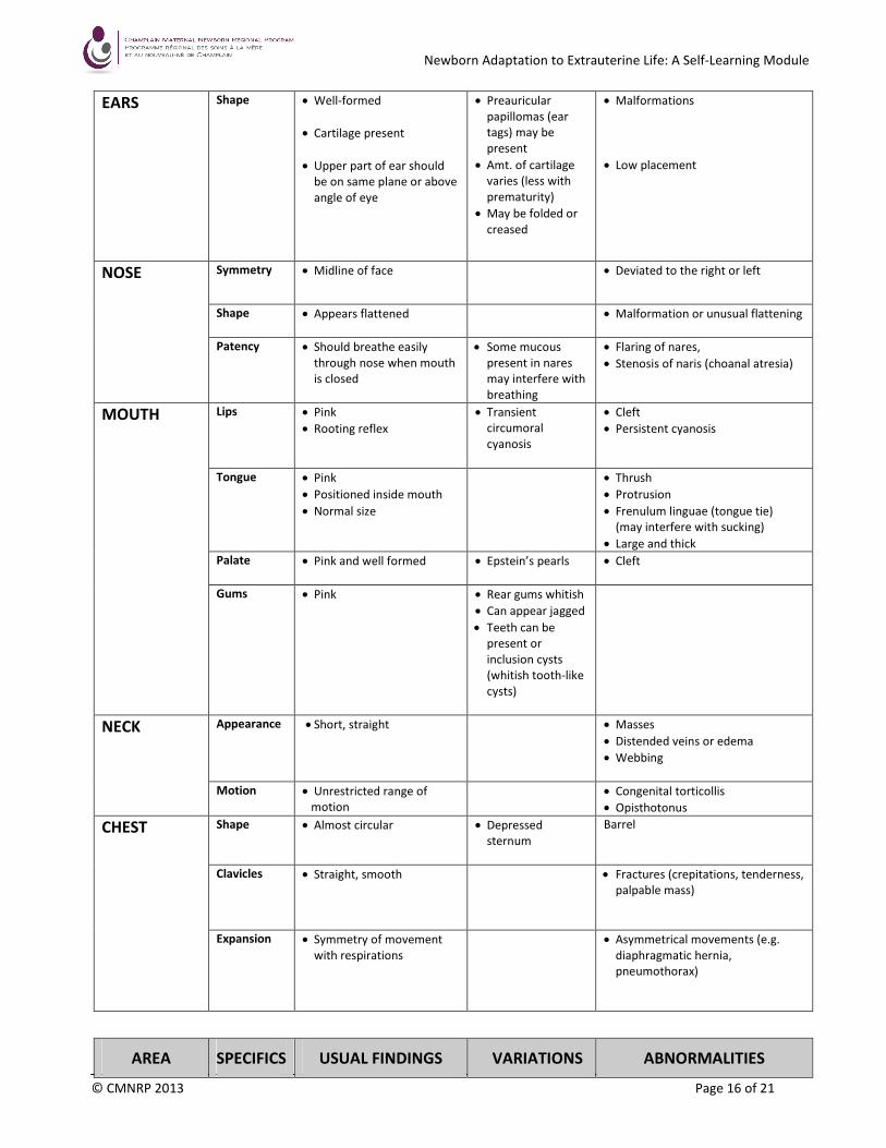
EARS Shape • Well-formed
• Cartilage present
• Upper part of ear should
be on same plane or above
angle of eye
• Preauricular
papillomas (ear
tags) may be
present
• Amt. of cartilage
varies (less with
prematurity)
• May be folded or
creased
• Malformations
• Low placement
NOSE Symmetry • Midline of face
• Deviated to the right or left
Shape • Appears flattened • Malformation or unusual flattening
Patency • Should breathe easily
through nose when mouth
is closed
• Some mucous
present in nares
may interfere with
breathing
• Flaring of nares,
• Stenosis of naris (choanal atresia)
MOUTH Lips • Pink
• Rooting reflex
• Transient
circumoral
cyanosis
• Cleft
• Persistent cyanosis
Tongue • Pink
• Positioned inside mouth
• Normal size
• Thrush
• Protrusion
• Frenulum linguae (tongue tie)
(may interfere with sucking)
• Large and thick
Palate • Pink and well formed
• Epstein’s pearls • Cleft
Gums • Pink
• Rear gums whitish
• Can appear jagged
• Teeth can be
present or
inclusion cysts
(whitish tooth-like
cysts)
NECK Appearance • Short, straight • Masses
• Distended veins or edema
• Webbing
Motion • Unrestricted range of
motion
• Congenital torticollis
• Opisthotonus
CHEST Shape • Almost circular
• Depressed
sternum
Barrel
Clavicles • Straight, smooth
• Fractures (crepitations, tenderness,
palpable mass)
Expansion • Symmetry of movement
with respirations
• Asymmetrical movements (e.g.
diaphragmatic hernia,
pneumothorax)
AREA SPECIFICS USUAL FINDINGS VARIATIONS ABNORMALITIES
Newborn Adaptation to Extrauterine Life: A Self-Learning Module
© CMNRP 2013 Page 17 of 21
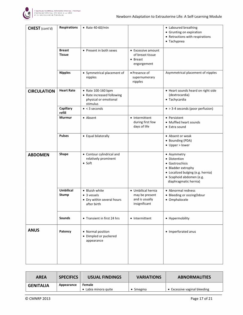
CHEST (cont’d) Respirations • Rate 40-60/min
• Laboured breathing
• Grunting on expiration
• Retractions with respirations
• Tachypnea
Breast
Tissue
• Present in both sexes
• Excessive amount
of breast tissue
• Breast
engorgement
Nipples
• Symmetrical placement of
nipples
• Presence of
supernumerary
nipples
Asymmetrical placement of nipples
CIRCULATION Heart Rate • Rate 100-160 bpm
• Rate increased following
physical or emotional
stimulus
• Heart sounds heard on right side
(dextrocardia)
• Tachycardia
Capillary
refill
• < 3 seconds
• > 3-4 seconds (poor perfusion)
Murmur • Absent • Intermittent
during first few
days of life
• Persistent
• Muffled heart sounds
• Extra sound
Pulses • Equal bilaterally • Absent or weak
• Bounding (PDA)
• Upper > lower
ABDOMEN Shape
• Contour cylindrical and
relatively prominent
• Soft
• Asymmetry
• Distention
• Gastroschisis
• Bladder extrophy
• Localized bulging (e.g. hernia)
• Scaphoid abdomen (e.g.
diaphragmatic hernia)
Umbilical
Stump
• Bluish white
• 3 vessels
• Dry within several hours
after birth
• Umbilical hernia
may be present
and is usually
insignificant
• Abnormal redness
• Bleeding or oozingOdour
• Omphalocele
Sounds
• Transient in first 24 hrs
• Intermittent
• Hypermobility
ANUS
Patency
• Normal position
• Dimpled or puckered
appearance
• Imperforated anus
AREA SPECIFICS USUAL FINDINGS VARIATIONS ABNORMALITIES
GENITALIA Appearance Female
• Labia minora quite
• Smegma
• Excessive vaginal bleeding
Newborn Adaptation to Extrauterine Life: A Self-Learning Module
© CMNRP 2013 Page 18 of 21
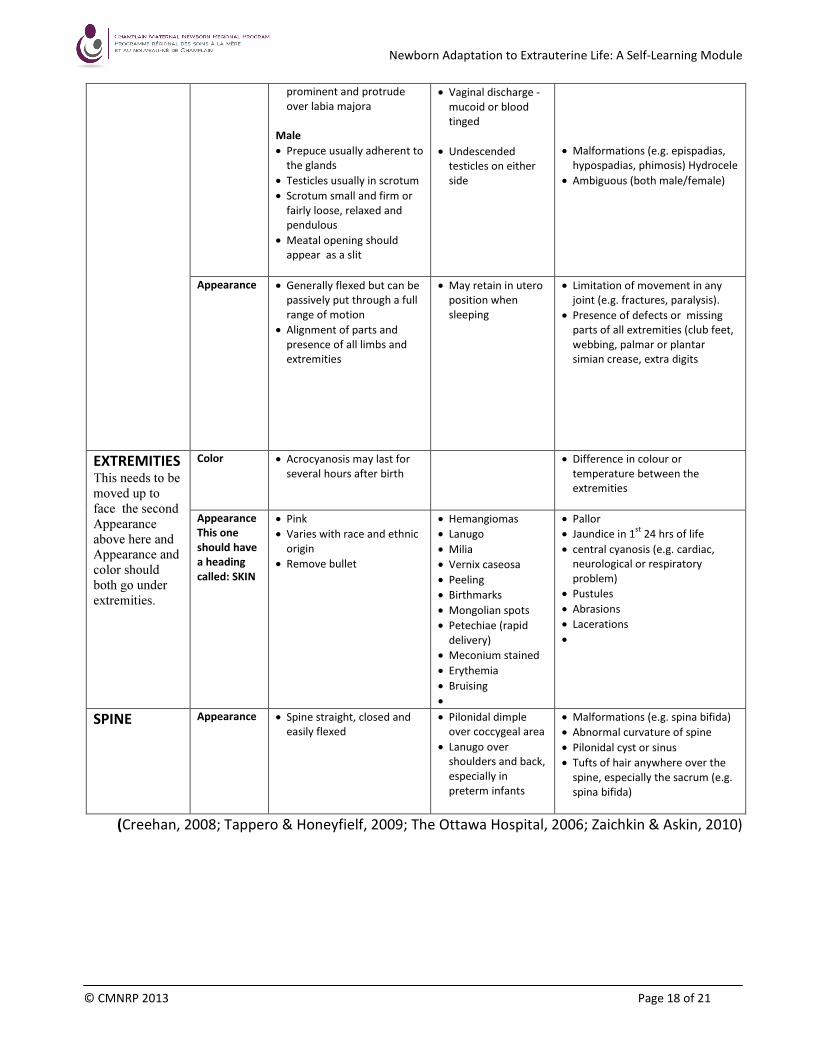
(Creehan, 2008; Tappero & Honeyfielf, 2009; The Ottawa Hospital, 2006; Zaichkin & Askin, 2010)
prominent and protrude
over labia majora
Male
• Prepuce usually adherent to
the glands
• Testicles usually in scrotum
• Scrotum small and firm or
fairly loose, relaxed and
pendulous
• Meatal opening should
appear as a slit
• Vaginal discharge -
mucoid or blood
tinged
• Undescended
testicles on either
side
• Malformations (e.g. epispadias,
hypospadias, phimosis) Hydrocele
• Ambiguous (both male/female)
Appearance • Generally flexed but can be
passively put through a full
range of motion
• Alignment of parts and
presence of all limbs and
extremities
• May retain in utero
position when
sleeping
• Limitation of movement in any
joint (e.g. fractures, paralysis).
• Presence of defects or missing
parts of all extremities (club feet,
webbing, palmar or plantar
simian crease, extra digits
EXTREMITIES This needs to be
moved up to
face the second
Appearance
above here and
Appearance and
color should
both go under
extremities.
Color • Acrocyanosis may last for
several hours after birth
• Difference in colour or
temperature between the
extremities
Appearance
This one
should have
a heading
called: SKIN
• Pink
• Varies with race and ethnic
origin
• Remove bullet
• Hemangiomas
• Lanugo
• Milia
• Vernix caseosa
• Peeling
• Birthmarks
• Mongolian spots
• Petechiae (rapid
delivery)
• Meconium stained
• Erythemia
• Bruising
•
• Pallor
• Jaundice in 1st
24 hrs of life
• central cyanosis (e.g. cardiac,
neurological or respiratory
problem)
• Pustules
• Abrasions
• Lacerations
•
SPINE Appearance • Spine straight, closed and
easily flexed
• Pilonidal dimple
over coccygeal area
• Lanugo over
shoulders and back,
especially in
preterm infants
• Malformations (e.g. spina bifida)
• Abnormal curvature of spine
• Pilonidal cyst or sinus
• Tufts of hair anywhere over the
spine, especially the sacrum (e.g.
spina bifida)
Newborn Adaptation to Extrauterine Life: A Self-Learning Module
© CMNRP 2013 Page 19 of 21
ACoRN Editorial Board. (2010). Acute care of at-Risk Newborns: A resource and learning tool for
health care Professionals. (1st ed.). Vancouver, BC: author.
Alvaro, R. E. & Rigatto, H. (2005). Cardiorespiratory adjustements at birth. In: Avery’s neonatalogy
pathophysiology & management of the newborn (6th
ed.) (pp. 285-303). Philadelphia, PA:
Lippincott Williams & Wilkins.
Askin, D. (2008). Newborn adaptation to extrauterine life. In: K. R. Simpson & P. A. Creehan (Eds).
AWHONN’s Perinatal Nursing (3rd
ed.) (pp. 527-545). Philadelphia, PA: Lippincott Williams &
Wilkins.
Askin, D. (2009). Fetal-to-neonatal transition – What is normal and what is not? Part 1: The
physiology of transition. Neonatal Network, 28(3), e33-e40.
Canadian Paediatric Society & College of Family Physicians of Canada. (2004). Routine administration
of vitamin K to newborns. Retrieved from http://www.cps.ca/English/statements/FN/fn97-
01.htm
Creehan, P. A. (2008). Newborn physical assessment. In: K. R. Simpson & P. A. Creehan (Eds).
AWHONN’s Perinatal Nursing (3rd
ed.) (pp. 556-574). Philadelphia, PA: Lippincott Williams &
Wilkins
Health Canada. (2000). Family-centred maternity and newborn care - National guidelines. Retrieved
from http://www.phac-aspc.gc.ca/dca-dea/publications/fcmc06_e.html
Kattwinkel, J. (Ed.). (2010). Textbook of neonatal resuscitation (6
th Ed.). Elk Grove, IL: American
Academy of Pediatrics and American Heart Association.
Kenner, C. (2003). Resuscitation and stabilization of the newborn. In C. Keener & J. QW. Lott (Eds.),
Comprehensive neonatal nursing: A physiologic perspective (3rd
ed.) (pp. 210-227). Philadelphia,
PA: Saunders.
Moore, E. R., Anderson, G. C., Bergman, N. (2007). Early skin-to-skin contact for mothers and their
healthy newborn infants. Cochrane Database of Systematic Reviews, Issue 3. Art. No.: CD003519.
DOI: 10.1002/14651858.CD003519.pub2.
Service Ontario (2011). Health Protection and Promotion Act. R. S. O. 1990, Chapter H. 7. Retrieved
from http://www.e-laws.gov.on.ca/html/statutes/english/elaws_statutes_90h07_e.htm#BK39
The Ottawa Hospital (2006). Assessment of infant. Ottawa: Author.
References
Newborn Adaptation to Extrauterine Life: A Self-Learning Module
© CMNRP 2013 Page 20 of 21
Zaichkin, J., & Fraser, D. (2010). The healthy newborn. In: R. J. Evans, M. K. Evans, Y. M. R. Brown, & S.
A. Orshan (Eds.).(2010). Canadian Maternity, Newborn, & Women’s Health Nursing. (1st
ed.) (773-
851). Philadelphia, PA: Lippincott Williams & Wilkins.
American Academy of Pediatrics - www.aap.org
Canadian Pediatric Society - www.cps.ca
Champlain Maternal Newborn Regional Program (CMNRP) www.cmnrp.ca
Evans, R. J., Evans, M. K., Brown, Y. M. R., & Orshan, S. A. (2010). Canadian Maternity, Newborn, &
Women’s Health Nursing. (1st
Ed.). Philadelphia, PA: Lippincott Williams & Wilkins.
Merenstein, G. B., & Gardner, S. L. (2006). Handbook of Neonatal Intensive Care. (6th
Ed.) St-Louis,
MI: Mosby.
Simpson, K. R., & Creehan, P. A. (2008). AWHONN’s Perinatal Nursing (3rd
Ed.). Philadelphia, PA:
Lippincott Williams & Wilkins.
Tappero, E. P. & Honeyfielf, M. E. (2009). Physical Assessment of the Newborn: A Comprehensive
Approach to the Art of Physical Examination. (4th
ed.). Santa Rosa, CA: NICU INK Book Publishers.
Resources
Newborn Adaptation to Extrauterine Life: A Self-Learning Module
© CMNRP 2013 Page 21 of 21
The Champlain Maternal Newborn Regional Program (CMNRP) would like to thank the members of
the Joint Orientation Sub-Committee for their work on the development of this Newborn Adaptation
Self-Learning Module.
CMNRP also acknowledges the work of the following groups and health care professionals who have
provided feedback and expertise:
� Members of the Interprofessional Education & Research Committee (IERC)
� Pediatricians and Neonatologists
� CMNRP Perinatal Consultants and Neonatal Nurse Practitioners
Acknowledgments