Embed Size (px)
Citation preview
Transcriptomic and Genetic Analysis of Direct InterspeciesElectron Transfer
Pravin Malla Shrestha, Amelia-Elena Rotaru, Zarath M. Summers,* Minita Shrestha, Fanghua Liu, Derek R. Lovley
Department of Microbiology, University of Massachusetts, Amherst, Massachusetts, USA
The possibility that metatranscriptomic analysis could distinguish between direct interspecies electron transfer (DIET) and H2
interspecies transfer (HIT) in anaerobic communities was investigated by comparing gene transcript abundance in cocultures inwhich Geobacter sulfurreducens was the electron-accepting partner for either Geobacter metallireducens, which performs DIET,or Pelobacter carbinolicus, which relies on HIT. Transcript abundance for G. sulfurreducens uptake hydrogenase genes was7-fold lower in cocultures with G. metallireducens than in cocultures with P. carbinolicus, consistent with DIET and HIT, respec-tively, in the two cocultures. Transcript abundance for the pilus-associated cytochrome OmcS, which is essential for DIET butnot for HIT, was 240-fold higher in the cocultures with G. metallireducens than in cocultures with P. carbinolicus. The pilin genepilA was moderately expressed despite a mutation that might be expected to repress pilA expression. Lower transcript abun-dance for G. sulfurreducens genes associated with acetate metabolism in the cocultures with P. carbinolicus was consistent withthe repression of these genes by H2 during HIT. Genes for the biogenesis of pili and flagella and several c-type cytochrome geneswere among the most highly expressed in G. metallireducens. Mutant strains that lacked the ability to produce pili, flagella, or theouter surface c-type cytochrome encoded by Gmet_2896 were not able to form cocultures with G. sulfurreducens. These resultsdemonstrate that there are unique gene expression patterns that distinguish DIET from HIT and suggest that metatranscriptom-ics may be a promising route to investigate interspecies electron transfer pathways in more-complex environments.
Interspecies electron transfer is essential to the proper function-ing of a diversity of anaerobic microbial communities, but there
is little information on the actual mechanisms by which microor-ganisms cooperate to exchange electrons in most anaerobic envi-ronments. The best-studied mechanism for interspecies electronexchange is H2 interspecies transfer (HIT) in which the electron-donating partner reduces protons to produce H2 and the electron-accepting partner oxidizes H2 with the reduction of an electronacceptor (1–4). Alternatively, in formate interspecies transfer(FIT), formate can serve as the electron carrier rather than H2
(3–5). Organic compounds with quinone moieties (6, 7) and sul-fur compounds (8–10) can also serve as interspecies electron shut-tles in laboratory cultures, but their environmental significance isnot known.
Direct interspecies electron transfer (DIET) is an alternativestrategy whereby electron exchange does not require soluble mol-ecules. For example, adaptive evolution of a coculture of Geobactermetallireducens and Geobacter sulfurreducens, grown under condi-tions in which the two species had to exchange electrons in orderto utilize ethanol as an electron donor with fumarate as an electronacceptor, led to a coculture in which electrons were transferreddirectly through electrically conductive cell aggregates rather thanvia HIT or FIT (11). Gene deletion and biochemical studies dem-onstrated that important components of DIET in the coculturewere the conductive pili of G. sulfurreducens (12–15) and OmcS(16, 17), a multiheme c-type cytochrome aligned along the pili(11). The possibility of HIT or FIT could be ruled out by thefinding that the coculture had a poor capacity for H2 or formatemetabolism and the rapid formation of cocultures initiated withmutants of G. sulfurreducens unable to utilize H2 and formate(11, 18).
DIET also appeared to be the mechanism for electron ex-change in natural methanogenic communities that formed ag-gregates in an anaerobic wastewater digester converting brew-
ery wastes to methane (19). The aggregates had electricalconductivity comparable to that of Geobacter cocultures, andthe temperature-dependent response of the conductivity sug-gested that long-range electron transport in the aggregates waslike metallic electron transport (19), similar to the metal-likeconductivity of G. sulfurreducens pili (12–15). Geobacter spe-cies were abundant within the aggregates and oxidized ethanoland fatty acids to drive methanogenesis. Conductive materialssuch as magnetite and granular activated carbon can facilitateDIET (7, 20–22).
DIET has the potential to be more efficient than HIT or FIT(23). However, not all microorganisms can make biological elec-trical connections with other species. This was apparent in a studyin which Pelobacter carbinolicus was grown in culture with G. sul-furreducens (11, 18). Unlike G. metallireducens, which could trans-fer electrons to a strain of G. sulfurreducens unable to use H2 andformate just as effectively as to wild-type G. sulfurreducens (11,18), P. carbinolicus required a G. sulfurreducens partner that coulduse at least one of these electron transfer molecules (18). Theinability of P. carbinolicus to function via DIET is consistent withits poor ability to transfer electrons directly to other external elec-tron acceptors such as electrodes (24) or Fe(III) oxides (25).
Received 12 December 2012 Accepted 25 January 2013
Published ahead of print 1 February 2013
Address correspondence to Pravin Malla Shrestha, [email protected].
* Present address: Zarath M. Summers, The BioTechnology Institute, University ofMinnesota, St. Paul, Minnesota, USA.
Supplemental material for this article may be found at http://dx.doi.org/10.1128/AEM.03837-12.
Copyright © 2013, American Society for Microbiology. All Rights Reserved.
doi:10.1128/AEM.03837-12
April 2013 Volume 79 Number 7 Applied and Environmental Microbiology p. 2397–2404 aem.asm.org 2397
on July 15, 2018 by guesthttp://aem
.asm.org/
Dow
nloaded from
The diversity of mechanisms by which species might exchangeelectrons leads to the question of how electrons are actually trans-ferred in anaerobic environments and the development of strate-gies to elucidate which mechanisms predominate. Metatranscrip-tomic analysis offers the means to query the physiological status ofmicroorganisms in natural environments (26–28). Therefore, in aproof-of-concept study, we compared the gene transcript abun-dance in cocultures exchanging electrons via HIT with that incocultures cooperating via DIET to determine whether metatran-scriptomic data can provide information diagnostic of these twodifferent modes of interspecies electron transfer.
MATERIALS AND METHODSCultures. Transcriptomic analyses were performed on previously de-scribed anaerobic cocultures in which G. sulfurreducens was the electron-accepting partner and either G. metallireducens (11) or P. carbinolicus (18)was the electron-donating partner. Both cocultures were grown in strictlyanaerobic, bicarbonate-buffered medium as previously described (11, 18)with ethanol (20 mM) as the electron donor and fumarate (40 mM) as theelectron acceptor. Metabolism of ethanol and reduction of fumarate weremonitored over time as previously described (18, 19, 29). Cultures wereharvested for transcriptomic or protein analysis at mid-log phase.
Mutant strains of G. metallireducens deficient in pilA, fliC (30), or theputative outer surface c-type cytochrome gene Gmet_2896 (31) were ob-tained from our laboratory culture collection maintained in pure culturesas previously described (30, 31) but adapted to ethanol (20 mM) insteadof acetate as the sole electron donor. To determine whether these strainscould grow in coculture with G. sulfurreducens, 0.5-ml cultures of eachpartner at mid-log phase were added to 10 ml anoxic medium containing20 mM ethanol and 40 mM fumarate and substrate consumption wasmonitored in parallel with that of a coculture established with the wild-type G. metallireducens strain.
Western blotting. For Western blotting, whole-cell lysate (5 �g) pro-teins were separated by 10% sodium dodecyl sulfate-polyacrylamide gelelectrophoresis (SDS-PAGE) and either visualized by Coomassie stainingwith the SeeBlue Plus prestained protein marker (Invitrogen) or trans-ferred to polyvinylidene difluoride immunoblotting membrane (Bio-Rad) for Western analysis, blocked in 3% bovine serum albumin in Tris-buffered saline with Tween for 1 h at room temperature, probed withPilA-specific antiserum diluted 1:1,000, washed twice in phosphate-buff-ered saline–Tween (10 mM sodium phosphate, 0.15 M NaCl, 0.05%Tween 20, pH 7.5), and incubated at 4°C overnight with a horseradishperoxidase (HRP)-coupled anti-rabbit secondary antibody diluted1:20,000 (Dako). Bands were then visualized with Ready Pour HRP–3,3=,5,5=-tetramethylbenzidine (Roche) by exposure to X-ray film.
Total mRNA extraction. For extraction of mRNA from G. metallire-ducens-G. sulfurreducens cocultures, the overlying medium of culturesgrown in 50 ml was removed anaerobically with a syringe and needle. Theaggregates left at the bottom of the bottle were mixed with RNAlater(Ambion) as previously described (27). The stopper was removed fromthe culture bottle, the aggregates were removed, and RNA was extractedwith TRIzol (Sigma) as previously described (18). For the P. carbinoli-cus-G. sulfurreducens cocultures, RNAlater (10% of the culture volume)was directly injected into the culture medium and cells were harvested at4°C with centrifugation at 6,000 � g for 20 min as previously described(18) and then extracted with TRIzol.
RNA was purified with the MinElute PCR purification kit (Qiagen)prior to recombinant DNase I (Ambion) digestion in accordance with themanufacturers’ protocols, followed by an additional treatment with theMinElute PCR purification kit. The absence of genomic DNA contami-nation was verified by 16S rRNA gene analysis as described previously(27). Aliquots of total RNA were preserved for real-time quantitative PCR(RT-qPCR), and the remainder was used for mRNA isolation. mRNA wasisolated with the MICROBExpress kit (Ambion) by following the manu-
facturer’s protocol. Aliquots of triplicate mRNA extracts were analyzedwith the Experion RNA HiSens kit (Bio-Rad). The electropherogram re-sult showed highly reproducible mRNA extracts (see Fig. S1 in the sup-plemental material). Equal concentrations of mRNA extracts from thetriplicate samples were then pooled to make composite samples (32–34)for each type of coculture for sequencing.
Illumina sequencing and data analysis. Illumina sequencing librarieswere prepared with the TruSeq RNA Sample Preparation kit by followingthe manufacturer’s protocol (Illumina). Briefly, 100 ng of the total mRNAwas chemically fragmented and converted into single-stranded cDNA byrandom-hexamer priming. Next, the second strand was generated to cre-ate double-stranded cDNA. The overhangs resulting from fragmentationwere converted to blunt ends by using an end repair mixture, and then the3= ends were adenylated. Adenylated products were ligated with individ-ual adapters containing unique hexameric indices/barcodes and enrichedwith a final 10-cycle PCR. The two enriched and purified samples con-taining unique barcodes and representing the two coculture types thatwere obtained were mixed in equimolar concentrations and used for hy-bridization in a HiSeq2000 flow cell for paired-end sequencing. Illuminasequence reads (100 bases) were functionally assigned by mapping againstthe published genomes of G. sulfurreducens strain PCA (35) and G. met-allireducens (36) with ArrayStar (DNASTAR), BOWTIE tools (37), andDeSeq (38). DeSeq is a free R software package (http://www.r-project.org/)that uses a method based on the negative binomial distribution with vari-ance and mean linked by local regression. DeSeq allows significant-differ-ence calculation in RNA-seq experiments both with and without biolog-ical replicates (38). Reads belonging to 16S/23S rRNA, reads that matchedmore than one segment of a genome, and reads with more than twomismatches were discarded. The remaining mRNA reads were reanalyzedand normalized with the RPKM (reads assigned per kilobase of target permillion mapped reads) method (39, 40) by using ArrayStar. Expressionlevels were considered significant only when the log2 RPKM value was �8.This was equivalent to the log2 RPKM value of the housekeeping gene recA(GSU0145) in the present experiment, which was previously used as theinternal standard during RT-qPCR (18). The n-fold change in the expres-sion level was calculated by comparing the normalized reads from G.sulfurreducens-G.metallireducens cocultures with those of P. carbinoli-cus-G. sulfurreducens cocultures. The n-fold changes were computed onlyfor those genes that had an RPKM value of �8 in one of the samples, andonly �2-fold changes in expression (up or down) were considered signif-icant (see Fig. S2 in the supplemental material). In addition to this, signif-icant n-fold changes were also calculated by using DeSeq (see Table S1a inthe supplemental material).
RT-qPCR. To verify the differential expression detected by sequenc-ing, RT-qPCR was performed for some genes that were highly upregu-lated or downregulated in G. metallireducens-G. sulfurreducens versus P.carbinolicus-G. sulfurreducens cocultures. For the RT-qPCR, about 300 ngof total RNA was converted into WTA (whole-transcriptome amplifica-tion) cDNA libraries and enriched by WTA PCR with reagents and pro-tocols supplied with or recommended by the manufacturer (Sigma).Briefly, 300 ng of total RNA was mixed with 2.5 ml WTA library synthesisbuffer and 2.5 ml WTA library stabilization solution and the total volumewas adjusted to 24 �l with nuclease-free water; the mixture was heated at70°C for 5 min and immediately cooled. One microliter of library synthe-sis enzyme was added, and WTA cDNA libraries were synthesized by usingthe following thermocycler program: 24°C for 15 min, 42°C for 2 h, and95°C for 5 min. Aliquots (5 �l) were WTA PCR amplified with JumpStartTaq DNA polymerase (Sigma), WTA amplification master mix, and de-oxynucleoside triphosphate mix in accordance with the manufacturer’sprotocol, except that the number of cycles was reduced to 15. The en-riched product was purified with the QIAquick PCR purification kit (Qia-gen) and used as the template in a qPCR experiment (18, 41). RT-PCR wascarried out with the ABI prism 7900 (Applied Biosystems). Primers tar-geting the genes pilA, pilR, omcS, omcT, omcZ, and recA were designed(Table 1) from the G. sulfurreducens genome sequence (35), and represen-
Shrestha et al.
2398 aem.asm.org Applied and Environmental Microbiology
on July 15, 2018 by guesthttp://aem
.asm.org/
Dow
nloaded from
tative products from all of these primer sets were verified by sequencing.Reactions were performed in triplicate for each gene tested in a totalvolume of 20 �l as described previously (18). A t test was used to calculatesignificant differences in gene expression levels at a significance level ofP � 0.05.
Nucleotide sequence accession number. The sequence reads deter-mined in this study have been submitted to the EMBL databases underaccession no. ERP001972.
RESULTS AND DISCUSSION
In order to gain insight into physiological differences duringgrowth by DIET versus growth by HIT, the gene transcript abun-dances in G. sulfurreducens were compared in two coculture sys-tems that differ in their modes of interspecies electron exchange.In cocultures supplied with ethanol as the electron donor, G. sul-furreducens can reduce the electron acceptor, fumarate, with elec-trons obtained either from G. metallireducens via DIET (11) orfrom P. carbinolicus via HIT (18). In both types of cocultures, G.sulfurreducens has the option to use acetate derived from ethanolmetabolism as an additional electron donor but continued metab-olism of ethanol is not possible without removal of the electronsreleased from the oxidation of ethanol to acetate and carbon di-oxide.
Unique Illumina sequence reads of G. sulfurreducens tran-scripts in the cocultures ranged from 1.8 � 106 to 2.9 � 106,yielding 47- to 76-fold coverage of the G. sulfurreducens genome.Of the 3,466 predicted protein-coding genes of G. sulfurreducens(35), 910 had significant expression levels (RPKM � 8) in at leastone of the cocultures (see Fig. S2a and Table S1b in the supple-mental material) and 640 genes had �2-fold higher or lower tran-script abundance in one coculture set than in the other (see Fig.S2b). In this study, the focus was on genes predicted to encodeproteins localized at the outer cell surface and/or involved in themetabolism of electron donors.
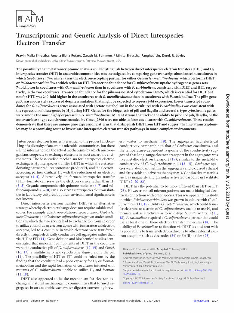
Evaluation of electron transfer via H2 or formate. G. sulfurre-ducens has only one hydrogenase that functions as an uptake hy-drogenase, membrane-bound Hyb (42). Transcripts of all five ofthe genes encoding Hyb subunits (GSU0782 to GSU0786) weresignificantly (P � 0.05) downregulated (�7-fold) in G. sulfurre-ducens growing in cocultures with G. metallireducens compared tothose in P. carbinolicus-G. sulfurreducens cocultures (Fig. 1; seeTable S1c in the supplemental material). Consistent with thelower transcript abundance of Hyb subunit genes in G. metallire-ducens-G. sulfurreducens cocultures was the lower expression
(�3-fold down) of the genes for Hyp (GSU0305 to GSU0309 andGSU0374), which are expected to catalyze Ni-Fe cofactor acquisi-tion by hydrogenases and are necessary for hydrogenase expres-sion (43). Other gene sets such as hya (GSU0120 to GSU0123), hox(GSU2717 to GSU2722), mvh (GSU2416 to GSU2423), hdr(GSU0085 to GSU0092), and ehr (GSU0739 to GSU0745), whichby annotation might be expected to function in H2 metabolism,had low expression levels in both types of cocultures (Fig. 1; seeTable S1c in the supplemental material), consistent with previousstudies indicating that they are not involved in H2 uptake (42, 44).
The finding that genes associated with H2 uptake were morehighly expressed in G. sulfurreducens growing with P. carbinolicusthan in G. sulfurreducens growing with G. metallireducens is con-sistent with the conclusions from other experimental results thatP. carbinolicus provides electrons from ethanol oxidation to G.sulfurreducens with H2 serving as the electron carrier whereas G.metallireducens transfers electrons to G. sulfurreducens throughdirect electrical connections (11, 18). Thus, the results presentedhere demonstrated that the transcriptomic approach could revealexpected differences in physiology under different syntrophicconditions and suggest that low expression of uptake hydrogenasegenes in potential electron-accepting partners might help rule outthis mode of interspecies electron transfer in environmentalstudies.
When P. carbinolicus is grown in coculture with a strain of G.sulfurreducens that cannot consume H2, formate can serve as analternative interspecies electron carrier (18). However, in the co-cultures investigated here, which had been transferred over 400
FIG 1 Heat map comparison of expression levels of genes associated withhydrogenase (black letter) and formate dehydrogenase (green letter) in cocul-tures of G. metallireducens-G. sulfurreducens (GS/GM) and G. sulfurredu-cens/P. carbinolicus (GS/PC). The n-fold change shown at the right representsthe gene transcript upregulation or downregulation in G. sulfurreducens in theGS/GM coculture compared to the transcript level in the GS/PC coculture. Then-fold change is presented only for G. sulfurreducens genes with significantexpression (log2 RPKM � 8) in one of the cocultures.
TABLE 1 Oligonucleotides used in this study
Primer Sequence (5=¡3=)Ampliconsize (bp)
pilAF ATCGGTATTCTCGCTGCAAT 117pilAR AATGCGGACTCAAGAGCAGTpilRF TTTCCGGGAGGATCTCTTTT 143pilRR TTATGATGCGGTCGCTGTAGomcSF TGGTTGGCGAAGGCATAGG 138omcSR CCATCAAGAACAGCGGTTCComcTF GGCTTCTGCGGTACTGATGT 145omcTR CCAGCAGATGAACAACGCTAomcZF AAGGTTGCTGACCTTGTTGG 158omcZR CCACCTATCAGCCCACTGTTrecAF CACCGGCATAATCTCCAAGT 150recAR ATCTTGCGGATATCGAGACG
Gene Expression Patterns for DIET
April 2013 Volume 79 Number 7 aem.asm.org 2399
on July 15, 2018 by guesthttp://aem
.asm.org/
Dow
nloaded from
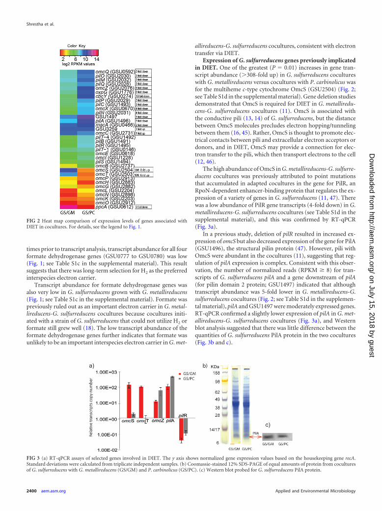
times prior to transcript analysis, transcript abundance for all fourformate dehydrogenase genes (GSU0777 to GSU0780) was low(Fig. 1; see Table S1c in the supplemental material). This resultsuggests that there was long-term selection for H2 as the preferredinterspecies electron carrier.
Transcript abundance for formate dehydrogenase genes wasalso very low in G. sulfurreducens grown with G. metallireducens(Fig. 1; see Table S1c in the supplemental material). Formate waspreviously ruled out as an important electron carrier in G. metal-lireducens-G. sulfurreducens cocultures because cocultures initi-ated with a strain of G. sulfurreducens that could not utilize H2 orformate still grew well (18). The low transcript abundance of theformate dehydrogenase genes further indicates that formate wasunlikely to be an important interspecies electron carrier in G. met-
allireducens-G. sulfurreducens cocultures, consistent with electrontransfer via DIET.
Expression of G. sulfurreducens genes previously implicatedin DIET. One of the greatest (P � 0.01) increases in gene tran-script abundance (�308-fold up) in G. sulfurreducens cocultureswith G. metallireducens versus cocultures with P. carbinolicus wasfor the multiheme c-type cytochrome OmcS (GSU2504) (Fig. 2;see Table S1d in the supplemental material). Gene deletion studiesdemonstrated that OmcS is required for DIET in G. metalliredu-cens-G. sulfurreducens cocultures (11). OmcS is associated withthe conductive pili (13, 14) of G. sulfurreducens, but the distancebetween OmcS molecules precludes electron hopping/tunnelingbetween them (16, 45). Rather, OmcS is thought to promote elec-trical contacts between pili and extracellular electron acceptors ordonors, and in DIET, OmcS may provide a connection for elec-tron transfer to the pili, which then transport electrons to the cell(12, 46).
The high abundance of OmcS in G. metallireducens-G. sulfurre-ducens cocultures was previously attributed to point mutationsthat accumulated in adapted cocultures in the gene for PilR, anRpoN-dependent enhancer-binding protein that regulates the ex-pression of a variety of genes in G. sulfurreducens (11, 47). Therewas a low abundance of PilR gene transcripts (4-fold down) in G.metallireducens-G. sulfurreducens cocultures (see Table S1d in thesupplemental material), and this was confirmed by RT-qPCR(Fig. 3a).
In a previous study, deletion of pilR resulted in increased ex-pression of omcS but also decreased expression of the gene for PilA(GSU1496), the structural pilin protein (47). However, pili withOmcS were abundant in the cocultures (11), suggesting that reg-ulation of pilA expression is complex. Consistent with this obser-vation, the number of normalized reads (RPKM � 8) for tran-scripts of G. sulfurreducens pilA and a gene downstream of pilA(for pilin domain 2 protein; GSU1497) indicated that althoughtranscript abundance was 5-fold lower in G. metallireducens-G.sulfurreducens cocultures (Fig. 2; see Table S1d in the supplemen-tal material), pilA and GSU1497 were moderately expressed genes.RT-qPCR confirmed a slightly lower expression of pilA in G. met-allireducens-G. sulfurreducens cocultures (Fig. 3a), and Westernblot analysis suggested that there was little difference between thequantities of G. sulfurreducens PilA protein in the two cocultures(Fig. 3b and c).
FIG 2 Heat map comparison of expression levels of genes associated withDIET in cocultures. For details, see the legend to Fig. 1.
FIG 3 (a) RT-qPCR assays of selected genes involved in DIET. The y axis shows normalized gene expression values based on the housekeeping gene recA.Standard deviations were calculated from triplicate independent samples. (b) Coomassie-stained 12% SDS-PAGE of equal amounts of protein from coculturesof G. sulfurreducens with G. metallireducens (GS/GM) and P. carbinolicus (GS/PC). (c) Western blot probed for G. sulfurreducens PilA protein.
Shrestha et al.
2400 aem.asm.org Applied and Environmental Microbiology
on July 15, 2018 by guesthttp://aem
.asm.org/
Dow
nloaded from
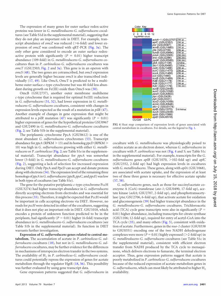
The expression of many genes for outer surface redox-activeproteins was lower in G. metallireducens-G. sulfurreducens cocul-tures (see Table S1d in the supplemental material), suggesting thatthey do not play an important role in DIET. For example, tran-script abundance of omcZ was reduced (�6-fold) and lower ex-pression of omcZ was confirmed with qRT-PCR (Fig. 3a). Theonly other gene considered to encode an outer surface redox-active protein with significantly (P � 0.01) higher transcriptabundance (109-fold) in G. metallireducens-G. sulfurreducens co-cultures than in P. carbinolicus-G. sulfurreducens cocultures wasomcT (GSU2503; Fig. 2 and 3a). This gene is in an operon withomcS (48). The two genes are cotranscribed, but omcS expressionlevels are generally higher because omcS is also transcribed indi-vidually (17, 49). Like OmcS, OmcT is predicted to be a multi-heme outer surface c-type cytochrome but was 40-fold less abun-dant during growth on Fe(III) oxide than OmcS was (50).
OmcB (GSU2737), another outer membrane multihemec-type cytochrome that is required for optimal Fe(III) reductionin G. sulfurreducens (51, 52), had lower expression in G. metalli-reducens-G. sulfurreducens cocultures, consistent with changes inexpression levels expected as the result of a mutation in pilR (47).Another example of changes in gene expression that might beattributed to a pilR mutation (47) was significantly (P � 0.01)higher expression of genes for the hypothetical proteins GSU0967and GSU2490 in G. metallireducens-G. sulfurreducens cocultures(Fig. 2; see Table S1b in the supplemental material).
The periplasmic cytochrome PpcA (GSU0612) is one of themost abundant G. sulfurreducens cytochromes (53). Transcriptabundance for ppcA (RPKM � 13) and its homolog ppcD (RPKM �10) was high in G. sulfurreducens growing with either G. metalli-reducens or P. carbinolicus (Fig. 2; see Table S1e in the supplemen-tal material). Transcript abundance for ppcA was somewhatlower (3-fold) in G. metallireducens-G. sulfurreducens cocultures(Fig. 2), suggesting a lack of selection for increased expressionduring DIET. Only PpcA and PpcD can accept and donate protonsalong with electrons (54). The expression level of the remaining threehomologs of ppcA in G. sulfurreducens (ppcB, ppcC, and ppcE) was lowin both types of cocultures (see Table S1e).
The gene for the putative periplasmic c-type cytochrome PccH(GSU3274) had higher transcript abundance in G. sulfurreducensdirectly accepting electrons from electrodes and was essential forthis process (55). Therefore, it might be expected that PccH wouldbe important in cells accepting electrons via DIET. However, noreads for pccH were detected in either of the cocultures, suggestingthat it does not play an important role in DIET. GSU1018, whichencodes a protein of unknown function predicted to be in theperiplasm, had significantly (P � 0.01) higher (6-fold) transcriptabundance in G. metallireducens-G. sulfurreducens cocultures (seeTable S1b in the supplemental material). Its function in DIETwarrants further investigation.
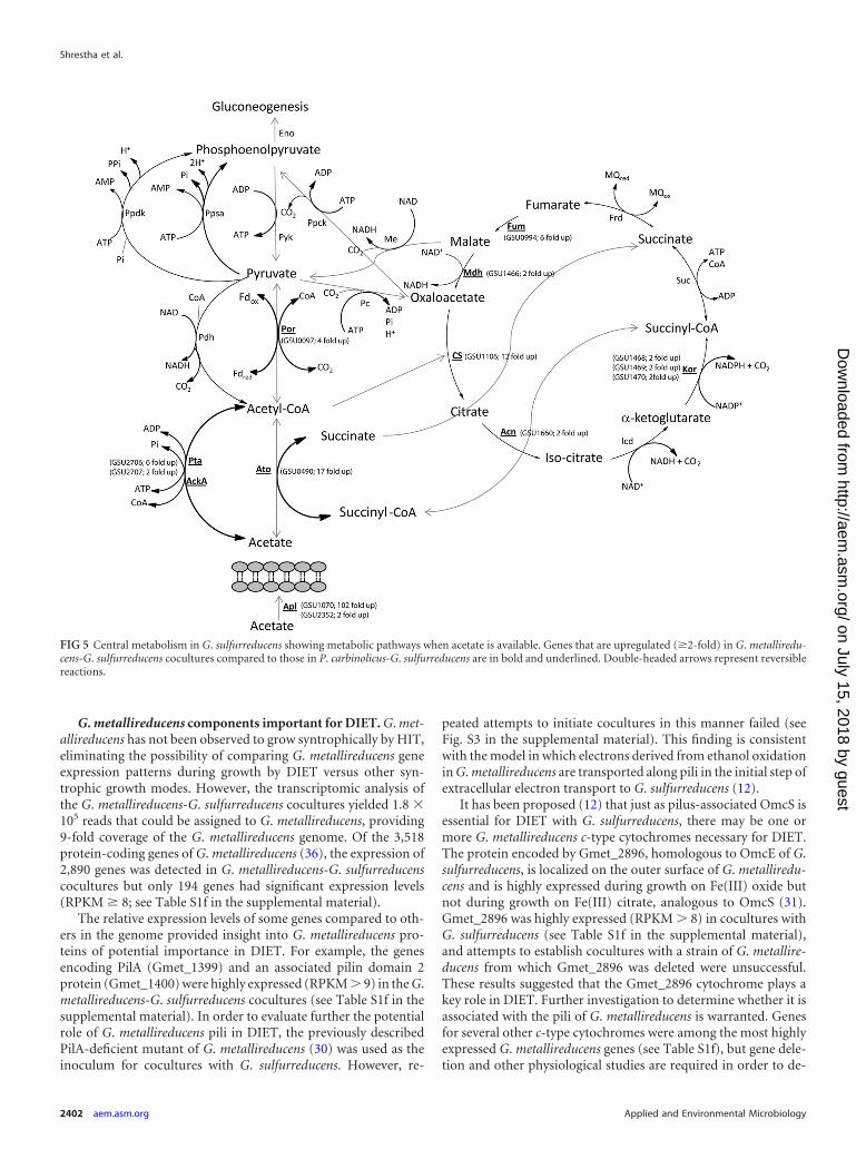
Expression of G. sulfurreducens genes related to central me-tabolism. The accumulation of acetate in P. carbinolicus-G. sul-furreducens cocultures (18), but not in G. metallireducens-G. sul-furreducens cocultures, may be further evidence for the differencesin mechanisms of interspecies electron transfer in the two systems.The availability of H2 in P. carbinolicus-G. sulfurreducens cocul-tures could potentially repress the expression of genes for acetatemetabolism through the regulator HgtR (18, 56). This possibilitywas further evaluated by using gene transcript data.
Gene expression patterns suggested that G. sulfurreducens in
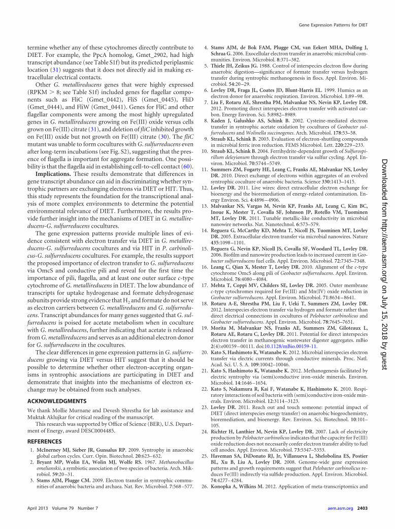
coculture with G. metallireducens was physiologically poised tooxidize acetate as an electron donor, whereas G. sulfurreducens incoculture with P. carbinolicus was not (Fig. 4 and 5; see Table S1cin the supplemental material). For example, transcripts for the G.sulfurreducens genes aplB (GSU1070, �102-fold up) and aplC(GSU2352, 2-fold up) had high expression levels in cocultureswith G. metallireducens. These genes, along with aplA (GSU1068),are associated with acetate uptake, and the expression of at leasttwo of these three genes is necessary for effective acetate uptake(57, 58).
G. sulfurreducens genes, such as those for succinyl:acetate co-enzyme A (CoA)-transferase (ato-1; GSU0490, 17-fold up), ace-tate kinase (ackA; GSU2707, 2-fold up), and phosphotransacety-lase (pta; GSU2706, 6-fold up), that activate acetate for oxidationand gluconeogenesis (59) had higher transcript abundance in theG. metallireducens-G. sulfurreducens cocultures. Trichloroaceticacid (TCA) cycle gene transcripts were also in significantly (P �0.01) higher abundance, including transcripts for citrate synthase(GSU1106; 12-fold up), required for entry of acetyl-CoA into theTCA cycle (35), and many others (Fig. 5), consistent with oxida-tion of acetate. Furthermore, genes in the nuo-1 cluster (GSU0338to GSU0351) encoding one of the two NADH dehydrogenasecomplexes were more (P � 0.01) highly expressed (�2-fold up) inG. metallireducens-G. sulfurreducens cocultures (see Table S1c inthe supplemental material), consistent with efficient electrontransfer from NADH produced by the TCA cycle to menaqui-none, which delivers electrons to fumarate, the terminal electronacceptor. Thus, gene expression patterns suggest that acetate ispoorly metabolized in P. carbinolicus-G. sulfurreducens coculturesbecause of the reduced expression of acetate metabolism genes inG. sulfurreducens, which can most likely be attributed to higher H2
availability.
FIG 4 Heat map comparison of expression levels of genes associated withcentral metabolism in cocultures. For details, see the legend to Fig. 1.
Gene Expression Patterns for DIET
April 2013 Volume 79 Number 7 aem.asm.org 2401
on July 15, 2018 by guesthttp://aem
.asm.org/
Dow
nloaded from
G. metallireducens components important for DIET. G. met-allireducens has not been observed to grow syntrophically by HIT,eliminating the possibility of comparing G. metallireducens geneexpression patterns during growth by DIET versus other syn-trophic growth modes. However, the transcriptomic analysis ofthe G. metallireducens-G. sulfurreducens cocultures yielded 1.8 �105 reads that could be assigned to G. metallireducens, providing9-fold coverage of the G. metallireducens genome. Of the 3,518protein-coding genes of G. metallireducens (36), the expression of2,890 genes was detected in G. metallireducens-G. sulfurreducenscocultures but only 194 genes had significant expression levels(RPKM � 8; see Table S1f in the supplemental material).
The relative expression levels of some genes compared to oth-ers in the genome provided insight into G. metallireducens pro-teins of potential importance in DIET. For example, the genesencoding PilA (Gmet_1399) and an associated pilin domain 2protein (Gmet_1400) were highly expressed (RPKM � 9) in the G.metallireducens-G. sulfurreducens cocultures (see Table S1f in thesupplemental material). In order to evaluate further the potentialrole of G. metallireducens pili in DIET, the previously describedPilA-deficient mutant of G. metallireducens (30) was used as theinoculum for cocultures with G. sulfurreducens. However, re-
peated attempts to initiate cocultures in this manner failed (seeFig. S3 in the supplemental material). This finding is consistentwith the model in which electrons derived from ethanol oxidationin G. metallireducens are transported along pili in the initial step ofextracellular electron transport to G. sulfurreducens (12).
It has been proposed (12) that just as pilus-associated OmcS isessential for DIET with G. sulfurreducens, there may be one ormore G. metallireducens c-type cytochromes necessary for DIET.The protein encoded by Gmet_2896, homologous to OmcE of G.sulfurreducens, is localized on the outer surface of G. metalliredu-cens and is highly expressed during growth on Fe(III) oxide butnot during growth on Fe(III) citrate, analogous to OmcS (31).Gmet_2896 was highly expressed (RPKM � 8) in cocultures withG. sulfurreducens (see Table S1f in the supplemental material),and attempts to establish cocultures with a strain of G. metallire-ducens from which Gmet_2896 was deleted were unsuccessful.These results suggested that the Gmet_2896 cytochrome plays akey role in DIET. Further investigation to determine whether it isassociated with the pili of G. metallireducens is warranted. Genesfor several other c-type cytochromes were among the most highlyexpressed G. metallireducens genes (see Table S1f), but gene dele-tion and other physiological studies are required in order to de-
FIG 5 Central metabolism in G. sulfurreducens showing metabolic pathways when acetate is available. Genes that are upregulated (�2-fold) in G. metalliredu-cens-G. sulfurreducens cocultures compared to those in P. carbinolicus-G. sulfurreducens are in bold and underlined. Double-headed arrows represent reversiblereactions.
Shrestha et al.
2402 aem.asm.org Applied and Environmental Microbiology
on July 15, 2018 by guesthttp://aem
.asm.org/
Dow
nloaded from
termine whether any of these cytochromes directly contribute toDIET. For example, the PpcA homolog, Gmet_2902, had hightranscript abundance (see Table S1f) but its predicted periplasmiclocation (31) suggests that it does not directly aid in making ex-tracellular electrical contacts.
Other G. metallireducens genes that were highly expressed(RPKM � 8; see Table S1f) included genes for flagellar compo-nents such as FliC (Gmet_0442), FliS (Gmet_0445), FliD(Gmet_0444), and FliW (Gmet_0441). Genes for FliC and otherflagellar components were among the most highly upregulatedgenes in G. metallireducens growing on Fe(III) oxide versus cellsgrown on Fe(III) citrate (31), and deletion of fliC inhibited growthon Fe(III) oxide but not growth on Fe(III) citrate (30). The fliCmutant was unable to form cocultures with G. sulfurreducens evenafter long-term incubations (see Fig. S2), suggesting that the pres-ence of flagella is important for aggregate formation. One possi-bility is that the flagella aid in establishing cell-to-cell contact (60).
Implications. These results demonstrate that differences ingene transcript abundance can aid in discriminating whether syn-trophic partners are exchanging electrons via DIET or HIT. Thus,this study represents the foundation for the transcriptional anal-ysis of more complex environments to determine the potentialenvironmental relevance of DIET. Furthermore, the results pro-vide further insight into the mechanisms of DIET in G. metallire-ducens-G. sulfurreducens cocultures.
The gene expression patterns provide multiple lines of evi-dence consistent with electron transfer via DIET in G. metallire-ducens-G. sulfurreducens cocultures and via HIT in P. carbinoli-cus-G. sulfurreducens cocultures. For example, the results supportthe proposed importance of electron transfer to G. sulfurreducensvia OmcS and conductive pili and reveal for the first time theimportance of pili, flagella, and at least one outer surface c-typecytochrome of G. metallireducens in DIET. The low abundance oftranscripts for uptake hydrogenase and formate dehydrogenasesubunits provide strong evidence that H2 and formate do not serveas electron carriers between G. metallireducens and G. sulfurredu-cens. Transcript abundances for many genes suggested that G. sul-furreducens is poised for acetate metabolism when in coculturewith G. metallireducens, further indicating that acetate is releasedfrom G. metallireducens and serves as an additional electron donorfor G. sulfurreducens in the cocultures.
The clear differences in gene expression patterns in G. sulfurre-ducens growing via DIET versus HIT suggest that it should bepossible to determine whether other electron-accepting organ-isms in syntrophic associations are participating in DIET anddemonstrate that insights into the mechanisms of electron ex-change may be obtained from such analyses.
ACKNOWLEDGMENTS
We thank Mollie Murnane and Devesh Shrestha for lab assistance andMuktak Aklujkar for critical reading of the manuscript.
This research was supported by Office of Science (BER), U.S. Depart-ment of Energy, award DESC0004485.
REFERENCES1. McInerney MJ, Sieber JR, Gunsalus RP. 2009. Syntrophy in anaerobic
global carbon cycles. Curr. Opin. Biotechnol. 20:623– 632.2. Bryant MP, Wolin EA, Wolin MJ, Wolfe RS. 1967. Methanobacillus
omelianskii, a symbiotic association of two species of bacteria. Arch. Mik-robiol. 59:20 –31.
3. Stams AJM, Plugge CM. 2009. Electron transfer in syntrophic commu-nities of anaerobic bacteria and archaea. Nat. Rev. Microbiol. 7:568 –577.
4. Stams AJM, de Bok FAM, Plugge CM, van Eekert MHA, Dolfing J,Schraa G. 2006. Exocellular electron transfer in anaerobic microbial com-munities. Environ. Microbiol. 8:371–382.
5. Thiele JH, Zeikus JG. 1988. Control of interspecies electron flow duringanaerobic digestion—significance of formate transfer versus hydrogentransfer during syntrophic methanogenesis in flocs. Appl. Environ. Mi-crobiol. 54:20 –29.
6. Lovley DR, Fraga JL, Coates JD, Blunt-Harris EL. 1999. Humics as anelectron donor for anaerobic respiration. Environ. Microbiol. 1:89 –98.
7. Liu F, Rotaru AE, Shrestha PM, Malvankar NS, Nevin KP, Lovley DR.2012. Promoting direct interspecies electron transfer with activated car-bon. Energy Environ. Sci. 5:8982– 8989.
8. Kaden J, Galushko AS, Schink B. 2002. Cysteine-mediated electrontransfer in syntrophic acetate oxidation by cocultures of Geobacter sul-furreducens and Wolinella succinogenes. Arch. Microbiol. 178:53–58.
9. Straub KL, Schink B. 2003. Evaluation of electron-shuttling compoundsin microbial ferric iron reduction. FEMS Microbiol. Lett. 220:229 –233.
10. Straub KL, Schink B. 2004. Ferrihydrite-dependent growth of Sulfurospi-rillum deleyianum through electron transfer via sulfur cycling. Appl. En-viron. Microbiol. 70:5744 –5749.
11. Summers ZM, Fogarty HE, Leang C, Franks AE, Malvankar NS, LovleyDR. 2010. Direct exchange of electrons within aggregates of an evolvedsyntrophic coculture of anaerobic bacteria. Science 330:1413–1415.
12. Lovley DR. 2011. Live wires: direct extracellular electron exchange forbioenergy and the bioremediation of energy-related contamination. En-ergy Environ. Sci. 4:4896 – 4906.
13. Malvankar NS, Vargas M, Nevin KP, Franks AE, Leang C, Kim BC,Inoue K, Mester T, Covalla SF, Johnson JP, Rotello VM, TuominenMT, Lovley DR. 2011. Tunable metallic-like conductivity in microbialnanowire networks. Nat. Nanotechnol. 6:573–579.
14. Reguera G, McCarthy KD, Mehta T, Nicoll JS, Tuominen MT, LovleyDR. 2005. Extracellular electron transfer via microbial nanowires. Nature435:1098 –1101.
15. Reguera G, Nevin KP, Nicoll JS, Covalla SF, Woodard TL, Lovley DR.2006. Biofilm and nanowire production leads to increased current in Geo-bacter sulfurreducens fuel cells. Appl. Environ. Microbiol. 72:7345–7348.
16. Leang C, Qian X, Mester T, Lovley DR. 2010. Alignment of the c-typecytochrome OmcS along pili of Geobacter sulfurreducens. Appl. Environ.Microbiol. 76:4080 – 4084.
17. Mehta T, Coppi MV, Childers SE, Lovley DR. 2005. Outer membranec-type cytochromes required for Fe(III) and Mn(IV) oxide reduction inGeobacter sulfurreducens. Appl. Environ. Microbiol. 71:8634 – 8641.
18. Rotaru A-E, Shrestha PM, Liu F, Ueki T, Summers ZM, Lovley DR.2012. Interspecies electron transfer via hydrogen and formate rather thandirect electrical connections in cocultures of Pelobacter carbinolicus andGeobacter sulfurreducens. Appl. Environ. Microbiol. 78:7645–7651.
19. Morita M, Malvankar NS, Franks AE, Summers ZM, Giloteaux L,Rotaru AE, Rotaru C, Lovley DR. 2011. Potential for direct interspecieselectron transfer in methanogenic wastewater digester aggregates. mBio2(4):e00159 – 00111. doi:10.1128/mBio.00159-11.
20. Kato S, Hashimoto K, Watanabe K. 2012. Microbial interspecies electrontransfer via electric currents through conductive minerals. Proc. Natl.Acad. Sci. U. S. A. 109:10042–10046.
21. Kato S, Hashimoto K, Watanabe K. 2012. Methanogenesis facilitated byelectric syntrophy via (semi)conductive iron-oxide minerals. Environ.Microbiol. 14:1646 –1654.
22. Kato S, Nakamura R, Kai F, Watanabe K, Hashimoto K. 2010. Respi-ratory interactions of soil bacteria with (semi)conductive iron-oxide min-erals. Environ. Microbiol. 12:3114 –3123.
23. Lovley DR. 2011. Reach out and touch someone: potential impact ofDIET (direct interspecies energy transfer) on anaerobic biogeochemistry,bioremediation, and bioenergy. Rev. Environ. Sci. Biotechnol. 10:101–105.
24. Richter H, Lanthier M, Nevin KP, Lovley DR. 2007. Lack of electricityproduction by Pelobacter carbinolicus indicates that the capacity for Fe(III)oxide reduction does not necessarily confer electron transfer ability to fuelcell anodes. Appl. Environ. Microbiol. 73:5347–5353.
25. Haveman SA, DiDonato RJ, Jr, Villanueva L, Shelobolina ES, PostierBL, Xu B, Liu A, Lovley DR. 2008. Genome-wide gene expressionpatterns and growth requirements suggest that Pelobacter carbinolicus re-duces Fe(III) indirectly via sulfide production. Appl. Environ. Microbiol.74:4277– 4284.
26. Konopka A, Wilkins M. 2012. Application of meta-transcriptomics and
Gene Expression Patterns for DIET
April 2013 Volume 79 Number 7 aem.asm.org 2403
on July 15, 2018 by guesthttp://aem
.asm.org/
Dow
nloaded from
proteomics to analysis of in situ physiological state. Front. Microbiol.3:184. doi:10.3389/fmicb.2012.00184.
27. Shrestha PM, Kube M, Reinhardt R, Liesack W. 2009. Transcriptionalactivity of paddy soil bacterial communities. Environ. Microbiol. 11:960 –970.
28. Marchetti A, Schruth DM, Durkin CA, Parker MS, Kodner RB, Ber-thiaume CT, Morales R, Allen AE, Armbrust EV. 2012. Comparativemetatranscriptomics identifies molecular bases for the physiological re-sponses of phytoplankton to varying iron availability. Proc. Natl. Acad.Sci. U. S. A. 109:E317–E325.
29. Nevin KP, Richter H, Covalla SF, Johnson JP, Woodard TL, Orloff AL,Jia H, Zhang M, Lovley DR. 2008. Power output and columbic efficien-cies from biofilms of Geobacter sulfurreducens comparable to mixed com-munity microbial fuel cells. Environ. Microbiol. 10:2505–2514.
30. Tremblay P-L, Aklujkar M, Leang C, Nevin KP, Lovley DR. 2011. Agenetic system for Geobacter metallireducens: role of the flagellin and pilinin the reduction of Fe(III) oxide. Environ. Microbiol. Rep. 4:82– 88.
31. Smith JA, Lovley DR, Tremblay P-L. 2013. Outer cell surface compo-nents essential for Fe(III) oxide reduction in Geobacter metallireducens.Appl. Environ. Microbiol. 79:901–907.
32. Shrestha PM, Kammann C, Lenhart K, Dam B, Liesack W. 2012.Linking activity, composition and seasonal dynamics of atmosphericmethane oxidizers in a meadow soil. ISME J. 6:1115–1126.
33. Jager M, Ott CE, Grunhagen J, Hecht J, Schell H, Mundlos S, Duda GN,Robinson PN, Lienau J. 2011. Composite transcriptome assembly ofRNA-seq data in a sheep model for delayed bone healing. BMC Genomics12:158. doi:10.1186/1471-2164-12-158.
34. Yu C, Li Y, Holmes A, Szafranski K, Faulkes CG, Coen CW, BuffensteinR, Platzer M, de Magalhaes JP, Church GM. 2011. RNA sequencingreveals differential expression of mitochondrial and oxidation reductiongenes in the long-lived naked mole-rat compared to mice. PLoS One6:e26729. doi:10.1371/journal.pone.0026729.
35. Methé BA, Nelson KE, Eisen JA, Paulsen IT, Nelson W, Heidelberg JF,Wu D, Wu M, Ward N, Beanan MJ, Dodson RJ, Madupu R, BrinkacLM, Daugherty SC, DeBoy RT, Durkin AS, Gwinn M, Kolonay JF,Sullivan SA, Haft DH, Selengut J, Davidsen TM, Zafar N, White O,Tran B, Romero C, Forberger HA, Weidman J, Khouri H, FeldblyumTV, Utterback TR, Van Aken SE, Lovley DR, Fraser CM. 2003. Genomeof Geobacter sulfurreducens: metal reduction in subsurface environments.Science 302:1967–1969.
36. Aklujkar M, Krushkal J, DiBartolo G, Lapidus A, Land ML, Lovley DR.2009. The genome sequence of Geobacter metallireducens: features of me-tabolism, physiology and regulation common and dissimilar to Geobactersulfurreducens. BMC Microbiol. 9:109. doi:10.1186/1471-2180-9-109.
37. Langmead B, Trapnell C, Pop M, Salzberg SL. 2009. Ultrafast andmemory-efficient alignment of short DNA sequences to the human ge-nome. Gen. Biol. 10:R25. doi:10.1186-Gb-2009-10-3-r25.
38. Anders S, Huber W. 2010. Differential expression analysis for sequencecount data. Gen. Biol. 11:R106. doi:10.1186-Gb-2010-11-10-r106.
39. Mortazavi A, Williams BA, Mccue K, Schaeffer L, Wold B. 2008.Mapping and quantifying mammalian transcriptomes by RNA-seq. Nat.Methods 5:621– 628.
40. Klevebring D, Bjursell M, Emanuelsson O, Lundeberg J. 2010. In-depthtranscriptome analysis reveals novel TARs and prevalent antisense tran-scription in human cell lines. PLoS One 5:e9762. doi:10.1371/journal.pone.0009762.
41. Tomlins SA, Mehra R, Rhodes DR, Shah RB, Rubin MA, Bruening E,Makarov V, Chinnaiyan AM. 2006. Whole transcriptome amplificationfor gene expression profiling and development of molecular archives.Neoplasia 8:153–162.
42. Coppi MV, O’Neil RA, Lovley DR. 2004. Identification of an uptakehydrogenase required for hydrogen-dependent reduction of Fe(III) andother electron acceptors by Geobacter sulfurreducens. J. Bacteriol. 186:3022–3028.
43. Sargent F, Ballantine SP, Rugman PA, Palmer T, Boxer DH. 1998.Reassignment of the gene encoding the Escherichia coli hydrogenase 2small subunit-identification of a soluble precursor of the small subunit ina hypB mutant. Eur. J. Biochem. 255:746 –754.
44. Tremblay PL, Lovley DR. 2012. Role of the NiFe hydrogenase Hya inoxidative stress defense in Geobacter sulfurreducens. J. Bacteriol. 194:2248 –2253.
45. Malvankar NS, Tuominen MT, Lovley DR. 2012. Lack of cytochromeinvolvement in long-range electron transport through conductive bio-films and nanowires of Geobacter sulfurreducens. Energy Environ. Sci.5:8651– 8659.
46. Lovley DR. 2012. Electromicrobiology. Annu. Rev. Microbiol. 66:391–409.
47. Juárez K, Kim BC, Nevin K, Olvera L, Reguera G, Lovley DR, MethéBA. 2009. PilR, a transcriptional regulator for pilin and other genes re-quired for Fe(III) reduction in Geobacter sulfurreducens. J. Mol. Microbiol.Biotechnol. 16:146 –158.
48. Mahadevan R, Yan B, Postier B, Nevin KP, Woodard TL, O’Neil R,Coppi MV, Methé BA, Krushkal J. 2008. Characterizing regulation ofmetabolism in Geobacter sulfurreducens through genome-wide expressiondata and sequence analysis. OMICS 12:33–59.
49. Holmes DE, Chaudhuri SK, Nevin KP, Mehta T, Methé BA, Liu A,Ward JE, Woodard TL, Webster J, Lovley DR. 2006. Microarray andgenetic analysis of electron transfer to electrodes in Geobacter sulfurredu-cens. Environ. Microbiol. 8:1805–1815.
50. Ding YH, Hixson KK, Aklujkar MA, Lipton MS, Smith RD, Lovley DR,Mester T. 2008. Proteome of Geobacter sulfurreducens grown with Fe(III)oxide or Fe(III) citrate as the electron acceptor. Biochim. Biophys. Acta1784:1935–1941.
51. Leang C, Coppi MV, Lovley DR. 2003. OmcB, a c-type polyheme cyto-chrome, involved in Fe(III) reduction in Geobacter sulfurreducens. J. Bac-teriol. 185:2096 –2103.
52. Leang C, Lovley DR. 2005. Regulation of two highly similar genes, omcBand omcC, in a 10 kb chromosomal duplication in Geobacter sulfurredu-cens. Microbiology 151:1761–1767.
53. Lloyd JR, Leang C, Myerson ALH, Coppi MV, Cuifo S, Methé B,Sandler SJ, Lovley DR. 2003. Biochemical and genetic characterization ofPpcA, a periplasmic c-type cytochrome in Geobacter sulfurreducens.Biochem. J. 369:153–161.
54. Morgado L, Bruix M, Pessanha M, Londer YY, Salgueiro CA. 2010.Thermodynamic characterization of a triheme cytochrome family fromGeobacter sulfurreducens reveals mechanistic and functional diversity. Bio-phys. J. 99:293–301.
55. Strycharz SM, Glaven RH, Coppi MV, Gannon SM, Perpetua LA, Liu A,Nevin KP, Lovley DR. 2011. Gene expression and deletion analysis ofmechanisms for electron transfer from electrodes to Geobacter sulfurredu-cens. Bioelectrochemistry 80:142–150.
56. Ueki T, Lovley DR. 2010. Genome-wide gene regulation of biosynthesisand energy generation by a novel transcriptional repressor in Geobacterspecies. Nucleic Acids Res. 38:810 – 821.
57. Risso C, Methé BA, Elifantz H, Holmes DE, Lovley DR. 2008. Highlyconserved genes in Geobacter species with expression patterns indicative ofacetate limitation. Microbiology 154:2589 –2599.
58. Mahadevan R, Palsson BO, Lovley DR. 2011. In situ to in silico and back:elucidating the physiology and ecology of Geobacter spp. using genome-scale modelling. Nat. Rev. Microbiol. 9:39 –50.
59. Segura D, Mahadevan R, Juárez K, Lovley DR. 2008. Computational andexperimental analysis of redundancy in the central metabolism of Geobac-ter sulfurreducens. PLoS Comput. Biol. 4:e36. doi:10.1371/journal.pcbi.0040036.
60. Kato S, Watanabe K. 2010. Ecological and evolutionary interactions insyntrophic methanogenic consortia. Microbes Environ. 25:145–151.
Shrestha et al.
2404 aem.asm.org Applied and Environmental Microbiology
on July 15, 2018 by guesthttp://aem
.asm.org/
Dow
nloaded from