Embed Size (px)
Citation preview

Trans-Synaptic Signaling by Activity-Dependent Cleavage
of Neuroligin-1
Rui Peixoto
Oeiras, Abril de 2011
Dissertation presented to obtain the Doutoramento (Ph.D.) degree in Neurobiology at the Instituto de Tecnologia Química e Biológica da Universidade Nova de Lisboa.

Com o apoio financeiro da FCT e do FSE no âmbito do Quadro Comunitário de apoio, BD nº SFRH/BD/15217/2004

Aos meus pais, pelo exemplo.


i
Table of contents
Table of contents i
Acknowledgements iii
Summary (English) v
Sumário (Português) ix
Introduction 1
Chapter 1 Acute regulation of synaptic Neuroligin-1 by Matrix-
Metalloprotease mediated cleavage
-Introduction 11
-Results 17
-Discussion 31
-Methods 37
-Supplementary Figures 43
-Acknowledgements 45

ii
Chapter 2 Signaling pathways Regulating Neuroligin-1 cleavage
-Introduction 47
-Results 53
-Discussion 67
-Methods 75
-Acknowledgements 79
Chapter 3 Acute Cleavage of Neuroligin-1 Destabilizes Presynaptic
Neurexin-1β and Reduces Neurotransmitter Release
-Introduction 81
-Results 87
-Discussion 101
-Methods 109
-Acknowledgements 113
Final Discussion 115
References 123

iii
Acknowledgements This project was undoubtedly the biggest endeavor I ever
embarked on and it is a pleasure to thank all those who helped
me along the way. First and foremost I want to thank my parents
and my sister for their unconditional love and support. It was their
examples of courage, dedication and perseverance that gave me
the strength to carry through all the frustration and frequent
disappointment that lie behind the work presented here. As
important to me was my wife Susana. Thank you for your
everlasting patience and for being the best friend, partner and
colleague one can wish for.
This thesis would have never been possible without the
guidance and continued support from my advisor Michael Ehlers
whose professionalism and rigor were truly inspiring and will
remain a beacon for all my future pursuits. A big thank you goes
as well to all the members of the lab, past and present, for their
company, friendship, and all the precious tips and tricks that
helped me get my experiments working. I am particularly grateful
to Ian Davison for going out of his way on many occasions and
for often being the voice of reason in more troubled times, and to
Cyril Hanus for always speaking his mind and sparking many
insightful discussions.
Some of the experiments presented here were done in
collaboration with Portia McCoy and Ben Philpot at the University
of North Carolina. Ben and Portia are amazing colleagues and it
was great to count with their enthusiasm and support. I also want

iv
to thank my committee members at Duke, Anne West, Ryohei
Yasuda and Richard Weinberg for all the helpful comments and
suggestions.
Regarding my previous advisors, I want to give a special
thanks to Sukalyan Chatterjee for the opportunities and trust he
gave me and for broadening my horizons at an important time in
my life. I am also in debt to my professor Carlos Duarte for
always being the most generous colleague and showing me early
on that there is more to science than memorizing endless
amounts of information.
I would also like to thank everyone involved in the
Gulbenkian PhD program and particularly to Miguel Seabra (and
Sukalyan) for letting me take part in it. The PGDB was a truly life
changing experience and I will be forever grateful for this
remarkable opportunity.
At last, I want to thank all my family and friends for their
friendship and support. Being abroad is definitely hard
sometimes, but it does make one appreciate that some things are
even wider the ocean.

v
Summary
Throughout the brain, patterns of activity in postsynaptic neurons
influence the properties of synaptic inputs. Such feedback
regulation is central to neural network stability that underlies
proper information processing and feature representation in the
central nervous system. At the cellular level, tight coupling of
presynaptic and postsynaptic function is fundamental to neural
computation and synaptic plasticity. The cohort of protein
complexes at the pre and postsynaptic membrane allows for tight
synapse-specific segregation and integration of diverse
molecular and electrical signals. Bridging these scaffolding
complexes are trans-synaptic adhesion molecules that organize
and stabilize synaptic specializations. Indeed, adhesive contact
between pre and post synaptic neurons initiates synapse
formation during brain development and provides a natural
means of trans-synaptic signaling. Numerous adhesion
molecules and their role during synapse development have been
described in detail, however, once established, the mechanisms
of adhesive disassembly and its function in acute regulation of
synaptic transmission remain uncertain.
Among the different classes of synaptic adhesion
molecules, Neuroligins (NLGs) have emerged as key regulators
of synaptic function. Recent evidence has also implicated
Neuroligins as critical proteins involved in the etiology of austistic
spectrum disorders (ASDs). Through association with
presynaptic Neurexins (NRXs), Neuroligins are able to cluster
and organize both pre and postsynaptic molecular scaffolding
vi
complexes involved in synaptic transmission. During
development, total NLG levels are correlated with the overall
number of synapses generated reflecting the strong
synaptogenic potential of this protein family. Furthermore, NLGs
regulate synaptic transmission by modulating postsynaptic
function and exocytosis of synaptic vesicles at the nerve
terminals and have been implicated in synaptic plasticity.
Moreover, NLG function is itself dependent of NMDA glutamate
receptors (NMDAR) and Ca2+/calmodulin-dependent protein
kinase (CaMK) activity indicating that these molecules are
themselves regulated by synaptic activity. However, despite the
numerous studies addressing NLG function, the basic molecular
and cellular mechanisms regulating NLG levels at synapses
remain mostly unknown.
Here, I report that neuronal depolarization induces acute
loss of NLG1 from mature synapses due to overall protein
degradation. This observation provides evidence that synaptic
NLG1 is acutely regulated by activity. Using a combination of
biochemical and microscopic analysis I show that under these
conditions, NLG1 degradation does not occur via the canonical
lysosomal pathway but is instead mediated by Matrix
Metalloprotease (MMP)-dependent proteolytic cleavage. MMP
cleavage of NLG1 is ubiquitous throughout the brain and is
regulated over development. Moreover, this process is
upregulated by sensory experience in visual cortex during early
development, which implicates NLG cleavage in the mechanisms
underlying the activity dependent synaptic refinement during
critical periods of circuit maturation.

vii
To characterize the molecular mechanisms regulating
NLG1 cleavage I developed a novel biochemical method based
on surface protein labeling with a biotin conjugate that allows the
enrichment and isolation of cleaved protein fragments from the
culture media. Using this technique I found that activity
dependent NLG1 cleavage requires NMDA receptor activation
and Ca2+/calmodulin-dependent protein kinase signaling.
Moreover, using pharmacological manipulation of protease
activity I determined that NLG1 cleavage occurs in its
juxtamembrane extracellular region and is mediated by Matrix
Metalloprotease-9 (MMP9). Interestingly, MMP9-dependent
cleavage of NLG1 is also upregulated in vivo, in the
hippocampus, during pilocarpine induced epileptic seizures,
unveiling a potential link between NLG function and
epileptogenesis.
Due to the redundant and promiscuous nature of MMP
activity regulation it has been difficult to address how acute
cleavage of adhesion molecules affects synaptic function. To
overcome this limitation I developed a new in vitro system based
on the application of an exogenous protease that allows the
specific and temporally controlled cleavage of transmembrane
proteins. Combining this method with real-time microscopy
imaging analysis, I found that acute NLG1 shedding at the
plasma membrane causes rapid destabilization of its presynaptic
partner Neurexin-1β (NRX1β). Interestingly, this effect is specific
and is not caused by the structural collapse of pre synaptic
terminals or due to loss of synaptic vesicles. In turn,
electrophysiological measurements of synaptic properties

viii
showed that NLG1 cleavage rapidly depresses synaptic
transmission by abruptly reducing presynaptic release probability.
Together, these results indicate that postsynaptic activity
influences presynaptic function through NLG1 cleavage.
Furthermore, this work describes a new post-translational
mechanism of NLG1 regulation that contributes to synapse
plasticity, and may provide a general paradigm for trans-synaptic
signaling in diverse neural circuits. Moreover, this work implicates
NLG function in epilepsy and critical period plasticity. Given the
high association of NLG and NRX mutations with ASDs, this work
may contribute for the understanding of the pathophysiology
underlying ASDs.

ix
Sumário
Os padrões de actividade neuronal influenciam as propriedades
funcionais dos aferentes pré-sinápticos ao nível do sistema
nervoso central. Este mecanismo regulador de feedback é
essencial para a estabilização de redes neuronais e para o
processamento de informação no cérebro. Ao nível celular, a
correcta justaposição das especializações pré- e pós-sinápticas
é essencial para a integração e transmissão de informação entre
neurónios. Múltiplas famílias de proteínas de adesão trans-
sinápticas estão envolvidas na organização e estabilização de
sinapses. De facto, a formação de sinapses entre neurónios
durante o desenvolvimento é iniciada pela interacção entre
moléculas de adesão, o que desencadeia cascatas de
sinalização que levam à eventual agregação e estabilização de
proteínas sinápticas. Têm sido descrito em detalhe o papel de
diversas proteínas de adesão existentes no sistema nervoso,
assim como a sua função durante o desenvolvimento. No
entanto, existe pouca informação acerca dos mecanismos
responsáveis pela regulação destas moléculas no contexto de
plasticidade sináptica.
As Neuroliguinas (NLGs) são moléculas de adesão com
uma função reguladora importante ao nível sináptico. Diversos
estudos recentes demonstraram também a associação entre
diversas nas NLGs e doenças de espectro austístico, o que
evidenciou a importância destas proteínas para o normal
funcionamento do sistema nervoso central. Através da
interacção com Neurexinas (NRXs), que são proteínas de

x
adesão pré-sinápticas, as NLGs induzem a justaposição e a
organização funcional de múltiplas proteínas nas sinapses.
Durante o desenvolvimento, os níveis globais de NLG estão
correlacionados com o número total de sinapses formadas, o
que revela o elevado potencial sinaptogénico destas moléculas.
Além disso, estudos recentes indicaram que as NLGs estão
envolvidas na regulação de propriedades pós-sinápticas, na
modulação de exocitose de vesículas sinápticas e em processos
de plasticidade sináptica. Outros estudos demonstraram ainda
que a função das NLGs depende da activação de receptores do
glutamato do tipo NMDA (NMDAR) e da proteína cinase
dependente de cálcio e calmodulina (CaMK), indicando que
estas moléculas são elas próprias reguladas pela actividade
sináptica. No entanto, apesar do elevado número de estudos
centrados na função de NLGs, os mecanismos celulares e
moleculares responsáveis pela regulação dos níveis de NLG nas
sinapses permanecem desconhecidos.
Neste trabalho, demonstrou-se que a actividade neuronal
induz a perda da Neuroligin-1 (NLG1) de sinapses maduras.
Esta observação evidencia que os níveis de NLG1 em sinapses
podem ser regulados pela actividade neuronal de uma forma
aguda. Utilizando métodos bioquímicos e microscópicos
mostrou-se que a perda de NLG1 dá-se por clivagem proteolítica
mediada por metaloproteinases (MMPs) e não através de
degradação em lisosomas. A clivagem de NLG1 ocorre em
diversas regiões do cérebro e é regulada durante o
desenvolvimento neuronal. Este processo é ainda potenciado no
córtex visual em resposta a manipulações da actividade
sensorial durante o período crítico de desenvolvimento cortical.

xi
De forma a caracterizar os mecanismos moleculares
responsáveis pela regulação da clivagem de NLG1 foi
desenvolvida uma nova técnica baseada na marcação de
proteínas de superfície por biotina que permite o isolamento de
fragmentos proteicos clivados presentes no meio de cultura.
Usando este método identificou-se que a clivagem de NLG1
induzida por actividade neuronal depende da activação de
receptores NMDA e da proteína cinase CaMK. Além disso,
através de manipulações farmacológicas demonstrou-se que a
clivagem de NLG1 é mediada pela actividade proteolítica da
metaloproteinase 9 (MMP9) e ocorre na região proximal
extracelular da proteína. A clivagem de NLG1 através da acção
de MMP9 acontece também in vivo no hipocampo e é
potenciada num modelo animal de epilepsia induzida
farmacologicamente, o que revela uma potencial associação
entre este mecanismo e processos epileptogénicos.
A elevada redundância e reduzida especificidade da
acção das MMPs tem dificultado o estudo dos efeitos específicos
mediados pela clivagem de proteínas de adesão sinápticas. De
forma a ultrapassar esta limitação, desenvolveu-se uma nova
metodologia in vitro baseada na aplicação de uma protease
exógena que permite a clivagem específica e controlada de
proteínas de membrana. Combinando esta técnica com métodos
de microscopia em tempo real, demonstrou-se que a proteólise
aguda de NLG1 induz a desestabilização rápida da sua parceira
de interacção situada ao nível pré-sináptico, Neurexina-1β
(NRX1β). Este efeito é específico e não resulta do colapso
estrutural de terminais pré-sinápticos, ou da redução do número
de vesículas pré-sinápticas. Além disso, através da análise de

xii
propriedades electrofisiológicas, demonstrou-se que a clivagem
de NLG1 origina uma depressão da transmissão sináptica por
redução aguda da probabilidade de exocitose de vesículas ao
nível pré-sináptico.
Este estudo identifica assim um novo mecanismo de
regulação pós-traducional que regula NLG1, que revela o
envolvimento destas moléculas em processos de plasticidade
sináptica e evidencia o papel que a clivagem de proteínas de
adesão pode ter na mediação de sinalização retrógrada em
sinapses maduras. Além disso, este trabalho revela o
envolvimento de NLGs em epilepsia e em mecanismos de
plasticidade sináptica que ocorrem durante os períodos críticos
de maturação de circuitos neuronais. Devido à elevada
associação de mutações de NLGs e NRXs em doenças do
espectro autístico, este estudo pode assim contribuir para a
elucidação de mecanismos moleculares subjacentes à
patofisiologia deste tipo de doenças.
1
Introduction
The vertebrate brain is arguably the most complex multicellular
system in all Eukarya. In humans, the average brain contains
approximately 100 billion neurons each participating in thousands
of precise inter-cellular connections resulting in a network of
staggering complexity (Kandel and Schwartz, 1985). Until the
early 1900’s the brain was conceived as a continuous reticular
network of interlacing nerve fibers where electricity could flow
freely to and from different regions of the body. This “reticular”
theory found a notable opponent in Ramon y Cajal, that by using
a novel staining method was able to demonstrate that the brain
was instead formed by an ordered array of multiple individual
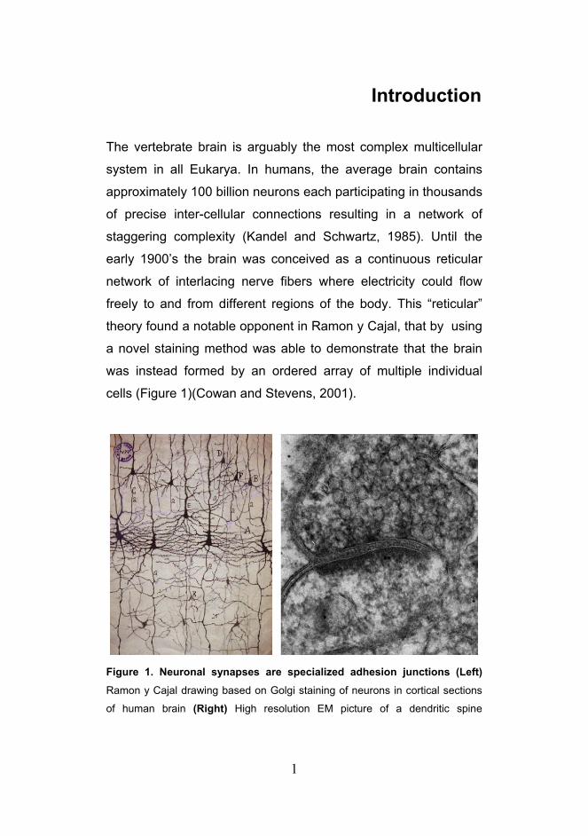
cells (Figure 1)(Cowan and Stevens, 2001).
Figure 1. Neuronal synapses are specialized adhesion junctions (Left)
Ramon y Cajal drawing based on Golgi staining of neurons in cortical sections
of human brain (Right) High resolution EM picture of a dendritic spine

2
highlighting the electron dense material in the synaptic cleft (courtesy of Cam
Robinson).
_______________________________________________________________
Crucial for the establishment of the new “neuron” theory
was the identification of the points of contact between different
neurons, sub-micron sized structures named synapses. From a
structural perspective neuronal synapses are highly specialized
adhesion junctions, whose function is to ensure the proper
transfer of information between neurons (Figure 1).
Morphologically, synapses resemble tight junctions with closely
juxtaposed membranes coated with electron dense material
(Stevenson and Keon, 1998). These correspond to the
presynaptic active zone of neurotransmitter release where
synaptic vesicles fuse and release their cargo and the
postsynaptic region where neurotransmitter receptors and
signaling molecules are enriched (Garner et al., 2000). In
excitatory synapses of the central nervous system (CNS) this last
region has a characteristic dense thickening called the
postsynaptic density (PSD) (Sheng, 2001). Between the pre and
postsynaptic membranes lies the synaptic cleft which performs
both mechanical and signaling functions (Garner et al., 2000). In
terms of signaling, the synaptic cleft is the region where
neurotransmitters and other signaling molecules diffuse and
effectively transduce information between the two cells.
Mechanically, the cleft orients and maintains the relative
apposition between the two synaptic plasma membranes.
Interestingly, regardless of their shape or size the tight apposition
between pre and postsynaptic elements is observed in all mature
synapses of the brain (Lisman and Harris, 1993). This
3
observation long suggested the presence of strong trans-synaptic
adhesive elements that would maintain the juxtaposition of both
synaptic sides. Indeed, ultrastructural analysis of neuronal
synapses revealed that the synaptic cleft is not empty, but is
instead filled with electron dense material (Gray, 1959), which is
now recognized to be composed mainly by highly glycosylated
membrane proteins and synaptic cell adhesion molecules
(CAMs).
Cell adhesion molecules are transmembrane proteins that
span across the synaptic cleft and undergo homophilic and
heterophilic interactions (Gerrow and El-Husseini, 2006).
Importantly, the formation of a mature synapse, or
synaptogenesis, is a multi-step process that gets initiated with
the transient adhesive contact between two neurons. This initial
contact is followed by the establishment of stable sites of cell-cell
contact and subsequent recruitment of scaffolding proteins,
which in turn contributes for the stabilization of neurotransmitter
receptors, voltage-gated ion channels, and various second-
messenger signaling molecules (Garner et al., 2002). Hence,
CAMs are key structural players in synapses that provide
anchoring points to intracellular cytoskeletal and scaffolding
elements, and extracellular trans-synaptic adhesive interactions.
Early studies in synaptosomes clearly demonstrated that
the adhesive apparatus of excitatory synapses is extremely
strong. These structures are pinched-off nerve endings that form
spontaneously upon homogenization of brain tissue and retain
the postsynaptic density and part of the postsynaptic plasma
membrane. Notably, synaptosomes are stable in solution for

4
days, even in complete absence of ATP (Gray and Whittaker,
1962). Furthermore, the association between pre and
postsynaptic sites resists the presence of high salt
concentrations and even urea (Cotman and Taylor, 1972).
Together, this indicates that the trans-synaptic adhesive complex
is extremely strong and thermodynamically stable. Yet, neuronal
synapses distinguish themselves from other cellular adhesive
specializations for their capacity to undergo extremely fast
functional and structural changes in response to brief stimuli (Citri
and Malenka, 2008; Trachtenberg et al., 2002). The activity-
dependent modification of synaptic properties, or synaptic
plasticity, underlies the capacity of neuronal networks to adapt to
changes in the environment and effectively process and transmit
information. However, this highly dynamic nature of synapse
remodeling is in stark contrast with the structural stability of the
synaptic adhesive complex observed in synaptosomes. This
suggests that the remodeling or disassembly of synaptic
structures requires the involvement of active processes.
However, the cellular and molecular basis for adhesive
disassembly and its potential function in regulating synaptic
transmission is still unknown and many questions remain about
how nanomolar high-avidity adhesive interactions are uncoupled
across the synaptic cleft in contexts of acute synaptic plasticity or
elimination.
There are numerous forms of synaptic plasticity that can
induce both short-term (ranging from milliseconds to several
minutes) (Zucker and Regehr, 2002) to long-term (ranging from
hours to years) changes in synaptic properties (Citri and
Malenka, 2008). The two most widely studied forms of long-term

5
plasticity, long-term potentiation (LTP) and long-term depression
(LTD), are thought to represent the cellular correlate of learning
and memory, and can have different expression mechanisms
depending on the neuronal circuits in which they operate (Citri
and Malenka, 2008; Malenka and Bear, 2004). Other forms of
plasticity act on a much broader regulatory scale. For example,
homeostatic synaptic plasticity serves as a negative feedback
mechanism in response to global changes in neuronal network
activity, resulting in a compensatory and uniform scaling of all
synaptic strengths (Pozo and Goda, 2010; Turrigiano, 1999,
2008).
Many synaptic CAMs have important roles in the
development and maturation of synapses. Such examples
include Neurexins and Neuroligins (Sudhof, 2008), Ephs and
ephrins, the immunoglobulin superfamily adhesion molecules,
cadherins (Dalva et al., 2007) and integrins (McGeachie et al.,
2011). Interestingly, these same classes of proteins are also
involved in the regulation of synaptic transmission in mature
stages. For instance, blockade of extracellular N-cadherin
adhesion with antibodies or peptides impairs LTP at Schaeffer
collateral–CA1 synapses without affecting basal synaptic
properties (Tang et al., 1998). Moreover, pharmacological
induction of LTP upregulates N-Cadherin expression and
increases the number of cadherin-positive synaptic puncta
(Bozdagi et al., 2000). N-Cadherin localization and dimerization
are in turn regulated by NMDAR activation, suggesting that
Cadherin based adhesion can be dynamically regulated by
activity-induced mechanisms (Tanaka et al., 2000). Interestingly,
N-cadherin can also regulate presynaptic function and short-term
6
plasticity as described in studies using embryonic stem cell-
derived neurons from N-cadherin-null mice (Jungling et al.,
2006). EphB receptors directly interact with NMDARs
extracellularly (Dalva et al., 2000) and regulate receptor function
and localization via intracellular enzymatic activity. EphBs also
regulate the localization of AMPARs (Kayser et al., 2006)
although the consequences of this regulation still require further
inquiry. However, several studies have now implicated Eph
receptors in several forms of hippocampal synaptic plasticity
(Grunwald et al., 2001; Henderson et al., 2001). Multiple lines of
evidence also implicate NCAM, a member of the immunoglobulin
superfamily of adhesion molecules, in the regulation of synaptic
function (Luthl et al., 1994; Venero et al., 2006). Initial studies
using blocking antibodies or synthetic peptides to inhibit NCAM-
mediated adhesion found normal basal synaptic transmission but
reduced LTP in the hippocampus (Luthl et al., 1994; Ronn et al.,
1995). In addition, NCAM null mice show hippocampus-
dependent long term memory defects (Bukalo et al., 2004;
Cremer et al., 1994) and impairment of LTP expression in CA1
area (Muller et al., 2000; Muller et al., 1996). Another class of
synaptic adhesion molecules implicated in synaptic plasticity is
the integrin protein family. Impairment of integrin function using
blocking peptides in acute hippocampal slices compromised the
late phase of LTP, which decayed back to baseline levels after
15–30 min of induction, while the baseline transmission was not
affected (Staubli et al., 1990). These results provided the first
evidence that the integrins have a role in the stabilization of LTP.
This was further confirmed by subsequent studies based on
genetic ablation of several integrin subunits (Chan et al., 2003).
In addition, Neuroligins, postsynaptic adhesion molecules that

7
were initially thought to play an important role during
synaptogenesis (Chih et al., 2005; Graf et al., 2004; Scheiffele et
al., 2000), have recently been implicated as critical regulators of
synaptic function (Chubykin et al., 2007; Futai et al., 2007;
Varoqueaux et al., 2006) and shown to be important for LTP
expression in the amygdala (Jung et al., 2010; Kim et al., 2008).
Together, these studies clearly demonstrate that cell adhesion
molecules play important roles in the regulation of mature
synapses. Hence the elucidation of the molecular mechanisms
capable of acutely regulating CAMs at synapses is an important
step to understand how these structural elements are regulated
in the context of rapid synaptic remodeling.
Interestingly, several synaptic CAMs have been shown to
undergo regulated ectodomain shedding. For example,
NCAM isoforms can undergo proteolytic cleavage via a
disintegrin and metalloprotease (ADAM) family of proteases,
resulting in soluble extracellular and intracellular domain
fragments (Diestel et al., 2005; Hinkle et al., 2006; Hubschmann
et al., 2005). Similarly, ephrins are cleaved by ADAM10 in
response to Eph receptor binding (Janes et al., 2005), while
EphB2 receptors themselves are cleaved by MMP2/MMP9 in
response to ephrin B2 ligation (Lin et al., 2008). Moreover,
ADAM10 also cleaves N-Cadherin via its metalloproteinase
domain and is responsible for initial step of N-cadherin proteolytic
processing (Reiss et al., 2005; Uemura et al., 2006). This
mechanism also generates extracellular soluble N-terminal
fragments (NTFs) and intracellular C-terminal fragments (CTFs).
Together these results suggest that ectodomain shedding is an
important regulatory mechanism capable of controlling cell

8
adhesion molecules expressed at the plasma membrane.
Moreover, given its acute and irreversible effects, proteolytic
cleavage is a plausible mechanism for CAM regulation at
synapses during synaptic plasticity. A recent study has employed
the application of tissue inhibitor of metalloproteinase-1 (TIMP-1),
an ADAM10 inhibitor, and a cell-permeable peptide capable of
interfering with ADAM10 synaptic localization and activity to
study the effects of N-Cadherin shedding at synapses. ADAM10
inhibition using these methods decreased N-cadherin cleavage,
induced a significant increase in size of dendritic spines and
potentiated AMPAR currents (Malinverno et al., 2010). However,
ADAM10 is capable of targeting several proteins (Janes et al.,
2005; Reiss et al., 2005) and TIMP-1 blocks multiple proteases
including for example, MMP9 (Ethell and Ethell, 2007). Hence
these results, despite showing that ADAM10 activity causes
changes in synaptic properties, are inconclusive when it comes
to providing information about the specific effects of N-Cadherin
shedding at synapses. In fact, this highly redundant nature of
ADAM and MMP activity has undermined the study of adhesion
molecule shedding and its consequences in synapse maturation
and function. Although manipulations of adhesion molecule levels
and binding properties can alter synaptic transmission and
influence synaptic plasticity, there is virtually no information on
the consequences of acute shedding of trans-synaptic adhesion
in response to neuronal activation.
This thesis is focused on the regulation of Neuroligins at
synapses and describes a previously unknown mechanism of
trans-synaptic signaling whereby synaptic activity induces acute
proteolytic cleavage of Neuroligin-1 (NLG1), which in turn causes

9
a direct reduction of synaptic transmission by decreasing
presynaptic function. The first chapter describes the initial
observation that cleavage of NLG1 occurs in response to
neuronal depolarization and is the major form of NLG1 regulation
under these conditions. Moreover, I also present data showing
that NLG1 cleavage occurs over development and is upregulated
in the visual cortex in response to sensory experience during
early development. Chapter 2 is centered on the characterization
of the signaling mechanisms regulating NLG1 cleavage using a
newly developed biochemical method based on surface
biotinylation. Using this technique I found that NLG1 cleavage is
bi-directionally regulated by activity and is mediated by NMDAR
and CaMK signaling. Moreover, I also identified the region where
NLG1 is cleaved and that the protease involved in activity
dependent NLG1 cleavage in vitro and in vivo is the Matrix
Metalloprotease-9 (MMP9). To finalize, in Chapter 3, I describe
the development of a new technique that allows the cleavage of
any transmembrane protein in a specific and temporally
controlled manner. Using this novel approach in combination with
real time microscopy I show that acute cleavage of NLG1 causes
rapid destabilization of its presynaptic partner Neurexin-1β
(NRX1β), which in turn depresses synaptic transmission by
abruptly reducing presynaptic release probability.

10

11
Chapter 1
Acute regulation of synaptic Neuroligin-1 by Matrix-
Metalloprotease mediated ectodomain shedding Introduction In the mammalian brain, neuronal synapses are highly
specialized adhesion junctions maintained by a complex network
of adhesion molecules that span the synaptic cleft and juxtapose
the presynaptic active zone of neurotransmitter release and the
postsynaptic density (Dalva et al., 2007; Shapiro et al., 2007;
Yamagata et al., 2003). Among these, Neuroligins (NLGs) and
Neurexins (NRXs) have emerged as critical regulators of proper
circuit development and function (Sudhof, 2008).
Neuroligins are postsynaptic adhesion molecules that
interact with presynaptic Neurexins with nanomolar binding
affinities (Arac et al., 2007; Chen et al., 2008; Comoletti et al.,
2006; Ichtchenko et al., 1995; Song et al., 1999). Structurally,
NLGs are type I transmembrane proteins presenting a large
extracellular globular domain that undergoes Ca2+ dependent
dimerization, a highly glycosylated stalk domain and a shorter
cytoplasmic tail containing a PDZ binding domain (Ichtchenko et
al., 1995). In mammals, there are 4 genes expressing NLGs with
NLG3 and NLG4 localized in the X chromosome. NLGs are
alternatively spliced at a single canonical site (A) with NLG1
containing an extra splice site (B) (Ichtchenko et al., 1996).

12
Interestingly, each NLG isoform exhibits a specific pattern of
expression and subcellular distribution. In particular, NLG1 and
NLG2 are exclusively localized to excitatory and inhibitory
synapses, respectively (Graf et al., 2004; Song et al., 1999;
Varoqueaux et al., 2004), whereas NLG3 can be present in both
(Budreck and Scheiffele, 2007).
Neurexins were identified as receptors for α-latrotoxin
(Ushkaryov et al., 1992), a toxin present in the venom of the
black widow spider that induces massive release of
neurotransmitters (Ushkaryov et al., 2008). The mammalian
genome contains 3 NRX genes each encoding a long α-protein
and a shorter β-protein from independent promoters (Tabuchi
and Sudhof, 2002). Moreover, NRXs are highly polymorphic and
through extensive alternative splicing at 5 canonical sites have
the potential to generate over 3000 possible isoforms (Ullrich et
al., 1995). Interestingly, different NRX splice variants are
differentially expressed in specific neuronal types (Ullrich et al.,
1995) and have different binding affinities to different NLG
isoforms (Chih et al., 2006) (Comoletti et al., 2006) (Ushkaryov
and Sudhof, 1993) indicating that NRXs may provide a structural
adhesive code at synapses.
NLGs are sufficient to induce functional maturation of
presynaptic terminals (Dean et al., 2003; Prange et al., 2004;
Scheiffele et al., 2000; Wittenmayer et al., 2009), and transgenic
expression of NLG1 results in extended active zones and
increased reserve pool size of synaptic vesicles (Dahlhaus et al.,
2010). Reciprocally, NRXs trigger the aggregation and clustering

13
of postsynaptic components (Graf et al., 2004; Heine et al., 2008;
Nam and Chen, 2005) and regulate NMDA receptor function
trans-synaptically (Kattenstroth et al., 2004). This synaptogenic
potential of NLGs and NRXs is due in part to the fact that both
these proteins contain intracellular domains that interact with
important synaptic scaffold proteins such as PSD95 and CASK
(Hata et al., 1996; Irie et al., 1997). Adhesion between NLGs and
NRXs thus provides a direct structural bridge between pre- and
postsynaptic scaffolding machinery.
The ability to induce trans-aggregation of synaptic
components suggested that NLGs and NRXs were critical
mediators of synapse formation. This hypothesis was further
supported by in vitro studies showing that NLG expression levels
are correlated with the number of synapses generated during
development (Chih et al., 2005; Dean et al., 2003; Graf et al.,
2004; Levinson et al., 2005; Prange et al., 2004). However,
despite presenting severe deficits in synaptic transmission,
NLG1-3 triple knockout (KO) mice exhibit normal synaptogenesis
(Varoqueaux et al., 2006). These results demonstrated that in
vivo, NLGs are not required for the initial stages of synapse
formation but are instead critical regulators of synaptic function.
In addition to these results, it was later found that NLG1
overexpression in dissociated neuronal cultures increases AMPA
and NMDA excitatory postsynaptic currents (EPSCs) (Chubykin
et al., 2007). Moreover, recent studies have also shown that
NLG1 is required for LTP in the amygdala (Jung et al., 2010; Kim
et al., 2008), which reinforces the notion that NLG1 is an
important functional component of mature synapses and is able
to modulate synaptic transmission in adult stages.

14
Interestingly, regardless of no apparent change in the
number of total synapses generated, NLG1-3 triple KO neurons
present reduced evoked excitatory postsynaptic currents
(eEPSCs) and decreased miniature EPSCs (mEPSCs)
frequency, which is consistent with impaired presynaptic function
(Varoqueaux et al., 2006). Moreover, overexpression of NLG1 in
hippocampal slices and cultured neurons results in increased
release probability through a NRX-dependent mechanism (Futai
et al., 2007; Ko et al., 2009b; Stan et al., 2010), suggesting that
NLG1 modulates presynaptic function trans-synaptically.
Consistent with this hypothesis, neurons lacking αNRX1-3 exhibit
deficits in synaptic transmission due to severe impairment of N-
type Ca2+ channel function (Missler et al., 2003), while disruption
of endogenous NLG-NRX interactions with soluble Fc-NRX
fragments decreases mEPSC frequency and release probability
(Levinson et al., 2005). Taken together, these results suggested
a new model by which NLGs and NRXs validate and stabilize
synapses during development by modulating synaptic
transmission at the presynaptic level.
The importance of the NRX-NLG trans-synaptic complex
for normal brain development is emphasized by the strong
association of several NLG and NRX mutations with autistic
spectrum disorders (ASDs) (Sudhof, 2008). A typical feature of
ASDs is that they usually affect the brain during the second or
third year of life, which is a period of extensive synapse
remodeling and circuit refinement in humans (Lord et al., 2000;
Pardo and Eberhart, 2007). The association of NLGs and NRXs
with ASDs may then reflect an important role of these molecules
during the synaptic activity-dependent maturation processes that

15
occur during early development (Hensch, 2004, 2005b). As such,
the elucidation of the molecular and cellular pathways regulating
NLGs may provide new insights regarding the function of these
proteins in the etiology of ASDs and in broader terms, the role of
adhesion molecules during the functional maturation of neuronal
circuits. Despite the numerous studies addressing how NLG
affects synaptic properties, little is known about how endogenous
NLGs are themselves regulated at synapses. It is also still
unclear if NLGs are stable structural elements in synapses or if
they can be regulated by changes in neuronal activity.
Here, I address how NLG1 is affected in response to
neuronal depolarization. Based on a combination of biochemical
methods and immunocytochemistry I show that synaptic NLG1
can be acutely regulated by changes in neuronal activity through
Matrix Metalloprotease dependent ectodomain shedding.
Moreover, NLG1 cleavage occurs in vivo, is regulated over
development and is modulated by sensory experience during
critical periods of circuit maturation.

16

17
Results To determine the effect of neuronal activity on synaptic NLGs, we
treated dissociated hippocampal cultures at 21 days in vitro
(DIV21) with 30 mM KCl for 2 h, a paradigm that elicits robust
depolarization and is widely used as a model for activity-
dependent neuronal signaling (Kim et al., 2010; Murase et al.,
2002; Sheng et al., 1990).
Figure 1. Neuronal activity triggers loss of Neuroligins from synapses. (A)
Hippocampal neurons (DIV21) were incubated in Neurobasal media (Control) or
Neurobasal media supplemented with 30 mM KCl (KCl) for 2 h, fixed, and
immunolabeled for endogenous PSD-95 and pan-NLG (NLG1-4). Neurons were
transfected with mCherry as a cell fill. Solid arrows show decreased NLG1-4
labeling at PSD-95 positive synapses following KCl incubation. Open arrows
depict synaptic NLG1-4 labeling under control conditions. Scale bar, 5 µm. (B)
Data indicate means ± SEM of NLG1-4 fluorescence intensity in PSD-95
positive dendritic spines (synaptic NLG1-4) or (C) over the entire neuron (total
NLG1-4) normalized to controls. Control, n = 435 spines from 8 neurons; KCl, n
= 546 spines from 9 neurons. * p < 0.01.

18
The level of endogenous NLGs at synapses was
assessed by immunocytochemistry using a pan-NLG antibody
targeted against the C-terminal domain of NLG1-4 together with
immunolabeling for PSD-95 to identify excitatory synapses.
Following KCl incubation, pan-NLG labeling at excitatory
synapses was reduced by 30 ± 5% (Figures 1A and 1B)
indicating that synaptic levels of NLGs are reduced after
increased activity. Interestingly, we also observed a similar
change in total average fluorescence of pan-NLG signal across
neurons (29 ± 1%) indicating that the loss of NLG signal from
PSD95 positive sites is not due to a redistribution of NLGs to
extra synaptic sites, but due to overall protein degradation
(Figure 1C). Among the different NLG isoforms, NLG1
exclusively partitions to and regulates excitatory synapses
(Chubykin et al., 2007; Graf et al., 2004; Ko et al., 2009b; Song
et al., 1999). Hence, the extensive loss of pan-NLG fluorescent
signal from PSD95 positive puncta prompted us to test whether
neuronal depolarization depletes NLG1 from the glutamatergic
synapses. Due to the lack of NLG1-specific antibodies suitable
for immunocytochemistry, we performed the same experiments
and measured NLG1 levels in biochemical fractions of PSDs
from dissociated cortical cultures treated with KCl and
Neurobasal media alone (Ehlers, 2003) using an antibody
targeted against the extracellular N-terminal domain (4C12).

19
Figure 2. Neuronal activity triggers loss of NLG1 from excitatory
synapses. (A) Immunoblot analysis of NLG1 and β-Tubulin in membrane
fractions isolated from control or KCl-treated cortical neurons (DIV21). EXT,
whole cell extract; SPM, synaptic plasma membrane; PSDI,II, III; sequential
postsynaptic density fractions. Note that 4-fold less protein by mass was loaded
in PSD fraction lanes. See Experimental Procedures for details. (B) Data
indicate means ± SEM of NLG1 proteins levels in indicated fractions relative to
the total extract control. n = 3, *p < 0.05, **p<0.005, ***p<0.001.
Immunoblot analysis of isolated fractions revealed an
extensive enrichment of NLG1 in the PSD (Figure S1).
Consistent with the immunocytochemistry data using the pan-
NLG antibody, KCl depolarization resulted in a significant loss
(48 ± 2%) of NLG1 from total neuronal extracts (Figures 2A and
2B). This reduction in NLG1 was also observed in the synaptic
plasma membrane (SPM) and PSD fractions (31 ± 2% decrease
in SPM; 24 ± 7% in PSDI; 45 ± 5% in PSDIII) and was
particularly pronounced in PSDII fractions (73 ± 5% decrease),
where NLG1 is most highly enriched (Figure S1). Thus, together
with the immunocytochemistry data using pan-NLG antibody,

20
these results indicate that the neuronal levels of NLG1 are
extensively reduced following 2h of neuronal depolarization.
Several synaptic membrane proteins and receptors are
degraded in response to changes in neuronal activity through the
lysosomal pathway upon regulated endocytosis (Ehlers, 2000).
To address whether the decrease in NLG1 levels is due to
increased internalization and lysosomal degradation, we
measured internalization rates of NLG1 in dissociated cortical
cultures during KCl depolarization using a biochemical strategy
based on surface biotinylation of proteins (Ehlers, 2000) (Figure
3A). In these experiments, neurons were pre-incubated with
leupeptin for 1 h to inhibit lysosomal proteolysis.
Figure 3. Neuronal depolarization does not increase NLG1 internalization. (A) Surface biotinylation based assay of endocytosis performed on cultured
cortical neurons. Surface proteins are covalently labeled with 1mg/ml Sulfo-
NHS-SS-Biotin for 10min, incubated for 1 h or 2 h at 37°C in neurobasal media

21
(Ctrl) or in media supplemented with 30 mM KCl (KCl), and subsequently
treated with 50mM Glutathione pH8 for 30min to remove surface biotin label (B) Immunoblot of GluA1 and NLG1 indicate that GluA1 internalization was
increased by KCl while NLG1 was not. (C) Quantitative analysis of GluA1 and
(D) NLG1 internalization over time. Immunoblot values were compared to a
calibration standard of total surface protein at time zero (Surf) to quantify
percent internalization.
_________________________________________________________
Under basal conditions 5.3 ± 1.2% of surface NLG1 was
internalized over 2h (Figures 3B and 3D). This low internalization
rate was unaltered by KCl incubation (5.7 ± 0.8% of surface
NLG1) indicating that KCl-induced NLG1 loss is not due to
increased internalization. The GluA1 AMPA receptor was used
as a positive control and exhibited a marked increase in
internalization under KCl stimulation (Figure 3B and 3C, 7.4 ±
2.4% of total surface protein internalized in control conditions; 25
± 4.7% in KCl), similar to previous reports (Ehlers, 2000). These
results suggested that KCl induced NLG1 loss is not caused by
protein internalization and lysosomal degradation.
Moreover, to further address whether if this effect was
sensitive to proteasome or lysosome inhibition we treated DIV21
dissociated cortical cultures with 30mM KCl for 2h in the
presence of proteosome inhibitor MG132 and/or lysosomal
enzyme inhibitor leupeptin, respectively, and measure how NLG1
degradation was affected.

22
Figure 4. Activity dependent loss of NLG1 is mediated by Metalloproteases. (A) Immunoblot analysis of NLG1 in total lysates from
cortical cultures following 2 h incubation in neurobasal medium (Ctrl) or medium
supplemented with 30 mM KCl alone (KCl) or with MG132 (10 µM), leupeptin
(200 μM), leupeptin plus MG132 (Leup/MG) or GM6001 (10 µM). Note that
GM6001 prevents KCl-induced loss of total NLG1. (B) Data indicate means ±
SEM of total NLG1 levels under the indicated conditions. n = 3, *p < 0.05.
As previously shown (Figure 2) depolarization induced by
KCl incubation resulted in a 48 ± 3% reduction in total NLG1
levels. Interestingly, this effect was unaffected by proteasome
inhibition (50 μM MG132), blockade of lysosomal degradation
(200 µM leupeptin), or both together (46 ± 7% decrease with
MG132, 52 ± 6% with leupeptin, 47 ± 4% with both; Figures 4A
and 4B). These results indicated that NLG1 degradation was
occurring through a different degradation pathway and prompted
us to test an alternative hypothesis. Several membrane proteins
are targeted and degraded by Matrix Metalloproteases, which are
a large family of secreted proteases. Indeed, incubation with the
broad-spectrum Matrix Metalloprotease (MMP) inhibitor GM6001
(10 μM) abolished the KCl-induced loss of NLG1 (102.9 ± 1.1%
of control; Figures 4A and 4B). This important result indicated
that NLG1 can be cleaved by MMPs and that MMP-dependent
23
proteolysis is the major regulatory mechanism mediating the
rapid and extensive degradation of NLG1 in response to KCl
depolarization.
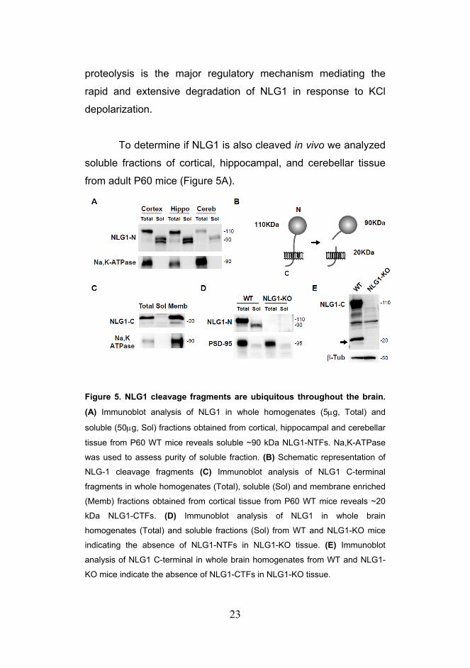
To determine if NLG1 is also cleaved in vivo we analyzed
soluble fractions of cortical, hippocampal, and cerebellar tissue
from adult P60 mice (Figure 5A).
Figure 5. NLG1 cleavage fragments are ubiquitous throughout the brain.
(A) Immunoblot analysis of NLG1 in whole homogenates (5μg, Total) and
soluble (50μg, Sol) fractions obtained from cortical, hippocampal and cerebellar
tissue from P60 WT mice reveals soluble ~90 kDa NLG1-NTFs. Na,K-ATPase
was used to assess purity of soluble fraction. (B) Schematic representation of
NLG-1 cleavage fragments (C) Immunoblot analysis of NLG1 C-terminal
fragments in whole homogenates (Total), soluble (Sol) and membrane enriched
(Memb) fractions obtained from cortical tissue from P60 WT mice reveals ~20
kDa NLG1-CTFs. (D) Immunoblot analysis of NLG1 in whole brain
homogenates (Total) and soluble fractions (Sol) from WT and NLG1-KO mice
indicating the absence of NLG1-NTFs in NLG1-KO tissue. (E) Immunoblot
analysis of NLG1 C-terminal in whole brain homogenates from WT and NLG1-
KO mice indicate the absence of NLG1-CTFs in NLG1-KO tissue.

24
Interestingly, several bands of approximately 90 kDa were
recognized by the N-terminal NLG1 antibody in the soluble
fractions of extracts of different brain regions, suggesting that
multiple NLG1 extracellular cleavage fragments are generated in
vivo. A logical outcome of the ectodomain shedding of NLG1 is
the generation of corresponding intracellular C-terminal
fragments of approximately 20kDa (Figure 5B). Analysis of
mouse cortical fractions using an antibody targeted against the
C-terminal domain of NLG1 (NLG1-C) revealed in fact, multiple
membrane bound bands of approximately 20KDa, a size
consistent with the predicted mass based on the size of the N-
terminal cleavage fragments (Figures 5B and 5C). To further
confirm these findings we expressed a dual labeled version of
NLG1 with GFP tagged to the N-terminus and HA tagged to the
C-terminus (GFP-NLG1-HA) in COS7 cells (Figure S2).
Immunoblot analysis of cell extracts using an anti-HA antibody
revealed the presence of similar ~20 kDa bands that were absent
in extracts of COS7 cells transfected with GFP-NLG1.
To exclude possible nonspecific interactions recognized
by the NLG1 antibodies, we tested whether similar bands were
detected in extracts of NLG1 null mice (Varoqueaux et al., 2006).
Both 110 kDa full form and 90 kDa NLG1 N-terminal fragments
(NLG1-NTFs) were absent from NLG1 KO brain extracts and
respective soluble fraction (Figure 5D). Similarly, NLG1 C-
terminal fragments (NLG1-CTFs) were absent from NLG1 KO
brain extracts (Figure 5E). Together, these results indicate that
the NTFs and CTFs recognized by the NLG1 antibodies are
indeed cleavage fragments of NLG1 and not an artifact due to
non-specific antibody binding.
25
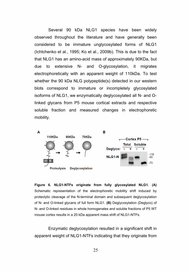
Several 90 kDa NLG1 species have been widely
observed throughout the literature and have generally been
considered to be immature unglycosylated forms of NLG1
(Ichtchenko et al., 1995; Ko et al., 2009b). This is due to the fact
that NLG1 has an amino-acid mass of approximately 90KDa, but
due to extensive N- and O-glycosylation, it migrates
electrophoretically with an apparent weight of 110kDa. To test
whether the 90 kDa NLG polypeptide(s) detected in our western
blots correspond to immature or incompletely glycosylated
isoforms of NLG1, we enzymatically deglycosylated all N- and O-
linked glycans from P5 mouse cortical extracts and respective
soluble fraction and measured changes in electrophoretic
mobility.
Figure 6. NLG1-NTFs originate from fully glycosylated NLG1. (A)
Schematic representation of the electrophoretic mobility shift induced by
proteolytic cleavage of the N-terminal domain and subsequent deglycosylation
of N- and O-linked glycans of full form NLG1. (B) Deglycosylation (Deglyco) of
N- and O-linked residues in whole homogenates and soluble fractions of P5 WT
mouse cortex results in a 20 kDa apparent mass shift of NLG1-NTFs.
Enzymatic deglycosylation resulted in a significant shift in
apparent weight of NLG1-NTFs indicating that they originate from

26
glycosylated forms of NLG1 (Figures 6A and 6B). Importantly,
whereas deglycosylation of total cortical extracts collapsed NLG1
to both a 90 kDa and 70 kDa species corresponding to full-length
and N-terminal cleaved NLG1, respectively, deglycosylation of
soluble fractions produced only a 70 kDa species (Figures 6B),
confirming that all soluble NLG1-NTFs correspond to cleavage
fragments of mature, fully glycosylated NLG1.
Together, the data presented here indicates that overall
NLG1 levels are modulated by neuronal activity via MMP
dependent ectodomain shedding. Interestingly, previous studies
have implicated tissue plasminogen activator (tPA) dependent
proteolytic mechanisms as critical processes regulating synapse
remodeling during early development (Mataga et al., 2004;
Mataga et al., 2002). Given the involvement of NLG1 in synapse
maturation we also characterized the developmental profile of
NLG1 cleavage and addressed if this mechanism is regulated
over brain development. Indeed, immunoblot analysis of mouse
cortical fractions at various time points from birth to adulthood
(P1-P60, Figure 7A) revealed that NLG1-NTFs are present
throughout development and are particularly enriched during
early development (P1-P7) where they are as abundant as the
full form protein (Figures 7A and 7B). This indicated that NLG1
cleavage is not only regulated by neuronal activity changes
during mature stages but is also an important mechanism during
early development.

27
Figure 7. NLG1-NTFs are highly abundant during early development and decrease with age. (A) Developmental profile of NLG1-NTFs from WT mice
cortical extracts. Postnatal days (P) are shown at the top. Total, 10 μg whole
cortical homogenates; Sol, 50 µg soluble fractions from mouse cortex. (B)
Analysis of the relative abundance of full form NLG1 and NLG1-NTFs present in
whole cortical extracts from P1 and P60 mice. Note that NLG1 cleavage
products are more abundant during early developmental stages. The graph
plots signal intensity along the lines shown on the blots on the left.
The refinement of synaptic connections by activity
dependent processes during early development is crucial for the
proper formation of functional neuronal circuits (Hensch, 2004,
2005b). To address if NLG1 cleavage is regulated by sensory
experience during critical periods of development we subjected
mice to 5 days of dark rearing (DR) and subsequently re-exposed
them to light for a brief period of 2 hours (DR+2hL). This protocol
induces rapid synaptic remodeling in the primary region of the
visual cortex and results in extensive molecular functional and
structural synaptic changes (Brakeman et al., 1997; Cotrufo et
al., 2003; Philpot et al., 2001; Tropea et al., 2010; Viegi et al.,
2002). Dark rearing of mice for 5 days resulted in a significant
decrease in the levels of NLG1-NTFs present in V1 cortex

28
compared with control animals (LR) reared in normal light cycle
(DR group - 0.71 ± 0.04 NLG1-NTFs normalized to LR group)
(Figures 8B and 8C). Notably, 2 hours of re- exposure to light
after dark rearing caused a robust increase in NLG1 cleavage
(1.51 ± 0.14 compared with LR group).
Figure 8. NLG1 cleavage is regulated during the critical period of visual cortex development. (A) Schematic representation of the experimental
paradigm. Mice are reared in normal 12h day/night cycle (LR) or submitted to
dark rearing for 5 days from P21-P26 (DR). DR+2hL indicates experimental
group submitted to a brief 2h light exposure following light deprivation. (B) immunoblot of P26 mouse primary visual cortex of animals reared under
different conditions. Note that 2h of light exposure after dark rearing induces
increase in NLG1-NTF levels. (C) Data indicate means ± SEM of NLG1-NTFs
levels under the indicated conditions normalized to LR animals. n = 12 animals
per condition, *p < 0.005.
These results indicate that NLG1 cleavage is acutely
regulated by changes in sensory experience in vivo and suggest

29
that this mechanism may be involved in the shaping of neuronal
circuits during development. Taken together, the results
presented here unveil a new activity dependent form of post-
translational regulation of NLG1 that is ubiquitous in the brain
and present throughout all developmental stages.

30

31
Discussion
Although highly studied for its role in synapse maturation and
stabilization, it has been unclear whether the NLG-NRX trans-
synaptic complex undergoes dynamic regulation and
dissociation. Here we have shown that acute depolarization leads
to a robust decrease in synaptic NLG1 levels. MMP inhibitors but
not lysosome or proteasome inhibitors block this effect, indicating
that under these conditions NLG1 is predominantly regulated by
proteolytic cleavage. The extent of NLG1 loss observed using
biochemical methods (Figures 2 and 4) is somewhat greater than
that observed by immunostaining (Figure 1A). This disparity may
be attributed to the broad specificity of the pan-NLG antibody
used for immunolabeling or to the fact that it targets the C-
terminal domain of NLGs, an epitope that may be differently
regulated after ectodomain shedding. In addition, the presence of
a residual C-terminal fragment (CTF) of cleaved NLG-1 (Figure
5C) raises the possibility that further intracellular processing of
NLG1 may trigger downstream signaling cascades similar to
what has been described for other membrane proteins such as
Notch and E-Cadherin (Marambaud et al., 2002; Mumm et al.,
2000). Moreover, these results suggest that caution is warranted
in interpreting changes in Neuroligin synaptic abundance
measured exclusively by an N-terminal or C-terminal antibody.
The N-terminal AChE homolog domain of Neuroligins
binds the extracellular domain of Neurexins and this interaction
has, to date, been thought to occur exclusively through
membrane-associated Neuroligin (Arac et al., 2007; Chen et al.,
2008; Comoletti et al., 2006). Here we have shown that the

32
Neurexin-binding domain of NLG-1 is released as a soluble N-
terminal fragment (NTF) and is abundant in brain. Glutamatergic
synapses are densely packed in brain tissue in vivo and, much
like other soluble factors such as brain-derived neurotrophic
factor (BDNF) (Desai et al., 1999; Rutherford et al., 1998), NLG1-
NTFs could potentially mediate heterosynaptic effects by
diffusing to neighboring synapses. Intriguingly, hetereosynaptic
depression of neurotransmitter release has been reported at
several CNS synapses, although the molecular basis for such
depression has not always been clear (Chistiakova and
Volgushev, 2009; Grover and Teyler, 1993; Huang et al., 2008).
Indeed, cleavage of the Neural Cell Adhesion Molecule (NCAM)
originates soluble extracellular fragments (NCAM-EC) that induce
several structural and functional defects in cortical neurons
(Brennaman and Maness, 2008; Pillai-Nair et al., 2005).
Moreover, cleavage of E-cadherin by MMP3 and MMP7 results in
disrupted aggregation and cell migration in neighboring cells due
to diffusion of soluble cleavage products (Lochter et al., 1997;
Noe et al., 2001). We further note that several studies have
employed exogenous application of recombinant soluble NRX
extracellular fragments as a means to disrupt Neuroligin
interactions (Levinson et al., 2005; Scheiffele et al., 2000) raising
the possibility that NLG-NTFs present in soluble brain fractions
may exert similar functions. Alternatively, release of soluble NLG-
NTFs could act to locally suppress pro-synaptogenic NLG-NRX
interactions during brain development, ensuring proper synapse
patterning and providing a potentially novel mechanism for
synapse competition.

33
Neuroligins have been strongly implicated in the
maturation of synapses during development. The regulation of
NLG1-NTF levels by sensory experience during the critical period
of primary visual cortex maturation indicates that NLG1 cleavage
is a physiological process regulated by activity in vivo and
suggests that this mechanism may be involved in the shaping of
neuronal circuits during early development. There is wide
evidence that neuronal circuits are shaped and refined by
sensory experience during critical periods of heightened plasticity
(Hensch, 2004, 2005b). During this process, single neurons tune
their functional properties in order to ultimately create stable and
functional neuronal networks (Hensch, 2004, 2005a). A key
requirement for this process is the establishment of the proper
balance between excitation and inhibition (Bavelier et al., 2010;
Hensch and Fagiolini, 2005; Hensch et al., 1998). Notably, the
relative levels of NLG1 and NLG2 determines the overall balance
between excitation and inhibition (Levinson et al., 2005; Prange
et al., 2004) suggesting that differential cleavage of different NLG
isoforms may contribute for the establishment of proper E/I ratio.
Moreover, manipulation of NLG levels during development
determines the overall number of synapses generated (Chih et
al., 2005; Dean et al., 2003; Graf et al., 2004; Levinson et al.,
2005; Prange et al., 2004) suggesting that NLGs may directly
regulate the stability of synapses. Our results indicate that NLG
cleavage is highly upregulated during the first postnatal week and
declines over time reaching a minimum at around 2 weeks of
age. Interestingly, this is the developmental period where more
dendritic spines are observed in neurons indicating that the
decrease in NLG1-NTFs is correlated with the stabilization
synapses during development. Moreover, previous studies have

34
shown that tissue plasminogen activator (tPA), a potent activator
of proteolytic cascades, is involved in the stabilization and
pruning of synapses during early development (Mataga et al.,
2004; Mataga et al., 2002). In addition, visual experience during
the critical period affects dendritic spine morphology in visual
cortex and leads to an increase in the fraction of thin spines and
filopodia (Tropea et al., 2010). Together, this raises the
interesting possibility that NLG cleavage may contribute for the
maturation of synapses during cortical development.
Moreover, manipulation of sensory experience during
critical period alters the ratio of NR2A/NR2B-only NMDARs layer
2/3 neurons of visual cortex (Philpot et al., 2001). The regulation
of this balance alters the metaplastic state of excitatory synapses
with NR2A increasing and NR2B decreasing the threshold for
LTP induction (Philpot et al., 2007). Interestingly, NLG1
overexpression increases the ratio of NMDAR/AMPAR currents
in cultured neurons (Chubykin et al., 2007). In addition, NLG1 KO
animals have lower NMDAR/AMPAR ratio and exhibit decreased
NR1 expression and NMDAR-mediated synaptic transmission
(Chubykin et al., 2007; Jung et al., 2010; Varoqueaux et al.,
2006), suggesting that NLG1 is important for recruitment of
NMDAR to postsynaptic sites. A recent study has also suggested
that NLG1 may induce clustering of NMDARs directly without the
presence of PSD95 (Barrow et al., 2009). Hence it is possible
that NLG cleavage may induce a shift in NMDAR subunits in
synapses, either by preferentially binding a particular type of
receptor or by enabling a structural remodeling of the
postsynaptic density that allows for a shift in synaptic receptors,
which subsequently could lead to synaptic plasticity.
35
Together the findings presented here describe a novel
post-translational mechanism that acutely regulates NLG1 in
synapses and that implicates this class of molecules in the
molecular mechanisms underlying critical period plasticity. Given
the strong association between several NLG and NRX mutations
with ASDs (Sudhof, 2008) the acute proteolytic regulation of
Neuroligins may provide novel insight into the pathophysiological
mechanisms and therapeutic strategies for synaptic dysfunction
in ASDs. More broadly, such proteolytic mechanism may provide
a general paradigm for regulation of cell-cell trans-synaptic
adhesion in the context of synaptic plasticity.

36

37
Methods
Reagents and Antibodies
Dissociated primary neuronal cultures were prepared from
hippocampi or cortex of embryonic day 18 or 19 Wistar rats.
Tissue was dissociated by enzymatic papain digestion followed
by brief mechanical trituration. Cultures were grown in
Neurobasal media supplemented with B27 and Glutamax. For
biochemical experiments 700k cortical cells were plated onto
60mm petri dishes. For immunocytochemistry, 100k hippocampal
cells were plated onto 12 well plates containing Poly-L-Lysine
coated 18mm glass coverslips. Plasmid transfection was done
using lipid mediated gene transfer using Lipofectamine 2000
(Invitrogen). Briefly, for each coverslip, 1μg total DNA was mixed
with 1μl Lipofectamine in 100 μl of Neurobasal media for 15min.
Following incubation time 500 μl of conditioned media was mixed
to the reaction and added to cell cultures for 30min. After this
period, lipofectamine containing media was removed and
replaced with a 1:1 mixture of conditioned media + fresh growth
media. COS7 cells were grown in DMEM (Invitrogen)
supplemented with 10% fetal bovine serum. Plasmid tranfections
were performed at approximately 70% confluency using 3 μg of
DNA and 3 μl of Lipofectamine per 60mm petri dish and let
express for 2 days.
Commercial antibodies used include N-terminal NLG1
antibody clone 4C12 (mouse, Synaptic Systems), C-terminal
NLG1 polyclonal antibody (rabbit, Synaptic Systems), PSD-95

38
(mouse, Chemicon), Na+,K+-ATPase (rabbit, Cell Signaling), GFP
(mouse, Millipore), HA.11 (mouse, Covance). Rabbit anti-
panNLG antibody used for immunocytochemistry was a gift from
Peter Scheiffele (Biozentrum).
Imaging
Confocal images of fixed samples were obtained using a Perkin
Elmer Ultraview spinning disc confocal microscope with either a
40x 1.3 N.A. objective or a 60x 1.4 N.A. objective. Images were
analyzed using Metamorph software (Molecular devices,
Universal Imaging Corporation). Maximum projections of z series
(0.5 mm steps) were used for quantification. For
immunocytochemistry, DIV21 hippocampal neurons were fixed in
4% paraformaldehyde/4% sucrose in PBS for 20 min,
permeabilized with -20ºC methanol for 10 min, and incubated
with indicated antibodies at 1:250 (panNLG) or 1:1000 (PSD-95)
dilution overnight at 4ºC. Quantification of integrated
fluorescence intensity at individual puncta was performed using
fixed region sizes. For PSD95 positive puncta quantification only
regions containing 3x background level of PSD95
immunofluorescence were considered for analyzes. For each
neuron the average fluorescence intensity for all puncta was
determined. For determination of total pan-NLG fluorescence cell
was traced based on mCherry cell fill and average intensity of
pan-NLG fluorescence was determined. Results depicted
correspond to the average of all neurons per condition.

39
Biochemical Analysis of Brain and PSD Fractions
Mouse brains or brain regions were homogenized in
homogenization buffer (4 mM HEPES, 0.32 M sucrose pH 7.4)
with protease and phosphatase inhibitors (Roche). Crude
membrane fractionation was performed by centrifugation at
150,000 x g for 30 min. High density cortical cultures were
incubated in Neurobasal media or media supplemented with 30
mM KCl for 2 h. Whole cell, synaptic plasma membrane, and
PSD fractions from cultured cortical neurons were prepared as
described previously (Ehlers, 2003) and immunoblotted for
proteins of interest. For PSD fractions, 5 μg of protein was
loaded per lane, while for remaining fractions 20 μg protein was
loaded. Deglycosylation was performed using an enzymatic
deglycosylation kit according to the manufacturer’s instructions
(Calbiochem). All protein concentrations were measured with Dc
protein assay (Bio-Rad).
Biotinylation-Based Internalization Assay
High-density cortical neuron cultures were incubated with 100
μg/ml of the lysosomal protease inhibitor leupeptin beginning 1 hr
prior to biotinylation with 1 mg/ml sulfo- NHS-SS-biotin (Ehlers,
2000). Leupeptin was present throughout all steps and
incubations except the 4ºC biotinylation reaction. Neurons were
then incubated at either 4ºC to block membrane trafficking or
37ºC for various times to allow endocytosis to occur. The
remaining surface biotin was cleaved by reducing its disulfide

40
linkage with glutathione cleavage buffer (50 mM glutathione in 75
mM NaCl and 10 mM EDTA containing 1% BSA and 0.075 N
NaOH) two times for 15 min each at 4ºC. Whole cell extracts
were prepared, and biotinylated proteins were precipitated
essentially as described (Ehlers, 2000). Biotinylated receptors
were detected by immunoblot (ECL Plus, Amersham), and
quantification was performed on an LAS-3000 gel reader
(Fujifilm), using Multigauge 3.0 software. The percent receptor
internalized was determined by measuring the band intensity
after 37ºC incubation, subtracting the nonspecific band intensity
obtained after 4ºC incubation (always <5%), and comparing to
the total surface receptor calibration curve.
Dark rearing experiments
Male and female mice were reared in a normal light dark cycle
(12 h light/12 h light) from birth until P26 (LR group) or were
transferred to a dark room in complete darkness at P21 (DR
group). Handling of animals in the dark room was done with the
aid of night vision goggles and infra-red light. Mice were all
sacrificed at P26 at the peak of the critical period of visual cortex
development (Gordon and Stryker, 1996). One experimental
group (DR+2hL) was re-exposed to light for 2 hours before being
euthanized. Animals were sacrificed after 2 hours of light
exposure and primary visual cortex was extracted, homogenized
in homogenization buffer (4 mM HEPES, 0.32 M sucrose pH 7.4)
containing protease and phosphatase inhibitors (Roche). Soluble
fraction was obtained by centrifugation at 150,000 x g for 30 min.
Protein concentration was determined using the Bradford reagent

41
(Bio-Rad) and 50μg of protein were loaded on western blot and
probed for NLG1-NTFs using a NLG1 specific antibody targeted
against the N-terminal domain (clone 4C12, Synaptic Systems).

42

43
Supplementary Figures Figure S1
Figure S1. Biochemical Fractionation of Cultured Cortical Neurons.
Shown are immunoblots of biochemical fractions from cortical neurons cultures.
EXT, whole cell extract; S1, supernatant; SPM, synaptic plasma membranes;
S3, synaptic vesicle fraction; PSDI, Triton-washed SPM pellet; PSDII, Triton-
washed PSDI pellet; PSDIII, Sarcosyl-washed PSDI pellet. Note that 4-fold less
protein by mass was loaded in PSD fraction lanes. See Chapter1 Experimental
Procedures for details.
Figure S2
Figure S2. Generation of NLG1-CTFs in COS7 cells.
Lysates from COS7 cells expressing GFP-NLG1 or GFP-NLG1-HA were
analysed by immunoblot using anti-GFP and anti-HA antibodies. Anti-HA
immunoblot revealed a fragment of approximately 20 kDa corresponding to a C-
terminal fragment.

44

45
Acknowledgements
For the realization of this work, Irina Lebedeva and Marguerita
Klein prepared the neuronal cultures from dissociated
hippocampus and cortex. The dark rearing experiments were
done at the laboratory of Ben Philpot at the University of North
Carolina. In these experiments Portia McCoy was responsible for
breeding and housing the animals in the dark room and helping
with tissue extraction.

46
47
Chapter 2
Signaling Pathways Regulating Neuroligin-1 Cleavage Introduction Neuroligins and Neurexins undergo high affinity molecular
interactions and subsequently activate multiple trans-synaptic
signaling mechanisms resulting in stabilization and maturation of
synapses during development (Dean et al., 2003; Graf et al.,
2004; Heine et al., 2008; Ichtchenko et al., 1995; Nam and Chen,
2005; Prange et al., 2004; Scheiffele et al., 2000; Song et al.,
1999; Wittenmayer et al., 2009). Furthermore, several studies
indicate that manipulation of NLG levels in vitro and in vivo alters
synaptic function (Chubykin et al., 2007; Futai et al., 2007;
Varoqueaux et al., 2006). However, the detailed molecular and
cellular mechanisms regulating endogenous NLGs in neuronal
synapses remain mostly unknown.
In the previous chapter I demonstrated that Matrix
Metalloprotease (MMP) dependent ectodomain shedding is an
important mechanism regulating NLG1 in response to neuronal
depolarization and sensory experience during critical periods of
cortical development. MMPs are a large family of Zn2+-dependent
proteolytic enzymes that were first identified for their capacity to
cleave and remodel the extracellular matrix. However, growing
evidence indicates that MMPs can also target multiple pericellular
proteins including proteinases, growth factors, cytokines, cell
48
surface receptors and cell adhesion molecules (Hubschmann et
al., 2005; Maretzky et al., 2005; Monea et al., 2006; Sternlicht
and Werb, 2001). Hence, MMPs can regulate a large array of
biological processes in diverse tissues from cell migration and
tissue morphogenesis to wound repair and inflammation (Ethell
and Ethell, 2007; Sternlicht and Werb, 2001). Moreover, the
expression profile of different MMPs can be tuned in response to
specific stimuli or different cellular contexts. Given the potential
deleterious effects caused by unrestrained proteolytic activity,
MMPs are tightly regulated at the level of transcription,
compartmentalization and enzymatic activity. Importantly, MMPs
are secreted as inactive zymogens containing an auto-inhibitory
N-terminal prodomain with a cysteine residue that blocks Zn2+ in
the active catalytic site. Removal of this prodomain via
proteolysis or S-nitrosylation exposes the catalytic site and
activates the protein (Gu et al., 2002; Morgunova et al., 1999;
Van Wart and Birkedal-Hansen, 1990).
There are 24 identified MMPs in the mammalian genome
with distinct yet overlapping target specificities (Ethell and Ethell,
2007). MMP2, MMP3 and MMP9 are the most abundant MMPs
in the brain and have been implicated in synapse formation,
multiple forms of synaptic plasticity and numerous
neuropathological conditions (Yong, 2005). In neurons, several
studies have reported an increased expression of MMP3 and
MMP9 in response to chemically induced seizures in the dentate
gyrus (Jourquin et al., 2003; Szklarczyk et al., 2002). Moreover,
MMP9 is located in the synaptic cleft of glutamatergic synapses
and in putative secretory vesicles in dendritic spines and
dendrites suggesting that it can be rapidly activated and secreted

49
in response to activity (Sbai et al., 2010; Wilczynski et al., 2008).
This is further supported by recent work based on high resolution
in situ zymography showing that gelatinase activity is increased
in glutamatergic synapses of hippocampal neurons 2h after
kainite (KA) induced seizures (Gawlak et al., 2009).
Epileptogenesis, the process that leads to recurrent seizures
after the initial epileptic episode, involves extensive circuit
remodeling and fiber sprouting in the hippocampus (Pitkanen and
Sutula, 2002). Thus, the robust upregulation of MMP9 levels and
activity after seizures long suggested a potential involvement of
this protease in synaptic remodeling. This hypothesis has been
recently confirmed in a study showing that MMP9-KO mice do
not exhibit synapse remodeling following seizures (Wilczynski et
al., 2008). This significant result indicates that cleavage of MMP9
targets constitutes an important regulatory step mediating
synapse stability in the hippocampus after status epilepticus.
Besides being involved in the molecular signaling
cascades induced by epileptogenic activity, MMP9 is also
upregulated in CA1 after LTP induction in acute hippocampal
slices (Nagy et al., 2006) and in vivo (Bozdagi et al., 2007).
Moreover, MMP-9 blockade impairs LTP under these conditions
whereas recombinant MMP9 is sufficient to induce LTP (Wang et
al., 2008). The substrates of MMP-9 associated with
hippocampal LTP remain however, unknown. Interestingly,
MMP9 cleaves the NRX binding partner β-dystroglycan (Sugita et
al., 2001) in vitro in response to glutamate or bicuculline
incubation and in vivo following KA induced seizures (Michaluk et
al., 2007). Moreover, MMP-9 can activate TrkB signaling in
cultured cortical neurons, whereas MMP inhibitors reduce zinc-

50
mediated TrkB activation (Hwang et al., 2005). In fact, zinc has
been shown to be released from presynaptic sites during high-
frequency stimulation, suggesting that this may be an additional
regulatory mechanism of MMP activity at synapses (Vogt et al.,
2000). In addition, NMDAR antagonists also block the MMP9
effect in LTP suggesting that MMP9 dependent proteolysis can
be regulated by synaptic activity (Nagy et al., 2006).
Interestingly, the synaptogenic potential of NLG1 is
dependent on NMDAR signaling, as incubation with NMDAR
receptor antagonist APV abrogates the increase in synapse
number caused by NLG1 overexpression in cultured neurons
(Chubykin et al., 2007). Moreover, APV and CaMK inhibitor KN93
also abrogate the increase in EPSCs mediated by NLG1
overexpression. These results indicate that NLG function at
synapses is regulated by neuronal activity and is dependent of
NMDAR and CaMK signaling. In addition, high frequency field
stimulation induces fast morphological changes of NLG1 puncta
further supporting that NLG1 can be acutely regulated by activity
(Gutierrez et al., 2009). However the mechanisms mediating this
effect remain unknown. Recent work has also shown that
induction of LTD using (S)-3,5-Dihydroxyphenylglycine (DHPG)
in both hippocampal slices and dissociated cultures, acutely
reduces the surface levels of NLG1/3 through a putative dynein
dependent process (Schapitz et al., 2010). Conversely, chemical
LTP induction using Forskolin/Rolipram increases surface
NLG1/3 levels. These results suggest that the trafficking of NLGs
can also be acutely regulated by activity. Moreover, brief
stimulation with 50mM KCl increased NLG1 and NRX1 surface
expression in hippocampal neurons by decreasing internalization

51
(Thyagarajan and Ting, 2010). Interestingly, this effect is also
blocked by NMDAR antagonist APV suggesting that NMDAR can
regulate NLG1 trafficking. Hence, several lines of evidence
suggest that NLG levels at synapses can be modulated in
response to changes in neuronal activity via NMDAR dependent
processes.
The previous results described in Chapter 1 indicate that
NLG1 cleavage is modulated by neuronal depolarization in
mature neurons and is differentially regulated throughout
development. However, the molecular mechanisms and specific
proteases regulating NLG1 cleavage remain unknown. Here,
using biochemical labeling of surface proteins and
pharmacological manipulation of enzymatic activity, I
demonstrate that activity dependent NLG1 cleavage is bi-
directionally regulated by neuronal activity, requires NMDA
receptor activation and Ca2+/calmodulin-dependent protein
kinase signaling. Moreover, NLG1 cleavage occurs near its
juxtamembrane extracellular region and is mediated by Matrix
Metalloprotease-9 (MMP9). Interestingly, MMP9 dependent
cleavage of NLG1 is also upregulated in vivo, in the
hippocampus, during pilocarpine induced epileptic seizures
furthering reinforcing the notion that NLG1 cleavage is an
important mechanism regulating neuronal function in vivo.

52

53
Results
To dissect the signaling mechanisms regulating NLG1 cleavage,
we developed a novel in vitro assay based on surface
biotinylation that allows the purification of protein fragments
cleaved and released to the culture media (Figure 1A).
Figure 1. Surface biotinylation based assay reveals NLG1-NTFs in neuronal cultures. (A) Schematic diagram indicating the biotinylation-based
cleavage assay. Media from cultured neurons previously subjected to surface
biotinylation is collected and soluble biotin conjugates isolated by streptavidin
precipitation. (B) Precipitation of soluble biotinylated proteins released to the
media followed by immunoblot detection reveals time-dependent release of
soluble ~90 kDa NLG1 N-terminal fragment (NLG-NTF). The ~110 kDa full
form NLG1 in a total lysate (Lys, 10% of precipitate) is shown. Molecular mass
markers in kDa are shown on the right. (C) Deglycosylation (Deglyco) of N- and
O-linked glycans induces an equivalent mass shift of both full-length NLG1
(lysate) (D) Collection and 30 kDa mass cutoff filtration of media collected from
cultured cortical neurons (DIV21) reveals the presence of ~90 kDa NLG1-NTFs
similar to those isolated by surface biotinylation and strepavidin pull-down

54
(Biotin pp). The slight increase in molecular mass of the streptavidin-isolated
NLG1-NTF relative to filter-isolated NLG1-NTF is likely due to biotinylation.
_______________________________________________________________
Briefly, neuronal cultures are covalently labeled with cell
impermeable biotin (Sulfo-LC-Biotin-NHS, 1 mg/ml) for 10 min to
exclusively label surface proteins. Following incubation at 37ºC,
culture media was collected, centrifuged, and incubated
overnight with streptavidin beads to isolate released soluble
biotin-conjugates prior to immunoblot analysis. Compared with
traditional filtration and protein precipitation techniques this
method specifically isolates protein fragments that originate from
proteins expressed at the plasma membrane at the time of
labeling and excludes peptides released via the secretory
pathway. Moreover, the labeling of surface proteins allows the
use of conditioned media during experiments given that previous
accumulation of NTFs in the media does not confound the
results.
Using this assay we detected a set of ~90 kDa
polypeptide(s) recognized by N-terminal NLG1 antibody that
accumulated in a time-dependent manner (Figure 1B).
Deglycosylation of the precipitated fragments resulted in a
migration shift for both full-length NLG1 and the soluble 90 kDa
NLG1-NTF species (Figure 1C), identifying the latter as a
cleaved N-terminal fragment rather than an immature under-
glycosylated species (See Chapter 1, Figure 6). To control for
possible artifacts generated by surface biotinylation, we also
filtered conditioned media from neuronal culture media using a
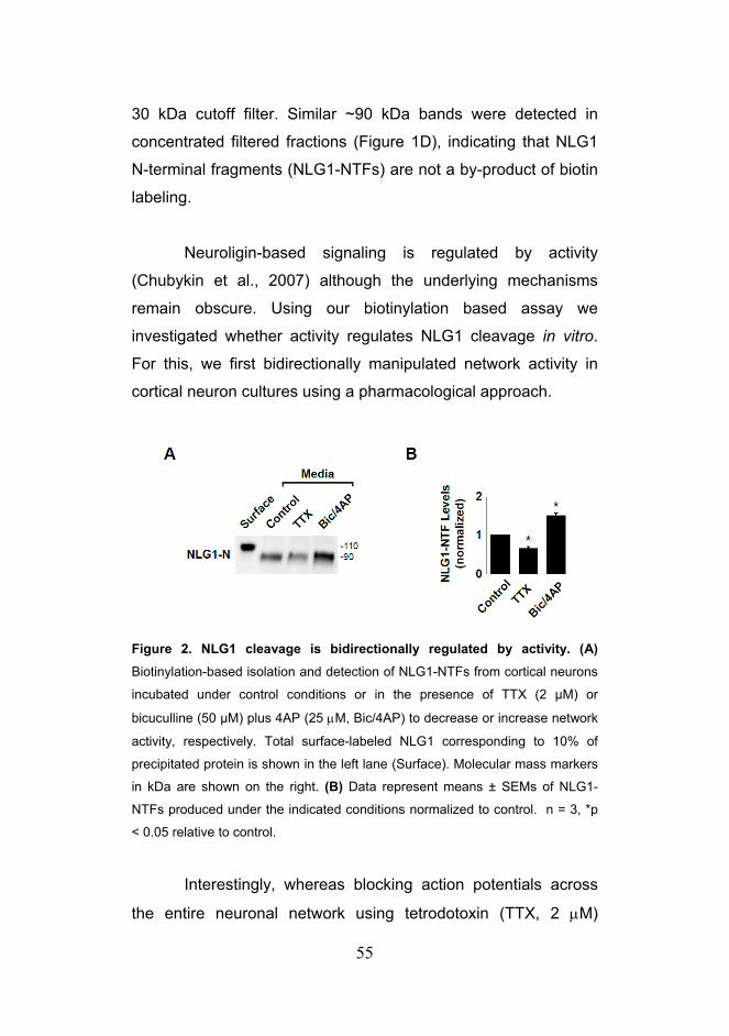
55
30 kDa cutoff filter. Similar ~90 kDa bands were detected in
concentrated filtered fractions (Figure 1D), indicating that NLG1
N-terminal fragments (NLG1-NTFs) are not a by-product of biotin
labeling.
Neuroligin-based signaling is regulated by activity
(Chubykin et al., 2007) although the underlying mechanisms
remain obscure. Using our biotinylation based assay we
investigated whether activity regulates NLG1 cleavage in vitro.
For this, we first bidirectionally manipulated network activity in
cortical neuron cultures using a pharmacological approach.
Figure 2. NLG1 cleavage is bidirectionally regulated by activity. (A)
Biotinylation-based isolation and detection of NLG1-NTFs from cortical neurons
incubated under control conditions or in the presence of TTX (2 µM) or
bicuculline (50 µM) plus 4AP (25 μM, Bic/4AP) to decrease or increase network
activity, respectively. Total surface-labeled NLG1 corresponding to 10% of
precipitated protein is shown in the left lane (Surface). Molecular mass markers
in kDa are shown on the right. (B) Data represent means ± SEMs of NLG1-
NTFs produced under the indicated conditions normalized to control. n = 3, *p
< 0.05 relative to control.
Interestingly, whereas blocking action potentials across
the entire neuronal network using tetrodotoxin (TTX, 2 μM)

56
resulted in decreased generation of NLG1-NTFs (0.65 ± 0.09 of
control), increasing network activity by simultaneously blocking
inhibitory synaptic transmission with the GABAA receptor
antagonist bicuculline (50 μM) and K+ channels with 4-
aminopyridine (4AP, 25 μM) significantly increased NLG1
cleavage (Bic/4AP, 1.5 ± 0.1 of control) (Figures 2A and 2B).
Besides confirming our previous data showing that NLG1
cleavage is increased by neuronal depolarization these results
also show that this process is bidirectionally regulated and can
be reduced in response to decreased neuronal activity.
To mimic conditions that lead to robust loss of synaptic
NLG1 (See Chapter 1, Figures 1 and 2), we depolarized cortical
neurons by incubation with KCl (30 mM, 2 h).
Figure 3. Activity Induces NLG1 Cleavage Through NMDARs and CaMK. (A) Biotinylation-based isolation and detection of NLG1-NTFs from cortical
neurons incubated under control conditions or in the presence of KCl (30 mM, 2
h) with or without indicated pharmacological agents. Note the robust increase in
NLG1-NTFs induced by KCl depolarization that is blocked by the NMDA
receptor antagonist APV and CaMK inhibitors KN93 and KN62. (B) Data
represent means ± SEMs of NLG1-NTFs produced under the indicated
conditions normalized to control. n = 4, * p< 0.05, ** p < 0.01.

57
As previously described (Chapter 1, Figures 1 and 2) KCl
mediated depolarization led to a dramatic increase in NLG1-
NTFs (4.4 ± 0.5 fold) compared to control conditions (Figures 3A
and 3B). Interestingly, this depolarization-induced cleavage of
NLG1 was abolished by the NMDA receptor antagonist APV (50
μM). Activation of NMDA receptors elicits Ca2+ influx and
activation of Ca2+/calmodulin-dependent kinases (CaMK)
including CaMKII (Fink and Meyer, 2002). Accordingly, inhibition
of CaMK using either KN93 (5 μM) or KN62 (10 μM), but not the
inactive isomer KN92 (5 μM), significantly reduced activity-
induced cleavage of NLG1 (KN93, 1.5 ± 0.6; KN62, 1.5 ± 0.5;
KN92, 5.0 ± 1.1 fold increase in NLG1-NTFs relative to control).
These results show that activity-induced cleavage of NLG1 is
triggered by NMDA receptor activation and requires
Ca2+/calmodulin-dependent kinase activity.
What enzyme is responsible for NLG1 cleavage? Among
the diverse cellular proteolytic enzymes, MMPs comprise a large
family of secreted endoproteases that mediate cell migration,
signaling, morphogenesis, and development in diverse tissues
(Ethell and Ethell, 2007; Sternlicht and Werb, 2001). Our
previous results described in Chapter 1 demonstrated that the
KCl-induced loss of full-length NLG1 in cortical neuron lysates
was abrogated by the broad spectrum MMP inhibitor GM6001
(See Chapter 1, Figure 4). This result was confirmed using our
biotinylation-based isolation method where incubation with
GM6001 (10 μM) reduced basal NLG1 cleavage and prevented
KCl-induced cleavage of NLG1 (fold increase relative to control:
KCl, 2.62 ± 0.20; KCl + GM6001, 0.53 ± 0.05; Figures 4A and

58
4B). In order to determine the specific Matrix Metalloprotease
involved in NLG1 ectodomain shedding we followed a candidate
approach based on pharmacological inhibition of MMP activity.
MMP-2, -3 and -9 are the most abundant MMPs in the brain and
have been implicated in synaptic maturation and multiple forms
of synaptic plasticity (Ethell and Ethell, 2007; Yong, 2005). This
prompted us to address how these specific proteases affected
KCl-induced cleavage of NLG1.
Figure 4. MMP9 Mediates Activity-Dependent Cleavage of NLG1. (A) Isolation and detection of ~90 kDa NLG1-NTFs following biotinylation of cortical
neurons incubated under control conditions or in the presence of KCl with or
without MMP inhibitors. Molecular mass markers in kDa are shown on the right.
(B) Data represent means ± SEMs of NLG1-NTFs produced under the indicated
conditions normalized to control. n = 4, *p < 0.01.

59
Incubation with the selective MMP9 inhibitor I (0.5 μM) or
MMP3 inhibitor III (50 μM) significantly reduced KCl-induced
cleavage (0.68 ± 0.02 and 1.27 ± 0.27 fold increase in NLG1-
NTFs relatively to control, respectively), with MMP9 inhibitor I
eliciting a more robust reduction. Conversely, the MMP1 inhibitor
GM1489 (5 nM), MMP2 inhibitor III (50 μM) and MMP8 inhibitor I
(10 μM) had no significant effect on activity-induced NLG1
cleavage (2.82 ± 0.14, 2.40 ± 0.19 and 1.92 ± 0.37 relatively to
control, respectively).
MMPs are secreted as inactive zymogens and require
further processing to become enzymatically active (Ethell and
Ethell, 2007; Van Wart and Birkedal-Hansen, 1990). MMP3 is an
upstream activator of MMP9 capable of cleaving its inhibitory
prodomain (Ogata et al., 1992). This raised the possibility that the
partial blockade of KCl-induced NLG1 cleavage by MMP3
inhibition could be due to compromised MMP9 activation. To test
this hypothesis and determine which MMP is the terminal
protease cleaving NLG1 we treated neurons with 4-
aminophenylmercuric acetate (APMA), a mercury-based
compound that indiscriminately activates all MMPs by cleaving
their prodomain (Van Wart and Birkedal-Hansen, 1990), and
tested the effect of specific MMP inhibitors on APMA mediated
cleavage. Since APMA activates all MMPs, only inhibition of the
final effector protease will impair NLG1 cleavage.

60
Figure 5. MMP9 Mediates Activity-Dependent Cleavage of NLG1. (A) Following surface biotinylation, cortical neurons were treated with the
nonselective MMP activator APMA (0.5 mM, 15 min) with or without MMP
inhibitors. Lysates (Lys) and soluble media (Med) were subjected to streptavidin
precipitation prior to immunoblot analysis for NLG1 using an N-terminal
antibody (4C12). Note that MMP9 inhibitors block APMA-induced cleavage. (B)
Data represent means ± SEMs of NLG1-NTFs produced under the indicated
conditions normalized to control. n = 3, *p < 0.05 relative to APMA alone.
Indeed, brief incubation of cortical neurons with 0.5 mM
APMA for 15 min induced robust generation of NLG1-NTFs
(Figures 5A and 5B). Co-incubation with MMP9 inhibitor I almost
completely blocked APMA-induced cleavage while inhibition of
MMP3 or MMP2 had no effect. Based on these results we
conclude that MMP9 is most likely the terminal effector protease
responsible for cleavage of NLG1 while MMP3 might be an
upstream component of the signaling cascade activating MMP9.
Deglycosylation of endogenous NLG1-NTFs produces a
~70 kDa species (Chapter 1, Figure 6) which, based on amino
acid mass, indicates that cleavage occurs in the extracellular
juxtamembrane domain of NLG1. To determine the specific
region of NLG1 targeted for cleavage, we generated a series of

61
mutants with sequential deletions and amino acid replacements
in its juxtamembrane domain (Figure 6A).
Figure 6. Extracellular Cleavage of NLG1 Occurs Near Its Transmembrane
Domain. (A) Model illustrating the location of the NLG1-ΔSD substitution
mutants in relation to NLG1 domains: AEHD, acetylcholinesterase homology
domain; DD, dimerization domain; SD, stalk domain; TM, transmembrane
domain; CT, C-terminal domain. Numbers indicate amino acids. (B)
Biotinylation-based cleavage assay of COS7 cells expressing the indicated
NLG1 mutants. Total lysates (Lys) or media (Med) were collected and subjected
to immunoblot analysis following treatment with the MMP activator APMA with
or without the broad spectrum MMP inhibitor GM6001 (GM). Both NLG1-
ΔSDfull and NLG-ΔSD3 were resistant to APMA-induced cleavage indicating
that MMP cleavage sites lie between residues 672 and 695. Molecular mass
markers are shown at the right.
These newly generated NLG1 mutants were tested for
their resistance to APMA-induced cleavage using biotinylation-
based labeling and isolation of NLG1-NTFs in COS7 cells (Figure
6). The presence of surface biotinylation labeling indicates that
surface trafficking and expression of all mutants is not
compromised. Brief incubation with APMA results in extensive
ectodomain shedding of wild-type GFP-NLG1 (Figure 6B). This
effect was fully blocked by GM6001 confirming that NLG1

62
cleavage is MMP-dependent under these conditions (Figure 6B).
Substituting 60 amino acids (aa) of the NLG1 stalk domain (aa
636-695) with the polylinker GAAAAA resulted in a mutant
(NLG1-ΔSDfull) that is resistant to APMA-induced cleavage
(Figures 6A and 6B). Within this 60 residue stretch, deletion of
amino acids 672-695 (NLG1-ΔSD3) and replacement with the
polylinker GAAAAA likewise abolished APMA-induced cleavage
whereas mutation of more membrane-distal sequences did not
(aa 636-660, NLG-ΔSD1; aa 654-677, NLG-ΔSD2). This
indicates that NLG1 cleavage occurs in it juxtramembrane region
within the 24 aminoacid sequence upstream of the
transmembrane domain. We attempted to further resolve the
precise cleavage site, but shorter deletions or different single site
substitution mutants were all cleaved upon APMA treatment
(data not shown), suggesting the presence of multiple MMP
target sequences within this 24 aa domain. However, these
results indicate that cleavage of NLG1 occurs in the region
corresponding to amino acids 672-695 of NLG1.
Different NLG isoforms partition to and regulate different
subsets of synapses (Budreck and Scheiffele, 2007; Graf et al.,
2004; Song et al., 1999; Varoqueaux et al., 2004). Hence, the
differential regulation of NLG isoforms may have important
implications for neuronal function (Dahlhaus et al., 2010; Hines et
al., 2008; Prange et al., 2004). To determine if all NLGs are
processed similarly in response to activity we measure total NLG
levels following KCl depolarization (Figure 7A).

63
Figure 7. Differential regulation of NLG2 cleavage. (A) Immunoblot analysis
of NLG1, NLG2 and NLG3 in total lysates from cortical cultures following 2 h
incubation in neurobasal medium (Ctrl) or medium supplemented with 30 mM
KCl alone (KCl) or with MG132 (10 µM), leupeptin (200 μM), leupeptin plus
MG132 (Leup/MG) or GM6001 (10 µM). Note that GM6001 prevents KCl-
induced loss of total NLG1 and NLG3 but not NLG2. (B) Immunoblot analysis of
NLG1, NLG2 and NLG3 in total lysates from cortical cultures following 15min
incubation in neurobasal medium (Ctrl) or medium supplemented with 0.5 mM
APMA (APMA). Note that NLG2 levels are not reduced by APMA (C)
Biotinylation-based cleavage assay of COS7 cells expressing the indicated NLG
isoform HA tagged mutants. Total lysates (Lys) or media (Med) were collected
and subjected to immunoblot analysis following treatment with the MMP
activator APMA. Note the presence of residual soluble NLG2-HA bands in the
media.
All NLGs exhibit a robust decrease in overall levels
following KCl treatment that is unaffected by lysosome and
proteasome inhibition. This suggests that all NLG isoforms are
processed by similar mechanisms upon KCl depolarization.
However, only degradation of NLG1 and NLG3, but not NLG2,
could be completely rescued by application of the broad
spectrum MMP inhibitor GM6001 (Figure 7A). This indicates that
NLG1 and NLG3 are cleaved my MMPs under these conditions
and suggests that NLG2 is regulated by a different mechanism.
Moreover, incubation of cortical cultures with 0.5mM APMA for

64
15 min resulted in a dramatic reduction of NLG1 and NLG3
levels, but not NLG2 (Figure 7B). Again, this is consistent with a
differential regulation of NLG2 compared with NLG1 and NLG3.
Unfortunately, due to the lack of NLG2 specific antibodies that
target the N-terminal domain of the protein we could not address
NLG2 cleavage based on surface biotinylation. However, to
address if NLG2 also undergoes ectodomain shedding we
expressed HA-tagged forms of NLG2 and NLG3 in COS7 cells
and measured the release of NLG-NTFs after APMA treatment
using surface biotinylation (Figure 7C). While surface NLG3-HA
was completely cleaved and released to the extracellular media
in response to APMA stimulation, NLG2-HA remained mostly
associated with the membrane fraction. This result is consistent
with lack of NLG2 degradation following APMA incubation in
neuronal cultures (Figure 7B). However, residual amounts of
soluble NLG2-HA fragments could still be detected in the media
of APMA treated cultures (Figure 7C, top panel) indicating that
NLG2 can also undergo ectodomain shedding. Nevertheless,
despite being cleaved this data indicates that NLG2 is
differentially regulated when compared with other NLG isoforms,
potentially by a different set of proteases that are not blocked by
GM6001 and not activated by APMA.
The upregulation of MMP9 in response to seizures
(Szklarczyk et al., 2002) suggested that NLG1 cleavage could
also be regulated by activity during adult stages. To address this
hypothesis we measured NLG1-NTFs generated during
pilocarpine induced status epilepticus (PSE) in mice. This
paradigm is characterized by recurrent activation of hippocampal
circuits (Curia et al., 2008).

65
Figure 8. NLG1 is cleaved in response to epileptic seizures in the hippocampus by MMP-9. (A) Schematic of the experimental conditions (B) WT
and MMP9 KO mice were injected with pilocarpine (315 mg/kg) to induce status
epilepticus (Pilocarpine) or saline (Control). After 2 h, hippocampi were isolated
and homogenized and either total extracts (5 μg) or soluble fractions (Sol, 50
μg) were immunoblotted for NLG1. Note the increased NLG1-NTFs after
pilocarpine-induced seizures in WT and the abrogation of this response in
MMP9-KO animals. Pilocarpine upregulates Arc in both wildtype and MMP9 KO
animals. (C) Data represent means ± SEMs of NLG1-NTFs produced under the
indicated conditions normalized to control. n = 9, *p < 0.05.
Intraperitoneal administration of pilocarpine in P60 mice
induced robust epileptic seizures and resulted in a 3.12 ± 0.47
fold increase of soluble NLG1-NTFs in the hippocampus after 2h
of PSE induction (Figures 8B and 8C). These findings indicate
that increased neuronal activity also upregulates NLG1 cleavage
in vivo. To address if MMP9 also mediates NLG1 cleavage under
pilocarpine induced status epilepticus, we repeated these
experiments in MMP9 null mice. Interestingly, soluble fractions of

66
MMP9-KO hippocampus under basal conditions still exhibit
normal levels of NLG1-NTFs indicating that NLG1 cleavage still
occurs in the absence of MMP9. However, 2h of PSE
characterized by robust seizures failed to elevate soluble NLG1-
NTFs in hippocampus when compared to saline controls (Figures
8B and 8C). As a control for seizure activity we measured levels
of the activity-regulated cytoskeleton-associated protein (Arc)
and both wild-type and MMP9 KO mice exhibited significant Arc
upregulation after pilocarpine administration (Figure 8B). These
findings indicate that activity triggers NLG1 cleavage in vivo
through MMP9 activation. Moreover, these results suggest a
potential link between NLG1 cleavage and circuit remodeling
during epileptogenesis.

67
Discussion
In this section, we have characterized the molecular and
signaling pathways regulating activity-dependent NLG1 cleavage.
Using a newly developed assay based on biotinylation of surface
proteins we found that cleavage of NLG1 at the plasma
membrane is bidirectionally regulated by neuronal activity,
requires NMDA receptor signaling, Ca2+/calmodulin-dependent
kinase activity and is mediated by the activity-regulated and
brain-expressed Matrix Metalloprotease-9.
Figure 9. Schematic model for activity-induced MMP9 cleavage of NLG1.
Increased neuronal activity activates a proteolytic cascade of MMPs via
NMDAR and CaMK signaling. Ultimately, MMP9 activation cleaves NLG1
releasing NLG1-NTFs to the media. NMDAR, N-methyl-D-aspartate receptor;
CaMK, Ca2+/calmodulin-dependent protein kinase; Scissors represent MMP9.

68
Moreover, these results define a novel link between NLG-
NRX adhesion and a NMDAR-CaMK-MMP9 signaling cascade.
In addition, we found that cleavage of NLG1 is highly upregulated
in the hippocampus during epileptic seizures. Previous data
described in Chapter 1 already has shown that NLG1 cleavage
can be regulated by changes in neuronal activity in vivo. Besides
providing additional evidence that NLG1 shedding is dynamically
regulated by activity in vivo, the increase of NLG1-NTFs levels
during epileptic seizures indicates that this process can also
occur in adult brains outside developmental periods of circuit
maturation. Indeed, recent work has implicated NLG1 in the
expression of LTP in the amygdala suggesting that NLG1 is not
only involved in the maturation and stabilization of synapses but
is also required for synaptic function in later stages of
development (Jung et al., 2010; Kim et al., 2008). Moreover, the
increase in NLG1-NTFs following pilocarpine injection was
absent in MMP9 KO mice indicating that MMP9 is required for
seizure-induced cleavage of NLG1 in the hippocampus. This
result is consistent with our findings in dissociated cultures where
MMP9 inhibition abrogated the increase in NLG1-NTFs induced
by KCl stimulation. Thus, MMP9 is likely an important protease
regulating NLG1 cleavage in contexts of increased neuronal
activity.
Epileptogenic patterns of activity in the hippocampus lead
to extensive fiber sprouting and circuit remodeling which are
thought to underlie the generation of recurrent seizures in later
stages of the disease (Isokawa, 1998, 2000). Interestingly, not
only is MMP9 upregulated in response to epileptic seizures but it
is also required for the synaptic pruning observed in the days

69
following seizure induction (Szklarczyk et al., 2002; Wilczynski et
al., 2008). This important finding suggests that the molecular
targets of MMP9 in hippocampal neurons may be critical
regulators of synapse maintenance and stability during
epileptogenesis. Indeed, MMP7, an upstream activator of MMP-
9, induces morphological changes in dendritic spines converting
them to elongated filopodia-like structures (Bilousova et al.,
2006). Conversely, MMP9 inhibition accelerates spine maturation
in neuronal cultures and increases the total fraction of mushroom
spines (Bilousova et al., 2009). Together these results suggest
that MMP9 activity may indeed be an important regulator of
synapse maintenance.
Besides interacting with NLGs, presynaptic NRXs also
bind to other postsynaptic molecules such as β-dystroglycan
(Sugita et al., 1999; Sugita et al., 2001). Interestingly, β-
dystroglycan is also cleaved by MMP9 in response to kainate
induced seizures (Michaluk et al., 2007), suggesting that MMP9
may be an important regulator of NRX function by targeting
multiple of its postsynaptic binding partners. Given the well-
established role of NLGs and NRXs in synaptic maturation and
maintenance (Chubykin et al., 2007; Varoqueaux et al., 2006),
MMP9 could then potentially regulate synapse stability by
controlling NLG-NRX function. Moreover, the developmental
profile of NLG1-NTFs shown in Chapter1 (Figure 8) indicates that
NLG1 cleavage is highly upregulated during early developmental
stages and declines during the first 2 postnatal weeks, which is
the period of synapse formation in the neocortex. This hypothesis
is further supported by upregulation of NLG1-NTFs observed in

70
response to visual experience during critical period, a paradigm
that also elicits subsequent synapse remodeling. Hence,
cleavage of NLG1 emerges as a potential general mechanism
associated with synaptic remodeling during development and
disease.
Alternatively, cleavage of NLG1 induced by strong activity
could also provide a protective mechanism to ensure proper
balance between excitation and inhibition in complex circuits. In
fact, we found that NLG1 cleavage is bidirectionally regulated by
activity both in vitro, in response to Bic/4AP and TTX treatment
(Figure 2) and in vivo in response to dark rearing and brief light
exposure after dark rearing (Chapter 1, Figure 8). Given that
NLG1 increases overall excitatory drive by preferentially
stabilizing (Chubykin et al., 2007) and augmenting the release
probability of excitatory synapses (Futai et al., 2007), the
bidirectional control of NLG1 levels by proteolytic regulation could
mediate homeostatic compensatory changes in synaptic
transmission to balance overall excitability. Interestingly, our data
indicates that NLG2 cleavage exhibits a different and specific
pattern of regulation compared with NLG1 and NLG3. Since
NLG2 and NLG1/3 exhibit a preferential enrichment in inhibitory
and excitatory synapses, respectively (Budreck and Scheiffele,
2007; Graf et al., 2004; Song et al., 1999; Varoqueaux et al.,
2004), the activation of specific proteolytic pathways targeting
NLG2 could also in principle, affect the balance between
excitation and inhibition. In fact, deletion of NLG2 decreases
IPSC amplitudes in acute cortical slices (Chubykin et al., 2007)
and increases the excitatory-to-inhibitory ratio in the
hippocampus (Poulopoulos et al., 2009) and cortex (Gibson et
71
al., 2009), whereas transgenic overexpression of NLG2 leads to
increased mIPSC frequency in prefrontal cortical regions (Hines
et al., 2008). This interesting hypothesis will certainly be the
focus of future studies.
Excitotoxicity driven by unrestrained glutamate release
contributes to neuronal degeneration in many acute CNS
diseases, including ischemia, trauma, and epilepsy (Arundine
and Tymianski, 2003). In particular, excessive NMDAR
dependent Ca2+ influx has been shown to be prone to induce
deleterious effects in neurons (Arundine and Tymianski, 2004).
Interestingly, NLG1 levels in dissociated neurons are directly
correlated with NMDAR EPSC amplitudes (Chubykin et al.,
2007). Moreover, αNRX deletion induces loss of NMDAR
function in cortical slices (Kattenstroth et al., 2004). Hence, an
acute loss of NLG1 in response to increased activity could
mediate decreased NMDAR dependent Ca2+ influx, protecting
neurons from glutamate excitotoxicity. Consistent with this
hypothesis, it has been shown that MMP9 activity increases
synaptic NMDAR mobility which potentially decreases NMDAR
synaptic currents (Michaluk et al., 2009). This effect has been
attributed to Integrin-β1 signaling however integrin function was
assessed by overnight antibody cross linking which may cause
indirect secondary effects. Thus, other mechanisms may be
involved in the destabilization of synaptic NMDAR by MMP9.
It is important to note that basal levels of NLG1-NTFs are
still present in MMP9 KO brain extracts. This indicates that even
in the absence of MMP9 NLG1 cleavage still occurs which

72
indicates the existence of parallel proteolytic mechanisms
targeting NLG1 under basal conditions. Consistent with this idea,
none of the pharmacological MMP inhibitors tested in our in vitro
assays was able to fully block NLG1 cleavage. Moreover,
multiple soluble NLG1-NTF species can be detected throughout
the brain (Chapter 1, Figure 5), indicating that NLG1 probably
contains multiple cleavage sites. This observation provides a
plausible explanation for why single amino acid point mutations in
the stalk region of NLG1 failed to generate mutants that would
resist APMA induced MMP-dependent cleavage, which instead
required substitution of a longer 24 amino acid segment (Figures
6A and 6B). Hence, these results suggest that the regulation of
NLG1 cleavage is probably under control of multiple proteolytic
mechanisms that may be differently regulated depending of
cellular context. It will be important for future studies to define in
detail the activity-independent and MMP9-independent
mechanisms responsible for NLG1 cleavage and their potential
role in synapse development and plasticity.
Finally, our results indicate that the activity-induced
cleavage of NLG1 requires NMDAR and CaMK signaling.
Previous studies have shown that the synaptogenic effect and
functional increase in EPSCs caused by NLG1 overexpression in
cultured neurons are also abrogated by APV and CaMK inhibitor
KN93 (Chubykin et al., 2007), however these experiments were
based on chronic application of APV for 4 days. Nevertheless,
our data suggest that NMDAR signaling controls NLG1 levels at
synapses through MMP activity. Consistent with this hypothesis,
APV blocks MMP9 upregulation in response to LTP suggesting
that NMDAR controls MMP9 function in other activity-dependent

73
contexts (Nagy et al., 2006). Recent work has further shown that
in response to brief depolarization, NLG1 internalization is halted
via NMDAR signaling (Thyagarajan and Ting, 2010). Taken
together, this data suggests that NLG1 is tightly regulated by
NMDAR and CaMK activity at multiple levels and that the
regulation of proteolysis and trafficking of NLGs may occur in
tandem though similar signaling mechanisms.
Taken together, the results discussed here indicate that
cleavage of NLGs is a highly regulated process that is involved in
multiple activity-dependent cellular contexts and has the potential
to generate different functional outcomes due to specific
regulation of each NLG isoform. Moreover, our data implicates
NLG processing during the early stages of epileptogenesis which
may provide new insights regarding the pathophysiology of
epileptic disorders.

74

75
Methods Reagents and Antibodies
Dissociated primary neuronal cultures were prepared from cortex
of embryonic day 18 or 19 Wistar rats. Tissue was dissociated by
enzymatic papain digestion followed by brief mechanical
trituration. Cultures were grown in Neurobasal media
supplemented with B27 and Glutamax. For surface biotinylation
experiments 700k cortical cells were plated onto 60mm petri
dishes. Commercial antibodies used include N-terminal NLG1
antibody clone 4C12 (mouse, Synaptic Systems), C-terminal
NLG2 polyclonal antibody (rabbit, Synaptic Systems), C-terminal
NLG3 polyclonal antibody (rabbit, Synaptic Systems), Arc (C-7,
rabbit, Santa Cruz) and HA.11 (mouse, Covance).
Pharmacological inhibitors used include KN93 (Calbiochem),
KN62 (Calbiochem), KN92 (Calbiochem), APV (Tocris), TTX
(Tocris), GM6001 (Calbiochem), MMP9 inhibitor I (Calbiochem),
MMP3 inhibitor III (Calbiochem), GM1489 (Calbiochem), MMP2
inhibitor III (Calbiochem), and MMP8 inhibitor I (Calbiochem),
MG132 (SIGMA), Leupeptin (SIGMA) and APMA (SIGMA).
NLG1-ΔSDfull mutant was obtained by substitution of
endogenous 636-695 residues with GAAAAA linker using primers
5’-ggcgccgcagcagcagcactaagtgtcacaatcgcagtg-3’ and 5’-
tgctgctgctgcggcgccattatgcagatgaggtacc-3’; NLG1-ΔSD1 (636-
660) 5’-ggcgccgcagcagcagcaaggaaaaattccactccag-3’ and 5’-
tgctgctgctgcggcgccattatgcagatgaggtacc-3; NLG1-ΔSD2 (654-
677) 5’-ggcgccgcagcagcagcacccaaacaacaaccaagtc-3’ and 5’-

76
tgctgctgctgcggcgccgtctgttgatggcactttag-3’; NLG1-ΔSD3 (672-
695) 5’-ggcgccgcagcagcagcactaagtgtcacaatcgcagtg-3’and 5’-
tgctgctgctgcggcgccgggaaaggctgatgtgactgg-3’. For GFP-NLG1-
HA the HA peptide YPYDVPDYA was inserted at the end of
GFP-NLG1(ΔA) using primer 5’- gcatctcgagctaagcgtaatctggaaca
tcgtatgggtataccctggttgttgaatgtg-3’. In all reactions the two self-
annealing PCR products were templates in a reaction with primer
5’-gttggagctggtacctcatc-3’ containing a KpnI site and 5’-
gtcactcgagctataccctggttgttgaatg-3’. The respective product was
subcloned into pcDNA3-GFP-NLG1 using KpnI and XhoI. Surface Biotinylation and Isolation of Soluble Cleavage Products
For surface protein labeling, cortical cultures (700,000 cells/6cm
plate) were incubated in PBS/Ca2+ supplemented with 1 mg/ml
Sulfo-LC-Biotin-NHS (Pierce) for 10 min at 37ºC. Cultures were
washed in PBS/Ca2+ containing 10 mM Tris pH7.4 and rinsed
twice in PBS/Ca2+ and incubated in conditioned media at 37ºC for
the indicated experimental time period. Pharmacological
inhibitors were added 5 minutes prior to manipulations of
neuronal activity. Following incubation at 37ºC, culture media
was collected, supplemented with protease inhibitor cocktail
(Roche) and centrifuged at 16,000 x g for 20 min. For surface-
labeled controls, cultures were washed twice with PBS/Ca2+,
immediately lysed with precipitation buffer (PBS with 1% Triton-
X100, 5 mM EDTA, 10 mM L-lysine pH7.4), sonicated and
centrifuged at 16,000 x g for 20 min. Supernantants were
incubated overnight with 50 µl NeutrAvidin Plus UltraLink Resin
77
(Pierce) at 4ºC and precipitated biotin conjugates subsequently
washed twice and liberated by boiling in 2x sample buffer.
Precipitated proteins were then resolved by SDS-PAGE prior to
immunoblot analysis with the indicated antibodies
Pilocarpine-Induced Status Epilepticus
Two month female FVB or FVB.Cg-Mmp9tm1Tvu/J (MMP9 KO)
mice (~18–25 g) were injected intraperitoneally (i.p.) with 1 mg/kg
scopolamine methyl-nitrate (Sigma) 30 min prior to the
experiment. Status epilepticus was induced by i.p. injection of
315 mg/kg pilocarpine hydrochloride (Sigma) while controls were
injected with vehicle alone (sterile 0.9% NaCl). The experiment
was terminated two hours after pilocarpine injection and
hippocampal tissue was collected and flash frozen. In the
pilocarpine group, only animals that exhibited class IV or higher
seizures during the course of the experiment were used for
analysis.

78

79
Acknowledgements For the realization of this work, Irina Lebedeva and Marguerita
Klein prepared the neuronal cultures from dissociated mouse
hippocampus and cortex. Susana da Silva kindly provided
assistance in the pilocarpine induced seizure experiments.

80

81
Chapter 3
Acute Cleavage of Neuroligin-1 Destabilizes Presynaptic
Neurexin-1β and Reduces Neurotransmitter Release
Introduction The close association between pre and postsynaptic elements
has long suggested that changes on either side of the synapse
could be transmitted trans-synaptically (Lisman and Harris,
1993). In fact, whether the expression of Hebbian-forms of long
term synaptic plasticity involve retrograde modulation of
presynaptic function has been a source of heated debate for
more than 20 years and remains an open question in the field
(Bear and Malenka, 1994; Enoki et al., 2009; Malenka and Bear,
2004). However, a less controversial form of trans-synaptic
signaling occurs during the homeostatic adaptation of
neurotransmitter release to chronic changes in neuronal activity.
In mature hippocampal neurons, suppression of
excitability by expression of an inward rectifier potassium channel
(Kir2.1) increases mEPSC frequency (Burrone et al., 2002).
Moreover, chronic silencing of network activity using tetrodotoxin
(TTX) increases the size of presynaptic terminals and their
respective pool of synaptic vesicles, with a concomitant increase
in release probability (Murthy et al., 2001). Interestingly, a similar
response is observed in response to chronic inhibition of AMPAR
currents using the antagonist 3-dihydroxy-6-nitro-7-sulfamoyl-
82
benzo[f]quinoxaline-2,3-dione (NBQX) suggesting that retrograde
signals may be involved in the generation of this response
(Thiagarajan et al., 2005). Conversely, increased KCl
concentration during development results in lower release
probability and loss of excitatory inputs (Moulder et al., 2004),
whereas 2h of 2Hz field stimulation of dissociated cultures
causes an acute decrease in release probability (Branco et al.,
2008). Importantly, this latter effect is blocked by NMDAR and
AMPAR antagonists suggesting that this homeostatic response is
induced postsynaptically and subsequently transduced by
retrograde messengers. A similar adaptive response has been
described in the Drosophila neuromuscular junction where
blockade of postsynaptic glutamate receptors (Frank et al.,
2006), or reduced cellular depolarization due to Kir2.1 expression
(Paradis et al., 2001) increase presynaptic release of
neurotransmitter without altering postsynaptic properties. Hence,
several lines of evidence indicate that chronic changes in
postsynaptic activity result in a homeostatic compensation of
afferent neurotransmitter release.
Retrograde signaling can be mediated by many types of
molecular mechanisms. Diffusible retrograde messengers
including nitric oxide and endocannabinoids (eCB) are two well
characterized systems capable of modulating neurotransmitter
release in response to changes in postsynaptic activity (Regehr
et al., 2009). However, given their confined localization at
synapses and restricted number of interactions, adhesion
molecules offer the potential for tight local control over
presynaptic function. For instance, postsynaptic ablation of N-
Cadherin induces defects in synaptic vesicle release under

83
conditions of high-release activity (Jungling et al., 2006), whereas
acute activation of presynaptic ephrin-B2 by soluble forms of
EphB2 induces a rapid increase in release probability in optic
tectum neurons (Lim et al., 2008).
Recently, NLGs have emerged as important trans-
synaptic signaling mediators. NLGs are capable of inducing
functional maturation of presynaptic terminals (Dean et al., 2003;
Prange et al., 2004; Scheiffele et al., 2000; Wittenmayer et al.,
2009), while transgenic expression of NLG1 results in extended
active zones and increased reserve pool size (Dahlhaus et al.,
2010). Interestingly, overexpression of NLG1 in hippocampal
slices and cultured neurons results in increased release
probability through a Neurexin (NRX) dependent mechanism
(Futai et al., 2007; Ko et al., 2009b; Stan et al., 2010), suggesting
that NLG1 can modulate presynaptic function trans-synaptically.
Indeed, neurons lacking NLG1-3 present decreased miniature
EPSCs (mEPSCs) frequency consistent with impaired
presynaptic function (Varoqueaux et al., 2006). Moreover,
neurons lacking αNRX1-3 exhibit deficits in synaptic transmission
due to impaired N-type Ca2+ channel function despite normal
synaptic vesicle readily releasable pool size (Missler et al., 2003).
In addition, chronic disruption of endogenous NLG-NRX
interactions with long term incubation of soluble Fc-NRX
fragments decreases mEPSC frequency and release probability
(Levinson et al., 2005). Taken together, these results suggest
that NLGs and NRXs are important regulators of presynaptic
function and that NLGs can signal retrogradely through NRXs.
However, a limitation of these studies is that they all rely on long
term manipulations of NLG and NRX function, which may

84
generate indirect compensatory mechanisms. Hence, whether
trans-synaptic NLG-NRX adhesion can acutely regulate synapse
transmission remains unclear.
The work described in the previous chapters,
demonstrates that NLGs can be rapidly degraded in response to
increased neuronal activity via MMP dependent proteolysis.
Considering the strong association between NLGs and NRXs,
acute proteolytic cleavage of NLGs could be an effective way to
disassociate the NLG-NRX interaction and mediate fast synaptic
signaling. In fact, acute proteolysis has been shown to elicit fast
changes in synaptic transmission. For instance, acute application
of recombinant MMP9 is able to induce LTP in hippocampal
slices (Nagy et al., 2006) and increase NMDAR mobility in matter
of minutes (Michaluk et al., 2009). Moreover, acute inhibition or
displacement of the metalloprotease ADAM10 from synapses
induces a rapid expansion of dendritic spine size and increase
AMPA EPSCs (Malinverno et al., 2010). However, the
promiscuous nature of MMP activity, where each MMP can
cleave multiple targets and activate parallel proteolytic cascades
(Ethell and Ethell, 2007), has been a limitation to address the
specific functional consequences of cell adhesion molecule
cleavage.
To overcome this limitation and address the functional
consequence of NLG1 cleavage we developed a novel strategy
using an exogenous protease that allows the cleavage of specific
membrane proteins in a temporally controlled manner.
Combining this strategy with real time confocal microscopy and
electrophysiological measurements of neuronal activity we

85
demonstrate that acute cleavage of NLG1 results in
destabilization and loss of its presynaptic partner Neurexin-1β.
This effect is specific and not an indirect consequence of pre
synaptic terminal collapse or loss of synaptic vesicle pools. In
turn, NLG1 cleavage rapidly depresses synaptic transmission by
abruptly reducing presynaptic release probability. Together,
these results indicate that postsynaptic activity influences
presynaptic function through NLG1 cleavage unveiling a novel
form of trans-synaptic signaling that may be involved in multiple
forms of synaptic plasticity.

86

87
Results To address the functional effect of acute cleavage of NLG1 we
developed a system to acutely and selectively cleave NLG1
(Figure 1A).

88
Figure 1. Acute cleavage of NLG1 using Thrombin. (A) Schematic of
thrombin-cleavable NLG1 construct. Red region represents insertion of
thrombin recognition sequence LVPRGS in the stalk domain of NLG1. (B)
Quantification of time lapse experiment measuring fluorescence intensity before
and after thrombin application of neurons expressing GFP-Thr-NLG1. (C)
Hippocampal neuron (DIV21) expressing GFP-Thr-NLG1 and mCherry imaged
live by confocal microscopy before and during thrombin incubation (5U/ml, 30
min). Thrombin application is indicated by the white circle. Note the robust loss
of GFP fluorescence due to GFP-Thr-NLG1 cleavage. Time is indicated in
minutes. Scale bar, 20 μm. _____________________________________________________
To achieve this, we inserted the thrombin recognition
sequence LVPRGS at the stalk domain of NLG1 replacing the
endogenous sequence TTTKVP, downstream of the dimerization
domain. In these experiments, the NLG1ΔA splice variant lacking
splice site A was chosen due to its stringent partition into
excitatory synapses (Chih et al., 2006). The thrombin-cleavable
mutant GFP-Thr-NLG1 targeted to synapses in a manner
indistinguishable from wild-type GFP-NLG1 (Figure 1C).
Incubation with 5U/ml thrombin for 30 min results in a marked
(80.4 ± 3.4%) reduction in GFP-Thr-NLG1 fluorescence (Figures
1B and 1C). Control neurons transfected with GFP-NLG1 lacking
a thrombin recognition sequence exhibited no change in GFP
fluorescence after thrombin treatment (Figure 1B, 99.9 ± 1.5% of
initial intensity), indicating that the reduced GFP fluorescence
was not due to photobleaching. This can also be inferred by the
fact that the internal pools of NLG1-GFP are not affected by
thrombin incubation (see lower panel in Figure 1C - GFP signal is
present in the soma after thrombin application potentially
corresponding to NLG1-GFP pool present in the secretory
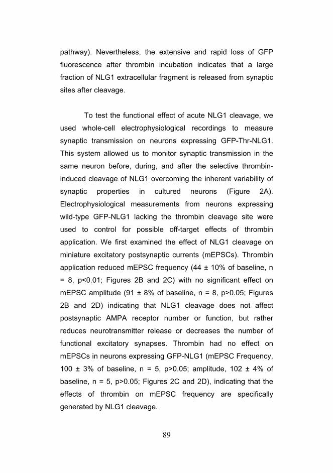
89
pathway). Nevertheless, the extensive and rapid loss of GFP
fluorescence after thrombin incubation indicates that a large
fraction of NLG1 extracellular fragment is released from synaptic
sites after cleavage.
To test the functional effect of acute NLG1 cleavage, we
used whole-cell electrophysiological recordings to measure
synaptic transmission on neurons expressing GFP-Thr-NLG1.
This system allowed us to monitor synaptic transmission in the
same neuron before, during, and after the selective thrombin-
induced cleavage of NLG1 overcoming the inherent variability of
synaptic properties in cultured neurons (Figure 2A).
Electrophysiological measurements from neurons expressing
wild-type GFP-NLG1 lacking the thrombin cleavage site were
used to control for possible off-target effects of thrombin
application. We first examined the effect of NLG1 cleavage on
miniature excitatory postsynaptic currents (mEPSCs). Thrombin
application reduced mEPSC frequency (44 ± 10% of baseline, n
= 8, p<0.01; Figures 2B and 2C) with no significant effect on
mEPSC amplitude (91 ± 8% of baseline, n = 8, p>0.05; Figures
2B and 2D) indicating that NLG1 cleavage does not affect
postsynaptic AMPA receptor number or function, but rather
reduces neurotransmitter release or decreases the number of
functional excitatory synapses. Thrombin had no effect on
mEPSCs in neurons expressing GFP-NLG1 (mEPSC Frequency,
100 ± 3% of baseline, n = 5, p>0.05; amplitude, 102 ± 4% of
baseline, n = 5, p>0.05; Figures 2C and 2D), indicating that the
effects of thrombin on mEPSC frequency are specifically
generated by NLG1 cleavage.

90
Figure 2. NLG1 cleavage acutely dampens excitatory synaptic transmission. (A) Schematic representation of the experimental design.
Electrophysiological recordings of hippocampal neurons (DIV18) expressing
either thrombin-cleavable GFP-Thr-NLG1 or non-cleavable GFP-NLG1 control
were performed before and after thrombin incubation. (B) Representative
miniature EPSCs (mEPSCs) recorded before (Baseline) and after (Post)
thrombin. (C) Quantification showing that thrombin-induced cleavage of NLG1
reduces mEPSC frequencies without affecting (D) amplitudes. Thrombin has no
effect on mEPSC frequency or amplitude in neurons expressing NLG1-EGFP
lacking a thrombin cleavage site. n = 5, 8 for GFP-Thr-NLG1 and GFP-NLG,
respectively. *p < 0.01, unpaired t-test.

91
Interestingly, NLG1 cleavage failed to alter mIPSC
frequency or amplitude (mIPSC Frequency, 103 ± 3%; mIPSC
Amplitude, 98 ± 8% of baseline, n = 6, p>0.05; Figure 3),
indicating that NLG1 cleavage does not alter neurotransmitter
release at inhibitory synapses. These results are consistent with
previous studies demonstrating that NLG1 predominantly
localizes to and regulates glutamatergic synapses (Song et al.,
1999) (Chubykin et al., 2007; Graf et al., 2004).
Figure 3. NLG1 cleavage has no effect in inhibitory synaptic transmission. (A) Representative mIPSCs before (Baseline) or after (Post) thrombin treatment
in neurons expressing GFP-Thr-NLG1. (B) Quantification showing that NLG1
cleavage does not affect mIPSC frequency or (C) amplitude (right). Data in bar
graphs represent mean ± SEM. n = 6.
The reduction observed in mEPSC frequency (Figures 2B
and 2C) without any visible loss of dendritic spines (Figures 1C
and 5A-C) suggested that NLG1 cleavage could be affecting
presynaptic function.

92
Figure 4. Cleavage of NLG1 reduces release probability at excitatory synapses. (A) Representative paired-pulse evoked EPSCs (eEPSCs) recorded
before (Baseline) or after (Post) thrombin treatment in hippocampal neurons
(DIV18) expressing either thrombin-cleavable GFP-Thr-NLG1 or GFL-NLG1
lacking a thrombin cleavage site. Insets depict traces before and after thrombin
treatment, normalized to the amplitude of the first EPSP to indicate the increase
in paired pulse ratio (PPR) after thrombin cleavage in cells expressing GFP-Thr-
NLG1. (B) NLG1 cleavage reduces eEPSC amplitude. Data represent means ±
SEM of eEPSC amplitudes after thrombin treatment normalized to amplitudes
prior to thrombin treatment. n's = 6; *p < 0.05, unpaired t-test. (C) NLG1
cleavage increases paired pulse ratio (PPR). Data indicate means ± SEM of
paired-pulse ratios after (Post) and before (Baseline) thrombin normalized to the
GFP-NLG1 control. n's = 6; *p < 0.05, unpaired t-test. Note that an increase in
PPR is consistent with a decrease in release probability.

93
To test this hypothesis further, we examined the effects of
thrombin-induced NLG1 cleavage on evoked excitatory synaptic
currents (eEPSCs) and their paired-pulse responses (Dobrunz
and Stevens, 1997). Evoked responses were elicited by
stimulating nearby cells with an interstimulus interval of 100 msec
(Figure 4A). Recordings were made on the same cell before and
after thrombin application. Thrombin treatment of neurons
expressing GFP-Thr-NLG1 reduced the amplitude of the first
response and increased the paired-pulse ratio (PPR) (eEPSC
amplitude, 50 ± 18% of baseline, n = 6, p < 0.05; PPR, 139 ± 8%
of baseline, n = 6, p <0.05, Figure 4), strongly suggesting that
NLG1 cleavage decreases the probability of neurotransmitter
release (Dobrunz and Stevens, 1997). This effect was not
attributable to thrombin treatment per se, as there was no change
in amplitude or PPR in neurons expressing wild type NLG1-
EGFP lacking a thrombin cleavage sequence (eEPSC amplitude,
95 ± 8% of baseline, n = 6, p > 0.05; PPR, 106 ± 6% of baseline,
n = 6, p>0.05; Figure 4). Together with the observed decrease in
mEPSC frequency (Figure 2C), these findings demonstrate that
juxtamembrane cleavage of postsynaptic NLG1 acutely dampens
synaptic transmission due to a reduction in presynaptic
neurotransmitter release. Thus, activity-dependent cleavage of
Neuroligin defines a retrograde signal for rapid trans-synaptic
tuning of synaptic strength.
To determine the molecular mechanism underlying this
retrograde modulation of neurotransmitter we performed a series
of studies based on microscopic analysis of different synaptic
proteins during acute NLG1 cleavage. We initially addressed
whether postsynaptic morphology was affected by thrombin
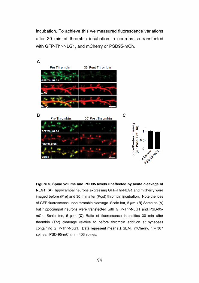
94
incubation. To achieve this we measured fluorescence variations
after 30 min of thrombin incubation in neurons co-transfected
with GFP-Thr-NLG1, and mCherry or PSD95-mCh.
Figure 5. Spine volume and PSD95 levels unaffected by acute cleavage of NLG1. (A) Hippocampal neurons expressing GFP-Thr-NLG1 and mCherry were
imaged before (Pre) and 30 min after (Post) thrombin incubation. Note the loss
of GFP fluorescence upon thrombin cleavage. Scale bar, 5 μm. (B) Same as (A)
but hippocampal neurons were transfected with GFP-Thr-NLG1 and PSD-95-
mCh. Scale bar, 5 μm. (C) Ratio of fluorescence intensities 30 min after
thrombin (Thr) cleavage relative to before thrombin addition at synapses
containing GFP-Thr-NLG1. Data represent means ± SEM. mCherry, n = 307
spines; PSD-95-mCh, n = 403 spines.

95
Interestingly, no significant changes in fluorescence intensity
were detected after thrombin treatment in mCherry cell fill (ΔF/F;
1.04 ± 0.03; n=307 spines; Figures 5A and 5C) and PSD95-mCh
puncta (ΔF/F; 0.97 ± 0 .02; n=403 spines; Figures 5B and 5C).
This indicates that acute NLG1 cleavage has no gross impact in
PSD structure or dendritic spine volume. These results are
consistent with the invariance observed in mEPSC amplitude
after thrombin incubation and reinforce the hypothesis that NLG1
cleavage predominantly affects presynaptic function.
NLG1 forms a high affinity trans-synaptic complex with
the presynaptic membrane protein Neurexin-1β (NRX1β), thereby
regulating presynaptic assembly and function (Arac et al., 2007;
Chen et al., 2008; Comoletti et al., 2007; Dean et al., 2003;
Fabrichny et al., 2007; Futai et al., 2007; Ichtchenko et al., 1995;
Prange et al., 2004; Scheiffele et al., 2000; Varoqueaux et al.,
2006; Wittenmayer et al., 2009). To test whether acute cleavage
of NLG1 alters the stability of the Neuroligin-Neurexin trans-
synaptic adhesion complex we sequentially transfected cultures
with GFP-Thr-NLG1 and NRX1β−mCh lacking splice site 4 in
separate days. This method results in the transfection of two
distinct neuronal populations with each construct, resulting in the
labeling of pre and postsynaptic pairs with NRX1β-mCh and
GFP-Thr-NLG, respectively (Figure 6A). At dually-labeled
synapses, acute thrombin incubation caused a rapid and
pronounced decrease in NRX1β-mCh fluorescence (Figures 6B
and 6C; 0.51 ± 0.04 of initial fluorescence after 30 min of
thrombin, n=87 synaptic pairs, p<0.05).

96
Figure 6. Acute NLG1 cleavage destabilizes presynaptic NRX1β. (A)
Hippocampal cultures were sequentially transfected with GFP-Thr-NLG1 and
NRX1β-mCh to label distinct postsynaptic and presynaptic neurons,
respectively. Neurons were incubated with thrombin and imaged live by
confocal microscopy. Solid arrowheads indicate presynaptic NRX1β-mCh
apposed to postsynaptic GFP-Thr-NLG1 puncta. Note the thrombin-induced
loss of NRX1β-mCh. Open arrows show newly generated mobile NRX1β-mCh
puncta along the axon. Scale bar, 5 μm. (B) Timelapse of the highlighted region
depicted in (A). The red arrowhead shows the time of thrombin (Thr)
application. Note that thrombin-induced cleavage of GFP-Thr-NLG1 is followed
by rapid loss of NRX1β-mCh. Scale bar, 5 μm. (C) Quantification of time lapse
experiments as in (B). Plotted is fluorescence intensity before and after
thrombin application at synapses containing presynaptic NRX1β-mCh along

97
with postsynaptic GFP-Thr-NLG1. The red arrowhead shows the time of
thrombin (Thr) application. n = 87 synaptic pairs. (D) Same as (C) except
neurons expressed GFP-NLG1 lacking a thrombin cleavage site. n = 76
synaptic pairs.
_______________________________________________________________
These results demonstrate that NLG1 cleavage
destabilizes presynaptic NRX1β-mCh and suggest that the
decrease in release probability observed after NLG1 shedding is
due, at least in part, to loss of presynaptic Neurexins.
Importantly, this destabilization is not an artifact caused by
thrombin off-target effects as NRX1β-mCh puncta opposed to
NLG1-GFP expressing synapses were unaffected by thrombin
incubation (Figures 6A and 6D).
Interestingly, we noted the appearance of several mobile
puncta containing high levels of NRX1β-mCh after thrombin
incubation which raised the possibility that NLG1 cleavage could
result in overall presynaptic terminal collapse or loss of synaptic
vesicles from synaptic sites. To address this possibility we
repeated the same experiments using Synaptophysin-mCh
instead of NRX1β-mCh. We detected no changes in presynaptic
Synaptophysin-mCh signal intensity (0.99 ± 0.08, n=43 synaptic
pairs, Figures 7A and 7B) indicating that destabilization of
NRX1β by NLG1 cleavage is specific and not due to indirect
structural changes in presynaptic terminals.

98
Figure 7. Acute NLG1 cleavage does not affect presynaptic Synaptophysin-mCh. (A) Hippocampal cultures were sequentially transfected
with GFP-Thr-NLG1 and Synaptophysin-mCh (Sph-mCh). Neurons were
incubated with thrombin and imaged live by confocal microscopy. Note that
cleavage of postsynaptic NLG1 does not elicit loss of presynaptic Sph-mCh .
Scale bar, 5 μm. (B) Ratio of fluorescence intensities 30 min after thrombin
(Thr) cleavage relative to before thrombin at synapses containing GFP-Thr-
NLG1. Data represent means ± SEM. Sph-mCh, n = 43 synaptic pairs; NRX1β-
mCh, n = 87 synaptic pairs, *p <0.05.
These results were further confirmed by measuring
endogenous synaptic vesicle pool size by immunolabeling of
vesicular glutamate transporter VGLUT1 in neurons expressing
GFP-NLG1 or GFP-Thr-NLG1 before and after 30 minutes of
thrombin incubation. Consistent with the stability of
Synaptophysin-mCh puncta observed after NLG1 cleavage, we
detected no change in VGLUT1 puncta number or intensity in
both neuronal populations after thrombin treatment (Figure 8),
indicating that NLG1 cleavage affects presynaptic Neurexin
levels specifically.

99
Figure 8. Neuroligin-1 cleavage does not alter presynaptic VGLUT1. (A)
Hippocampal neurons (DIV21) expressing mCherry and either GFP-NLG1 or
GFP-Thr-NLG1 were incubated in 5U/ml thrombin for 30 min, fixed, and
immunolabeled for endogenous VGLUT1. Arrows indicate VGLUT1-positive
presynaptic terminals. Note that VGLUT1 staining is unaffected by the thrombin-
induced loss of GFP-Thr-NLG1. Scale bar, 5 µm. (B) Data represent means ±
SEM of VGLUT1 puncta intensity apposing GFP-Thr-NLG1 or GFP-NLG1 after
thrombin treatment normalized to the GFP-NLG1 group. GFP-NLG1, n = 2316
puncta from 9 neurons; GFP-Thr-NLG1, n = 2408 puncta from 8 neurons. (C)
Data represent means ± SEM of VGLUT1 puncta density after thrombin
normalized to GFP-NLG1 group. n values as in (B).
Together, the results presented here define a novel trans-
synaptic signaling mechanism induced by NLG1 cleavage. NLG1

100
shedding results in destabilization and loss of presynaptic
NRX1β with concomitant reduction of release probability. Given
the involvement of NLG1 cleavage during early development and
synaptic plasticity described in previous chapters these findings
indicate that acute regulation of NLG1 at synapses has direct
consequences over synaptic transmission and provide new
insight about how NLGs regulate mature synapses.

101
Discussion Neurexins and Neuroligins are highly synaptogenic proteins
capable of aggregating pre and postsynaptic scaffolding
complexes (Dean et al., 2003; Graf et al., 2004; Heine et al.,
2008; Nam and Chen, 2005; Prange et al., 2004; Scheiffele et al.,
2000; Wittenmayer et al., 2009). Moreover, recent work has also
shown that NLGs can regulate synaptic transmission (Chubykin
et al., 2007; Futai et al., 2007; Varoqueaux et al., 2006) and are
required for Hebbian forms of synaptic plasticity (Jung et al.,
2010; Kim et al., 2008). However, these studies were based on
chronic long-term manipulations of NLG and NRX levels, which
are susceptible to indirect compensatory mechanisms. Hence, it
remained unclear whether acute modulation of the NLG-NRX
trans-synaptic interaction can directly affect synaptic
transmission. The experiments described in previous chapters
unveiled a new form of NLG1 regulation based on ectodomain
shedding by MMP9. Due to the acute and irreversible nature of
MMP proteolysis, this mechanism has the potential to elicit rapid
and long lasting changes in synaptic function. However, the
functional consequences induced by shedding of specific
adhesion molecules have been difficult to address due to the
highly redundant and promiscuous nature of MMP activity. Here,
we have developed a novel strategy capable of specifically and
acutely cleaving surface NLG1. Using this method we
demonstrate that acute cleavage of NLG1 causes rapid loss of
presynaptic NRX1β and reduces synaptic transmission by
decreasing presynaptic release probability (Figure 9).

102
Figure 9. Schematic model for synaptic effects induced by cleavage of
NLG1. NLG1 cleavage decreases NRX1β stability and reduces transmitter
release probability (Pr) without inducing changes in overall presynaptic structure
or synaptic vesicle pool size. NMDAR, N-methyl-D-asparate receptor; CaMK,
Ca2+/calmodulin-dependent protein kinase; NTFs, N-terminal fragments; NLG1-
CTFs, NLG1 C-terminal fragments. Scissors represent MMP9.
These results provide first evidence that NLGs can
acutely regulate synaptic transmission and suggest that cleavage
of other adhesion molecules may elicit similar effects. Indeed,
acute activation of presynaptic ephrin-B2 by soluble forms of
EphB2 induces a rapid increase in release probability in optic
tectum neurons (Lim et al., 2008). This suggests that EphB2-
ephrin interaction has the potential to mediate acute retrograde
modulation of presynaptic function. Interestingly, MMP9/MMP2
have been shown to target and cleave EphB2 (Lin et al., 2008)
suggesting that MMP9 activation can potentially activate parallel
signaling cascades that converge in the silencing of presynaptic
vesicle release. In addition to Eph cleavage by MMPs, ephrins
can also be cleaved by ADAM10 in response to Eph binding

103
(Janes et al., 2005). This indicates that the same trans-synaptic
adhesive complex may be under control of different proteolytic
pathways. Another cell adhesion molecule that has been shown
to regulate presynaptic function is N-Cadherin (NCAD). NCAD
deficient neurons have defects in synaptic vesicle release from
incoming afferents under conditions of high neuronal activity
(Jungling et al., 2006). Notably, ADAM10 can also cleave N-
cadherin, although the potential effect of NCAD cleavage in
presynaptic function has not been addressed. Together, these
results demonstrate that shedding of adhesion molecules has the
potential to induce retrograde modulation of presynaptic release
probability, but also illustrate the highly complex regulatory
network that these processes will likely entail. Nevertheless, the
strategies presented here may provide significant aid in
addressing how cleavage of specific adhesion molecules affects
synaptic function.
Previous studies have shown that deletion of αNRXs in
mice reduces action-potential-evoked neurotransmitter release
due to impaired presynaptic Ca2+ channel function (Missler et al.,
2003). However, due to the role of NRXs in synapse maturation
(Dean et al., 2003; Prange et al., 2004; Scheiffele et al., 2000;
Wittenmayer et al., 2009) it has been unclear whether this effect
is due to indirect developmental effects or reflects an ongoing
role for NRXs in neurotransmitter release. Using the highly
selective thrombin-induced cleavage of NLG1, we have
demonstrated an overall reduction in excitatory transmission and
release probability concurrent with NRX1β loss. This finding is
consistent with a role of NRXs as regulators of neurotransmitter
release (Futai et al., 2007; Missler et al., 2003; Zhang et al.,

104
2005) and further suggests that NRX-dependent regulation of
presynaptic function can occur in time scales of minutes.
Interestingly, the destabilization of NRX1β observed after acute
NLG1 cleavage is specific, since presynaptic protein
Synaptophysin was unaffected under similar conditions. This also
indicates that acute ectodomain shedding of NLG1 is not
accompanied by gross structural changes of presynaptic
terminals or by loss of synaptic vesicles. Besides interacting with
NLGs, NRXs bind to several other postsynaptic transmembrane
proteins such as members of the Leucine Rich Repeat
Transmembrane Molecule (LRRTM) family (de Wit et al., 2009;
Ko et al., 2009a), Cerebellin precursor proteins (Joo et al., 2011;
Matsuda and Yuzaki, 2011), Neurexophilins (Petrenko et al.,
1996), β-Dystroglycan (Sugita et al., 2001) and also to GABA(A)
receptors (Zhang et al., 2010). The function of NRX interaction
with each of these specific postsynaptic partners and its potential
contribution for synaptic regulation remains unclear. However, in
contrast to NLGs no functional effect of binding of Neurexophilin
or β-Dystroglycan to NRXs has been observed so far (Sudhof,
2008). Nevertheless, our results suggest that postsynaptic NLG1
is a major stabilizing element controlling the recruitment and
abundance of NRX1β at synapses. Moreover, the increase in
PPR observed after NLG1 cleavage indicates that the NLG-NRX
interaction directly contributes for the regulation of release
probability.
Neurons undergo compensatory, homeostatic synaptic
changes in order to maintain their overall firing rate (Davis, 2006;
Turrigiano, 2008). In mature hippocampal and cortical cultures

105
postsynaptic receptor blockade causes an increase in mEPSC
frequency (Burrone et al., 2002; Thiagarajan et al., 2005;
Wierenga et al., 2006) and augments presynaptic terminal size
and release probability (Murthy et al., 2001; Thiagarajan et al.,
2005). In contrast, KCl-induced depolarization results in lower
release probability and loss of excitatory inputs (Moulder et al.,
2004) indicating that homeostatic regulation of presynaptic
function is a bidirectional process. Elegant experiments
performed in dissociated cultures further demonstrated that local
dendritic activity regulates release probability of presynaptic
terminals (Branco et al., 2008) implying the existence of trans-
synaptic signals capable of transducing postsynaptic messages.
The identity of the molecular signals involved in this response
remains however, unknown. At the Drosophila neuromuscular
junction, compromised postsynaptic activity results in a
compensatory increase of presynaptic neurotransmitter release
(Davis, 2006) with two distinct phases: an initial protein
synthesis-independent phase operating within minutes, and a
slower protein synthesis-dependent phase that is required for the
long term maintenance of the presynaptic adaptation (Frank et
al., 2006). Interestingly, the latter component involves modulation
of Cav2.1 calcium channels by Eph receptor activation (Frank et
al., 2009), resembling the effect of αNRX1-3 ablation on N-type
Ca2+ channel function (Missler et al., 2003). Although quite
different in molecular mechanism, these observations together
with the findings presented here suggest an evolutionarily
conserved module whereby postsynaptic activity signals
retrogradely by adhesion molecule coupling to presynaptic
voltage-gated Ca2+ channels. Hence, we propose that
bidirectional regulation of NLG1 cleavage by neuronal activity

106
may provide a trans-synaptic signal for homeostatic plasticity that
dampens or augments presynaptic function in response to
increased or decreased postsynaptic activity, respectively.
Several other retrograde signaling mechanisms can
directly affect presynaptic function. Notable examples are
mediated by nitric oxide (NO) and the endocannabinoid signaling
system (eCB). Both these signaling mechanisms are activated by
elevated postsynaptic Ca2+ levels and ultimately result in
changes in presynaptic function. Production of eCBs is triggered
by intracellular calcium and Gq-coupled receptor activation, which
in turn drive the conversion of Phosphatidylinositol 4,5-
bisphosphate (PIP2) into Diacylglycerol (DAG) by phospholipase-
C beta (PLCβ), and is subsequently converted into the
endocannabinoid 2-AG by DAG lipase (Regehr et al., 2009). In
turn, 2-AG is released from the postsynaptic cell and activates
Cannabinoid Receptors type 1 (CB1Rs) located in presynaptic
terminals. Similarly, NMDAR signaling activates NO production
by Nitric Oxide Synthetase (iNOS) in postsynaptic cells
(Garthwaite, 2008) that is able to freely diffuse across
membranes and activate multiple targets in presynaptic terminals
such as soluble guanylyl cyclase (Garthwaite et al., 1989).
Activation of eCB and NO signaling pathways have direct
implications for presynaptic function. For example, CBR1
agonists suppress synaptic transmission by decreasing release
probability through Ca2+ channel regulation (Brown et al., 2003;
Brown et al., 2004), and eCBs have been shown to suppress
seizures in the hippocampus (Monory et al., 2006). Conversely,
NO activity has been implicated in several forms of presynaptic
LTP (Nugent et al., 2007). Hence, there are multiple signaling

107
mechanisms capable of transducing retrograde messages across
the synapse. However, in comparison with signaling mechanisms
based on small diffusible messengers, shedding of adhesion
molecules has the potential for increased temporal and spatial
control over trans-synaptic signaling. Indeed, eCB and NO have
been shown to induce heterosynaptic effects (Lancaster, 1994;
Wilson and Nicoll, 2001). Moreover, long-term signaling requires
continuous production of intermediate messengers which may be
unfavorable from an energetic perspective. Signaling through
adhesion molecule cleavage may then offer the possibility to
induce permanent structural changes in synapses which may
then be remodeled by new incoming molecules. Future work will
be required to characterize in detail how different retrograde
signaling mechanisms are regulated and how they specifically
affect synaptic function. Interestingly, NO can regulate enzymatic
function in many target proteins through S-nitrosylation including
MMP9 proteolytic activity (Gu et al., 2002). It is tempting to
speculate that some of the reported effects of NO may occur in
tandem with, or even be caused by, MMP9 activation.
Consistent with results demonstrating that NLG1
predominantly regulates excitatory synaptic function (Chubykin et
al., 2007; Ko et al., 2009b), we found no effect of NLG1 cleavage
on mIPSCs (Figure 3). Moreover, mEPSC amplitudes were
unaffected by acute NLG1 cleavage, indicating that postsynaptic
function was largely intact. This is consistent with live-cell
imaging data showing that dendritic spines and PSD95-mCh
puncta are unaffected by acute NLG1 cleavage. Our results
described in Chapter 2 indicate that NLG2 can also be cleaved,
but through a different proteolytic pathway. It will be interesting to

108
address if cleavage of NLG2 may be involved in the regulation of
inhibitory synapse function. Notably, long term potentiation of
GABAergic synapses on dopaminergic neurons in the ventral
tegmental area (VTA) is expressed presynaptically, as evidenced
by a change in paired-pulse ratio, yet it can be impaired by
blocking postsynaptic calcium elevations with BAPTA, suggesting
a requirement for a retrograde messenger (Nugent et al., 2007).
Hence, the future elucidation of the mechanisms regulating
cleavage of different NLG isoforms may have important
implications for understanding retrograde signaling at different
synapses.
Together, the results discussed here indicate that
postsynaptic activity influences presynaptic function by NLG1
cleavage. Furthermore, these findings reveal a new post-
translational mechanism of NLG1 regulation that contributes to
synapse plasticity and may provide a general paradigm for acute
retrograde signaling in diverse neural circuits.

109
Methods
Reagents and Antibodies
Dissociated primary neuronal cultures were prepared from
hippocampi of embryonic day 18 or 19 Wistar rats. The
hippocampi were dissociated by enzymatic papain digestion
followed by brief mechanical trituration. Cultures were grown in
Neurobasal media supplemented with B27 and Glutamax. For
immunocytochemistry, 100k hippocampal cells were plated onto
12 well plates containing Poly-L-Lysine coated 18mm glass
coverslips. Plasmid transfection was done using lipid mediated
gene transfer using Lipofectamine 2000 (Invitrogen). Briefly, for
each coverslip, 1μg total DNA was mixed with 1μl Lipofectamine
in 100 μl of Neurobasal media for 15min. Following incubation
time 500 μl of conditioned media was mixed to the reaction and
added to cell cultures for 30min. After this period, lipofectamine
containing media was removed and replaced with a 1:1 mixture
of conditioned media + fresh growth media. Commercial
antibodies used include VGLUT1 (mouse, Synaptic Systems).
DNA Constructs and Antibodies
PSD95-mCh was a gift from Thomas Blanpied (University of
Maryland). mCherry-N1 vector was a gift from Roger Tsien
(University of California, San Diego). Synaptophysin-mCh was a
gift from Matthew Kennedy (Duke University). Rat GFP-NLG1 in
pcDNA3 (ΔA splice variant) was kindly provided by Anne Marie

110
Craig (University of British Columbia), and NRX1β-mCh was a
gift from Thomas Sudhof (Stanford University). Thrombin
inducible GFP-Thr-NLG1 was generated by replacing the
endogenous sequence TTTKVP from GFP-NLG1(ΔA) with the
thrombin recognition sequence LVPRGS. This was achieved by
sequential PCR using primers 5’-ctagtacctagaggatcctcaacagaca
tcactctcag-3’ and 5’-ggatcctctaggtactagcgaggtatactgagaaatgtca-
3’, and primers 5’-ggatcctctaggtactagctgagaaatgtcattgag-3’ and
5’-gttggagctggtacctcatc-3’. The two self-annealing PCR products
were templates in a reaction with primer 5’-gttggagctggtacctcatc-
3’ containing KpnI site and 5’-gtcactcgagctataccctggttgttgaatg-3’.
The respective product was subcloned into pcDNA3-GFP-NLG1
using KpnI and XhoI.
Imaging
Confocal images of live cells were obtained using a Perkin Elmer
Ultraview spinning disc confocal microscope with a 60x 1.4 N.A.
objective. Images were analyzed using Metamorph software
(Molecular Devices, Universal Imaging Corporation). For
immunocytochemistry, DIV21 hippocampal neurons were fixed in
4% paraformaldehyde/4% sucrose in PBS for 20 min,
permeabilized with 0.2% Triton X-100 for 15 min, and incubated
with 1:1000 VGLUT1 antibody in 3% goat serum/ 3% BSA
overnight at 4ºC. For live imaging, cells were imaged in E4
media (10 mM HEPES, 120 mM NaCl, 3 mM KCl, 10 mM
glucose, 2 mM CaCl2, 2 mM MgCl2) at 37ºC. Images were taken
every 3 min and 5U/ml thrombin was added at t = 20 min.

111
Quantification of fluorescence intensity at individual puncta was
performed before and 30 min after thrombin application. For
each neuron the average fluorescence intensity ratios for all
puncta were determined. Results depicted correspond to the
average of all neurons per condition.
Electrophysiological Recordings
For EPSC recordings, patch pipettes (6-8 MΩ tip resistance)
were filled with internal solution (30 mM CsSO4, 70 mM K2SO4,
25 mM HEPES, 25 mM N-methly-D-glucamine, 0.1 mM CaCl2, 1
mM EGTA, 2 mM (Na)ATP, and 0.1 mM leupeptin, pH7.2, ~300
mOsm). Cells were bathed in external solution (150 mM NaCl, 5
mM KCl, 10 mM HEPES, 1 mM MgCl2, 30 mM D-glucose, 2 mM
CaCl2, and 30 μM bicuculline). Only cells with series resistance <
30 MΩ and having <20% change in input resistance, series
resistance, and holding current were included for analyses.
EPSCs were evoked from a concentric bipolar stimulating
electrode. Paired-pulse responses, with an inter-pulse interval of
100 ms were evoked every 30 sec by extracellular stimulation
(200 μs duration) of nearby cells. Currents were recorded in
neurons voltage-clamped at -70 mV. Similar conditions were
used to record mEPSCs, except the external solution additionally
contained 1 μM TTX (to block action potentials mediated by
voltage-gated sodium channels). For mIPSC recordings, patch
pipettes were filled with internal solution (150 mM KCl, 3 mM
MgCl2, 15 mM HEPES, 0.1 mM CaCl2, 1 mM EGTA, 2 mM
Na2ATP, and 0.1 mM leupeptin, pH7.2, ~300 mOsm). Cells were

112
bathed in external solution containing (150 mM NaCl, 5 mM KCl,
10 mM HEPES, 1 mM MgCl2, 30 mM D-glucose, 2 mM CaCl2, 1
μM TTX (to block voltage-gated sodium channels), 50 μM D-APV
(to block NMDA receptors), and 10 μM CNQX (to block
AMPA/kainate receptors). Only cells with series resistance < 30
MΩ and having <20% change in input resistance, series
resistance, and holding current were included for analyses.
Currents were recorded in neurons voltage-clamped at -70 mV.

113
Acknowledgements For the realization of this work, Portia McCoy performed all the
electrophysiological recordings and analyzed the data from those
experiments. Irina Lebedeva and Marguerita Klein prepared the
neuronal cultures from dissociated mouse hippocampus. I also
thank Matt Kennedy for generously providing the Synaptophysin-
mCh construct.

114

115
Final Discussion
Although highly studied for their role during synapse maturation
(Chih et al., 2005; Graf et al., 2004; Levinson et al., 2005;
Scheiffele et al., 2000; Varoqueaux et al., 2006; Wittenmayer et
al., 2009), it has remained unclear whether Neuroligins acutely
regulate synaptic function and whether the Neuroligin-Neurexin
trans-synaptic complex undergoes dynamic dissociation. In this
study, we have shown that neuronal activity triggers cleavage
and loss of NLG1 at glutamatergic synapses. Cleavage of NLG1
requires NMDA receptor activation and Ca2+/calmodulin-
dependent kinase activity, and is mediated by the activity-
regulated brain-expressed protease MMP9. Cleavage of NLG1
occurs within its juxtamembrane ectodomain, releasing soluble
N-terminal cleavage fragments that are present in brain and are
mostly abundant during early postnatal development.
Furthermore, acute cleavage of NLG1 causes rapid loss of
presynaptic NRX1β, reduces synaptic strength, and decreases
presynaptic release probability. These results define a novel
trans-synaptic signaling mechanism coupling postsynaptic
activity to presynaptic function, and link Neuroligin-Neurexin
synaptic adhesion to a NMDAR-CaMK-MMP9 signaling cascade.
Moreover, these findings suggest that acute cleavage of synaptic
adhesion molecules by activity-regulated proteolysis may provide
a general mechanism linking synaptic activity to structural
remodeling of mature and developing neuronal circuits.
Taken together, the data presented here suggests that
NLG1 cleavage is under complex regulation and may have

116
multiple functional outcomes depending on cellular context.
Moreover, it is likely that NLG1 cleavage may be regulated by
other proteases besides MMP9. In mature synapses, NLG1
cleavage is upregulated in response to increased neuronal
activity in vitro and in vivo resulting in acute downregulation of
synaptic transmission due to changes in presynaptic release
probability. As discussed in Chapters 2 and 3, this effect may
underlie a homeostatic mechanism of negative feedback
responsible for maintaining proper levels of excitation in neurons.
However, our results also demonstrate that NLG1 cleavage is
particularly prominent during the first weeks of life and can be bi-
directionally regulated by sensory experience during critical
periods of cortical development. This suggests that besides
mediating acute trans-synaptic modulation of presynaptic activity,
NLG cleavage may also regulate synapse formation and
maturation at a different scale. Moreover, it is worth noting that
although the effects of NLG1 cleavage described here are
exclusively presynaptic, it remains to be determined whether
prolonged loss or further processing of NLG1 can impact
postsynaptic function.
Despite initial studies suggesting that NLG could be
involved in the early steps of synaptogenesis (Chih et al., 2005;
Graf et al., 2004; Levinson et al., 2005; Scheiffele et al., 2000;
Varoqueaux et al., 2006; Wittenmayer et al., 2009), recent data
suggests different model whereby NLGs regulate synapse
maintenance and pruning during later stages of development
through activity dependent processes (Chubykin et al., 2007;
Varoqueaux et al., 2006). Interestingly, the synaptogenic effect of
NLG1 overexpression is blocked by NMDAR antagonist and

117
CaMK inhibitors, indicating that the same signaling pathways
regulating NLG1 cleavage also control NLG-induced
synaptogenesis. Hence, it is possible that MMP-dependent
cleavage of NLG is involved in synapse maturation.
Thrombospondins (TSPs) are secreted glycoproteins that
promote synapse formation in CNS neurons (Christopherson et
al., 2005). A recent study has shown that TSP1 can bind to NLG1
and that TSP1 induced synapse formation is abrogated in the
absence of NLG1 (Xu et al., 2010). This suggests that TSP1 acts
through NLG1 to promote synapse stabilization, although the
detailed molecular mechanisms mediating this response remain
unknown. Interestingly, TSP1 blocks MMP9 activity (Bein and
Simons, 2000) raising the interesting possibility that TSP1
synaptogenic properties may be due, at least in part, to inhibition
of MMP9-dependent NLG1 cleavage. Moreover, as previously
discussed in Chapter 2, MMP9 inhibition accelerates spine
maturation in neuron cultures and increases the total fraction of
mushroom spines (Bilousova et al., 2009), whereas MMP7, an
upstream activator of MMP9, induces morphological changes in
dendritic spines reverting them to elongated filopodia-like
structures (Bilousova et al., 2006). Thus, although our work was
centered on the function of NLG1 cleavage in mature synapses,
the characterization of the specific molecular mechanisms and
proteases regulating NLG cleavage during development may
provide important insight regarding the molecular mechanisms
controlling synapse maturation and neuronal circuit formation
during development.

118
It is also important to note that NLG1 cleavage is
upregulated during epileptic seizures in the hippocampus and
also in the visual cortex in response to light stimulation after dark
rearing. Interestingly, both these experimental paradigms result
in synapse remodeling and circuit rearrangements that last for
days after stimuli induction (Suzuki et al., 1997; Tropea et al.,
2010). Moreover, activity induced processes leading to synaptic
depression have been associated with synaptic pruning and loss
(Bastrikova et al., 2008; Becker et al., 2008; Mysore et al., 2007).
It is then tempting to speculate that persistent NLG1
downregulation, perhaps coupled with other signals, could lead to
synapse disassembly and elimination. In this putative model
(Figure 1), specific patterns of neuronal activity would decrease
overall surface levels of NLGs through MMP-dependent
cleavage. In turn, loss of synaptic NLGs would result in silencing
of synapses due to reduced release probability. The decreased
levels of surface NLGs would then trigger synaptic competition
for the remaining pools of the protein. It is still unclear whether
MMP-dependent NLG cleavage operates at a local or global
scale. However, synaptic NLG1 molecules exhibit a relatively fast
exchange rate (τ=100s) and a significant mobile fraction at
excitatory synapses (40% of total) (data not shown), indicating
that NLG molecules remaining at the plasma membrane can
diffuse and be redistributed among neighboring synapses. In this
scenario, synapses able to sequester and recruit remaining
NLGs would restore normal levels of activity and be maintained,
while others unable to do so, would be eliminated.
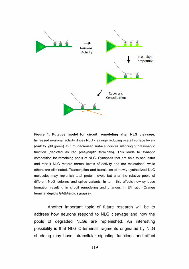
119
Figure 1. Putative model for circuit remodeling after NLG cleavage.
Increased neuronal activity drives NLG cleavage reducing overall surface levels
(dark to light green). In turn, decreased surface induces silencing of presynaptic
function (depicted as red presynaptic terminals). This leads to synaptic
competition for remaining pools of NLG. Synapses that are able to sequester
and recruit NLG restore normal levels of activity and are maintained, while
others are eliminated. Transcription and translation of newly synthesized NLG
molecules may replenish total protein levels but alter the relative pools of
different NLG isoforms and splice variants. In turn, this affects new synapse
formation resulting in circuit remodeling and changes in E/I ratio (Orange
terminal depicts GABAergic synapse).
Another important topic of future research will be to
address how neurons respond to NLG cleavage and how the
pools of degraded NLGs are replenished. An interesting
possibility is that NLG C-terminal fragments originated by NLG
shedding may have intracellular signaling functions and affect

120
downstream transcription similar to other biologically active CTFs
(Mumm et al., 2000; Schettini et al., 2010). Moreover, different
NLG isoforms partition to and regulate specific synaptic subsets
differently (Chubykin et al., 2007; Gibson et al., 2009) and in
addition, variations in alternative splicing can have a high impact
in the binding for affinity between different NLG and NRX
isoforms (Boucard et al., 2005; Chih et al., 2006; Ullrich et al.,
1995). Hence, a shift in the relative levels of different NLG
isoforms or splice variants caused by changes in transcriptional
regulation could have profound implications in the types of new
synapses generated, which could in turn lead to circuit
rearrangements or changes in overall E/I ratio.
Finally, several NLG and NRX mutations have been
linked with autism spectrum disorders (ASDs) (Sudhof, 2008). A
typical feature of ASDs is that they develop before two or three
years of age which corresponds to the period of heightened
synaptic maturation in humans (Lord et al., 2000; Pardo and
Eberhart, 2007). Moreover, 30% of autistic patients have epileptic
seizures and many more may suffer from subclinical epileptoform
conditions (Lewine et al., 1999). In addition, neuroimaging and
postmortem studies of patients with autism suggest that changes
in the ratio of excitation and inhibition in neural circuits may
underlie the etiology of ASDs (Rubenstein and Merzenich, 2003).
Together, these results suggest that at least a subset of ASDs
may arise from deficient synaptic maturation during critical
periods of development that in turn destabilize circuit function by
altering the E/I ratio. Interestingly, NLG levels during
development control overall excitability and regulate the ratio of
excitation and inhibition in neurons (Chubykin et al., 2007;

121
Gibson et al., 2009; Poulopoulos et al., 2009; Prange et al.,
2004). Our results indicate that NLG cleavage is regulated by
sensory experience during critical periods of circuit maturation
and in response to epileptic seizures in the hippocampus.
Although the characterizations of the detailed molecular
mechanisms regulating NLG in these different contexts will
require further inquiry, this data suggests a potential link between
NLG function, critical period plasticity and epileptogenesis.
Hence, the proteolytic regulation of NLGs reported here may
provide novel insight into the pathophysiological mechanisms
and therapeutic strategies for synaptic dysfunction in ASDs.

122
123
References Arac, D., Boucard, A.A., Ozkan, E., Strop, P., Newell, E., Sudhof, T.C., and Brunger, A.T. (2007). Structures of neuroligin-1 and the neuroligin-1/neurexin-1 beta complex reveal specific protein-protein and protein-Ca2+ interactions. Neuron 56, 992-1003.
Arundine, M., and Tymianski, M. (2003). Molecular mechanisms of calcium-dependent neurodegeneration in excitotoxicity. Cell Calcium 34, 325-337.
Arundine, M., and Tymianski, M. (2004). Molecular mechanisms of glutamate-dependent neurodegeneration in ischemia and traumatic brain injury. Cell Mol Life Sci 61, 657-668.
Barrow, S.L., Constable, J.R., Clark, E., El-Sabeawy, F., McAllister, A.K., and Washbourne, P. (2009). Neuroligin1: a cell adhesion molecule that recruits PSD-95 and NMDA receptors by distinct mechanisms during synaptogenesis. Neural Dev 4, 17.
Bastrikova, N., Gardner, G.A., Reece, J.M., Jeromin, A., and Dudek, S.M. (2008). Synapse elimination accompanies functional plasticity in hippocampal neurons. Proc Natl Acad Sci U S A 105, 3123-3127.
Bavelier, D., Levi, D.M., Li, R.W., Dan, Y., and Hensch, T.K. (2010). Removing brakes on adult brain plasticity: from molecular to behavioral interventions. J Neurosci 30, 14964-14971.
Bear, M.F., and Malenka, R.C. (1994). Synaptic plasticity: LTP and LTD. Curr Opin Neurobiol 4, 389-399.
Becker, N., Wierenga, C.J., Fonseca, R., Bonhoeffer, T., and Nagerl, U.V. (2008). LTD induction causes morphological changes of presynaptic boutons and reduces their contacts with spines. Neuron 60, 590-597.
Bein, K., and Simons, M. (2000). Thrombospondin type 1 repeats interact with matrix metalloproteinase 2. Regulation of metalloproteinase activity. J Biol Chem 275, 32167-32173.
Bilousova, T.V., Dansie, L., Ngo, M., Aye, J., Charles, J.R., Ethell, D.W., and Ethell, I.M. (2009). Minocycline promotes dendritic spine maturation and improves behavioural performance in the fragile X mouse model. J Med Genet 46, 94-102.
Bilousova, T.V., Rusakov, D.A., Ethell, D.W., and Ethell, I.M. (2006). Matrix metalloproteinase-7 disrupts dendritic spines in hippocampal neurons through NMDA receptor activation. J Neurochem 97, 44-56.

124
Boucard, A.A., Chubykin, A.A., Comoletti, D., Taylor, P., and Sudhof, T.C. (2005). A splice code for trans-synaptic cell adhesion mediated by binding of neuroligin 1 to alpha- and beta-neurexins. Neuron 48, 229-236.
Bozdagi, O., Nagy, V., Kwei, K.T., and Huntley, G.W. (2007). In vivo roles for matrix metalloproteinase-9 in mature hippocampal synaptic physiology and plasticity. J Neurophysiol 98, 334-344.
Bozdagi, O., Shan, W., Tanaka, H., Benson, D.L., and Huntley, G.W. (2000). Increasing numbers of synaptic puncta during late-phase LTP: N-cadherin is synthesized, recruited to synaptic sites, and required for potentiation. Neuron 28, 245-259.
Brakeman, P.R., Lanahan, A.A., O'Brien, R., Roche, K., Barnes, C.A., Huganir, R.L., and Worley, P.F. (1997). Homer: a protein that selectively binds metabotropic glutamate receptors. Nature 386, 284-288.
Branco, T., Staras, K., Darcy, K.J., and Goda, Y. (2008). Local dendritic activity sets release probability at hippocampal synapses. Neuron 59, 475-485.
Brennaman, L.H., and Maness, P.F. (2008). Developmental regulation of GABAergic interneuron branching and synaptic development in the prefrontal cortex by soluble neural cell adhesion molecule. Mol Cell Neurosci 37, 781-793.
Brown, S.P., Brenowitz, S.D., and Regehr, W.G. (2003). Brief presynaptic bursts evoke synapse-specific retrograde inhibition mediated by endogenous cannabinoids. Nat Neurosci 6, 1048-1057.
Brown, S.P., Safo, P.K., and Regehr, W.G. (2004). Endocannabinoids inhibit transmission at granule cell to Purkinje cell synapses by modulating three types of presynaptic calcium channels. J Neurosci 24, 5623-5631.
Budreck, E.C., and Scheiffele, P. (2007). Neuroligin-3 is a neuronal adhesion protein at GABAergic and glutamatergic synapses. Eur J Neurosci 26, 1738-1748.
Bukalo, O., Fentrop, N., Lee, A.Y., Salmen, B., Law, J.W., Wotjak, C.T., Schweizer, M., Dityatev, A., and Schachner, M. (2004). Conditional ablation of the neural cell adhesion molecule reduces precision of spatial learning, long-term potentiation, and depression in the CA1 subfield of mouse hippocampus. J Neurosci 24, 1565-1577.
Burrone, J., O'Byrne, M., and Murthy, V.N. (2002). Multiple forms of synaptic plasticity triggered by selective suppression of activity in individual neurons. Nature 420, 414-418.
Chan, C.S., Weeber, E.J., Kurup, S., Sweatt, J.D., and Davis, R.L. (2003). Integrin requirement for hippocampal synaptic plasticity and spatial memory. J Neurosci 23, 7107-7116.

125
Chen, X., Liu, H., Shim, A.H., Focia, P.J., and He, X. (2008). Structural basis for synaptic adhesion mediated by neuroligin-neurexin interactions. Nat Struct Mol Biol 15, 50-56.
Chih, B., Engelman, H., and Scheiffele, P. (2005). Control of excitatory and inhibitory synapse formation by neuroligins. Science 307, 1324-1328.
Chih, B., Gollan, L., and Scheiffele, P. (2006). Alternative splicing controls selective trans-synaptic interactions of the neuroligin-neurexin complex. Neuron 51, 171-178.
Chistiakova, M., and Volgushev, M. (2009). Heterosynaptic plasticity in the neocortex. Exp Brain Res.
Christopherson, K.S., Ullian, E.M., Stokes, C.C., Mullowney, C.E., Hell, J.W., Agah, A., Lawler, J., Mosher, D.F., Bornstein, P., and Barres, B.A. (2005). Thrombospondins are astrocyte-secreted proteins that promote CNS synaptogenesis. Cell 120, 421-433.
Chubykin, A.A., Atasoy, D., Etherton, M.R., Brose, N., Kavalali, E.T., Gibson, J.R., and Sudhof, T.C. (2007). Activity-dependent validation of excitatory versus inhibitory synapses by neuroligin-1 versus neuroligin-2. Neuron 54, 919-931.
Citri, A., and Malenka, R.C. (2008). Synaptic plasticity: multiple forms, functions, and mechanisms. Neuropsychopharmacology 33, 18-41.
Comoletti, D., Flynn, R.E., Boucard, A.A., Demeler, B., Schirf, V., Shi, J., Jennings, L.L., Newlin, H.R., Sudhof, T.C., and Taylor, P. (2006). Gene selection, alternative splicing, and post-translational processing regulate neuroligin selectivity for beta-neurexins. Biochemistry 45, 12816-12827.
Comoletti, D., Grishaev, A., Whitten, A.E., Tsigelny, I., Taylor, P., and Trewhella, J. (2007). Synaptic arrangement of the neuroligin/beta-neurexin complex revealed by X-ray and neutron scattering. Structure 15, 693-705.
Cotman, C.W., and Taylor, D. (1972). Isolation and structural studies on synaptic complexes from rat brain. J Cell Biol 55, 696-711.
Cotrufo, T., Viegi, A., Berardi, N., Bozzi, Y., Mascia, L., and Maffei, L. (2003). Effects of neurotrophins on synaptic protein expression in the visual cortex of dark-reared rats. J Neurosci 23, 3566-3571.
Cowan, W.M., Sudhof, T.C., Stevens, F.S. (2001). Synapses. The Johns Hopkins University Press.
Cremer, H., Lange, R., Christoph, A., Plomann, M., Vopper, G., Roes, J., Brown, R., Baldwin, S., Kraemer, P., Scheff, S., et al. (1994). Inactivation of the N-CAM gene in mice results in size reduction of the olfactory bulb and deficits in spatial learning. Nature 367, 455-459.
Curia, G., Longo, D., Biagini, G., Jones, R.S., and Avoli, M. (2008). The pilocarpine model of temporal lobe epilepsy. J Neurosci Methods 172, 143-157.

126
Dahlhaus, R., Hines, R.M., Eadie, B.D., Kannangara, T.S., Hines, D.J., Brown, C.E., Christie, B.R., and El-Husseini, A. (2010). Overexpression of the cell adhesion protein neuroligin-1 induces learning deficits and impairs synaptic plasticity by altering the ratio of excitation to inhibition in the hippocampus. Hippocampus 20, 305-322.
Dalva, M.B., McClelland, A.C., and Kayser, M.S. (2007). Cell adhesion molecules: signalling functions at the synapse. Nat Rev Neurosci 8, 206-220.
Dalva, M.B., Takasu, M.A., Lin, M.Z., Shamah, S.M., Hu, L., Gale, N.W., and Greenberg, M.E. (2000). EphB receptors interact with NMDA receptors and regulate excitatory synapse formation. Cell 103, 945-956.
Davis, G.W. (2006). Homeostatic control of neural activity: from phenomenology to molecular design. Annu Rev Neurosci 29, 307-323.
de Wit, J., Sylwestrak, E., O'Sullivan, M.L., Otto, S., Tiglio, K., Savas, J.N., Yates, J.R., 3rd, Comoletti, D., Taylor, P., and Ghosh, A. (2009). LRRTM2 interacts with Neurexin1 and regulates excitatory synapse formation. Neuron 64, 799-806.
Dean, C., Scholl, F.G., Choih, J., DeMaria, S., Berger, J., Isacoff, E., and Scheiffele, P. (2003). Neurexin mediates the assembly of presynaptic terminals. Nat Neurosci 6, 708-716.
Desai, N.S., Rutherford, L.C., and Turrigiano, G.G. (1999). BDNF regulates the intrinsic excitability of cortical neurons. Learn Mem 6, 284-291.
Diestel, S., Hinkle, C.L., Schmitz, B., and Maness, P.F. (2005). NCAM140 stimulates integrin-dependent cell migration by ectodomain shedding. J Neurochem 95, 1777-1784.
Dobrunz, L.E., and Stevens, C.F. (1997). Heterogeneity of release probability, facilitation, and depletion at central synapses. Neuron 18, 995-1008.
Ehlers, M.D. (2000). Reinsertion or degradation of AMPA receptors determined by activity-dependent endocytic sorting. Neuron 28, 511-525.
Ehlers, M.D. (2003). Activity level controls postsynaptic composition and signaling via the ubiquitin-proteasome system. Nat Neurosci 6, 231-242.
Enoki, R., Hu, Y.L., Hamilton, D., and Fine, A. (2009). Expression of long-term plasticity at individual synapses in hippocampus is graded, bidirectional, and mainly presynaptic: optical quantal analysis. Neuron 62, 242-253.
Ethell, I.M., and Ethell, D.W. (2007). Matrix metalloproteinases in brain development and remodeling: synaptic functions and targets. J Neurosci Res 85, 2813-2823.
Fabrichny, I.P., Leone, P., Sulzenbacher, G., Comoletti, D., Miller, M.T., Taylor, P., Bourne, Y., and Marchot, P. (2007). Structural analysis of the synaptic

127
protein neuroligin and its beta-neurexin complex: determinants for folding and cell adhesion. Neuron 56, 979-991.
Fink, C.C., and Meyer, T. (2002). Molecular mechanisms of CaMKII activation in neuronal plasticity. Curr Opin Neurobiol 12, 293-299.
Frank, C.A., Kennedy, M.J., Goold, C.P., Marek, K.W., and Davis, G.W. (2006). Mechanisms underlying the rapid induction and sustained expression of synaptic homeostasis. Neuron 52, 663-677.
Frank, C.A., Pielage, J., and Davis, G.W. (2009). A presynaptic homeostatic signaling system composed of the Eph receptor, ephexin, Cdc42, and CaV2.1 calcium channels. Neuron 61, 556-569.
Futai, K., Kim, M.J., Hashikawa, T., Scheiffele, P., Sheng, M., and Hayashi, Y. (2007). Retrograde modulation of presynaptic release probability through signaling mediated by PSD-95-neuroligin. Nat Neurosci 10, 186-195.
Garner, C.C., Kindler, S., and Gundelfinger, E.D. (2000). Molecular determinants of presynaptic active zones. Curr Opin Neurobiol 10, 321-327.
Garner, C.C., Zhai, R.G., Gundelfinger, E.D., and Ziv, N.E. (2002). Molecular mechanisms of CNS synaptogenesis. Trends Neurosci 25, 243-251.
Garthwaite, J. (2008). Concepts of neural nitric oxide-mediated transmission. Eur J Neurosci 27, 2783-2802.
Garthwaite, J., Garthwaite, G., Palmer, R.M., and Moncada, S. (1989). NMDA receptor activation induces nitric oxide synthesis from arginine in rat brain slices. Eur J Pharmacol 172, 413-416.
Gawlak, M., Gorkiewicz, T., Gorlewicz, A., Konopacki, F.A., Kaczmarek, L., and Wilczynski, G.M. (2009). High resolution in situ zymography reveals matrix metalloproteinase activity at glutamatergic synapses. Neuroscience 158, 167-176.
Gerrow, K., and El-Husseini, A. (2006). Cell adhesion molecules at the synapse. Front Biosci 11, 2400-2419.
Gibson, J.R., Huber, K.M., and Sudhof, T.C. (2009). Neuroligin-2 deletion selectively decreases inhibitory synaptic transmission originating from fast-spiking but not from somatostatin-positive interneurons. J Neurosci 29, 13883-13897.
Gordon, J.A., and Stryker, M.P. (1996). Experience-dependent plasticity of binocular responses in the primary visual cortex of the mouse. J Neurosci 16, 3274-3286.
Graf, E.R., Zhang, X., Jin, S.X., Linhoff, M.W., and Craig, A.M. (2004). Neurexins induce differentiation of GABA and glutamate postsynaptic specializations via neuroligins. Cell 119, 1013-1026.

128
Gray, E.G. (1959). Axo-somatic and axo-dendritic synapses of the cerebral cortex: an electron microscope study. J Anat 93, 420-433.
Gray, E.G., and Whittaker, V.P. (1962). The isolation of nerve endings from brain: an electron-microscopic study of cell fragments derived by homogenization and centrifugation. J Anat 96, 79-88.
Grover, L.M., and Teyler, T.J. (1993). Presynaptic mechanism for heterosynaptic, posttetanic depression in area CA1 of rat hippocampus. Synapse 15, 149-157.
Grunwald, I.C., Korte, M., Wolfer, D., Wilkinson, G.A., Unsicker, K., Lipp, H.P., Bonhoeffer, T., and Klein, R. (2001). Kinase-independent requirement of EphB2 receptors in hippocampal synaptic plasticity. Neuron 32, 1027-1040.
Gu, Z., Kaul, M., Yan, B., Kridel, S.J., Cui, J., Strongin, A., Smith, J.W., Liddington, R.C., and Lipton, S.A. (2002). S-nitrosylation of matrix metalloproteinases: signaling pathway to neuronal cell death. Science 297, 1186-1190.
Gutierrez, R.C., Flynn, R., Hung, J., Kertesz, A.C., Sullivan, A., Zamponi, G.W., El-Husseini, A., and Colicos, M.A. (2009). Activity-driven mobilization of post-synaptic proteins. Eur J Neurosci 30, 2042-2052.
Hata, Y., Butz, S., and Sudhof, T.C. (1996). CASK: a novel dlg/PSD95 homolog with an N-terminal calmodulin-dependent protein kinase domain identified by interaction with neurexins. J Neurosci 16, 2488-2494.
Heine, M., Thoumine, O., Mondin, M., Tessier, B., Giannone, G., and Choquet, D. (2008). Activity-independent and subunit-specific recruitment of functional AMPA receptors at neurexin/neuroligin contacts. Proc Natl Acad Sci U S A 105, 20947-20952.
Henderson, J.T., Georgiou, J., Jia, Z., Robertson, J., Elowe, S., Roder, J.C., and Pawson, T. (2001). The receptor tyrosine kinase EphB2 regulates NMDA-dependent synaptic function. Neuron 32, 1041-1056.
Hensch, T.K. (2004). Critical period regulation. Annu Rev Neurosci 27, 549-579.
Hensch, T.K. (2005a). Critical period mechanisms in developing visual cortex. Curr Top Dev Biol 69, 215-237.
Hensch, T.K. (2005b). Critical period plasticity in local cortical circuits. Nat Rev Neurosci 6, 877-888.
Hensch, T.K., and Fagiolini, M. (2005). Excitatory-inhibitory balance and critical period plasticity in developing visual cortex. Prog Brain Res 147, 115-124.
Hensch, T.K., Fagiolini, M., Mataga, N., Stryker, M.P., Baekkeskov, S., and Kash, S.F. (1998). Local GABA circuit control of experience-dependent plasticity in developing visual cortex. Science 282, 1504-1508.

129
Hines, R.M., Wu, L., Hines, D.J., Steenland, H., Mansour, S., Dahlhaus, R., Singaraja, R.R., Cao, X., Sammler, E., Hormuzdi, S.G., et al. (2008). Synaptic imbalance, stereotypies, and impaired social interactions in mice with altered neuroligin 2 expression. J Neurosci 28, 6055-6067.
Hinkle, C.L., Diestel, S., Lieberman, J., and Maness, P.F. (2006). Metalloprotease-induced ectodomain shedding of neural cell adhesion molecule (NCAM). J Neurobiol 66, 1378-1395.
Huang, Y., Yasuda, H., Sarihi, A., and Tsumoto, T. (2008). Roles of endocannabinoids in heterosynaptic long-term depression of excitatory synaptic transmission in visual cortex of young mice. J Neurosci 28, 7074-7083.
Hubschmann, M.V., Skladchikova, G., Bock, E., and Berezin, V. (2005). Neural cell adhesion molecule function is regulated by metalloproteinase-mediated ectodomain release. J Neurosci Res 80, 826-837.
Hwang, J.J., Park, M.H., Choi, S.Y., and Koh, J.Y. (2005). Activation of the Trk signaling pathway by extracellular zinc. Role of metalloproteinases. J Biol Chem 280, 11995-12001.
Ichtchenko, K., Hata, Y., Nguyen, T., Ullrich, B., Missler, M., Moomaw, C., and Sudhof, T.C. (1995). Neuroligin 1: a splice site-specific ligand for beta-neurexins. Cell 81, 435-443.
Ichtchenko, K., Nguyen, T., and Sudhof, T.C. (1996). Structures, alternative splicing, and neurexin binding of multiple neuroligins. J Biol Chem 271, 2676-2682.
Irie, M., Hata, Y., Takeuchi, M., Ichtchenko, K., Toyoda, A., Hirao, K., Takai, Y., Rosahl, T.W., and Sudhof, T.C. (1997). Binding of neuroligins to PSD-95. Science 277, 1511-1515.
Isokawa, M. (1998). Remodeling dendritic spines in the rat pilocarpine model of temporal lobe epilepsy. Neurosci Lett 258, 73-76.
Isokawa, M. (2000). Remodeling dendritic spines of dentate granule cells in temporal lobe epilepsy patients and the rat pilocarpine model. Epilepsia 41 Suppl 6, S14-17.
Janes, P.W., Saha, N., Barton, W.A., Kolev, M.V., Wimmer-Kleikamp, S.H., Nievergall, E., Blobel, C.P., Himanen, J.P., Lackmann, M., and Nikolov, D.B. (2005). Adam meets Eph: an ADAM substrate recognition module acts as a molecular switch for ephrin cleavage in trans. Cell 123, 291-304.
Joo, J.Y., Lee, S.J., Uemura, T., Yoshida, T., Yasumura, M., Watanabe, M., and Mishina, M. (2011). Differential interactions of cerebellin precursor protein (Cbln) subtypes and neurexin variants for synapse formation of cortical neurons. Biochem Biophys Res Commun.
Jourquin, J., Tremblay, E., Decanis, N., Charton, G., Hanessian, S., Chollet, A.M., Le Diguardher, T., Khrestchatisky, M., and Rivera, S. (2003). Neuronal

130
activity-dependent increase of net matrix metalloproteinase activity is associated with MMP-9 neurotoxicity after kainate. Eur J Neurosci 18, 1507-1517.
Jung, S.Y., Kim, J., Kwon, O.B., Jung, J.H., An, K., Jeong, A.Y., Lee, C.J., Choi, Y.B., Bailey, C.H., Kandel, E.R., et al. (2010). Input-specific synaptic plasticity in the amygdala is regulated by neuroligin-1 via postsynaptic NMDA receptors. Proc Natl Acad Sci U S A 107, 4710-4715.
Jungling, K., Eulenburg, V., Moore, R., Kemler, R., Lessmann, V., and Gottmann, K. (2006). N-cadherin transsynaptically regulates short-term plasticity at glutamatergic synapses in embryonic stem cell-derived neurons. J Neurosci 26, 6968-6978.
Kandel ER, Schwartz JH (1985) Principles of Neural Science, 2nd Edition: Elsevier.
Kattenstroth, G., Tantalaki, E., Sudhof, T.C., Gottmann, K., and Missler, M. (2004). Postsynaptic N-methyl-D-aspartate receptor function requires alpha-neurexins. Proc Natl Acad Sci U S A 101, 2607-2612.
Kayser, M.S., McClelland, A.C., Hughes, E.G., and Dalva, M.B. (2006). Intracellular and trans-synaptic regulation of glutamatergic synaptogenesis by EphB receptors. J Neurosci 26, 12152-12164.
Kim, J., Jung, S.Y., Lee, Y.K., Park, S., Choi, J.S., Lee, C.J., Kim, H.S., Choi, Y.B., Scheiffele, P., Bailey, C.H., et al. (2008). Neuroligin-1 is required for normal expression of LTP and associative fear memory in the amygdala of adult animals. Proc Natl Acad Sci U S A 105, 9087-9092.
Kim, T.K., Hemberg, M., Gray, J.M., Costa, A.M., Bear, D.M., Wu, J., Harmin, D.A., Laptewicz, M., Barbara-Haley, K., Kuersten, S., et al. (2010). Widespread transcription at neuronal activity-regulated enhancers. Nature 465, 182-187.
Ko, J., Fuccillo, M.V., Malenka, R.C., and Sudhof, T.C. (2009a). LRRTM2 functions as a neurexin ligand in promoting excitatory synapse formation. Neuron 64, 791-798.
Ko, J., Zhang, C., Arac, D., Boucard, A.A., Brunger, A.T., and Sudhof, T.C. (2009b). Neuroligin-1 performs neurexin-dependent and neurexin-independent functions in synapse validation. EMBO J 28, 3244-3255.
Lancaster, J.R., Jr. (1994). Simulation of the diffusion and reaction of endogenously produced nitric oxide. Proc Natl Acad Sci U S A 91, 8137-8141.
Levinson, J.N., Chery, N., Huang, K., Wong, T.P., Gerrow, K., Kang, R., Prange, O., Wang, Y.T., and El-Husseini, A. (2005). Neuroligins mediate excitatory and inhibitory synapse formation: involvement of PSD-95 and neurexin-1beta in neuroligin-induced synaptic specificity. J Biol Chem 280, 17312-17319.

131
Lewine, J.D., Andrews, R., Chez, M., Patil, A.A., Devinsky, O., Smith, M., Kanner, A., Davis, J.T., Funke, M., Jones, G., et al. (1999). Magnetoencephalographic patterns of epileptiform activity in children with regressive autism spectrum disorders. Pediatrics 104, 405-418.
Lim, B.K., Matsuda, N., and Poo, M.M. (2008). Ephrin-B reverse signaling promotes structural and functional synaptic maturation in vivo. Nat Neurosci 11, 160-169.
Lin, K.T., Sloniowski, S., Ethell, D.W., and Ethell, I.M. (2008). Ephrin-B2-induced cleavage of EphB2 receptor is mediated by matrix metalloproteinases to trigger cell repulsion. J Biol Chem 283, 28969-28979.
Lisman, J.E., and Harris, K.M. (1993). Quantal analysis and synaptic anatomy--integrating two views of hippocampal plasticity. Trends Neurosci 16, 141-147.
Lochter, A., Galosy, S., Muschler, J., Freedman, N., Werb, Z., and Bissell, M.J. (1997). Matrix metalloproteinase stromelysin-1 triggers a cascade of molecular alterations that leads to stable epithelial-to-mesenchymal conversion and a premalignant phenotype in mammary epithelial cells. J Cell Biol 139, 1861-1872.
Lord, C., Cook, E.H., Leventhal, B.L., and Amaral, D.G. (2000). Autism spectrum disorders. Neuron 28, 355-363.
Luthl, A., Laurent, J.P., Figurov, A., Muller, D., and Schachner, M. (1994). Hippocampal long-term potentiation and neural cell adhesion molecules L1 and NCAM. Nature 372, 777-779.
Malenka, R.C., and Bear, M.F. (2004). LTP and LTD: an embarrassment of riches. Neuron 44, 5-21.
Malinverno, M., Carta, M., Epis, R., Marcello, E., Verpelli, C., Cattabeni, F., Sala, C., Mulle, C., Di Luca, M., and Gardoni, F. (2010). Synaptic localization and activity of ADAM10 regulate excitatory synapses through N-cadherin cleavage. J Neurosci 30, 16343-16355.
Marambaud, P., Shioi, J., Serban, G., Georgakopoulos, A., Sarner, S., Nagy, V., Baki, L., Wen, P., Efthimiopoulos, S., Shao, Z., et al. (2002). A presenilin-1/gamma-secretase cleavage releases the E-cadherin intracellular domain and regulates disassembly of adherens junctions. EMBO J 21, 1948-1956.
Maretzky, T., Schulte, M., Ludwig, A., Rose-John, S., Blobel, C., Hartmann, D., Altevogt, P., Saftig, P., and Reiss, K. (2005). L1 is sequentially processed by two differently activated metalloproteases and presenilin/gamma-secretase and regulates neural cell adhesion, cell migration, and neurite outgrowth. Mol Cell Biol 25, 9040-9053.
Mataga, N., Mizuguchi, Y., and Hensch, T.K. (2004). Experience-dependent pruning of dendritic spines in visual cortex by tissue plasminogen activator. Neuron 44, 1031-1041.

132
Mataga, N., Nagai, N., and Hensch, T.K. (2002). Permissive proteolytic activity for visual cortical plasticity. Proc Natl Acad Sci U S A 99, 7717-7721.
Matsuda, K., and Yuzaki, M. (2011). Cbln family proteins promote synapse formation by regulating distinct neurexin signaling pathways in various brain regions. Eur J Neurosci.
McGeachie, A.B., Cingolani, L.A., and Goda, Y. (2011). A stabilising influence: Integrins in regulation of synaptic plasticity. Neurosci Res.
Michaluk, P., Kolodziej, L., Mioduszewska, B., Wilczynski, G.M., Dzwonek, J., Jaworski, J., Gorecki, D.C., Ottersen, O.P., and Kaczmarek, L. (2007). Beta-dystroglycan as a target for MMP-9, in response to enhanced neuronal activity. J Biol Chem 282, 16036-16041.
Michaluk, P., Mikasova, L., Groc, L., Frischknecht, R., Choquet, D., and Kaczmarek, L. (2009). Matrix metalloproteinase-9 controls NMDA receptor surface diffusion through integrin beta1 signaling. J Neurosci 29, 6007-6012.
Missler, M., Zhang, W., Rohlmann, A., Kattenstroth, G., Hammer, R.E., Gottmann, K., and Sudhof, T.C. (2003). Alpha-neurexins couple Ca2+ channels to synaptic vesicle exocytosis. Nature 423, 939-948.
Monea, S., Jordan, B.A., Srivastava, S., DeSouza, S., and Ziff, E.B. (2006). Membrane localization of membrane type 5 matrix metalloproteinase by AMPA receptor binding protein and cleavage of cadherins. J Neurosci 26, 2300-2312.
Monory, K., Massa, F., Egertova, M., Eder, M., Blaudzun, H., Westenbroek, R., Kelsch, W., Jacob, W., Marsch, R., Ekker, M., et al. (2006). The endocannabinoid system controls key epileptogenic circuits in the hippocampus. Neuron 51, 455-466.
Morgunova, E., Tuuttila, A., Bergmann, U., Isupov, M., Lindqvist, Y., Schneider, G., and Tryggvason, K. (1999). Structure of human pro-matrix metalloproteinase-2: activation mechanism revealed. Science 284, 1667-1670.
Moulder, K.L., Meeks, J.P., Shute, A.A., Hamilton, C.K., de Erausquin, G., and Mennerick, S. (2004). Plastic elimination of functional glutamate release sites by depolarization. Neuron 42, 423-435.
Muller, D., Djebbara-Hannas, Z., Jourdain, P., Vutskits, L., Durbec, P., Rougon, G., and Kiss, J.Z. (2000). Brain-derived neurotrophic factor restores long-term potentiation in polysialic acid-neural cell adhesion molecule-deficient hippocampus. Proc Natl Acad Sci U S A 97, 4315-4320.
Muller, D., Wang, C., Skibo, G., Toni, N., Cremer, H., Calaora, V., Rougon, G., and Kiss, J.Z. (1996). PSA-NCAM is required for activity-induced synaptic plasticity. Neuron 17, 413-422.
Mumm, J.S., Schroeter, E.H., Saxena, M.T., Griesemer, A., Tian, X., Pan, D.J., Ray, W.J., and Kopan, R. (2000). A ligand-induced extracellular cleavage

133
regulates gamma-secretase-like proteolytic activation of Notch1. Mol Cell 5, 197-206.
Murase, S., Mosser, E., and Schuman, E.M. (2002). Depolarization drives beta-Catenin into neuronal spines promoting changes in synaptic structure and function. Neuron 35, 91-105.
Murthy, V.N., Schikorski, T., Stevens, C.F., and Zhu, Y. (2001). Inactivity produces increases in neurotransmitter release and synapse size. Neuron 32, 673-682.
Mysore, S.P., Tai, C.Y., and Schuman, E.M. (2007). Effects of N-cadherin disruption on spine morphological dynamics. Front Cell Neurosci 1, 1.
Nagy, V., Bozdagi, O., Matynia, A., Balcerzyk, M., Okulski, P., Dzwonek, J., Costa, R.M., Silva, A.J., Kaczmarek, L., and Huntley, G.W. (2006). Matrix metalloproteinase-9 is required for hippocampal late-phase long-term potentiation and memory. J Neurosci 26, 1923-1934.
Nam, C.I., and Chen, L. (2005). Postsynaptic assembly induced by neurexin-neuroligin interaction and neurotransmitter. Proc Natl Acad Sci U S A 102, 6137-6142.
Noe, V., Fingleton, B., Jacobs, K., Crawford, H.C., Vermeulen, S., Steelant, W., Bruyneel, E., Matrisian, L.M., and Mareel, M. (2001). Release of an invasion promoter E-cadherin fragment by matrilysin and stromelysin-1. J Cell Sci 114, 111-118.
Nugent, F.S., Penick, E.C., and Kauer, J.A. (2007). Opioids block long-term potentiation of inhibitory synapses. Nature 446, 1086-1090.
Ogata, Y., Enghild, J.J., and Nagase, H. (1992). Matrix metalloproteinase 3 (stromelysin) activates the precursor for the human matrix metalloproteinase 9. J Biol Chem 267, 3581-3584.
Paradis, S., Sweeney, S.T., and Davis, G.W. (2001). Homeostatic control of presynaptic release is triggered by postsynaptic membrane depolarization. Neuron 30, 737-749.
Pardo, C.A., and Eberhart, C.G. (2007). The neurobiology of autism. Brain Pathol 17, 434-447.
Petrenko, A.G., Ullrich, B., Missler, M., Krasnoperov, V., Rosahl, T.W., and Sudhof, T.C. (1996). Structure and evolution of neurexophilin. J Neurosci 16, 4360-4369.
Philpot, B.D., Cho, K.K., and Bear, M.F. (2007). Obligatory role of NR2A for metaplasticity in visual cortex. Neuron 53, 495-502.
Philpot, B.D., Sekhar, A.K., Shouval, H.Z., and Bear, M.F. (2001). Visual experience and deprivation bidirectionally modify the composition and function of NMDA receptors in visual cortex. Neuron 29, 157-169.

134
Pillai-Nair, N., Panicker, A.K., Rodriguiz, R.M., Gilmore, K.L., Demyanenko, G.P., Huang, J.Z., Wetsel, W.C., and Maness, P.F. (2005). Neural cell adhesion molecule-secreting transgenic mice display abnormalities in GABAergic interneurons and alterations in behavior. J Neurosci 25, 4659-4671.
Pitkanen, A., and Sutula, T.P. (2002). Is epilepsy a progressive disorder? Prospects for new therapeutic approaches in temporal-lobe epilepsy. Lancet Neurol 1, 173-181.
Poulopoulos, A., Aramuni, G., Meyer, G., Soykan, T., Hoon, M., Papadopoulos, T., Zhang, M., Paarmann, I., Fuchs, C., Harvey, K., et al. (2009). Neuroligin 2 drives postsynaptic assembly at perisomatic inhibitory synapses through gephyrin and collybistin. Neuron 63, 628-642.
Pozo, K., and Goda, Y. (2010). Unraveling mechanisms of homeostatic synaptic plasticity. Neuron 66, 337-351.
Prange, O., Wong, T.P., Gerrow, K., Wang, Y.T., and El-Husseini, A. (2004). A balance between excitatory and inhibitory synapses is controlled by PSD-95 and neuroligin. Proc Natl Acad Sci U S A 101, 13915-13920.
Regehr, W.G., Carey, M.R., and Best, A.R. (2009). Activity-dependent regulation of synapses by retrograde messengers. Neuron 63, 154-170.
Reiss, K., Maretzky, T., Ludwig, A., Tousseyn, T., de Strooper, B., Hartmann, D., and Saftig, P. (2005). ADAM10 cleavage of N-cadherin and regulation of cell-cell adhesion and beta-catenin nuclear signalling. EMBO J 24, 742-752.
Ronn, L.C., Bock, E., Linnemann, D., and Jahnsen, H. (1995). NCAM-antibodies modulate induction of long-term potentiation in rat hippocampal CA1. Brain Res 677, 145-151.
Rubenstein, J.L., and Merzenich, M.M. (2003). Model of autism: increased ratio of excitation/inhibition in key neural systems. Genes Brain Behav 2, 255-267.
Rutherford, L.C., Nelson, S.B., and Turrigiano, G.G. (1998). BDNF has opposite effects on the quantal amplitude of pyramidal neuron and interneuron excitatory synapses. Neuron 21, 521-530.
Sbai, O., Ould-Yahoui, A., Ferhat, L., Gueye, Y., Bernard, A., Charrat, E., Mehanna, A., Risso, J.J., Chauvin, J.P., Fenouillet, E., et al. (2010). Differential vesicular distribution and trafficking of MMP-2, MMP-9, and their inhibitors in astrocytes. Glia 58, 344-366.
Schapitz, I.U., Behrend, B., Pechmann, Y., Lappe-Siefke, C., Kneussel, S.J., Wallace, K.E., Stempel, A.V., Buck, F., Grant, S.G., Schweizer, M., et al. (2010). Neuroligin 1 is dynamically exchanged at postsynaptic sites. J Neurosci 30, 12733-12744.
Scheiffele, P., Fan, J., Choih, J., Fetter, R., and Serafini, T. (2000). Neuroligin expressed in nonneuronal cells triggers presynaptic development in contacting axons. Cell 101, 657-669.

135
Schettini, G., Govoni, S., Racchi, M., and Rodriguez, G. (2010). Phosphorylation of APP-CTF-AICD domains and interaction with adaptor proteins: signal transduction and/or transcriptional role--relevance for Alzheimer pathology. J Neurochem 115, 1299-1308.
Shapiro, L., Love, J., and Colman, D.R. (2007). Adhesion molecules in the nervous system: structural insights into function and diversity. Annu Rev Neurosci 30, 451-474.
Sheng, M. (2001). Molecular organization of the postsynaptic specialization. Proc Natl Acad Sci U S A 98, 7058-7061.
Sheng, M., McFadden, G., and Greenberg, M.E. (1990). Membrane depolarization and calcium induce c-fos transcription via phosphorylation of transcription factor CREB. Neuron 4, 571-582.
Song, J.Y., Ichtchenko, K., Sudhof, T.C., and Brose, N. (1999). Neuroligin 1 is a postsynaptic cell-adhesion molecule of excitatory synapses. Proc Natl Acad Sci U S A 96, 1100-1105.
Stan, A., Pielarski, K.N., Brigadski, T., Wittenmayer, N., Fedorchenko, O., Gohla, A., Lessmann, V., Dresbach, T., and Gottmann, K. (2010). Essential cooperation of N-cadherin and neuroligin-1 in the transsynaptic control of vesicle accumulation. Proc Natl Acad Sci U S A 107, 11116-11121.
Staubli, U., Vanderklish, P., and Lynch, G. (1990). An inhibitor of integrin receptors blocks long-term potentiation. Behav Neural Biol 53, 1-5.
Sternlicht, M.D., and Werb, Z. (2001). How matrix metalloproteinases regulate cell behavior. Annu Rev Cell Dev Biol 17, 463-516.
Stevenson, B.R., and Keon, B.H. (1998). The tight junction: morphology to molecules. Annu Rev Cell Dev Biol 14, 89-109.
Sudhof, T.C. (2008). Neuroligins and neurexins link synaptic function to cognitive disease. Nature 455, 903-911.
Sugita, S., Khvochtev, M., and Sudhof, T.C. (1999). Neurexins are functional alpha-latrotoxin receptors. Neuron 22, 489-496.
Sugita, S., Saito, F., Tang, J., Satz, J., Campbell, K., and Sudhof, T.C. (2001). A stoichiometric complex of neurexins and dystroglycan in brain. J Cell Biol 154, 435-445.
Suzuki, F., Makiura, Y., Guilhem, D., Sorensen, J.C., and Onteniente, B. (1997). Correlated axonal sprouting and dendritic spine formation during kainate-induced neuronal morphogenesis in the dentate gyrus of adult mice. Exp Neurol 145, 203-213.
Szklarczyk, A., Lapinska, J., Rylski, M., McKay, R.D., and Kaczmarek, L. (2002). Matrix metalloproteinase-9 undergoes expression and activation during dendritic remodeling in adult hippocampus. J Neurosci 22, 920-930.

136
Tabuchi, K., and Sudhof, T.C. (2002). Structure and evolution of neurexin genes: insight into the mechanism of alternative splicing. Genomics 79, 849-859.
Tanaka, H., Shan, W., Phillips, G.R., Arndt, K., Bozdagi, O., Shapiro, L., Huntley, G.W., Benson, D.L., and Colman, D.R. (2000). Molecular modification of N-cadherin in response to synaptic activity. Neuron 25, 93-107.
Tang, L., Hung, C.P., and Schuman, E.M. (1998). A role for the cadherin family of cell adhesion molecules in hippocampal long-term potentiation. Neuron 20, 1165-1175.
Thiagarajan, T.C., Lindskog, M., and Tsien, R.W. (2005). Adaptation to synaptic inactivity in hippocampal neurons. Neuron 47, 725-737.
Thyagarajan, A., and Ting, A.Y. (2010). Imaging activity-dependent regulation of neurexin-neuroligin interactions using trans-synaptic enzymatic biotinylation. Cell 143, 456-469.
Trachtenberg, J.T., Chen, B.E., Knott, G.W., Feng, G., Sanes, J.R., Welker, E., and Svoboda, K. (2002). Long-term in vivo imaging of experience-dependent synaptic plasticity in adult cortex. Nature 420, 788-794.
Tropea, D., Majewska, A.K., Garcia, R., and Sur, M. (2010). Structural dynamics of synapses in vivo correlate with functional changes during experience-dependent plasticity in visual cortex. J Neurosci 30, 11086-11095.
Turrigiano, G.G. (1999). Homeostatic plasticity in neuronal networks: the more things change, the more they stay the same. Trends Neurosci 22, 221-227.
Turrigiano, G.G. (2008). The self-tuning neuron: synaptic scaling of excitatory synapses. Cell 135, 422-435.
Uemura, K., Kihara, T., Kuzuya, A., Okawa, K., Nishimoto, T., Ninomiya, H., Sugimoto, H., Kinoshita, A., and Shimohama, S. (2006). Characterization of sequential N-cadherin cleavage by ADAM10 and PS1. Neurosci Lett 402, 278-283.
Ullrich, B., Ushkaryov, Y.A., and Sudhof, T.C. (1995). Cartography of neurexins: more than 1000 isoforms generated by alternative splicing and expressed in distinct subsets of neurons. Neuron 14, 497-507.
Ushkaryov, Y.A., Petrenko, A.G., Geppert, M., and Sudhof, T.C. (1992). Neurexins: synaptic cell surface proteins related to the alpha-latrotoxin receptor and laminin. Science 257, 50-56.
Ushkaryov, Y.A., Rohou, A., and Sugita, S. (2008). alpha-Latrotoxin and its receptors. Handb Exp Pharmacol, 171-206.
Ushkaryov, Y.A., and Sudhof, T.C. (1993). Neurexin III alpha: extensive alternative splicing generates membrane-bound and soluble forms. Proc Natl Acad Sci U S A 90, 6410-6414.

137
Van Wart, H.E., and Birkedal-Hansen, H. (1990). The cysteine switch: a principle of regulation of metalloproteinase activity with potential applicability to the entire matrix metalloproteinase gene family. Proc Natl Acad Sci U S A 87, 5578-5582.
Varoqueaux, F., Aramuni, G., Rawson, R.L., Mohrmann, R., Missler, M., Gottmann, K., Zhang, W., Sudhof, T.C., and Brose, N. (2006). Neuroligins determine synapse maturation and function. Neuron 51, 741-754.
Varoqueaux, F., Jamain, S., and Brose, N. (2004). Neuroligin 2 is exclusively localized to inhibitory synapses. Eur J Cell Biol 83, 449-456.
Venero, C., Herrero, A.I., Touyarot, K., Cambon, K., Lopez-Fernandez, M.A., Berezin, V., Bock, E., and Sandi, C. (2006). Hippocampal up-regulation of NCAM expression and polysialylation plays a key role on spatial memory. Eur J Neurosci 23, 1585-1595.
Viegi, A., Cotrufo, T., Berardi, N., Mascia, L., and Maffei, L. (2002). Effects of dark rearing on phosphorylation of neurotrophin Trk receptors. Eur J Neurosci 16, 1925-1930.
Vogt, K., Mellor, J., Tong, G., and Nicoll, R. (2000). The actions of synaptically released zinc at hippocampal mossy fiber synapses. Neuron 26, 187-196.
Wang, X.B., Bozdagi, O., Nikitczuk, J.S., Zhai, Z.W., Zhou, Q., and Huntley, G.W. (2008). Extracellular proteolysis by matrix metalloproteinase-9 drives dendritic spine enlargement and long-term potentiation coordinately. Proc Natl Acad Sci U S A 105, 19520-19525.
Wierenga, C.J., Walsh, M.F., and Turrigiano, G.G. (2006). Temporal regulation of the expression locus of homeostatic plasticity. J Neurophysiol 96, 2127-2133.
Wilczynski, G.M., Konopacki, F.A., Wilczek, E., Lasiecka, Z., Gorlewicz, A., Michaluk, P., Wawrzyniak, M., Malinowska, M., Okulski, P., Kolodziej, L.R., et al. (2008). Important role of matrix metalloproteinase 9 in epileptogenesis. J Cell Biol 180, 1021-1035.
Wilson, R.I., and Nicoll, R.A. (2001). Endogenous cannabinoids mediate retrograde signalling at hippocampal synapses. Nature 410, 588-592.
Wittenmayer, N., Korber, C., Liu, H., Kremer, T., Varoqueaux, F., Chapman, E.R., Brose, N., Kuner, T., and Dresbach, T. (2009). Postsynaptic Neuroligin1 regulates presynaptic maturation. Proc Natl Acad Sci U S A 106, 13564-13569.
Xu, J., Xiao, N., and Xia, J. (2010). Thrombospondin 1 accelerates synaptogenesis in hippocampal neurons through neuroligin 1. Nat Neurosci 13, 22-24.
Yamagata, M., Sanes, J.R., and Weiner, J.A. (2003). Synaptic adhesion molecules. Curr Opin Cell Biol 15, 621-632.

138
Yong, V.W. (2005). Metalloproteinases: mediators of pathology and regeneration in the CNS. Nat Rev Neurosci 6, 931-944.
Zhang, C., Atasoy, D., Arac, D., Yang, X., Fucillo, M.V., Robison, A.J., Ko, J., Brunger, A.T., and Sudhof, T.C. (2010). Neurexins physically and functionally interact with GABA(A) receptors. Neuron 66, 403-416.
Zhang, W., Rohlmann, A., Sargsyan, V., Aramuni, G., Hammer, R.E., Sudhof, T.C., and Missler, M. (2005). Extracellular domains of alpha-neurexins participate in regulating synaptic transmission by selectively affecting N- and P/Q-type Ca2+ channels. J Neurosci 25, 4330-4342.
Zucker, R.S., and Regehr, W.G. (2002). Short-term synaptic plasticity. Annu Rev Physiol 64, 355-405.