Embed Size (px)
Citation preview
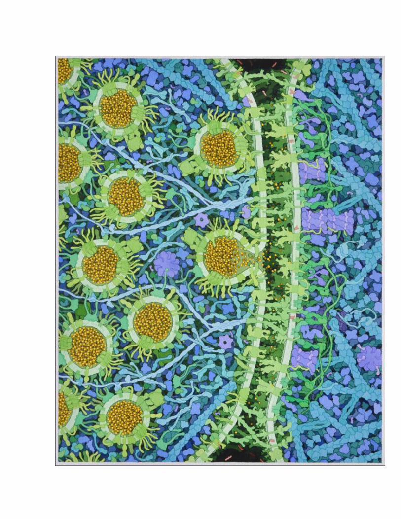
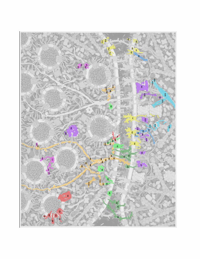
Goodsell Landscape….Glutamate Signaling Synapse. The overall process of neurotransmitter release forms a cycle, with neurotransmitters stored in small vesicles, which fuse with the synaptic membrane and release neurotransmitters, and finally, the empty vesicles are endocytosed and regenerated [1, 2]. The illustration includes this entire cycle, based on transmission EM images of thin sections, in particular, Figure 3A of reference [3]. EM images reveal a clustering of synaptic vesicles in the presynaptic cell, held in place and guided to the membrane by strings of proteins. A protein-rich region just inside the membrane is termed the "presynaptic density." A narrow synapse separates the two cells. Inside the postsynaptic cell, a dense network of proteins, termed the "postsynaptic density" or PSD, organizes reception of the signal. The synaptic vesicles and SNARE complexes are based on the same sources as used for vesicles in the neuromuscular synapse illustration [4]. This includes molecules involved in fusion synaptobrevin, synaptophysin, and synaptotagnin; the transporter vGlut; vesicular ATPase; and Rab3 (described below). Two additional proteins were added. Synuclein (Q16143, 134 amino acids) associates with synaptobrevin, and is based on an NMR structure of the protein bound to micelles [5]. SV2A (Q7L0J3, 742 amino acids) plays a modulatory role in vesicle exocytosis, but its precise role is unclear. It is based on an EM tomograph [6]. Syntaxin and SNAP25 are included in the presynaptic membrane. Key - shown in red (synaptic vesicle) A1. synaptobrevin A2. synaptophysin A3. synaptotagnin A4. vGlut A5. vesicular ATPase A6. SV2A A7. synuclein shown in green (presynaptic membrane) D2. syntaxin D3. SNAP25 Presynaptic Density A complex infrastructure of proteins is used to store and deliver synaptic vesicles to the synapse. These are seen in the EM to be protein-dense complexes near the synaptic membrane, extending to long filaments that connect the vesicles [7-9] The illustration is based on schematic diagrams from several papers. The primary reference is Figure 4 from [10], which shows both presynaptic and postsynaptic structures. This diagram includes LAR and neurexin in the membrane, connected to CASK and liprin-alpha, which then associate with RIM, ELKS, Bassoon, Piccolo (as well as Veli and Mint), and ultimately associate with vesicles through PRA1 and Rab3. Figure 3 of [11] adds a connection to the membrane-bound calcium channel through RIM, also including RIM-BP in the interaction, and also includes Munc13 bound to RIM and Rab3. The figure also shows an inactive, soluble, dimeric form of Munc13. Figure 1 of [12] and Figure 1 of [13] are largely consistent with this arrangement. Figure 4 of [14] shows RIM and RIM-BP associated with Rab3 on a docking vesicle, which I have included on the left side of the illustration. Figure 5 of [15] shows Munc13 assisting with the formation of the SNARE complex through interaction with Munc18. The structures of these proteins are based largely on domain assignments in UniProt. LAR (P10586, 1907 amino acids) has a large extracellular portion with three IG domains and eight fibronectin domains, a short transmembrane segment, and two phosphatase cytoplasmic domains. Neurexin is discussed below in "Cell Adhesion Molecules". RIM (Q9U026, 1411 amino acids) has a Rab-binding domain at the N-terminal end, a PDZ and C2 domain near the center, and a C-terminal C2 domain. RIM-BP (O15034, 1052 amino acids) has three SH3 domains and three fibronectin domains. CASK (O14936, 926 amino acids) has kinase domains at the two ends, and L27, PDZ and SH3 domains in the center. Munc13 (O14795, 1591 amino acids) also contains several types of domains and is thought to dimerize through a short coiled-coil domain at its center. Several of the proteins are predicted to have long coiled-coil sections. Liprin-alpha (O75145, 1194 amino acids) has three SAM domains at the C-terminus, which forms a homodimer or heterodimer with liprin-beta. The association of the SAM domains and interaction with CASK is taken from PDB entry 3tad. The coiled-coil segment, if it forms a single alpha helix, would extend 90 nm. ELKS (Q8IUD1, 1116 amino acids) has a small domain at the C-terminus and the remaining portion is predicted to be largely coiled-coil. In an alpha helix, this would extend 130 nm. Bassoon (Q9UPA5, 3926 amino acids) and

piccolo (Q9Y6V0, 5065 amino acids) are huge proteins depicted as long tethers that extend away from the membrane. They have several zinc fingers near the N-termini, and piccolo has several PDZ domains at the C-terminus. In the illustration, I have drawn all of these proteins as a speculative alpha-helical bundle, held together by liprin SAM domains and forming two branches for bassoon and piccolo. PRA1 (Q9UI14, 185 amino acids) forms the connection to Rab3 and the vesicles. Key - shown in orange B1. LAR B2. CASK B3. liprin alpha and beta B4. ELKS B5. piccolo B6. bassoon B7. PRA1 B8. RIM B9. RIM-BP B10. Munc13 B10i. inactive Munc13 B11. Munc18 Synapsin and Rab3A Synapsin is involved in storage and release of synaptic vesicles by tethering vesicles to the actin cytoskeleton and linking neighboring vesicles [16]. It is plentiful, at about 8 molecules per vesicle. Its function is regulated through phosphorylation by CaMKII [17], shown here tethered to synapsin [16]. Synapsin (P17600, 705 amino acids) has several domains. A large globular domain at the center (the "C" domain, PDB entry 1auv) mediates dimerization, interacts with actin, and has amphipathic sequences that insert into the surface of the vesicle membrane. The N-terminal end interacts with phospholipids, and a proline-rich C-terminal region interacts with many other proteins. Based on Figures from [16], the illustration shows synapsin forming a bridge to actin and forming dimers between neighboring vesicles. Rab3, Rabphilin 3A and GDI are involved in regulation of synaptic vesicle fusion [2]. The complex of Rab3 (P20336, 220 amino acids) and the rab-binding domain of rabphilin 3A (Q9Y2J0, 694 amino acids) are included in PDB entry 1zbd. The structure of GDI (P31150, 447 amino acids) is included in PDB entry 1d5t. In the illustration, rab3 is shown mediating connections between vesicles in storage and the proteins synapsin and PRA (and on to bassoon and piccolo), which are released through association with rabphilin. Then, GDI extracts rab3 from the docked vesicles and delivers it back to vesicles in storage. Key - shown in magenta C1. synapsin C2. Rab3 C3. rabphilin C4. GDI Presynaptic Membrane The presynaptic membrane includes machinery for vesicle docking and fusion, cell adhesion molecules, and several channels/transporters. The illustration includes voltage-gated calcium channels [18, 19], which are involved in neurotransmitter release and also serve to organize proteins in the presynaptic density. Syntaxin and SNAP25 are also included in the membrane, as well as NSF in the cytoplasm, which unfolds the complex after fusion. These are all based on the same sources as the neuromuscular synapse illustration. Uptake of glutamate is medated by excitory amino acid transporters (EAAT, P43003, 542 amino acids. They are found mostly in the astroglia, as part of a cyclic system where glutamate is imported into astroglia, converted to glutamine (which is not active as a neurotransmitter) and released, and the glutamine is taken up by the nerve cell and converted back to glutamate [20]. Some of the transporters are found outside the PSD region, and one is shown there in the illustration based on the aspartate transporter in PDB entry 2nwl. Key - shown in green D1. voltage-gated calcium channel D2. syntaxin
D3. SNAP25 D4. EAAT D5. NSF Synaptic Cleft and Cell Adhesion Molecules The cleft measures 20 nm in conventional EM and 24 nm in cryo-EM [21]. It is densely filled with protein, most of which is thought to be proteins forming transsynaptic connections. (DSG: I haven't been able to find information about extracellular matrix proteins in the synapse.) A variety of proteins form transsynaptic connections [22]. LAR (P10586, 1907 amino acids) binds to NGL-3 (Q9NT99, 713 amino acids) [23]. LAR has a large extracellular domain with 3 Ig domains and 8 fibronectin domains (PDB entry 2yd5 includes one of the Ig domains), a short transmembrane segment, and two phosphatase domains on the cytoplasmic side (PDB entry 1lar). NGL-3 has a large extracellular domain with many leucine rich repeats and an Ig domain (included in PDB entry 2id5), a short transmembrane segment and just over 100 amino acids on the cytoplasmic side. NGL-3 also binds to netrin G2 (Q96CW9, 530 amino acids), which has a laminin domain, three EGF domains, and is lipidated at the C-terminal end. PDB entry 3tbd includes the laminin domain and the first EGF domain. Neurexin (Q9ULB1, 1447 amino acids) has a string of laminin and EGF domains and a short transmembrane segment. Based on the Uniprot annotation, the second laminin domain is shown associated with neuroligin (Q8N2Q7, 840 amino acids, which has a ~690 amino acid extracellular domain, a transmembrane segment, and a cytoplasmic domain that interacts with PSD-95 in the PSD [24]. The complex of extracellular domains of neurexin and neuroligin is included in PDB entry 1biw. Key - shown in dark green E1. NGL-3 E2. neuroligin E3. neurexin E4. netrin E5. cadherin E6. catenins Receptors Several receptors are included in the illustration, based on Figure 4 of [21]. The primary receptors are NMDAR and AMPAR, which are found in the region of the postsynaptic density. Each is composed of four similar chains, with a large portion in the synaptic cleft, three transmembrane segments, and a variable cytoplasmic tail at the C-terminal end [25]. The NMDAR tails are long and involved in interaction with proteins in the PSD [26], for instance, NMDAR-1 (Q05586, 938 amino acids) has 104 amino acids in the tail and NMDAR-2A (Q12879, 1464 amino acids) has 626 amino acids in the tail. The AMPAR tails are shorter, but it associates with stargazin (O88602, 323 amino acids), which has four transmembrane segments and a cytoplasmic tail of 120 amino acids that associates with proteins in the PSD. The inward rectifier potassium ion channel (P63252, 427 amino acids) is also included in the PSD area, drawn as a tetramer based on PDB entry 3sph. A metabotropic glutamate receptor (Q13255, 1194 amino acids) is included in the region flanking the PSD [21], drawn as a homodimer based on annotations in UniProt, and based on the crystal structure of other GPCR. The protein HOMER forms a link to SHANK in the PSD. Retrograde signaling by endocannabinoids [27] is shown in the regions flanking the synapse. The presynaptic membrane includes phospholipase Cbeta (Q01970, 1234 amino acids), which converts phosphatidylinositol to DAG (diacylglycerol), and diacylglycerol lipase (Q8NCG7, 672 amino acids), which converts DAG to 2-AG (2-arachidonoylglycerol). The diacylglycerol lipase structure is based on annotations of transmembrane segments in the UniProt entry, and the phospholipase structure is based on the structure in PDB entry 4gnk, with linkages to SHANK based on the UniProt annotation. An endocannabinoid receptor is shown in the presynaptic membrane, along with a G-protein and adenylate cyclase. Monoacylglycerol lipase (Q99685, 303 amino acids), which breaks down 2-AG, is also included in the presynaptic membrane, based on PDB entry 1mt5. Key - shown in yellow F1. NMDAR F2. AMPAR F3. stargazin F4. potassium ion channel F5. metabotropic glutamate receptor
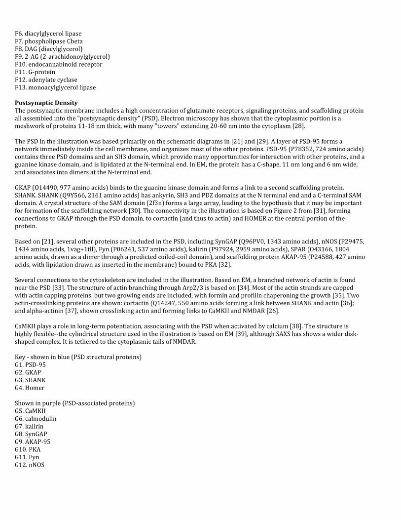
F6. diacylglycerol lipase F7. phospholipase Cbeta F8. DAG (diacylglycerol) F9. 2-AG (2-arachidonoylglycerol) F10. endocannabinoid receptor F11. G-protein F12. adenylate cyclase F13. monoacylglycerol lipase Postsynaptic Density The postsynaptic membrane includes a high concentration of glutamate receptors, signaling proteins, and scaffolding protein all assembled into the "postsynaptic density" (PSD). Electron microscopy has shown that the cytoplasmic portion is a meshwork of proteins 11-18 nm thick, with many "towers" extending 20-60 nm into the cytoplasm [28]. The PSD in the illustration was based primarily on the schematic diagrams in [21] and [29]. A layer of PSD-95 forms a network immediately inside the cell membrane, and organizes most of the other proteins. PSD-95 (P78352, 724 amino acids) contains three PSD domains and an SH3 domain, which provide many opportunities for interaction with other proteins, and a guanine kinase domain, and is lipidated at the N-terminal end. In EM, the protein has a C-shape, 11 nm long and 6 nm wide, and associates into dimers at the N-terminal end. GKAP (O14490, 977 amino acids) binds to the guanine kinase domain and forms a link to a second scaffolding protein, SHANK. SHANK (Q9Y566, 2161 amino acids) has ankyrin, SH3 and PDZ domains at the N terminal end and a C-terminal SAM domain. A crystal structure of the SAM domain (2f3n) forms a large array, leading to the hypothesis that it may be important for formation of the scaffolding network [30]. The connectivity in the illustration is based on Figure 2 from [31], forming connections to GKAP through the PSD domain, to cortactin (and thus to actin) and HOMER at the central portion of the protein. Based on [21], several other proteins are included in the PSD, including SynGAP (Q96PV0, 1343 amino acids), nNOS (P29475, 1434 amino acids, 1vag+1tll), Fyn (P06241, 537 amino acids), kalirin (P97924, 2959 amino acids), SPAR (O43166, 1804 amino acids, drawn as a dimer through a predicted coiled-coil domain), and scaffolding protein AKAP-95 (P24588, 427 amino acids, with lipidation drawn as inserted in the membrane) bound to PKA [32]. Several connections to the cytoskeleton are included in the illustration. Based on EM, a branched network of actin is found near the PSD [33]. The structure of actin branching through Arp2/3 is based on [34]. Most of the actin strands are capped with actin capping proteins, but two growing ends are included, with formin and profilin chaperoning the growth [35]. Two actin-crosslinking proteins are shown: cortactin (Q14247, 550 amino acids forming a link between SHANK and actin [36]; and alpha-actinin [37], shown crosslinking actin and forming links to CaMKII and NMDAR [26]. CaMKII plays a role in long-term potentiation, associating with the PSD when activated by calcium [38]. The structure is highly flexible--the cylindrical structure used in the illustration is based on EM [39], although SAXS has shows a wider disk-shaped complex. It is tethered to the cytoplasmic tails of NMDAR. Key - shown in blue (PSD structural proteins) G1. PSD-95 G2. GKAP G3. SHANK G4. Homer Shown in purple (PSD-associated proteins) G5. CaMKII G6. calmodulin G7. kalirin G8. SynGAP G9. AKAP-95 G10. PKA G11. Fyn G12. nNOS
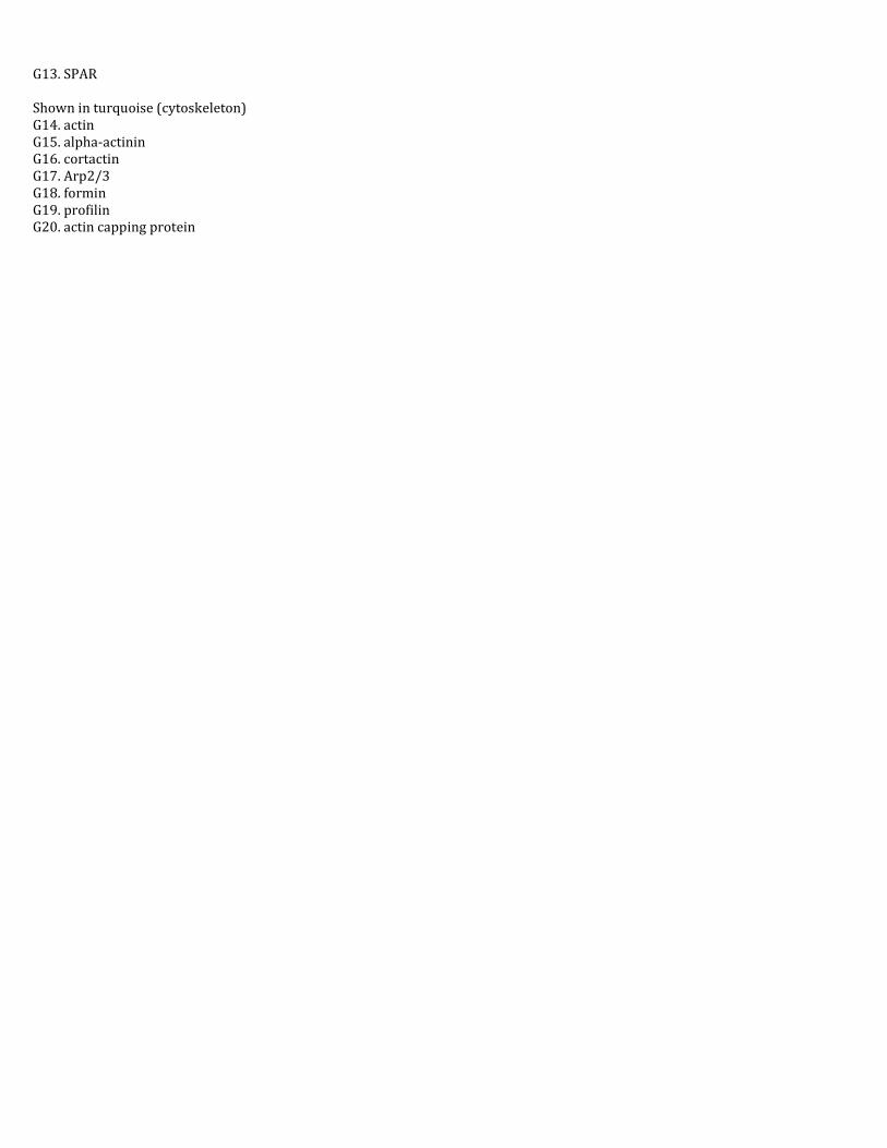
G13. SPAR Shown in turquoise (cytoskeleton) G14. actin G15. alpha-actinin G16. cortactin G17. Arp2/3 G18. formin G19. profilin G20. actin capping protein