Embed Size (px)
Citation preview
29
Toxic anterior segment syndromeSíndrome tóxica do segmento anterior
Luiz Filipe de Albuquerque Alves1, Marcelo Jarczun Kac2, Tiago Bisol3, Bruno Franco Fernandes4,Demian Temponi Eskenazi5
ABSTRACT
Toxic anterior segment syndrome is acute inflammatory reaction caused by a noninfectious substance that enters the anterior segment,resulting in extracellular damage with necrosis and apoptosis during an immune response. We have the report of a case of toxic anteriorsegment syndrome (TASS), in which the authors seek to emphasize the most common causes of the appearance of these syndrome. Theypoint out the care that must be taken in the process of sterilization of surgical material, in addition to reviewing the best conduct whenfaced with these cases. In conclusion, it was noted that the main focus should be on prevention, as treatment only seeks to suppress thesecondary inflammatory response. Treatment in cases of toxic anterior segment syndrome (TASS) consists of intense instillation oftopical steroids with strict follow-up and control of late complications such as glaucoma.
Keywords: Anterior eye segment/drug effects; Anterior eye segment/pathology; Ophthalmic solutions/adverse effects; Syndrome;Case reports
RESUMO
A síndrome tóxica do segmento anterior (STSA) é uma severa reação inflamatória aguda causada por agente não infeccioso queentra no segmento anterior, resultando em lesão celular tóxica com necrose e apoptose mediado por resposta imunológica. Nesterelato de caso de STSA são enfatizadas as causas mais comuns para o aparecimento da síndrome, apontam para os cuidados quedevem ser tomados no processo de esterilização do material cirúrgico além de revisar a melhor conduta diante desses casos. Emconclusão notou-se que o foco principal deve ser a prevenção, pois o tratamento busca apenas suprimir a resposta inflamatóriasecundária. O tratamento nos casos de STSA consiste em intensa instilação de esteróides tópicos com seguimento rigoroso e contro-le de complicações tardias como o glaucoma.
Descritores: Segmento anterior do olho/efeito de drogas e patologia; Soluções oftálmicas/efeitos adversos; Síndrome; Relatode caso
1Air Force Central Hospital (HCA), Rio de Janeiro (RJ), Brazil.2Glaucoma Sectors of Pedro Ernesto University Hospital and Antônio Pedro University Hospital, Rio de Janeiro (RJ), Brazil.3Catholic University of Rio de Janeiro (PUC-RJ), Rio de Janeiro (RJ), Brazil.4Department of Ophthalmology & Pathology, Henry C. Witelson Ocular Pathology Laboratory. McGill University, Montreal, Canada5Retina and Vitreous Sector of the Federal Hospital of the Civil Servants of the State of Rio de Janeiro, Rio de Janeiro (RJ), Brazil.
ORIGINAL ARTICLE
The authors declare no conflicts of interest
Rev Bras Oftalmol. 2013; 72 (1): 29-33
Received for publication: 5/4/2012 - Accepted for publication:15/7/2012
30
INTRODUCTION
Cataract extraction with lens implant is currently the mostcommon surgical procedure in the United States(1). Dueto its small surgical trauma, phacoemulsification has great
success rates, with minimal postoperative inflammation and short-term visual recovery. In recent years, the toxic anterior segmentsyndrome (TASS) has been described as a sterile endophthalmitisaffecting the anterior chamber. The syndrome is related to toxicsubstances injected into or around the eye or even used topicallyduring the procedure(2).
Toxic anterior segment syndrome is a severe acuteinflammatory reaction caused by non-infectious agents that enterthe anterior segment resulting in toxic cell damage with necrosisand apoptosis mediated by an immune response(2). The processtypically begins 12 to 48 hours after anterior segment surgeryand often improves with steroid use. Gram staining and a negativeculture exclude infectious endophthalmitis, which is the primarydifferential diagnosis(3).
The aim of this report is to describe the investigationconducted during the postoperative period of a routine cataractsurgery performed in a public federal hospital without operativecomplications, which progressed with intense inflammatoryreaction, swelling of the cornea and depigmentation of the iris.The authors emphasise the most common causes of TASS, pointingto the necessary precautions in terms of sterilisation of surgicalmaterials and the need to review the best treatment for thesecases.
Case report
White, female, 76 year-old patient with hypertension andcardiac arrhythmia, using amiodarone and having usedhydroxychloroquine for 17 years without a confirmed diagnosis ofrheumatoid arthritis or systemic lupus erythematosus. She soughtophthalmic care due to senile cataract with reduced vision andblurring. On ophthalmic examination (biomicroscopy) senile cataract(grade II) was found in both eyes (BE); no abnormalities were seenin the anterior chamber (AC); intraocular pressure by applanationtonometry was 15 mmHg in BE; corrected visual acuity was 20/40
J2 in the right eye (RE) and 20/30 J2 in the left eye (LE); fundoscopyshowed macular epiretinal membrane (macular pucker) in the RE,a normal posterior pole in the LE, and a periphery without significantchanges in BE; potential acuity (PAM) was 20/20 with lensexchange in the RE and 20/20 in the LE.
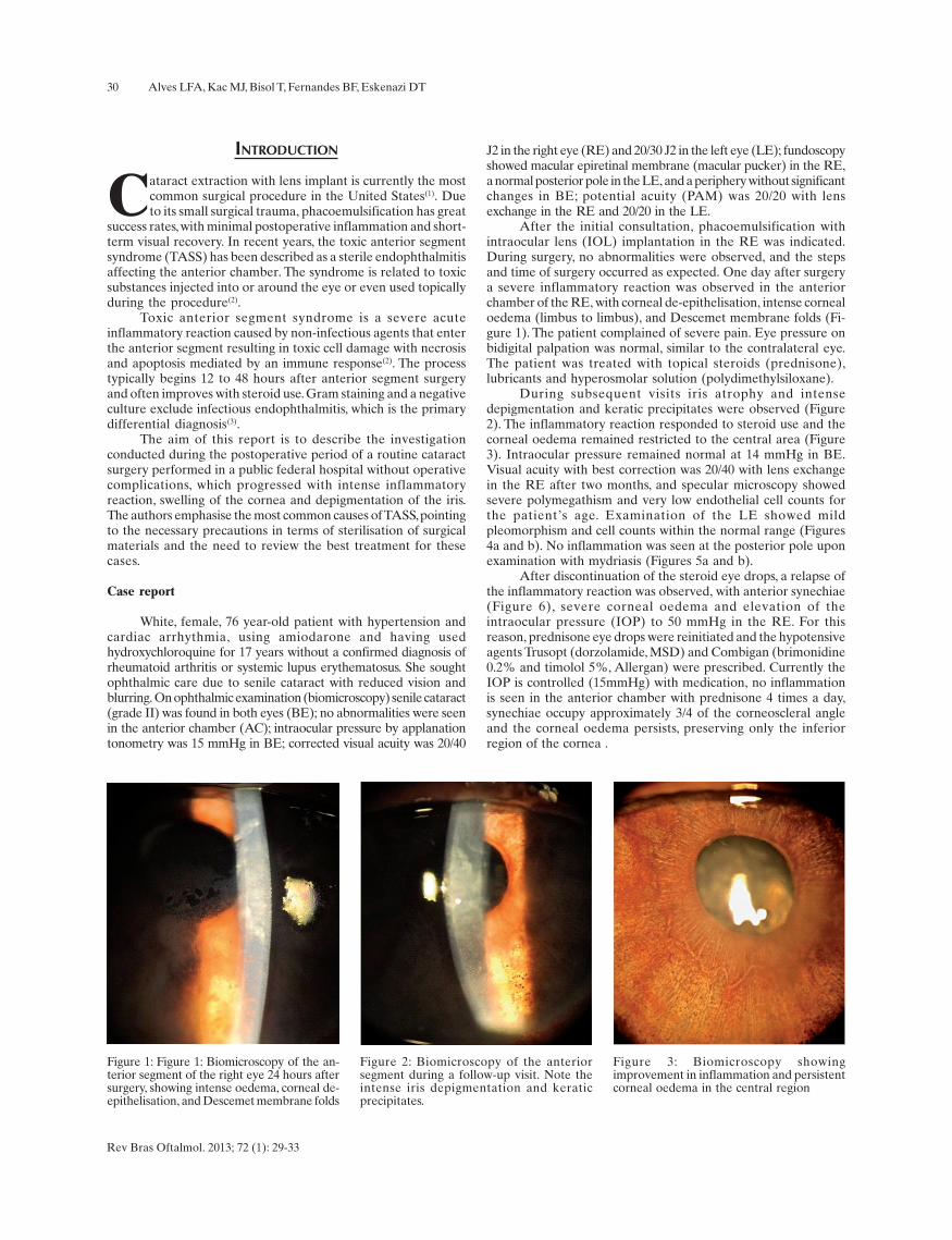
After the initial consultation, phacoemulsification withintraocular lens (IOL) implantation in the RE was indicated.During surgery, no abnormalities were observed, and the stepsand time of surgery occurred as expected. One day after surgerya severe inflammatory reaction was observed in the anteriorchamber of the RE, with corneal de-epithelisation, intense cornealoedema (limbus to limbus), and Descemet membrane folds (Fi-gure 1). The patient complained of severe pain. Eye pressure onbidigital palpation was normal, similar to the contralateral eye.The patient was treated with topical steroids (prednisone),lubricants and hyperosmolar solution (polydimethylsiloxane).
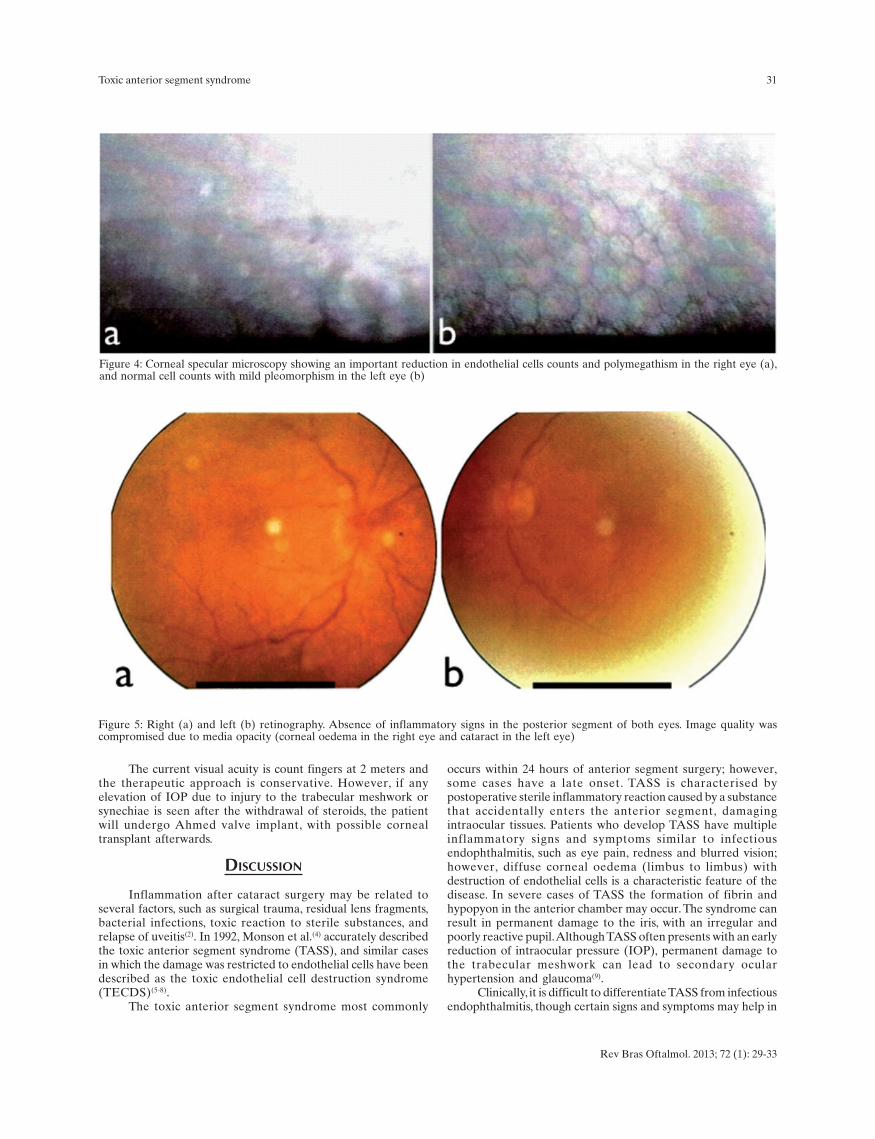
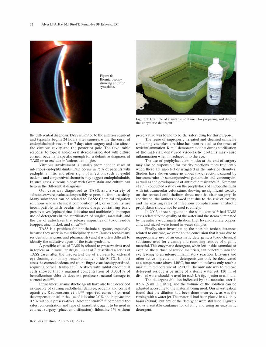
During subsequent visits iris atrophy and intensedepigmentation and keratic precipitates were observed (Figure2). The inflammatory reaction responded to steroid use and thecorneal oedema remained restricted to the central area (Figure3). Intraocular pressure remained normal at 14 mmHg in BE.Visual acuity with best correction was 20/40 with lens exchangein the RE after two months, and specular microscopy showedsevere polymegathism and very low endothelial cell counts forthe patient’s age. Examination of the LE showed mildpleomorphism and cell counts within the normal range (Figures4a and b). No inflammation was seen at the posterior pole uponexamination with mydriasis (Figures 5a and b).
After discontinuation of the steroid eye drops, a relapse ofthe inflammatory reaction was observed, with anterior synechiae(Figure 6), severe corneal oedema and elevation of theintraocular pressure (IOP) to 50 mmHg in the RE. For thisreason, prednisone eye drops were reinitiated and the hypotensiveagents Trusopt (dorzolamide, MSD) and Combigan (brimonidine0.2% and timolol 5%, Allergan) were prescribed. Currently theIOP is controlled (15mmHg) with medication, no inflammationis seen in the anterior chamber with prednisone 4 times a day,synechiae occupy approximately 3/4 of the corneoscleral angleand the corneal oedema persists, preserving only the inferiorregion of the cornea .
Figure 1: Figure 1: Biomicroscopy of the an-terior segment of the right eye 24 hours aftersurgery, showing intense oedema, corneal de-epithelisation, and Descemet membrane folds
Figure 2: Biomicroscopy of the anteriorsegment during a follow-up visit. Note theintense iris depigmentation and keraticprecipitates.
Figure 3: Biomicroscopy showingimprovement in inflammation and persistentcorneal oedema in the central region
Rev Bras Oftalmol. 2013; 72 (1): 29-33
Alves LFA, Kac MJ, Bisol T, Fernandes BF, Eskenazi DT
31
The current visual acuity is count fingers at 2 meters andthe therapeutic approach is conservative. However, if anyelevation of IOP due to injury to the trabecular meshwork orsynechiae is seen after the withdrawal of steroids, the patientwill undergo Ahmed valve implant, with possible cornealtransplant afterwards.
DISCUSSION
Inflammation after cataract surgery may be related toseveral factors, such as surgical trauma, residual lens fragments,bacterial infections, toxic reaction to sterile substances, andrelapse of uveitis(2). In 1992, Monson et al.(4) accurately describedthe toxic anterior segment syndrome (TASS), and similar casesin which the damage was restricted to endothelial cells have beendescribed as the toxic endothelial cell destruction syndrome(TECDS)(5-8).
The toxic anterior segment syndrome most commonly
occurs within 24 hours of anterior segment surgery; however,some cases have a late onset. TASS is characterised bypostoperative sterile inflammatory reaction caused by a substancethat accidentally enters the anterior segment, damagingintraocular tissues. Patients who develop TASS have multipleinflammatory signs and symptoms similar to infectiousendophthalmitis, such as eye pain, redness and blurred vision;however, diffuse corneal oedema (limbus to limbus) withdestruction of endothelial cells is a characteristic feature of thedisease. In severe cases of TASS the formation of fibrin andhypopyon in the anterior chamber may occur. The syndrome canresult in permanent damage to the iris, with an irregular andpoorly reactive pupil. Although TASS often presents with an earlyreduction of intraocular pressure (IOP), permanent damage tothe trabecular meshwork can lead to secondary ocularhypertension and glaucoma(9).
Clinically, it is difficult to differentiate TASS from infectiousendophthalmitis, though certain signs and symptoms may help in
Figure 4: Corneal specular microscopy showing an important reduction in endothelial cells counts and polymegathism in the right eye (a),and normal cell counts with mild pleomorphism in the left eye (b)
Figure 5: Right (a) and left (b) retinography. Absence of inflammatory signs in the posterior segment of both eyes. Image quality wascompromised due to media opacity (corneal oedema in the right eye and cataract in the left eye)
Rev Bras Oftalmol. 2013; 72 (1): 29-33
Toxic anterior segment syndrome
32
the differential diagnosis. TASS is limited to the anterior segmentand typically begins 24 hours after surgery, while the onset ofendophthalmitis occurs 4 to 7 days after surgery and also affectsthe vitreous cavity and the posterior pole. The favourableresponse to topical and/or oral steroids associated with diffusecorneal oedema is specific enough for a definitive diagnosis ofTASS or to exclude infectious aetiologies.
Vitreous involvement is usually prominent in cases ofinfectious endophthalmitis. Pain occurs in 75% of patients withendophthalmitis, and other signs of infection, such as eyelidoedema and conjunctival chemosis may suggest endophthalmitis.In such cases, vitreous biopsy with Gram stain and culture canhelp in the differential diagnosis.
Our case was diagnosed as TASS, and a variety ofsubstances were evaluated as possibly responsible for the toxicity.Many substances can be related to TASS: Chemical irrigationsolutions whose chemical composition, pH, or osmolality areincompatible with ocular tissues, drugs containing toxicpreservatives (epinephrine, lidocaine and antibiotics), improperuse of detergents in the sterilisation of surgical materials, andthe use of autoclaves that release impurities or toxic residue(copper, zinc, nickel, and silica)(9,10).
TASS is a problem for ophthalmic surgeons, especiallybecause they work in multidisciplinary team (nurses, technicians,residents, physicians, and pharmacists) and it is often difficult toidentify the causative agent of the toxic syndrome.
A possible cause of TASS is related to preservatives usedin topical or intraocular drugs. Liu et al.(7) described a series ofTASS cases after the inadvertent use of a cream for externaleye cleaning containing benzalkonium chloride 0.01%. In mostcases the corneal oedema and count-finger visual acuity persisted,requiring corneal transplant(7). A study with rabbit endothelialcells showed that a maximal concentration of 0.0001% ofbenzalkonium chloride does not produce structural damage tocorneal cells(11).
Intracamerular anaesthetic agents have also been describedas capable of causing endothelial damage, oedema and cornealopacities. Kadonosono et al.(12) reported cases of cornealdecompensation after the use of lidocaine 2.0% and bupivacaine0.5% without preservatives. Another study(13,14) compared thesafest concentration and type of anaesthetic agent to be used incataract surgery (phacoemulsification); lidocaine 1% without
Figure 6:Biomicroscopyshowing anteriorsynechiae.
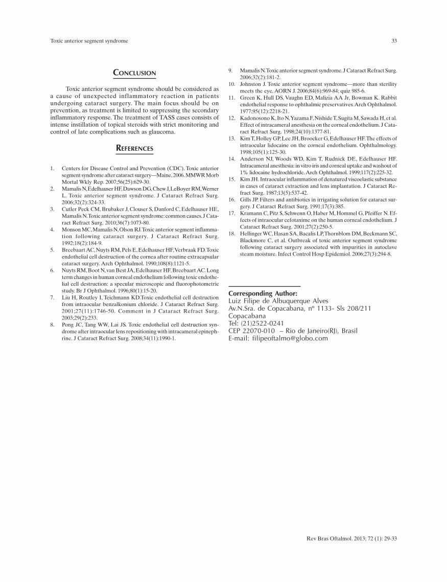
Figure 7: Example of a suitable container for preparing and dilutingthe enzymatic detergent.
Rev Bras Oftalmol. 2013; 72 (1): 29-33
Alves LFA, Kac MJ, Bisol T, Fernandes BF, Eskenazi DT
preservative was found to be the safest drug for this purpose.The reuse of improperly irrigated and cleansed cannulae
containing viscoelastic residue has been related to the onset oftoxic inflammation. Kim(15) demonstrated that during sterilisationof the material, denatured viscoelastic proteins may causeinflammation when introduced into the eye.
The use of prophylactic antibiotics at the end of surgerymay also be responsible for toxicity reactions, more frequentlywhen these are injected or irrigated in the anterior chamber.Studies have shown concerns about toxic reactions caused byintracamerular or subconjunctival gentamicin and vancomycin,as well as the development of antibiotic resistance(16). Kramannet al.(17) conducted a study on the prophylaxis of endophthalmitiswith intracamerular cefotaxime, showing no significant toxicityon the corneal endothelium three months after surgery. Inconclusion, the authors showed that due to the risk of toxicityand the existing rates of infectious complications, antibioticprophylaxis should not be used routinely.
In 2002, three surgeons in the same centre(18) had TASScases related to the quality of the water and the steam eliminatedby the autoclave during sterilisation. High levels of sulfate, copper,zinc, and nickel were found in water samples.
Finally, after investigating the possible toxic substancesrelated to our case, we came to the conclusion that it was due toinappropriate use of an enzymatic detergent, a toxic chemicalsubstance used for cleaning and removing residue of organicmaterial. This enzymatic detergent, when left inside cannulae oreven on the surface of reused materials, can penetrate into theeye leading to an intense inflammatory reaction. Enzymes andother active ingredients in detergents can only be deactivatedat a temperature above 140°C, but most autoclaves only reach amaximum temperature of 120°C(9). The only safe way to removedetergent residue is by using of a sterile water jet; 120 ml ofdistilled water should be used for each I/A tip, injector or cannula.
The detergent dilution indicated by the manufacturer is0.5% (5 ml in 1 litre), and the volume of the solution can beadjusted according to the material being used. Our investigationfound that the dilution had been done incorrectly, as was therinsing with a water jet. The material had been placed in a kidneybasin (500ml), but 5ml of the detergent were still used. Figure 7shows a suitable container for diluting and using an enzymaticdetergent.
33
Corresponding Author:Luiz Filipe de Albuquerque AlvesAv.N.Sra. de Copacabana, nº 1133- Sls 208/211CopacabanaTel: (21)2522-0241CEP 22070-010 – Rio de Janeiro(RJ), BrasilE-mail: [email protected]
Rev Bras Oftalmol. 2013; 72 (1): 29-33
Toxic anterior segment syndrome
CONCLUSION
Toxic anterior segment syndrome should be considered asa cause of unexpected inflammatory reaction in patientsundergoing cataract surgery. The main focus should be onprevention, as treatment is limited to suppressing the secondaryinflammatory response. The treatment of TASS cases consists ofintense instillation of topical steroids with strict monitoring andcontrol of late complications such as glaucoma.
REFERENCES
1. Centers for Disease Control and Prevention (CDC). Toxic anteriorsegment syndrome after cataract surgery—Maine, 2006. MMWR MorbMortal Wkly Rep. 2007;56(25):629-30.
2. Mamalis N, Edelhauser HF, Dawson DG, Chew J, LeBoyer RM, WernerL. Toxic anterior segment syndrome. J Cataract Refract Surg.2006;32(2):324-33.
3. Cutler Peck CM, Brubaker J, Clouser S, Danford C, Edelhauser HE,Mamalis N. Toxic anterior segment syndrome: common causes. J Cata-ract Refract Surg. 2010;36(7):1073-80.
4. Monson MC, Mamalis N, Olson RJ. Toxic anterior segment inflamma-tion following cataract surgery. J Cataract Refract Surg.1992;18(2):184-9.
5. Breebaart AC, Nuyts RM, Pels E, Edelhauser HF, Verbraak FD. Toxicendothelial cell destruction of the cornea after routine extracapsularcataract surgery. Arch Ophthalmol. 1990;108(8):1121-5.
6. Nuyts RM, Boot N, van Best JA, Edelhauser HF, Breebaart AC. Longterm changes in human corneal endothelium following toxic endothe-lial cell destruction: a specular microscopic and fluorophotometricstudy. Br J Ophthalmol. 1996;80(1):15-20.
7. Liu H, Routley I, Teichmann KD.Toxic endothelial cell destructionfrom intraocular benzalkonium chloride. J Cataract Refract Surg.2001;27(11):1746-50. Comment in J Cataract Refract Surg.2003;29(2):233.
8. Pong JC, Tang WW, Lai JS. Toxic endothelial cell destruction syn-drome after intraocular lens repositioning with intracameral epineph-rine. J Cataract Refract Surg. 2008;34(11):1990-1.
9. Mamalis N. Toxic anterior segment syndrome. J Cataract Refract Surg.2006;32(2):181-2.
10. Johnston J. Toxic anterior segment syndrome—more than sterilitymeets the eye. AORN J. 2006;84(6):969-84; quiz 985-6.
11. Green K, Hull DS, Vaughn ED, Malizia AA Jr, Bowman K. Rabbitendothelial response to ophthalmic preservatives. Arch Ophthalmol.1977;95(12):2218-21.
12. Kadonosono K, Ito N, Yazama F, Nishide T, Sugita M, Sawada H, et al.Effect of intracameral anesthesia on the corneal endothelium. J Cata-ract Refract Surg. 1998;24(10):1377-81.
13. Kim T, Holley GP, Lee JH, Broocker G, Edelhauser HF. The effects ofintraocular lidocaine on the corneal endothelium. Ophthalmology.1998;105(1):125-30.
14. Anderson NJ, Woods WD, Kim T, Rudnick DE, Edelhauser HF.Intracameral anesthesia: in vitro iris and corneal uptake and washout of1% lidocaine hydrochloride. Arch Ophthalmol. 1999;117(2):225-32.
15. Kim JH. Intraocular inflammation of denatured viscoelastic substancein cases of cataract extraction and lens implantation. J Cataract Re-fract Surg. 1987;13(5):537-42.
16. Gills JP. Filters and antibiotics in irrigating solution for cataract sur-gery. J Cataract Refract Surg. 1991;17(3):385.
17. Kramann C, Pitz S, Schwenn O, Haber M, Hommel G, Pfeiffer N. Ef-fects of intraocular cefotaxime on the human corneal endothelium. JCataract Refract Surg. 2001;27(2):250-5.
18. Hellinger WC, Hasan SA, Bacalis LP, Thornblom DM, Beckmann SC,Blackmore C, et al. Outbreak of toxic anterior segment syndromefollowing cataract surgery associated with impurities in autoclavesteam moisture. Infect Control Hosp Epidemiol. 2006;27(3):294-8.