Embed Size (px)
Citation preview
Hindawi Publishing CorporationCase Reports in Ophthalmological MedicineVolume 2013, Article ID 910342, 3 pageshttp://dx.doi.org/10.1155/2013/910342
Case ReportFulminant Panuveitis following Iris Suture Fixation ofPosterior Chamber Intraocular Lens
Ahmad M. Mansour1, 2 and Shady T. Awwad1
1 Department of Ophthalmology, American University of Beirut, P.O. Box 1136044, Beirut, Lebanon2Department of Ophthalmology, Rafic Hariri University Hospital, P.O. Box 11-5344, Beirut, Lebanon
Correspondence should be addressed to Ahmad M. Mansour; [email protected]
Received 17 December 2012; Accepted 15 January 2013
Academic Editors: A. A. Bialasiewicz and C. G. Kiss
Copyright © 2013 A. M. Mansour and S. T. Awwad. This is an open access article distributed under the Creative CommonsAttribution License, which permits unrestricted use, distribution, and reproduction in any medium, provided the original work isproperly cited.
We present a case of fulminant panuveitis following iris suture fixation of a posterior chamber intraocular lens. We hypothesizethat the zonular dehiscence allowed the inflammatory cells in the anterior compartment to gain access to the posterior segmentmimicking endophthalmitis or toxic anterior segment syndrome. Also certain bulky lens designs, like the current Raynerhydrophilic acrylic lens, are difficult to manipulate and hold in the optic capture position, and hence the iris fixation of theselenses can be traumatic and lengthy. It is advised to exchange such lenses with 3-piece intraocular lenses that are easy to fixate.
1. Introduction
The concept of iris suture fixation for posterior chamberintraocular lenses dates back to 1976, whenMalcolmMcCan-nel, M.D., described his trans-corneal suture technique tostabilize subluxated posterior intraocular lenses. Since then,iris suture fixation has become a well-established effectivemeans for stabilizing posterior chamber lenses in the lack ofadequate capsular support [1–8]. The technique consisted ofa McCannel 10-0 polypropylene suture which was used tofixate the haptics to the iris using the Siepser sliding knot[5]. In a series of 46 patients [6], the main complicationsof iris suture fixation included transient low-grade uveitisin 3 (6.5%), transient pigment dispersion in 3 (6.5%), andintraocular lens dislocation in 2 (4.3%). Additionally in asecond series of 17 eyes of 9 children [7], other complicationsof iris suture fixation included hyphema in 1 case and sterileendophthalmitis in another case. A case of severe uveitis andsevere visual loss after iris suture fixation is described.
2. Case Report
This 46-year-old Iraqi lady had prior anterior chamberintraocular lens implantation for familial lens subluxationand previous pars plana vitrectomy for retinal detachment
ending with poor vision in the right eye. The left eye under-went scleral buckle for rhegmatogenous retinal detachmentwith findings of severe scleral thinning. Subsequently she hadphacoemulsification with hydrophilic acrylic intraocular lens(Superflex, Rayner Intraocular Lenses Ltd, East Sussex, UK;6.25mm optic and 12.5mm overall length) implantation inthe bag with zonular dehiscence. Visual acuity was 6/9 inthe left eye with mild decentration (Figure 1). The patientwas referred for scleral fixation of the lens. Because of scleralthinning and history of retinal detachment, we proceededwith iris suture fixation. Two surgeons were working simul-taneously through several keratome incisions, and it wasnecessary to place repeated viscoelastic (2.4mL of sodiumhyaluronate) to avoid corneal touch by the lens. The lens wasfixated by the first surgeon using intraocular forceps usedin vitreous surgery in the right hand and a Sinskey hook tomaintain optic capturewith the left hand.The second surgeonperformed iris suture fixation superiorly and inferiorly. Theinferior suture led to lens tilt that was not relieved by gentleiris massage around the suture and thereafter that suture wasremoved. Surgery was done at night and lasted 75 minutes.Ten hours postoperatively, the patient had finger counting at10 cm with fibrinous panuveitis (Figure 2) and dense echoeson B-scan (Figure 3). The possibility of endophthalmitiswas discussed with the patient who declined immediate
2 Case Reports in Ophthalmological Medicine
Figure 1: Preoperative anterior segment photograph of the left eyeshows mild subluxation of Rayner Superflex hydrophilic acrylicintraocular lens with uncorrected visual acuity of 6/9.
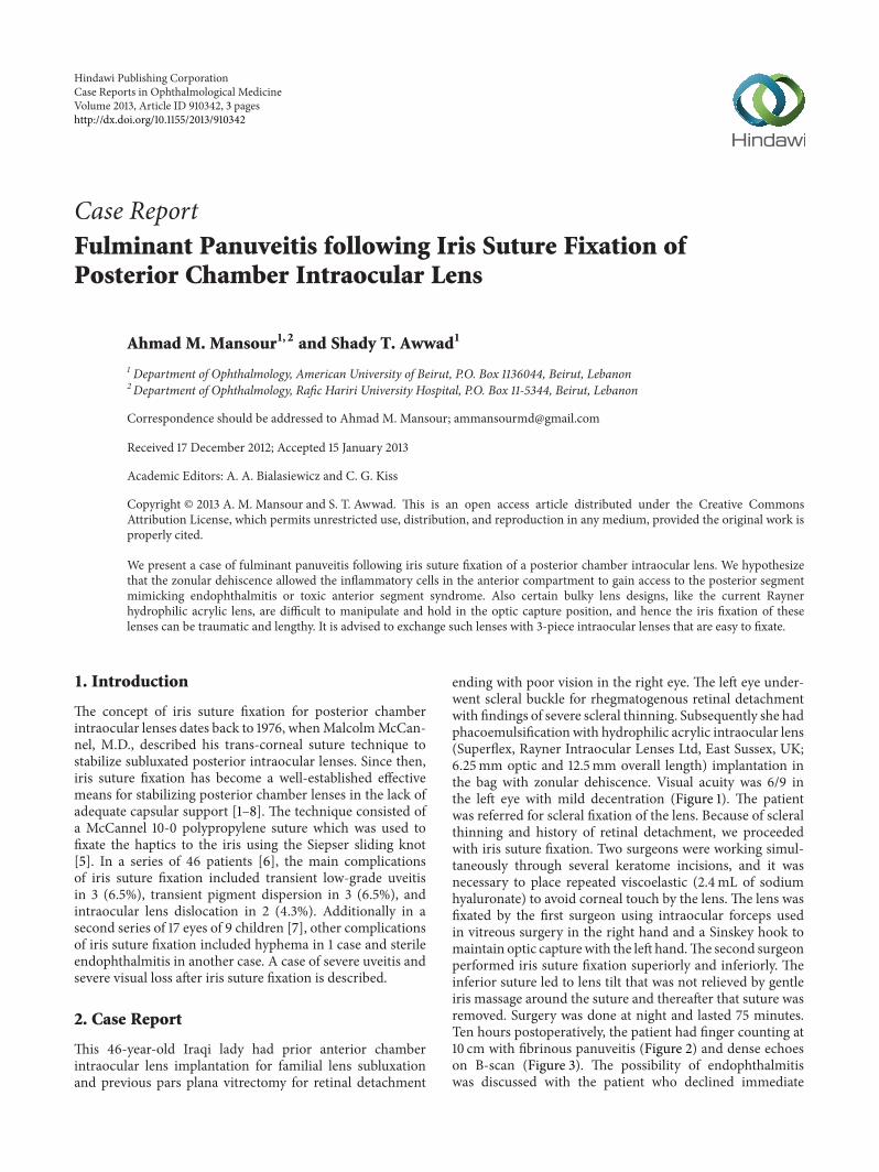
Figure 2: Ten hours after surgery, the left eye has fibrinous iritis withvisual acuity of finger counting at 10 cm.The cornea hasmild stromaledema andDescemetmembrane folds.The pupil ismid-constricted.
aqueous paracentesis for culture purposes. She was thereforefollowed up several times daily initially. Intensive oral andtopical corticosteroid therapy with topical nonsteroidal anti-inflammatory drops controlled the inflammation as earlyas 24 hours after surgery. Visual acuity improved to 6/21one week postoperatively with minimal iritis (Figure 4). Oraland topical corticosteroids (dexamethasone drop every halfhour and 100mg prednisone daily) were substituted withloteprednol etabonate because of severe ocular hypertension(intraocular pressure of 38mmHg). Intraocular pressuredropped to 12mmHg and visual acuity dropped to 6/24 fromresidual mild uveitis on discharge 2 weeks after surgery. Thepatient travelled to Iraq on loteprednol and non-steroidalanti-inflammatory drop. She reported a gradual recovery ofpreoperative vision 5 weeks after surgery. Rheumatologicworkupwas negative (including rheumatology consult, antin-uclear antibodies, and rheumatoid arthritis latex test).
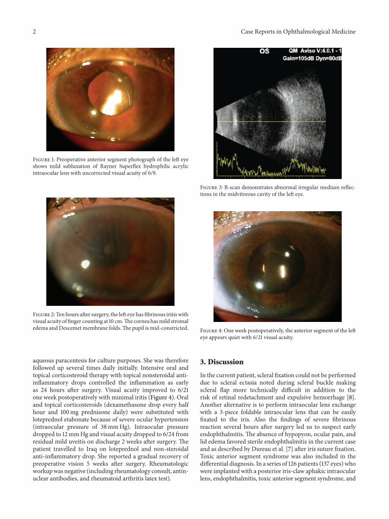
Figure 3: B-scan demonstrates abnormal irregular medium reflec-tions in the midvitreous cavity of the left eye.
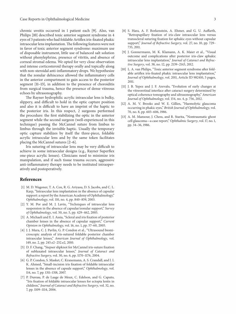
Figure 4: One week postoperatively, the anterior segment of the lefteye appears quiet with 6/21 visual acuity.
3. Discussion
In the current patient, scleral fixation could not be performeddue to scleral ectasia noted during scleral buckle makingscleral flap more technically difficult in addition to therisk of retinal redetachment and expulsive hemorrhage [8].Another alternative is to perform intraocular lens exchangewith a 3-piece foldable intraocular lens that can be easilyfixated to the iris. Also the findings of severe fibrinousreaction several hours after surgery led us to suspect earlyendophthalmitis. The absence of hypopyon, ocular pain, andlid edema favored sterile endophthalmitis in the current caseand as described by Dureau et al. [7] after iris suture fixation.Toxic anterior segment syndrome was also included in thedifferential diagnosis. In a series of 126 patients (137 eyes) whowere implanted with a posterior iris-claw aphakic intraocularlens, endophthalmitis, toxic anterior segment syndrome, and
Case Reports in Ophthalmological Medicine 3
chronic uveitis occurred in 1 patient each [9]. Also, vanPhilips [10] described toxic anterior segment syndrome in 4eyes of 3 patients who had foldable Artiflex iris-fixated phakicintraocular lens implantation.The following featureswere notin favor of toxic anterior segment syndrome: maximum useof disposable instrument, little use of balanced salt solutionwithout phenylephrine, presence of vitritis, and absence ofcorneal stromal edema. We opted for very close observationand intense corticosteroid therapy orally and topically alongwith non-steroidal anti-inflammatory drops. We hypothesizethat the zonular dehiscence allowed the inflammatory cellsin the anterior compartment to gain access to the posteriorsegment [11–13], in addition to the presence of choroiditisfrom surgical trauma, hence the presence of dense vitreousechoes by ultrasonography.
The Rayner hydrophilic acrylic intraocular lens is bulky,slippery, and difficult to hold in the optic capture positionand also it is difficult to have an imprint of the haptic tothe posterior iris. In this respect, 2 surgeons performedthe procedure: the first stabilizing the optic in the anteriorsegment while the second surgeon (well-experienced in thistechnique) passing the McCannel suture from limbus tolimbus through the invisible haptic. Usually the temporaryoptic capture stabilizes by itself the three-piece, foldableacrylic intraocular lens and by the same token facilitatesplacing the McCannel sutures [2–6].
Iris suturing of intraocular lens may be very difficult toachieve in some intraocular designs (e.g., Rayner Superflexone-piece acrylic lenses). Clinicians need to minimize irismanipulation, and if such tissue trauma occurs, aggressiveanti-inflammatory therapy needs to be instituted intraoper-atively and postoperatively.
References
[1] M. D. Wagoner, T. A. Cox, R. G. Ariyasu, D. S. Jacobs, and C. L.Karp, “Intraocular lens implantation in the absence of capsularsupport: a report by theAmericanAcademyofOphthalmology,”Ophthalmology, vol. 110, no. 4, pp. 840–859, 2003.
[2] Y. M. Por and M. J. Lavin, “Techniques of intraocular lenssuspension in the absence of capsular/zonular support,” Surveyof Ophthalmology, vol. 50, no. 5, pp. 429–462, 2005.
[3] A. Michaeli and E. I. Assia, “Scleral and iris fixation of posteriorchamber lenses in the absence of capsular support,” CurrentOpinion in Ophthalmology, vol. 16, no. 1, pp. 57–60, 2005.
[4] J. J. Mura, C. J. Pavlin, G. P. Condon et al., “Ultrasound biomi-croscopic analysis of iris-sutured foldable posterior chamberintraocular lenses,” American Journal of Ophthalmology, vol.149, no. 2, pp. 245.e2–252.e2, 2010.
[5] D. F. Chang, “Siepser slipknot for McCannel iris-suture fixationof subluxated intraocular lenses,” Journal of Cataract andRefractive Surgery, vol. 30, no. 6, pp. 1170–1176, 2004.
[6] G. P. Condon, S.Masket, C. Kranemann, A. S. Crandall, and I. I.K. Ahmed, “Small-incision iris fixation of foldable intraocularlenses in the absence of capsule support,” Ophthalmology, vol.114, no. 7, pp. 1311–1318, 2007.
[7] P. Dureau, P. de Laage de Meux, C. Edelson, and G. Caputo,“Iris fixation of foldable intraocular lenses for ectopia lentis inchildren,” Journal of Cataract and Refractive Surgery, vol. 32, no.7, pp. 1109–1114, 2006.
[8] S. Hara, A. F. Borkenstein, A. Ehmer, and G. U. Auffarth,“Retropupillary fixation of iris-claw intraocular lens versustransscleral suturing fixation for aphakic eyes without capsularsupport,” Journal of Refractive Surgery, vol. 27, no. 10, pp. 729–735, 2011.
[9] J. Gonnermann, M. K. Klamann, A. K. Maier et al., “Visualoutcome and complications after posterior iris-claw aphakicintraocular lens implantation,” Journal of Cataract and Refrac-tive Surgery, vol. 38, no. 12, pp. 2139–2143, 2012.
[10] L. A. van Philips, “Toxic anterior segment syndrome after fold-able artiflex iris-fixated phakic intraocular lens implantation,”Journal of Ophthalmology, vol. 2011, Article ID 982410, 5 pages,2011.
[11] J. B. Yepez and J. F. Arevalo, “Evolution of early changes atthe vitreoretinal interface after cataract surgery determined byoptical coherence tomography and ultrasonography,” AmericanJournal of Ophthalmology, vol. 154, no. 4, p. 756, 2012.
[12] A. M. V. Brooks and W. E. Gillies, “Haemolytic glaucomaoccurring in phakic eyes,” British Journal of Ophthalmology, vol.70, no. 8, pp. 603–606, 1986.
[13] A. M. Mansour, J. Chess, and R. Starita, “Nontraumatic ghostcell glaucoma—a case report,”Ophthalmic Surgery, vol. 17, no. 1,pp. 34–36, 1986.

Submit your manuscripts athttp://www.hindawi.com
Stem CellsInternational
Hindawi Publishing Corporationhttp://www.hindawi.com Volume 2014
Hindawi Publishing Corporationhttp://www.hindawi.com Volume 2014
MEDIATORSINFLAMMATION
of
Hindawi Publishing Corporationhttp://www.hindawi.com Volume 2014
Behavioural Neurology
EndocrinologyInternational Journal of
Hindawi Publishing Corporationhttp://www.hindawi.com Volume 2014
Hindawi Publishing Corporationhttp://www.hindawi.com Volume 2014
Disease Markers
Hindawi Publishing Corporationhttp://www.hindawi.com Volume 2014
BioMed Research International
OncologyJournal of
Hindawi Publishing Corporationhttp://www.hindawi.com Volume 2014
Hindawi Publishing Corporationhttp://www.hindawi.com Volume 2014
Oxidative Medicine and Cellular Longevity
Hindawi Publishing Corporationhttp://www.hindawi.com Volume 2014
PPAR Research
The Scientific World JournalHindawi Publishing Corporation http://www.hindawi.com Volume 2014
Immunology ResearchHindawi Publishing Corporationhttp://www.hindawi.com Volume 2014
Journal of
ObesityJournal of
Hindawi Publishing Corporationhttp://www.hindawi.com Volume 2014
Hindawi Publishing Corporationhttp://www.hindawi.com Volume 2014
Computational and Mathematical Methods in Medicine
OphthalmologyJournal of
Hindawi Publishing Corporationhttp://www.hindawi.com Volume 2014
Diabetes ResearchJournal of
Hindawi Publishing Corporationhttp://www.hindawi.com Volume 2014
Hindawi Publishing Corporationhttp://www.hindawi.com Volume 2014
Research and TreatmentAIDS
Hindawi Publishing Corporationhttp://www.hindawi.com Volume 2014
Gastroenterology Research and Practice
Hindawi Publishing Corporationhttp://www.hindawi.com Volume 2014
Parkinson’s Disease
Evidence-Based Complementary and Alternative Medicine
Volume 2014Hindawi Publishing Corporationhttp://www.hindawi.com