Embed Size (px)
Citation preview

© 2017. Published by The Company of Biologists Ltd.
TOPLESS mediates brassinosteroid control of shoot boundaries and root
meristem development in Arabidopsis thaliana
Ana Espinosa-Ruiz1,*, Cristina Martínez1,*, Miguel de Lucas1,†, Norma
Fàbregas2, Nadja Bosch2, Ana I. Caño-Delgado2 and Salomé Prat1.
1Department of Plant Molecular Genetics. Centro Nacional de Biotecnología-
CSIC. Darwin 3, Madrid E-28049, Spain.
2Department of Molecular Genetics. Centre for Research in Agricultural
Genomics (CRAG) CSIC-IRTA-UAB-UB. Barcelona E-08193, Spain.
*These authors made equal contributions to the manuscript
† Current address: School of Biological and Biomedical Sciences, Durham
University, UK.
Corresponding author: Salomé Prat
E-mail: [email protected]
Phone: +34 91 5854916
FAX: +34 91 5854506
Salomé Prat ([email protected]) is responsible for distribution of materials
integral to the findings presented in this article.
Key words: BR-signaling, BES1, EAR domain, TOPLESS, organ boundary, QC
quiescence
Summary Statement:
BES1 recruitment of the co-repressor TOPLESS to the CUC3 and BRAVO
promoters plays a critical role in BR-mediated control of organ boundary
formation and root QC quiescence.
Dev
elo
pmen
t • A
dvan
ce a
rtic
le
http://dev.biologists.org/lookup/doi/10.1242/dev.143214Access the most recent version at First posted online on 20 March 2017 as 10.1242/dev.143214
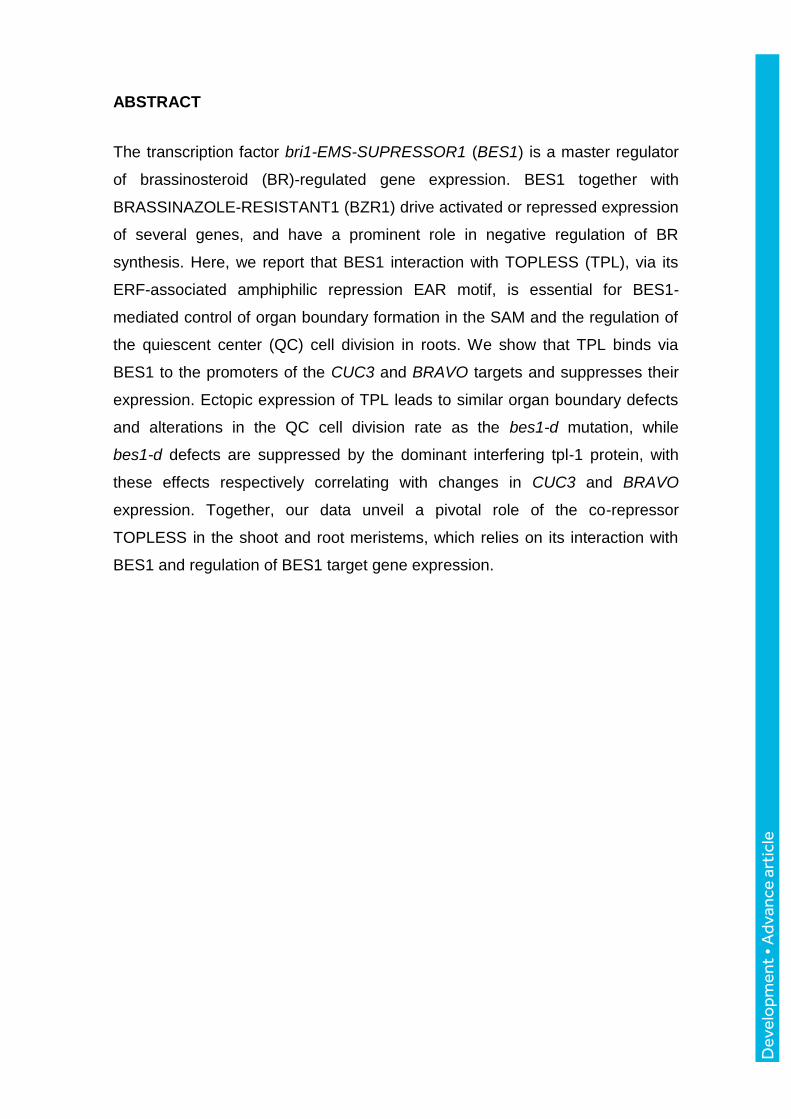
ABSTRACT
The transcription factor bri1-EMS-SUPRESSOR1 (BES1) is a master regulator
of brassinosteroid (BR)-regulated gene expression. BES1 together with
BRASSINAZOLE-RESISTANT1 (BZR1) drive activated or repressed expression
of several genes, and have a prominent role in negative regulation of BR
synthesis. Here, we report that BES1 interaction with TOPLESS (TPL), via its
ERF-associated amphiphilic repression EAR motif, is essential for BES1-
mediated control of organ boundary formation in the SAM and the regulation of
the quiescent center (QC) cell division in roots. We show that TPL binds via
BES1 to the promoters of the CUC3 and BRAVO targets and suppresses their
expression. Ectopic expression of TPL leads to similar organ boundary defects
and alterations in the QC cell division rate as the bes1-d mutation, while
bes1-d defects are suppressed by the dominant interfering tpl-1 protein, with
these effects respectively correlating with changes in CUC3 and BRAVO
expression. Together, our data unveil a pivotal role of the co-repressor
TOPLESS in the shoot and root meristems, which relies on its interaction with
BES1 and regulation of BES1 target gene expression.
Dev
elo
pmen
t • A
dvan
ce a
rtic
le

INTRODUCTION
Brassinosteroids (BR) are steroid plant hormones with an essential role in plant
growth and development (Clouse 2011; Guo et al. 2013). In tight connection
with environmental cues and other plant hormones, BRs control shoot and root
growth and distinct developmental programs such as photomorphogenesis,
organ boundary formation and vascular differentiation (Ibanes et al. 2009; Bell
et al. 2012; Gendron et al. 2012; Wang et al. 2012). BR perception triggers a
signalling cascade that ultimately leads to activation and accumulation of two
homologous transcription factors, bri1-EMS-SUPPRESSOR1 (BES1) and
BRASSINAZOLE RESISTANT1 (BZR1). In the nucleus, BES1 and BZR1
modulate the expression of thousands of genes with a role in cell elongation,
BR synthesis, and in the control of multiple cellular processes (He et al. 2005;
Yin et al. 2005). Such wide range of transcriptional effects relies on BES1 and
BZR1 ability to interact with different families of transcriptional factors, which
partly modify their DNA recognition motif and switch their transcriptional activity
from a repressor to activation function (Yin et al. 2005; Oh et al. 2012). Although
early studies showed that BZR1 binds a conserved BRRE (CGTGC/TG)
element in the promoters of BR-biosynthetic genes (He et al. 2005), whereas
BES1 activates gene expression by recognizing as a complex with the BIM1
(BES1 INTERACTING MYC1) bHLH factor an E-box (CANNTG) element in its
target promoters (Yin et al. 2005), more recent studies have established that
both factors have similar DNA binding and transcriptional activities (Sun et al.
2010; Yu et al. 2011). BES1 and BZR1 interact with the PHYTOCHROME-
INTERACTING FACTOR (PIF) family of bHLH factors to co-regulate a large
number of light and BR-responsive genes (Oh et al. 2012; Bernardo et al.
2014), and are blocked by the DELLA repressors via a similar sequestration
mechanism as PIFs (Bai et al. 2012; Gallego-Bartolomé et al. 2012; Li et al.
2012). However, BES1 and BZR1 also play independent roles in other
processes, like BES1-mediated attenuation of ABA signalling (Ryu et al. 2014)
or BZR1 negative regulation of immune signalling (Lozano-Duran et al. 2013).
BES1 and BZR1 share a conserved ERF-associated amphiphilic repression
(EAR) motif in the C-terminal end, recent studies showing that the repressive
function of these factors involves direct interaction with the co-repressor
Dev
elo
pmen
t • A
dvan
ce a
rtic
le

TOPLESS (Oh et al. 2014; Ryu et al. 2014). TOPLESS (TPL) and its
TOPLESS-RELATED (TPR) homologues belong to the family of Groucho/TUP1
transcriptional co-repressors (Long et al. 2006), found to bind a wide range of
transcription factors via the EAR motifs to repress their downstream targets
(Kagale and Rozwadowski 2011; Causier et al. 2012). Repression by
TPL/TPR has been associated to the recruitment of HISTONE DEACETYLASE
19 and 6 (HDA19 and HDA6), two closely related deacetylases that promote
chromatin compaction and transcriptional inactivation (Long et al. 2006; Krogan
et al. 2012; Wang et al. 2013). TPL/TPRs regulate gene expression in multiple
hormone response pathways, including auxin, jasmonate and strigolactone,
through their interaction with the Aux/IAA, JAZ and SMLX transcriptional
repressors (Szemenyei et al. 2008; Pauwels et al. 2010; Wang et al. 2015); in
addition to play a role in the central oscillator, through interaction with PRR5,
PRR7 and PRR9 (Wang et al. 2013). Likewise, TPL modulates BZR1-regulated
cell elongation (Oh et al. 2014), and mediates antagonistic effects of BRs on
ABA signaling, a response that is specifically controlled by BES1 (Ryu et al.
2014).
BR-signaling is also critical to the control of cell proliferation in the shoot and
root meristems. In the SAM, BRs specifically modulate limited growth of organ
boundaries, a group of small rarely dividing cells that separate new forming
organs from the meristem (Fletcher 2002; Reddy et al. 2004; Barton 2010).
BZR1 fusions to the YFP fluorescent protein revealed that this factor is depleted
in the boundaries, in opposite to bzr1-1D-CFP that shows uniform distribution in
the SAM and boundary cells (Gendron et al. 2012). BZR1 directly represses
expression of the organ boundary identity CUP-SHAPED COTYLEDON1, 2 and
3 (CUC1-3) genes, with constitutive bzr1-1D mutants found to display organ
fusion defects indicative of impaired organ boundaries separation (Bell et al.
2012; Gendron et al. 2012).
Reduced BR signaling is likewise required to maintain quiescence at the root
stem cell niche (Gonzalez-Garcia et al. 2011; Heyman et al. 2013). BRs
promote QC cell division through a cell autonomous pathway that is
independent of auxin and ethylene signalling (Gonzalez-Garcia et al. 2011; Lee
et al. 2015) and that is mediated by the R2R3 MYB transcription factor BRAVO
Dev
elo
pmen
t • A
dvan
ce a
rtic
le
(BRASSINOSTEROIDS AT VASCULAR AND ORGANIZING CENTRE).
BRAVO is specifically expressed in the QC and stele initials and maintains QC
quiescence downstream from BRI1 (Vilarrasa-Blasi et al. 2014).
While recent studies evidenced a function of TPL/TPR in BES1/BZR1-mediated
control of cell elongation, it is at present unknown whether this family of co-
repressors is also involved in the promotion of cell proliferation in response to
BR-signalling. Here, we show that mutation of the EAR domain in the bes1-D
protein reverses both the organ boundary and the QC defects of bes1-D over-
expressors. Increased TPL gene dosage aggravates the organ fusion and QC
cell division phenotype of bes1-D mutants, while overexpression of the mutant
tpl-1 protein largely overrides bes1-D effects. We show that TPL binds to
conserved BRRE and G-box elements in the CUC3 and BRAVO promoters
through complex formation with BES1, and that pTPL::TPL seedlings display
similar organ fusion defects and increased QC division rates as bes1-D
mutants. Together, these results unveil a pivotal role of the co-repressor TPL in
BR-regulated expression in the root and shoot meristems, and demonstrate that
this function is essential to organ boundary initiation and maintenance, and to
the preservation of low QC cell division rates.
RESULTS
BES1-TPL interaction is required for BES1 transcriptional activity
BES1, BZR1 and BEH1 to BEH4, all contain a conserved EAR domain (LXLXL)
at their C-terminal region. Since EAR domain proteins were identified in
complexes with the co-repressor TOPLESS (Kagale et al. 2010), we
investigated whether BES1 directly interacts with TPL. We analyzed interaction
of these proteins, in in vitro yeast two-hybrid assay, and in vivo, by using
bimolecular fluorescence complementation (BiFC) and co-immunoprecipitation
(co-IP) studies in Nicotiana benthamiana leaves. As shown in Fig. S1A, BES1
and TPL were observed to interact in yeast cells, and this interaction is fully
dependent on the presence of an intact EAR domain. Fluorescence of the
reconstituted split YFP protein was observed in the nucleus of leaf cells co-
Dev
elo
pmen
t • A
dvan
ce a
rtic
le

transfected with the BES1-eYFPN and TPL-eYFPC constructs, but not in cells
expressing PIF4-eYFPN and TPL-eYFPC, used as negative control (Fig. S1B).
TPL-HA was also pulled-down out of leaf extracts co-expressing the BES1-GFP
and TPL-HA proteins, after BES1-GFP immunoprecipitation. By contrast, a
mutated version of BES1, where the three Leu residues in the EAR domain
were replaced by Ala (BES1-EARm-GFP), was unable to pull-down TPL-HA
(Fig. S1C), demonstrating that TPL and BES1 interact via the BES1 EAR
domain.
To test whether this domain is required for BES1 function, we analyzed
repressive activity of the wild-type and BES1-EARm proteins in transient
assays, using the pDWF4::LUC construct as a reporter. N. benthamiana leaves
were agro-infiltrated with the pDWF4::LUC construct alone, or in combination
with 35S constructs for the BES1, bes1-D, BES1-EARm or bes1-D-EARm
proteins, and leaf discs were used to measure LUC activity. As shown in Fig.
S2A, expression of the BES1 and bes1-D proteins efficiently repressed the
DWF4 promoter, but this repressive effect was not observed for the BES1-
EARm or bes1-D-EARm mutated proteins. Also, expression of the TPL co-
repressor reduced LUC activity driven by the DWF4 promoter, and enhanced
the repressive effects of BES1 (Figure S2B), in opposite to a partial reversion of
BES1 inhibitory effects observed on expression of the mutant tpl-1 protein.
These effects were not observed when TPL or tpl-1 were co-expressed with
BES1-EARm, in support of a function of TPL in repressing DWF4 expression
via interaction with the BES1 EAR motif (Figure S2B).
To confirm these results in vivo, we generated 35S::bes1-D-GFP and
35S::bes1-D-EARm-GFP transgenic lines, and two bes1-D-EARm lines (L13
and L33) were further characterized (Fig. S3A). As expected, over-expression
of the bes1-D-GFP protein phenocopied the bes1-D mutant, with a decreased
response to the biosynthetic inhibitor brassinazole (BRZ), and the characteristic
bent petioles and curled leaves of adult bes1-D plants (Fig. S3B-D). However,
none of these phenotypes were recapitulated in bes1-D-EARm-GFP lines,
neither in the stronger over-expressor (Fig. S3B-D), indicating that the EAR
domain is essential for BES1 function.
Dev
elo
pmen
t • A
dvan
ce a
rtic
le

BR biosynthetic gene expression confirmed that mutation of the EAR domain
abolishes bes1-D ability to repress CPD, DWF4 and ROT3 genes (Fig. S3E).
IAA19 and PRE5 gene activation was also impaired in bes1D-EARm lines,
suggesting that the EAR domain is not only essential for bes1-D repressive
activity but for the transcriptional activation of its target genes. Together, these
results establish that the EAR domain is essential for BES1 transcriptional
activity, mutation of this domain inactivating bes1-D function.
Loss of TOPLESS function abolishes the constitutive BR response
phenotype of bes1-D mutants
TPL and the four TPL-related (TPR) Groucho/Tup1 co-repressors were
identified by isolation of the temperature-sensitive tpl-1 mutant, which shows
severe apical/basal axis defects and fused cotyledons, and at restrictive
temperatures, the replacement of the shoot by an apical root (Long et al. 2006).
The tpl-1 mutation has a semi-dominant character due to the dominant negative
effect of the N176H substitution over the rest of TPL/TPRs proteins, (Long et al.
2006). Inactivation of all five TPL/TPR genes is indeed required to recapitulate
the tpl-1 phenotype, identical phenotypic alterations being also observed in lines
ectopically expressing tpl-1 (Wang et al. 2013).
To obtain additional genetic evidence for BES1 and TPL interaction, we
generated double tpl-1 OX;bes1-D and TPL;bes1-D lines, by crossing plants
over-expressing the mutant tpl-1 protein (tpl-1 OX) or that expressed an extra
copy of the TPL gene (pTPL::TPL) into the bes1-D mutant background. As
shown in Fig. 1A,B, over-expression of tpl-1 abolished the BRZ-insensitive
phenotype of the constitutive bes1-D mutants, tpl-1 OX;bes1-D seedlings
showing shorter hypocotyls than bes1-D or the WT, and a similar growth
inhibition response to BRZ as WT plants. Expression of this mutant protein
caused by itself the inhibition of hypocotyl elongation, and a hypersensitive
response to BRZ, indicative of a function of TPL in BR-dependent promotion of
hypocotyl growth.
BES1 and BZR1 promote plant growth via direct activation of multiple cell wall
remodeling and auxin signalling genes, like IAA19, SAUR15 and PRE5 (Sun et
al. 2010; Oh et al. 2012). Expression of these gene targets was significantly
Dev
elo
pmen
t • A
dvan
ce a
rtic
le

reduced in tpl-1 OX plants, the tpl-1 protein also suppressing activation of these
genes in the bes1-D background (Fig. 1C). Moreover, as reported for the
tpl;tpr1;tpr4 triple mutant (Oh et al., 2014), bes1-D caused a milder repression
of the BR-biosynthetic DWF4, ROT3 and CPD genes in tpl-1 OX seedlings
than in the wild-type background (Fig. 1E).
Notably, tpl-1 over-expression rescues the bent petioles and curly leaf
phenotype of adult bes1-D plants, tpl-1 OX lines showing smaller and more
compact rosettes, because of their shorter petioles (Fig. 1D). Upon flowering
transition, tpl-1 OX inflorescences were also smaller and more compact than
WT, and more detailed phenotypic studies showed that their compact aspect is
associated to defects in pedicel elongation. By contrast, bes1-D inflorescences
were larger than WT (Fig. 1E), and had bigger flowers as a result of increased
expansion of sepals and petals (Fig. S4). All these phenotypes were rescued by
tpl-1, inflorescences of tpl-1 OX;bes1-D plants being identical to those of tpl-1
OX plants (Fig. 1E and Fig. S4). Together, these results indicate that impaired
TPL function interferes with BES1 transcriptional activity, and abolishes not only
BES1 repressive function, but its ability to activate gene expression.
Increased TOPLESS dosage results in organ fusion defects
Lines with an increased TPL dosage, due to expression of an extra TPL gene
copy (pTPL::TPL), displayed similar organ fusion defects as bes1-D mutants
(Fig. 2A,C). Fusion of the cauline leaves and pedicels to the main stem, and
fused sepals and stamens (Fig. 2A,C) were observed in both bes-1D and
pTPL::TPL lines, suggesting that an excess of TPL or BES1 function interferes
with proper organ boundary formation. Similar defects were previously reported
in bzr1-1D mutants (Gendron et al. 2012), indicating that BES1 and BZR1
redundantly control organ boundary formation.
Boundary cells are characterized by expressing a specific set of genes (Tian et
al. 2014), including the CUP-SHAPED COTYLEDON 1-3 (CUC1-3) boundary
identity genes. This NAC-type family of transcription factors restricts cellular
proliferation and differentiation, and plays a pivotal role in organ separation
during both the vegetative and reproductive stages (Takada et al. 2001;
Vroemen et al. 2003). CUC1-3 have overlapping functions in boundary
Dev
elo
pmen
t • A
dvan
ce a
rtic
le

maintenance, as indicated by the lack of phenotype of single loss-of-function
mutants (Vroemen et al. 2003; Laufs et al. 2004; Hibara et al. 2006; Burian et
al. 2015). Likewise, incomplete penetrance of their organ fusion defects suggest
that other pathways converge to the control of boundaries (Johnston et al. 2014;
Colling et al. 2015; Hepworth and Pautot 2015).
As for cuc mutants, sporadic organ fusion defects such as pedicel-stem fusions
(Fig. 2A), fused stamens (Fig. 2B), and partially fused sepals (Fig. 2C), were
observed in both bes1-D and pTPL::TPL plants. Penetrance of this phenotype
was similar in pTPL::TPL and bes1-D plants (2-4%, see Table 1), but was
sensibly increased in the double TPL;bes1-D background (11% and 18%, see
Table 1), suggesting a cooperative function of the BES1 and TPL proteins in
mediating these alterations. Expression of the tpl-1 mutant protein, on the other
hand, rescued the organ fusion phenotype of bes1-D plants, none of these
defects being observed in tpl-1 OX or tpl-1 OX;bes1-D plants (Table 1).
A few percent of bes1-D (6%) and pTPL::TPL (11%) plants displayed floral
patterning defects, such as extra petals, or a reduced number of petals of
dissimilar size (Fig. 2D and Table S1). A related phenotype has been described
in the EARLY EXTRA PETALS 1 (EEP1) mutant, encoding MIR164c that post-
transcriptionally regulates CUC1 and CUC2, with eep1 mutants failing to
repress CUC1 and CUC2 expression in the second whorl (Laufs et al. 2004;
Baker et al. 2005). Although tpl-1 OX rescued the patterning defects of bes1-D
plants, penetrance of these alterations was not increased in the TPL;bes1-D
background (Table S1), suggesting that TPL controls petal initiation also via
BES1-independent pathways, likely via regulation of auxin-signaling
(Szemenyei et al. 2008).
TOPLESS regulates organ separation via BES1 mediated CUC3 gene
repression
To assess that fusion defects in bes-1D and pTPL::TPL lines were associated
to down-regulation of the CUC genes, we examined the spatial pattern of CUC3
expression in these plants. To this aim, a pCUC3::GUS reporter line (Kwon et
al. 2006) was crossed into the tpl-1 OX, pTPL::TPL, bes1-D and TPL;bes1-D
backgrounds and GUS expression was analyzed by staining of the
Dev
elo
pmen
t • A
dvan
ce a
rtic
le
inflorescences (Fig. 2E,F). During floral transition, the SAM is converted to an
inflorescence meristem. This process involves the formation of meristem-organ
boundaries between the central inflorescence meristem and the floral primordia,
and organ-organ boundaries that separate the four concentric whorls and
adjacent organs within a whorl. CUC3 is reported to be expressed in each of
these boundaries (Vroemen et al. 2003) and, in agreement with previous
reports, GUS expression in WT inflorescences was restricted to the adaxial side
of the pedicel axils and to the boundaries between floral primordia in the SAM.
In floral buds, it formed a ring at the bases of sepals and petals, and marked the
boundaries between ovule primordia in the gynoecium (Fig. 2F). Notably, tpl-1
OX increased CUC3 expression in all these boundary regions, while GUS
expression was reduced in both pTPL::TPL and bes1-D plants. Moreover,
TPL;bes1-D plants showed an additive inhibition of GUS expression, indicating
that TPL and bes1-D synergistically suppress the CUC3 gene (Fig. 2E,F).
In paraclade junctions between primary and secondary stems, CUC3
expression was restricted to the bases of the cauline leaf and the emerging
axillary shoot (Fig. 3B). GUS activity was strongly reduced in bes1-D mutants,
correlating with defective axillary branch separation (Fig. 3A,B). Reduced GUS
expression was likewise detected in pTPL::TPL lines, in opposite to tpl-1 OX
that showed an expanded area of CUC3 expression (Fig. 3B). Also, increased
TPL dosage resulted in stronger CUC3 inhibition and more severe cauline leaf-
branch fusions in TPL;bes1-D plants, whereas tpl-1 OX alleviated the fusion
defects of bes1-D mutants (Fig. 3C). Similar trends in CUC3 expression were
observed by RT-qPCR analyses of young seedlings, with reduced CUC3
transcript levels detected in bes1-D, pTPL::TPL and TPL;bes-1D lines, while in
the tpl-1 OX;bes1-D and bes1-D-EARm backgrounds expression levels were
similar to the WT (Fig. 3E). In these analyses, CUC3 levels in tpl-1 OX
seedlings were found to be slightly lower than the WT, likely due to the delayed
leaf differentiation in this genotype. Altogether, these results demonstrate that
TPL and BES1 act in concert to repress CUC3 expression, impaired TPL
function in tpl-1 over-expressors abolishing bes1-D mediated suppression of
CUC3.
Dev
elo
pmen
t • A
dvan
ce a
rtic
le

TPL is recruited to specific DNA promoter regions through interaction with
different families of DNA-binding transcription factors. To test if TPL binds the
same CUC3 promoter elements as BES1, we performed chromatin
immunoprecipitation (ChIP) assays using both 35S::BES1-GFP plants and
transgenic lines expressing the pTPL::TPL construct in the bes1-D mutant
background. BES1-GFP ChIP-PCR studies confirmed that BES1 binds the
CUC3 and DWF4 promoters with similar affinities, and associates to the same
CUC3 promoter region as BZR1 (Fig. 3D) (Gendron et al. 2012). These two
promoter fragments were also enriched by TPL-HA, although binding to the
BES1-recognition sites was less efficient than for BES1-GFP (Fig. 3D),
consistent with an indirect association of TPL to DNA. Together, these results
demonstrate that BES1 recruits the TPL protein to the DWF4 and CUC3
promoters, pointing to a pivotal function of the TPL-BES1 module in the control
of organ boundary maintenance, through direct repression of the CUC1-3
genes.
TOPLESS modulates root meristem organization through BES1-mediated
suppression of BRAVO
Reduced BR-signaling is critical to the control of cell cycle progression in the
root stem cell niche and to the correct organization of the meristem; in opposite
to the role of increased BR-signaling in promoting cell elongation and
differentiation in the root transition-elongation zone (Gonzalez-Garcia et al.
2011; Chaiwanon and Wang 2015). The BAS1 and SOB7 BR catabolic
enzymes are expressed in the root cap, and reduce availability of bioactive BRs
in the adjacent stem cell niche (Chaiwanon and Wang 2015). In the QC, BR-
signaling targets the BRASSINOSTEROIDS AT VASCULAR AND
ORGANIZING CENTRE (BRAVO) and ETHYLENE RESPONSE FACTOR 115
(ERF115) factors, which regulate QC quiescence in opposite ways (Heyman et
al. 2013; Vilarrasa-Blasi et al. 2014). BRAVO is expressed in the QC and stele
initials, and acts as a cell-specific repressor of QC division (Vilarrasa-Blasi et al.
2014). BRAVO is a repression target of BES1 and BZR1, reduced expression of
this gene in bes1-D and bzr1-1D mutants leading to ectopic activation of QC
division (Vilarrasa-Blasi et al. 2014; Chaiwanon and Wang 2015). BRAVO also
Dev
elo
pmen
t • A
dvan
ce a
rtic
le

physically interacts and inactivates BES1, this negative feed-back loop enabling
high levels of QC expression, at the same time that prevents its suppression as
a result of fortuitous activation of BR signaling (Vilarrasa-Blasi et al. 2014).
To assess whether TPL function was required to BR-mediated control of cell
progression in the root meristem, we examined QC cell division in pTPL::TPL
and tpl-1 OX roots. As shown in Fig. 4, expression of an extra TPL copy
sensibly increased the number of plants with a divided QC, two QC cell layers
being observed in 25% pTPL::TPL roots as compared to 5% in WT roots. In
contrast, no QC cell divisions were observed in any of the tpl-1 OX roots
analysed. Moreover, pTPL::TPL expression greatly increased the frequency of
divided QC cells in bes1-D plants, a double QC layer or partially duplicated cells
seen in 90% TPL;bes1-D roots (Fig. 4). Lines expressing the bes1-D-EARm
protein, on the other hand, displayed a WT behavior, indicating that the EAR
domain is required for BES1 promotion of QC cell division (Fig. 4A,C). To
further prove that TPL and tpl-1 OX effects on QC division depend on BR-
signaling, we tested whether altered QC division in these genotypes was
restituted by BL or BRZ application. As shown in Figure 4B,C, increased QC
division rates were observed in tpl-1 OX roots upon BL treatment, although
divided cells were still less than in the WT, while the increased QC division
phenotype of TPL roots was partially rescued by the inhibitor BRZ. Hence,
altogether these results are consistent with a cooperative action of BES1 and
TPL in promoting QC cell division.
We next analyzed if TPL effects on QC cell division correlated with suppressed
BRAVO expression by crossing pBRAVO::GFP reporter lines into the
pTPL::TPL and tpl-1 OX backgrounds. Unfortunately, pBRAVO::GFP was
silenced in tpl-1 OX lines and we were unable to examine tpl-1 effects on
expression of this gene. However, a notable decrease in GFP activity was
observed in pTPL::TPL lines, evidencing that an increased TPL dosage leads to
BRAVO suppression (Fig. 5A). Due to increased QC division, these plants
displayed disorganized root meristems (see Fig. 5A), and such a phenotype
was reverted by BRZ application (Fig. 5A,B). Western blot studies of
pBRAVO::GFP and pBRAVO::GFP;TPL roots confirmed that TPL causes a
similar reduction in BRAVO expression as seen in the WT in response to BL. In
Dev
elo
pmen
t • A
dvan
ce a
rtic
le

addition, BL further suppressed BRAVO expression in pTPL::TPL roots (Fig.
5C), suggesting an additive effect of TPL and BL in BRAVO suppression.
Finally, we tested whether TPL is recruited to the BRAVO promoter by
performing ChIP-PCR studies on TPL;bes1-D lines. BRAVO contains a G-box
and several BRRE elements in its 2.1 kb upstream region (Fig. 5D) and
significant enrichment was observed for a promoter fragment including the G-
box and one of the BRRE elements, previously shown to be recognized by
BES1 (Vilarrasa-Blasi et al. 2014), indicating that TPL is recruited to this
promoter region by BES1 (Fig. 5E). Additionally, ChIP-PCR experiments on
pTPL::TPL and TPL;bes1-D seedlings grown on BRZ showed that BRZ
impaired TPL binding to the BRAVO and CUC3 promoters in pTPL::TPL plants,
but not in the BRZ-insensitive TPL;bes1-D background (Figure 5F), hence
establishing that BES1 is required for TPL recruitment to these promoters.
Altogether, our results demonstrate that interaction with TPL via its conserved
EAR domain is essential for BES1 function in promoting QC cell division, and
show that BL effects on QC division depend to a large extent on BES1 direct
repression of the transcription factor BRAVO. Thus, these data unveil a novel
cell-specific function of TPL in the root stem cell niche.
DISCUSSION
BES1 is a pivotal factor in BR signaling with dual roles as both transcriptional
activator and repressor. Here, we show that the BES1 EAR domain is essential
for its transcriptional activity, and that this conserved domain mediates
interaction with the co-repressor TOPLESS (TPL), consistent with recent
reports (Oh et al., 2014; Ryu et al., 2014).
Notably, over-expression of the mutant tpl-1 protein, caused de-repression of
BES1/BZR1-repressed targets, such as DWF4, ROT3 and CPD, and impaired
activation of the induced PRE5, IAA19 and SAUR15 targets (Fig. 1C,E),
suggesting that TPL is as well required for BES1/BZR1 transcriptional activation
function. This effect was more evident in tpl-1 OX;bes1-D plants, in which tpl-1
partially suppressed constitutive activation of these targets, in particular of
PRE5. tpl-1 OX plants in fact showed shorter hypocotyls and petioles than WT,
Dev
elo
pmen
t • A
dvan
ce a
rtic
le

and displayed a hypersensitive response to BRZ, while tpl-1 suppressed the
BRZ-insensitive phenotype of bes1-D mutants, suggesting that the dominant
negative function of tpl-1 impairs BR response.
Recent determination of the TPD crystal structure showed that the N176H
substitution in tpl-1 does not play a relevant role in dimerization or EAR binding
(Ke et al. 2015). Although the molecular basis for the dominant nature of this
mutation is not well understood, our finding that tpl-1 interferes with BES1-target
gene activation, suggest that TPL is implicated both in BR-repressed and
activated gene expression. Related findings were also obtained by fusion of the
bes1-D-mEAR protein to SDRX, TPL or HDA19 (Ryu et al., 2014), which
restitutes constitutive BR-signaling activity of the protein and leads to elongated
hypocotyl growth on BRZ, thus further supporting of a function of TPL in
BES1/BZR1 target gene activation.
A role for TPL in shoot meristem maintenance has been previously reported
through its interaction with the WUSCHEL (WUS) homeodomain and
RAMOSA1 zinc-finger transcription factors (Kieffer et al., 2006; Sablowski 2007;
Yadav et al. 2011; Gavallotti et al., 2010). Here, we provided biochemical and
genetic evidence for a function of the BES1-TPL complex in direct suppression
of the CUC3 and BRAVO genes, acting as cell-specific repressors of cell
proliferation in the meristem boundaries and the root QC. We showed that
increased TPL dosage causes similar organ fusion and QC division alterations
as the constitutive BR-response bes-1D mutation. Moreover, TPL and bes1-D
have synergistic effects in inhibiting boundary formation and QC quiescence,
whereas tpl-1 expression abolishes bes-1D defects. Our findings show that
BES1 recruits TPL to the CUC3 and BRAVO promoters, to repress boundary
and QC cell specific expression of these genes.
Comparative analyses of BR-responsive gene expression and organ boundary
specific transcriptomes (Tian et al. 2014) evidenced a significant overlap
between boundary enriched transcripts and BR signaling repressed genes (Fig.
S5). Most of the BR-repressed transcription factors were reported as BES1
and/or BZR1 direct targets, suggesting that BES1 and BZR1 modulate the
expression of other boundary specific regulators in addition to CUCs.
Interestingly, similar comparative studies of the QC cell transcriptome showed
Dev
elo
pmen
t • A
dvan
ce a
rtic
le

that the only transcription factors targeted by BZR1 and repressed by BL were
the BRAVO, MONOPOLE and PLETHORA genes (PLT1, BABYBOOM/PLT4)
(Chaiwanon and Wang 2015), supporting a main function of BRAVO
downstream of BES1/BZR1 in the root QC.
Reduced division of boundary cells is critical to the separation of young organs
from the central meristem, and to the maintenance and organization of the
meristem. Boundary cells express a specific set of genes that restrict cell
division and auxin efflux carrier activity, while promote meristematic gene
expression (Hepworth and Pautot 2015). These cells, similar to the root QC,
function as a mode of organizing center regulating the patterning and
development of adjacent organs (Zadnikova and Simon 2014; Yu and Huang
2016), thus highlighting a pivotal role of TPL in the organization of the shoot and
root meristems. Consistent with this function, TPL is expressed to higher levels
in the SAM and root meristem zone, and in young actively dividing tissues (Fig.
S6). Moreover, our results provide evidence for a prevalent function of the
BES1/BZR1-TPL module in coordinating the balance between cell proliferation
and differentiation in both the root meristem and shoot boundary domains,
therefore linking organogenesis to the maintenance of meristem activity.
A further intriguing question is why TPL activity is required for the activation
function of BES1 and BZR1. Groucho/Tup1 co-repressors are believed to
function as binding scaffolds for histone deacetylases and chromatin
remodeling complexes (Long et al. 2006; Zhu et al. 2010; Krogan et al. 2012),
but their exact mechanism of action is not yet understood. Although genetic
evidence suggests that TPL acts through HDA19 (Long et al. 2006), high-
throughput yeast two-hybrid approaches failed to identify HDA19 as a direct
TPL interactor (Causier et al. 2012), while interaction of these proteins was
observed in plant extract pull-down experiments (Zhu et al. 2010). This would
indicate that additional factors bridge TPL and HDA19 and, in fact, yeast two-
hybrid studies showed that TPL/TPR directly bind PKR1, an homolog of the
PICKLE (PKL)/ ENHANCED PHOTOMORPHOGENIC1 (EPP1) chromatin-
remodeling factor (Causier et al. 2012). Interestingly, PKL/EPP1 was recently
shown to associate with PIF3 and BZR1, that recruit this chromatin-remodeling
factor to the promoters of the IAA19 and PRE1 genes (Zhang et al. 2014).
Dev
elo
pmen
t • A
dvan
ce a
rtic
le

Thus, it is possible that TPL forms chromatin modification complexes with
opposite transcriptional outputs depending on its interaction with BES1 or the
BES1-PIF heterodimer, an important task to the future being the identification of
such complexes.
MATERIALS AND METHODS
Plant materials and growth conditions
tpl-1 OX, pTPL::TPL (Wang et al. 2013), pCUC3::GUS (Kwon et al. 2006) and
pTPL::GUS (Tao et al. 2013) genotypes are in the Col-0 background. tpl-1 OX
and pTPL::TPL plants were crossed to the bes1-D mutant (introgressed into
Col-0, Ibanes et al. 2009) to obtain TPL;bes1-D and tpl-1 OX;bes1-D,
respectively.
Seeds were surface-sterilized for 15 minutes in 70% (v/v) ethanol and 0,01%
(v/v) Triton X-100, followed by two washes of 2 minutes in 96% (v/v) ethanol. Air
dried seeds were then sown on half strength MS-agar plates with 1% sucrose
and stratified for 3 days at 4ºC in the dark. BL (24-epibrassinolide, Sigma-
Aldrich) and brassinazole (Tokyo Chemical Industry, Japan) treatments were
performed at 1.0 μM and 0.8 μM, respectively. Hypocotyls were measured
using the ImageJ software.
Plasmid constructs
Full-length coding regions for the Arabidopsis BES1, TPL and PIF4 proteins
were amplified with primers BES1-F/BES1-R, TPL-F/TPL-R and
PIF4YFPf/PIF4YFPr, respectively. The bes1-D mutant ORF was amplified from
an Arabidopsis bes1-D mutant cDNA, using primers BES1-F and BES1-R. To
obtain the BES1-EARm and bes1-D-EARm constructs, primers BES1-F and
BES1-EARm-R were used to introduce the EAR mutation into the
corresponding ORFs, using as a template wild type and bes1-D cDNA,
respectively. The PCR amplified fragments were cloned into pENTR/D-TOPO
(Invitrogen) and used for subsequent LR reactions.
Dev
elo
pmen
t • A
dvan
ce a
rtic
le

BES1, BES1-EARm, bes1-D and bes1-D-EARm full-length coding regions were
cloned by LR clonase (Invitrogen) recombination into pGWB5, to obtain the
35S::BES1-GFP, 35S::BES1-EARm-GFP, 35S::bes1-D-GFP and 35S::bes1-D-
EARm-GFP constructs.
The TPL coding region was inserted by LR clonase recombination into
pGWB14, to create the 35S::TPL-HA binary vector.
The DWF4 promoter region was amplified using primers pDWF4-F and pDWF4-
R, and cloned into LucTrap-3 to obtain the pDWF4::LUC reporter plasmid.
Transgenic plants
35S::bes1-D-GFP and 35S::bes1-D-EARm-GFP constructs were transformed
into the Agrobacterium tumefaciens strain GV3101. Arabidopsis transformation
was performed through the floral dip method. Homozygous Arabidopsis lines
were identified by kanamycin resistance and lines with appropriate expression
of the transgene selected by western blot immunodetection using an anti-GFP
antibody (Roche)
Bimolecular Fluorescence Complementation assay (BiFC)
The TPL, PIF4 and BES1 coding sequences were inserted by LR-reaction
(Invitrogen) into pBiFC binary vectors containing the N- and C- terminal YFP
fragments (YFPN43 and YFPC43).. Plasmids were transformed into the
Agrobacterium tumefaciens GV3101 strain and infiltrated into Nicotiana
benthamiana leaves. The p19 protein was used to suppress gene silencing.
Two days after infiltration, leaves were observed under a Leica TCS SP5 laser
scanning confocal microscope.
Co-immunoprecipitation
N. benthamiana leaves were co-infiltrated with Agrobacterium cultures bearing
the 35S::BES1-GFP, 35S::BES1-EARm-GFP and 35S::TPL-HA plasmids in the
appropriate combinations. After 48 hours leaves were homogenized in protein
extraction buffer (20 mM Tris-HCl pH 7.5, 5 mM MgCl2, 75 mM NaCl, 2.5 mM
EDTA, 25mM β-glycerophosphate, 0.1% Nonidet P-40, 10 mM NaF, 0,05%
sodium deoxycholate, 5 mM β-mercaptoethanol, 10 M MG-132, 1 mM PMSF
and protease inhibitors (Roche)). Extracts were cleared by centrifugation at
Dev
elo
pmen
t • A
dvan
ce a
rtic
le

13,000g for 15 min at 4ºC, and 1 ml of the supernatant was incubated at 4ºC for
3 h with 50 μl of anti-GFP magnetic beads (μMACS Epitope Tag, Miltenyi
Biotec). Beads were bound with the help of a magnet and washed five times
with 500 μl extraction buffer. Immunocomplexes were eluted by boiling for 2
minutes in 50 μl of 2x SDS loading buffer. Antibodies anti-HA-Peroxidase
(Roche) and anti-GFP-Peroxidase (Miltenyi Biotec) were used for
immunodetection.
Yeast Two-Hybrid Assay
Yeast two-hybrid interaction assays were performed with the GAL4 Two-Hybrid
System (Clontech). The complete ORFs for the TPL, BES1 and BES1-EARm
proteins were introduced by LR clonase recombination into the pGADT7 and
pGBKT7 Gateway compatible vectors (Clontech). The NINJA-pGBT9 plasmid
was a kind gift from Dr. Roberto Solano. Appropriate plasmid combinations
were transformed into the yeast strain AH109 by the lithium acetate method and
reporter gene activation was assayed by selection on SD-LWHA plates.
Luciferase activity assays
Nicotiana benthamiana leaves were co-infiltrated with Agrobacterium
tumefaciens cultures bearing the pDWF4::LUC reporter construct, alone or in
combination with 35S::BES1-GFP, 35S::BES1-EARm-GFP, 35S::bes1-D-GFP
or the 35S::bes1-D-EARm-GFP effector constructs. Two days after inoculation,
0.5 cm diameter leaf discs were collected and transferred to 96 well microtiter
plates filled with 165 µL 0.5X MS liquid media and 35 µL of 1x D-Luciferin
substrate (20 µg/ml). At least 12 discs were measured per sample. Luciferase
activity was measured with the LB 960 Microplate Luminometer Center
(Berthold) using the MikroWin software.
Quantitative RT-PCR analysis
Total RNA was extracted from whole seedlings using the High Pure Isolation kit
(Roche). One g of RNA was used for first-strand cDNA synthesis using the
SuperScript II Reverse Transcriptase (Invitrogen). One l of the cDNA reaction
was used for quantitative PCR using the FastStart Universal SYBR Green
Master mix (Roche) and a 7500 Real Time PCR System (Applied Biosystems),
Dev
elo
pmen
t • A
dvan
ce a
rtic
le
following manufacturer’s instructions. Expression levels were calculated relative
to the PP2A gene, using the ΔΔ threshold cycle (Ct) method (Applied
Biosystems). Primers used are listed in Supplementary Table S2. Results
correspond to three biological replicates.
GUS Staining
Freshly harvested plant material was placed in cold 90% acetone for 20
minutes, washed once with water and transferred to staining solution (50 mM
NaHPO4 buffer pH 7.2, 2 mM potassium ferricyanide, 2 mM potassium
ferrocyanide, 2 mM X-glucuronide, and 0.2% Triton X-100). After 5 minutes
vacuum infiltration, samples were placed at 37 °C overnight. Next day, they
were incubated for 30 minutes in 20, 30 and 50% ethanol, fixed in FAA (50%
ethanol, 5% formaldehyde, 10% acetic acid) and kept in 70% ethanol until
visualization with a stereomicroscope.
Chromatin Immunoprecipitation assays
ChIP assays were performed as described previously (Lee et al. 2007). 3 grams
of 6 days old Col-0, 35S::BES-GFP and pTPL-TPL-HA;bes1-D-GFP seedlings
were used for chromatin preparation. The chromatin pellet was sonicated at 4ºC
with a Diagenode Bioruptor to achieve an average DNA fragment size of 0.3-
to 0.8-kb. 1 l of anti-GFP (MBL), 1 l of anti-HA (2,2g) and 10 l of protein G
coupled to magnetic beads (Invitrogen) were used for chromatin
immunoprecipitation. DNA was purified using the MiniElute Reaction CleanUp
kit (Qiagen). An aliquot of untreated sonicated chromatin was reverse cross-
linked and used as input DNA control for PCR amplification.
Confocal microscopy
Analysis of QC cell division rates and visualization of columella cells starch
granules was carried out by imaging fixed stained primary roots obtained
through a modified PseudoSchiff-PI staining method (Truernit and Haseloff
2008). For in vivo imaging experiments, roots were stained in 10 µg/ml
propidium iodide for 1 minute, rinsed and mounted in dH2O. A Leica TCS SP5
laser scanning confocal microscope, with an Excitation Beam Splitter TD
488/561/633 and an Emission band width between 495 and 556nm was used to
Dev
elo
pmen
t • A
dvan
ce a
rtic
le

visualize the samples. Dividing cells in the QC were manually counted from
confocal stacks.
Western blot analysis.
Seedlings or roots were homogenized in extraction buffer (1XPBS, 0.1% SDS,
0.1% Triton X-100, 100 μM PMSF, 5 μM -mercaptoethanol and protease
inhibitors (Roche)). Extracts were cleared by centrifugation at 13,000 rpms for
15 min, and the protein concentration determined by Bradford assay (Bio-Rad).
Protein samples were boiled in 2xSDS loading buffer, and loaded on SDS-
PAGE gels. Blots were immunodetected with an anti-GFP antibody (Roche) and
a secondary antibody peroxidase-conjugated. Anti-RPT5 was used as a loading
control.
Dev
elo
pmen
t • A
dvan
ce a
rtic
le

ACKNOWLEDGMENTS
We thank Dr. David Sommers (Ohio State University) for tpl-1 OX and
pTPL::TPL seeds, Dr. Patrick Laufs (INRA Versailles) for pCUC3::GUS seeds,
and Dr. Genji Qin (Peking University) for the pTPL::GUS seeds.
COMPETING INTERESTS
No competing interests are declared.
AUTHOR CONTRIBUTIONS
S.P., A.E. and C.M. designed the experiments. M.L. performed initial studies.
A.E. obtained the double mutants and performed their phenotypic and
molecular characterization. A.E. and C.M. analyzed TPL and BES1 interaction
and carried out the GUS expression studies. A.E. and S.P. performed the LUC
transactivation studies and C.M. the ChiP experiments. BRAVO repression was
analyzed by C.M.. N.F., N.B. and A.I.C.-D. studied QC cell division. A.E. and
S.P. wrote the manuscript. All authors revised the manuscript.
FUNDING
C.M. was initially supported by a Juan de la Cierva contract from the Spanish
Ministry of Science and Innovation. This work was supported by grants
BIO2011-30546 and BIO2014-60064-R from the Spanish MINECO. A.I.C.-D.
lab is funded by a BIO2013-43873 grant from the Spanish Ministry of Economy
and Competitiveness and by an ERC Consolidator Grant (ERC-2015-CoG-
683163).
Dev
elo
pmen
t • A
dvan
ce a
rtic
le

REFERENCES
Bai, M. Y., Shang, J. X., Oh, E., Fan, M., Bai, Y., Zentella, R., Sun, T. P. and
Wang, Z. Y. (2012). Brassinosteroid, gibberellin and phytochrome impinge on a
common transcription module in Arabidopsis. Nat Cell Biol. 14, 810-7.
Baker, C. C., Sieber, P., Wellmer, F. and Meyerowitz, E. M. (2005). The early
extra petals1 mutant uncovers a role for microRNA miR164c in regulating petal
number in Arabidopsis. Curr. Biol. 15, 303-15.
Barton, M. K. (2010). Twenty years on: the inner workings of the shoot apical
meristem, a Developmental dynamo. Dev. Biol. 341, 95-113.
Bell, E. M., Lin, W. C., Husbands, A. Y., Yu, L., Jaganatha, V., Jablonska,
B., Mangeon, A., Neff, M. M., Girke, T. and Springer, P. S. (2012).
Arabidopsis lateral organ boundaries negatively regulates brassinosteroid
accumulation to limit growth in organ boundaries. Proc. Natl. Acad. Sci. U. S. A.
109, 21146-51.
Bernardo-Garcia, S., de Lucas, M., Martinez, C., Espinosa-Ruiz, A.,
Daviere, J. M. and Prat, S. (2014). BR-dependent phosphorylation modulates
PIF4 transcriptional activity and shapes diurnal hypocotyl growth. Genes Dev.
28, 1681-94.
Burian, A., Raczynska-Szajgin, M., Borowska-Wykret, D., Piatek, A., Aida,
M. and Kwiatkowska, D. (2015). The CUP-SHAPED COTYLEDON2 and 3
genes have a post-meristematic effect on Arabidopsis thaliana phyllotaxis. Ann.
Bot. 115, 807-20.
Causier, B., Ashworth, M., Guo, W. and Davies, B. (2012). The TOPLESS
interactome: a framework for gene repression in Arabidopsis. Plant Physiol.
158, 423-38.
Chaiwanon, J. and Wang, Z. Y. (2015). Spatiotemporal brassinosteroid
signaling and antagonism with auxin pattern stem cell dynamics in Arabidopsis
roots. Curr. Biol. 25, 1031-42.
Clouse, S. D. (2011). Brassinosteroid signal transduction: from receptor kinase
activation to transcriptional networks regulating plant development. Plant Cell
Dev
elo
pmen
t • A
dvan
ce a
rtic
le

23, 1219-30.
Colling, J., Tohge, T., De Clercq, R., Brunoud, G., Vernoux, T., Fernie, A.
R., Makunga, N. P., Goossens, A. and Pauwels, L. (2015). Overexpression of
the Arabidopsis thaliana signalling peptide TAXIMIN1 affects lateral organ
development. J Exp. Bot. 66, 5337-49.
Fletcher, J. C. (2002). Shoot and floral meristem maintenance in arabidopsis.
Annu. Rev. Plant Biol. 53, 45-66.
Gallego-Bartolome, J., Minguet, E. G., Grau-Enguix, F., Abbas, M.,
Locascio, A., Thomas, S. G., Alabadi, D. and Blazquez, M. A. (2012).
Molecular mechanism for the interaction between gibberellin and
brassinosteroid signaling pathways in Arabidopsis. Proc. Natl. Acad. Sci. U. S.
A. 109, 13446-51.
Gendron, J. M., Liu, J. S., Fan, M., Bai, M. Y., Wenkel, S., Springer, P. S.,
Barton, M. K. and Wang, Z. Y. (2012). Brassinosteroids regulate organ
boundary formation in the shoot apical meristem of Arabidopsis. Proc. Natl.
Acad. Sci. U. S. A. 109, 21152-7.
Goda, H., Sawa, S., Asami, T., Fujioka, S., Shimada, Y..and.Yoshida, S.
(2004). Comprehensive comparison of auxin-regulated and brassinosteroid-
regulated genes in Arabidopsis. Plant Physiol. 134, 1555-73.
Gonzalez-Garcia, M. P., Vilarrasa-Blasi, J., Zhiponova, M., Divol, F., Mora-
Garcia, S., Russinova, E. and Caño-Delgado, A. I. (2011). Brassinosteroids
control meristem size by promoting cell cycle progression in Arabidopsis roots.
Development 138, 849-59.
Guo, H., Li, L., Aluru, M., Aluru, S. and Yin, Y. (2013). Mechanisms and
networks for brassinosteroid regulated gene expression. Curr. Opin Plant Biol.
16, 545-53.
He, J. X., Gendron, J. M., Sun, Y., Gampala, S. S., Gendron, N., Sun, C. Q.
and Wang, Z. Y. (2005). BZR1 is a transcriptional repressor with dual roles in
brassinosteroid homeostasis and growth responses. Science 307, 1634-8.
Hepworth, S. R. and Pautot, V. A. (2015). Beyond the Divide: Boundaries for
Patterning and Stem Cell Regulation in Plants. Front. Plant Sci. 6, 1052.
Dev
elo
pmen
t • A
dvan
ce a
rtic
le

Heyman, J., Cools, T., Vandenbussche, F., Heyndrickx, K. S., Van Leene,
J., Vercauteren, I., Vanderauwera, S., Vandepoele, K., De Jaeger, G., Van
Der Straeten, D. et al. (2013). ERF115 controls root quiescent center cell
division and stem cell replenishment. Science 342, 860-3.
Hibara, K., Karim, M. R., Takada, S., Taoka, K., Furutani, M., Aida, M. and
Tasaka, M. (2006). Arabidopsis CUP-SHAPED COTYLEDON3 regulates
postembryonic shoot meristem and organ boundary formation. Plant Cell 18,
2946-57.
Ibanes, M., Fabregas, N., Chory, J. and Caño-Delgado, A. I. (2009).
Brassinosteroid signaling and auxin transport are required to establish the
periodic pattern of Arabidopsis shoot vascular bundles. Proc. Natl. Acad. Sci. U.
S. A. 106, 13630-5.
Johnston, R., Wang, M., Sun, Q., Sylvester, A. W., Hake, S. and Scanlon,
M. J. (2014). Transcriptomic analyses indicate that maize ligule development
recapitulates gene expression patterns that occur during lateral organ initiation.
Plant Cell 26, 4718-32.
Kagale, S., Links, M. G. and Rozwadowski, K. (2010). Genome-wide analysis
of ethylene-responsive element binding factor-associated amphiphilic
repression motif-containing transcriptional regulators in Arabidopsis. Plant
Physiol. 152, 1109-34.
Kagale, S. and Rozwadowski, K. (2011). EAR motif-mediated transcriptional
repression in plants: an underlying mechanism for epigenetic regulation of gene
expression. Epigenetics 6, 141-6.
Ke, J., Ma, H., Gu, X., Thelen, A., Brunzelle, J. S., Li, J., Xu, H. E. and
Melcher, K. (2015). Structural basis for recognition of diverse transcriptional
repressors by the TOP.LESS family of corepressors. Sci Adv 1, e1500107.
Kieffer, M., Stern, Y., Cook, H., Clerici, E., Maulbetsch, C., Laux, T. and
Davies, B. (2006). Analysis of the transcription factor WUSCHEL and its
functional homologue in Antirrhinum reveals a potential mechanism for their
roles in meristem maintenance. Plant Cell 18, 560-73.
Krogan, N. T., Hogan, K. and Long, J. A. (2012). APETALA2 negatively
Dev
elo
pmen
t • A
dvan
ce a
rtic
le

regulates multiple floral organ identity genes in Arabidopsis by recruiting the co-
repressor TOPLESS and the histone deacetylase HDA19. Development 139,
4180-90.
Kwon, C. S., Hibara, K., Pfluger, J., Bezhani, S., Metha, H., Aida, M.,
Tasaka, M. and Wagner, D. (2006). A role for chromatin remodeling in
regulation of CUC gene expression in the Arabidopsis cotyledon boundary.
Development 133, 3223-30.
Laufs, P., Peaucelle, A., Morin, H. and Traas, J. (2004). MicroRNA regulation
of the CUC genes is required for boundary size control in Arabidopsis
meristems. Development 131, 4311-22.
Lee, H. S., Kim, Y., Pham, G., Kim, J. W., Song, J. H., Lee, Y., Hwang, Y. S.,
Roux, S. J. and Kim, S. H. (2015). Brassinazole resistant 1 (BZR1)-dependent
brassinosteroid signalling pathway leads to ectopic activation of quiescent cell
division and suppresses columella stem cell differentiation. J Exp. Bot. 66,
4835-49.
Lee, J., He, K., Stolc, V., Lee, H., Figueroa, P., Gao, Y., Tongprasit, W.,
Zhao, H., Lee, I. and Deng, X. W. (2007). Analysis of transcription factor HY5
genomic binding sites revealed its hierarchical role in light regulation of
development. Plant Cell 19, 731-49.
Li, Q. F., Wang, C., Jiang, L., Li, S., Sun, S. S. and He, J. X. (2012). An
interaction between BZR1 and DELLAs mediates direct signaling crosstalk
between brassinosteroids and gibberellins in Arabidopsis. Sci. Signal. 5, ra72.
Long, J. A., Ohno, C., Smith, Z. R. and Meyerowitz, E. M. (2006). TOPLESS
regulates apical embryonic fate in Arabidopsis. Science 312, 1520-3.
Lozano-Duran, R., Macho, A. P., Boutrot, F., Segonzac, C., Somssich, I. E.
and Zipfel, C. (2013). The transcriptional regulator BZR1 mediates trade-off
between plant innate immunity and growth. Elife 2, e00983.
Oh, E., Zhu, J. Y., Ryu, H., Hwang, I. and Wang, Z. Y. (2014). TOPLESS
mediates brassinosteroid-induced transcriptional repression through interaction
with BZR1. Nat. Commun. 5, 4140.
Oh, E., Zhu, J. Y. and Wang, Z. Y. (2012). Interaction between BZR1 and PIF4
Dev
elo
pmen
t • A
dvan
ce a
rtic
le

integrates brassinosteroid and environmental responses. Nat. Cell Biol. 14, 802-
9.
Pauwels, L., Barbero, G. F., Geerinck, J., Tilleman, S., Grunewald, W.,
Perez, A. C., Chico, J. M., Bossche, R. V., Sewell, J., Gil, E. et al. (2010).
NINJA connects the co-repressor TOPLESS to jasmonate signalling. Nature
464, 788-91.
Reddy, G. V., Heisler, M. G., Ehrhardt, D. W. and Meyerowitz, E. M. (2004).
Real-time lineage analysis reveals oriented cell divisions associated with
morphogenesis at the shoot apex of Arabidopsis thaliana. Development 131,
4225-37.
Ryu, H., Cho, H., Bae, W. and Hwang, I. (2014). Control of early seedling
development by BES1/TPL/HDA19-mediated epigenetic regulation of ABI3. Nat.
Commun. 5, 4138.
Sablowski, R. (2007). Flowering and determinacy in Arabidopsis. J Exp Bot.
58, 899-907.
Sun, Y., Fan, X. Y., Cao, D. M., Tang, W., He, K., Zhu, J. Y., He, J. X., Bai, M.
Y., Zhu, S., Oh, E. et al. (2010). Integration of brassinosteroid signal
transduction with the transcription network for plant growth regulation in
Arabidopsis. Dev. Cell 19, 765-77.
Sun, Y., Fan, X. Y., Cao, D. M., Tang, W., He, K., Zhu, J. Y., He, J. X., Bai, M.
Y., Zhu, S., Oh, E. et al. (2010). Integration of brassinosteroid signal
transduction with the transcription network for plant growth regulation in
Arabidopsis. Dev. Cell 19, 765-77.
Szemenyei, H., HAnn.on, M. and Long, J. A. (2008). TOPLESS mediates
auxin-dependent transcriptional repression during Arabidopsis embryogenesis.
Science 319, 1384-6.
Takada, S., Hibara, K., Ishida, T. and Tasaka, M. (2001). The CUP-SHAPED
COTYLEDON1 gene of Arabidopsis regulates shoot apical meristem formation.
Development 128, 1127-35.
Tao, Q., Guo, D., Wei, B., Zhang, F., Pang, C., Jiang, H., Zhang, J., Wei, T.,
Gu, H., Qu, L. J. et al. (2013). The TIE1 transcriptional repressor links TCP
Dev
elo
pmen
t • A
dvan
ce a
rtic
le
transcription factors with TOPLESS/TOPLESS-RELATED corepressors and
modulates leaf development in Arabidopsis. Plant Cell 25, 421-37.
Tian, C., Zhang, X., He, J., Yu, H., Wang, Y., Shi, B., Han, Y., Wang, G.,
Feng, X., Zhang, C. et al. (2014). An organ boundary-enriched gene regulatory
network uncovers regulatory hierarchies underlying axillary meristem initiation.
Mol. Syst. Biol. 10, 755.
Truernit, E. and Haseloff, J. (2008). A simple way to identify non-viable cells
within living plant tissue using confocal microscopy. Plant Methods 4, 15.
Vert, G., Nemhauser, J. L., Geldner, N., Hong, F. and Chory, J. (2005).
Molecular mechanisms of steroid hormone signaling in plants. Annu. Rev. Cell
Dev. Biol. 21, 177-201.
Vilarrasa-Blasi, J., Gonzalez-Garcia, M. P., Frigola, D., Fabregas, N.,
Alexiou, K. G., Lopez-Bigas, N., Rivas, S., Jauneau, A., Lohmann., J. U.,
Benfey, P. N. et al. (2014). Regulation of plant stem cell quiescence by a
brassinosteroid signaling module. Dev. Cell 30, 36-47.
Vroemen, C. W., Mordhorst, A. P., Albrecht, C., Kwaaitaal, M. A. and de
Vries, S. C. (2003). The CUP-SHAPED COTYLEDON3 gene is required for
boundary and shoot meristem formation in Arabidopsis. Plant Cell 15, 1563-77.
Wang, L., Kim, J. and Somers, D. E. (2013). Transcriptional corepressor
TOP.LESS complexes with pseudoresponse regulator proteins and histone
deacetylases to regulate circadian transcription. Proc. Natl. Acad. Sci. U. S. A.
110, 761-6.
Wang, L., Wang, B., Jiang, L., Liu, X., Li, X., Lu, Z., Meng, X., Wang, Y.,
Smith, S. M. and Li, J. (2015). Strigolactone Signaling in Arabidopsis
Regulates Shoot development by Targeting D53-Like SMXL Repressor Proteins
for Ubiquitination and Degradation. Plant Cell 27, 3128-42
Wang, Z. Y., Bai, M. Y., Oh, E. and Zhu, J. Y. (2012). Brassinosteroid
signaling network and regulation of photomorphogenesis. Annu. Rev. Genet.
46, 701-24.
Yadav, R. K., Perales, M., Gruel, J., Girke, T., Jonsson, H. and Reddy, G. V.
(2011). WUSCHEL protein movement mediates stem cell homeostasis in the
Dev
elo
pmen
t • A
dvan
ce a
rtic
le
Arabidopsis shoot apex. Genes Dev. 25, 2025-30.
Yin, Y., Vafeados, D., Tao, Y., Yoshida, S., Asami, T. and Chory, J. (2005). A
new class of transcription factors mediates brassinosteroid-regulated gene
expression in Arabidopsis. Cell 120, 249-59.
Yin, Y., Wang, Z. Y., Mora-Garcia, S., Li, J., Yoshida, S., Asami, T. and
Chory, J. (2002). BES1 accumulates in the nucleus in response to
brassinosteroids to regulate gene expression and promote stem elongation. Cell
109, 181-91.
Yu, H. and Huang, T. (2016). Molecular Mechanisms of Floral Boundary
Formation in Arabidopsis. Int. J. Mol. Sci. 17
Yu, X., Li, L., Zola, J., Aluru, M., Ye, H., Foudree, A., Guo, H., Anderson, S.,
Aluru, S., Liu, P. et al. (2011). A brassinosteroid transcriptional network
revealed by genome-wide identification of BESI target genes in Arabidopsis
thaliana. Plant J. 65, 634-46.
Zadnikova, P. and Simon, R. (2014). How boundaries control plant
Dev.elopment. Curr. Opin. Plant Biol. 17, 116-25.
Zhang, D., Jing, Y., Jiang, Z. and Lin, R. (2014). The Chromatin-Remodeling
Factor PICKLE Integrates Brassinosteroid and Gibberellin Signaling during
Skotomorphogenic Growth in Arabidopsis. Plant Cell 26, 2472-2485.
Zhu, Z., Xu, F., Zhang, Y., Cheng, Y. T., Wiermer, M., Li, X. and Zhang, Y.
(2010). Arabidopsis resistance protein SNC1 activates immune responses
through association with a transcriptional corepressor. Proc. Natl. Acad. Sci. U.
S. A. 107, 13960-5.
Dev
elo
pmen
t • A
dvan
ce a
rtic
le

Figures
Dev
elo
pmen
t • A
dvan
ce a
rtic
le
Fig. 1. Characterisation of tpl-1 OX and tpl-1 OX;bes1-D plants. (A)
Phenotype of 7-day-old WT, tpl-1 OX, bes1-D and tpl-1 OX;bes1-D seedlings
grown under short day conditions on MS growth media (Mock) or MS media
supplemented with 0.5 µM brassinazole (BRZ). (B) Hypocotyl lengths of
seedlings grown in the same conditions as (A). Hypocotyl growth reduction in
BRZ compared to mock. Asterisks indicate significant difference in the BRZ
response of tpl-1 OX compared to the corresponding background genotype
(p<0.01, Student’s t-test). Bars represent s. d. (n=20). (C) and (E) Quantitative
real-time PCR analysis of BES1-activated and BR-biosynthesis repressed
genes. Gene expression levels were normalized to PP2A. Error bars indicate
s.d. (n=3 biological replicates). Asterisks indicate significant difference between
each genotype and the WT (p<0.05, Student’s t-test). Numbers in (E) represent
the relative expression levels. (D) Phenotypes of 4-week-old plants (F)
Inflorescences of WT, tpl-1 OX, bes1-D and tpl-1 OX;bes1-D plants
Dev
elo
pmen
t • A
dvan
ce a
rtic
le

Fig. 2. TPL and BES1 regulate postembryonic organ separation and
cooperatively repress CUC3 expression. Defects in organ boundary
formation and maintenance observed in pTPL::TPL plants (TPL in the figure):
pedicel fusion to the stem (A), fused stamens (B), partially fused sepals (C),
flowers with 3 or 5 petals and petals of different size (D). (E) pCUC3::GUS
expression in the inflorescence and branch junctions of tpl-1 OX, WT,
pTPL::TPL (TPL), bes1-D and pTPL::TPL;bes1-D (TPL;bes1-D) plants. First
primary inflorescences were collected for each genotype and used for staining.
(F) Detail showing the boundary associated pattern of pCUC3::GUS expression
in WT, TPL and TPL;bes1-D inflorescences.
Dev
elo
pmen
t • A
dvan
ce a
rtic
le

Fig. 3. TPL and BES1 control leaf-branch separation and bind the CUC3
promoter. (A) Junction between the main stem, axillary branch and cauline leaf
in the indicated phenotypes. (B) Detail showing pCUC3::GUS expression in the
stem-branch junction of the indicated genotypes. (C) Length of fused region
between the branch and the cauline leaf. Measurements were made on the
lowest cauline leaf axil of plants of the same age (n=20). Significant differences
by the Tukey’s multiple comparison test are indicated by letters above bars
(p<0.01). (D) ChIP-qPCR analysis of BES1 and TPL binding to the CUC3
promoter region. DWF4 is included as a positive control, and the ORF of CUC3
and ACT as negative controls. The experiment was repeated three times with
similar results. (E) Quantitative real-time PCR analysis of CUC3 expression.
RNA was extracted from 6-days-old seedlings grown under long day conditions.
Gene expression levels were normalized to those of PP2A. Error bars indicate
s.d. (n=3 biological replicates). Asterisks indicate significant difference
compared to the WT (p<0.05, Student’s t-test).
Dev
elo
pmen
t • A
dvan
ce a
rtic
le

Dev
elo
pmen
t • A
dvan
ce a
rtic
le

Fig. 4. TPL regulates QC cell division. (A) (B) Microscopy images of mPS-PI
stained 6-day-old root tips of the indicated genotypes, grown in long day
conditions on MS media (A) and MS media supplemented with 0.4 nM BL or 1
µM BRZ (B). Red arrows mark the QC position. The 35S::bes1-DEARm line
corresponds to EARm L33. (C) Quantification of the QC cell divisions expressed
as percentage, (n > 50 seedlings for each genotype).
Dev
elo
pmen
t • A
dvan
ce a
rtic
le

Fig. 5. TPL and BES1 repress BRAVO expression. (A) Microscopy images of
propidium iodide stained root tips of 6-days-old WT and pTPL::TPL (TPL)
seedlings showing pBRAVO::GFP expression in QC cells and stele initials.
Scale bar represents 30 m. (B) Percentage of roots with disorganized
meristems of the indicated genotypes. Seedlings were grown on MS medium for
four days and 2 additional days on MS medium (Mock) or MS + 0.5 µM BRZ.
Dev
elo
pmen
t • A
dvan
ce a
rtic
le

n>15, error bars represent s. d. (p<0.05, Student’s t-test). Asterisks indicate
statistically significance difference between TPL and the WT in mock, and TPL
seedlings grown in mock and BRZ (C) Western blot showing pBRAVO::GFP
expression levels in WT and TPL roots. Hybridization with an anti-RPT5
antibody is included as a loading control. (D) Schematic representation of the
BRAVO and DWF4 promoter regions showing the putative BES1/BZR1 binding
elements. Bar indicates the region selected for qPCR. (E) ChIP-qPCR assay
showing binding of TPL to the BRAVO promoter region indicated in (D). 6-days-
old pTPL::TPL;bes1-D (TPL;bes1-D) seedlings were used to the assay. DWF4
amplification is included as positive control and the ORF of actin (ACT) as a
negative control. (F) ChIP-qPCR assay showing differences in binding of TPL to
the CUC3 and BRAVO promoters in pTPL::TPL (TPL) compared to TPL;bes1-D
plants. 6-days-old seedlings grown in 0.5 µM BRZ were used. Experiments
were repeated twice with similar results. Figures represent one of the two
biological replicates.
Dev
elo
pmen
t • A
dvan
ce a
rtic
le
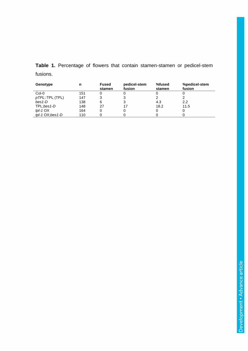
Table 1. Percentage of flowers that contain stamen-stamen or pedicel-stem
fusions.
Genotype n Fused stamen
pedicel-stem fusion
%fused stamen
%pedicel-stem fusion
Col-0 151 0 0 0 0 pTPL::TPL (TPL) 147 3 3 2 2 bes1-D 138 6 3 4.3 2.2 TPL;bes1-D 148 27 17 18.2 11.5 tpl-1 OX 164 0 0 0 0 tpl-1 OX;bes1-D 110 0 0 0 0
Dev
elo
pmen
t • A
dvan
ce a
rtic
le

Fig. S1. BES1 interacts with TPL through the EAR domain. (A) Yeast two-hybrid assay showing TPL and BES1 interaction. TPL interacts with the intact BES1 protein but not with BES1-EARm (EARm), where core Leucine residues in the EAR motif (LXLXL) were substituted by Alanine. TPL interaction with NINJA is included as a positive control. Yeast cells were grown on the synthetic dropout minimal medium lacking Leu and Trp (-LW), or synthetic dropout without Leu, Trp, His and Ade (-LWAH). (B) BiFC showing BES1 and TPL interaction. Nuclear YFP fluorescence is observed in N. benthamiana leaves infiltrated with the BES1-eYFPN and TPL- eYFPC constructs. PIF4-eYFPN and TPL-eYFPC are included as a negative control. Red fluorescence corresponds to chlorophyll. Scale bars represent 14 µm in the BES1-TPL panel and 50 µm in the PIF4-TPL panel. (C) Co-IP assay. TPL-HA is pulled-down by immunoprecipitation of GFP-tagged BES1, but not by BES1-EARm (EARm-GFP). N. benthamiana leaves were agroinfiltrated with BES1-GFP, BES1-EARm-GFP and TPL-HA. Two days after agroinfiltration, total protein extracts were immunoprecipitated with an anti-GFP antibody. TPL-HA was detected in these fractions with an anti-HA antibody.
Development 144: doi:10.1242/dev.143214: Supplementary information
Dev
elo
pmen
t • S
uppl
emen
tary
info
rmat
ion

Fig. S2. The EAR domain is required for the repressor activity of the BES1 factor. (A) The DWF4 promoter containing two BRRE elements was fused to the LUC reporter gene and co-transfected with the 35S::BES1, 35S::bes1-D, 35S::BES1-EARm or 35S::bes1-D-EARm effector constructs into N. benthamiana leaves. (B) TPL regulation of DWF4 expression. Leaves were co-infiltrated with the DWF4 reporter and combinations of Agrobacteria expressing the pTPL::TPL, 35S::BES1, 35S::BES1+pTPL::TPL, 35S::BES1+tpl-1 OX, 35S::BES1-EARm, 35S::BES1-EARm+ pTPL::TPL or 35S::BES1-EARm+ tpl-1 OX, as indicated. Leaf discs were collected 48 hours after infiltration and luciferase activity was measured. Error bars represent SD (n=20). Schematic representation of the reporter construct shows the position of the BRRE elements. Asterisks indicate significant difference compared to the DWF4 reporter (p<0.05, Student’s t-test).
Development 144: doi:10.1242/dev.143214: Supplementary information
Dev
elo
pmen
t • S
uppl
emen
tary
info
rmat
ion

Development 144: doi:10.1242/dev.143214: Supplementary information
Dev
elo
pmen
t • S
uppl
emen
tary
info
rmat
ion

Fig. S3. The EAR domain is essential for BES1 function. (A) Western blot of 2-weeks old WT, 35S::bes1-D-GFP (bes1-D) and 35S::bes1-D-EARm transgenic seedlings (EARm13 and EARm33). (B) Phenotype of adult WT, 35S::bes1-D-GFP and 35S::bes1-D-EARm-GFP plants. Plants were grown for 4-weeks under long day conditions (16h light/8h dark). (C) Phenotype of WT, 35S::bes1-D-GFP and 35S::bes1-D-EARm-GFP seedlings grown on BRZ. Seedlings were grown for 6 days under short day conditions (Light) or in continuous dark (Dark) in MS growth media (Mock) or MS media supplemented with 0.5 µM brassinazole (BRZ). (D) Hypocotyl length measurements of plants grown in (C). Bars represent s. d. (n=20). Similar results were obtained in three independent experiments. Asterisks indicate significant difference compared to the WT genotype (p<0.05, Student’s t-test). (E) Expression levels of the CPD, DWF4, ROT3, IAA19 and PRE5 genes in plants grown in short day conditions as in (D). Samples were taken at ZT0. Bars represent the standard deviation of three independent biological replicates. Asterisks indicate significant difference compared to the WT genotype (p<0.05, Student’s t-test).
Development 144: doi:10.1242/dev.143214: Supplementary information
Dev
elo
pmen
t • S
uppl
emen
tary
info
rmat
ion

Fig. S4. Phenotypic characterization of the inflorescences. Significant differences by the Tukey’s multiple comparison test are indicated by letters above bars (P < 0.05). (A) A representative flower of the indicated genotypes. (B) Measurement of the pedicel, bud, sepal and petal length of flowers in the primary inflorescences (n=10 plants and 20 flowers of each genotype). F: first open flower; 1, 2 and 3: three oldest buds.
Development 144: doi:10.1242/dev.143214: Supplementary information
Dev
elo
pmen
t • S
uppl
emen
tary
info
rmat
ion
Development 144: doi:10.1242/dev.143214: Supplementary information
Dev
elo
pmen
t • S
uppl
emen
tary
info
rmat
ion

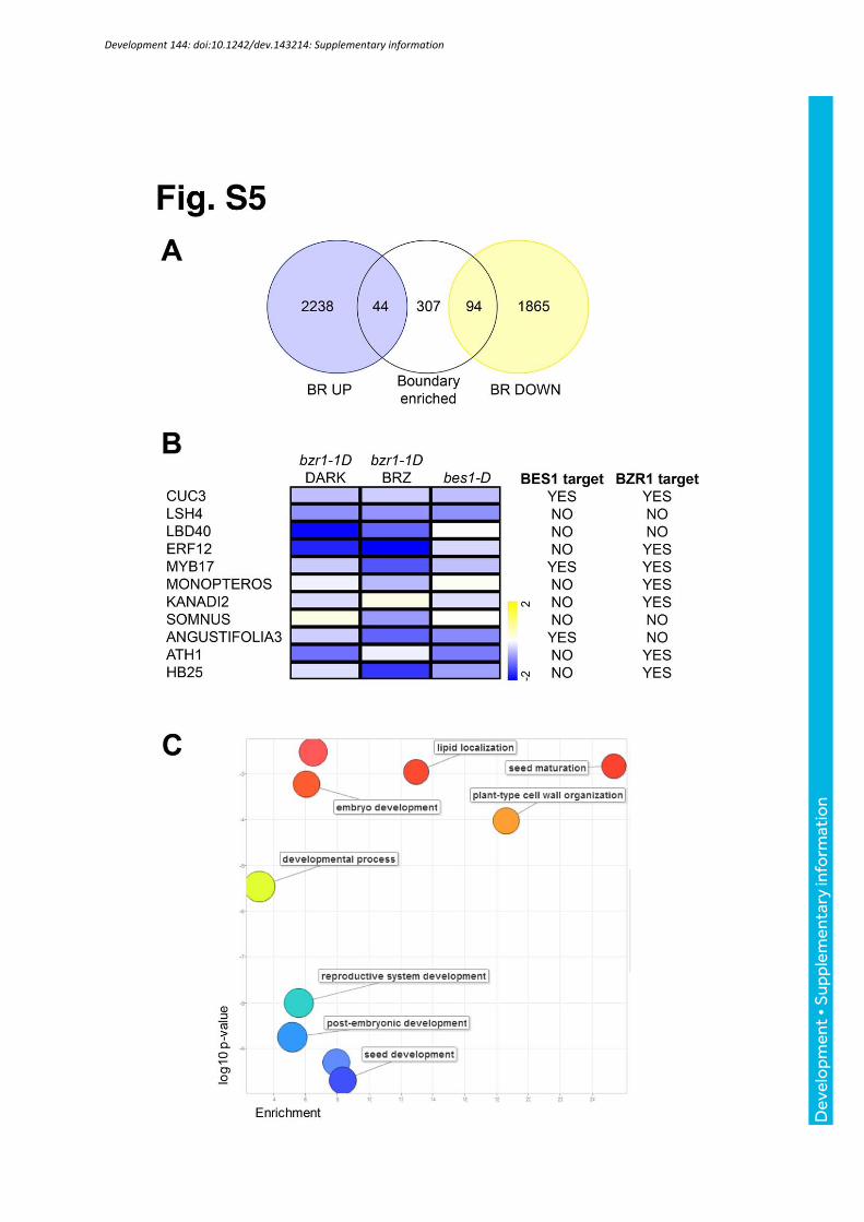
Fig. S5. Boundary-enriched BES1/BZR1-repressed targets include several boundary and patterning regulators. (A) Venn diagram showing the overlap of BR differentially expressed genes (Goda et al., 2004; Vert et al., 2005; Sun et al., 2010; Oh et al., 2012) and boundary enriched transcripts (Tian et al., 2014). BR-induced genes are not significantly enriched (44 found, 33 expected, hypergeometric test, p-value >0.01), while BR-repressed genes are strongly enriched in boundary-specific transcripts (94 found, 26 expected, hypergeometric test, p-value = 1.5 x 10⁻²⁵). (B) Heat-map representation of the expression profiles of the BR-repressed, boundary expressed transcription factors: bzr1-1D (Sun et al., 2010), bzr1-1D BRZ (Oh et al., 2012) and bes1-D (our own data). 8 out of 11 transcription factors were identified as direct BES1- or BZR1- targets (Sun et al., 2010; Yu et al., 2011). (C) Gene ontology analysis of the 94 boundary enriched, BR-repressed genes visualized with ReviGO.
Development 144: doi:10.1242/dev.143214: Supplementary information
Dev
elo
pmen
t • S
uppl
emen
tary
info
rmat
ion

Fig. S6. TPL is expressed in the shoot and root meristems. GUS staining of 6-day old pTPL::GUS seedlings. In the root, GUS staining was strong in the cell division and elongation zones. In aerial tissues, strong GUS staining was observed in the SAM and young, actively growing tissues.
Development 144: doi:10.1242/dev.143214: Supplementary information
Dev
elo
pmen
t • S
uppl
emen
tary
info
rmat
ion

Supplemental Table 1. Percentage of flowers that contain altered petal number.
Genotype n 3 petals 5 petals % more or less petals Col-0 152 1 0 0.7 pTPL::TPL 142 4 12 11.9 bes1-D 145 5 4 6.2 pTPL::TPL;bes1-D 159 3 11 8.8 tpl1-OX 120 0 1 0.8 tpl1-OX;bes1-D 105 0 0 0
Development 144: doi:10.1242/dev.143214: Supplementary information
Dev
elo
pmen
t • S
uppl
emen
tary
info
rmat
ion

Supplemental Table 2. List of primers used in this study.
Primers used for constructs
BES1-F CACCATGACGTCTGACGGAGCAAC
BES1-R ACCCGGGCAACTATGAGCTTTACCATTTCC
TPL-F CACCATGTCTTCTCTTAGTAGAGAGCTC
TPL-R TCTCTGAGGCTGATCAGATGC
PIF4-YFP-F CACCATGGAACACCAAGGTTGG
PIF4-YFP-R TCCGTGGTCCAAACGAGAACCGTC
BES1-EARm-R TCAACTATGAGCTTTACCATTTCCAGCCGTGGCCTCTGCATCCTCCATAGCC
pDWF4-F CACCGAATTACCGGTTGTTATGTAAATATAG
pDWF4-R TGGAGCTAGTTTCTCTCTCTCTCTCACT
Primers used for RT-qPCR
CPD-F TGAAACAACCTCCACGATCA
CPD-R TGCCCTAATCTTTTCATGCTCT
DWF4-F CGACGTGGGGAAACAACTAC
DWF4-R CTGAACCAGCACATAGCCTTG
ROT3-F ATCCGTGGAGATGGGACA
ROT3-R AACCAGGACATAGCCTTTGC
IAA19-F
GAGCTGAGATTGGGGCTTC
IAA19-R CCGACGACGTCATATTCATCT
PRE5-F TGCTTCGAGGATCTCCGATGACCA
PRE5-R GCCGTTCGTGAATCTCCGGCA
SAUR15-F AAGGGAATCATCGTCGACAC
SAUR15-R
AAGTATGAAACCGGCACCAC
Primers used for ChIP-PCR
CUC3p-F TGCATAGTCGTGCCAATTACTAA
CUC3p-R TACTGGGACAGACGAAGCCTT
CUC3 ORF-F
ACAGAGCAGTCTTCGAACGGTA
CUC3 ORF-R
GCTGGAATCCTAAAGGACATGG
PP2A-F GCCTTAAGCTCCGTTTCCTACTT
PP2A-R CGGCTTTCATGATTCCCTCT
DWF4p-2F GCCAAAAGTCTACGGGTTTG
DWF4p-2R TATGGGAAAAGGGTGGGCTC
BRAVOp-F
GCACGTGTAGAGAGAGAGAGC
BRAVOp-R AACCGCACGAAAACATTAAATATTC
Development 144: doi:10.1242/dev.143214: Supplementary information
Dev
elo
pmen
t • S
uppl
emen
tary
info
rmat
ion