Embed Size (px)
Citation preview
Scanning Microscopy Scanning Microscopy
Volume 1 Number 3 Article 27
4-23-1987
Tooth Eruption: The Regulation of a Localized, Bilaterally Tooth Eruption: The Regulation of a Localized, Bilaterally
Symmetrical Metabolic Event in Alveolar Bone Symmetrical Metabolic Event in Alveolar Bone
S. C. Marks Jr. University of Massachusetts Medical School
Follow this and additional works at: https://digitalcommons.usu.edu/microscopy
Part of the Life Sciences Commons
Recommended Citation Recommended Citation Marks, S. C. Jr. (1987) "Tooth Eruption: The Regulation of a Localized, Bilaterally Symmetrical Metabolic Event in Alveolar Bone," Scanning Microscopy: Vol. 1 : No. 3 , Article 27. Available at: https://digitalcommons.usu.edu/microscopy/vol1/iss3/27
This Article is brought to you for free and open access by the Western Dairy Center at DigitalCommons@USU. It has been accepted for inclusion in Scanning Microscopy by an authorized administrator of DigitalCommons@USU. For more information, please contact [email protected].
Scanning Microscopy, Vol. 1, No. 3, 1987 (Pages 1125-1133) 0891-7035/87$3.00+.00 Scanning Microscopy International, Chicago (AMF O'Hare), IL 60666 USA
TOOTH ERUPTION: THE REGULATION OF A LOCALIZED, BILATERALLY SYMMETRICAL METABOLIC EVENT IN ALVEOLAR BONE
S. C. Marks, Jr.
Department of Anatomy, University of Massachusetts Medical School, Worcester, Massachusetts 01605
(Received for publication February 14, 1987, and in revised form April 23, 1987)
Abstract
Tooth eruption is a complicated process by which developing teeth are moved within the jaws to their functional position. The usual model chosen to study this process, the erupted rodent incisor, differs both structurally and functionally from the human dentition and conclusions drawn from these studies are not directly applicable to tooth eruption in human beings. We have studied the eruption of developing permanent premolars in dogs and present evidence by scanning electron microscopy for regional differences in metabolic activities on bone surfaces of the crypt during eruption. We review evidence that these polarizations of alveolar bone metabolism are cell-mediated, dependent upon the dental follicle, independent of root formation or the tooth it self and that tooth eruption depends on coordination of these activities by the dental follicle. We conclude that tooth eruption is a localized, bilaterally symmetrical event in alveolar bone and that this is an excellent model system in which to study the regulation of alveolar bone metabolism.
KEY WORDS: Tooth eruption, alveolar bone, dental follicle, bone resorption, bone formation, osteoclast, osteoblast, prostaglandin E2, indomethacin, epidermal growth factor.
Address for correspondence: Sandy C. Marks, Jr. Department of Anatomy University of Mass. Medical School 55 Lake Avenue North Worcester, MA 01605 Phone No. (617) 856-3848
1125
Tooth Eruption - the historical perspective
Tooth eruption can be defined as the process of migration of a tooth from its site of development within bone to its functional position within the oral cavity (Massler and Schour 1941). It is a process that exhibits both exquisite localization and bilateral symmetry within the jaws. Like the formation of a tooth, its eruption involves the contact of diverse cell and tissue types and, despite considerable investigative effort, there is presently no clear consensus about the mechanism(s) of tooth eruption. The numerous theories advanced to explain eruption have involved almost every tissue in and around the tooth. Thus, root growth, dentin formation, cellular proliferation or vascular changes within the pulp, contraction of tissues around different parts of the tooth and alveolar bone growth have been proposed at one time or another as essential events in tooth eruption (Brash 1928; 8eertsen et al. 1974; Berkovitz and Thomas 1969; Bellows et al. 1981; Pitaru et al. 1976; Ten Cate 1969; Thomas 1976). Most comprehensive reviews of the subject have concluded that tooth eruption is a multifactorial process in which cause and effect are difficult to separate. Thus, no single theory offers an adequate explanation of all events (Magnusson 1968; Moxham and Berkovitz 1982).
The favorite ~xperimental model for studies of tooth eruption has been the erupted rodent incisor (Berkovitz et al. 1982). This tooth is sufficiently large to be amenable to surgical interventions and the choice of rodents makes experimentation relatively inexpensive. In the continuously erupting rodent incisor the periodontal ligament (the fibrous attachment of the tooth to bone) can cause eruption by itself. After transection of the root of this erupted tooth and immobilizing the proximal (apical) segment, the distal portion continues to erupt and is eventually lost (Berkovitz and Thomas 1969). This surgical procedure eliminates both root growth and pulpal pressure and the authors have concluded that the periodontal ligament
S. C. Marks , Jr.
(POL) was responsible for eruption of the distal fragment (Berkovitz 1971). While these conclusions concerning the continued eruption of the rodent incisor are warranted, this model has several unique features which make inferences about eruption of the human dentition hazardous. First, the rodent incisor and human dentition differ significantly with respect to the distribution of enamel and the POL (Matena 1972; 1973) and the rodent incisor grows and erupts at a rapid rate throughout life. These anatomical and physiological differences make extrapolation s from rodent incisors to the human dentition difficult. Second, one cannot assume that the mechanism for continued eruption of an erupted rodent incisor is the same as that responsible for its initial eruption through bone. For example, incisors in osteopetrotic rats and mice have a well-developed POL but fail to erupt because of a generalized reduction in bone resorption (Marks 1976; 1984). Finally, canine, primate and human teeth made rootless by irradiation (Gowgiel 1961; Carl and Wood 1980) or surgical procedures (Cahill and Marks 1980; Marks and Cahill 1984) are not prevented from erupting. Since rootless teeth by definition have no POL, the eruption of the succedaneous, noncontinuously erupting dentition in other vertebrates appears to be by a different mechanism that that of the continuously erupting rodent incisor.
Changes in Alveolar Bone during Tooth Eruption
Alterations in alveolar bone during tooth eruption have long been documented. Brash (1928) and Landsberger (1924) described changes in the bony crypt during development as teeth moved around within the jaws prior to eruption and noticed that an isolated failure of tooth development led to alveolar hypoplasia in that localized segment of the jaw. Thus, development of alveolar bone depends upon tooth development and eruption . Later studies of alveolar bone (Gregg and Avery 1964; Gregg 1965; Kenney and Ramfjord 1969; Sicher and Weinmann 1944) extended these ideas about the interdependence of tooth eruption and alveolar bone formation and showed that alveolar bone surfaces near the apex of developing teeth were sites of intense cell proliferation and bone formation during tooth eruption. Studies of temporarily impacted teeth have provided additional evidence that formation of alveolar bone at the base of erupting teeth may play a role in eruption (Cahi 11 1969; 1970). Permanent premolars temporarily impacted by transmandibular wires in dogs erupted at a faster rate when released from impaction than unimpacted contralateral teeth . This rapid, catch-up eruption was manifested histologically by the appearance of trabeculae of alveolar bone aligned below the tooth in its lonq axis. Elonqated trabeculae of bone
1126
also occur below erupting teeth, can expand to compensate for a failure or reduction in root formation, and develop on schedule even in the absence of a tooth provided the dental follicle is present (Cahill and Marks 1980· Marks and Cahi 11 1984). These data taken • together suggest that alveolar bone formation and tooth eruption are interdependent.
That the bone overlying a developing tooth must be resorbed during tooth eruption is self-evident. However, the mechanism by which this is accomplished is not . One thing seems to be certain. Pressure P.ff se is not a sufficient stimulus. When eruptive movement of dog premolars is blocked by transmandibular wires (Cahill 1969), development of an eruption pathway along with root resorption and exfoliation of deciduous precursors proceeds on schedule compared with contralateral unoperated controls. The eruption pathway develops through the activity of osteoclasts (Cahill 1974) and needs only the presence of the dental follicle not the development of roots (Cahill and Marks 1980) nor even the presence of the tooth itself (Marks and Cahill 1984). These observations indicate that alveolar bone resorption is coordinated with but not dependent upon tooth eruption. That tooth eruption depends upon bone resorption is shown by the state of the dentition in conditions of compromised bone re sorption such as congenital osteopetrosis (Marks 1984). In this disease tooth eruption i s either absent or retarded (Schour et al. 1949; Marks 1976) and bone resorption reduced (Marks 1973). Additional studies have shown that the failure of eruption is not due to the failure of tooth development (Marks 1976) but to the persistence of unresorbed bone. Tooth eruption occurs after neonatal restoration of bone resorption (Marks 1981), indicating that the initial failure resided within the resorptive apparatus not the POL.
These observations show that formation and resorption are localized, coordinated metabolic activities in alveolar bone that occur on opposite sides of erupting teeth .
Scanning Electron Microscopy of Alveolar Bone during Tooth Eruption
Mandibular permanent premolars in dogs begin eruption in the fifteenth postnatal week and the process is completed by week 23 (Cahill and Marks 1982). Examination of the third and fourth premolars, prepared for light and scanning electron microscopy as previously described (Cahill 1969; Marks et al. 1983; Marks and Cahill 1986), reveal the characteristic features of the process . An erupting permanent premolar at 20 weeks (late eruption) is shown in Figure l . Notice that the roots of this tooth are about half formed, that root and bone resorption above the permanent tooth has created an eruption pathway and that bone has formed between its two roots. An SEM montage of mineralized surfaces of part of the crypt at this stage of
TOOTH ERUPTION AND ALVEOLAR BONE METABOLISM
~" '
I.; • , · /. .
• , • • , , • • . I '
, I • I ' 1• \': ,- .... · ' <1 . '\, ', " ,, ~ 2\ ' ' ,'-,1,' · '·,:,
, .'' I ·'1,.-• · I · \ • · ·r ; (· I ,,):I:•: i;_
~ '\ I ' J 't- ., .... ~ (f' Mt · ·rJ.. · · ;· ~:: ,. :·· · - .·-
-\) .JI( - •• ~.,<"I: . . ·-.. 1 , l , , . / · . · •
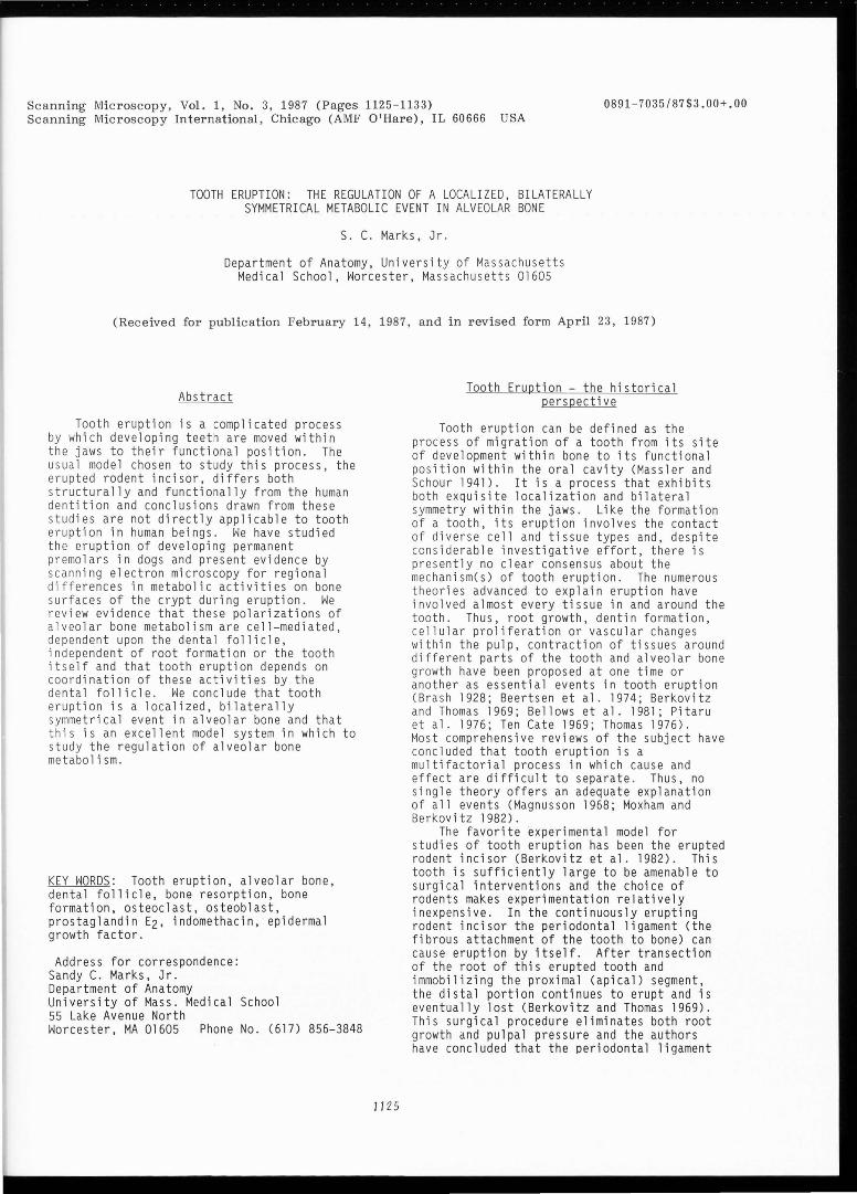
Figure 1. Photomicrograph of an erupting permanent mandibular premolar (P3) at 20 weeks. D3=deciduous third premolar; MC=mandibular canal; EP=eruption pathway and asterisk=dental follicle. The areas examined by SEM (Figures 4-9) in other specimens are indicated by arrows 1 and 2. Reprinted with permission from Arch. Oral Biol. 29, No. 4, Marks and Cahill, copyright 1984,Pergamon Journals Ltd. Bar=l mm.
Figure 2. A montage of scanning electron micrographs showing the bony surfaces of the crypt and adjacent areas of an erupting permanent premolar at 20 weeks. The areas and surfaces, from top to bottom are 1) root of deciduous tooth, 2) the eruption pathway for the permanent tooth, produced by bone resorption in the coronal part of its crypt, 3) a small area of resting bone surface, characterized by neither bone resorption n~r formation and 4) formation of trabecular bone at the base of the crypt. The hole at the bottom is the foramen connecting the crypt to the mandibular canal which transmits the blood and nerve supply to the erupting tooth. The area shown is the rectangular area of Figure 1 after removal of the permanent tooth (P3) and all organic matter . Bar=2 mm.
11 Z7
eruption is shown in Figure 2. Even at this low magnification one can see that the bone surface exhibits three distinct morphologies; an upper pitted surface, an intermediate smooth surface and a lower trabeculated surface. The following SEM photomicrographs demonstrate that these are the typical appearances by SEM of resorbing, resting and forming bone surfaces, respectively (Boyde and Hobdell 1969; Jones and Boyde 1974).
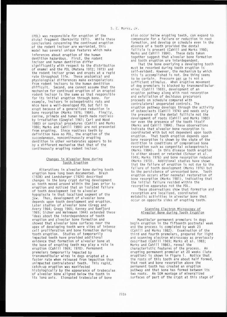
The crypt of a permanent premolar early (16 wk) in eruption is shown in Figure 3. In this broad view of an erupting tooth these three distinct surface appearances of alveolar bone are easily appreciated. In the upper (coronal) part of the crypt (Figs. 4 and 5) the scalloped surfaces characteristic of bone resorption cover the entire area except where numerous vascular channels penetrate the crypt wall . About two-thirds the way down the crypt (Fig. 6) a thin band of smooth bone surface is
Figure 3. Low power SEM of the crypt of the left third permanent mandibular premolar from a 16-wk-old beagle dog. The crypt has been sectioned transversely and this view shows the mineralized anterior (mesial) surface of the crypt after removal of all soft tissues with NaOCl. The arrow indicates the long axis of the crown of the permanent tooth. Bone surfaces early in eruption exhibit three morphologically distinct appearances (numbered l, 2 and 3 from coronal to apical). L=lateral cortical plate of mandible, R=root of deciduous third premolar, asterisk=roof of mandibular canal. Bar=200 µm.
S. C. Marks, Jr.
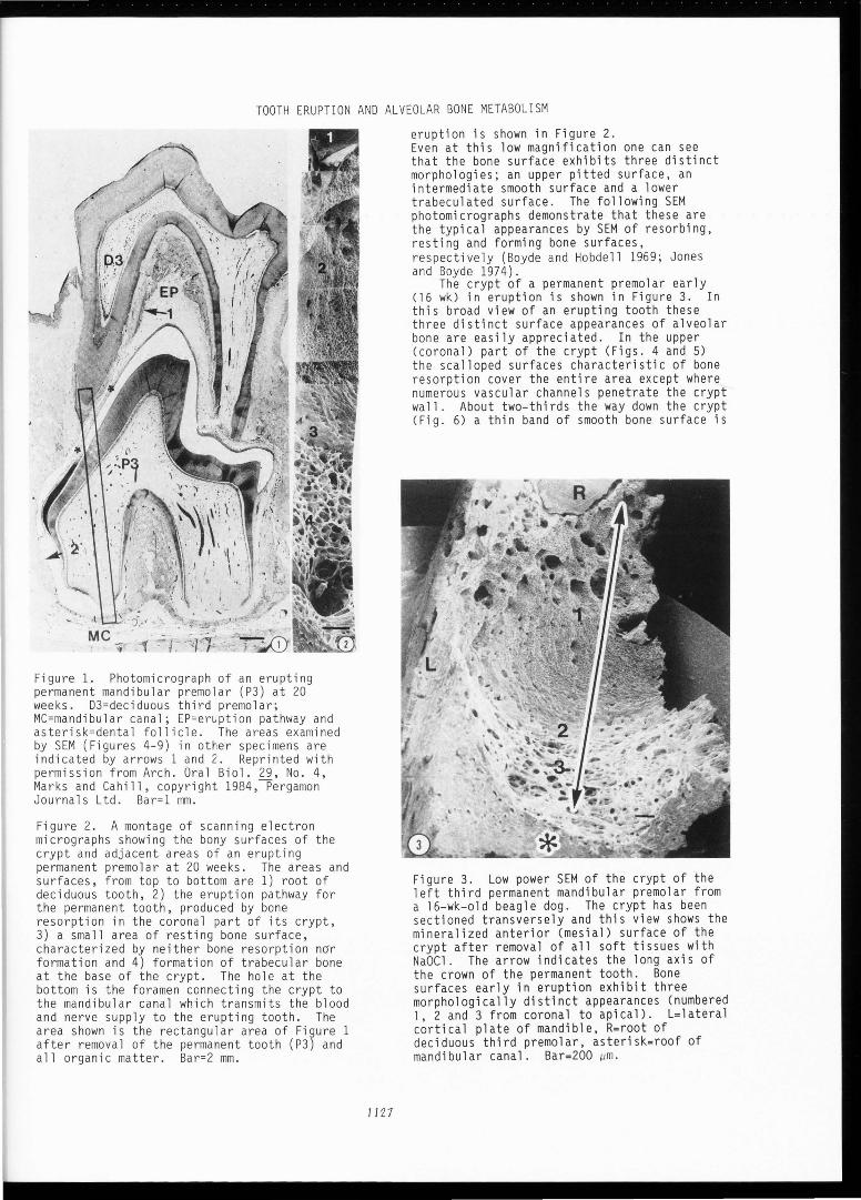
Figure 4. Higher power of region 1 of Figure 3 showing scalloped bone surfaces typical of resorption. V=vascular channels. Bar=lOO µm.
Figure 5. Typical scalloped surfaces of the resorbing bone characteristic of coronal aspects of crypts of erupting premolars at higher magnification show pitted surfaces probably representing osteocytic canaliculi . V=vascular channels. Bar=50 µm.
1128
Figure 6. Transition zone between resorbing (1) and resting (2) surfaces of crypt of an erupting premolar. See Figure 3 for or ientation . V=vascular channels . Bar=lOO µm.
Figure 7. Transition zone between resting (2) and formative (3) crypt surfaces during tooth eruption. See Figure 3 for orientation . V=vascular channels . Bar=lOO µm.
encountered . This is the typical appearance of resting bone surfaces, i.e . , those undergoing neither formation or degradation. At the base of the crypt (Figs. 7,8,9) is a trabeculated lattice of newly formed bone, typified by knobby surface projections and the lacunae of newly forming osteocytes (Fig. 9).
These three alveolar bone surface phenotypes are observed around erupting teeth f rom just before eruption until late in the proces s when penetration of the al~eolar bone cortex is completed (Marks and Cahill 1986).

TOOTH ERUPTION AND ALVEOLAR BONE METABOLISM
Figure 8. Higher magnification of trabeculated surfaces of alveolar bone at the base of the crypt of an erupting third premolar. V=vascular channels. Bar=50 µm.
Figure 9. High magnification of trabeculae of bone at arrow in Figure 8. These knobby surfaces are characteristic of bone formation and include several depressions Co) indicating partially formed lacunae of osteocytes. Bar=25 µm.
The Regulation of Alveolar Bone Metabolism during Tooth Eruption
Additional studies have shown that these typical topographical features of crypt surfaces of erupting teeth have the appropria te cellular components. Osteoclasts cover more than half of coronal crypt surfaces, osteoblasts form a thick carpet on
1129
basal bone surfaces throughout eruption (Marks et al. 19B3) and these cells exhibit ultrastructural features of intense activity (Marks and Cahill 19B6). Furthermore, the erupt ion sequence depends only on the presence of the dental follicle, a thin vascular connective tissue investment of the developing tooth, not on root growth, the POL or even the tooth itself (Cahill and Marks 1980; Marks and Cahill 1984). After removal of the coronal part of the dental follicle root formation occurs but an eruption pathway does not develop and eruption fails. Removal of the basal part of the follicle interferes with formation of bone below the developing tooth and, while root growth occurs and an eruption pathway forms, eruption does not take place (Marks and Cahill 1987). These observations suggest that alveolar bone metabolism during tooth eruption is dependent upon and coordinated by the adjacent parts of the dental follicle . What are the mechanisms by which coronal bone resorption and basal alveolar bone formation are regulated?
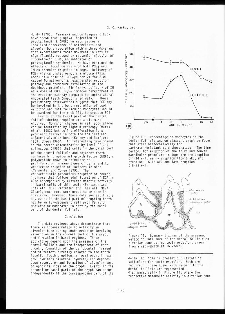
Events in the coronal part of the crypt appear to be regulated by a population of mononuclear cells which increase 15-fold in this part of the follicle just prior to eruption (Marks et al. 1983). These cells have ultrastructural features of monocytes and contain dense-core, membrane-bound cytoplasmic granules (Wise et al . 1985) described by others as characteristics of mononuclear osteoclast precursors (Scott 1967; Rifkin et al. 1980). Some of these cells develop before eruption the ability to produce tartrateresistant acid phosphatase (TRAP), a cytochemical marker for osteoclasts and their precursors (Baron et al. 1986; Minkin 1982; van de Wijngaert and Burger 1986), and both the number and percentage of mononuclear cells expressing TRAP increase early in eruption (Marks and Grolman 1987). Mononuclear cells expressing TRAP also occur next to osteoclasts on coronal surfaces of the crypt during eruption. The changes in TRAP-positive mononuclears in the coronal part of the dental follicle and adjacent bone surfaces during eruption is shown in Figure 10. The significant increase in TRAP-positive cells in the follicle between 14 and 16 weeks precedes a dramatic increase in osteoclasts on adjacent crypt surfaces (Marks et al. 1983). These data suggest that the regulatio n of the bone resorption of tooth eruption by the coronal part of the dental follicle (Marks and Cahill 1987) is mediated by a population of mononuclear cells which enter the follicle just prior to the extensive osteoclastmediated resorption of bone that occurs early in the eruption sequence. Whether these cells are osteoclast precursors or affect osteoclast function by the local release of mediator s of bone resorption remains to be demonstrated. One such local mediator of resorption is prostaglandin E1 (Dietrich et al . 1975), and monocytes are a known source of this potent stimulator of bone resorption (Yoneda and
S. C. Marks , Jr.
Mundy 1979). Yamasaki and colleagues (1980) have shown that gingival injection of prostaglandin E (PGE) in rats causes a localized appearance of osteoclasts and alveolar bone resorption within three days and that experimental tooth movement in rats is significantly reduced by systemic injection of indomethacin (IM), an inhibitor of prostaglandin synthesis . We have examined the effects of local delivery of both PGE1 and IM on premolar eruption in dogs. Delivery of PGE1 via canulated osmotic minipump (Alza Corp) at a dose of 100 µgm per wk for 3 wk caused formation of an exaggerated eruption pathway and premature exfoliation of the deciduous premolar. Similarly, delivery of IM at a dose of 800 µgm/ wk impeded development of the eruption pathway compared to contralateral unoperated teeth (unpublished data) . These preliminary observations suggest that PGE may be involved in the bone resorption of tooth eruption and that follicular monocytes should be examined for their ability to produce PGE.
Events in the basal part of the dental follicle during eruption are a bit more elusive. No major changes in cell population s can be identified by light microscopy (Marks et al. 1983) but cell proliferation is a prominent feature in both the follicle and adjacent alveolar bone (Kenney and Ramfjord 1969; Gregg 1965). An intere sting development is the recent demonstration by Thesleff and colleagues (1987) that cells in the basal part of the dental follicle and adjacent tooth surfaces bind epidermal growth factor (EGF), a polypeptide known to stimulate cell proliferation in many types of cells and to accelerate eruption of incisor s in mice (Carpenter and Cohen 1979). The characteristic precocious eruption of rodent incisors that follows administration of EGF i s al so accompanied by elevated mitotic activit y in basal cells of this tooth (Partanen and Thesleff 1987; Rihtniemi and Thesleff 1987). Clearly much more work needs to be done in thi s area. However, these data suggest that a key event in the basal part of erupting teeth may be an EGF- dependent cell proliferation mediated or moderated in part by the basal part of the dental follicle .
Conclusion
The data reviewed above demonstrate that there is intense metabolic activity in alveolar bone during tooth eruption involving resorption in the coronal part of the crypt and formation in basal regions . These activities depend upon the presence of the dental follicle and are independent of root growth, formation of the periodontal ligament and of factors directly related to the tooth itself . Tooth eruption, a local event in each jaw, exhibits bilateral symmetry and depends upon resorption and formation of alveolar bone on opposite sides of the crypt . Events in the coronal or basal parts of the crypt can occur independently if the corresponding part of the
1130
1-- 50 z
I------I-1~ UJ u II: UJ
o. 40 I CRYPT
CJ)
UJ I-> g 30 / f\ ""'I'---. f I z 0 :=;;
o.: 20 <t t / FOLLICLE
--1 \ II:
~
10
•-i II /12 14 15 16 18 20
AGE IN WEEKS
Figure 10. Percentage of monocytes in the dental follicle and on adjacent crypt surfaces that stain histochemically for tartrate-resistant acid phosphatase. The time periods for eruption of the third and fourth mandibular premolars in dogs are pre-eruption (11-14 wk), early eruption (15-16 wk), mid eruption (16-18 wk) and late eruption (18-23 wk).
9ub1?.rnoculurn d1?.nt1s
4p
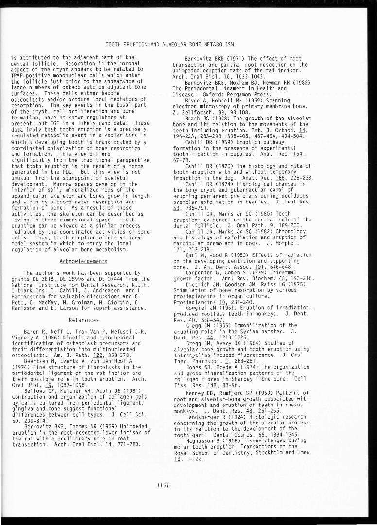
Figure 11. Summary diagram of the presumed metabolic influence of the dental follicle on alveolar bone during tooth eruption, drawn from a radiograph at 16 weeks.
dental follicle is present but neither is sufficient for tooth eruption. Both are required. These ideas with respect to the dental follicle are represented diagrammatically in Figure 11, where the respective metabolic activity in alveolar bone
TOOTH ERUPTION AND ALVEOLAR BONE METABOLISM
is attributed to the adjacent part of the dental follicle . Resorption in the coronal aspect of the crypt appears to be related to TRAP-positive mononuclear cells which enter the follicle just prior to the appearance of large numbers of osteoclasts on adjacent bone surfaces. These cells either become osteoclasts and/or produce local mediators of resorption . The key events in the basal part of the crypt, cell proliferation and bone formation, have no known regulators at present, but EGF is a likely candidate. These data imply that tooth eruption is a precisely regulated metabolic event in alveolar bone in which a developing tooth is translocated by a coordinated polarization of bone resorption and formation. This view differs significantly from the traditional perspective that tooth eruption is the result of a force generated in the POL. But this view is not unusual from the standpoint of skeletal development. Marrow spaces develop in the interio r of solid mineralized rods of the appendicular skeleton and bones grow in length and width by a coordinated resorption and formation of bone. As a result of these activities, the skeleton can be described as moving in three-dimensional space. Tooth eruption can be viewed as a similar process mediated by the coordinated activities of bone cells. Thus, tooth eruption offers an ideal model system in which to study the local regulation of alveolar bone metabolism.
Acknowledgements
The author's work has been supported by grants DE 3818, DE 05996 and DE 07444 from the National Institute for Dental Researc h, N.I. H. I thank Ors. D. Cahill, J. Andreasen and L. Hammarstrom for valuable discussions and C. Peto, C. MacKay, M. Grolman, M. Giorgio, C. Karlsson and E. Larson for superb assistance .
References
Baron R, Neff L, Tran Van P, Nefussi J-R, Vignery A (1986) Kinetic and cytochemical identification of osteoclast precursors and their differentiation into multinucleated osteoclasts. Am. J. Path . ill, 363-378.
Beertsen W, Everts V, van den Hoof A (1974) Fine structure of fibroblasts in the periodontal ligament of the rat incisor and their possible role in tooth eruption. Arch. Oral Biol. 19., 1087-1098.
Bellows CF, Melcher AH, Aubin JE (1981) Contraction and organization of collagen gels by cells cultured from periodontal ligament, gingiva and bone suggest functional differences between eel l types. J. Cell Sci. 50, 299-314.
Berkovitz BKB, Thomas NR (1969) Unimpeded eruption in the root-resected lower incisor of the rat with a preliminary note on root transection. Arch. Oral Biol. 11, 771-780.
1131
Berkovitz BKB (1971) The effect of root transection and partial root resection on the unimpeded eruption rate of the rat incisor . Arch. Oral Biol. 16., 1033-1043.
Berkovitz BKB, Moxham BJ, Newman HN (1982) The Periodontal Ligament in Health and Disease. Oxford: Pergamon Press.
Boyde A, Hobdell MH (1969) Scanning electron microscopy of primary membrane bone. Z. Zellforsch . 99, 98-108.
Brash JC (1928) The growth of the alveolar bone and its relation to the movements of the teeth including eruption. Int. J. Orthod. 11, 196-223, 283-293, 398-405, 487-494, 494-504.
Cahill DR (1969) Eruption pathway formation in the presence of experimental tooth impaction in puppies. Anat. Rec. 16..1, 67-78.
Cahill DR (1970) The histology and rate of tooth eruption with and without temporary impaction in the dog. Anat. Rec. ill, 225-238.
Cahill DR (1974) Histological changes in the bony crypt and gubernacular canal of erupting permanent premolars during deciduous premolar exfoliation in beagles. J. Dent Res. 53, 786-791.
Cahill DR, Marks Jr SC (1980) Tooth eruption: evidence for the central role of the dental follicle . J. Oral Path . .2., 189-200.
Cahill DR, Marks Jr SC (1982) Chronology and histology of exfoliation and eruption of mandibular premolars in dogs. J . Morphol. ill, 213-218.
Carl W, Wood R (1980) Effects of radiation on the developing dentition and supporting bone. J. Am. Dent. Assoc. l.Ql, 646-648.
Carpenter G, Cohen S (1979) Epidermal growth factor. Ann. Rev. Biochem. 48, 193-216.
Dietrich JW, Goodson JM, Raisz LG (1975) Stimulation of bone resorption by various prostaglandins in organ culture . Prostaglandins l.Q, 231-240.
Gowgiel JM (1961) Eruption of irradiationproduced rootless teeth in monkeys. J . Dent. Res. 40, 538-547.
Gregg JM (1965) Immobilization of the erupt ing molar in the Syrian hamster. J . Dent. Res. 44, 1219-1226.
Gregg JM, Avery JK (1964) Studies of alveolar bone growth and tooth eruption using tetracycline-induced fluorescence. J . Oral Ther. Pharmacol. l, 268-281.
Jones SJ, Boyde A (1974) The organization and gross mineralization patterns of the collagen fibres in Sharpey fibre bone. Cell Tiss. Res. ill, 83-96.
Kenney EB, Ramfjord SP (1969) Patterns of root and alveolar-bone growth associated with development and eruption of teeth in rhesus monkeys. J . Dent. Res. 48, 251-256.
Landsberger R (1924) Histologic research concerning the growth of the alveolar proces s in its relation to the development of the tooth germ. Dental Cosmos. 66, 1334-1345.
Magnusson B (1968) Tissue changes during molar tooth eruption. Transactions of the Royal School of Dentistry, Stockholm and Umea Ll, 1-122.

S. C. Marks, Jr.
Marks Jr SC (1973) Pathogenesis of osteo petrosi s in the ia rat: Reduced bone resorption due to reduced osteoclast function. Am. J. Anat. ill, 165-189.
Marks Jr SC (1976) Tooth eruption and bone resorption: experimental investigation of the ia (osteopetrotic) rat as a model for studying their relationships. J. Oral Path . .5., 149-163.
Marks Jr SC (1981) Tooth eruption depends on bone resorption: Experimental evidence from osteopetrotic (ia) rats. Metab. Bone Dis. Rel. Res. J, 107-115.
Marks Jr SC (1984) Congenital osteopetrotic mutations as probes of the origin, structure and function of osteoclasts. Clin. Orthoped. Rel. Res. ill, 239-263.
Marks Jr SC, Cahill DR, Wise GE (1983) The cyto logy of the dental follicle and adjacent alveolar bone during tooth eruption. Am. J. Anat. 168, 277-289.
Marks Jr SC, Cahill DR (1984) Experimental study in the dog of the non-active role of the tooth in the eruptive process. Arch Oral. Biol. 29, 311-322.
Marks Jr SC, Cahill DR (1986) Ultrastructure of alveolar bone during tooth eruption in the dog. Am. J . Anat. ill, 427-438.
Marks Jr SC, Cahill DR (1987) Regional control of alveolar bone metaboli sm by the dental follicle during tooth eruption. J. Oral Path. in press.
Marks Jr SC, Grolman ML (1987) Tartrate-resistant acid phosphatase in mononuclear and multinuclear cells during the bone resorption of tooth eruption. J. Histochem. Cytochem. in press.
Massler M, Schour I (1941) Studies in tooth development: Theories of eruption. Am. J. Orthodont. Oral Surg. 27, 552-576.
Matena V (1972) The periodontium of the enamel aspects of the rat incisor. J. Periodont. 43, 311-315.
Matena V (1973) Periodontal ligament of a rat incisor tooth. J. Periodont. 44, 629-635.
Minkin C (1982) Bone acid phosphatase: tartrate-resistant acid phosphatase as a marker of osteoclast function. Calcif. Tissue Int. 34, 285-290.
Moxham BJ, Berkovitz BKB (1982) The periodontal ligament and physio logical tooth movements. In: The Periodontal Ligament in Health and Disease, Berkovitz BKB, Moxham BJ, Newman HN (eds) New York: Pergamon Press. Chap. 10, pp. 215-240.
Partanen A-M, Thesleff I (1987) Localization and quantitation of l25I-epidermal growth factor binding in mouse embryonic tooth and other embryonic tissues at different developmental stages. Devel. Biol. ill, in press.
Pitaru S, Michaeli Y, Zajicek G, Weinreb MM (1976) Role of attrition and occlusal contact in the physiology of the rat incisor. X. The part played by the periodontal ligament in the eruptive process. J. Dent. Res. 55, 819-824.
1132
Rifkin BR, Brand JS, Chushing JE, Coleman SJ, Sanavi F (1980) Fine structure of fetal rat calvarium; provisional identification of preosteoclasts. Calcif. Tissue Int. n. 21-28.
Rihtniemi L, Thesleff I (1987) The EGF-induced precocious mouse incisor eruption is associated with stimulation of cell proliferation in the root sheath region . Arch. Oral Biol. in press .
Schour I, Bhaskar SN, Greep RO, Weinmann JP (1949) Odontome-like formations in a mutant strai n of rats. Am. J. Anat. 85, 73-112.
Scott BL (1967) Occurrence of specific cytoplasmic granules in the osteoclast. J. Ultrastruct. Res . .12, 417-431.
Sicher H, Weinmann JP (1944) Bone growth and physiologic tooth movement. Am. J. Orthodont. Oral Surg. 30, 109-132.
Ten Cate AR (1969) The mechanism of tooth eruption. In Biology of the Periodontium, Melcher AH, Bowen WH (eds) New York: Academic Press, pp. 91-103.
Thesleff I, Partanen A-M, Rihtniemi L (1987) Localization of epidermal growth factor receptors in mouse incisors and human premolars during eruption. Euro. J. Orthodont. ~. in press.
Thomas NR (1976) Collagen as the generator of tooth eruption. In: The Eruption and Occlusion of Teeth, Proceedings of the 27th Symposium of the Colston Research Society. Poole DFG, Stack MV (eds), -London: Butterworths, pp. 290-301.
van de Wijngaert FP, Burger EH (1986) Demonstration of tartrate-resistant acid phosphatase in undecalcified glycolmethacrylate-embedded mouse bone: a possible marker for (pre) osteoclast identification . J. Histochem. Cytochem. 34, 1317-1323.
Wise GE, Marks Jr SC, Cahill DR (1985) Ultrastructural features of the dental follicle associated with formation of the tooth eruption pathway in the dog. J. Oral Path . .11, 15-26.
Yamasaki K, Miura F, Suda T (1980) Prostaglandin as a mediator of bone resorption induced by experimental tooth movement in rats. J. Dent. Res. 59, 1635-1642.
Yoneda T, Mundy GR (1979) Monocytes regulate osteoclast-activating factor production by releasing prostaglandins. J. Exp. Med. l.5..Q, 1635-1642.
Discussion with Reviewers
M.B. Engel: Can you speculate on how the metabolic events in the coronal and basal follicle are transformed into forces which transport the tooth th rough the jaw into the ora l cavity? Author: I believe that the linkage between follicular events and tooth eruption lies eventually in an understanding of local cellular events and mediators, which have only been recently discovered and acknowledged. Thus, the roles of prostaglandins,
TOOTH ERUPTION AND ALVEOLAR BONE METABOLISM
interleukins, and growth factors in the cellular events of eruption need to be explored before a basis for speculation can be established. The use of the term "force" with respect to eruption is unduly restrictive, primarily because of its mechanical implications. I prefer to think of the skeletal events in eruption in terms of movements of bone surfaces in space, similar to formation of marrow spaces.
A.R. Ten Cate: Is tooth eruption solely the result of a coordinated activity of bone cells? If so, how does bony remodeling actually bring about tooth movement, given that a forming root must be accommodated? Author: I don't know anyone who is prepared to attribute eruption solely to skeletal events. However, with respect to the early (intraosseous) course of eruption, I can see no reasonable alternatives at present. Once the tooth emerges through bone and gingiva there may well be a role for the periodontal ligament, whose most coronal fibers are by this time organized and attached to bone. This phase of eruption, as you suggest, needs more careful study .
B.R. Rifkin: You have presented data that indomethacin impeded development of the eruption pathway. However, Rosenberg, L. et al (J . Dent. Res. 62, 501a, 1983) using 5-10 mg/kg indomethacin were unable to alter clinical eruption schedules for first and second rodent molars. Could you comment on this? Author: I suspect that the differences are ones of route of administration (systemic vs. local) and dose.
B.R. Rifkin : You indicated that indomethacin impeded the development of the eruption pathway. Could you be more quantitative? What is the effect on the clinical eruption schedules of the premolars? There is data that PGE can augment bone formation. Did you identify, following PGE administration, any changes in the basal part of the dental follicle or corresponding basal region of the crypt? Can PGE levels be measured biochemically in the coronal portion of the dental follicle? Author: Ours are only preliminary observations and we do not have sufficient data for quantitative analyses. PGE levels in the follicle and osteogenic effects of PGE are presently under investigation .
B.R. Rifkin: You indicated that there is a marked increase in mononuclear cells in the coronal part of the follicle prior to eruption and that Qil..Ly some of these cells are TRAP positive. Does this mean there are subpopulations of mononuclear cells? Do only the more mature mononuclear cells stain positive for TRAP?
1133
Author: Based on the presence of perivascular (newly arrived?) TRAP-positive cells, I suspect that we have identified a subpopulation of cells in the follicle which are mononuclear by light microscopic criteria. However, as you suggest, the expression of TRAP could be a late event in mononuclear cell differentiation . This possibility can be examined using EM histochemistry or a combination of cDNA probe and cytochemistry.