Embed Size (px)
Citation preview
Tolerogen-Producing Cells in Allogeneic Bone
Marrow Chimeras Established with
Spontaneously Leukemia-Prone Mice
Machiko Mishima ,Mari Hirano ,Taiki Morohashi ,Noriko Arase ,
Hayase Shisa ,Hiroshi Hiai ,Manabu Ato and Kazunori OnoeDivision of Immunobiology,Institute for Genetic Medicine,Hokkaido UniversityLaboratory of Basic Cancer Study,Saitama Cancer Center Research InstituteDepartment of Pathology and Biology of Diseases,Graduate School of Medicine,Kyoto University
Using SL/Kh mice and AKR/J mice,which are animal models for spontaneous pre-B-cell leukemia
and thymic lymphoma, respectively, we studied the protective influence of allogeneic bone marrow
transplantation (BMT)and the induction of tolerance to Mls-1,a host antigen. When BM cells from
allogeneic C57BL/6 mice were used to reconstitute self-tolerance SL/Kh mice,these[B6→SL]chimeric
mice survived for a longer time than non-treated SL or[SL→SL]syngeneic chimeras. These findings
are compatible with results previously obtained for[B6→AKR]chimeras. In[B10.D2→SL]and[B10.
D2→AKR]chimeras,Vβ6 T-cells reactive to Mls-1 were eliminated 5 weeks after BMT. On the other
hand,minor graft versus host reaction(GVHR)abrogated the clonal elimination of Vβ6 T-cells in both[B10.D2→SL]and[B10.D2→AKR]chimeras. The cause of this abrogation was attributed to the early
disappearance of Mls-1-producing host T-cells in the GVHR chimeras. The cells responsible for the Mls-1 production were revealed to be mainly CD8 CD44 T-cells,by in vitro mixed lymphocyte reaction
(MLR)and in vivo tolerance induction. The present findings indicate that host CD8 CD44 T-cells
constitute the major source of Mls-1 antigens in the[Mls-1→Mls-1]BM chimera system.
Key words allogeneic bone marrow chimera,minor lymphocyte stimulatory antigen 1 ,negative selec-tion,leukemia-prone mice
INTRODUCTION
T-cells undergo both negative and positive
selections in the thymus . To elucidate the selec-
tion mechanism,especially negative selection,the
minor lymphocyte stimulatory (Mls)-1 antigen
(Ag)system,an intrinsic super-antigen,has been
widely employed. T-cell reactivity to Mls-1 Ag
correlates with the expression of certain T-cell
Ag receptor(TCR)-Vb regions . The expression
of Mtv-7,an endogenous mammary tumor virus
gene which determines the Mls-1 phenotype,
results in the deletion among developing
thymocytes, of T-cells that express Vβ6, Vβ7,
Vβ8.1,or Vβ9 . Using[Mls-1→Mls-1]bone
marrow (BM) chimeras, we demonstrated that
Mls-1-reactive T-cells are eliminated from the
developing thymocyte population that is derived
from the donor BM . Furthermore,the presence
of thymic stromal cells derived from the donor
BM has been shown to be the primary require-
ment for the effective deletion of Mls-1-reactive
thymocytes . We have reported that activated
CD8 and CD4 T-cells both produce Mls-1 Ag
in vitro, although only CD8 T-cells, not CD4
T-cells, can produce Mls-1 Ag under non-
stimulated conditions . Taking into account
thatclonal deletion is a major mechanism for
inducing and maintaining self-tolerance, it is
important to determine the source of the relevant
tolerogen in this chimera system,paying particu-
lar attention to the role of T-cells. In the present
study,we first analyzed the protective influence
of allogeneic bone marrow transplantation
(BMT) in SL/Kh mice, an animal model for
pre-B-cell leukemia. We then analyzed which
cell components in lethally irradiated recipient
mice provides Mls-1 Ag and ultimately contrib-
ute to clonal elimination of the Mls-1-reactive
T-cell repertoire in the BM chimeric mice. We
Received:December 27,2000
Revised :February 5,2001
Accepted:February 5,2001
show here that the residual radio-resistant recipi-
ent T-cells are the cells responsible for this
intrathymic clonal elimination.
AND
AKR/J (AKR)(H-2, Mls-1, Thy1.1)mice
were obtained from the Jackson Laboratory(Bar
Harbor, ME). C57BL/6 (B6) (H-2, Mls-1,
Thy1.2), B10. BR/SgSnSlc (BR) (H-2, Mls-1,
Thy1.2),and B10.D2(D2)(H-2,Mls-1,Thy1.2)
mice were obtained from JAPAN SLC Co.
(Hamamatsu, Japan). SL/Kh (SL) (K , A , E ,
Dq) and (AKR×BR) F mice were bred and
maintained in our animal facility at Hokkaido
University.
Eight-week-old female AKR or SL mice were
subjected to either 10 or 11 Gy X-ray irradiation.
Twenty four hours later,these mice were treated
to achieve hematopoietic and immunologic recon-
stitution with 2×10 BM cells taken from 8-week-old B6, D2 or syngeneic mice. Prior to
BMT,BM cells were treated in vitro with anti-
Thy1.2 (F7D5, Olac, Bicester, UK) monoclonal
antibody(mAb)plus selected rabbit complement
(C) . To induce subclinical GVHR, BM cells
treated with anti-Thy1.2 mAb alone were inject-
ed intravenously into the recipient mice . As
reported earlier ,these GVHR chimeras scarcely
showed overt signs of GVHR (ruffled fur, hun-
ched back,significant loss of weight),but clonal
elimination of self-reactive T-cells (i. e. against
recipient’s Ag)was abrogated. Chimeras prepar-
ed by injecting T-cell-depleted BM cells alone
will be referred to as[donor→recipient]chimer-
as or control chimeras. Chimeras which were
given BM cells pretreated with anti-Thy1.2 mAb
alone will be referred to as GVHR[donor→recip-
ient]chimeras. In some experiments,2×10AKR
T-cell subsets obtained from untreated (AKR×
BR)F mice were introduced intravenously into[BR→BR]syngeneic BM chimeras one week
after BMT.[BR→BR]chimera mice were pre-
pared as described above.
Spleen cells were passed over nylon wool
columns and the purified T-cells were treated
with either anti-CD4 mAb or anti-CD8 mAb and
selected rabbit C. These cells were further pur-
ified by M-450 Dynabeads (Dynal Inc.,Norway)
coated with anti-rat immunoglobulin G (IgG)and
anti-mouse IgG antibodies(Ab). The purified cell
fraction showed more than 99% relevant cells.
The CD4 and CD8 T-cell fractions were then
treated with anti-CD44 mAb and stained cells
were sorted using the FACStar system as de-
scribed elsewhere .
Three-color FACS analyses were carried out
as previously described . Thymocytes from
chimeric mice were treated in vitro before analy-
sis with the following primary mAbs:anti-
Thy1.1 (T11D7e. Olac), anti-CD3ε(2C11), anti-
Vβ6 (44-22-1) or anti-Vβ8.1, 2, 3 (F23.1).
Biotinylated anti-mouse and anti-rat IgG secon-
dary Ab (Cappel,West Chester,PA,USA)were
also used, followed by TANDEM-streptavidin
(Southern Biotech., Birmingham, AL, USA) to
treat the thymocytes. After blockingbinding sites
of these secondary Ab,phycoerythrin (PE)-anti-
CD4 and fluorescein isothiocyanate (FITC)-anti-
CD8 (Becton-Dickinson, Mountain View, CA)
were reacted with the thymocytes. When the
population of host-derived T-cells was analyzed,
cells were first incubated with biotinylated anti-
mouse IgG followed by PE-streptavidin. Then,
anti-Thy1.1 or anti-Thy1.2 mAb and FITC-anti
mouse IgG were added. Stained cells were anal-
yzed with a FACScan system(Becton-Dickinson).
-
Total cellular RNA was extracted from
spleen cells using the guanidinium isothiocyanate
method . Reverse-transcription(RT)was perfor-
med,and the Mls-1-specific sequence contained
within the ORF of the 3’-LTR of MTV-7 cDNA
was amplified using the following Mls-1-
specific primers:5’-primer GTCAAAGAACAG-
GTGCAAGGAC and 3’-primer AAGGGATC-
GAAGCCAACGCG. Theβ-Actin cDNA was
amplified for control (5-primer TGGAATCCT-
GTGGCATCCATGAAAC and 3’-primer
Journal of Clinical and Experimental Hematopathology Vol.41 No.1 M.Mishima M.Hirano T.Morohashi N.Arase H.Shisa H.Hiai M.Ato and K.Onoe
TAAAACGCAGCTCAGTAACAGTCCG).
MLR was performed as described else-
where . BR T-cells were stimulated with AKR
or (AKR×BR)F T-cell subsets in the presence
of mitomycin-treated BR spleen cells as antigen-
presenting cells(APC). In some experiments,Vβ
8.2-T-cells of BR mice were used.
Statistical analyses were carried out using
Student’s t test. P values of less than 0.05 were
considered significant.
RESULTS
The SL mouse, which develops pre-B-cell
leukemia, was established in Japan . It has
been reported that allogeneic BMT prevents leu-
kemogenesis in spontaneous and radiation-
induced models . Thus, we attempted to
determine whether allogeneic BMT also prevents
leukemogenesis in the SL mouse, a pre-B-cell
leukemia model.
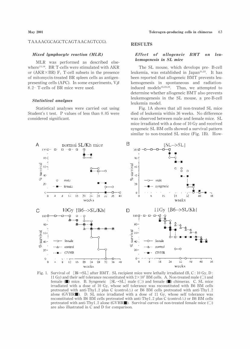
Fig.1A shows that all non-treated SL mice
died of leukemia within 36 weeks. No difference
was observed between male and female mice. SL
mice irradiated with a dose of 10 Gy and received
syngeneic SL BM cells showed a survival pattern
similar to non-treated SL mice (Fig.1B). How-
May 2001 Tolerogen-producing cells in chimeras
Fig.1. Survival of B6→SL after BMT. SL recipient mice were lethally irradiated(B,C:10 Gy,D :11 Gy)and their self tolerance reconstituted with 2 10 BM cells. A.Non-treated male(○)and
female (■)mice. B.Syngeneic SL→SL male (○)and female (■)chimeras. C.SL mice
irradiated with a dose of 10 Gy,whose self tolerance was reconstituted with B6 BM cells
pretreated with anti-Thy1 2 plus C (control )or B6 BM cells pretreated with anti-Thy1 2
alone (GVHR■). D. SL mice irradiated with a dose of 11 Gy, whose self tolerance was
reconstituted with B6 BM cells pretreated with anti-Thy1 2 plus C (control )or B6 BM cells
pretreated with anti-Thy1 2 alone(GVHR■). Survival curves of non-treated female mice(○)are also illustrated in C and D for comparison.
ever,when allogeneic (B6)BM cells were trans-
planted to SL mice irradiated with doses of 10 Gy
or 11 Gy,approximately 70% or 50% of the mice,
respectively,survived more than 36 weeks after
BMT (Fig.C,D). No difference in the survival
curve was detected between the[B6→SL](con-
trol)and the GVHR[B6→SL]chimeras.
β [ →
] [ → ]
We reported that Vβ6 T-cells reactive to
Mls-1 plus MHC class II were eliminated in the
thymus and the spleen of[B10.AQR→AKR]and[B10.BR→AKR]chimeras as the result of nega-
tive selection . The elimination of Vβ6 T-
cells,however,was abrogated in GVHR chimer-
as . Since the SL background partially
containsAKR genes,it was assumed that SL mice
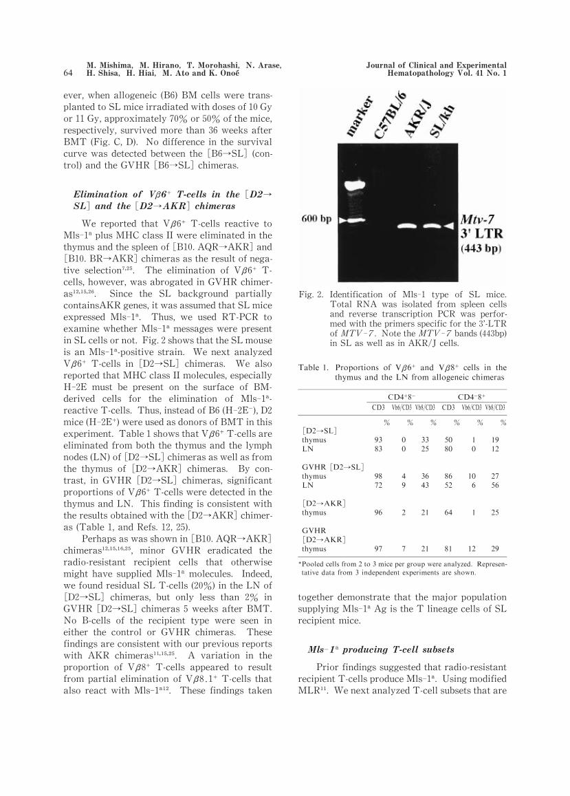
expressed Mls-1. Thus, we used RT-PCR to
examine whether Mls-1 messages were present
in SL cells or not. Fig.2 shows that the SL mouse
is an Mls-1-positive strain. We next analyzed
Vβ6 T-cells in[D2→SL]chimeras. We also
reported that MHC class II molecules,especially
H-2E must be present on the surface of BM-
derived cells for the elimination of Mls-1-
reactive T-cells. Thus,instead of B6(H-2E ),D2
mice(H-2E )were used as donors of BMT in this
experiment. Table 1 shows that Vβ6 T-cells are
eliminated from both the thymus and the lymph
nodes(LN)of[D2→SL]chimeras as well as from
the thymus of[D2→AKR]chimeras. By con-
trast, in GVHR[D2→SL]chimeras, significant
proportions of Vβ6 T-cells were detected in the
thymus and LN. This finding is consistent with
the results obtained with the[D2→AKR]chimer-
as (Table 1,and Refs.12,25).
Perhaps as was shown in[B10.AQR→AKR]
chimeras , minor GVHR eradicated the
radio-resistant recipient cells that otherwise
might have supplied Mls-1 molecules. Indeed,
we found residual SL T-cells (20%)in the LN of[D2→SL]chimeras, but only less than 2% in
GVHR[D2→SL]chimeras 5 weeks after BMT.
No B-cells of the recipient type were seen in
either the control or GVHR chimeras. These
findings are consistent with our previous reports
with AKR chimeras . A variation in the
proportion of Vβ8 T-cells appeared to result
from partial elimination of Vβ8.1 T-cells that
also react with Mls-1 . These findings taken
together demonstrate that the major population
supplying Mls-1 Ag is the T lineage cells of SL
recipient mice.
-
Prior findings suggested that radio-resistant
recipient T-cells produce Mls-1. Using modified
MLR . We next analyzed T-cell subsets that are
Fig.2. Identification of Mls 1 type of SL mice.Total RNA was isolated from spleen cells
and reverse transcription PCR was perfor
med with the primers specific for the 3’-LTR
of MTV 7. Note the MTV 7 bands(443bp)in SL as well as in AKR J cells.
-
β β
→
→
→
→
Journal of Clinical and Experimental Hematopathology Vol.41 No.1 M.Mishima M.Hirano T.Morohashi N.Arase H.Shisa H.Hiai M.Ato and K.Onoe
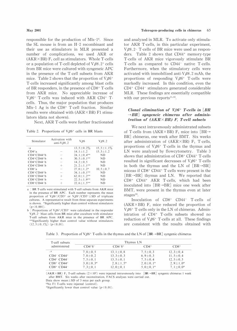
responsible for the production of Mls-1. Since
the SL mouse is from an H-2 recombinant and
their use as stimulators in MLR presented a
number of complications, we used AKR or
(AKR×BR)F cell as stimulators. Whole T-cells
or a population of T-cell depleted of Vβ8.2 cells
from BR mice were cultured with syngeneic APC
in the presence of the T-cell subsets from AKR
mice. Table 2 shows that the proportion of Vβ6
T-cells increased significantly among blast cells
of BR responders,in the presence of CD8 T-cells
from AKR mice. No appreciable increase of
Vβ6 T-cells was induced with AKR CD4 T-
cells. Thus,the major population that produces
Mls-1 Ag is the CD8 T-cell fraction. Similar
results were obtained with (AKR×BR)F1 stimu-
lators (data not shown).
Next,AKR T-cells were further fractionated
and analyzed in MLR. To activate only stimula-
tor AKR T-cells, in this particular experiment,
Vβ8.2-T-cells of BR mice were used as respon-
ders. Table 2 shows that CD44 memory-type
T-cells of AKR mice vigorously stimulate BR
T-cells as compared to CD44 native T-cells.
Furthermore, when the stimulatory cells were
activated with immobilized anti-Vβ8.2 mAb,the
proportions of responding Vβ6 T-cells were
markedly increased. In this condition,even the
CD4 CD44 stimulators generated considerable
MLR. These findings are essentially compatible
with our previous reports .
β [
→ ]
×
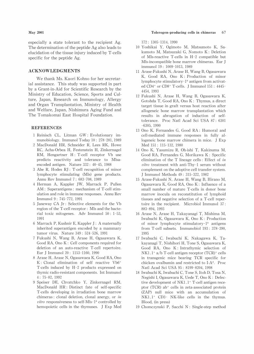
We next intravenously administrated subsets
of T-cells from (AKR×BR)F mice into[BR→
BR]chimeras,one week after BMT. Six weeks
after administration of (AKR×BR) F T-cells,
proportions of Vβ6 T-cells in the thymus and
LN were analyzed by flowcytometry. Table 3
shows that administration of CD8 CD44 T-cells
resulted in significant decreases of Vβ6 T-cells
in both the thymus and the LN of[BR→BR]
miceas if CD8 CD44 T-cells were present in the[BR→BR]thymus and LN. We reported that
CD8 CD44 AKR T-cells, which had been
inoculated into[BR→BR]mice one week after
BMT,were present in the thymus even at later
stages .
Inoculation of CD8 CD44 T-cells of
(AKR×BR) F mice reduced the proportion of
Vβ6 T-cells only in the LN of chimeras. Admin-
istration of CD4 T-cells subsets showed no
reduction of Vβ6 T-cells at all. These findings
are consistent with the results obtained with
β →
→
β
ββ β
β β
ββ
May 2001 Tolerogen-producing cells in chimeras

MLR and our previous study in which the T-cell
repertoire was analyzed at later periods after
BMT . The present results indicate again that
CD8 CD44 T-cells are the most potent Mls-1-producing cells.
DISCUSSION
BMT is one of the most promising therapies
for many hematopoietic and immunodeficiency
diseases that can not otherwise be treated effec-
tively . In the present study,we showed that
allogeneic BMT prevented leukemogenesis in SL
mice, an animal model for spontaneous pre-
B-cell leukemia,as was shown in AKR/J mice,an
animal model for spontaneous thymic
lymphoma . It has been reported that GVHR
may exert beneficial influences on the reconstitu-
tion of recipient hematopoietic and lymphoid
tissues by donor-derived cells (graft enhance-
ment) . In addition,GVHR may be associated
with graft versus leukemia (GVL)effect . How-
ever,we could not detect any appreciable influ-
ences of minor GVHR on the survival of SL
chimeras in the conditions we tested. Similar
survival curves were observed in the control and
the GVHR[B6→SL]chimeras.
We have reported that minor GVHR resulted
in the abrogation of negative selection of T-cells
reactive to recipient Ag . In the SL chimer-
a system,we also found that minor GVHR led to
the failure of clonal elimination of Mls-1-
reactive T-cells. At first, the abrogation of the
negative selection was attributed to the lack of
Mls-1 Ag-producing cells in GVHR[Mls-1→
Mls-1]chimeras. However,we found recently
that GVHR resulted in the failure of clonal elimi-
nation of T-cells reactive to donor Ag .
Thus, itmay be concluded that the GVHR
also induces functional changes to the thymus.
Similar observation was reported by Desbarats
and Lapp .
We reported that the acute GVHR induced in
AKR recipients shifted the T-cell responses to the
Th2 dominant state . This early Th2 shift
appeared to be associated with the subsequent
T-cell responsiveness, since T-cells recovered
from acute GVHR showed the Th2 dominant
state. Thus,T-cells from such chimeras promi-
nently producedIL-4 but not IFN-γupon stimula-
tion. In addition,these T-cells exhibited signifi-
cant MLR but not cytotoxic T-lymphocyte
responses to the recipient Ag (split tolerance) .
Although we did not analyze T-cell responsive-
ness in[B6→SL]chimeras,a similar functional
state appeared to be generated in these GVHR
chimeras., since significant proportions of Mls-1-reactive T-cells were detected in GVHR[D2→SL]mice but not in control[D2→SL]chimer-
as. We first expected that these Mls-1-reactive
T-cells might be exerting the GVL effect. How-
ever,as described above,no difference was obser-
ved in the survival rate between[B6→SL]and
GVHR[B6→SL]chimeras. It is possible that
these Mls-1-reactive T-cells induced neither
harmful GVHR responses nor beneficial GVL
responses in GVHR[B6→SL]chimeras. It seems
important to elucidate the basic mechanism
underlying the immunological alteration induced
by GVHR in further studies.
In the present study,we demonstrated that
the major population of Mls-1-producing cells of
the recipients were CD8 CD44 T-cells,although
these findings were obtained with the AKR
chimera system but not with the SL chimera
system. These findings are essentially compat-
ible with our previous reports . In our previ-
ous studies , we demonstrated that CD8
CD44 T-cells expressed larger amounts of MTV-7 mRNA than CD8 CD44 T-cells. Since cells
of donor mice cannot produce Mls-1,it is clear
that Mls-1 Ag derived from radio-resistant
recipient cells (mainly CD8 CD44 )are transfer-
red to and presented on the surface of donor
MHC class II cells in the thymus (cross-
presentation). We and others have reported that
these Mls-1 Ag plus MHC class II on the surface
of donor BM-derived cells (APC) eliminate the
Mls-1-reactive T-cells between 2 and 3 weeks
after BMT in the thymic medulla .
Although we analyzed here T-cell
reactivities to a superantigen,GVHR appears to
be induced by various allogeneic protein Ags
including MHC Ag. It is now understood that the
major GVHR is not induced by the recipient
MHC Ag alone but by complexes of MHC and
peptide Ag bound in the Ag-binding groove of the
MHC . These recipient peptides with a spe-
cific motif for binding to the MHC appeared to
be derived from cellular components of the recipi-
ent. Thus, identification of the peptide Ag
involved in the rather complex GVHR is essential
to explain the influence of the GVHR on the
development of the recipient immune system,
Journal of Clinical and Experimental Hematopathology Vol.41 No.1 M.Mishima M.Hirano T.Morohashi N.Arase H.Shisa H.Hiai M.Ato and K.Onoe
especially a state tolerant to the recipient Ag.
The determination of the peptide Ag also leads to
elucidation of the tissue injury induced by T-cells
specific for the peptide Ag.
ACKNOWLEDGMENTS
We thank Ms.Kaori Kohno for her secretar-
ial assistance. This study was supported in part
by a Grant-in-Aid for Scientific Research by the
Ministry of Education, Science, Sports and Cul-
ture, Japan, Research on Immunology, Allergy
and Organ Transplantation,Ministry of Health
and Welfare,Japan,Nishimura Aging Fund and
The Tomakomai East Hospital Foundation.
REFERENCES
1 Reinisch CL, Litman GW :Evolutionary im-
munobiology. Immunol Today 10:278-281,1989
2 MacDonald HR, Schneider R, Lees RK, Howe
RC,Acha-Orbea H,Festenstein H,Zinkernagel
RM, Hengartner H :T-cell receptor Vb use
predicts reactivity and tolerance to Mlsa-
encoded antigen. Nature 332:40-45,1988
3 Abe R,Hodes RJ:T-cell recognition of minor
lymphocyte stimulating (Mls) gene products.
Annu Rev Immunol 7:683-708,1989
4 Herman A, Kappler JW, Marrack P, Pullen
AM :Superantigens:mechanism of T-cell stim-
ulation and role in immune responses. Annu Rev
Immunol 9 :745-772,1991
5 Janeway CA Jr:Selective elements for the Vb
region of the T-cell receptor:Mls and the bacte-
rial toxic mitogenes. Adv Immunol 50:1-53,
1991
6 Marrack P,Kushnir E,Kappler J:A maternally
inherited superantigen encoded by a mammary
tumor virus. Nature 349 :524-526,1991
7 Fukushi N, Wang B, Arase H, Ogasawara K,
Good RA,Ono K:Cell components required for
deletion of an auto-reactive T-cell repertoire.
Eur J Immunol 20:1153-1160,1990
8 Arase H,Arase N,Ogasawaea K,Good RA,Ono
K:Clonal elimination of self reactive Vb6
T-cells induced by H-2 products expressed on
thymic radio-resistant components. Int Immunol
4:75-82,1992
9 Speiser DE, Chvatchko Y, Zinkernagel RM,
MacDonald HR:Distinct fate of self-specific
T-cells developing in irradiation bone marrow
chimeras:clonal deletion, clonal anergy, or in
vitro responsiveness to self-Mls-1 controlled by
hemopoietic cells in the thymuses. J Exp Med
172:1305-1314,1990
10 Yoshikai Y, Ogimoto M, Matsumoto K, Sa-
kumoto M,Matsuzaki G,Nomoto K:Deletion
of Mls-reactive T-cells in H-2 compatible but
Mls-incompatible bone marrow chimeras. Eur J
immunol 19 :1009-1013,1989
11 Arase-Fukushi N,Arase H,Wang B,Ogasawara
K, Good RA, Ono K:Production of minor
lymphocyte stimulatory-1 antigen from activat-
ed CD4 or CD8 T-cells. J Immunol 151:4445-
4454,1993
12 Fukushi N, Arase H, Wang B, Ogasawara K,
Gotohda T,Good RA,Ono K:Thymus,a direct
target tissue in graft versus host reaction after
allogeneic bone marrow transplantation which
results in abrogation of induction of self-
tolerance. Proc Natl Acad Sci USA 87:6301-6305,1990
13 Ono K, Fernandes G, Good RA:Humoral and
cell-mediated immune responses in fully al-
logeneic bone marrow chimera in mice. J Exp
Med 151:115-132,1980
14 Ono K, Yasumizu R, Oh-ishi T, Kakinuma M,
Good RA,Fernandes G,Morikawa K:Specific
elimination of the T lineage cells:Effect of in
vitro treatment with anti-Thy-1 serum without
complement on the adoptive cell transfer system.
J Immunol Methods 49 :315-322,1982
15 Arase-Fukushi N,Arase H,Wang B,Hirano M,
Ogasawara K,Good RA,Ono K:Influence of a
small number of mature T-cells in donor bone
marrow inocula on reconstitution of lymphoid
tissues and negative selection of a T-cell reper-
toire in the recipient. Microbiol Immunol 37:
883-894,1993
16 Arase N,Arase H,Takayanagi T,Mishima M,
Iwabuchi K,Ogasawara K,Ono K:Production
of minor lymphocyte stimulatory-1 antigens
from T-cell subsets. Immunobiol 193:378-390,
1995
17 Iwabuchi C, Iwabuchi K, Nakagawa K, Ta-
kayanagi T,Nishihori H,Tone S,Ogasawara K,
Good RA, Ono K:Intrathymic selection of
NK1.1 a/b T-cell antigen receptor(TCR) cells
in transgenic mice bearing TCR specific for
chicken ovalbumin and restricted to I-A . Proc
Natl Acad Sci USA 95:8199-8204,1998
18 Iwabuchi K,Iwabuchi C,Tone S,Itoh D,Tosa N,
Negishi I,Ogasawara K,Uede T,Ono K:Defec-
tive development of NK1.1 T-cell antigen rece-
ptor (TCR)ab cells in zeta-associated protein
(ZAP) null mice with an accumulation of
NK1.1 CD3- NK-like cells in the thymus.
Blood,(in press)
19 Chomczynski P, Sacchi N :Single-step method
May 2001 Tolerogen-producing cells in chimeras
of RNA isolation by acid guanidinium
thiocyanate-phenol-chloroform extraction. Anal
Biochem 162:156-159,1987
20 Sambrook J,Fritsch EF,Maniatis T :In Molec-
ular Cloning :A Laboratory Manual. Vol. 1.
Cold Spring Harbor Laboratory Press, Cold
Spring Harbor,NY.1989
21 Shimada MO, Yamada Y, Nakakuki Y,
Okamoto K, Fukumoto M, Honjo T, Hiai H :
SL/KH strain of mice:a model of spontaneous
pre-B-lymphomas. Leukemia Res 17:573-578,
1993
22 Hiai H,Kaneshima H,Nakamura H,Oguro YB,
Moriwaki K,Nishizuka Y:Unusually early and
high rate of spontaneous occurrence of nonth-
ymic leukemia in SL/Kh mice,a subline of SL
strain. Jpn J Cancer Res 73:704-712,1982
23 Aizawa S, Sado T :Graft-versus-leukemia
effect in MHC-compatible and incompatible
allogeneic bone marrow transplantation of
radiation-induced, leukemia-bearing mice.
Transplantation 52:885-889,1991
24 Tanaka T,Obata Y,Fernandes G,Ono K,Stock-
ert E,Good RA:Prevention of leukemia in leth-
ally irradiated AKR mice by CBA-H marrow
transplantation. Proc Am Assoc Cancer Res 20:
114,1979 (Abstr.).
25 Hirano M, Arase H, Arase-Fukushi N, Ogas-
awara K, Iwabuchi K, Miyazaki T, Good RA,
Ono K:Reconstitution of lymphoid tissues
under the influence of subclinical level of graft
versus host reaction induced by bone marrow
T-cells or splenic T-cell subsets. Cell Immunol
151:118-132,1993
26 Ono K, Arase N, Arase H, Takayanagi T,
Nishihori H, Iwabuchi K, Ogasawara K, Good
RA:Influence of graft versus host reaction on
the T-cell repertoire differentiating from bone
marrow precursors following allogeneic bone
marrow transplantation. Transplant Immunol
5:75-82,1997
27 Good RA:Toward safer marrow transplanta-
tion. New Eng J Med 306:421-423,1982
28 Good RA,Kapoor N,Reisner Y:Bone marrow
transplantation-an expanding approach to tret-
ment of many diseases. Cell Immunol 82:36-54,
1983
29 Sykes M, Sheard MA, Sachs DH :Effects of
T-cell depletion in radiation bone marrow
chimeras II.Requirement for allogeneic T-cells
in the reconstituting bone marrow inoculum for
subsequent resistance to breaking tolerance. J
Exp Med 168:661-673,1988
30 Murphy WJ,Kumar V,Cope JC,Bennett M :An
absence of T-cells in murine bone marrow allo-
grafts leads to an increased susceptibility to
rejection by natural killer cells and T-cells. J
Immunol 144:3305-3311,1990
31 Horowitz MM,Gale RP, Sondel PM,Goldman
JM, Kersey J, Kolb HJ, Rim AA, Ringden O,
Rozman C, Speck B :Graft-versus-leukemia
reactions after bone marrow transplantation.
Blood 75:555-562,1990
32 Morohashi T,Ogasawara K,Kitaichi N,Iwabu-
chi K,Ono K:Abrogation of negative selection
by GVHR induced by minor histocompatibility
antigens or H-2D antigen alone. Immunobiol
202:368-379,2000
33 Desbarats J, Lapp WS:Thymic selection and
thymic major histocompatibility complex class
II expression are abnormal in mice undergoing
graft-versus-host reaction. J Exp Med 178:805-
814,1993
34 Takayanagi T,Nishihori H,Matsuki N,Iwabu-
chi K, Ogasawara K, Ono K:Effects of non-
major histocompatibility antigens on acute
graft-versus-host reaction after allogeneic bone
marrow transplantation. Bone Marrow Trans-
plant 20:297-304,1997
35 Morohashi T,Ogasawara K.Kitaichi N,Iwabu-
chi K, Ono K:Significant MLR but not CTL
responses against recipient antigens generated in
T-cells from bone marrow chimeras recovered
from GVHD. Bone Marrow Transplant 26:
1069-1076,2000
36 den Haan JMM, Sherman NE, Blokland E,
Huczko E,Koning F, Drijfhout JW, Skipper J,
Shabonowitz J,Hunt DF,Engelhard VH :Identi-
fication of a graft versus host disease-associated
human minor histocompatibility antigen. Sci-
ence 268:1476-1480,1995
37 Leibnitz RR,Lipsky PL,Thiele DL:Protection
from T helper cell mediated graft-versus-host
disease by the presence of an MHC class I
alloantigen is associated with perturbation of
MHC class II-restricted responses by class I-
derived peptide. J Immunol 155:1784-1795,1995
38 Ogasawara K,Ono K:MHC binding motifs and
design of peptide-based vaccines. Trends Mi-
crobiol 1:276-279,1993
Journal of Clinical and Experimental Hematopathology Vol.41 No.1 M.Mishima M.Hirano T.Morohashi N.Arase H.Shisa H.Hiai M.Ato and K.Onoe