Embed Size (px)
Citation preview

1
Title page
TITLE: Selected ginsenosides of the protopanaxdiol series are novel positive allosteric modulators
of P2X7 receptors
Authors: R Helliwell1, C Ong ShioukHuey2, K Dhuna2, J C Molero1, J-M Ye1, C C Xue1, L
Stokes2
1School of Health Sciences, Health Innovations Research Institute, RMIT University, Melbourne,
Australia. 2School of Medical Sciences, Health Innovations Research Institute, RMIT University,
Melbourne, Australia.
Running title: Ginsenosides potentiate P2X7 receptors
Address for Correspondence: Dr L. Stokes, School of Medical Sciences, Health Innovations
Research Institute, RMIT University, Bundoora West Campus, Bundoora VIC 3083, Australia.
Email: [email protected]
Author contributions:
RH, COS, KD, LS performed the research
LS, RH, JCM, CCX, and J-M Y designed the study
CCX and J-M Y contributed essential reagents
LS, COS and RH analysed the data
RH, LS and CCX wrote the manuscript.

2
List of non-standard abbreviations PPD, protopanaxdiol; PPT, protopanaxtriol; HEK-hP2X7, HEK-293 cells stably expressing human P2X7; AM, acetoxymethyl.

3
SUMMARY
Background and purpose
P2X7 is an ATP-gated ion channel predominantly expressed in immune cells and plays a key role in
inflammatory processes. Ginseng is a well-known Chinese herb with both pro- and anti-
inflammatory properties where many of these actions have been ascribed to constituent
ginsenosides. We performed a screen to determine if ginsenoside compounds could display
pharmacological activity at P2X7 that might contribute to the reported immunomodulatory actions
of ginseng.
Experimental approach
We used several assays to measure P2X7 responses; ATP-mediated dye uptake, intracellular
calcium measurement and whole cell patch clamp recordings. HEK-293 cells stably expressing
human P2X7 were used in addition to mouse macrophages endogenously expressing P2X7.
Key results
Four ginsenosides of the protopanaxdiol series, Rb1, Rh2, Rd and the metabolite compound K (CK)
potentiated P2X7 dye uptake responses whereas other ginsenosides tested were ineffective (1 -
10µM). The potentiation was rapid in onset, required a threshold concentration of ATP (> 50 µM)
and had an EC50 of 1.08 µM. CK markedly enhanced ATP activated P2X7 currents, likely via an
extracellular site of action. One of the consequences of this potentiation effect is a sustained rise in
intracellular Ca2+ that could account for the decrease in cell viability in mouse macrophages after a
combination of 500 µM ATP and 10 µM CK that are non-toxic when applied alone.
Conclusions and implications
This study identifies selected ginsenosides as novel potent allosteric modulators of P2X7 channels
that may account for some of the reported immune modulatory actions of protopanaxdiol
ginsenosides in vivo.

4
INTRODUCTION
Ginseng has been used for at least 2000 years in China and other Asian countries to support vitality
and a long life. In traditional Chinese medicine it is often described as a ‘precious tonic’ that
stimulates natural resistance to infection and maintains homeostasis (Hanley et al., 2012a; Kumagai
et al., 2013). It is typically extracted from the roots of Panax ginseng C Mayer (Asian) and Panax
quinfolious (American) and contains a complex mixture of bioactive compounds; the most
extensively studied being the ‘steroid-like’ dammarane triterpenoid glycosides, termed
ginsenosides. Ginsenosides are further subdivided into protopanaxdiols (PPD) (examples shown in
Figure 1) or protopanaxtriols (PPT) depending on the location of the sugar moieties on the
dammarane carbon skeleton. Both glycosylated classes may have sugar moieties on carbon (C) 20
but protopanaxadiols can have additional sugar moieties on C-3 whereas protopanaxatriols are on
C-6. The aglycones of both classes differ only in the presence of an extra hydroxyl group on C-6 in
the triol (Tomasinsig et al., 2008; Chotjumlong et al., 2013). Ginseng is commercially available in a
variety of forms (capsules, tablets, oils) where the type and amounts of constituent ginsenosides are
precisely controlled. The G115 formulation tested in this study contains 4% w/w ginsenosides and
is a principle constituent of Ginsana® and Gincosan® medications (Ginsana SA).
We are interested in understanding the mechanisms underlying the reported immuno-
modulatory effects of ginseng/ginsenosides as they may translate into effective medications for the
prevention and treatment of infective and inflammatory diseases. In vivo studies using ginseng
formulations have been shown to protect against lung infections caused by Pseudomonas
Aeruginosa improving pulmonary bacterial clearance (Song et al., 1997a; Song et al., 1997b; Song
et al., 2010) and a clinical trial demonstrated a significant reduction in the number of cases of
influenza when given as an adjuvant with vaccine that correlated with increased antibody titres and
NK cell activity (Scaglione et al., 1996). Similar adjuvant effects have also been consistently
5
demonstrated with ginsenosides in vivo (Rivera et al., 2005; Han et al., 2013). In vitro,
ginsenosides have been shown to either trigger or prevent apoptosis depending on cell type or the
specific ginsenoside used (Ham et al., 2006; Zhang et al., 2008; Li et al., 2012; Zhang et al., 2013;
Zheng et al., 2014). A number of studies have also demonstrated that ginsenosides can act via either
genomic Eβ or glutocorticoid receptors in the sub-micromolar range to differentially regulate
angiogenesis in endothelial cells (Sengupta et al., 2004; Leung et al., 2006; Leung et al., 2007;
Leung et al., 2009). However, similar to some endogenous steroids, ginsenosides have also been
shown to rapidly and reversibly interact with ligand gated ion channels which points to additional
non-genomic sites of action (Nah, 2014). These include both inhibitory actions on nicotinic
acetylcholine receptors (Lee et al., 2003), 5-HT3 receptors (Choi et al., 2003a), NMDA receptors
(Kim et al., 2002), GABAA receptors (Lee et al., 2012), TRPV1 channels (Huang et al., 2012) and
potentiating actions on glycine (Noh et al., 2003), GABAA receptors (Choi et al., 2003b), and
TRPV1 channels (Jung et al., 2001).
In this study we tested the effects of ginsenosides on a subtype of ATP-gated ion channels
of the P2X family, P2X7 receptors, given their important role in regulating immune cell function
(Bartlett et al., 2014). Studies have demonstrated P2X7 to play a role in Ca2+ signalling, reactive
oxygen species generation and cell death pathways and regulation of such signalling pathways may
control immune responses (Bartlett et al., 2014). We report that several glycosylated PPD
ginsenosides, but not PPT ginsenosides, appear to be positive allosteric modulators of the ATP
activated P2X7 channel which can lead to enhanced Ca2+ influx and subsequent apoptosis in
macrophages. Due to the relatively high expression levels of P2X7 in immune cells, this action
may account for some of the reported immune modulatory actions of PPD ginsenosides in vivo.

6
MATERIALS AND METHODS
Materials
The P2X7 agonists ATP and BzATP were obtained from Sigma Aldrich (Ryde, NSW Australia).
P2X7 antagonists AZ-10606120 and A-438079 hydrochloride were obtained from Tocris
Biosciences (Bristol, UK). ATP was prepared as a 100 mM stock in double distilled water, pH to
7.4 with NaOH and was stored at -80 °C until the day of experiment. AZ-10606120 and A-438079
(10 mM in DMSO) were stored at -20 °C. Ginsenosides (certified as 98 % pure) were obtained from
Chengdu Mansite Pharmaceutical Co Ltd (PPT, Rg1, Rg3, Rb1, Rb2, CK), Sichuan Weikeqi
Biological Technology Co. Ltd, (PPD, Rf), Chengdu Biopurify Phytochemicals Ltd (Rc, Rd, Re)
and Shanghai E Star Bio Technology Co Ltd (Rh1, Rh2). Each compound was prepared as a 50 mM
stock in DMSO and stored at -80 °C until the day of the experiment. All ginsenoside stocks were
further diluted in DMSO to 1000X final concentration so that when diluted in assay buffers the final
concentration of DMSO was 0.1%.
Cell Culture
HEK-293 cells stably transfected with the human P2X7 (standard nomenclature conforms to
guidelines (Alexander et al., 2013)) plasmid (clone pJB3) were maintained in DMEM:F12 media
(Life Technologies catalogue number 11320-033) supplemented with 10% foetal bovine serum
(FBS, French origin, Bovogen, Australia), 1% Glutamax, 10 000 U. ml-1 penicillin and 10 mg. ml-1
streptomycin with selection under 400 µg. mL-1 G418 (Life Technologies). The J774 murine
macrophage cell line was maintained in RPMI 1640 media supplemented with 10% FBS, 1%
Glutamax, and 1% penicillin and streptomycin (Bhaskaracharya et al., 2014).
Animals

7
Animal care and procedures were performed with permission of the local RMIT Animal Ethics
Committee (approval number AEC1312) and in accordance with Australian guidelines for the use
of animals. All studies involving animals are reported in accordance with the ARRIVE guidelines
(Kilkenny et al., 2010). Adult male C57BL/6 mice were maintained in a 12h light/dark cycle and
fed standard diet with water ad libitum. Mice were killed by CO2 asphyxiation and peritoneal
macrophages were obtained by flushing the peritoneal cavity with cold PBS (5 ml).
Dye uptake experiments
Cells were plated at 2.5 x 10⁴ cells per well the day before experiments into poly-D-lysine coated
96-well plates (ThermoFisher). YOPRO-1 iodide (Life Technologies) was used as the membrane
impermeant dye with a final concentration of 2 µM in low divalent buffer (145 mM NaCl, 5 mM
KCl, 0.2 mM CaCl2, 13 mM glucose, 10 mM HEPES, pH 7.3, osm 300-310). A Flexstation III plate
reader (Molecular Devices) was used to acquire data using the following settings – excitation
wavelength 490 nm, emission wavelength 520 nm and 6 reads per well. Data was analysed as slope
of dye uptake or area under curve between 40 and 300 seconds using SoftMax Pro software
(Molecular Devices).
Calcium measurements
HEK-293 cells were plated at 2.5 x 10⁴ cells per well the day before experiments into poly-D-lysine
coated 96-well plates. Cells were loaded with 1 µM Fluo-4AM calcium indicator dye in low
divalent buffer for 30 minutes at 37 °C. This solution was then removed and replaced with standard
extracellular assay buffer (145 mM NaCl, 5 mM KCl, 2 mM CaCl2, 1mM MgCl2, 13 mM glucose,
10 mM HEPES, pH 7.3). A Flexstation III plate reader was used to acquire data using the following

8
settings – excitation wavelength 490nm, emission wavelength 520 nm and 6 reads per well. ATP
was injected automatically after 40 seconds.
J774 mouse macrophages were plated at 2.5 x 10⁴ cells per well into poly-D-lysine coated
96-well plates. Cells were loaded with 2.5 µM Fura-2AM calcium indicator dye plus an equal
volume of pluronic acid in HBSS buffer for a total of 30 minutes at 37°C. This loading solution was
removed and replaced with standard extracellular assay buffer. A Flexstation III plate reader was
used to acquire data using the following settings; excitation wavelengths 340 nm and 380 nm with
an emiision wavelength of 520 nm. 6 reads per well was used and ATP (10X concentration) was
injected automatically after 40 seconds.
Peritoneal macrophages were plated onto 12 mm glass coverslips at 10,000 cells per slip and
cultured overnight. Macrophages were loaded with 1 µM Fluo-4AM in low divalent buffer for 30
minutes at 37 °C and then placed into a heated (36 °C) 35 mm bath chamber on a Nikon Eclipse Ti-
U fluorescent microscope. Cells were continuously perfused with low divalent assay buffer by
gravity feed and calcium signals measured using a CoolSnap HQ2 CCD camera recording green
fluorescence at 520 nm. A time-lapse recording was generated with exposure time of 300 ms and
gain of 500 controlled through NIS Elements software (Nikon Instruments). Regions of interest
were drawn around 150-170 individual cells to measure fluorescence at each time point,
background subtracted and the mean fluorescence value calculated.
Patch clamp electrophysiology
Stably expressing HEK-hP2X7 cells were plated onto 13-mm glass coverslips 4 – 24 hours before
use. Membrane currents were recorded in the whole-cell patch clamp configuration using an EPC10
amplifier (HEKA, Lambrecht, Germany) and borosilicate glass electrodes (Clark Electromedical),
resistance 3-8 MOhm when filled with standard internal solution. Cells were continually perfused
by gravity feed with standard divalent buffer solution (145 mM NaCl, 5 mM KCl, 2 mM CaCl2,

9
1mM MgCl2, 13 mM glucose, 10 mM HEPES, pH 7.3) prior to seal formation and with low
divalent buffer solution in the majority of the experiments. Standard internal buffer solution
contained NaCl 145 mM, HEPES 10 mM, EGTA 10 mM, pH 7.3 with NaOH 10M. In certain
experiments, investigating permeability changes of P2X7 channels, all cations in the external
solution were substituted with the large organic cation N-methyl-D-glucamine. This solution
contained NMDG 154 mM, HEPES 10 mM, Glucose 13 mM, pH 7.3 with HCl 10M. Series
resistance compensation was routinely applied up to a maximum of 80% to minimize voltage errors.
ATP and drugs were applied using a computer controlled fast-flow system (Bio-Logic Instruments,
Claix, France) with the perfusion capillaries placed in close proximity to the cell under
investigation.
Cell viability assays
Cells were plated at a density of 5 x 10⁴ cells/well (50 µl) in a 96 well plate (Costar) in triplicate.
Compounds (or vehicle control) were added to the plate at 2X final concentration and cells were
treated for 24 hours. Cell viability was assessed using the CellTiter Glo Aqueous One solution
(Promega) which was added to the wells for the last 4 hours of the experiment. Absorbance was
read at 490 nm using a BMG Labtech Clariostar plate reader.
Data plot and statistical analysis
Graphs were plotted using GraphPad Prism version 6 (La Jolla, USA). Concentration-response
curves were fitted using a log (agonist) vs response – variable slope (four parameter) best-fit
equation. Data was analysed for statistical significance using either unpaired t-tests or one-way
ANOVA with post-tests as appropriate. Significance was taken as P<0.05.

10
RESULTS
Ginsenosides increase the rate of dye uptake through activated P2X7
To establish any potential effects of ginseng on P2X7 we used a standard screening assay that relies
upon the uptake of the membrane impermeant dye YOPRO-1 iodide through the P2X7-dependent
permeability pathway activated with the agonist ATP (Jursik et al., 2007; Bhaskaracharya et al.,
2014). The initial screening of compounds used an approximate EC50 concentration of ATP (200
µM) in order to see either potentiation or inhibition of the response. We first tested a standard
formulation of ginseng known as G115 since it is one of the main formulations used in the clinic.
Pre-treatment of HEK cells expressing human P2X7 (HEK-hP2X7) for 10 minutes with 100 µg.ml-1
G115 enhanced the rate of ATP-induced dye uptake by around 2-fold (Figure 1A, B red trace).
G115 alone did not induce dye uptake in HEK-hP2X7 cells (Supplementary Figure 1) suggesting
this was not due to a non-specific effect on the cells.
G115 contains a fixed amount (4% w/w) of eight ginsenosides from both PPD and PPT
chemical classes, namely Rb1, Rc, Rd, Re (PPD ginsenosides) and Rb2, Rf, Rg1, Rg2 (PPT
ginsenosides). It was important to establish whether the observed effects with G115 resulted from
one or more specific ginsenoside(s) in the formulation. We tested 14 purified ginsenosides in a
screening assay encompassing the eight ginsenosides in G115 plus the principle intestinal
metabolites Rh1, Rh2, Rg3, compound K (CK) and the two aglycones PPD and PPT. All
ginsenosides were tested at a concentration of 10 µM and were added 10 minutes prior to the
addition of ATP (200 µM). None of the ginsenosides directly stimulated dye uptake
(Supplementary Figure 1) however, four PPD ginsenosides, Rb1, Rd, Rh2 and CK, significantly
increased the rate of dye uptake after ATP addition (Figure 1C). In contrast, ginsenosides of the
PPT series (labelled green in Figure 1C) had no significant effect on the ATP-induced dye uptake.
The chemical structures of the four ginsenosides with effect on P2X7 are shown in Figure 1D.
11
A glucopyranoside sugar residue in CK is essential for rapid P2X7 potentiation
Since the PPD aglycone was ineffective in potentiating P2X7 responses at concentrations up to 50
µM (data not shown) there is an absolute requirement for at least one sugar residue in this structure.
Based on our data this could be located at C-3 (Rh2) or C-20 (CK). As CK appeared to be the most
potent in this assay and is one of the principle metabolites of ginseng reaching plasma
concentrations of around 70 ng.ml-1 in humans (Kim et al., 2013) we focused our investigation on
this ginsenoside to examine the potentiating action on P2X7 in more detail.
The potentiation of ATP responses at P2X7 by CK was also demonstrated in buffer
containing physiological divalent ion concentrations (2 mM Ca2+, 1 mM Mg2+). Under these
conditions much higher concentrations of ATP (>500 µM) are required to elicit any significant dye
uptake most likely due to the actions of these divalent ions in blocking P2X7 responses (Virginio et
al., 1997). CK could potentiate both 0.5 mM and 3 mM ATP responses (Figure 2A). The onset of
potentiation of P2X7 responses by CK was rapid with immediate effects observed following co-
injection of a pre-mixed cocktail of the agonist ATP and CK (Figure 2B). The response induced by
ATP and CK was solely dependent on P2X7 as it could be completely abolished by pre-treatment
with either of two selective P2X7 antagonists; AZ10606120 (10 µM) and A-438079 (10 µM)
(Figure 2C).
Our initial experiments suggested that CK was not a direct activator or agonist of P2X7
since CK could not directly stimulate dye uptake in HEK-hP2X7 cells (Supplementary Fig 1). The
EC50 value of the potentiation effect of CK on hP2X7 was calculated as 1.08 µM (95% CI 0.86 µM
to 1.35 µM) (Figure 3A). In contrast the aglycone PPD was ineffective at all concentrations tested
(0.1 – 50 µM). To determine if CK was acting as a positive allosteric modulator of P2X7 we
performed a full concentration-response curve for ATP in the absence and presence of 10 µM CK
(Figure 3B). Potentiation by CK left-shifted the concentration-response curve reducing the EC50

12
from 244 µM to 71 µM, increased the maximum ATP response, and required a threshold
concentration of ATP (~50 µM), In contrast when BzATP was used as a full agonist at human
P2X7 (Surprenant et al., 1996), CK did not increase the maximum response but did cause a left-
shift in EC50 from 19 µM to 4.4 µM (Figure 3B). Both leftward shifts were statistically significant
(P<0.01).
Ginsenoside CK increases sustained calcium influx through P2X7
Due to their high permeability to calcium, P2X7 activation can result in a sustained rise in
intracellular Ca2+ leading to either proliferation/activation or cell death, depending on the magnitude
and extent of channel activation (Adinolfi et al., 2005b). Therefore it was important to establish
whether the ginsenoside potentiation of P2X7 responses observed in dye uptake experiments lead to
physiologically relevant sustained increases in intracellular Ca2+ concentration. We first
investigated differences in intracellular Ca2+ responses between HEK-hP2X7 cells and the parental
HEK-293 cell line (Figure 4A) using fluo-4AM loaded cells. In both cell lines there was an initial
transient rise in intracellular Ca2+ following ATP addition which is due to the activation of G-
protein coupled P2Y receptors. However only HEK-hP2X7 cells showed an additional sustained
elevation in intracellular Ca2+ on addition of ATP concentrations > 100 µM (Figure 4A). In the
presence of ginsenoside CK, this sustained calcium response was observed with 100 µM ATP
(Figure 4B, blue trace). Quantification of the changes in fluo-4 relative fluorescence units (both
peak response and sustained response) show that CK significantly increases the ATP-mediated rise
in intracellular Ca2+ only in P2X7-expressing cells (Figure 4C).
Ginsenosides are potent modulators of ATP-evoked P2X7 currents
To definitively demonstrate that PPD ginsenosides enhanced ionic flux through activated P2X7
channels we used the whole-cell patch-clamp technique and delivery of ATP -/+ ginsenosides via

13
an 8-channel fast flow system. Our standard patch protocol used an initial application of 200 µM
ATP for 5 seconds then switching to 200 µM ATP plus ginsenoside for 5 seconds before briefly
returning to ATP alone (2 seconds) prior to wash off (Figure 5A – D). Representative traces for CK,
Rd, Rb1, PPD, PPT, Rg1 and Rh1 are shown. Similar to the dye uptake experiments, we observed
only glycosylated PPD ginsenosides potentiated ATP responses whereas the PPD aglycone and all
PPT ginsenosides had no effect at 50 µM, the highest concentration tested. The rank order of
potency was CK > Rd >Rb1 with thresholds for effect around 50 nM, 0.5 µM and 10 µM
respectively. These effects were rapid in onset occurring in <1 second of switching from ATP alone
to ATP plus ginsenoside. Similarly the effects were rapidly reversible on wash-out of the
ginsenoside in the continued presence of ATP. Consistent with the dye uptake data, the most potent
ginsenoside CK did not directly activate P2X7 channels, only after prior activation with ATP (data
not shown). The magnitude of potentiation was quantified by dividing the current amplitude after 5
seconds in ATP + ginsenoside by the amplitude in ATP alone immediately prior to ginsenoside
addition (Figure 5E). Although all the PPD ginsenosides had a dose-dependent effect in regard to
potentiating ATP evoked currents, it was not possible to demonstrate a maximal effect where
further increases in ginsenoside concentration lead to no further increases in the potentiation. This
was due to the fact that at higher concentrations of PPD ginsenosides the potentiating effect was so
large that the cells could not be effectively voltage-clamped.
Given the rapid onset and reversibility of the potentiation it was likely that the site of
interaction with P2X7 was extracellular. This was confirmed by comparing the amplitude of P2X7
currents evoked by 5 second pulses of 200 µM ATP in HEK-hP2X7 dialysed with standard pipette
solution or pipette solution containing 1 µM CK for 5 minutes (Figure 6A). In both groups the
amplitude of successive P2X7 currents (2 min apart) evoked by external ATP was similar; 221 ± 59
pA (n= 8) and 264 ± 48 pA (n=3) respectively (P>0.05, unpaired t –test). To confirm that the HEK-
hP2X7 cells containing intracellular CK pipette solution were still responsive to extracellular CK,

14
500 nM CK was applied in addition to ATP for the fifth pulse (Figure 6A, open triangles) and
compared to a fifth successive pulse of ATP alone in the control group. The ATP-evoked current in
the presence of CK was 1953 ± 552 pA (n = 3), significantly larger than after ATP alone, 261 ± 82
pA (n =6 cells, P<0.05, unpaired t-test).
To confirm that the CK potentiation of the ATP activated current was solely mediated by
P2X7 channels we first applied 200 µM ATP for 5 seconds followed by a 5 second addition of CK
in the continued presence of ATP (Figure 6B). This resulted in a marked potentiation of the current
that was almost completely and irreversibly blocked by a subsequent addition of the selective P2X7
antagonist AZ10606120 (10 µM; 92.6 ± 2% inhibition, n=4 cells) in the continued presence of CK
and ATP (Figure 6B).
Ginsenoside CK does not induce an immediate permeability shift to large cations
The fact that PPD ginsenosides accelerated YO-PRO dye uptake in the presence of ATP might
suggest that an underlying increase in the permeability of P2X7 channels to large molecular weight
cations is associated with the potentiating action of ginsenosides. To investigate this possibility
cells were bathed in a solution that contained NMDG as the only extracellular cation and dialysed
with standard 145 mM NaCl-containing internal solution. Under these conditions, we applied 500
msec voltage ramps from -100 mV to +50 mV every 1 second. Fast application of ATP for 10
seconds evoked outward currents at all voltages positive to around -80 mV. The mean reversal
potential (Erev) at the end of the 10s ATP addition (Figure 7, black bar) was -81 ± 2.1 mV (n=4
cells) and the mean outward current amplitude at -30 mV was 1186 ± 416 pA. Solution containing
ATP and 1 µM CK was then applied (Figure 7, hatched bar) which resulted in marked increase in
the amplitude of the current at -30 mV within 2-3 seconds of addition (mean amplitude was 3667 ±
1017 pA). Hence under these conditions CK potentiated the amplitude of the outward current by
around 3-fold but this was not associated with any measurable change in permeability (Erev -81.3 ±

15
2.4 mV). This is illustrated in Figure 7 (inset) where representative ramps in one cell are shown at
the end of the first 10s addition of ATP (ATP Ramp) and 2-3s after the subsequent addition of ATP
+ 1 µM CK (ATP + CK Ramp). In the continued presence of ATP + 1 µM CK there was a
progressive positive shift in Erev which reached -74 ± 2 mV by the end of the 10s addition (Figure
7). These data suggest that permeability changes do not contribute to the fast potentiation of the
P2X7-mediated current after CK addition.
Potentiation of P2X7 by ginsenoside CK is not dependent on cations or membrane voltage
We further used patch clamp recordings of HEK-hP2X7 cells to determine whether the presence of
extracellular divalent cations (Ca2+, Mg2+) could interact with potentiation of the ATP response.
The degree of potentiation of ATP-evoked inward currents was similar in standard extracellular
solution (2 mM Ca2+) although a higher concentration of ATP was required; 1 mM rather than 200
µM in 0.2 mM external Ca2+ solution (Figure 8A). There was no voltage-dependence to the effect
of CK as demonstrated by linear I-V relationships for ATP or ATP in the presence of CK (Figure
8B).
Ginsenoside CK potentiates endogenous P2X7 responses in mouse macrophages
We next established whether similar effects of the ginsenoside CK on endogenous P2X7 could be
demonstrated in mouse macrophages. We determined that CK could potentiate mouse P2X7
expressed in HEK-293 cells (Supplementary Figure 2) thus ruling out a species-specific effect of
ginsenosides. The EC50 value for CK potentiation of mouse P2X7 was 0.45 µM (Supplementary
Figure 2), similar to the CK effect on human P2X7. Using the J774 mouse macrophage cell line
(Figure 9A) and primary mouse peritoneal macrophages (Figure 9B) we demonstrated enhanced
sustained intracellular calcium responses to 500 µM ATP in the presence of 10 µM CK compared
to 500 µM ATP alone. Furthermore, patch clamp recordings from individual macrophages revealed

16
a similar rapid onset of potentiation of ATP-mediated inward currents in J774 and peritoneal
macrophages (Figure 9C).
Finally, we investigated whether this potentiation of ATP-induced responses could translate
to a significant downstream functional effect such as induction of apoptotic cell death. We used
J774 macrophages and treated them with either CK alone (10 µM) or a non-lethal concentration of
ATP (500 µM) for 24 hours. We determined any effect on cellular viability through an MTS
viability assay. Neither 10 µM CK, DMSO or 500 µM ATP alone induced any detrimental effect on
cell viability (n=4 experiments; Figure 10). In contrast, a high concentration of ATP (3 mM) for 24
hours could induce marked cell death resulting in a significant reduction of cell viability (to 16.7 ±
4.3 % of control, n=4). Treating J774 macrophages with a combination of 10 µM CK and
previously non-lethal ATP (500 µM) together could also significantly reduce cell viability to 49.6 ±
11.3 % of control (Figure 10B).

17
DISCUSSION In this study we have shown that certain ginsenosides (Rb1, Rh2, Rd and CK) markedly enhance
P2X7 responses after prior activation with exogenous ATP. These effects were only manifest after
previous P2X7 activation by orthostatic agonists (ATP, BzATP) and were first characterized in a
HEK cell line expressing P2X7 and corroborated in a macrophage cell line (J774) and mouse
peritoneal macrophages. The consequences of the interaction were increased Ca2+ influx and a
subsequent decrease in cell viability to lower concentrations of ATP. Given the widespread
distribution of P2X7 channels on immune cells and the fact that effects could be observed in the
sub-micromolar range with one of the principle metabolites of ginseng CK, it is possible that this
mechanism may account for some of the reported immune modulatory actions of ginseng in vivo.
Since the potentiation of P2X7 responses appears to be unique to glycosylated PPD
ginsenosides and can be observed in the low to sub-micromolar range (EC50 of 0.45 – 1.08 µM), it
is unlikely to be a non-specific interaction resulting from changes in membrane fluidity as has been
suggested for certain ginsenoside actions reflecting the amphipathic nature of these steroid-like
saponins (Attele et al., 1999). In addition, the site of action is likely to be extracellular and may
involve a direct interaction with the P2X7 channel since effects are not observed with intracellular
application of CK (1 µM). The potentiation is rapid in onset and is rapidly reversible. However, the
kinetics of onset/reversibility were not specifically addressed in this study but are at least within 1
second based on our patch clamp experiments using a ‘fast’ drug application system.
The potentiating effects of PPD ginsenosides on P2X7 are novel for this family of ligand-
gated ion channels but do show some similarities to reported effects of ginsenosides on GABAA
(Choi et al., 2003b) and glycine currents (Noh et al., 2003) in Xenopus oocytes. Although the
concentrations of ginsenosides necessary for the observed effects were much higher on GABA and
glycine channels (around 50 µM), these studies also indicated that PPD ginsenosides were more
potent than PPT ginsenosides. Such studies of effects of ginsenosides on ion channels have been

18
extensively reviewed in (Nah 2014). One PPD ginsenoside, Rg3, was additionally found to directly
activate GABA channels containing the γ2 subunit (Lee et al., 2013). In our study we demonstrate
that the aglycone compounds PPD and PPT were ineffective at potentiating P2X7 which points to
an absolute requirement of at least one sugar residue in PPD ginsenosides. Interestingly both PPD
and PPT alglycones have been shown to inhibit rather than potentiate GABAA currents (Lee et al.,
2012). However, we did not observe any inhibitory effects on P2X7 responses with any ginsenoside
compound. Further investigations are required to determine if there are any shared structural
features between the ginsenoside binding sites on P2X7 and GABAA channels. This may also
apply to HERG channels as it has been reported that PPD ginsenosides were typically more
effective than PPT ginsenosides in potentiating tail currents and PPT/PPD aglycones were
ineffective (Choi et al., 2011).
Glycosylated PPD ginsenosides, such as the principal ginseng metabolite CK, are novel
potent positive allosteric modulators of human P2X7. Several other positive modulators of P2X7
have been described including clemastine, tenidap, polymixin B, and ivermectin (Sanz et al., 1998;
Ferrari et al., 2004; Norenberg et al., 2011; Nörenberg et al., 2012). There are some similarities
between the action of clemastine and CK on P2X7 channels in that their action is rapid (< 100ms),
reversible, calcium- and voltage-independent, and uses an extracellular site. Similar to CK,
modulators such as clemastine have no direct effects on P2X7 channels and required the presence of
the agonist (Norenberg et al., 2011). Although Norenberg et al reported an accelerated change in
reversal potential occurring over tens of seconds, reflecting an increased rate in the permeability to
NMDG+ in the presence of ATP, they recognized that this cannot account for the rapid (<1 s)
potentiating effect and reversibility of clemastine (Norenberg et al., 2011). The most plausible
explanation, which may also be applicable to PPD ginsenosides, is that such modulators increase
the mean-open time of P2X7 channels. Furthermore, such a net increase in channel activation is
known to accelerate pore dilation. Hence this hypothesis may reconcile our patch data which clearly

19
indicates that the rapid/reversible effects of ginsenosides are not associated with significant changes
in permeability and our YOPRO data where ginsenosides clearly increase the rate of dye uptake.
An important consequence of the PPD ginsenoside action on P2X7 channels is enhanced
sustained Ca2+ influx in both macrophages and HEK cells stably expressing human P2X7. The use
of CK as a positive allosteric modulator of P2X7 reduces the concentration of ATP required to
generate a sustained Ca2+ response (Figures 4, 8). Many downstream consequences of P2X7
activation have been demonstrated to depend on sustained Ca2+ signaling (Bartlett et al., 2014).
With regard to cell viability, brief additions of high concentrations of ATP (<5 min, >1 mM) can
lead to a transient ‘ pseudoapoptosis’ that does not lead to cell death (Mackenzie et al., 2005) or a
delayed cell death occurring after a number of hours (Hanley et al., 2012b). Higher concentrations
of ATP and /or prolonged applications on the other hand lead to cell death within minutes due to
massive Ca2+ influx (Mackenzie et al., 2005). In contrast, lower concentrations of ATP (<1 mM)
can have the opposite effect, stimulating proliferation and prolonging cell survival (Adinolfi et al.,
2005a). Consistent with this we have shown that enhancing Ca2+ influx via P2X7 in macrophages
through the use of CK can effectively convert a sub-lethal dose of 500 µM ATP into a lethal
concentration as measured by a significant decrease in cell viability after 24h (Figure 10). However
given the fact that the timing and extent of Ca2+ influx via P2X7 can lead to different functional
outcomes further studies are warranted with different ATP/PPD ginsenoside combinations and
examination of other parameters in addition to cell viability.
In conclusion, the present study identifies selected ginsenosides as novel positive allosteric
modulators of P2X7 channels. Our findings together suggest that the modulation of P2X7 may
account for some of the reported immune modulatory actions of protopanaxdiol ginsenosides in
vivo.
20
ACKNOWLEDGEMENTS
LS is supported by an RMIT University Vice Chancellor’s Research Fellowship. RH is supported
by funding from Guangdong Provincial Academy of Chinese Medical Sciences. COS was
supported by a studentship from Singapore Polytechnic to visit RMIT University. KD is supported
by an RMIT University PhD scholarship. We gratefully acknowledge the help of Dr Joanne Hart
(RMIT University) for the provision of animal tissue.

21
REFERENCES
Adinolfi E, Callegari MG, Ferrari D, Bolognesi C, Minelli M, Wieckowski MR, et al. (2005a).
Basal activation of the P2X7 ATP receptor elevates mitochondrial calcium and potential, increases
cellular ATP levels, and promotes serum-independent growth. Molecular biology of the cell 16(7):
3260-3272.
Adinolfi E, Pizzirani C, Idzko M, Panther E, Norgauer J, Di Virgilio F, et al. (2005b). P2X(7)
receptor: Death or life? Purinergic signalling 1(3): 219-227.
Alexander SPH, Benson HE, Faccenda E, Pawson AJ, Sharman JL, Spedding M, Peters JA, Harmar
AJ, CGTP Collaborators (2013) The Concise Guide to Pharmacology: Ligand-gated Ion Channels.
British Journal of Pharmacology 170: 1582-1606.
Attele AS, Wu JA, Yuan CS (1999). Ginseng pharmacology: multiple constituents and multiple
actions. Biochemical pharmacology 58(11): 1685-1693.
Bartlett R, Stokes L, Sluyter R (2014). The P2X7 Receptor Channel: Recent Developments and the
Use of P2X7 Antagonists in Models of Disease. Pharmacological Reviews 66(3): 638-675.
Bhaskaracharya A, Dao-Ung P, Jalilian I, Spildrejorde M, Skarratt KK, Fuller SJ, et al. (2014).
Probenecid blocks human P2X7 receptor-induced dye uptake via a pannexin-1 independent
mechanism. PLoS One 9(3): e93058.
Choi S-H, Shin T-J, Hwang S-H, Lee B-H, Kang J, Kim H-J, et al. (2011). Differential effects of
ginsenoside metabolites on HERG k channel currents. J Ginseng Res 35(2): 191-199.

22
Choi S, Lee J-H, Oh S, Rhim H, Lee S-M, Nah S-Y (2003a). Effects of ginsenoside Rg2 on the 5-
HT3A receptor-mediated ion current in Xenopus oocytes. Molecules and cells 15(1): 108-113.
Choi SE, Choi S, Lee JH, Whiting PJ, Lee SM, Nah SY (2003b). Effects of ginsenosides on
GABA(A) receptor channels expressed in Xenopus oocytes. Archives of pharmacal research 26(1):
28-33.
Chotjumlong P, Bolscher JG, Nazmi K, Reutrakul V, Supanchart C, Buranaphatthana W, et al.
(2013). Involvement of the P2X7 purinergic receptor and c-Jun N-terminal and extracellular signal-
regulated kinases in cyclooxygenase-2 and prostaglandin E2 induction by LL-37. Journal of innate
immunity 5(1): 72-83.
Ferrari D, Pizzirani C, Adinolfi E, Forchap S, Sitta B, Turchet L, et al. (2004). The Antibiotic
Polymyxin B Modulates P2X7 Receptor Function. The Journal of Immunology 173(7): 4652-4660.
Ham Y-M, Lim J-H, Na H-K, Choi J-S, Park B-D, Yim H, et al. (2006). Ginsenoside-Rh2-induced
mitochondrial depolarization and apoptosis are associated with reactive oxygen species- and Ca2+-
mediated c-Jun NH2-terminal kinase 1 activation in HeLa cells. J Pharmacol Exp Ther 319(3):
1276-1285.
Han Y, Rhew KY (2013). Ginsenoside Rd induces protective anti-Candida albicans antibody
through immunological adjuvant activity. Int Immunopharmacol 17(3): 651-657.

23
Hanley PJ, Kronlage M, Kirschning C, del Rey A, Di Virgilio F, Leipziger J, et al. (2012a).
Transient P2X7 receptor activation triggers macrophage death independent of Toll-like receptors 2
and 4, caspase-1, and pannexin-1 proteins. J Biol Chem 287(13): 10650-10663.
Huang J, Ding L, Shi D, Hu J-H, Zhu Q-G, Gao S, et al. (2012). Transient receptor potential
vanilloid-1 participates in the inhibitory effect of ginsenoside Rg1 on capsaicin-induced interleukin-
8 and prostaglandin E2 production in HaCaT cells. J Pharm Pharmacol 64(2): 252-258.
Jung SY, Choi S, Ko YS, Park CS, Oh S, Koh SR, et al. (2001). Effects of ginsenosides on
vanilloid receptor (VR1) channels expressed in Xenopus oocytes. Molecules and cells 12(3): 342-
346.
Jursik C, Sluyter R, Georgiou JG, Fuller SJ, Wiley JS, Gu BJ (2007). A quantitative method for
routine measurement of cell surface P2X7 receptor function in leucocyte subsets by two-colour
time-resolved flow cytometry. J Immunol Methods 325(1-2): 67-77.
Kilkenny C, Browne W, Cuthill IC, Emerson M, Altman DG (2010). Animal research: Reporting in
vivo experiments: The ARRIVE guidelines. British Journal of Pharmacology 160: 1577-1579.
Kim JS, Kim Y, Han S-H, Jeon J-Y, Hwang M, Im Y-J, et al. (2013). Development and validation
of an LC-MS/MS method for determination of compound K in human plasma and clinical
application. J Ginseng Res 37(1): 135-141.

24
Kim S, Ahn K, Oh TH, Nah S-Y, Rhim H (2002). Inhibitory effect of ginsenosides on NMDA
receptor-mediated signals in rat hippocampal neurons. Biochemical and biophysical research
communications 296(2): 247-254.
Kumagai S, Matsui K, Kawaguchi H, Yamashita T, Mohri T, Fujio Y, et al. (2013). Cathelicidin
antimicrobial peptide inhibits fibroblast migration via P2X7 receptor signaling. Biochemical and
biophysical research communications 437(4): 609-614.
Lee B-H, Choi S-H, Shin T-J, Hwang S-H, Kang J, Kim H-J, et al. (2012). Effects of Ginsenoside
Metabolites on GABAA Receptor-Mediated Ion Currents. J Ginseng Res 36(1): 55-60.
Lee BH, Kim HJ, Chung L, Nah SY (2013). Ginsenoside Rg(3) regulates GABAA receptor channel
activity: involvement of interaction with the gamma(2) subunit. European journal of pharmacology
705(1-3): 119-125.
Lee J-H, Jeong SM, Lee B-H, Kim D-H, Kim J-H, Kim J-I, et al. (2003). Differential effect of
bovine serum albumin on ginsenoside metabolite-induced inhibition of alpha3beta4 nicotinic
acetylcholine receptor expressed in Xenopus oocytes. Archives of pharmacal research 26(10): 868-
873.
Leung KW, Cheung LW, Pon YL, Wong RN, Mak NK, Fan TP, et al. (2007). Ginsenoside Rb1
inhibits tube-like structure formation of endothelial cells by regulating pigment epithelium-derived
factor through the oestrogen beta receptor. British journal of pharmacology 152(2): 207-215.

25
Leung KW, Leung FP, Mak NK, Tombran-Tink J, Huang Y, Wong RN (2009). Protopanaxadiol
and protopanaxatriol bind to glucocorticoid and oestrogen receptors in endothelial cells. British
journal of pharmacology 156(4): 626-637.
Leung KW, Pon YL, Wong RNS, Wong AST (2006). Ginsenoside-Rg1 induces vascular
endothelial growth factor expression through the glucocorticoid receptor-related
phosphatidylinositol 3-kinase/Akt and beta-catenin/T-cell factor-dependent pathway in human
endothelial cells. J Biol Chem 281(47): 36280-36288.
Li W, Chu Y, Zhang L, Yin L, Li L (2012). Ginsenoside Rg1 prevents SK-N-SH neuroblastoma
cell apoptosis induced by supernatant from Abeta1-40-stimulated THP-1 monocytes. Brain research
bulletin 88(5): 501-506.
Mackenzie AB, Young MT, Adinolfi E, Surprenant A (2005). Pseudoapoptosis induced by brief
activation of ATP-gated P2X7 receptors. The Journal of biological chemistry 280(40): 33968-
33976.
Nah SY (2014). Ginseng ginsenoside pharmacology in the nervous system: involvement in the
regulation of ion channels and receptors. Frontiers in physiology 5: 98.
Noh J-H, Choi S, Lee J-H, Betz H, Kim J-i, Park C-S, et al. (2003). Effects of ginsenosides on
glycine receptor alpha1 channels expressed in Xenopus oocytes. Molecules and cells 15(1): 34-39.

26
Norenberg W, Hempel C, Urban N, Sobottka H, Illes P, Schaefer M (2011). Clemastine potentiates
the human P2X7 receptor by sensitizing it to lower ATP concentrations. The Journal of biological
chemistry 286(13): 11067-11081.
Nörenberg W, Sobottka H, Hempel C, Plötz T, Fischer W, Schmalzing G, et al. (2012). Positive
allosteric modulation by ivermectin of human but not murine P2X7 receptors. British journal of
pharmacology 167(1): 48-66.
Rivera E, Ekholm Pettersson F, Inganas M, Paulie S, Gronvik KO (2005). The Rb1 fraction of
ginseng elicits a balanced Th1 and Th2 immune response. Vaccine 23(46-47): 5411-5419.
Sanz JM, Chiozzi P, Di Virgilio F (1998). Tenidap enhances P2Z/P2X7 receptor signalling in
macrophages. European journal of pharmacology 355(2-3): 235-244.
Scaglione F, Cattaneo G, Alessandria M, Cogo R (1996). Efficacy and safety of the standardised
Ginseng extract G115 for potentiating vaccination against the influenza syndrome and protection
against the common cold [corrected]. Drugs under experimental and clinical research 22(2): 65-72.
Sengupta S, Toh SA, Sellers LA, Skepper JN, Koolwijk P, Leung HW, et al. (2004). Modulating
angiogenesis: the yin and the yang in ginseng. Circulation 110(10): 1219-1225.
Song Z, Johansen HK, Faber V, Moser C, Kharazmi A, Rygaard J, et al. (1997a). Ginseng treatment
reduces bacterial load and lung pathology in chronic Pseudomonas aeruginosa pneumonia in rats.
Antimicrob Agents Chemother 41(5): 961-964.

27
Song Z, Kong KF, Wu H, Maricic N, Ramalingam B, Priestap H, et al. (2010). Panax ginseng has
anti-infective activity against opportunistic pathogen Pseudomonas aeruginosa by inhibiting
quorum sensing, a bacterial communication process critical for establishing infection.
Phytomedicine : international journal of phytotherapy and phytopharmacology 17(13): 1040-1046.
Song ZJ, Johansen HK, Faber V, Hoiby N (1997b). Ginseng treatment enhances bacterial clearance
and decreases lung pathology in athymic rats with chronic P. aeruginosa pneumonia. APMIS : acta
pathologica, microbiologica, et immunologica Scandinavica 105(6): 438-444.
Surprenant A, Rassendren F, Kawashima E, North RA, Buell G (1996). The cytolytic P2Z receptor
for extracellular ATP identified as a P2X receptor (P2X7). Science 272(5262): 735-738.
Tomasinsig L, Pizzirani C, Skerlavaj B, Pellegatti P, Gulinelli S, Tossi A, et al. (2008). The human
cathelicidin LL-37 modulates the activities of the P2X7 receptor in a structure-dependent manner.
The Journal of biological chemistry 283(45): 30471-30481.
Virginio C, Church D, North RA, Surprenant A (1997). Effects of divalent cations, protons and
calmidazolium at the rat P2X7 receptor. Neuropharmacology 36(9): 1285-1294.
Zhang G, Liu A, Zhou Y, San X, Jin T, Jin Y (2008). Panax ginseng ginsenoside-Rg2 protects
memory impairment via anti-apoptosis in a rat model with vascular dementia. J Ethnopharmacol
115(3): 441-448.
28
Zhang Y-L, Zhang R, Xu H-L, Yu X-F, Qu S-C, Sui D-Y (2013). 20(S)-protopanaxadiol triggers
mitochondrial-mediated apoptosis in human lung adenocarcinoma A549 cells via inhibiting the
PI3K/Akt signaling pathway. Am J Chin Med 41(5): 1137-1152.
Zheng Z-Z, Ming Y-L, Chen L-H, Zheng G-H, Liu S-S, Chen Q-X (2014). Compound K-induced
apoptosis of human hepatocellular carcinoma MHCC97-H cells in vitro. Oncol Rep 32(1): 325-331.

29
FIGURE LEGENDS
Figure 1. G115 ginseng formulation and purified ginsenosides potentiate ATP-induced
responses at the human P2X7 receptor.
(A) ATP-induced dye uptake was measured at 37 °C using YOPRO-1 (2 µM) as the membrane
impermeant dye. Relative Fluorescence Units (RFU) were measured following excitation at 490 nm
and emission recorded at 520 nm using a fluorescent plate reader (Flexstation III). HEK-hP2X7
cells were pre-treated with 10 or 100 µg. ml-1 G115 in a low divalent buffer for 10 minutes at 37 °C.
ATP (200 µM) was then injected to elicit a P2X7 response. The mean of 5 individual wells is
plotted. (B) Mean slope data (n=10-20 wells) is plotted for buffer control, ATP, or ATP in the
presence of 10 or 100 µg. ml-1 G115. Error bars are SEM. ** denotes P<0.001 using ANOVA with
Dunnett’s multiple comparison test. (C) A total of 14 purified ginsenoside compounds were tested
for potentiation of P2X7 responses at a concentration of 10 µM. All compounds were prepared in
DMSO and were added to the low divalent buffer. Compounds were pre-incubated for 10 minutes
prior to the addition of ATP (200 µM). (D) Chemical structures of ginsenosides with potentiating
effect on human P2X7.
Figure 2. Ginsenoside CK acts rapidly to potentiate P2X7 responses and is prevented by
selective antagonists.
(A) CK can potentiate P2X7 responses in buffer containing physiological concentrations of CaCl2
and MgCl2. Data is collated from 5-9 individual wells. ** represents P<0.05 from one-way
ANOVA with Dunnett’s post test. (B) Dye uptake plot showing co-injection of 100 µM ATP plus
10 µM CK (blue) compared with 100 µM ATP alone (black). The mean ± SEM response from 3
individual wells is plotted. (C) Mean data from 7-8 wells measuring ATP-induced dye uptake.

30
Selective antagonists AZ10606120 and A-438079 inhibit the response. ** represents P<0.05 from
unpaired t-test (Welch’s corrected).
Figure 3. CK acts as a positive allosteric modulator of P2X7.
(A) A concentration-response curve for the potentiating effect of CK. PPD is included as a control.
(B) Concentration response curves were generated for ATP over the range 10 µM to 1 mM in the
absence (black) and presence (blue) of 10 µM CK. Data is the mean of 5 independent experiments.
(C) Concentration response curves were generated for BzATP over the range 1 µM to 0.3 mM in
the absence (black) and presence (blue) of 10 µM CK. Error bars are SEM. Sigmoidal dose
responses were fitted in GraphPad Prism.
Figure 4. CK enhances the sustained calcium response associated with hP2X7 activation.
(A) Intracellular Ca2+ responses were measured in fluo-4AM loaded HEK-hP2X7 cells or
untransfected HEK-293 cells. Baseline values were recorded for 15 seconds and then ATP was
applied. Increasing concentrations of ATP from 100 µM (open circles), 200 µM (black squares) to
500 µM (grey triangles) allows a sustained Ca2+response to be measured in P2X7-expressing cells.
(B) HEK-hP2X7 or untransfected HEK cells were treated with 100 µM ATP (black), 100 µM ATP
+ 0.1% DMSO (green) or 100 µM ATP + 10 µM CK (blue) and fluo-4 responses measured over
time. (C) Quantitative measures of peak Ca2+ response (max – baseline) and sustained Ca2+
response (mean fluorescence between 140 seconds and 300 seconds) in both HEK-hP2X7 and HEK
cells. ** indicates P<0.05 from one-way ANOVA with Dunnett’s post test.
Figure 5. PPD ginsenosides rapidly and reversibly potentiate ATP–activated P2X7 currents.
HEK-hP2X7 cells were voltage clamped at -60 mV and ATP was rapidly applied for 5 seconds
(black bars), followed by a 5 second application of ginsenoside + ATP (coloured bars) before
31
briefly returning to ATP alone for 2 seconds prior to wash off. Representative traces are shown for
CK (A), Rd (B), Rb1 and PPD (C), PPT, Rg1, Rh1 (D). (E) Quantification of the dose-dependence
of the potentiation by calculating I2/I1 (inset) and plotting the Ratio vs [Ginsenoside] µM. Solid
symbols are PPD ginsenosides and open symbols are PPT ginsenosides. PPD and all members of
the PPT series (PPT, Rg1 and Rh1) do not potentiate P2X7 currents at 50 µM.
Figure 6. CK potentiation of P2X7 currents is via an extracellular site
(A) One to five successive external applications of 200 µM ATP (5 second duration at 2 minute
intervals) produced peak inward currents of similar amplitudes (control group, solid squares). With
1 µM CK added to the pipette solution, successive external applications of 200 µM ATP produced
peak inward current amplitudes (open circles) similar to those recorded in the control group.
Application of external CK (0.5 µM) with ATP at the 5th application in the test group of cells
produced a significant potentiation of the peak inward current (open triangle). (B) A representative
trace showing the rapid and irreversible block of the CK induced potentiation with the selective
P2X7 antagonist AZ10606120 (AZ106). ATP (200 µM) was applied for 5 seconds (black bar) prior
to the addition of ATP 200 µM plus CK 0.5µM for a further 5 seconds (hatched bar). AZ106
(10µM) was then applied in the continued presence of ATP and CK for 5 seconds (dark hatched
bar) prior to wash out in ATP and CK only to assess reversibility. CK was then washed off in ATP
alone for 2s (2nd black bar).
Figure 7. CK potentiation does not involve a significant change in permeability to NMDG.
(A) Cells were bathed in NMDG+ external solution and a similar drug addition protocol to that in
Figure 5A, using 1 µM CK (drugs applied for 10s rather than 5s). Voltage ramps (500ms) were
applied from -100 mV to +50 mV every second throughout the experiment. Typical leak –
subtracted currents (inset) are shown after 10s in 200 µM ATP (ATP) and then after 2-3s in 200 µM

32
ATP +1 µM CK (ATP + CK). Under both conditions currents were outward at most voltages
reflecting outward movement of Na+ and negligible inward NMDG+ movement. Even though there
was a large potentiation of the ATP-evoked outward current in the presence of CK the reversal
potential did not change significantly (Mean Erev in ATP = -81 ±2.1 mV vs Mean Erev in ATP + CK
= – 81.3 ±2.4 mV, n=4 cells). Maintaining the cells in ATP + CK for a further 10s lead to a
progressive positive shift in Ere reflecting a progressive increase in NMDG permeability and
therefore pore dilation. Re addition of ATP alone for a further 10s did not appear to affect the
progressive positive shift in Erev
Figure 8. Ginsenoside CK potentiation of ATP-Evoked P2X7 currents is observed at
physiological extracellular [Ca2+] and is voltage-independent . (A) Using the same drug
application protocol as depicted in Figure 5, CK (1 µM) potentiated ATP (1 mM) P2X7 currents in
2 mM extracellular Ca2+ to a similar extent as ATP-mediated P2X7 currents in 0.2 mM extracellular
Ca2+. Representative traces in 0.2 mM (left trace) and 2 mM extracellular Ca2+ (right trace) are
shown and (B) potentiation was quantified as a ratio using the method shown in Figure 5E, inset
(bar graph). The mean ratio ±S.E.M in 0.2 mM and 2 mM extracellular Ca2+was 8.39 ± 1.4 (n=15
cells) and 12.7 ± 2.3 (n=6 cells) respectively. (C) Voltage ramps (1s duration) were continuously
applied from -100 mV to +40 mV (every 1.25 seconds), from a holding potential of -60 mV. (D)
Leak-subtracted ramp currents were obtained after 10s in ATP only and after 10s subsequent
addition of ATP and 0.5 µM CK and plotted against voltage. Ramp current voltages are shown for
typical cells in the presence of either 0.2 mM (left graph) or 2 mM extracellular Ca2+ (middle
graph). From these ramp current/voltage relationships the ratio of current evoked after ATP + 0.5
µM CK and ATP alone was calculated at -100 mV and then at 20 mV increments up to +40 mV.
For each cell the ratio was normalized to the ratio at +40 mV and the mean calculated. Open circles,

33
200µM ATP in 0.2mM extracellular Ca2+ and solid circles 1000µM ATP in 2mM extracellular Ca2+
(n=4, right graph).
Figure 9. CK potentiates ATP-induced calcium responses and inward currents in J774
macrophages and mouse peritoneal macrophages. (A) Intracellular Ca2+ responses were
measured in Fura-2AM loaded J774 macrophages. Baseline values were taken for 15 seconds and
then ATP (500 µM) was applied in the absence (black) or presence (blue) of 10 µM CK. Buffer
control is shown in open circles. (B) Mouse peritoneal macrophages on 12 mm glass coverslips
were loaded with fluo-4AM and intracellular Ca2+ responses measured using a CCD camera
mounted on a Nikon Eclipse Ti-U microscope. ATP (500 µM) was applied in the absence (black) or
presence (blue) of 10 µM CK. Buffer control is shown in grey circles. Quantitative measures of
peak Ca2+response (max – baseline) and sustained Ca2+ response (mean fluorescence between 140
seconds and 300 seconds) in peritoneal macrophages is shown. (C) Whole cell patch clamp
recordings of J774 macrophages (top) and peritoneal macrophages (bottom) showing rapid
potentiation of ATP-induced inward currents in the presence of 1 µM CK. Mean current density
from 5-6 cells is displayed. ** indicates P<0.05 from one-way ANOVA with Dunnett’s post test.
Figure 10. CK enhances the ability of ATP to cause cell death in J774 mouse macrophages.
J774 cells were plated at 5 x 10⁴ cells/well in triplicate in 96 well plates and treated with 0.1%
DMSO, 10 µM CK, 500 µM ATP + 0.1% DMSO, 500 µM ATP + 10 µM CK or 3 mM ATP for 24
hours. CellTiter Aqueous ONE solution was added for the last 4 hours of treatment and absorbance
was read at 490 nm using a BMGLabTech Clariostar plate reader. (A) shows cells after 18 hours of
treatment and (B) is a representative cell viability experiment, representative of three independent
experiments.

buffe
rATP
10mg.
mL-1
100
mg.m
L-1
0.0
0.2
0.4
0.6
Slo
pe
of
dye
up
take
(RF
U.s
-1)
****
0 100 200 3000
100
200
300
Time (seconds)
RF
U
ATP
100 mg.mL-1 G115 + ATP
ATP
Rb1
Rd Rh2
CK
A B
C D
buffer
ATPRb1
Rb2 Rc RdRg3
Rh2CK
PPD Re RfRg1
Rh1Rg2
PPT0.0
0.5
1.0
1.5
Slo
pe
of
dy
eu
pta
ke(R
FU
.s-1
)
**
****
**
Figure 1

Buffe
r
500
mMATP
500
mMATP
+10
mMCK
3m
MATP
3m
MATP
+10
mMCK
0
5000
10000
15000
20000
Dy
eu
pta
ke(a
rea
un
de
ru
pta
ke
cu
rve)
**
**A
B
2mM Ca2+
1mM Mg2+
Figure 2
0 100 200 3000
50
100
150
200
250
Time (seconds)
Dy
eu
pta
ke(R
FU
)
ATP
ATP + 10 mM CK
C
0.0
0.2
0.4
0.6
Slo
pe
of
dye
up
take
(RF
U.s
-1)
10 mM CK - - - + + +ATP + + + + + +AZ106 - + - - + -A438079 - - + - - +
** ******

-7 -6 -5 -4 -30.0
0.5
1.0
1.5
2.0
2.5
log[ATP], M
Slo
pe
of
dye
up
take
(RF
U.s
-1)
ATP
ATP+ 10 mM CK
-7 -6 -5 -4 -30.0
0.5
1.0
1.5
2.0
2.5
log[BzATP] M
Slo
pe
of
dye
up
take
(RF
U.s
-1)
BzATP
BzATP+ 10 mM CK
Figure 3
A
B
C
[ginsenoside] mM
Slo
pe
of
dye
up
take
(RF
U.s
-1)
-9 -8 -7 -6 -5 -40.0
0.5
1.0
1.5 CK
PPD

0 100 200 3000
50
100
150
200
250
Time (seconds)
Flu
o-4
flu
ore
scen
ceA
rbit
rary
un
its
0 100 200 3000
50
100
150
200
250
Time (seconds)
Flu
o-4
flu
ore
scen
ceA
rbit
rary
un
its
100 mM ATP200 mM ATP500 mM ATP
0 100 200 3000
50
100
150
200
250
Time (seconds)
Flu
o-4
flu
ore
scen
ceA
rbit
rary
un
its
HEK-hP2X7 HEK0
50
100
150
200
Pea
kca
lciu
mre
spo
nse
-b
ase
lin
e(R
FU
)
**
HEK-hP2X7 HEK0
50
100
150
200
Su
stai
ned
resp
on
seM
ean
flu
ore
scen
ce(1
40-3
00se
cs)
100 ìM ATP
100 ìM ATP DMSO
100 ìM ATP 10 ìM CK**
HEKhP2X7
0 100 200 3000
50
100
150
200
250
Time (seconds)
Flu
o-4
flu
ore
scen
ceA
rbit
rary
un
its
100 ATP100 ATP + DMSO100 ATP + CK
HEKA
B
Figure 4
Peak Sustained phaseC
HEKhP2X7 HEK


0 100 200 3001.5
2.0
2.5
3.0
3.5
Time (seconds)
F34
0/F
380
Rat
io
500 mM ATP
Buffer
500 mM ATP+10 mM CK
Buffer 10 mM CK ATP ATP+CK0.0
0.5
1.0
1.5
DF
340/
F38
0R
ati
o
**
0 100 200 3000
1000
2000
3000
4000
Time (seconds)
Flu
o-4
flu
ore
scen
ce(A
rbit
rary
un
its
)
Buffer500 mM ATP500 mM ATP+10 mM CK
Figure 9
Peritoneal macrophages
J774 Per Mac0
500
1000
1500
Cu
rre
nt
de
ns
ity
(pA
/pF
)
ATP
ATP + 1 ?M CK
**
**
J774A
B
C J774
Buffe
rATP
ATP+CK
Buffe
rATP
ATP+CK
0
1000
2000
3000
Flu
o-4
flu
ore
scen
ce(A
rbit
rary
un
its
)Peak Sustained
Peritoneal macrophages
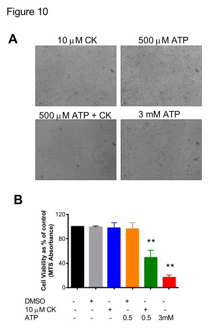
Figure 10
B
A 10 mM CK 500 mM ATP
500 mM ATP + CK 3 mM ATP
0
40
80
120
Cel
lVia
bili
tyas
%o
fco
ntr
ol
(MT
SA
bso
rban
ce)
DMSO - + - + - -10 mM CK - - + - + -ATP - - - 0.5 0.5 3mM
**
**

buffe
r
DMSO
ATPRb1 Rc Rd
Rg3Rh2 CK
PPDG11
50.00
0.05
0.10
0.15
0.20
0.25
Slo
pe
of
dye
up
take
(RF
U.s
-1)
Supplementary Figure 1
Supplementary Figure 1. Ginsenosides have no direct effect on rate ofdye uptake responses in HEK-hP2X7 cells.Dye uptake responses were measured at 37 °C using YOPRO-1 (2 ?M) as themembrane impermeant dye. Relative Fluorescence Units (RFU) weremeasured following excitation at 490 nm and emission recorded at 520 nmusing a fluorescent plate reader (Flexstation III). HEK-hP2X7 cells were treatedwith ginsenosides in a low divalent buffer at 37 °C. ATP (200 ?M) was used toelicit a P2X7 response as a positive control. The mean of 5 individual wells isplotted.

0 100 200 3000
50
100
150
Time (seconds)
RF
U
500 mM ATP
ATP + 10 mM CK
ATP
Mouse P2X7
Supplementary Figure 2
[ginsenoside] mM
Slo
pe
of
dye
up
take
(RF
U.s
-1)
-9 -8 -7 -6 -5 -40.0
0.2
0.4
0.6
0.8
CK
PPD
Supplementary Figure 2. CK ginsenoside potentiates ATP-induced responses at themouse P2X7 receptor.(A) ATP-induced dye uptake was measured at 37 °C using YOPRO-1 (2 ?M) as the membraneimpermeant dye. Relative Fluorescence Units (RFU) were measured following excitation at 490nm and emission recorded at 520 nm using a fluorescent plate reader (Flexstation III). HEK293cells stably expressing mouse P2X7 cells were pre-treated with 10 ?M CK in a low divalentbuffer for 10 minutes at 37 °C. ATP (500 ?M) was then injected to elicit a P2X7 response. Themean of 6-10 individual wells is plotted from three independent experiments. (B) Aconcentration-response curve for CK potentiation on mouse P2X7. Data is mean of 6 wells foreach concentration.
A B