Embed Size (px)
Citation preview
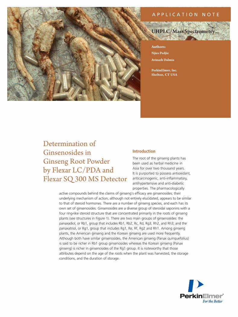
A P P L I C A T I O N N O T E
Introduction
The root of the ginseng plants has been used as herbal medicine in Asia for over two thousand years. It is purported to possess antioxidant, anticarcinogenic, anti-inflammatory, antihypertensive and anti-diabetic properties. The pharmacologically
active compounds behind the claims of ginseng's efficacy are ginsenosides; their underlying mechanism of action, although not entirely elucidated, appears to be similar to that of steroid hormones. There are a number of ginseng species, and each has its own set of ginsenosides. Ginsenosides are a diverse group of steroidal saponins with a four ring-like steroid structure that are concentrated primarily in the roots of ginseng plants (see structures in Figure 1). There are two main groups of ginsenosides: the panaxadiol, or Rb1, group that includes Rb1, Rb2, Rc, Rd, Rg3, Rh2, and Rh3; and the panaxatriol, or Rg1, group that includes Rg1, Re, Rf, Rg2 and Rh1. Among ginseng plants, the American ginseng and the Korean ginseng are used more frequently. Although both have similar ginsenosides, the American ginseng (Panax quinquefolius) is said to be richer in Rb1 group ginsenosides whereas the Korean ginseng (Panax ginseng) is richer in ginsenosides of the Rg1 group. It is noteworthy that those attributes depend on the age of the roots when the plant was harvested, the storage conditions, and the duration of storage.
Determination of Ginsenosides in Ginseng Root Powder by Flexar LC/PDA and Flexar SQ 300 MS Detector
UHPLC/Mass Spectrometry
Authors:
Njies Pedjie
Avinash Dalmia
PerkinElmer, Inc. Shelton, CT USA

2
This application note presents a robust LC method to simultaneously detect seven common ginsenosides . The analysis was achieved using a PerkinElmer® Flexar™ FX-15 with a PDA detector. The analysis was furthered with Flexar SQ 300 MS; the MS detector with its high sensitivity and ability to separate compounds by masses, lowered the threshold of detection and allowed for the separation of Rg1 and Re, which typically co-eluted.
LC/ PDA Analysis
ExperimentalA 1 mg/mL stock of each ginsenoside (Rg1, Rf, Rg2, Rb1, Rc, Rd, Rb2) was prepared by dissolving the appropriate net weight with 70:30 methanol/water (diluent) followed by one minute of vortexing. A working standard of 0.14 mg/mL was prepared by mixing 0.5 mL of each of the stock solutions.Precision was evaluated with five injections of the working standard. Linearity was determined across a 7- 140 µg/mL range. The accuracy of the method was assessed by spiking purified water with the working standard so as to obtain a solution with 7 µg/mL ginsenosides. The ginseng sample was prepared by transferring about 3 g of a panax ginseng powder from capsules into a 50 mL volumetric flask, 30 mL of diluent was added, followed by about a minute vortexing and 30 min. sonication. The solution was centrifuged at 5000 RPM for 10 min; the supernatant was transferred into a 50 mL volumetric flask and set aside. 15 mL of diluent was added to the remaining precipitate, followed by
vortexing, sonication and centrifugation as described above. This latter supernatant was collected and added to the supernatant collected earlier, the 50 mL volumetric flask was then brought to volume with diluents. Samples were mixed well and filtered through a 0.2 µm nylon membrane prior to testing.
LC/ PDA Chromatographic ConditionsAutosampler: Flexar FX UHPLC 50 µL loop and 15 µL needle volume, partial loop mode
Injector Wash: water
Injection Vol.: 2 µL
Sample Diluents: 70:30 methanol/water
PDA Detector: Analytical wavelength 203 nm
UHPLC Column: PerkinElmer Brownlee™ SPP C-18, 3 50 x 2.1 mm, 2.7 µm at 45 °C, Cat# N9308402
Mobile Phase: A: water B: acetonitrile Time Flow rate (min) (mL/min) B % Curve
2.5 0.4 30-35 1 3.5 0.4 35-50 1
3 min. equilibration after each run
Sampling Rate: 5 pt/s
Software: Chromera® Version 3.0
Results and Discussion
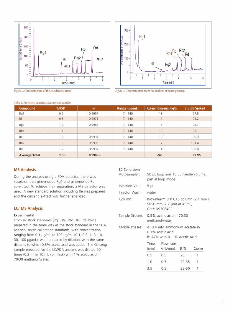
The optimal flow rate was determined to be 0.4 mL/min. at 45 °C, with pressure stabilizing around 5150 PSI (355 bar). All the peaks eluted within six minutes. Representative chromatograms of the standards solution and the Korean ginseng solution are shown in Figures 2 and 3. Excellent method performance was achieved with the coefficient of determinations no less than 0.998, and precisions with values ranging from 0.6% to 1.2%. RSD. The purified water spiked with working standard had a recovery of 99.9% with values ranging from 91.2% to 108.0%. Details of the method performance and results of the samples analyzed are presented in Table 1.
Figure 1. Molecular structure of ginsenosides.
Ginsenoside R3 R2 R1
Rb1 group -O-Glc(6-1)Glc -H -O-Glc(2-1)Glc
Rg1 group -OH -O-Glc O-Glc
Glc Glucopyranoside (β-D-Glucose).
3
MS Analysis
During the analysis using a PDA detector, there was suspicion that ginsenoside Rg1 and ginsenoside Re co-eluted. To achieve their separation, a MS detector was used. A new standard solution including Re was prepared and the ginseng extract was further analyzed.
LC/ MS Analysis
ExperimentalFrom six stock standards (Rg1, Re, Rb1, Rc, Rd, Rb2 ) prepared in the same way as the stock standard in the PDA analysis, seven calibration standards, with concentration ranging from 0.1 µg/mL to 100 µg/mL (0.1, 0.3, 1, 3, 10, 30, 100 µg/mL), were prepared by dilution, with the same diluents to which 0.5% acetic acid was added. The Ginseng sample prepared for the LC/PDA analysis was diluted 50 times (0.2 ml in 10 mL vol. flask) with 1% acetic acid in 70/30 methanol/water.
LC ConditionsAutosampler: 50 µL loop and 15 µL needle volume,
partial loop mode
Injection Vol.: 5 µL
Injector Wash: water
Column: Brownlee™ SPP C18 column (2.1 mm x 5050 mm, 2.7 μm) at 45 °C, Cat# N9308402
Sample Diluents: 0.5% acetic acid in 70:30 methanol/water
Mobile Phases: A: 0.4 mM ammonium acetate in 0.1% acetic acid B: ACN with 0.1 % Acetic Acid
Time Flow rate (min) (mL/min) B % Curve
0.5 0.5 20 1
1.0 0.5 20-35 1
3.5 0.5 35-50 1
Figure 2. Chromatogram of the standard solution. Figure 3. Chromatogram from the analysis of panax ginseng.
Table 1. Precision, linearity, accuracy and samples. Compound %RSD r² Range (µg/mL) Korean Ginseng mg/g 7 ppm Spiked Rg1 0.9 0.9997 7 - 140 13 97.5
Rf 0.6 0.9971 7 - 140 1 91.2
Rg2 1.2 0.9983 7 - 140 1 98.7
Rb1 1.1 1 7 - 140 10 102.1
Rc 1.2 0.9994 7 - 140 10 100.3
Rb2 1.0 0.9996 7 - 140 7 101.4
Rd 1.2 0.9997 7 - 140 4 108.0
Average/Total 1.0/- 0.9988/- -/46 99.9/-
4
Mass Spec Conditions Source: ESI in negative ion mode
Drying Gas Temp and flow: 350 °C and 15 L/min
Nebulizer Gas Pressure: 80 psi
SIM Dwell Time: 50 ms
SIM Mass: 799.5 for Rg1, 945.6 for Rd, Re, 1077.6 for Rb2, Rc, 1107.6 for Rb1
Capillary Exit Voltage: -150 V for Rg1, Rd, Re, -165 V forRb2,Rc,Rb1
m/z Range: 100-1200 Da
Scan Rate: 4000 Da/s
Capillary Exit Voltage: 180 V in scan mode
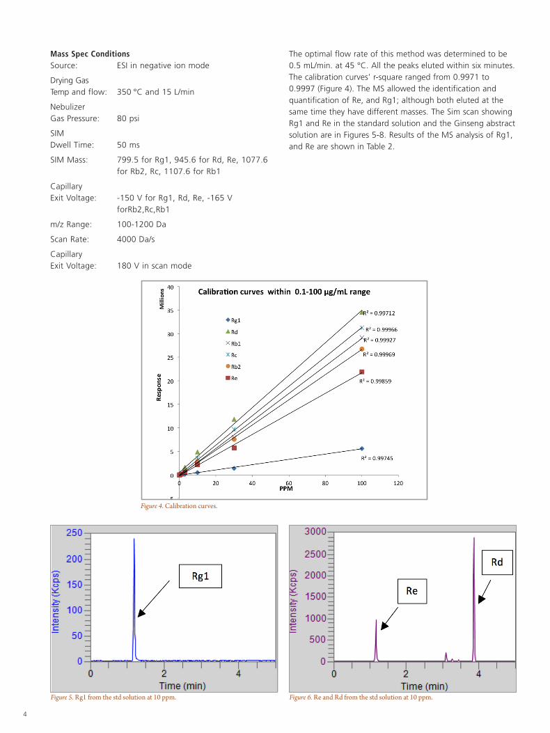
The optimal flow rate of this method was determined to be 0.5 mL/min. at 45 °C. All the peaks eluted within six minutes. The calibration curves’ r-square ranged from 0.9971 to 0.9997 (Figure 4). The MS allowed the identification and quantification of Re, and Rg1; although both eluted at the same time they have different masses. The Sim scan showing Rg1 and Re in the standard solution and the Ginseng abstract solution are in Figures 5-8. Results of the MS analysis of Rg1, and Re are shown in Table 2.
Figure 5. Rg1 from the std solution at 10 ppm.
Figure 4. Calibration curves.
Figure 6. Re and Rd from the std solution at 10 ppm.
For a complete listing of our global offices, visit www.perkinelmer.com/ContactUs
Copyright ©2009-2013, PerkinElmer, Inc. All rights reserved. PerkinElmer® is a registered trademark of PerkinElmer, Inc. All other trademarks are the property of their respective owners. 011195_01
PerkinElmer, Inc. 940 Winter Street Waltham, MA 02451 USA P: (800) 762-4000 or (+1) 203-925-4602www.perkinelmer.com
Conclusion
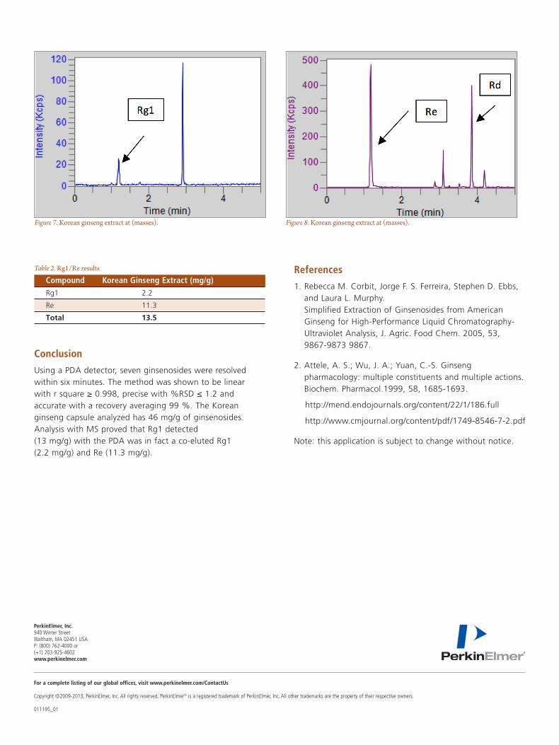
Using a PDA detector, seven ginsenosides were resolved within six minutes. The method was shown to be linear with r square ≥ 0.998, precise with %RSD ≤ 1.2 and accurate with a recovery averaging 99 %. The Korean ginseng capsule analyzed has 46 mg/g of ginsenosides. Analysis with MS proved that Rg1 detected (13 mg/g) with the PDA was in fact a co-eluted Rg1 (2.2 mg/g) and Re (11.3 mg/g).
References
1. Rebecca M. Corbit, Jorge F. S. Ferreira, Stephen D. Ebbs, and Laura L. Murphy. Simplified Extraction of Ginsenosides from American Ginseng for High-Performance Liquid Chromatography-Ultraviolet Analysis, J. Agric. Food Chem. 2005, 53, 9867-9873 9867.
2. Attele, A. S.; Wu, J. A.; Yuan, C.-S. Ginseng pharmacology: multiple constituents and multiple actions. Biochem. Pharmacol.1999, 58, 1685-1693.
http://mend.endojournals.org/content/22/1/186.full
http://www.cmjournal.org/content/pdf/1749-8546-7-2.pdf
Note: this application is subject to change without notice.
Figure 7. Korean ginseng extract at (masses). Figure 8. Korean ginseng extract at (masses).
Compound Korean Ginseng Extract (mg/g) Rg1 2.2
Re 11.3
Total 13.5
Table 2. Rg1/Re results