Embed Size (px)
Citation preview

UK Standards for Microbiology Investigations Investigation of Bone and Soft Tissue associated with Osteomyelitis
Issued by the Standards Unit, Microbiology Services, PHEBacteriology | B 42 | Issue no: dl +| Issue date: dd.mm.yy <tab+enter> | Page: 1 of 30
Investigation of Bone and Soft Tissue associated with Osteomyelitis
Bacteriology | B 42 | Issue no: dl + | Issue date: dd.mm.yy <tab+enter> | Page: 2 of 30UK Standards for Microbiology Investigations | Issued by the Standards Unit, Public Health England

Investigation of Bone and Soft Tissue associated with Osteomyelitis
AcknowledgmentsUK Standards for Microbiology Investigations (SMIs) are developed under the auspices of Public Health England (PHE) working in partnership with the National Health Service (NHS), Public Health Wales and with the professional organisations whose logos are displayed below and listed on the website http://www.hpa.org.uk/SMI/Partnerships. SMIs are developed, reviewed and revised by various working groups which are overseen by a steering committee (see http://www.hpa.org.uk/SMI/WorkingGroups).The contributions of many individuals in clinical, specialist and reference laboratories who have provided information and comments during the development of this document are acknowledged. We are grateful to the Medical Editors for editing the medical content.We also acknowledge Dr Bridget Atkins, of the Bone Infection Unit, Nuffield Orthopaedic Centre, Oxford University Hospitals NHS for her considerable specialist input.For further information please contact us at:Standards UnitMicrobiology Services Public Health England61 Colindale AvenueLondon NW9 5EQE-mail: [email protected]: http://www.hpa.org.uk/SMIUK Standards for Microbiology Investigations are produced in association with:
Bacteriology | B 42 | Issue no: dl + | Issue date: dd.mm.yy <tab+enter> | Page: 3 of 30UK Standards for Microbiology Investigations | Issued by the Standards Unit, Public Health England

Investigation of Bone and Soft Tissue associated with Osteomyelitis
Bacteriology | B 42 | Issue no: dl + | Issue date: dd.mm.yy <tab+enter> | Page: 4 of 30UK Standards for Microbiology Investigations | Issued by the Standards Unit, Public Health England

Investigation of Bone and Soft Tissue associated with Osteomyelitis
UK Standards for Microbiology Investigations: StatusUsers of SMIsThree groups of users have been identified for whom SMIs are especially relevant:
SMIs are primarily intended as a general resource for practising professionals in the field operating in the field of laboratory medicine in the UK. Specialist advice should be obtained where necessary.
SMIs provide clinicians with information about the standard of laboratory services they should expect for the investigation of infection in their patients and the documents provide information that aids the electronic ordering of appropriate tests from hospital wards.
SMIs also provide commissioners of healthcare services with the standard of microbiology investigations they should be seeking as part of the clinical and public health care package for their population.
Background to SMIsSMIs comprise a collection of recommended algorithms and procedures covering all stages of the investigative process in microbiology from the pre-analytical (clinical syndrome) stage to the analytical (laboratory testing) and post analytical (result interpretation and reporting) stages.Syndromic algorithms are supported by more detailed documents containing advice on the investigation of specific diseases and infections. Guidance notes cover the clinical background, differential diagnosis, and appropriate investigation of particular clinical conditions. Quality guidance notes describe essential laboratory methodologies which underpin quality, for example assay validation, quality assurance, and understanding uncertainty of measurement.Standardisation of the diagnostic process through the application of SMIs helps to assure the equivalence of investigation strategies in different laboratories across the UK and is essential for public health interventions, surveillance, and research and development activities. SMIs align advice on testing strategies with the UK diagnostic and public health agendas.
Involvement of Professional OrganisationsThe development of SMIs is undertaken within PHE in partnership with the NHS, Public Health Wales and with professional organisations.
UK Standards for Microbiology Investigations were formerly known as National Standard Methods.Microbiology is used as a generic term to include the two GMC-recognised specialties of Medical Microbiology (which includes Bacteriology, Mycology and Parasitology) and Medical Virology.
Bacteriology | B 42 | Issue no: dl + | Issue date: dd.mm.yy <tab+enter> | Page: 5 of 30UK Standards for Microbiology Investigations | Issued by the Standards Unit, Public Health England

Investigation of Bone and Soft Tissue associated with Osteomyelitis
The list of participating organisations may be found at http://www.hpa.org.uk/SMI/Partnerships. Inclusion of an organisation’s logo in an SMI implies support for the objectives and process of preparing SMIs. Representatives of professional organisations are members of the steering committee and working groups which develop SMIs, although the views of participants are not necessarily those of the entire organisation they represent.SMIs are developed, reviewed and updated through a wide consultation process. The resulting documents reflect the majority view of contributors. SMIs are freely available to view at http://www.hpa.org.uk/SMI as controlled documents in Adobe PDF format.
Quality AssuranceThe process for the development of SMIs is certified to ISO 9001:2008. NHS Evidence has accredited the process used by PHE to produce SMIs. Accreditation is valid for three years from July 2011. The accreditation is applicable to all guidance produced since October 2009 using the processes described in PHE’s Standard Operating Procedure SW3026 (2009) version 6. SMIs represent a good standard of practice to which all clinical and public health microbiology laboratories in the UK are expected to work. SMIs are well referenced and represent neither minimum standards of practice nor the highest level of complex laboratory investigation possible. In using SMIs, laboratories should take account of local requirements and undertake additional investigations where appropriate. SMIs help laboratories to meet accreditation requirements by promoting high quality practices which are auditable. SMIs also provide a reference point for method development. SMIs should be used in conjunction with other SMIs.UK microbiology laboratories that do not use SMIs should be able to demonstrate at least equivalence in their testing methodologies.The performance of SMIs depends on well trained staff and the quality of reagents and equipment used. Laboratories should ensure that all commercial and in-house tests have been validated and shown to be fit for purpose. Laboratories should participate in external quality assessment schemes and undertake relevant internal quality control procedures. Whilst every care has been taken in the preparation of SMIs, PHE, its successor organisation(s) and any supporting organisation, shall, to the greatest extent possible under any applicable law, exclude liability for all losses, costs, claims, damages or expenses arising out of or connected with the use of an SMI or any information contained therein. If alterations are made to an SMI, it must be made clear where and by whom such changes have been made. SMIs are the copyright of PHE which should be acknowledged where appropriate.
Bacteriology | B 42 | Issue no: dl + | Issue date: dd.mm.yy <tab+enter> | Page: 6 of 30UK Standards for Microbiology Investigations | Issued by the Standards Unit, Public Health England
Investigation of Bone and Soft Tissue associated with Osteomyelitis
Microbial taxonomy is up to date at the time of full review.
Equality and Information GovernanceAn Equality Impact Assessment on UK Standards for Microbiology Investigations is available at http://www.hpa.org.uk/SMI.PHE is a Caldicott compliant organisation. It seeks to take every possible precaution to prevent unauthorised disclosure of patient details and to ensure that patient-related records are kept under secure conditions.
Suggested citation for this document:Public Health England. (YYYY <tab+enter>). Investigation of Bone and Soft Tissue associated with Osteomyelitis. UK Standards for Microbiology Investigations. B 42 Issue dl +. http://ww.hpa.org.uk/SMI/pdf.
Bacteriology | B 42 | Issue no: dl + | Issue date: dd.mm.yy <tab+enter> | Page: 7 of 30UK Standards for Microbiology Investigations | Issued by the Standards Unit, Public Health England

Investigation of Bone and Soft Tissue associated with Osteomyelitis
ContentsACKNOWLEDGMENTS............................................................................2UK STANDARDS FOR MICROBIOLOGY INVESTIGATIONS: STATUS..............3AMENDMENT TABLE..............................................................................6SCOPE OF DOCUMENT...........................................................................7INTRODUCTION.....................................................................................7TECHNICAL INFORMATION/LIMITATIONS...............................................121 SPECIMEN COLLECTION, TRANSPORT AND STORAGE.....................132 SPECIMEN PROCESSING..............................................................143 REPORTING PROCEDURE.............................................................184 NOTIFICATION TO PHE................................................................18APPENDIX: INVESTIGATION OF BONE AND SOFT TISSUE ASSOCIATED
WITH OSTEOMYELITIS FLOWCHART.............................................19REFERENCES.......................................................................................20
Amendment Table
Bacteriology | B 42 | Issue no: dl + | Issue date: dd.mm.yy <tab+enter> | Page: 8 of 30UK Standards for Microbiology Investigations | Issued by the Standards Unit, Public Health England

Investigation of Bone and Soft Tissue associated with Osteomyelitis
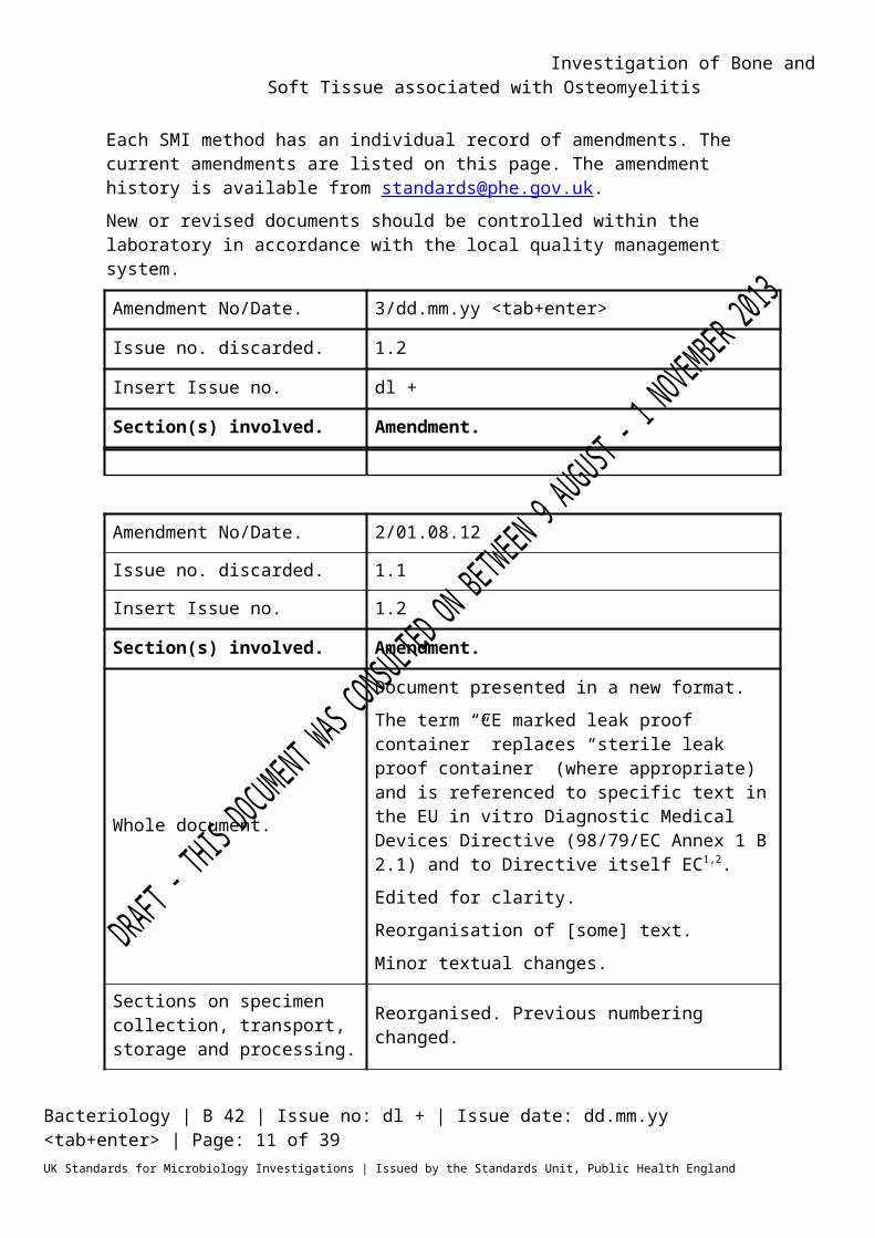
Each SMI method has an individual record of amendments. The current amendments are listed on this page. The amendment history is available from [email protected] or revised documents should be controlled within the laboratory in accordance with the local quality management system.Amendment No/Date. 3/dd.mm.yy <tab+enter>Issue no. discarded. 1.2Insert Issue no. dl +Section(s) involved. Amendment.
Amendment No/Date. 2/01.08.12Issue no. discarded. 1.1Insert Issue no. 1.2Section(s) involved. Amendment.
Whole document.
Document presented in a new format.The term “CE marked leak proof container” replaces “sterile leak proof container” (where appropriate) and is referenced to specific text in the EU in vitro Diagnostic Medical Devices Directive (98/79/EC Annex 1 B 2.1) and to Directive itself EC1,2.Edited for clarity.Reorganisation of [some] text.Minor textual changes.
Sections on specimen collection, transport, storage and processing.
Reorganised. Previous numbering changed.
References. Some references updated.
Bacteriology | B 42 | Issue no: dl + | Issue date: dd.mm.yy <tab+enter> | Page: 9 of 30UK Standards for Microbiology Investigations | Issued by the Standards Unit, Public Health England
Investigation of Bone and Soft Tissue associated with Osteomyelitis
Scope of Document Type of SpecimensIntra-operative samples of bone or bone biopsies sent for clinical diagnosis. For biopsies and aspirates sent for the investigation of prosthetic joint infections see B 44 – Investigation of prosthetic joint infection samples.
ScopeThis SMI describes the processing and microbiological investigation of bone and soft tissue associated with osteomyelitis.This SMI should be used in conjunction with other SMIs.
IntroductionOsteomyelitis is a progressive infection which results in inflammation of the bone and causes bone destruction, necrosis and deformation3. It may be acquired from a contiguous site or haematogenously via the bloodstream, and if left undiagnosed or untreated may lead to chronic infection4. Contiguous-focus osteomyelitis may be associated with vascular insufficiency (eg diabetic foot infections) or without vascular insufficiency 4. In addition it may be associated with an orthopaedic device such as a fracture fixation device or prosthesis. Haematogenous osteomyelitis results from bacteraemic seeding of the bone. In children the commonest site for infection is in the growing ends of long bones. In adults, the spine is the most common site. Risk factors for adult haematogenous osteomyelitis include sickle cell disease, immune deficiencies and intravenous drug use5. Gram positive bacteria (Staphylococcus aureus, coagulase negative staphylococci, Enterococcus species, etc) are most often isolated from bone and soft tissue samples; however Gram negative bacteria and fungi may also be isolated. Gram negative bacilli when isolated are of major clinical importance due to their antimicrobial resistance patterns6.
Acute contiguous-focus osteomyelitis In contiguous-focus osteomyelitis, the organisms may be inoculated at the time of trauma or during intra-operative or peri-operative procedures. Alternatively they may extend from an adjacent soft tissue focus of infection. Common predisposing factors include surgical reduction and fixation of fractures, prosthetic devices, open fractures and chronic soft tissue infections (see B 14 – Investigation of abscesses and post-operative wound and deep seated wound infections). In general the microbiology of contiguous osteomyelitis is more complex than that of haematogenous osteomyelitis and is commonly polymicrobial.
Bacteriology | B 42 | Issue no: dl + | Issue date: dd.mm.yy <tab+enter> | Page: 10 of 30UK Standards for Microbiology Investigations | Issued by the Standards Unit, Public Health England
Investigation of Bone and Soft Tissue associated with Osteomyelitis
Contiguous-focus osteomyelitis without vascular insufficiencyPuncture wounds of the foot through footwear such as training shoes are particularly associated with osteomyelitis due to Pseudomonas aeruginosa7-9. Osteomyelitis following human bites and tooth socket infections affecting the mandible are often caused by strict anaerobes for example Actinomyces species; in children anaerobic bone and joint infections are rare10,11. Many of the principles applied to prosthetic joint infection also apply to device-related, contiguous-focus osteomyelitis (B 44 – Investigation of prosthetic joint infection samples ) 12. Where a device is involved, skin flora such as coagulase-negative staphylococci, often regarded as contaminants in the laboratory, are common pathogens. Many devices in long bones and other sites are used to fix fractures; if these become infected, the presentation may be acute, with gross purulence.
Contiguous-focus osteomyelitis with vascular insufficiencyMost patients with contiguous-focus osteomyelitis associated with vascular insufficiency have diabetes mellitus. The bones and joints of the feet are most often affected4.Diabetic foot infections13-15
Diabetic foot infections are responsible for many hospital admissions and a significant number can end up with limb amputation and consequent disability. Neuropathy and vasculopathy (impaired blood supply) are complications of diabetes. The former means that protective sensation is lost, allowing skin injury to occur without it being perceived. In addition it can ultimately lead to fragmentation, destruction and dislocations of the bones of the foot (Charcot neuro-osteoarthropathy). Foot deformity in diabetics due to motor neuropathy is also a further strong risk factor for developing ulcers and infection. The basic principles in the treatment of diabetic foot infection are education and prevention with good glucose control, accommodative footwear, regular inspection and general compliance.Once infection has occurred, abscesses may need to be drained, diagnostic biopsies may be required to guide antibiotics and diseased bone may need to be resected. Acute infections in patients who have not recently received antimicrobials are often monomicrobial (almost always with aerobic gram-positive cocci such as S. aureus and β-haemolytic streptococci), whereas chronic infections are often polymicrobial. Cultures of specimens obtained from patients with such mixed infections generally yield 3–5 isolates, including Gram positive and Gram negative aerobes and anaerobes. These may include enterococci, various Enterobacteriaceae, obligate anaerobes, Pseudomonas aeruginosa and, sometimes, other non-fermentative Gram negative rods. Hospitalisation, surgical procedures, and, especially,
Bacteriology | B 42 | Issue no: dl + | Issue date: dd.mm.yy <tab+enter> | Page: 11 of 30UK Standards for Microbiology Investigations | Issued by the Standards Unit, Public Health England

Investigation of Bone and Soft Tissue associated with Osteomyelitis
prolonged or broad spectrum antibiotic therapy may predispose patients to colonisation and/or infection with antibiotic resistant organisms (eg meticillin resistant Staphylococcus aureus (MRSA) or vancomycin-resistant enterococci (VRE)). The impaired host defences around necrotic soft tissue or bone may allow low-virulence colonizers, such as coagulase-negative staphylococci and Corynebacterium species (“diphtheroids”), to assume a pathogenic role.Diabetic foot infections are often managed initially by podiatrists; they may perform a deep tissue debridement and send soft tissue for culture, although the presence of osteomyelitis may be indicated on the request card.In the immunocompromised or diabetic host, Nocardia species should also be considered as a rare cause of osteomyelitis.
Acute haematogenous osteomyelitis3,5,16,17
Haematogenous osteomyelitis has been classically described in childhood, but can occur in any age group especially when there are risk factors such as a recent intravascular device, haemodialysis, intravenous drug usage or recurrent infections elsewhere (such as urinary tract infections)5. In adults the vertebrae are most often affected, however the long bones, pelvis or clavicle may also be affected.18
In classical haematogenous osteomyelitis of childhood, the growing ends (metaphyses) of long bones are involved. The commonest organism is S. aureus; however β-haemolytic streptococci and HACEK organisms such as Kingella species are also important causes16. Organisms in the bloodstream gain access to bone by way of the nutrient artery. They pass though branches of this vessel to the small blind ended terminal vessels usually near the epiphyseal plate (growing end of the bone). This area is thought to have sluggish circulation, and bacteria can lodge here, starting the process of infection. Following this there is extension to other areas and the host inflammatory response is mobilised. Pus is created and expands under pressure thereby creating further impedance of the local circulation and death of bone. In certain areas such as the hip, where the epiphyseal plate is situated within the joint capsule, early joint involvement by infection is common. Pus under pressure may strip the periosteum (outer lining of bone). New immature bone is formed as a response to periosteal stripping, and, in severe cases, the entire shaft may be encased in a sheath of new bone referred to as an involucrum. Where a major portion of the shaft has been deprived of blood supply, a resulting sequestrum (dead bone) lies within the involucrum. Openings in the bone may permit escape of pus from bone causing abscesses, systemic sepsis and in some cases death.The bacterial species in haematogenous osteomyelitis are usually dependent on the age of the patient. In neonates, Group B streptococci, S. aureus and Escherichia coli cause infection3. Multiple sites of infection are
Bacteriology | B 42 | Issue no: dl + | Issue date: dd.mm.yy <tab+enter> | Page: 12 of 30UK Standards for Microbiology Investigations | Issued by the Standards Unit, Public Health England

Investigation of Bone and Soft Tissue associated with Osteomyelitis
common in neonates19. Between the ages of one and sixteen, S. aureus, and Haemophilus influenzae type B predominate (although the latter is rare after the age of 5 years and increasingly rare in children under 5 because of a successful vaccination campaign). Streptococcus pneumoniae is occasionally involved. In adult life, S. aureus is the commonest organism and, in the elderly, infection with aerobic Gram-negative rods may occur. Candida species may be found when intravenous devices are in use20. In acute haematogenous osteomyelitis a single pathogenic organism is usually isolated but in many cases of chronic osteomyelitis, particularly when associated with wounds and ulcers the disease can be polymicrobial.
Vertebral osteomyelitisIn adults haematogenous osteomyelitis most commonly involves the spine. Vertebral osteomyelitis may also result from trauma or complications during surgery. Risk factors include older age, a recent intravascular device, haemodialysis, diabetes and intravenous drug usage (a risk factor for Pseudomonas infection), infection and immunosupression21. Lumbar spine infections may originate from urinary tract infections, possibly by translocation of bacteria via a venous plexus (Batson’s plexus) that links the bladder with the spine. Following the initial infection, pus may break out of the cortex anteriorly to form a paravertebral abscess or posteriorly to form an epidural abscess. In addition weakening of the bone may cause vertebral collapse. Organisms causing vertebral infections include S. aureus, streptococci and aerobic Gram negative rods(associated with urinary tract infections)3. In patients with risk factors, tuberculosis should always be considered; microbiological diagnosis (with or without histology) is required for a definitive diagnosis22,23. Although infection most often occurs in the spine, extrapulmonary tuberculosis can occur in any bone or joint. Diagnosis is mainly by biopsy for histology and microbiology. Cultures are often positive and are crucial for determining the presence of resistance to anti-tuberculosis agents. However the decision to treat is often made on clinical and histopathological grounds in the first instance24,25. Other mycobacteria, such as Mycobacterium marinum, Mycobacterium avium-intracellulare, Mycobacterium fortuitum and Mycobacterium gordonae have also been associated with bone infections particularly in patients who are immunocompromised.In endemic areas Brucella species is a common cause of vertebral infection, therefore a travel history should always be sought. Other fastidious Gram negative rods eg the HACEK group (Haemophilus species, Actinobacillus actinomycetemcomitans, Cardiobacterium hominis, Eikenella corrodens and Kingella species; (see ID 12 – Identification of Haemophilus species and the HACEK group of organisms) may be occasional causes of vertebral osteomyelitis26.
Bacteriology | B 42 | Issue no: dl + | Issue date: dd.mm.yy <tab+enter> | Page: 13 of 30UK Standards for Microbiology Investigations | Issued by the Standards Unit, Public Health England
Investigation of Bone and Soft Tissue associated with Osteomyelitis
Sickle cell disease3,4
Adult haematogenous osteomyelitis in adults is often associated with sickle cell disease. Symptoms may mimic those of marrow crisis; culture results should therefore be used for confirmation of clinical diagnosis. Organisms often isolated include Salmonella species, S. aureus and streptococci. Salmonella species rarely cause osteomyelitis in patients who are immunocompetent; typically Salmonella species are associated with sickle cell anaemia and patients who are immunosuppressed. Infection caused by Salmonella species may be due to indirect contamination from an animal host27,28.
Haemodialysis patients4
As a result of the use of intravascular access devices in these patients, haematogenous infections can occur usually due to S. aureus or coagulase negative staphylococci. Gram negative infections are more common in haemodialysis patients than in the general population.
Intravenous drug users4,29
Septic arthritis, osteomyelitis of the long bones or vertebral discs is associated with haematogenous infection in intravenous drug users. Organisms often isolated include S. aureus, P. aeruginosa and Candida species.
Chronic osteomyelitis3,4,18
Chronic osteomyelitis may be haematogenous or secondary to contiguous-focus infection. Patients typically present with chronic pain and drainage, and may have a history of osteomyelitis. Treatment of infection may be challenging as surrounding tissue and bone will be of poor quality; antibiotic treatment alone is rarely sufficient to arrest infection. Risk factors include open fractures, bacteraemia and ischemic ulcers associated with diabetes, sickle cell disease and malnutrition. Organisms often associated with chronic osteomyelitis include S. aureus, Gram negative bacilli and anaerobic bacteria.
Secondary to contiguous-focus osteomyelitisIn chronic device related infections, organisms may be present in a biofilm that is associated with the device or diseased/necrotic bone. The presence of a biofilm has implications for sampling technique, processing and the interpretation of culture results. Low grade infection eg with skin flora such as a coagulase-negative staphylococcus can lead to delayed fracture union or non-union. The organisms may only be present in small numbers and culture often requires broth enrichment30. When a patient with a device has debridement, removal and/or exchange of the device, multiple (4-5) samples should be taken using separate instruments. If several are culture positive, whatever the organism(s), this is likely to represent true infection.
Bacteriology | B 42 | Issue no: dl + | Issue date: dd.mm.yy <tab+enter> | Page: 14 of 30UK Standards for Microbiology Investigations | Issued by the Standards Unit, Public Health England

Investigation of Bone and Soft Tissue associated with Osteomyelitis
Haematogenous Acute haematogenous osteomyelitis can lead to chronic osteomyelitis characterised by dead areas of bone and sinus tract31,32. This condition can fail to respond to treatment and persist for long periods33. Infections may recur many years after the first episode34
Brodie’s abscess35
Brodies’s abscess is an uncommon condition and is a chronic localised abscess of bone, most often in the distal part of the tibia. It is usually due to S. aureus and generally occurs in patients under 25 years of age. Surgery (surgical debridement) and culture-specific antibiotic therapy are usually effective in arresting infection.
Fungal osteomyelitis Fungal osteomyelitis is rare; however, some fungi endemic to certain areas can be associated with osteomyelitis. This includes Cryptococcus, Blastomyces and Sporothrix species. In patients who are immunocompromised or those with multiple previous surgical procedures at that site, more common fungi such as Candida or Aspergillus species can also cause osteomyelitis20,36. A mycetoma is a chronic granulomatous infection of the skin, subcutaneous tissues and in its advanced stages, bone. It is most prevalent in tropical and sub-tropical regions of Africa, Asia and Central America. Infection usually follows traumatic inoculation of organisms into subcutaneous tissue from soil or vegetable sources37. Various genera have been implicated including Madurella, Acremonium, Pseudoallescheria and Actinomadura species. Fungal mycetomas are termed eumycetomas in contrast to those caused by Nocardia species.
Diagnosis 4,15,16,18
The diagnosis of osteomyelitis usually requires a combination of a full clinical assessment, plain X-rays and further imaging (eg MRI scan, CT scan, ultra-sound), blood cultures (particularly in acute cases), bone and/or soft tissue biopsies and/or surgical sampling. For specific indications eg risk of Brucella infection, other tests such as serology may be required. When tuberculosis is suspected, a full clinical ‘work up’ including a chest X-ray is indicated.
Sample TypesRadiologically obtained percutaneous bone biopsiesThese may be taken in the radiology department where they can be guided by imaging such as ultrasound, fluoroscopy or CT. Usually a sample should also be sent to histology to confirm infection, provide pointers to unusual infections and/or exclude malignancy. It is not commonly possible to send more than one sample to microbiology, but when this is done, each should be processed separately. It is important that detailed clinical information is
Bacteriology | B 42 | Issue no: dl + | Issue date: dd.mm.yy <tab+enter> | Page: 15 of 30UK Standards for Microbiology Investigations | Issued by the Standards Unit, Public Health England

Investigation of Bone and Soft Tissue associated with Osteomyelitis
provided to ensure cultures are set up for appropriate organisms. This includes details such as the presence of a prosthetic device (where any organism eg a coagulase negative staphylococcus, may be the pathogen and also where prolonged cultures are required). It also includes any clinical suspicion or risk factors for tuberculosis, brucella, nocardia, atypical mycobacteria or fungi.Intra-operative bone biopsiesThese are taken in theatre either as primarily a diagnostic procedure, or as the first part of a larger debridement/resection procedure. Multiple (4-5) samples should be taken using separate sterile instruments for microbiological culture. Similar samples from similar sites should also be taken for histopathological examination. A risk-benefit assessment of antibiotic timing is required. Where infection is likely and/or a microbiological diagnosis is likely to significantly affect clinical outcome, prophylactic antibiotics can be withheld until immediately after sampling. The effect of a single dose of antibiotic on the sensitivity of microbiological culture is unknown. In addition to bone samples, deep soft tissue samples are usually taken at the same time. Sinus samples should be discouraged as colonising organisms cannot be differentiated from infecting organisms.Samples from around devices12
Samples of bone and soft tissues may be taken from around a prosthetic device, eg a fracture fixation plate or nail. This may be debrided and left in situ (debridement and retention) or removed. If the fracture has not united a further internal or external device is usually required. Samples associated with such a device should be processed with the same principles as those associated with prosthetic joint samples (B 44 – Investigation of prosthetic joint infection samples).The key to processing bone samples is to reduce manipulation to a minimum. Samples can be transferred to the laboratory using routine timescales (ie within hours rather than minutes). In order to reduce the risk of contamination it is desirable to process specimens in a Class 2 microbiological safety cabinet.Shaking with sterile glass (Ballotini) beads is relatively simple and therefore carries a low risk of contamination. Another method suggested for specimen processing is grinding. However, grinders can break and constitute a health and safety hazard, as can aerosol release from blenders. The problems of heat generated by the grinder (that may be sufficient to damage or kill some microbes), and insufficient strength in the operator to homogenise the tissue in the Griffiths grinding tubes also render this technique non-standardised and with unknown recovery characteristics. The impact of bone and cement fragments on shattering the Griffiths grinding tubes has also not been investigated. Sonication of devices is a means of disrupting bacterial biofilm in vascular and orthopaedic prostheses and has been shown to be useful in the detection of prosthetic joint infection38-40.
Bacteriology | B 42 | Issue no: dl + | Issue date: dd.mm.yy <tab+enter> | Page: 16 of 30UK Standards for Microbiology Investigations | Issued by the Standards Unit, Public Health England

Investigation of Bone and Soft Tissue associated with Osteomyelitis
Broth enrichment is important especially in chronic osteomyelitis, device related infections or where the patient has already received antibiotics30,41. Broth cultures can become contaminated and so if only one or two samples are taken, a positive culture from one broth is not interpretable42. If several (4-5) samples are taken then a definition of a positive microbiological result is easier to create for that patient. In the presence of a device or non-viable bone, any organism cultured in more than one sample may be relevant and should be identified, have extended sensitivities and be reported. Extended sensitivities are particularly important in patients with chronic osteomyelitis, or retained metalwork, where prolonged oral antibiotics are often required.
Management4,15,16,18
In acute presentations, surgery may be required to drain pus. In chronic osteomyelitis, areas of dead bone may need to be resected. Both need to be accompanied by specific antibiotic therapy depending on culture results. This is most often carried out intravenously, initially. In some cases, where the disease is too extensive to fully resect, the patient is too unfit for surgery or a device is retained, long term oral antibiotics may be required. Organisms need to be tested against a wide variety of antibiotic options as patients commonly are intolerant of one or more antibiotics.
Rapid techniques43-45
Rapid techniques including Nucleic Acid Amplification Tests (NAATs) (eg PCR, 16s rRNA gene PCR, PCR-electrospray ionization (ESI)/MS etc) and matrix-assisted laser desorption ionization time of flight analysis mass spectrometry (MALDI-TOFF/MS) have become available as a means of rapid, sensitive identification of organisms associated with bone and joint infection46. PCR methods require the extraction of DNA or RNA from the sample for analysis, whereas MALDI-TOFF differentiates organisms based on their mass/charge spectrum. PCR has been shown to be more sensitive than convention culture for the isolation of some fastidious organisms for example Kingella kingae, and PCR – hybridization after sonication has been shown to improve diagnosis of implant related infections 47,48.
Technical Information/LimitationsSpecimen containers1,2
SMIs use the term “CE marked leak proof container” to describe containers bearing the CE marking used for the collection and transport of clinical specimens. The requirements for specimen containers are given in the EU in vitro Diagnostic Medical Devices Directive (98/79/EC Annex 1 B 2.1) which states: “The design must allow easy handling and, where necessary, reduce as far as possible contamination of, and leakage from, the device during use and, in the case of specimen receptacles, the risk of contamination of the specimen. The manufacturing processes must be appropriate for these purposes”.
Bacteriology | B 42 | Issue no: dl + | Issue date: dd.mm.yy <tab+enter> | Page: 17 of 30UK Standards for Microbiology Investigations | Issued by the Standards Unit, Public Health England
Investigation of Bone and Soft Tissue associated with Osteomyelitis
1 Specimen Collection, Transport and Storage1,2
1.1 Safety considerations49-60
Use aseptic technique.Care should be taken to avoid accidental injury when using “sharps”.Collect specimens in appropriate CE marked leak proof containers and transport specimens in sealed plastic bags.Compliance with postal and transport regulations is essential.
1.2 Achieving optimal conditions 1.2.1 Time between specimen collection and
processingCollect specimens before antimicrobial therapy where possible.Specimens should be transported and processed as soon as possible.If possible stop all antibiotics at least 1-2 weeks prior to sampling and consider not giving routine surgical prophylaxis until after sampling. For more information regarding antimicrobial therapy and surgical sampling please refer to B 44 – Investigation of prosthetic joint infection samples.The volume of the specimen influences the transport time that is acceptable. Larger pieces of bone may maintain the viability of anaerobes for longer61. Samples should however not exceed the size of the available CE Marked leak proof containers.
1.2.2 Special considerations to minimise deterioration If processing is delayed, refrigeration is preferable to storage at ambient temperature60. Delays of over 48hr are undesirable.
1.3 Correct specimen type and method of collectionUnless otherwise stated, swabs for bacterial and fungal culture should then be placed in Amies transport medium with charcoal62.Collect specimens other than swabs into appropriate CE Marked leak proof containers and place in sealed plastic bags.Direct collection in theatres can be placed into a CE Marked leak proof container with Ringer’s or saline solution and Ballotini beads (as an option) which is placed into sealed plastic bags. However, microbiology and histology specimen pots can be confused leading to difficulties in processing samples).
1.4 Adequate quantity and appropriate number of specimens
Bacteriology | B 42 | Issue no: dl + | Issue date: dd.mm.yy <tab+enter> | Page: 18 of 30UK Standards for Microbiology Investigations | Issued by the Standards Unit, Public Health England

Investigation of Bone and Soft Tissue associated with Osteomyelitis
In chronic osteomyelitis collection of multiple (4-5) intra-operative samples with separate instruments (usually sterile forceps and scalpel) is important. Duplicate samples must be taken for histology. Swabs are not recommended.Minimum specimen size will depend on the number of investigations requested.Numbers and frequency of specimen collection are dependent on clinical condition of patient.
2 Specimen Processing1,2
2.1 Safety considerations49-60
Containment Level 2.Laboratory procedures that give rise to infectious aerosols must be conducted in a microbiological safety cabinet52. Refer to current guidance on the safe handling of all organisms documented in this SMI.The above guidance should be supplemented with local COSHH and risk assessments.
2.2 Test selectionSelect a representative portion of specimen for appropriate procedures such as culture for Mycobacterium species (B 40 - Investigation of specimens for Mycobacterium species ) or fungi depending on clinical details.
2.3 AppearanceN/A
2.4 Microscopy2.4.1 StandardGram stain (see TP 39 - Staining Procedures)If sufficient specimen is received prepare as recommended in Section 2.5. Using a sterile pipette place one drop of specimen on to a clean microscope slide.Spread this with a sterile loop to make a thin smear for Gram staining.
2.5 Culture and investigation2.5.1 Pre-treatmentExamine the specimen for the presence of any soft tissue. Remove soft tissue using a sterile scalpel or scissors and homogenise using, as appropriate, a sterile grinder (Griffith tube or unbreakable alternative), a
Bacteriology | B 42 | Issue no: dl + | Issue date: dd.mm.yy <tab+enter> | Page: 19 of 30UK Standards for Microbiology Investigations | Issued by the Standards Unit, Public Health England
Investigation of Bone and Soft Tissue associated with Osteomyelitis
sterile scalpel or (preferably) sterile scissors and Petri dish. The addition of a small volume (approximately 0.5 mL) of sterile filtered water, saline or nutrient Ringer’s will aid the homogenisation process.Homogenisation must be performed in a racked shaker for 15 minutes in a Class 1 exhaust protective cabinet.SupplementaryFungi and Mycobacterium species (B 40 - Investigation of specimens for Mycobacterium species ).
2.5.2 Specimen processingBone (percutaneous biopsy or intra-operative sample) or soft tissue associated with osteomyelitisThe objective should be to reduce manipulation to a minimum (for instance the number of times any container is opened), thereby minimising the risk of exposing the operative sample to potential contamination. For this reason centrifugation of the sample for concentration should not be performed, instead divide the whole sample in appropriate amounts for tests.In units with high workloads of this specimen type, the provision to the operating theatre of CE Marked leak proof containers in a sealed plastic bag with approximately 10 Ballotini beads and 5 mL broth, could be considered. In such circumstances, homogenisation can be carried out in the original container. It is not uncommon, however, for microbiology and histology specimen pots to be confused leading to difficulties in processing samples.Alternatively, samples may be sent to the laboratory in a plain CE Marked leak proof container in a sealed plastic bag. These samples require transfer, homogenisation and then further transfer to culture media, including liquid media. If this methodology is followed, particular care is necessary with asepsis when transferring, homogenising or processing the sample. Clean air provision is desirable. Homogenisation with Ballotini beads can be performed by shaking at 250 rpm for 10 minutes in a covered rack on an orbital shaker, or alternatively vortexing for 15 seconds (40 Hz). The diluent for the Ballotini beads and tissues should be Ringer’s solution or saline. If molecular analysis is to be carried out then sterile molecular grade water and new universal containers should be used. In the case of molecular work the volume should not exceed 2 mL.Inoculate each agar plate and a slide for Gram staining with a drop of the suspension using a sterile pipette (see Q 5 - Inoculation of culture media for bacteriology).For the isolation of individual colonies, spread inoculum using a sterile loop. Inoculate broth with the remainder of the suspension including any tissue fragments.
Bacteriology | B 42 | Issue no: dl + | Issue date: dd.mm.yy <tab+enter> | Page: 20 of 30UK Standards for Microbiology Investigations | Issued by the Standards Unit, Public Health England

Investigation of Bone and Soft Tissue associated with Osteomyelitis
2.5.3 Culture media, conditions and organismsClinical details/conditions
Standard media
Incubation Cultures read
Target organism(s)
Temp °C
Atmos Time
‡
Osteomyelitis,Bone biopsy,Brodie’s abscess,Diabetic foot osteomyelitis,Discitis+For debridement of fracture fixation device refer to B44 – Investigation of prosthetic joint
Blood agarandChocolate agar
35 - 37
5 - 10% CO2
40 - 48 hr Daily
StaphylococciStreptococciEnterobacteriaceaePseudomonadsHACEK groupNocardia species*
Fastidious anaerobic broth, cooked meat broth or equivalentSubculture when cloudy
35 - 37 air 5 d N/A
StaphylococciStreptococciEnterobacteriaceacePseudomonadsAnaerobes
Subculture plates
Fastidious anaerobic agar or
35 - 37
Anaerobic
40 - 48 hr Daily Anaerobes
Chocolate 35 - 37
5 - 10% CO2
40 - 48 hr Daily Any
For these situations, add the following:
Clinical details/ conditions
Supplementary media
Incubation Cultures read
Target organism(s)Temp
°CAtmos Time
Mycetoma, Sabouraud agar
35 - 37 air 2 - 5 d
40 hr and 5 d‡
Yeast and Moulds
Always consider other organisms such as Mycobacterium species (B 40 - Investigation of specimens for Mycobacterium species ), fungi and actinomycetes. Routine processing for mycobacteria should be considered for all non post-operative *May require incubation for a further 3 days+Most surgical cases with intra-operative biopsies eg fracture fixation devices or chronic osteomyelitis requires multiple samples. If an indistinguishable organism is isolated in two or more samples then it is likely to be clinically significant.‡ Extended incubation may be required (for up to 8 weeks) for certain species of fungi such as Cryptococcus species or Histoplasma species63,64.
Bacteriology | B 42 | Issue no: dl + | Issue date: dd.mm.yy <tab+enter> | Page: 21 of 30UK Standards for Microbiology Investigations | Issued by the Standards Unit, Public Health England
Investigation of Bone and Soft Tissue associated with Osteomyelitis
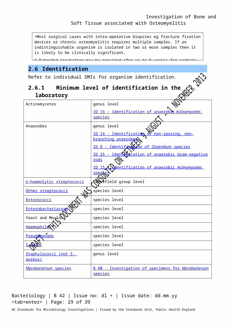
2.6 IdentificationRefer to individual SMIs for organism identification.
2.6.1 Minimum level of identification in the laboratoryActinomycetes genus level
ID 15 – Identification of anaerobic Actinomycetes species
Anaerobes genus levelID 14 - Identification of non-sporing, non-branching anaerobesID 8 - Identification of Clostridium species ID 25 - Identification of anaerobic Gram-negative rodsID 15 – Identification of anaerobic Actinomycetes species
-haemolytic streptococci Lancefield group levelOther streptococci species level
Enterococci species levelEnterobacteriaceae species level
Yeast and Moulds species levelHaemophilus species level
Pseudomonads species levelS. aureus species level
Staphylococci (not S. aureus) genus levelMycobacterium species B 40 - Investigation of specimens for Mycobacterium
species
Organisms may be further identified if clinically or epidemiologically indicated.Note: Any organism considered to be a contaminant may not require identification to species level.
2.7 Antimicrobial susceptibility testingRefer to British Society for Antimicrobial Chemotherapy (BSAC) guidelines. It is important to include a wide range of antibiotics particularly for those patients who may require prolonged oral treatment with biofilm active drugs (see Introduction). These antibiotics are not usually included in the common first line antimicrobials tested in most laboratories. On Gram-positive these may include a teicoplanin MIC plus antibiotics such as rifampicin, tetracyclines, quinolones, co-trimoxazole, fusidic acid, linezolid, quinupristin/dalfopristin and others.
2.8 Referral to reference laboratories Bacteriology | B 42 | Issue no: dl + | Issue date: dd.mm.yy <tab+enter> | Page: 22 of 30UK Standards for Microbiology Investigations | Issued by the Standards Unit, Public Health England

Investigation of Bone and Soft Tissue associated with Osteomyelitis
Contact appropriate reference laboratory for information on the tests available, turn around times, transport procedure and any other requirements for sample submission. Information regarding specialist and reference laboratories is available via the following websites:HPA - Specialist and Reference Microbiology Tests and ServicesHealth Protection Scotland – Reference LaboratoriesBelfast Health and Social Care Trust – Laboratory and Mortuary ServicesOrganisms with unusual or unexpected resistance and whenever there is a laboratory or clinical problem, or anomaly that requires elucidation, should be sent to the appropriate reference laboratory.
2.9 Referral for outbreak investigationsN/A
3 Reporting Procedure3.1 Microscopy3.1.1 Gram stainReport on WBCs and organisms detected.
3.1.2 Microscopy reporting timeWritten report: 16 - 72 hrUrgent microscopy: telephone when available
3.2 CultureReport any growth. Report absence of growth.Also, report results of supplementary investigations.
3.3 Antimicrobial susceptibility testingReport susceptibilities as clinically indicated. Prudent use of antimicrobials according to local and national protocols is recommended.
4 Notification to PHE65,66 The Health Protection (Notification) regulations 2010 require diagnostic laboratories to notify PHE when they identify the causative agents that are listed in Schedule 2 of the Regulations. Notifications must be provided in writing, on paper or electronically, within seven days. Urgent cases should be notified orally and as soon as possible, recommended within 24 hours. These should be followed up by written notification within seven days.
Bacteriology | B 42 | Issue no: dl + | Issue date: dd.mm.yy <tab+enter> | Page: 23 of 30UK Standards for Microbiology Investigations | Issued by the Standards Unit, Public Health England
Investigation of Bone and Soft Tissue associated with Osteomyelitis
For the purposes of the Notification Regulations, the recipient of laboratory notifications is the local PHE Health Protection Team. If a case has already been notified by a registered medical practitioner, the diagnostic laboratory is still required to notify the case if they identify any evidence of an infection caused by a notifiable causative agent.Notification under the Health Protection (Notification) Regulations 2010 does not replace voluntary reporting to PHE. The vast majority of NHS laboratories voluntarily report a wide range of laboratory diagnoses of causative agents to PHE and many PHE Health Protection Teams have agreements with local laboratories for urgent reporting of some infections. This should continue. Note: The Health Protection Legislation Guidance (2010) includes reporting of HIV & STIs, HCAIs and CJD under ‘Notification Duties of Registered Medical Practitioners’: it is not noted under ‘Notification Duties of Diagnostic Laboratories’.Other arrangements exist in Scotland67 and Wales68.
Bacteriology | B 42 | Issue no: dl + | Issue date: dd.mm.yy <tab+enter> | Page: 24 of 30UK Standards for Microbiology Investigations | Issued by the Standards Unit, Public Health England

Investigation of Bone and Soft Tissue associated with Osteomyelitis
Appendix: Investigation of Bone and Soft Tissue associated with Osteomyelitis Flowchart
Bacteriology | B 42 | Issue no: dl + | Issue date: dd.mm.yy <tab+enter> | Page: 25 of 30UK Standards for Microbiology Investigations | Issued by the Standards Unit, Public Health England

Investigation of Bone and Soft Tissue associated with Osteomyelitis
References1. European Parliament. UK Standards for Microbiology Investigations (SMIs) use the
term "CE marked leak proof container" to describe containers bearing the CE marking used for the collection and transport of clinical specimens. The requirements for specimen containers are given in the EU in vitro Diagnostic Medical Devices Directive (98/79/EC Annex 1 B 2.1) which states: "The design must allow easy handling and, where necessary, reduce as far as possible contamination of, and leakage from, the device during use and, in the case of specimen receptacles, the risk of contamination of the specimen. The manufacturing processes must be appropriate for these purposes".
2. Official Journal of the European Communities. Directive 98/79/EC of the European Parliament and of the Council of 27 October 1998 on in vitro diagnostic medical devices. 7-12-1998. p. 1-37.
3. Jorge LS, Chueire AG, Rossit AR. Osteomyelitis: a current challenge. Braz J Infect Dis 2010;14:310-5.
4. Calhoun JH, Manring MM. Adult osteomyelitis. Infect Dis Clin North Am 2005;19:765-86.
5. Gaujoux-Viala C, Zeller V, Leclerc P, Chicheportiche V, Mamoudy P, Desplaces N, et al. Osteomyelitis in adults: an underrecognized clinical entity in immunocompetent hosts. A report of six cases. Joint Bone Spine 2011;78:75-9.
6. Carvalho VC, Oliveira PR, Dal-Paz K, Paula AP, Felix CS, Lima AL. Gram-negative osteomyelitis: clinical and microbiological profile. Braz J Infect Dis 2012;16:63-7.
7. Rahn KA, Jacobson FS. Pseudomonas osteomyelitis of the metatarsal sesamoid bones. Am J Orthop 1997;26:365-7.
8. Niall DM, Murphy PG, Fogarty EE, Dowling FE, Moore DP. Puncture wound related pseudomonas infections of the foot in children. Ir J Med Sci 1997;166:98-101.
9. Laughlin TJ, Armstrong DG, Caporusso J, Lavery LA. Soft tissue and bone infections from puncture wounds in children. West J Med 1997;166:126-8.
10. Gaetti-Jardim E Jr, Landucci LF, de Oliveira KL, Costa I, Ranieri RV, Okamoto AC, et al. Microbiota associated with infections of the jaws. Int J Dent 2012;2012:369751.
11. Brook I. Joint and bone infections due to anaerobic bacteria in children. Pediatr Rehabil 2002;5:11-9.
12. Osmon DR, Berbari EF, Berendt AR, Lew D, Zimmerli W, Steckelberg JM, et al. Executive summary: diagnosis and management of prosthetic joint infection: clinical practice guidelines by the infectious diseases society of America. Clin Infect Dis 2013;56:1-10.
13. Lipsky BA, Berendt AR, Cornia PB, Pile JC, Peters EJ, Armstrong DG, et al. 2012 Infectious Diseases Society of America clinical practice guideline for the diagnosis and treatment of diabetic foot infections. Clin Infect Dis 2012;54:e132-e173.
Bacteriology | B 42 | Issue no: dl + | Issue date: dd.mm.yy <tab+enter> | Page: 26 of 30UK Standards for Microbiology Investigations | Issued by the Standards Unit, Public Health England
Investigation of Bone and Soft Tissue associated with Osteomyelitis
14. Lavery LA, Armstrong DG, Murdoch DP, Peters EJ, Lipsky BA. Validation of the Infectious Diseases Society of America's diabetic foot infection classification system. Clin Infect Dis 2007;44:562-5.
15. National Institute for Health and Clinical Excellence. Diabetic foot problems: Inpatient management of diabetic foot problems .
16. Dartnell J, Ramachandran M, Katchburian M. Haematogenous acute and subacute paediatric osteomyelitis: a systematic review of the literature. J Bone Joint Surg Br 2012;94:584-95.
17. Faust SN, Clark J, Pallett A, Clarke NM. Managing bone and joint infection in children. Arch Dis Child 2012;97:545-53.
18. Hatzenbuehler J, Pulling TJ. Diagnosis and management of osteomyelitis. Am Fam Physician 2011;84:1027-33.
19. Ish-Horowicz MR, McIntyre P, Nade S. Bone and Joint infections caused by multiple resistant Staphylococcus aureus in a neonatal intensive care unit. Pediatr Infect Dis 1992;11:82-7.
20. Mader JT, Calhoun J. Osteomyelitis. In: Mandell GL, Bennett JE, Dolin R, editors. Mandell Douglas and Bennett's Principles and Practice of Infectious Diseases. 5th ed. Edinburgh: Churchill Livingstone; 2000. p. 1182-96.
21. Holzman RS, Bishko F. Osteomyelitis in heroin addicts. Ann Intern Med 1971;75:693-6.
22. Fuentes FM, Gutierrez TL, Ayala RO, Rumayor ZM, del Prado GN. Tuberculosis of the spine. A systematic review of case series. Int Orthop 2012;36:221-31.
23. Merino P, Candel FJ, Gestoso I, Baos E, Picazo J. Microbiological diagnosis of spinal tuberculosis. Int Orthop 2012;36:233-8.
24. Sagoo RS, Lakdawala A, Subbu R. Tuberculosis of the elbow joint. JRSM Short Rep 2011;2:17.
25. Sandher DS, Al-Jibury M, Paton RW, Ormerod LP. Bone and joint tuberculosis: cases in Blackburn between 1988 and 2005. J Bone Joint Surg Br 2007;89:1379-81.
26. Farrington M, Eykyn SJ, Walker M, Warren RE. Vertebral osteomyelitis due to coccobacilli of the HB group. Br Med J (Clin Res Ed) 1983;287:1658-60.
27. Lebeaux D, Zarrouk V, Petrover D, Nicolas-Chanoine MH, Fantin B. Salmonella Colindale osteomyelitis in an immunocompetent female patient. Med Mal Infect 2012;42:36-7.
28. Kolker S, Itsekzon T, Yinnon AM, Lachish T. Osteomyelitis due to Salmonella enterica subsp. arizonae: the price of exotic pets. Clin Microbiol Infect 2012;18:167-70.
29. Allison DC, Holtom PD, Patzakis MJ, Zalavras CG. Microbiology of bone and joint infections in injecting drug abusers. Clin Orthop Relat Res 2010;468:2107-12.
30. Larsen LH, Lange J, Xu Y, Schonheyder HC. Optimizing culture methods for diagnosis of prosthetic joint infections: a summary of modifications and improvements reported since 1995. J Med Microbiol 2012;61:309-16.
Bacteriology | B 42 | Issue no: dl + | Issue date: dd.mm.yy <tab+enter> | Page: 27 of 30UK Standards for Microbiology Investigations | Issued by the Standards Unit, Public Health England

Investigation of Bone and Soft Tissue associated with Osteomyelitis
31. Mackowiak PA, Jones SR, Smith JW. Diagnostic value of sinus-tract cultures in chronic osteomyelitis. JAMA 1978;239:2772-5.
32. Mousa HA. Evaluation of sinus-track cultures in chronic bone infection. J Bone Joint Surg Br 1997;79:567-9.
33. Cierny G, Mader JT. Adult chronic osteomyelitis. Orthopaedics 1984;7:1557-64.
34. Waldvogel FA, Papageorgiou PS. Osteomyelitis: the past decade. N Engl J Med 1980;360-70.
35. Olasinde AA, Oluwadiya KS, Adegbehingbe OO. Treatment of Brodie's abscess: excellent results from curettage, bone grafting and antibiotics. Singapore Med J 2011;52:436-9.
36. Dirschl DR, Almekinders LC. Osteomyelitis. Common causes and treatment recommendations. Drugs 1993;45:29-43.
37. Medical Microbiology L A Guide to Microbial Infections. 15 ed. Edinburgh: Churchill Livingstone; 1997. p. 566
38. Monsen T, Lovgren E, Widerstrom M, Wallinder L. In vitro effect of ultrasound on bacteria and suggested protocol for sonication and diagnosis of prosthetic infections. J Clin Microbiol 2009;47:2496-501.
39. Piper KE, Jacobson MJ, Cofield RH, Sperling JW, Sanchez-Sotelo J, Osmon DR, et al. Microbiologic diagnosis of prosthetic shoulder infection by use of implant sonication. J Clin Microbiol 2009;47:1878-84.
40. Vergidis P, Greenwood-Quaintance KE, Sanchez-Sotelo J, Morrey BF, Steinmann SP, Karau MJ, et al. Implant sonication for the diagnosis of prosthetic elbow infection. J Shoulder Elbow Surg 2011;20:1275-81.
41. Hughes HC, Newnham R, Athanasou N, Atkins BL, Bejon P, Bowler IC. Microbiological diagnosis of prosthetic joint infections: a prospective evaluation of four bacterial culture media in the routine laboratory. Clin Microbiol Infect 2011;17:1528-30.
42. Morris AJ, Wilson SJ, Marx CE, Wilson ML, Mirrett S, Reller LB. Clinical impact of bacteria and fungi recovered only from broth cultures. J Clin Microbiol 1995;33:161-5.
43. Carbonnelle E, Mesquita C, Bille E, Day N, Dauphin B, Beretti JL, et al. MALDI-TOF mass spectrometry tools for bacterial identification in clinical microbiology laboratory. Clin Biochem 2011;44:104-9.
44. van Veen SQ, Claas ECJ, Kuijper EJ. High-Throughput Identification of Bacteria and Yeast by Matrix-Assisted Laser Desorption Ionization-Time of Flight Mass Spectrometry in Conventional Medical Microbiology Laboratories â–¿. J Clin Microbiol 2010;48:900-7.
45. Espy MJ, Uhl JR, Sloan LM, Buckwalter SP, Jones MF, Vetter EA, et al. Real-time PCR in clinical microbiology: applications for routine laboratory testing. Clin Microbiol Rev 2006;19:165-256.
46. Arciola CR, Montanaro L, Costerton JW. New trends in diagnosis and control strategies for implant infections. Int J Artif Organs 2011;34:727-36.
Bacteriology | B 42 | Issue no: dl + | Issue date: dd.mm.yy <tab+enter> | Page: 28 of 30UK Standards for Microbiology Investigations | Issued by the Standards Unit, Public Health England
Investigation of Bone and Soft Tissue associated with Osteomyelitis
47. Esteban J, Alonso-Rodriguez N, del-Prado G, Ortiz-Perez A, Molina-Manso D, Cordero-Ampuero J, et al. PCR-hybridization after sonication improves diagnosis of implant-related infection. Acta Orthop 2012;83:299-304.
48. Cherkaoui A, Ceroni D, Emonet S, Lefevre Y, Schrenzel J. Molecular diagnosis of Kingella kingae osteoarticular infections by specific real-time PCR assay. J Med Microbiol 2009;58:65-8.
49. Advisory Committee on Dangerous Pathogens. The Approved List of Biological Agents. Her Majesty's Stationery Office. Norwich. 2004. p. 1-21.
50. Centers for Disease Control and Prevention. Guidelines for Safe Work Practices in Human and Animal Medical Diagnostic Laboratories. MMWR Surveill Summ 2012;61:1-102.
51. Advisory Committee on Dangerous Pathogens. Infections at work: Controlling the risks. Her Majesty's Stationery Office. 2003.
52. Advisory Committee on Dangerous Pathogens. Biological agents: Managing the risks in laboratories and healthcare premises. Health and Safety Executive. 2005.
53. Health and Safety Executive. Control of Substances Hazardous to Health Regulations. The Control of Substances Hazardous to Health Regulations 2002. 5th ed. HSE Books; 2002.
54. Health and Safety Executive. Five Steps to Risk Assessment: A Step by Step Guide to a Safer and Healthier Workplace. HSE Books. 2002.
55. Health and Safety Executive. A Guide to Risk Assessment Requirements: Common Provisions in Health and Safety Law. HSE Books. 2002.
56. British Standards Institution (BSI). BS EN12469 - Biotechnology - performance criteria for microbiological safety cabinets. 2000.
57. British Standards Institution (BSI). BS 5726 - Microbiological safety cabinets. Part 2: Recommendations for information to be exchanged between purchaser, vendor and installer and recommendations for installation. 1992.
58. British Standards Institution (BSI). BS 5726 - Microbiological safety cabinets. Part 4: Recommendations for selection, use and maintenance. 1992.
59. Health Services Advisory Committee. Safe Working and the Prevention of Infection in Clinical Laboratories and Similar Facilities. HSE Books. 2003.
60. Department for transport. Transport of Infectious Substances, 2011 Revision 5. 2011.
61. Holden J. Collection and transport of clinical specimens for anaerobic culture. In: Isenberg HD, editor. Clinical Microbiology Procedures Handbook.Vol 1. Washington D.C.: American Society for Microbiology; 1992. p. 2.2.1-7.
62. Barber S, Lawson PJ, Grove DI. Evaluation of bacteriological transport swabs. Pathology 1998;30:179-82.
63. Bosshard PP. Incubation of fungal cultures: how long is long enough? Mycoses 2011;54:e539-e545.
Bacteriology | B 42 | Issue no: dl + | Issue date: dd.mm.yy <tab+enter> | Page: 29 of 30UK Standards for Microbiology Investigations | Issued by the Standards Unit, Public Health England
Investigation of Bone and Soft Tissue associated with Osteomyelitis
64. Morris AJ, Byrne TC, Madden JF, Reller LB. Duration of incubation of fungal cultures. J Clin Microbiol 1996;34:1583-5.
65. Health Protection Agency. Laboratory Reporting to the Health Protection Agency: Guide for Diagnostic Laboratories. 2010.
66. Department of Health. Health Protection Legislation (England) Guidance. 2010. p. 1-112.
67. Scottish Government. Public Health (Scotland) Act. 2008.
68. The Welsh Assembly Government. Health Protection Legislation (Wales) Guidance. 2010.
Bacteriology | B 42 | Issue no: dl + | Issue date: dd.mm.yy <tab+enter> | Page: 30 of 30UK Standards for Microbiology Investigations | Issued by the Standards Unit, Public Health England