Embed Size (px)
Citation preview
Use of Integrated PET CT in the Clinical Staging of Non Small Cell
Lung CancerLaura Myers, Harvard Medical School, Year III
Gillian Lieberman, MD
November 2010Laura Myers, 2010Gillian Lieberman, MD

2
Clinical Presentation• 79yo woman with cough productive of green
sputum worse than baseline cough, exertional SOB and 12 lb unintentional weight loss in 1 mo
• Denies fevers, night sweats or CP• PMH: COPD, HTN, CAD, DM2• SoH: 54 pack-yr hx of smoking cigarettes, quit 5
yrs ago• PE: afebrile, 02 sat 95% RA, good air movement,
mild end-exp wheezing, fine crackles in left lung that resolved after cough
Laura Myers, 2010Gillian Lieberman, MD
3
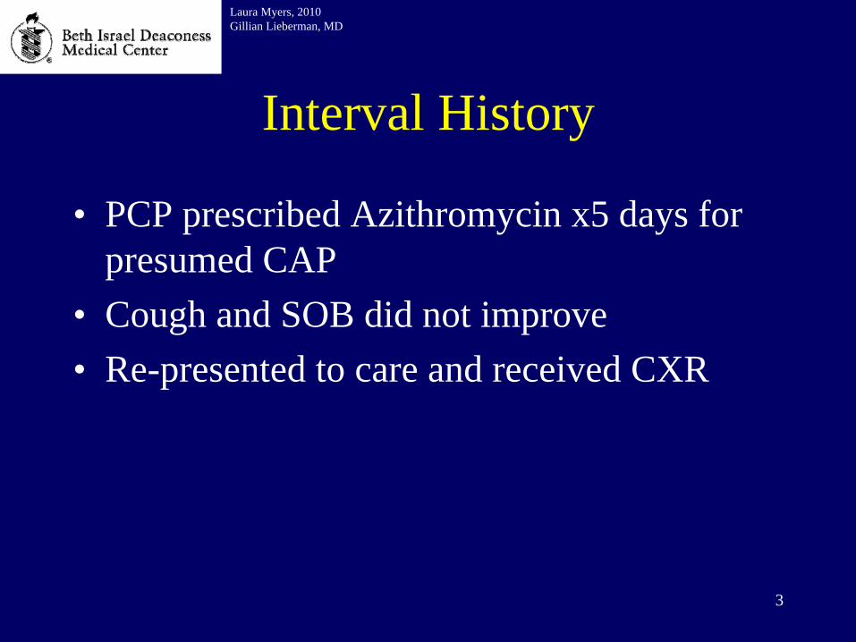
Interval History
• PCP prescribed Azithromycin x5 days for presumed CAP
• Cough and SOB did not improve• Re-presented to care and received CXR
Laura Myers, 2010Gillian Lieberman, MD
4
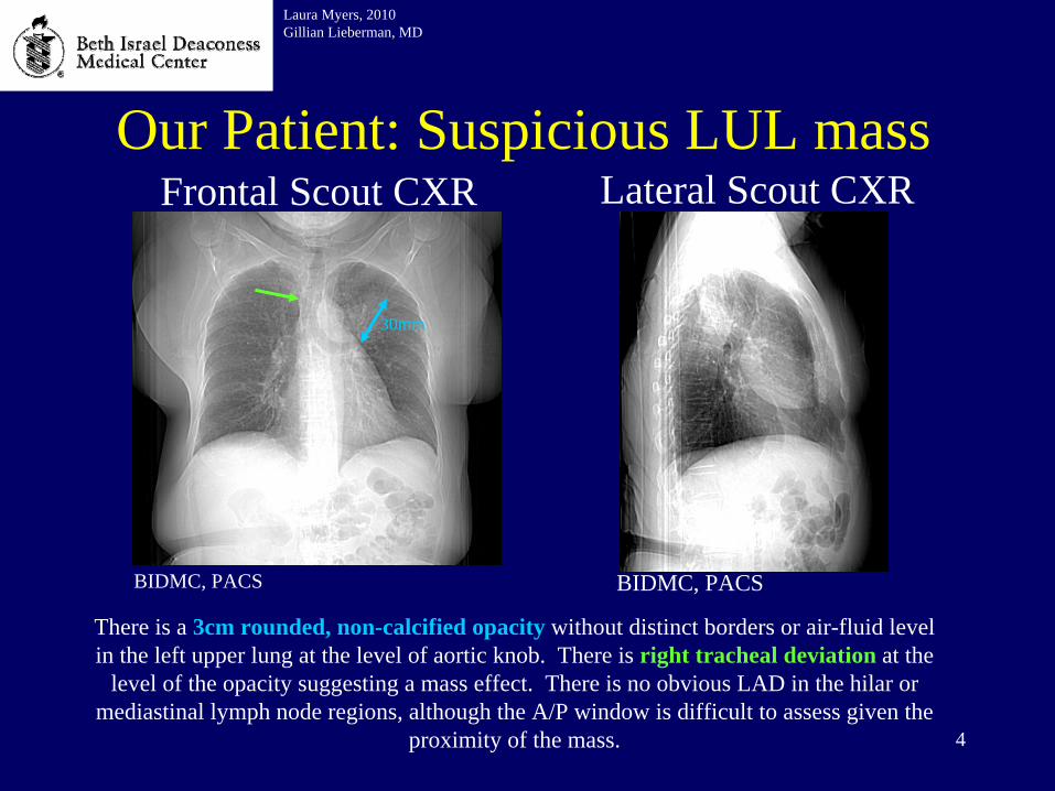
Our Patient: Suspicious LUL mass
Laura Myers, 2010Gillian Lieberman, MD
BIDMC, PACS BIDMC, PACS
30mm
Frontal Scout CXR Lateral Scout CXR
There is a 3cm rounded, non-calcified opacity without distinct borders or air-fluid level in the left upper lung at the level of aortic knob. There is right tracheal deviation at the
level of the opacity suggesting a mass effect. There is no obvious LAD in the hilar or mediastinal lymph node regions, although the A/P window is difficult to assess given the
proximity of the mass.
5
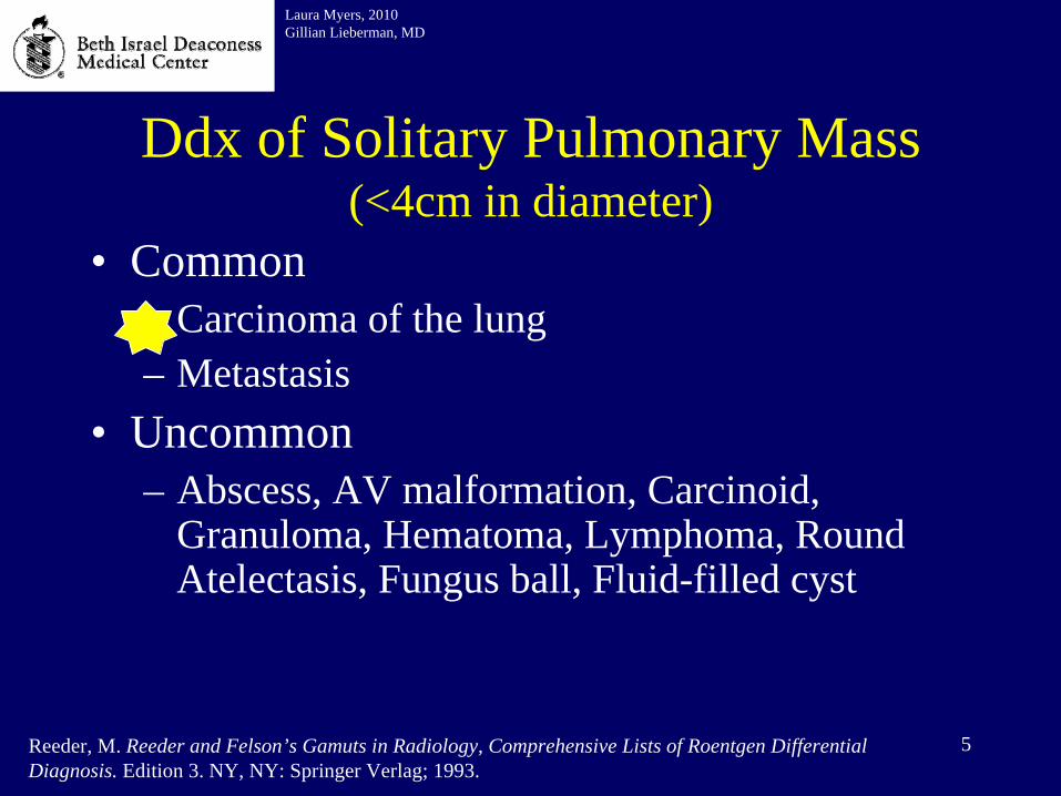
Ddx of Solitary Pulmonary Mass (<4cm in diameter)
Laura Myers, 2010Gillian Lieberman, MD
• Common– Carcinoma of the lung – Metastasis
• Uncommon – Abscess, AV malformation, Carcinoid,
Granuloma, Hematoma, Lymphoma, Round Atelectasis, Fungus ball, Fluid-filled cyst
Reeder, M. Reeder and Felson’s Gamuts in Radiology, Comprehensive Lists of Roentgen Differential Diagnosis. Edition 3. NY, NY: Springer Verlag; 1993.
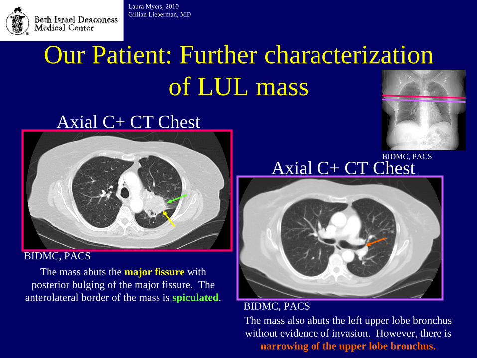
Our Patient: Further characterization of LUL mass
Laura Myers, 2010Gillian Lieberman, MD
BIDMC, PACS
BIDMC, PACS
BIDMC, PACS
Axial C+ CT Chest
Axial C+ CT Chest
The mass abuts the major fissure with posterior bulging of the major fissure. The
anterolateral border of the mass is spiculated.
The mass also abuts the left upper lobe bronchus without evidence of invasion. However, there is
narrowing of the upper lobe bronchus.
7
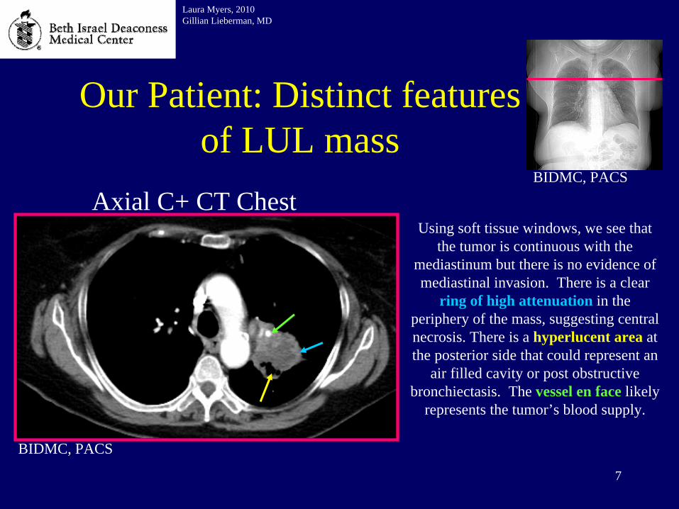
Our Patient: Distinct features of LUL mass
Laura Myers, 2010Gillian Lieberman, MD
BIDMC, PACS
BIDMC, PACS
Axial C+ CT ChestUsing soft tissue windows, we see that
the tumor is continuous with the mediastinum but there is no evidence of mediastinal invasion. There is a clear
ring of high attenuation in the periphery of the mass, suggesting central necrosis. There is a hyperlucent area at the posterior side that could represent an
air filled cavity or post obstructive bronchiectasis. The vessel en face likely
represents the tumor’s blood supply.

8
T in TNM Staging
Laura Myers, 2010Gillian Lieberman, MD
Up to date
• Based on these images, we know that our patient’s tumor has a diameter of 3cm so it is considered stage T1b.
9
Let’s determine this patient’s nodal stage.
Laura Myers, 2010Gillian Lieberman, MD

10
Our Patient: Bilateral hilar lymphadenopathy
Laura Myers, 2010Gillian Lieberman, MD
BIDMC, PACS
BIDMC, PACS
BIDMC, PACS
Axial C+ CT Chest Axial C+ CT Chest
15mm10mm
There are areas of soft tissue attenuation that are >1cm in diameter and located in the right and left hilar regions, likely representing bilateral hilar lymphadenopathy. While thoracic
lymph nodes can vary in size, a short axis diameter >1cm is considered abnormal.
11
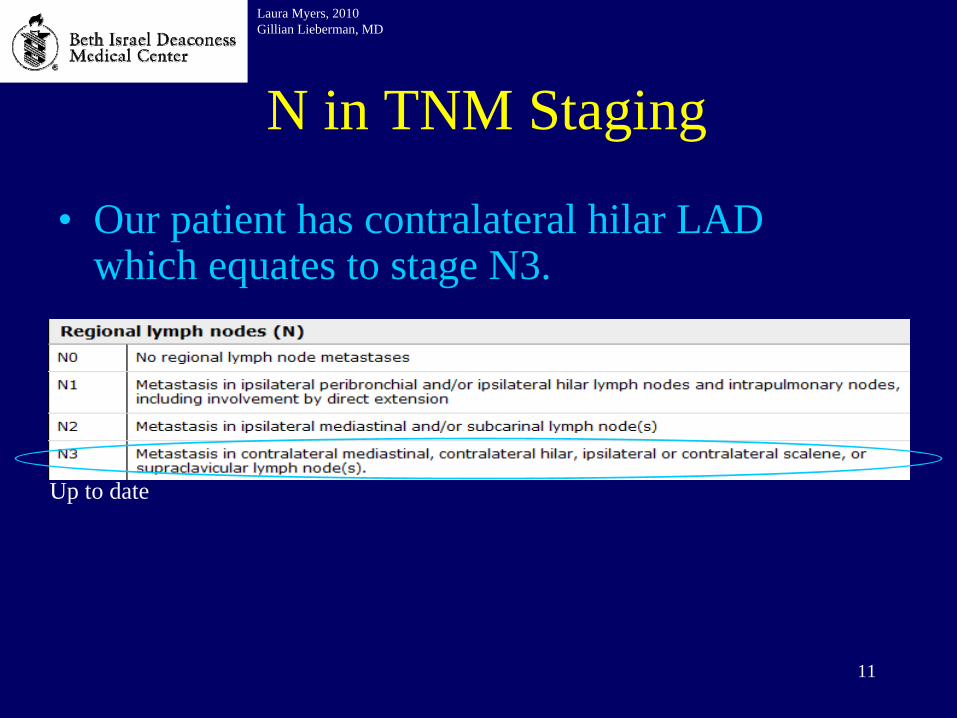
N in TNM Staging
Laura Myers, 2010Gillian Lieberman, MD
Up to date
• Our patient has contralateral hilar LAD which equates to stage N3.

Our patient: “Tree in bud”
Laura Myers, 2010Gillian Lieberman, MD
BIDMC, PACS
BIDMC, PACS
Axial C+ CT Chest
Eisenhuber, E. The Tree in Bud Sign. Radiology. March 2002. 222: 771-772.
Companion Patient 1
There are tubular and nodular opacities in the lung periphery consistent with a “Tree in bud” sign. A classic Tree in Bud is shown at right.
13
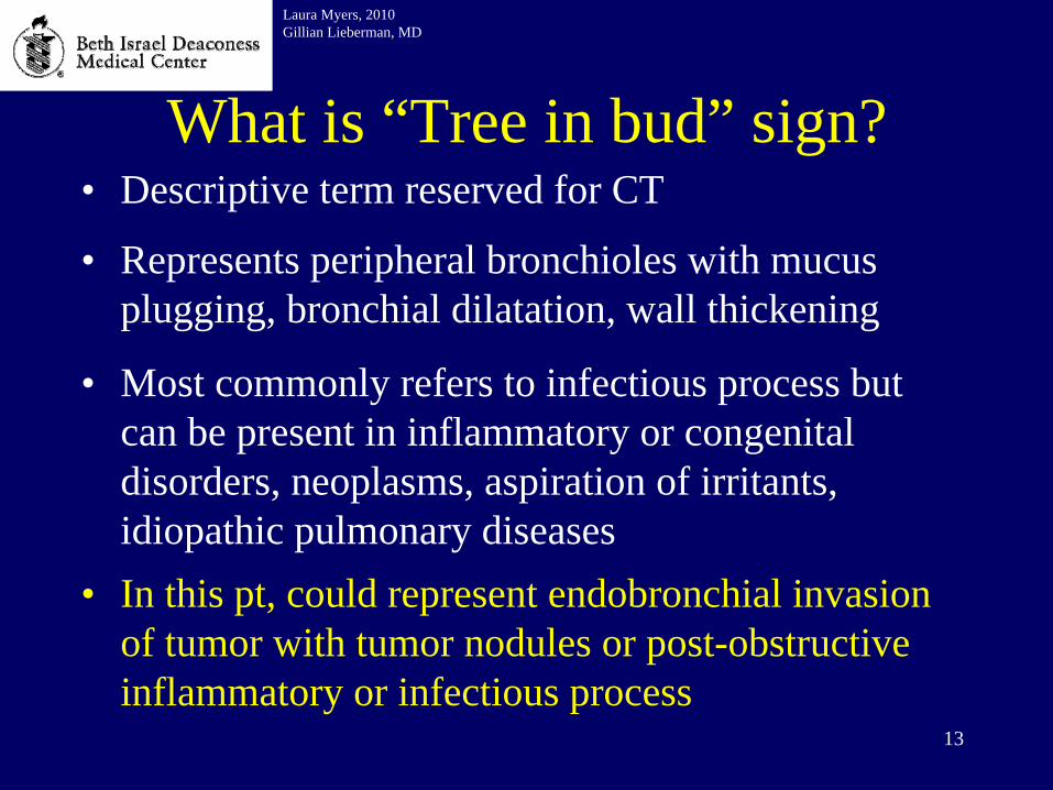
What is “Tree in bud” sign?
Laura Myers, 2010Gillian Lieberman, MD
• Descriptive term reserved for CT
• Represents peripheral bronchioles with mucus plugging, bronchial dilatation, wall thickening
• Most commonly refers to infectious process but can be present in inflammatory or congenital disorders, neoplasms, aspiration of irritants, idiopathic pulmonary diseases
• In this pt, could represent endobronchial invasion of tumor with tumor nodules or post-obstructive inflammatory or infectious process

14
Our Patient: Incidental finding of multiple renal masses
Laura Myers, 2010Gillian Lieberman, MD
BIDMC, PACS
BIDMC, PACS
BIDMC, PACSC+ CT Chest
C+ CT Chest
There are several small irregular heterogeneous areas of soft tissue attenuation seen on both of the images with thinning of the cortex and deformation of the normal kidney
architecture. MRI abdomen is needed to characterize these lesions.

15
Our Patient: Multiple enhancing renal masses
Laura Myers, 2010Gillian Lieberman, MD
Axial C+ MRI AbdomenPre-contrast
Axial C+ MRI AbdomenPost-contrast
BIDMC, PACS BIDMC, PACS
BIDMC, PACS
There is a rounded low signal area within the right kidney (left image) that enhances with contrast (right image), indicating that this is a vascularized area and not just a simple cyst.
16
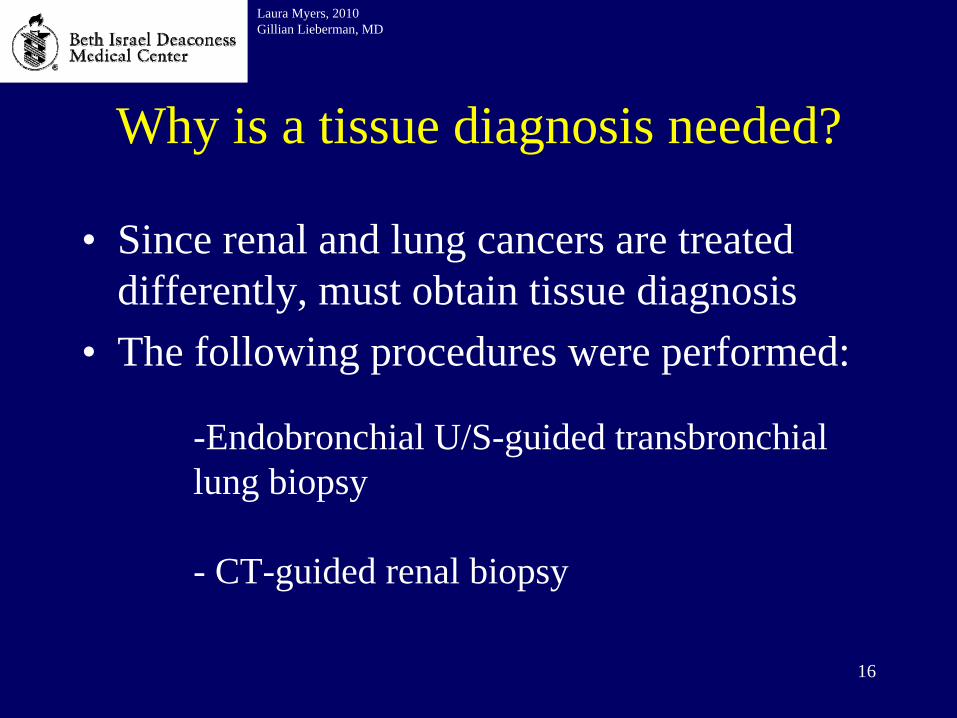
Why is a tissue diagnosis needed?
• Since renal and lung cancers are treated differently, must obtain tissue diagnosis
• The following procedures were performed:
Laura Myers, 2010Gillian Lieberman, MD
-Endobronchial U/S-guided transbronchial lung biopsy
- CT-guided renal biopsy

17
Our Patient: Pathology specimen
Laura Myers, 2010Gillian Lieberman, MD
Alveolar tissue is seen with occasional neutrophils. No
tumor is present.
Normal Tumor
Slide courtesy of Hannah Gilmore MD, Pathology Dept, BIDMC
Nest of malignant tumor cells with a
high N:C ratio, abundant mitoses and focal necrosis.
Consistent with metastatic
squamous cell carcinoma.
200X: H&E stained section of lung from transbronchial biopsy
40X: H&E stained section from core needle biopsy of kidney
200X: H&E stained section from core needle biopsy of kidney
Since SCC does not arise in the kidney, it is most likely primary lung cancer with renal metastasis.
18
Radiologic Features of SCC of Lung• Ranges in size from 1-10cm
Laura Myers, 2010Gillian Lieberman, MD
Patz, E. Imaging Bronchogenic Carcinoma. Chest. 200.117(4):90s-95s.
• Centrally located, although 1/3 of SCC are found beyond the segmental bronchus
• Because of the central location, can cause post-obstructive pneumonia or atelectasis which occurs in up to 50% of cases
• Cavitate in 10-20% of cases, particularly in large peripheral lesions
• Most common sites for metastases from SCC of the lung are adrenal, kidney, bone and liver
19
We believe this patient has renal metastases. Let’s confirm this and determine if there are other
sites of metastases.
Laura Myers, 2010Gillian Lieberman, MD

20
M in TNM Staging
Laura Myers, 2010Gillian Lieberman, MD
• In the past, has been done by PET alone or CT alone – Advantage of PET: detect smaller lesions that can’t be resolved by
CT; limit detection to fast-growing tumors, which are more likely to be malignant
– Advantage of CT: understand exact location of lesion
• Now combined PET CT is being used• Images can either be displayed side by side and
correlated visually by the radiologist or directly overlaid: 2003 NEJM trial showed that direct integration of images has better diagnostic accuracy than visual correlation

21
Integrated PET CT
Laura Myers, 2010Gillian Lieberman, MD
• Physiologic positives: brain, heart, GI tract, genitourinary tract, recently exercised muscle
• False positives: Benign tumors such as sclerosing hemangioma, leiomyoma, inflammatory pseudotumor, any areas of inflammation, atherosclerosis (aorta), granulation tissue, in pleura after pleurodesis, after placement of central lines, chest tubes, gastrostomy tubes, after mediastinoscopy
• False negatives: slow growing tumors such as BAC, carcinoid, mucoepidermoid carcinoma
Cannot be used reliably for brain metastases, so many undergo separate MRI brain

22
Sensitivity and Specificity of PET CT in NSCLC Staging
Laura Myers, 2010Gillian Lieberman, MD
Sensitivity Specificity
Tumor (Chest wall invasion)
38-90% 40-90%
Nodes(Mediastinal lymph
nodes)
79-85% 89-92%
Mets(to liver and adrenal
glands)
88-100% 100%
Adapted from: De Wever, W. Et al. Integrated PET/CT in the staging of nonsmall cell lung cancer: technical aspects and clinical integration. European Respiratory Journal. 33(1): 201-212.

23
Radiation Exposure in PET CT
Laura Myers, 2010Gillian Lieberman, MD
• More than either study alone • (~18mSv from CT, ~7mSv from FDG)• Effective dose depends on:
• Therefore, current protocols use 80mA current to optimize image quality but decrease radiation exp
24
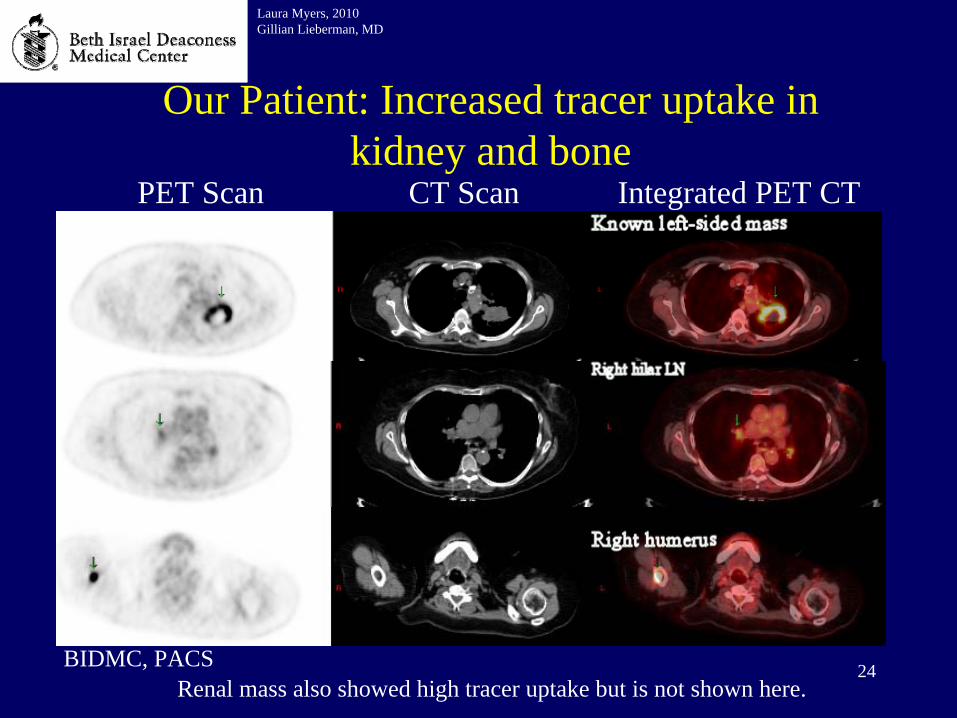
Our Patient: Increased tracer uptake in kidney and bone
Laura Myers, 2010Gillian Lieberman, MD
BIDMC, PACSRenal mass also showed high tracer uptake but is not shown here.
Integrated PET CTCT ScanPET Scan

25
M in TNM Staging
Laura Myers, 2010Gillian Lieberman, MD
Up to date
• Our patient has confirmed metastases to kidney and newly discovered metastases to bone, which equates to stage M1b.
26
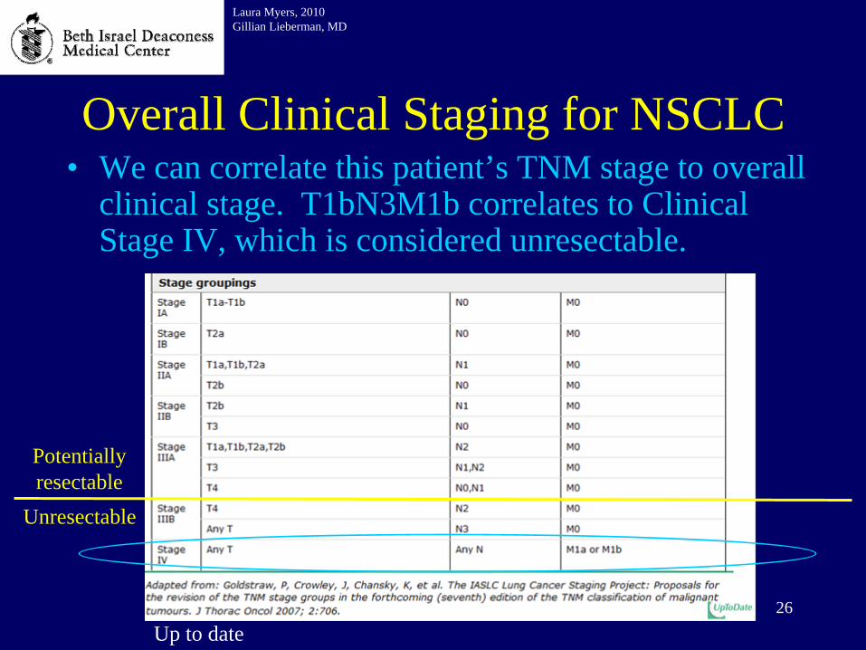
Overall Clinical Staging for NSCLC
Laura Myers, 2010Gillian Lieberman, MD
Up to date
Potentially resectable
Unresectable
• We can correlate this patient’s TNM stage to overall clinical stage. T1bN3M1b correlates to Clinical Stage IV, which is considered unresectable.

27
Treatment• For elderly patients with advanced NSCLC,
treatment depends on the individual’s goals of care
• However, platinum-based systemic chemotherapy is the chemotherapy regimen of choice when appropriate
• Our patient began cisplatin/gemcitabine regimen
Laura Myers, 2010Gillian Lieberman, MD
28
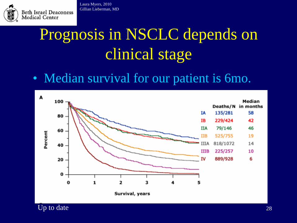
Prognosis in NSCLC depends on clinical stage
Laura Myers, 2010Gillian Lieberman, MD
Up to date
• Median survival for our patient is 6mo.

29
Summary• Common presenting symptoms of a pt with lung
cancer• Differential of a single rounded opacity on CXR• Normal anatomy of thoracic lymph nodes• Use of IR-guided procedures to obtain a tissue
diagnosis of a suspected malignancy • Sensitivity and specificity of integrated PET CT
and use of PET CT in clinical staging of NSCLC• Treatment of and prognosis for patients with
NSCLC
Laura Myers, 2010Gillian Lieberman, MD
30
ReferencesBrambilla E. et al. The new WHO classification of lung tumors. European Respiratory
Journal. 2001;(18):1059-1068.De Wever, W. et al. Integrated PET/CT in the staging of non-small cell lung cancer: technical aspects
and clinical integration. European Respiratory Journal. 2009;33(1):201-212.Eisenhuber, E. The Tree in Bud Sign. Radiology. 2002;222:771-772.Griffeth, L. Use of PET/CT scanning in cancer patients: technical and practical considerations. Baylor
University Medical Center Proceedings. 2005;18(4): 321-330.Hany, T. PET diagnostic accuracy: improvement with in-line PET-CT system: initial
results. Radiology. 2002;225(2):575-81.Lardinois D. et al. Staging of Non-Small Cell Lung Cancer with Integrated Positron-Emission
Tomography and Computed Tomography. NEJM. 2003;348:2500-2507.Patz, E. Imaging Bronchogenic Carcinoma. Chest. 2000;117(4):90s-95s.Reeder, M. Reeder and Felson’s Gamuts in Radiology, Comprehensive Lists of Roentgen Differential
Diagnosis. Edition 3. NY, NY: Springer Verlag; 1993.Sharma A. Patterns of Lymphadenopathy in Thoracic Malignancies. Radiographics. Education
Exhibit. 2004; 24(2):419-434.Smith, R. et al. Epidemiology of Lung Cancer. The Radiologic Clinics of North America. Vol 38,
Number 3. Philadelphia, PA: W.B. Saunders Company; 2000.Travis, W. et al. World Health Organization classification of tumors. Pathology and genetics of tumors
of the lung, pleura, thymus and heart. IARC Press. Lyon; 2004.
Laura Myers, 2010Gillian Lieberman, MD

31
Acknowledgements
• Gillian Lieberman, MD• Paul Spirn, MD• Iva Petvoska, MD• Olga Brooke, MD• Carole Ridge, MD• Hannah Gilmore, MD• Emily Hanson
Laura Myers, 2010Gillian Lieberman, MD