Embed Size (px)
Citation preview

Vivian GonzalezGillian Lieberman, MD
Lumbar Spine Trauma
Vivian Gonzalez, Harvard Medical School Year IIIGillian Lieberman, MD
Vivian GonzalezGillian Lieberman, MD
January 2002

2
Vivian GonzalezGillian Lieberman, MD
Agenda
• Anatomy and Biomechanics of Lumbar Spine
• Three-Column Concept• Classification of Fractures• Our Patient• Imaging Modalities• Role of Radiologist in Spinal Trauma

3
Vivian GonzalezGillian Lieberman, MD
Anatomy and Biomechanics of the Lumbar Spine
• Each vertebra articulates with adjacent vertebrae at three points– intervertebral disk – paired facet joints posteriorly
• Muscles attach to the lumbar vertebra- >strength and stability
• more mobility than thoracic spine due to sagittal orientation of facet joints and absence of ribs
4
Vivian GonzalezGillian Lieberman, MD
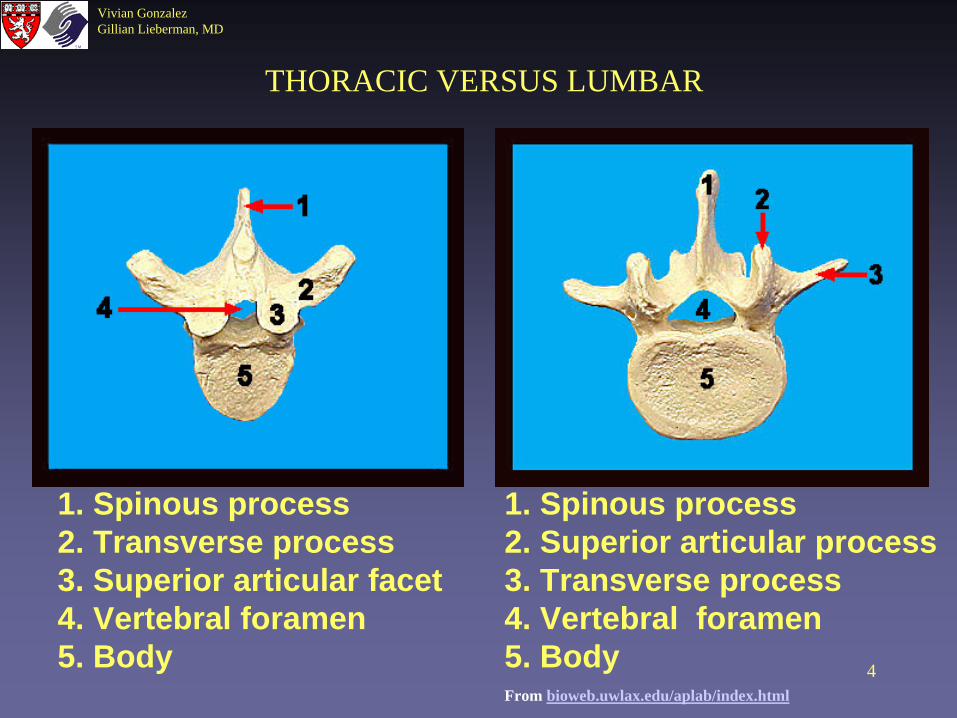
1. Spinous process2. Transverse process3. Superior articular facet4. Vertebral foramen5. Body
1. Spinous process2. Superior articular process3. Transverse process4. Vertebral foramen5. BodyFrom bioweb.uwlax.edu/aplab/index.html
THORACIC VERSUS LUMBAR

5
Vivian GonzalezGillian Lieberman, MD
Lumbar Ligaments
From Netter’s Atlas of Human Anatomy
6
Vivian GonzalezGillian Lieberman, MD
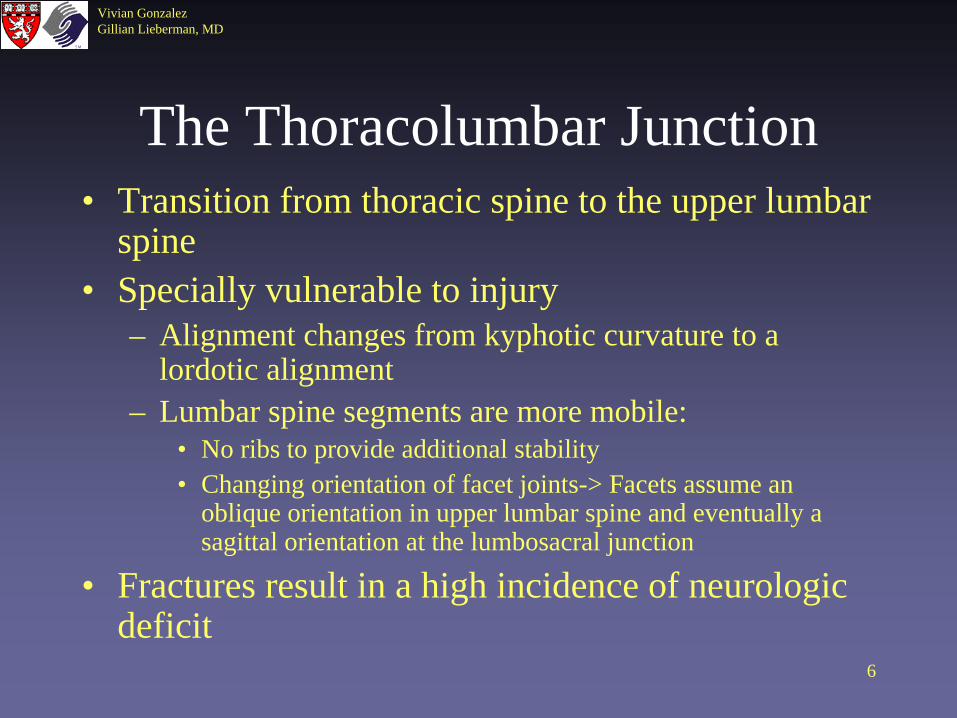
The Thoracolumbar Junction• Transition from thoracic spine to the upper lumbar
spine• Specially vulnerable to injury
– Alignment changes from kyphotic curvature to a lordotic alignment
– Lumbar spine segments are more mobile: • No ribs to provide additional stability• Changing orientation of facet joints-> Facets assume an
oblique orientation in upper lumbar spine and eventually a sagittal orientation at the lumbosacral junction
• Fractures result in a high incidence of neurologic deficit

7
Vivian GonzalezGillian Lieberman, MD
Denis’ THREE-COLUMN CONCEPT (1983)
• Determines fracture severity and predicts stability
• Fractures involving only the anterior columns are considered stable, while fractures that additionally involve the middle or all three columns are considered unstable.
http://www.hawaii.edu/medicine/pediatrics/pemxray/v6c13.html

8
Vivian GonzalezGillian Lieberman, MD
Major Lumbar Spine Fractures• Denis 4 Basic Types
– Compression Fracture– Burst Fracture– Seat Belt Injury– Fracture-Dislocation
• McAfee’s (based on CT appearance)– Wedge Compression Fracture– Stable Burst Fracture– Unstable Burst Fracture– Seat Belt-type Injury– Flexion Distraction Injury– Translation Injuries

9
Vivian GonzalezGillian Lieberman, MD
www.hawaii.edu/medicine/pediatrics/pemxray/v6c13.html
McAfee Classification of Major Fractures

10
Vivian GonzalezGillian Lieberman, MD
Compression Fractures
• Most common- 58% of major spine fractures
• Represent isolated failure in compression of the anterior column with the middle column remaining intact
• True compression fracture shouldn’t produce any neurologic defect
• STABLE
From www.hawaii.edu/medicine/pediatrics/pemxray/v6c13.html
•LOOK at posterior vertebral line→normally MILDLY CONVEX ANTERIORLY

11
Vivian GonzalezGillian Lieberman, MD
Burst Fractures• 17% of major spinal fractures• Like compression fractures, they occur during
hyperflexion and axial loading of a vertebra (e.g. MVA, fall from height)
• BURST= spreads out in all directions• Compressed disk adjacent to the affected vertebra
herniates into the vertebral body• Failure of ANTERIOR and MIDDLE columns
defines these as UNSTABLE
12
Vivian GonzalezGillian Lieberman, MD
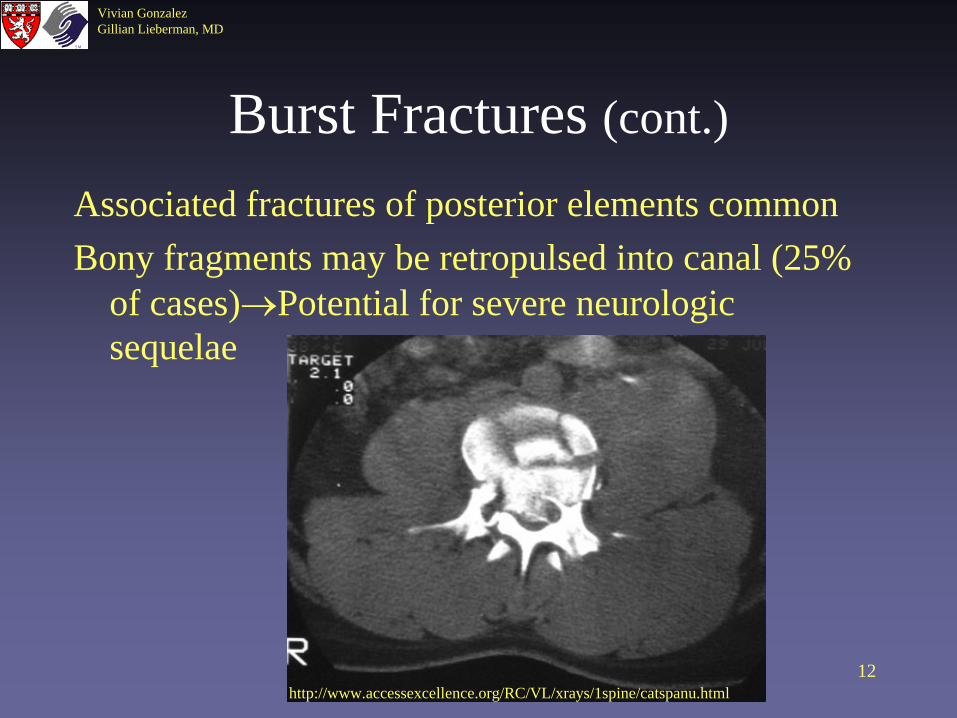
Burst Fractures (cont.)
Associated fractures of posterior elements commonBony fragments may be retropulsed into canal (25%
of cases)→Potential for severe neurologic sequelae
http://www.accessexcellence.org/RC/VL/xrays/1spine/catspanu.html

13
Vivian GonzalezGillian Lieberman, MD
Radiology of Burst Fractures
PLAIN FILMS• AP Vertebral Body
height ↓• If severe:
– Posterior column fractures
– widening of interpedicular distance
From Brandser EA, El-Khoury. Thoracic and Lumbar Spine Trauma.The Radiologic Clinics of North America 1997; 35: 533-557.

14
Vivian GonzalezGillian Lieberman, MD
Seat Belt Injuries6% of Major Spinal FracturesSpectrum of Ligamentous and Bony Injuries: Includes the classic CHANCE fractureMechanism of Injury
Restrained by lap belt w/o shoulder harnessPropelled forward but restrained at abdominal wallFulcrum for rotation moves anterior to spine and results in distraction force→ MIDDLEAND POSTERIOR COLUMNS FAIL
***UNSTABLE***

15
Vivian GonzalezGillian Lieberman, MD
Seat-belt Injuries
From www.rad.washington.edu/maintf/cases/unk41/answers.html

16
Vivian GonzalezGillian Lieberman, MD
Chance Fractures
www.medmedia.com/o11/198.htm
Fracture can extend through the pedicles into posterior elements AP shows involvement of
pedicles and lamina Fracture extension from posterior elements into vertebral body with buckle in anterior cortex
The Radiology Clinics of NA

17
Vivian GonzalezGillian Lieberman, MD
THE FOLLOWING WOULD HAVE TO BE TORN TO ALLOW THIS NEW POSITION • supraspinous
ligament• interspinous
ligaments• ligamenta
flava• capsular ligaments• posterior longitudinal ligament• possibly the posterior annulus fibrosus
Posterior ligamentous disruption, L4-5
www.rad.washington.edu/maintf/cases/unk4 1/answers.html
18
Vivian GonzalezGillian Lieberman, MD
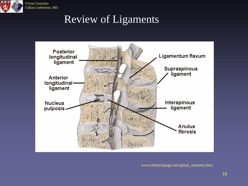
www.thebackpage.net/spinal_anatomy.htm
Review of Ligaments

19
Vivian GonzalezGillian Lieberman, MD
Fracture Dislocations• 19% of Major Fractures• Anterior and Cranial Displacement of Superior Vertebral
Body• Failure of all 3 COLUMNS!• Higher incidence of neurologic deficit
Widened interspinous distance
Anterior translation
From Brandser EA, El-Khoury. Thoracic and Lumbar Spine Trauma.The Radiologic Clinics of North America 1997; 35: 533-557.

20
Vivian GonzalezGillian Lieberman, MD
Fracture Dislocation of T11-T12
Juhl: Paul and Juhl’s Essentials of Radiologic Imaging, 7th edition
T11
T12

21
Vivian GonzalezGillian Lieberman, MD
Our patient
• Hx: 20 y.o. man with 30 feet fall off roof onto concrete
• Trauma Series• Started on steroids per spine injury protocol• No rectal tone, + urinary retention• Significant pain and tenderness over sacrum and
T12 vertebrae• Motor strength 5/5• Sensation intact

22
Vivian GonzalezGillian Lieberman, MD
Pelvic Film (part of Trauma Series) shows some obvious fractures
But look closely at right hemisacrum…
BIDMC.PACS
23
Vivian GonzalezGillian Lieberman, MD
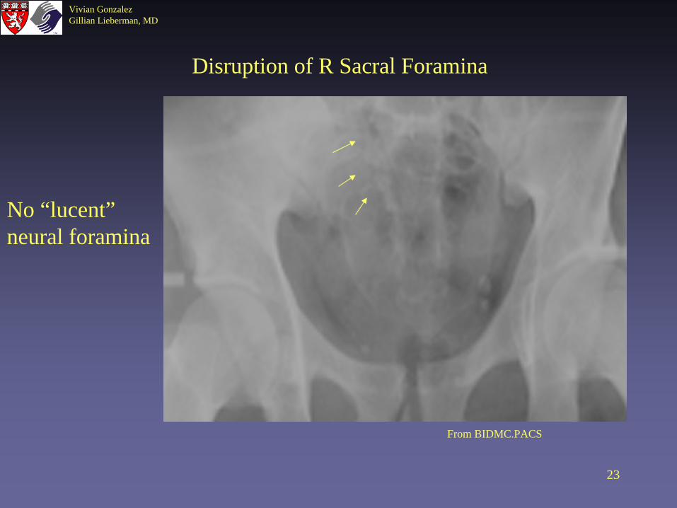
Disruption of R Sacral Foramina
From BIDMC.PACS
No “lucent” neural foramina
24
Vivian GonzalezGillian Lieberman, MD
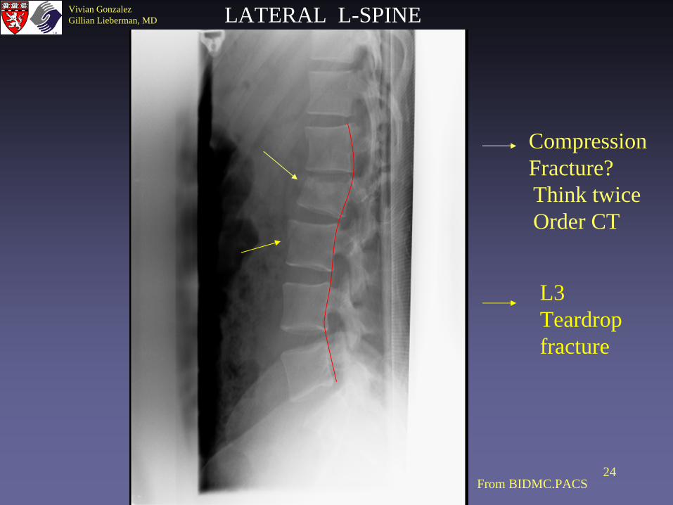
L3 Teardrop fracture
Compression Fracture?Think twiceOrder CT
LATERAL L-SPINE
From BIDMC.PACS

Vivian GonzalezGillian Lieberman, MD
Every compression fracture should be examined closely for
evidence of a retropulsed fragment!!!!!

26
Vivian GonzalezGillian Lieberman, MD
CT Axial 1
BIDMC.PACS

27
Vivian GonzalezGillian Lieberman, MD
CT Axial 2
BIDMC.PACS
28
Vivian GonzalezGillian Lieberman, MD
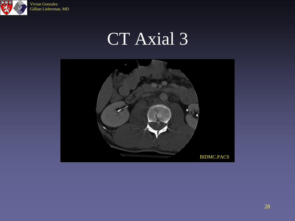
CT Axial 3
BIDMC.PACS

29
Vivian GonzalezGillian Lieberman, MD
CT Axial 4
BIDMC.PACS

30
Vivian GonzalezGillian Lieberman, MD
Coronal CT Reconstruction Showing Burst Fracture
From BIDMC.PACS

31
Vivian GonzalezGillian Lieberman, MD
CT Sagittal Reconstruction
Burst Fracture in L2
Teardrop fracture through theanterior superior endplate of L3

32
Vivian GonzalezGillian Lieberman, MD
MRI- Spine
• Should be used in addition to but not as a replacement of CT
• Better defines extent of injury to soft tissue elements: Muscles, ligaments, intervertebral disc, neural elements
• Cord Compression vs Cord injury• Cord edema vs. hemorrhage• Spinal malalignment

33
Vivian GonzalezGillian Lieberman, MD
MRI T2 Sagittal View
DAVID, Online Atlas of Human Anatomy for Clinical Imaging Diagnosis Developed by J.-C. Oberson MD. Copyright 1998. www.cid.ch/DAVID/LUM2/lum2sl01.html
BIDMC.PACS
Our Patient Normal

34
Vivian GonzalezGillian Lieberman, MD
RADIOLOGIST IMPRESSION: Severe comminuted L2 fracture with retropulsion and compression of the cauda equina and thecal sac with associated epidural hemorrhage causing mild mass effect upon the ventral thecal sac. From BIDMC.PACS
Cord edema would show up as area of high-signal on T2 images- None present in our patient
T2-Weighted MRI
Early stage hemorrhage=low-signal
BIDMC.PACS

35
Vivian GonzalezGillian Lieberman, MD
MRI Anatomy
DAVID, Online Atlas of Human Anatomy for Clinical Imaging Diagnosis Developed by J.-C. Oberson MD. Copyright 1998

36
Vivian GonzalezGillian Lieberman, MD
MRI vs CT
Axial View of L2 Burst Fracture in Our Patient
From BIDMC.PACS
From BIDMC.PACS

37
Vivian GonzalezGillian Lieberman, MD
CT Coronal ReconstructionCan appreciate fracture of posterior elements of L1 which is not evident in axial views
=
From BIDMC.PACS

38
Vivian GonzalezGillian Lieberman, MD
Transverse Process Fracture at L1
nondisplaced fracture through the right transverse process.
Other Spinal Fractures in this Patient
From BIDMC.PACS

39
Vivian GonzalezGillian Lieberman, MD
Fracture of the Posterior Elements of L2
Gross burst fracture, with retropulsion of a large fragment into the central canal, resulting in 75% invasion. This fracture continues posteriorly through the spinous process, in a saggital orientation.
From BIDMC.PACS

40
Vivian GonzalezGillian Lieberman, MD
Retroperitoneal Bleeding on CT
Obscuration of the retroperitoneal fat anterior to the major vessels suggests a retroperitoneal hematoma from the L2 burst fracture.
Observe how the small bowel is displaced anteriorly. There is a large retroperitoneal hematoma measuring approximately 6 x 6 cm at the level of the sacrum, secondary to a complex sacral fracture.
From BIDMC.PACS

41
Vivian GonzalezGillian Lieberman, MD
S/P Vertebrectomy and Recontruction with Internal Fixation
IVC Filter
From BIDMC.PACS

42
Vivian GonzalezGillian Lieberman, MD
Conclusion
• Lumbar fractures need to be characterized as either stable or unstable
• REMEMBER 3-column concept• Radiologist is instrumental in the evaluation
of patients with suspected spine fractures and can guide which imaging studies are performed and in what order.

43
Vivian GonzalezGillian Lieberman, MD
References• Brandser EA, El-Khoury. Thoracic and Lumbar Spine Trauma.The Radiologic Clinics of North
America: Imaging of Orthopedic Trauma 1997; 35: 533-557.• Quencer, RM. The Injured Spinal Cord: Evaluation with Magnetic Resonance and
Intraoperative Sonography.The Radiologic Clinics of North America: Imaging in Neuroradiology,Part II 1988; 26: 1025-1045.
• Wheeless, CR. Wheeless’ Textbook of Orthopedics. http://www.medmedia.com/orthoo/41.htm
Student NameGillian Lieberman, MD

44
Vivian GonzalezGillian Lieberman, MD
Acknowledgements
• Daniel Saurborn, MD• Larry Barbaras and Cara Lyn D’amour,
Webmasters• Gillian Lieberman, MD• Pamela Lepkowski• Virginia Hsu
Student NameGillian Lieberman, MD