Embed Size (px)
Citation preview
1
CHARACTERIZATIONOFBIOMATERIALUSING POWDER X-RAY
DIFFRACTION AND SEM-EDX ANALYSIS
R.SREELATHA1 ,THIRUPPATHI
2, 1Professor,Dept. of Physics
BIST, BIHER, Bharath university,Chennai-73, [email protected],
Bharathiyar Institute of Engineering for Women, Deviyakurichi, Salem District, Tamilnadu, India
Abstract
Threedifferent biomaterialare collected from various patients in the urology department
of Rajah Muthiah Medical College and Hospital, Annamalai University, Tamil Nadu, India.
They are analyzed by powder X-ray diffraction (XRD) and Scanning electron microscopy-
Energy dispersive X-ray spectroscopy (SEM-EDX). The present study has been carried out to
identify the minerals and minor trace elements are present in the urinary stones as well as
establish its morphological structure analyzed and results are discussed.
Keywords: Human urinary stones, spectroscopy, minerals,XRD, SEM-EDX.
PACS Nos.: 87.64.je; 87.64.-t
*Corresponding author, Email: [email protected]
Introduction
Urolithiasis is the third most common urological disease affecting men and women. The
genetic and environmental factors contribute to stone formation [1-9]. Urinary stones affect
most of the population, among industrialized nations, and cost billions of dollars a year in
diagnosis and treatment [10-16]. In addition, this debilitating condition contributes to loss of
profit and productivity in industry, reduces income for families, and places the afflicted
individual in extreme physical pain. The health and age of the patients range from the very
young to the old, with the majority of urinary calculi appearing in Caucasian males [17-21].
International Journal of Pure and Applied MathematicsVolume 119 No. 12 2018, 7085-7095ISSN: 1314-3395 (on-line version)url: http://www.ijpam.euSpecial Issue ijpam.eu
7085
2
Urolithiasis or the condition of formation of kidney stones is fairly a common condition,
affecting about up to 5% of the industrialized population, with the lifetime risk of passing a
kidney stone to be about 8-10% [22-29].Urinary stone is mentioned in different terms as urinary
lithiasis, urolithiasis, kidney stones, renal stones, nephrolithiasis and renal calculus. Renal calculus
refers to calculi formed any where inside the urinary tract between renal tubules, ureter and bladder
[30-37].Analytical methods such as XRD, XRF, PIXE, SEM with EDX and FT-IR can
provide vital information regarding chemical and structural formations of urinary calculi
[8-9]. Qualitative and quantitative estimations of human urinary stones containing calcium
oxalate monohydrate, calcium phosphate, magnesium ammonium phosphate, calcium
carbonate and uric acid can also be tried by FT-IR technique [38-41].
The X-ray diffraction patterns were recorded for human urinary stone samples. In the
present study, the mineral identification was carried out by comparing JCPDS file with XRD
data of the urinary stone samples[42-45]. The presences of calcium oxalate, calcium phosphate,
calcium phosphate hydroxide, magnesium ammonium phosphate and uric acid minerals are
identified.
Scanning electron microscopy with energy dispersive X-ray (SEM-EDX) is a well
established analytical tool for simultaneous determination of the morphological structure and
trace elemental status in human urinary stones. In the present study the morphological variation
of different samples of human urinary stones is evaluated using Scanning Electron Microscopic
technique. The urinary stone samples are analyzed for the percentage of elements like Ca, Cu,
Fe, K, Mg, Na, P, S and Zn. The results are discussed[46-50].
Materials and Methods
International Journal of Pure and Applied Mathematics Special Issue
7086
3
Three human urinary stone samples were collected from Department of urology, Rajah
Muthiah Medical College Hospital (RMMC&H), Annamalai University, Annamalainagar, Tamil
Nadu, South India. The samples were numbered sequentially S1-S3. The stones were washed with
deionised water to remove debris such as blood, mucous and casts and the urinary stones were
kept for further analysis. The oven dried at 60 °C for one hour to remove moisture content and
then the dried stone samples are ground into a fine powder by using an agate mortar. The XRD
patterns of the well powdered samples are characterized by powder X-ray diffraction analysis
using an X-ray diffract meter model Rigaku D/max-2500 with monochromatic Cu-Kα source
(=1.54056 Å) using tube voltage and current of 40 kV and 100mA, respectively. The samples
are scanned over the range (2) 10°-80. The X-ray diffraction of patterns of the urinary stone
samples are obtained at Rigaku D/max-2500 XRD diffractometer available at University of
Madras, Guindy campus, Chennai Tamil Nadu, South India.
The morphology structure and elemental composition of the human urinary stone samples
(S1-S12) are studied in JEOL-JEM-5610 LV Scanning Electron Microscope, equipped with an
energy dispersive X-ray analyzer (OXFORD EDS) available at Centralized Instrumentation and
Service Laboratory (CISL), Department of Physics, Annamalai University, Tamil Nadu, South
India.It is a reliable method for identifying the structure and crystalline composition, and permits
quantification of stone components.Analytical methods such as XRD, XRF, PIXE, SEM with
EDX and FT-IR can provide vital information regarding chemical and structural formations
of urinary calculi . The X-ray diffraction patterns were recorded for human urinary stone
samples. In the present study, the mineral identification was carried out by comparing JCPDS
file with XRD data of the urinary stone samples. The presences of calcium oxalate, calcium
International Journal of Pure and Applied Mathematics Special Issue
7087
4
phosphate, calcium phosphate hydroxide, magnesium ammonium phosphate and uric acid
minerals are identified.
Scanning electron microscopy with energy dispersive X-ray (SEM-EDX) is a well
established analytical tool for simultaneous determination of the morphological structure and
trace elemental status in human urinary stones. In the present study the morphological variation
of different samples of human urinary stones is evaluated using Scanning Electron Microscopic
technique. The urinary stone samples are analyzed for the percentage of elements like Ca, Cu,
Fe, K, Mg, Na, P, S and Zn. The results are discussed.
3. Result and discussion
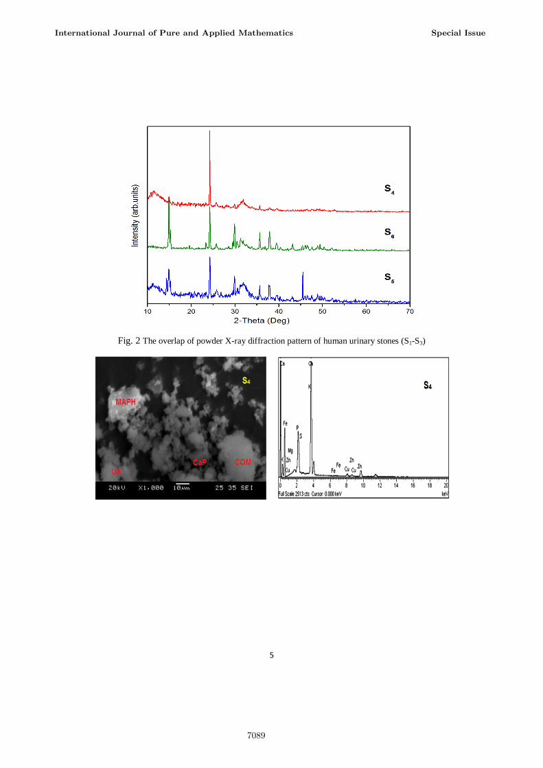
3.1 Powder X-ray diffraction method
Threehuman urinary stone samples were analysed for in this study using powder X-ray
diffraction technique. The stones were analyzed and then categories into four groups according
to their identification of minerals than compared to FT-IR method [10]. The samples were
numbered sequentially from each group S1–S3.In group II (S1–S3), three urinary stone samples
were found to be a mixture of calcium oxalate monohydrate (COM), calcium phosphate (apatite),
magnesium ammonium phosphate (MAPH) and uric acid with the major constituents of apatite
and MAPH and the minor constituents of COM and uric acid. The observed XRD pattern of
results indicate these components mostly contains calcium oxalate, calcium phosphate,
magnesium ammonium phosphate and uric acid (JCPDS-20-0231; 09-0432; 15-0762; 19-1996)
[12, 13] and are known to crystalline in monoclinic, hexagonal, orthorhombic and orthorhombic
system respectively. These results are good agreement with FT-IR [11] and SEM-EDX studies.
International Journal of Pure and Applied Mathematics Special Issue
7088
5
Fig. 2 The overlap of powder X-ray diffraction pattern of human urinary stones (S1-S3)
International Journal of Pure and Applied Mathematics Special Issue
7089
6
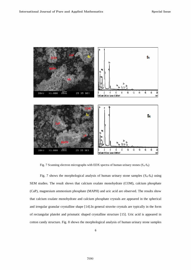
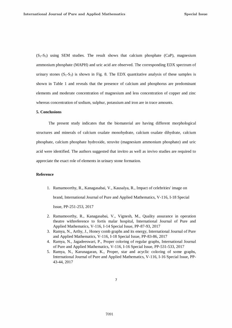
Fig. 7 Scanning electron micrographs with EDX spectra of human urinary stones (S4-S6)
Fig. 7 shows the morphological analysis of human urinary stone samples (S4-S6) using
SEM studies. The result shows that calcium oxalate monohydrate (COM), calcium phosphate
(CaP), magnesium ammonium phosphate (MAPH) and uric acid are observed. The results show
that calcium oxalate monohydrate and calcium phosphate crystals are appeared in the spherical
and irregular granular crystalline shape [14].In general struvite crystals are typically in the form
of rectangular platelet and prismatic shaped crystalline structure [15]. Uric acid is appeared in
cotton candy structure. Fig. 8 shows the morphological analysis of human urinary stone samples
International Journal of Pure and Applied Mathematics Special Issue
7090
7
(S1-S3) using SEM studies. The result shows that calcium phosphate (CaP), magnesium
ammonium phosphate (MAPH) and uric acid are observed. The corresponding EDX spectrum of
urinary stones (S1-S3) is shown in Fig. 8. The EDX quantitative analysis of these samples is
shown in Table 1 and reveals that the presence of calcium and phosphorus are predominant
elements and moderate concentration of magnesium and less concentration of copper and zinc
whereas concentration of sodium, sulphur, potassium and iron are in trace amounts.
5. Conclusions
The present study indicates that the biomaterial are having different morphological
structures and minerals of calcium oxalate monohydrate, calcium oxalate dihydrate, calcium
phosphate, calcium phosphate hydroxide, struvite (magnesium ammonium phosphate) and uric
acid were identified. The authors suggested that invitro as well as invivo studies are required to
appreciate the exact role of elements in urinary stone formation.
Reference
1. Ramamoorthy, R., Kanagasabai, V., Kausalya, R., Impact of celebrities' image on
brand, International Journal of Pure and Applied Mathematics, V-116, I-18 Special
Issue, PP-251-253, 2017
2. Ramamoorthy, R., Kanagasabai, V., Vignesh, M., Quality assurance in operation
theatre withreference to fortis malar hospital, International Journal of Pure and
Applied Mathematics, V-116, I-14 Special Issue, PP-87-93, 2017
3. Ramya, N., Arthy, J., Honey comb graphs and its energy, International Journal of Pure
and Applied Mathematics, V-116, I-18 Special Issue, PP-83-86, 2017
4. Ramya, N., Jagadeeswari, P., Proper coloring of regular graphs, International Journal
of Pure and Applied Mathematics, V-116, I-16 Special Issue, PP-531-533, 2017
5. Ramya, N., Karunagaran, K., Proper, star and acyclic coloring of some graphs,
International Journal of Pure and Applied Mathematics, V-116, I-16 Special Issue, PP-
43-44, 2017
International Journal of Pure and Applied Mathematics Special Issue
7091
8
6. Ramya, N., Muthukumar, M., On coloring of 4-regular graphs, International Journal of
Pure and Applied Mathematics, V-116, I-16 Special Issue, PP-491-494, 2017
7. Ramya, N., Muthukumar, M., On star and acyclic coloring of graphs, International
Journal of Pure and Applied Mathematics, V-116, I-16 Special Issue, PP-467-469,
2017
8. Ramya, N., Pavi, J., Coloring of book and gear graphs, International Journal of Pure
and Applied Mathematics, V-116, I-17 Special Issue, PP-401-402, 2017
9. Ramya, P., Hameed Hussain, J., Alteration framework for integrating quality of
service in internet real-time network, International Journal of Pure and Applied
Mathematics, V-116, I-8 Special Issue, PP-57-61, 2017
10. Ramya, P., Sriram, M., Tweet sarcasm: Peep, International Journal of Pure and
Applied Mathematics, V-116, I-10 Special Issue, PP-231-235, 2017
11. Sabarish, R., Meenakshi, C.M., Comparision of beryllium and CI connecting rod using
ansys, International Journal of Pure and Applied Mathematics, V-116, I-17 Special
Issue, PP-127-132, 2017
12. Sabarish, R., Rakesh, N.L., Outcome of inserts for enhancing the heat exchangers,
International Journal of Pure and Applied Mathematics, V-116, I-17 Special Issue, PP-
419-422, 2017
13. Sangeetha, M., Gokul, N., Aruls, S., Estimator for control logic in high level synthesis,
International Journal of Pure and Applied Mathematics, V-116, I-20 Special Issue, PP-
425-428, 2017
14. Sangeetha, M., Gokul, N., Aruls, S., Image steganography using a curvelet
transformation, International Journal of Pure and Applied Mathematics, V-116, I-20
Special Issue, PP-417-422, 2017
15. Saraswathi, P., Srinivasan, V., Peter, M., Research on financial supply chain from
view of stability, International Journal of Pure and Applied Mathematics, V-116, I-17
Special Issue, PP-211-213, 2017
16. Saravana Kumar, A., Hameed Hussain, J., Expanding the pass percentage in semester
examination, International Journal of Pure and Applied Mathematics, V-116, I-15
Special Issue, PP-45-48, 2017
17. Saravana, S., Arulselvi, S., AdaBoost SVM based brain tumour image segmentation
and classification, International Journal of Pure and Applied Mathematics, V-116, I-20
Special Issue, PP-399-403, 2017
18. Saravana, S., Arulselvi, S., Dynamic power management monitoring and controlling
system using wireless sensor network, International Journal of Pure and Applied
Mathematics, V-116, I-20 Special Issue, PP-405-408, 2017
19. Saravana, S., Arulselvi, S., Clustered morphic algorithm based medical image
analysis, International Journal of Pure and Applied Mathematics, V-116, I-20 Special
Issue, PP-411-415, 2017
20. Saravana, S., Arulselvi, S., Networks, International Journal of Pure and Applied
Mathematics, V-116, I-20 Special Issue, PP-393-396, 2017
International Journal of Pure and Applied Mathematics Special Issue
7092
9
21. Saritha, B., Chockalingam, M.P., Adsorptive removal of heavy metal chromium from
aqueous medium using modified natural adsorbent, International Journal of Civil
Engineering and Technology, V-8, I-8, PP-1382-1387, 2017
22. Saritha, B., Chockalingam, M.P., Adsorptive removal of brilliant green dye by
modified coconut shell adsorbent, International Journal of Pure and Applied
Mathematics, V-116, I-13 Special Issue, PP-211-215, 2017
23. Saritha, B., Chockalingam, M.P., Photodegradation of eriochrome black-T dye from
aqueous medium by photocatalysis, International Journal of Pure and Applied
Mathematics, V-116, I-13 Special Issue, PP-183-187, 2017
24. Saritha, B., Chockalingam, M.P., Photodradation of malachite green DYE using
TIO<inf>2</inf>/activated carbon composite, International Journal of Civil
Engineering and Technology, V-8, I-8, PP-156-163, 2017
25. Saritha, B., Chockalingam, M.P., Synthesis of photocatalytic composite Fe-C/TiO2 for
degradation of malachite green dye from aqueous medium, International Journal of
Pure and Applied Mathematics, V-116, I-13 Special Issue, PP-177-181, 2017
26. Saritha, B., Chockalingam, M.P., Removal of heavy X`X`l from aqueous medium
using modified natural adsorbent, International Journal of Pure and Applied
Mathematics, V-116, I-13 Special Issue, PP-205-210, 2017
27. Saritha, B., Chockalingam, M.P., Degradation of malachite green dye using a
semiconductor composite, International Journal of Pure and Applied Mathematics, V-
116, I-13 Special Issue, PP-195-199, 2017
28. Sartiha, B., Chockalingam, M.P., Photocatalytic decolourisationof textileindustry
wastewaterby TiO2, International Journal of Pure and Applied Mathematics, V-116, I-
18 Special Issue, PP-221-224, 2017
29. Sartiha, B., Chockalingam, M.P., Study on photocatalytic degradation of Crystal
Violet dye using a semiconductor, International Journal of Pure and Applied
Mathematics, V-116, I-18 Special Issue, PP-209-212, 2017
30. Shanthi, E., Nalini, C., Rama, A., The effect of highly-available epistemologies on
hardware and architecture, International Journal of Pharmacy and Technology, V-8, I-
3, PP-17082-17086, 2016
31. Shanthi, E., Nalini, C., Rama, A., Drith: Autonomous,random communication,
International Journal of Pharmacy and Technology, V-8, I-3, PP-17002-17006, 2016
32. Shanthi, E., Nalini, C., Rama, A., A case for replication, International Journal of
Pharmacy and Technology, V-8, I-3, PP-17234-17238, 2016
33. Shanthi, E., Nalini, C., Rama, A., Elve: A methodology for the emulation of robots,
International Journal of Pharmacy and Technology, V-8, I-3, PP-17182-17187, 2016
34. Shanthi, E., Nalini, C., Rama, A., Autonomous epistemologies for 802.11 mesh
networks, International Journal of Pharmacy and Technology, V-8, I-3, PP-17087-
17093, 2016
35. Sharavanan, R., Golden Renjith, R.J., Design and analysis of fuel flow in bend pipes,
International Journal of Pure and Applied Mathematics, V-116, I-15 Special Issue, PP-
59-64, 2017
International Journal of Pure and Applied Mathematics Special Issue
7093
10
36. Sharavanan, R., Jose Ananth Vino, V., Emission analysis of C.I engine run by
diesel,sunflower oil,2 ethyl hexyl nitrate blends, International Journal of Pure and
Applied Mathematics, V-116, I-14 Special Issue, PP-403-408, 2017
37. Sharavanan, R., Sabarish, R., Design of built-in hydraulic jack for light motor
vehicles, International Journal of Pure and Applied Mathematics, V-116, I-17 Special
Issue, PP-457-460, 2017
38. Sharavanan, R., Sabarish, R., Design and fabrication of aqua silencer using charcoal
and lime stone, International Journal of Pure and Applied Mathematics, V-116, I-14
Special Issue, PP-513-516, 2017
39. Sharmila, G., Thooyamani, K.P., Kausalya, R., A schoolwork on customer relationship
management with special reference to domain 2 host, International Journal of Pure and
Applied Mathematics, V-116, I-20 Special Issue, PP-199-203, 2017
40. Sharmila, S., Jeyanthi Rebecca, L., Anbuselvi, S., Kowsalya, E., Kripanand, N.R.,
Tanty, D.S., Choudhary, P., Swathy Priya, L., GC-MS analysis of biofuel extracted
from marine algae, Der Pharmacia Lettre, V-8, I-3, PP-204-214, 2016
41. Sidharth Raj, R.S., Sangeetha, M., Data embedding method using adaptive pixel pair
matching method, International Journal of Pure and Applied Mathematics, V-116, I-15
Special Issue, PP-417-421, 2017
42. Sidharth Raj, R.S., Sangeetha, M., Android based industrial fault monitoring,
International Journal of Pure and Applied Mathematics, V-116, I-15 Special Issue, PP-
423-427, 2017
43. Sidharth Raj, R.S., Sangeetha, M., Mobile robot system control through an brain
computer interface, International Journal of Pure and Applied Mathematics, V-116, I-
15 Special Issue, PP-413-415, 2017
44. Sivaraman, K., Sundarraj, B., Decisive lesion detection in digital fundus image,
International Journal of Pure and Applied Mathematics, V-116, I-10 Special Issue, PP-
161-164, 2017
45. Sridhar, J., Sriram, M., Cloud privacy preserving for dynamic groups, International
Journal of Pure and Applied Mathematics, V-116, I-8 Special Issue, PP-117-120, 2017
International Journal of Pure and Applied Mathematics Special Issue
7094

7095

7096