Embed Size (px)
Citation preview
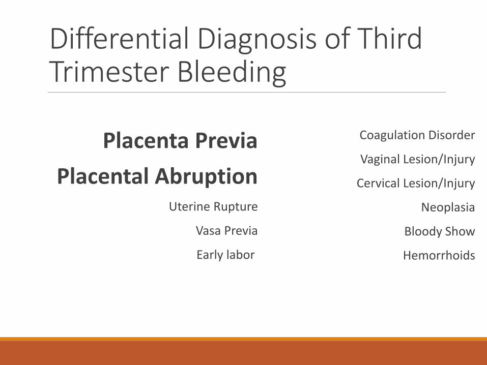
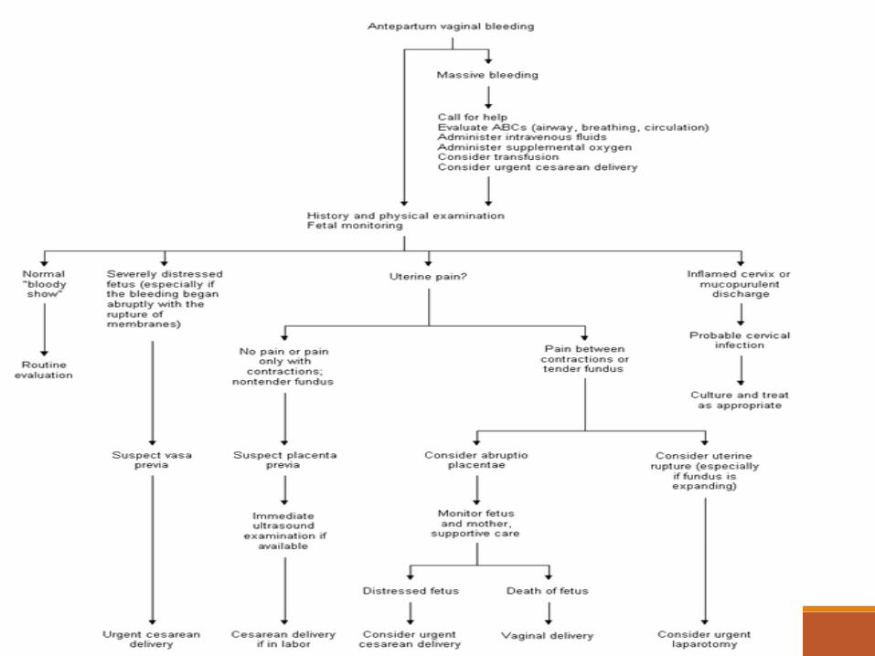
Differential Diagnosis of Third Trimester Bleeding
Placenta Previa
Placental AbruptionUterine Rupture
Vasa Previa
Early labor
Coagulation Disorder
Vaginal Lesion/Injury
Cervical Lesion/Injury
Neoplasia
Bloody Show
Hemorrhoids
Epidemiology of Third Trimester Bleeding
About 3.8 % of third trimester pregnancies ◦placenta previa - 22%; placental abruption - 31%
Serious problem in pregnancy associated with maternal and fetal risks
Require urgent initial assessment and, occasionaly, only partial diagnostic procedures
Oyelese Y, Smulian JC. Placenta Previa, Placenta Accreta, and Vasa Previa. Obstet Gynecol. 2006;107:927-941
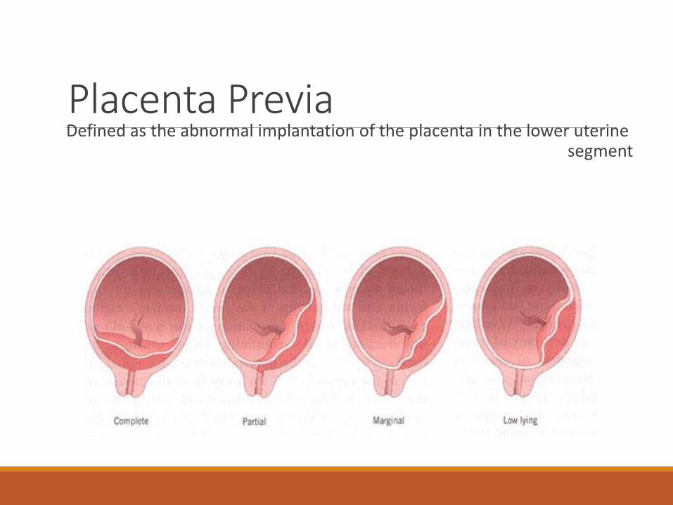
Placenta PreviaDefined as the abnormal implantation of the placenta in the lower uterine
segment
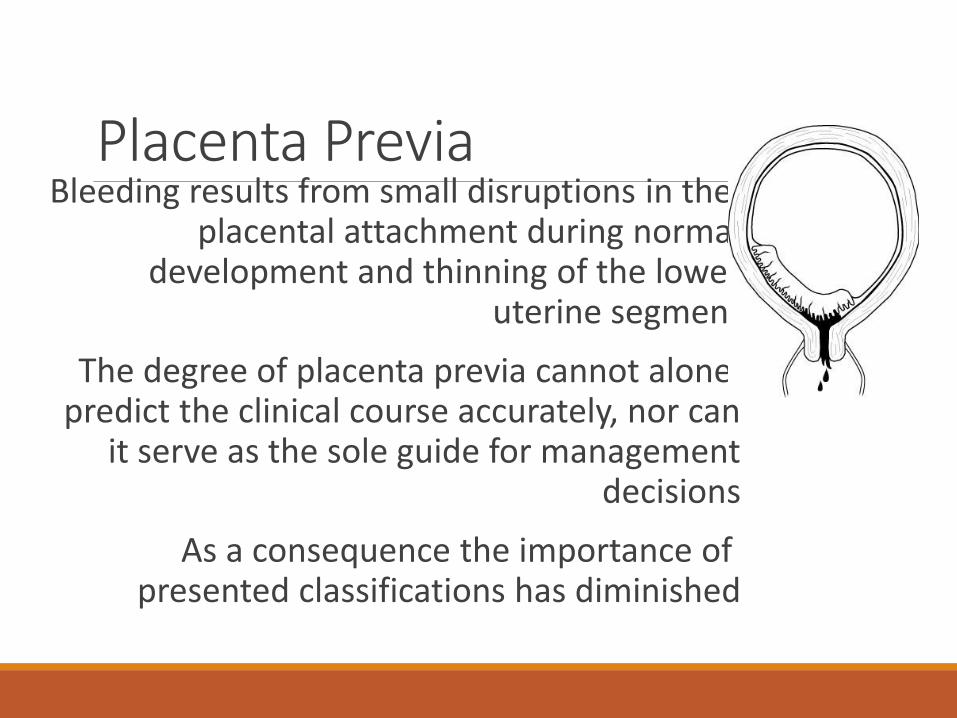
Placenta PreviaBleeding results from small disruptions in the
placental attachment during normal development and thinning of the lower
uterine segment
The degree of placenta previa cannot alone predict the clinical course accurately, nor can
it serve as the sole guide for management decisions
As a consequence the importance of presented classifications has diminished
Placenta previa - Epidemiology4 percent of ultrasound studies performed at 20 to 24
weeks
0,4% at term
The diagnosis of placenta previa is common before the third trimester, but up to 95% resolve before delivery
Placental migration ?
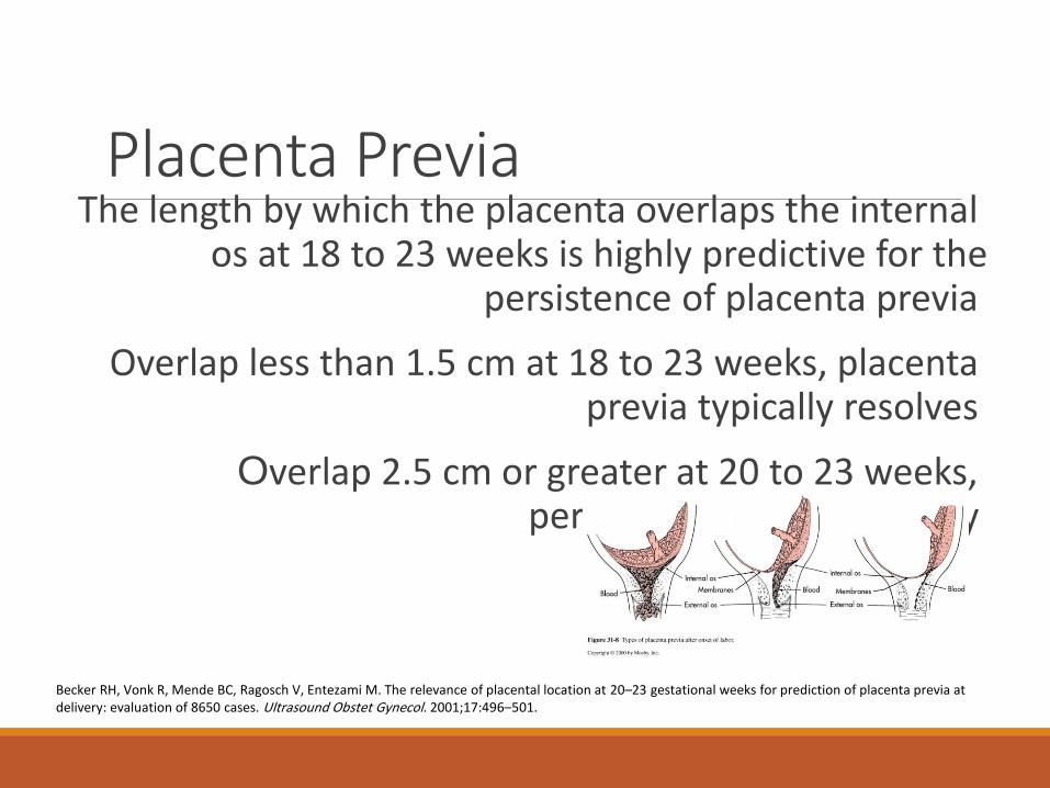
Placenta PreviaThe length by which the placenta overlaps the internal
os at 18 to 23 weeks is highly predictive for the persistence of placenta previa
Overlap less than 1.5 cm at 18 to 23 weeks, placenta previa typically resolves
Overlap 2.5 cm or greater at 20 to 23 weeks, persistence to term is likely
Becker RH, Vonk R, Mende BC, Ragosch V, Entezami M. The relevance of placental location at 20–23 gestational weeks for prediction of placenta previa at delivery: evaluation of 8650 cases. Ultrasound Obstet Gynecol. 2001;17:496–501.
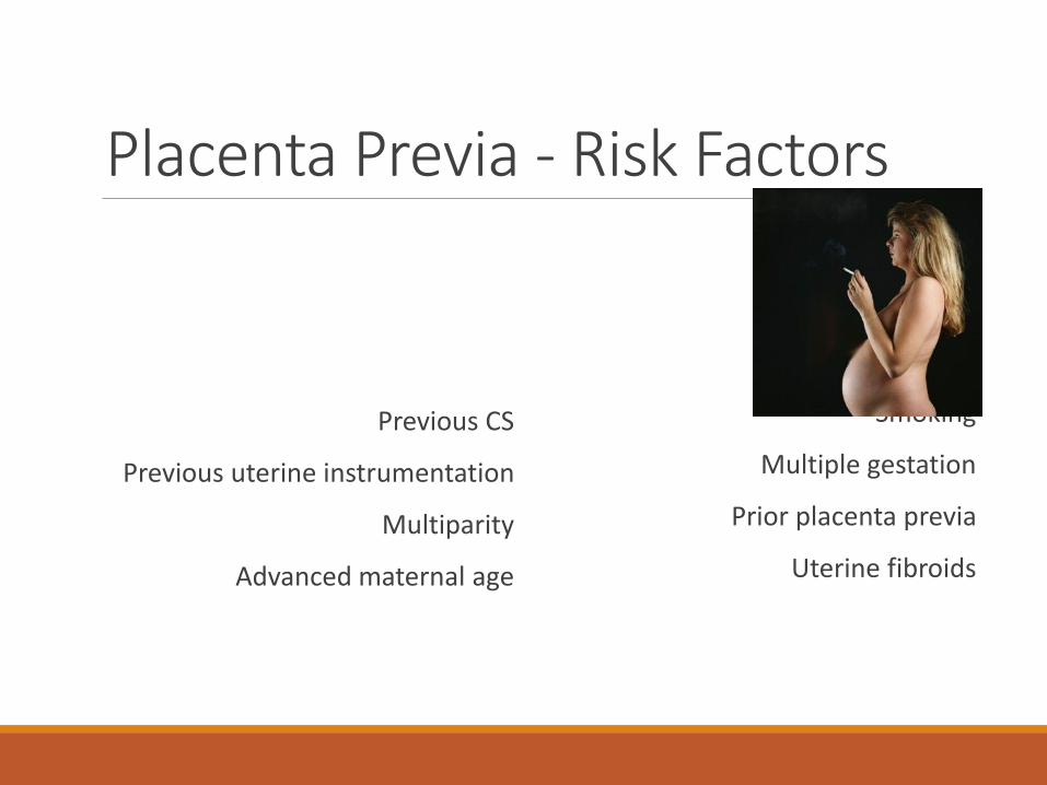
Placenta Previa - Risk Factors
Previous CS
Previous uterine instrumentation
Multiparity
Advanced maternal age
Smoking
Multiple gestation
Prior placenta previa
Uterine fibroids

Placenta Previa - Risk Factors 4.8‰
Risk of recurrent placenta previa is 4% to 8%
Risk of placenta previa increases with the number of prior cesarean sections, rising to 10% with four or more
For woman older than 40 years risk is 2%
Placenta Previa - Clinical presentation
Episode of bleeding has a peak incidence at about the 34th week of pregnancy
One-third of cases become symptomatic before the 30th week and one-third after the 36th week
Approximately 10% of cases, bleeding begins only with the onset of labor
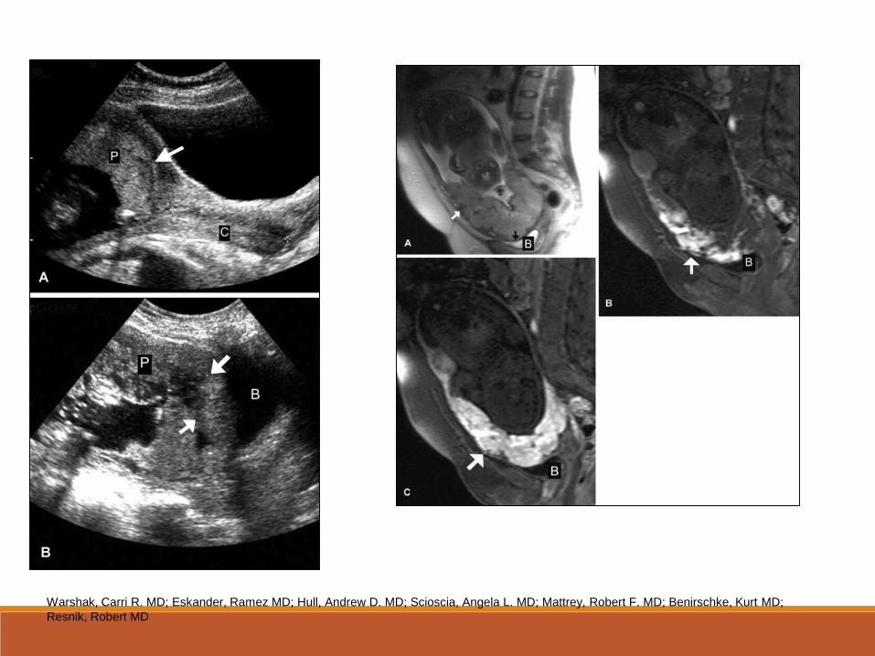
Placenta Previa - DiagnosisTransabdominal sonography
Transvaginal sonography
Translabial sonography
Magnetic resonance imaging
Warshak, Carri R. MD; Eskander, Ramez MD; Hull, Andrew D. MD; Scioscia, Angela L. MD; Mattrey, Robert F. MD; Benirschke, Kurt MD;
Resnik, Robert MD
Placenta Previa - Morbidity and Mortality
Placenta Previa is rarely a cause of life-threatening maternal hemorrhage unless instrumentation or digital exam is performed
The most common morbidity with this problem is the necessity for operative delivery and the risks associated with surgical
intervention
Perinatal morbidity and mortality are primarily related to the complications of prematurity, because the hemorrhage is
maternal.
Placenta Previa - Morbidity and MortalityReduction in both maternal and perinatal mortality rates over the past
40 years
Expectant management approach and the liberal use of cesarean section rather than vaginal delivery
Maternal mortality rate has fallen from between 25% and 30% to less than 1%.
Total perinatal mortality rate has fallen from between 60% and 70% to under 10%
Placenta Previa - Morbidity and MortalityGoal is to obtain the maximum fetal maturation possible while
minimizing the risk to both the fetus and the mother
In a significant proportion of cases delivery may be safely delayed to a more advanced stage of maturity
Placenta Previa - ManagementIt is reasonable to hospitalize women in the situation of acute bleeding
episode or uterine contractions
Women who present with bleeding in the second half of pregnancy should have a sonographic examination for placental location prior to
any attempt to perform a digital examination
Placenta Previa - ManagementWide-bore intravenous cannulas
Blood count and type and screen
At least 4 units of compatible packed red blood cells and coagulation factors at short notice
Rh immune globulin to Rh-negative women
Kleihauer-Bettke test for quantification of fetal-maternal transfusion in Rh-negative women
Placenta Previa - ManagementSteroids should be administered in women between 24 and 34 weeks of
gestation
Before 32 weeks of gestation, with no maternal or fetal compromise blood transfusions should be considered
Tocolysis ?
Cerclage ?
Placenta Previa - ManagementWhen the patient has had no further bleeding for 48 hours, she may be
considered for discharge
Women who are stable and asymptomatic, and who are reliable and have quick access to hospital, may be considered for outpatient
management
Placenta Previa - DeliveryCesarean delivery at 36-37 weeks of gestation - documentation of fetal
lung maturity by amniocentesis
Placental edge is 2 cm or more from the internal os at term - good chances to deliver vaginally
Regional anesthesia - less blood loss and requirements for blood transfusion
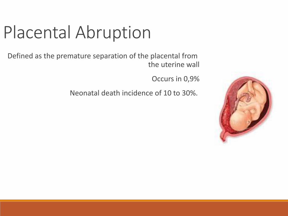
Placental AbruptionDefined as the premature separation of the placental from
the uterine wall
Occurs in 0,9%
Neonatal death incidence of 10 to 30%.
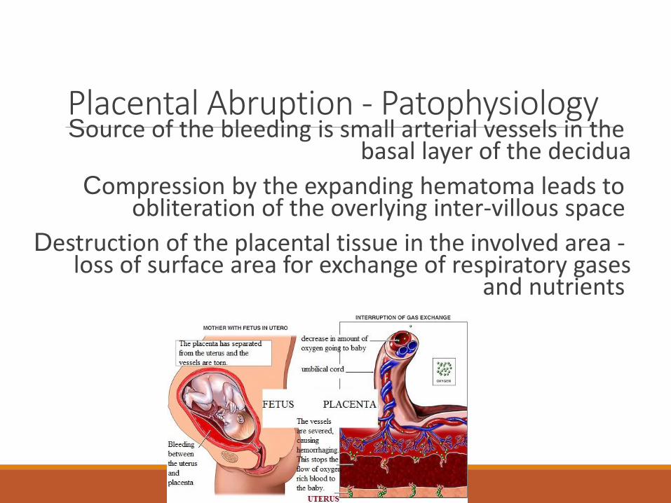
Placental Abruption - PatophysiologySource of the bleeding is small arterial vessels in the
basal layer of the decidua
Compression by the expanding hematoma leads to obliteration of the overlying inter-villous space
Destruction of the placental tissue in the involved area -loss of surface area for exchange of respiratory gases
and nutrients
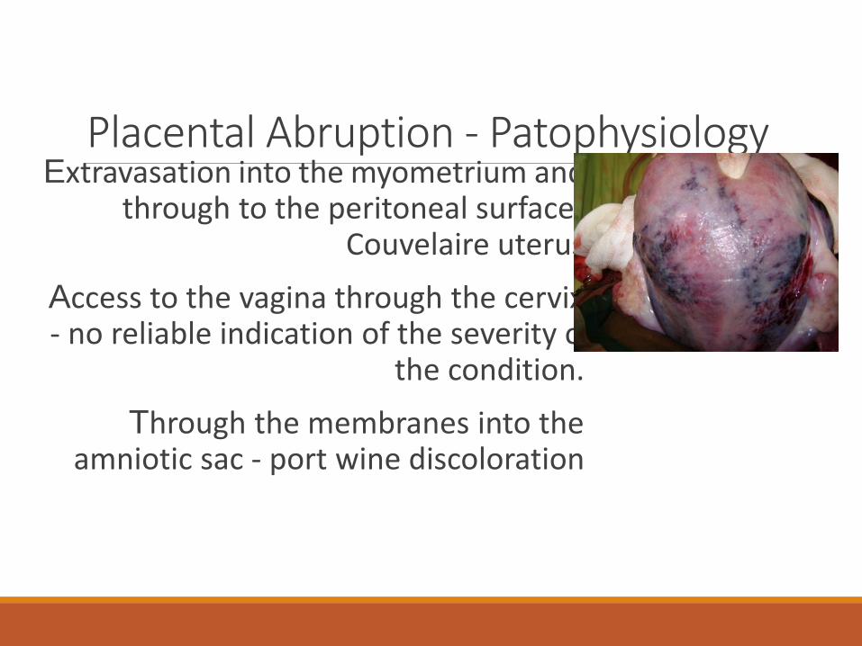
Placental Abruption - PatophysiologyExtravasation into the myometrium and
through to the peritoneal surface -Couvelaire uterus
Access to the vagina through the cervix - no reliable indication of the severity of
the condition.
Through the membranes into the amniotic sac - port wine discoloration
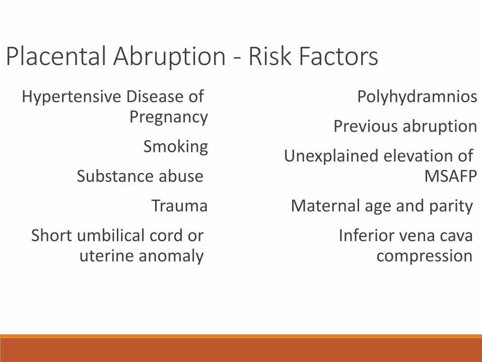
Placental Abruption - Risk Factors
Hypertensive Disease of Pregnancy
Smoking
Substance abuse
Trauma
Short umbilical cord or uterine anomaly
Polyhydramnios
Previous abruption
Unexplained elevation of MSAFP
Maternal age and parity
Inferior vena cava compression
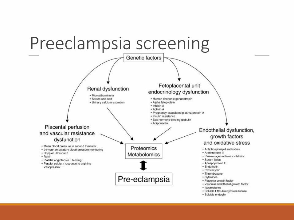
Preeclampsia screening
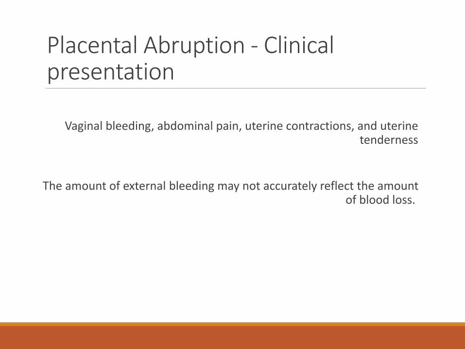
Placental Abruption - Clinical presentation
Vaginal bleeding, abdominal pain, uterine contractions, and uterine tenderness
The amount of external bleeding may not accurately reflect the amount of blood loss.
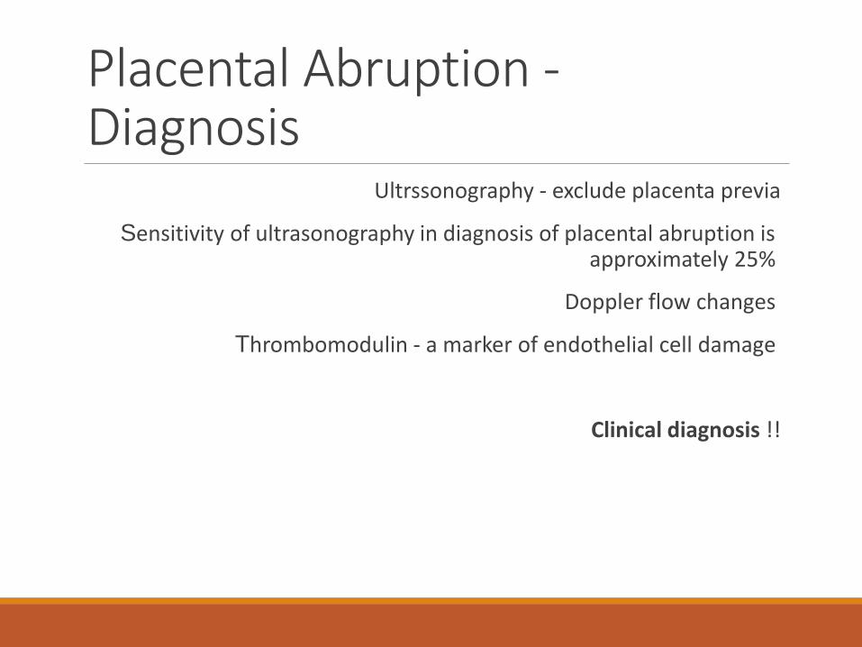
Placental Abruption -Diagnosis
Ultrssonography - exclude placenta previa
Sensitivity of ultrasonography in diagnosis of placental abruption is approximately 25%
Doppler flow changes
Thrombomodulin - a marker of endothelial cell damage
Clinical diagnosis !!
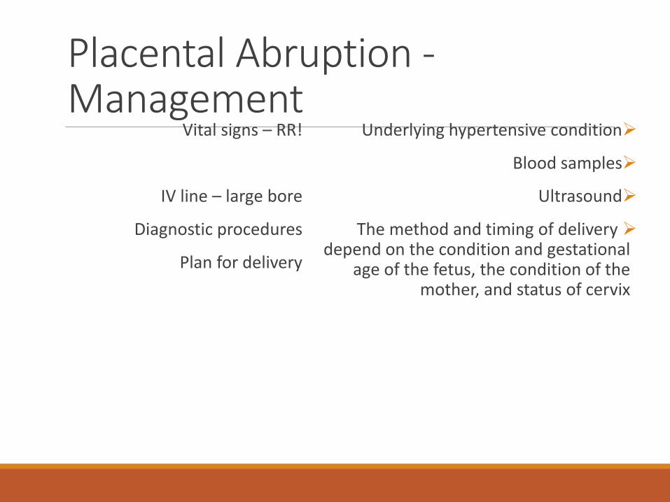
Placental Abruption -Management
Vital signs – RR!
IV line – large bore
Diagnostic procedures
Plan for delivery
Underlying hypertensive condition
Blood samples
Ultrasound
The method and timing of delivery depend on the condition and gestational
age of the fetus, the condition of the mother, and status of cervix
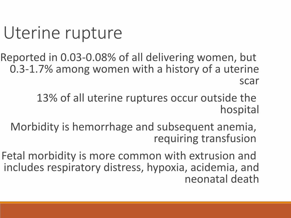
Uterine ruptureReported in 0.03-0.08% of all delivering women, but
0.3-1.7% among women with a history of a uterine scar
13% of all uterine ruptures occur outside the hospital
Morbidity is hemorrhage and subsequent anemia, requiring transfusion
Fetal morbidity is more common with extrusion and includes respiratory distress, hypoxia, acidemia, and
neonatal death
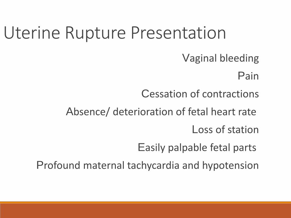
Uterine Rupture PresentationVaginal bleeding
Pain
Cessation of contractions
Absence/ deterioration of fetal heart rate
Loss of station
Easily palpable fetal parts
Profound maternal tachycardia and hypotension
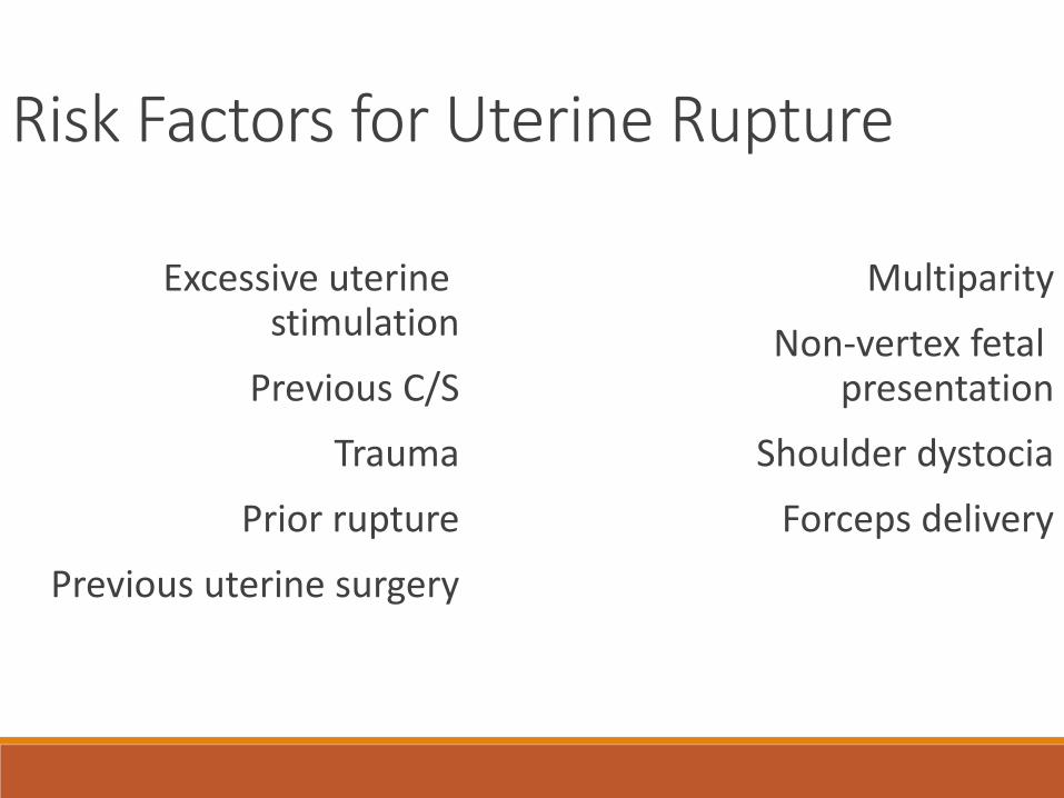
Risk Factors for Uterine Rupture
Excessive uterine stimulation
Previous C/S
Trauma
Prior rupture
Previous uterine surgery
Multiparity
Non-vertex fetal presentation
Shoulder dystocia
Forceps delivery
Uterine Rupture ManagementIn the case of sudden change in fetal baseline heart rate
or the onset of severe decelerations, the provider should initiate intrauterine resuscitation with maternal
position change, IVF hydration, discontinuation of oxitocin, O2 administration by re-breather mask
If the measures are ineffective, emergent laparotomy is indicated
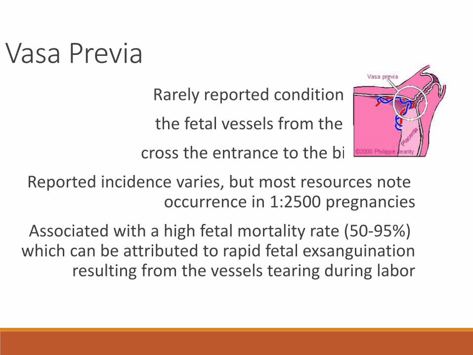
Vasa PreviaRarely reported condition in which
the fetal vessels from the placenta
cross the entrance to the birth canal
Reported incidence varies, but most resources note occurrence in 1:2500 pregnancies
Associated with a high fetal mortality rate (50-95%) which can be attributed to rapid fetal exsanguination
resulting from the vessels tearing during labor
Risk Factors for Vasa Previa
Bilobed and succenturiate placentas
Velamentous insertion of the cord
Low-lying placenta and/or placenta previa
Multiple gestation
Pregnancies resulting from in vitro fertilization
Palpable vessel on vaginal exam
Maternal history of uterine surgery
Vasa Previa - ManagementWhen vasa previa is detected prior to labor, the baby
has a much greater chance of surviving
It can be detected during pregnancy with use of transvaginal sonography, preferably in combination with
color Doppler
Some researchers have suggested screening color Doppler in the second trimesters of patients with risk
factors present on routine 20 week ultrasound
Vasa Previa - ManagementWhen vasa previa is diagnosed prior to labor, elective
caesarian delivery can save the baby’s life
The International Vasa Previa Foundation recommends hospitalization in the third trimester, delivery by 35
weeks, and immediate blood transfusion of the infant in the event of a rupture
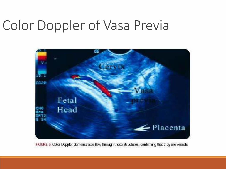
Color Doppler of Vasa Previa

Vasa Previa - Diagnosis in the Acute Setting
Clinical scenarios suggesting vasa previa:
-significant bleeding at the time of membrane rupture
-fetal heart rate abnormalities associated with vaginalbleeding
-palpable vessels on vaginal examination
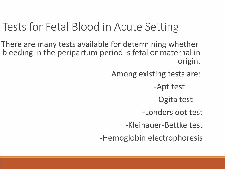
Tests for Fetal Blood in Acute Setting
There are many tests available for determining whether bleeding in the peripartum period is fetal or maternal in
origin.
Among existing tests are:
-Apt test
-Ogita test
-Londersloot test
-Kleihauer-Bettke test
-Hemoglobin electrophoresis
Take-home messageDevelopment of clinical guidelines and
protocols designed to provide early diagnosis
of patients at risk for major obstetric hemorrhage
and efficient care in emergency situations
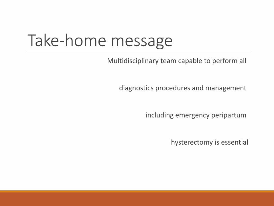
Take-home messageMultidisciplinary team capable to perform all
diagnostics procedures and management
including emergency peripartum
hysterectomy is essential