Embed Size (px)
Citation preview
8th Edition APGO Objectives for Medical Students
Third Trimester Bleeding
Rationale
Bleeding in the third trimester requires immediate patient evaluation. Thoughtful, prompt evaluation and management is necessary to reduce the threat to the lives of the mother and fetus.
ObjectivesThe student will be able to: Describe the approach to the patient with third-trimester
bleeding Compare symptoms, physical findings and diagnostic
methods that differentiate patients with placenta previa, abruptio placenta and other causes of third-trimester bleeding
Describe complications of placenta previa and abruptio placenta
Describe immediate management of shock secondary to third-trimester bleeding
Describe components of the various blood products and indications for their use
Placental abruption
Definition separation of placenta from uterine wall Incidence 1% of all pregnancies 20% of all third-trimester bleeders Recurrence risk
10% in first pregnancy 25% in second pregnancy
Placental abruption
Risk factors Chronic hypertension Cocaine Abdominal trauma Smoking Uterine decompression Prolonged PROM High parity Prior abruption
Placental abruption
Symptoms Vaginal bleeding Abdominal or back pain
Placental abruption
Physical findings Uterine contractions Hypertonus Tetany Vaginal bleeding Concealed hemorrhage Fetal distress
Placental abruption
Laboratory findings Anemia - may be out of proportion to
observed blood loss DIC in 10% (30% if severe abruption)
First, increase in FSP Followed by decrease in fibrinogen
Placental abruption
Diagnosis Clinical scenario Ultrasound - not good, except to rule out
placenta previa
Placental abruption
Treatment Continuous electronic fetal monitoring Ultrasound to confirm viability, gestational
age, no previa, fetal lie Expectant care and vaginal delivery, if
fetal tracing reassuring Available anesthesia, OR team for stat
cesarean delivery
Placenta previa
Definition
placental tissue covers cervical os
Placenta previa
Types Central/total - complete covers os Partial - partially covers internal os Marginal - placental edge at margin of
internal os Lateral/low-lying - placenta approaches
os
Placenta previa
Incidence ~ 1:20 at 24 wk. 1:200 at 40 wk.
Nulliparous - 0.2% Multiparous - 0.5%
Placenta previa
Risk factors Prior cesarean delivery/myomectomy Previous abortion Increased parity Multifetal gestation Advanced maternal age Abnormal presentation Smoking
Placenta previa
Symptom - painless vaginal bleeding Spontaneous After coitus
Placenta previa
Physical findings Abnormal lie If significant bleeding:
Tachycardia Postural hypertension
Placenta previa
Diagnosis
ultrasound
Placenta previa
Management Admit to hospital Observe in L&D IV access Electronic fetal monitoring
Placenta previa
Management (con’t) Less than 36 wk gestation - expectant
management Bed rest No vaginal exams Steroids for lung maturation
Greater than 36 wk gestation - schedule cesarean delivery with hysterectomy backup
Vasa previa
Definition
In cases of velamentous cord insertion fetal vessels cover cervical os
Vasa previa
Incidence0.1-1.0% Singleton - 0.2% Twins - 6-11% Triplets - 95%
Vasa previa
DiagnosisPainless vaginal bleeding Fetal bleeding
Positive Kleihauer Betke test Positive APT test
Vasa previa
Treatment
Emergent cesarean delivery
Other causes of third-trimester bleeding
Cervicitis Cervical erosion Trauma Cervical cancer Foreign body Bloody show
Perinatal mortality and morbidity
Mortality Previa - 40% Abruption - 0.4%; if severe abruption,
approaches 50% Vasa previa - about 50%
Perinatal mortality and morbidity
Morbidity Prematurity Fetal asphyxia Exsanguination (rare) Congenital malformations
Maternal mortality and morbidity
Mortality - rare
Morbidity Anemia Hemorrhage Shock DIC (coagulopathies) Couvelaire uterus Placenta accreta
Management of shock
Stabilize mother Large-bore IV x 2 Place patient in Trendelenburg position Crossmatch for 4 units of blood Rapidly infuse 5% dextrose in lactated
Ringer’s
Monitor urine output
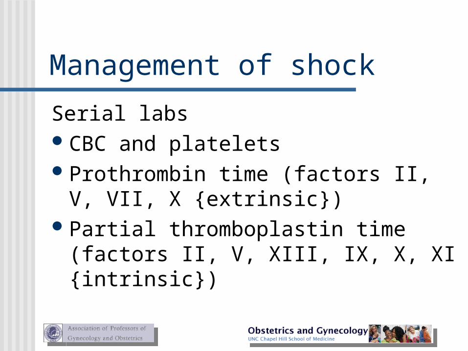
Management of shock
Serial labsCBC and platelets Prothrombin time (factors II, V, VII, X
{extrinsic}) Partial thromboplastin time (factors II, V,
XIII, IX, X, XI {intrinsic})
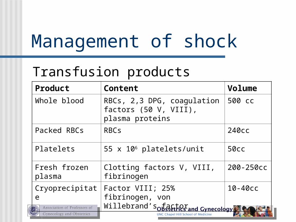
Management of shock
Transfusion products Product Content Volume
Whole blood RBCs, 2,3 DPG, coagulation factors (50 V, VIII), plasma proteins
500 cc
Packed RBCs RBCs 240cc
Platelets 55 x 106 platelets/unit 50cc
Fresh frozen plasma
Clotting factors V, VIII, fibrinogen 200-250cc
Cryoprecipitate Factor VIII; 25% fibrinogen, von Willebrand’s factor
10-40cc
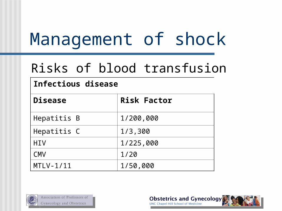
Management of shock
Risks of blood transfusionInfectious disease
Disease Risk Factor
Hepatitis B 1/200,000
Hepatitis C 1/3,300
HIV 1/225,000
CMV 1/20
MTLV-1/11 1/50,000
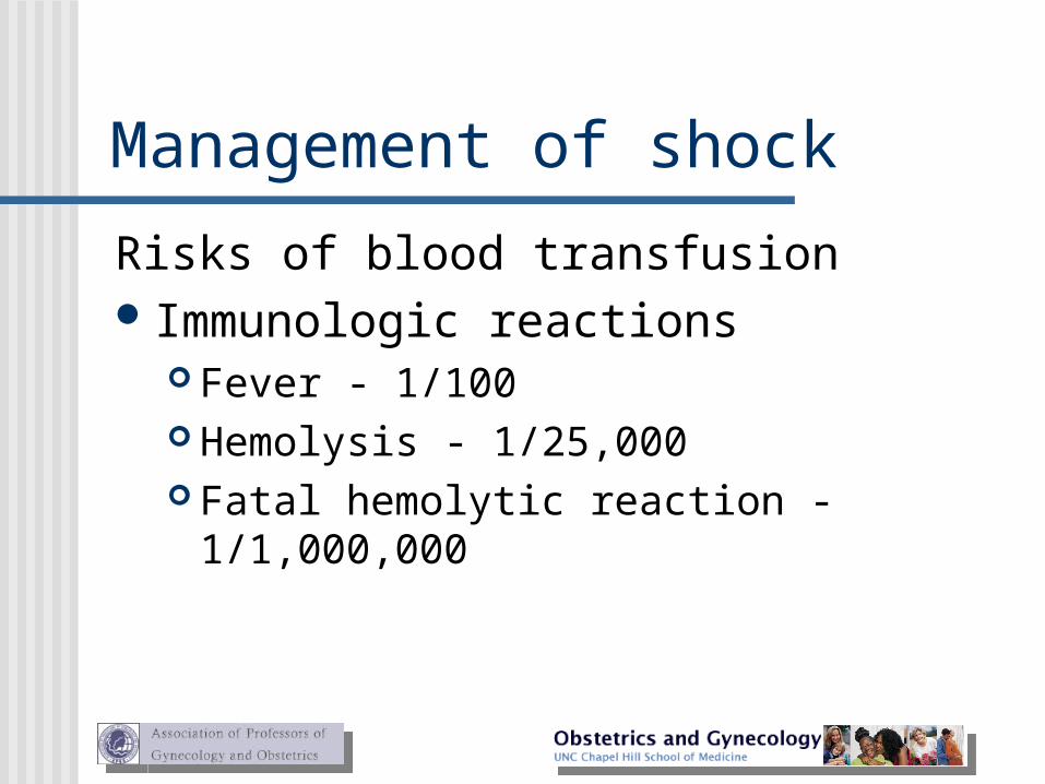
Management of shock
Risks of blood transfusion Immunologic reactions
Fever - 1/100 Hemolysis - 1/25,000 Fatal hemolytic reaction - 1/1,000,000
Management of shock
DeliveryVaginally unless other obstetrical
indication, i.e. fetal distress, herpes, etc. Best to stabilize mother before initiating
labor or going to delivery
References Baron F, Hill WC. “Placenta previa, placenta abruption”, Clinical
Obstetrics and Gynecology, Sep 1998 41(3) pp527-532. Benedetti T. Obstetric hemorrhage, in obstetrics: normal and
problem pregnancies, Gabbe S, Niebyl J, Simpson J, 3rd ed. New York: Churchill Livingston 1996, pp161-184.
Hertzberg B. “Ultrasound evaluation of third trimester bleeding,” The Radiologist, July 1997 4(4) pp227-234.
Sheiner E, Shohan-Vardi I. “Placenta previa: obstetric risk factors and pregnancy outcome,” Journal of Maternal-Fetal Medicine, December 2001 10(6) pp414-418.
Adapted from Association of Professors of Gynecology and Obstetrics Medical Student Educational Objectives, 7th edition, copyright 1997.
Clinical Case
Third Trimester Bleeding
ObjectivesAt the conclusion of this exercise, the student will be able
to1. Describe the approach to the patient with third
trimester bleeding2. Compare symptoms, physical findings and diagnostic
methods to differentiate between placenta previa, placental abruption and other causes of 3rd trimester bleeding
3. Describe immediate management of shock due to 3rd trimester bleeding
4. Understand complications of placental abruption and placenta previa
Patient presentationJoanne is a 25-year-old G2P1 female at 32 weeks gestation.
About an hour before arrival in labor and delivery, she was watching television when she noted a sudden gush of bright red blood vaginally. The bleeding was heavy and soaked through her clothes, though it has decreased since then. She denies any cramps or abdominal pain. She says that her last sexual intercourse was a week ago. A review of her prenatal chart finds nothing remarkable other than a borderline high blood pressure from her first prenatal visit that has not required medication. There is no mention of bleeding prior to this episode. She had an ultrasound to confirm pregnancy at 14 weeks, but none since.
Patient presentationPhysical examination reveals a very anxious woman whose
blood pressure is 138/90, pulse 90, respirations 22, temperature 99 F. Her abdomen is soft without guarding or rebound to palpation, and the uterus is nontender and firm, but not rigid. Fundal height is 33cm. Fetal heart tones are in the 140s with good variability. The external monitor reveals uterine irritability, but no discrete contractions are seen. There is a small amount of dark blood on her underwear, but no active bleeding is seen. Vaginal examination is deferred pending an ultrasound evaluation of placental location.
Patient presentationElectrolytes, liver enzymes and coagulation profile
are all within normal range. CBC reveals a hemoglobin of 8.0 gm/dl and a hematocrit of 24%; MCV, MCH and MCHC are all low normal. White blood cell and platelet counts are normal. Type and Rh confirm A negative blood type with negative antibody titer. Kleihauer-Betke test is negative for fetal blood.
Patient presentationAn ultrasound examination shows a singleton
fetus in cephalic position. Biparietal diameter, head circumference and femur length are all consistent with gestational age. No gross anomalies are seen. Amniotic fluid volume is normal. The placenta is low lying and extends to, but does not appear to cover, the cervical os.
Diagnosis
Third trimester bleeding with low-lying placenta
Assessment/plan
After being observed for several hours in labor and delivery, Joanne is admitted to the antepartum floor for further observation. She is placed at strict bed rest with fetal heart monitoring each nursing shift. She is instructed to report any further bleeding. Blood has been typed and crossed for 4 units and a repeat CBC ordered for the morning. Nursing orders are to notify you of any orthostatic changes or complaints of dizziness or weakness.
Assessment/plan
After 24 hours, there has been no further bleeding and Joanne’s vital signs have remained normal. She is discharged to home on iron replacement, to follow-up at the next scheduled appointment next week. She is advised to avoid intercourse or strenuous physical activities, though she may to continue to do her regular work as an accountant.
Teaching points There are several causes of third trimester bleeding. Placental
abruption describes separation of the placenta from the uterine wall. It occurs in about 20% of all third trimester bleeders and has a 25% recurrence risk in a subsequent pregnancy. Risk factors for placental abruption include chronic hypertension, cocaine use, abdominal trauma, sudden uterine compression (as with rupture of membranes), and high parity. Physical findings include frequent uterine contractions or hypertonicity, vaginal bleeding (sometimes catastrophic), and fetal distress. Disseminated intravascular coagulation occurs in 10% of cases, in 30% if the bleeding is severe. If the fetal heart tracing is reassuring, expectant management and vaginal delivery may be considered. If there are signs of maternal or fetal deterioration, an immediate cesarean delivery is required. Perinatal mortality approaches 50% in severe cases.
Teaching points Placenta previa occurs when placental tissue covers the cervical os. A
central or total placenta previa covers the os completely; as its name implies, a partial placenta previa partially covers the os. In a marginal previa, the placental edge is at the margin of the internal os while, with a low-lying placenta, the placenta approaches the os, but is not at its edge. At 24 weeks, about 1 pregnancy in 20 will demonstrate ultrasound evidence of a placenta previa, while, at 40 weeks, the incidence decreases to 1 in 200. Risk factors include prior cesarean delivery, history of myomectomy, previous abortion, increased parity, multiple gestation, advanced maternal age and smoking. Bleeding is usually painless and may occur after intercourse. Management includes observation in labor and delivery, IV access, continuous fetal monitoring and steroids for fetal lung maturation if needed. Cesarean delivery is the method of choice with hysterectomy backup if intraoperative bleeding cannot be controlled. Perinatal mortality can read 40%
Teaching points Vasa previa is a rare condition where the fetal
vessels of a velamentous cord insertion cover the cervical os. The incidence is less than 1% of all pregnancies, though it is increased in multiple gestations: up to 11% in twins and up to 95% in triplets. The diagnosis is suggested by painless vaginal bleeding in the absence of evidence of placenta previa or abruption. Treatment is delivery by cesarean section.
Teaching points Shock from blood loss is a very real possibility in 3rd
trimester bleeding from any cause. Management includes 2 large bore IVs, crossmatch for 4 or more units of blood; 5% dextrose in lactated ringers or normal saline should be infused rapidly. Urine output must be monitored and is a gauge of renal failure from hypoperfusion. Serial CBC and platelet counts, prothrombin time and partial thromboplastin time are all followed. Transfusion products are given as needed: whole blood, packed red blood cells, platelets, fresh frozen plasma and cryoprecipitate.