Embed Size (px)
Citation preview

Thermophilic Microbial Electrochemical Cells
by
Bradley Lusk
A Dissertation Presented in Partial Fulfillment
of the Requirements for the Degree
Doctor of Philosophy
Approved November 30, 2015 by the
Graduate Supervisory Committee:
César I. Torres, Chair
Bruce E. Rittmann
Rosa Krajmalnik-Brown
ARIZONA STATE UNIVERSITY
December 2015
ii
ABSTRACT
Microbial Electrochemical Cell (MXC) technology harnesses the power stored in
wastewater by using anode respiring bacteria (ARB) as a biofilm catalyst to convert the
energy stored in waste into hydrogen or electricity. ARB, or exoelectrogens, are able to
convert the chemical energy stored in wastes into electrical energy by transporting
electrons extracellularly and then transferring them to an electrode. If MXC technology is
to be feasible for ‘real world’ applications, it is essential that diverse ARB are discovered
and their unique physiologies elucidated- ones which are capable of consuming a broad
spectrum of wastes from different contaminated water sources.
This dissertation examines the use of Gram-positive thermophilic (60 ◦C) ARB in
MXCs since very little is known regarding the behavior of these microorganisms in this
setting. Here, we begin with the draft sequence of the Thermincola ferriacetica genome
and reveal the presence of 35 multiheme c-type cytochromes. In addition, we employ
electrochemical techniques including cyclic voltammetry (CV) and chronoamperometry
(CA) to gain insight into the presence of multiple pathways for extracellular electron
transport (EET) and current production (j) limitations in T. ferriacetica biofilms.
Next, Thermoanaerobacter pseudethanolicus, a fermentative ARB, is investigated
for its ability to ferment pentose and hexose sugars prior to using its fermentation
products, including acetate and lactate, for current production in an MXC. Using CA,
current production is tracked over time with the generation and consumption of
fermentation products. Using CV, the midpoint potential (EKA) of the T. pseudethanolicus
EET pathway is revealed.

iii
Lastly, a cellulolytic microbial consortium was employed for the purpose
ofassessing the feasibility of using thermophilic MXCs for the conversion of solid waste
into current production. Here, a highly enriched consortium of bacteria, predominately
from the Firmicutes phylum, is capable of generating current from solid cellulosic
materials.

iv
TABLE OF CONTENTS
Page
LIST OF TABLES…………………………………………………………………...… viii
LIST OF FIGURES……………………………………………………………………… ix
CHAPTER
1 INTRODUCTION TO MICROBIAL ELECTROCHEMICAL CELLS…….. 1
Overview………………………………………………………………..… 1
Biological Principles of Gram-Positive Thermophilic Bacteria………….. 9
Biological Principles of Thermincola ferriacetica………...………......... 15
Biological Principles of Thermonanaerobacter pseudethanolicus…........ 17
Biological Principles of Cellulose Fermentation………..………………. 19
Thermodynamic Principles of Thermophilic Microbial Electrochemical
Cells……………………………………………………………………... 20
References……………………………………………………………….. 29
2 DRAFT GENOME OF THE GRAM-POSITIVE THERMOPHILIC IRON
REDUCER THERMINCOLA FERRIACETICA STRAIN Z-0001 T………... 40
Overview……………………………………………………………...… 40
Results……...………………………………………………………........ 41
Nucleotide Accession Number................................................................. 43
References……………………………………………………………….. 46

v
CHAPTER Page
3 THE EFFECT OF PH AND BUFFER CONCENTRATION ON ANODE
BIOFILMS OF THERMINCOLA FERRIACETICA……………………...... 48
Overview………………………………………………………………… 48
Introduction……………………………………………………………… 49
Materials and Methods…………………………………………………... 51
Results and Discussion………………………………………………….. 55
Conclusion………………………………………………………….........66
References…………………………………………………………..........67
4 PH SHIFTS IN THE ANODE POTENTIAL RESPONSE OF
THERMINCOLA FERRIACETICA SUGGEST THE PRESENCE OF A
RATE LIMITING PROTON-COUPLED ELECTRON TRANSFER
PROTEIN …………………………………………………...………………. 70
Overview………………………………………………………………… 70
Introduction……………………………………………………………… 71
Materials and Methods…………………………………………………... 73
Results and Discussion………………………………………………….. 75
Conclusion…………………………………………………………......... 87
References………………………………………………………….......... 89
5 CHARACTERIZATION OF ELECTRICAL CURRENT-GENERATION
CAPABILITIES FROM THERMOPHILIC BACTERIUM
THERMOANAEROBACTER PSEUDETHANOLICUS USING XYLOSE,
GLUCOSE, CELLOBIOSE, OR ACETATE WITH FIXED ANODE
POTENTIALS………………………………………………………………. 92
Overview………………………………………………………………… 92
Introduction……………………………………………………………… 93

vi
CHAPTER Page
Materials and Methods…………………………………………………... 96
Results and Discussion………..…………………………......………….. 99
Conclusion…………………………………………………………....... 116
References…………………………………………………………........ 117
6 SIMULTANEOUS FERMENTATION OF CELLULOSE AND CURRENT
PRODUCTION WITH A HIGHLY ENRICHED MIXED CULTURE OF
THERMOPHILIC BACTERIA IN A MICROBIAL ELECTROLYSIS CELL
………………………………………………………………………………122
Overview……………………………………………………………….. 122
Introduction…………………………………………………………….. 123
Materials and Methods…………………………………………………. 126
Results………………..………………………………………………… 132
Discussion.……………………………………………………………... 146
References……………………………………………………………… 147
7 CONCLUSION AND FUTURE OUTLOOK……………………………... 154
Implications of MXC Research Using Thermophilic Bacteria………… 154
Future Research.……………………………...………………………... 156
References……………………………………………………………… 160
REFERENCES……………………………………………………………….... 165
APPENDIX
A ANOVA AND POST-HOC RESULTS FOR BICARBONATE BUFFER
EXPERIMENTS SHOWN IN FIGURE 3.2 FROM CHAPTER 3…………196
B CONFOCAL LASER SCANNING MICROSCOPY RAW IMAGES FROM
CHAPTER 3............................................................…………………………198

vii
Page
C ANOVA AND POST-HOC RESULTS FOR CONFOCAL LASER
SCANNING MICROSCOPY RESULTS SHOWN IN FIGURE 3.3 FROM
CHAPTER 3.................................................................................................. 204
BIOGRAPHICAL SKETCH…………………………………………………... 207

viii
LIST OF TABLES
Table Page
1.1. Effect of Temperature on pKa1 and pKa2 of Bicarbonate Buffered Solutions…...21
1.2. Potential Comparison for MFCs Under Standard, Mesophilic and Thermophilic
Conditions………………………………………………………………………...25
1.3. Potential Comparison for MECs Under Standard, Mesophilic and Thermophilic
Conditions……...…………………………………………………………………28
1.4. Temperature Dependency of mV Shift in Overpotential Per pH Unit Change…...29
2.1. Multiheme c-type Cytochromes for T. ferriacetica……………………………….44
3.1. Results from Reactors Used for Bicarbonate Buffer Experiments………………..59
4.1. Values for Reactors Used in Midpoint Experiments and Increasing (Up) vs
Decreasing (Down) Bicarbonate Buffer Concentration Experiments………….....81
7.1. List of Potential Thermophilic ARB for Study in MXCs………………….….....158

ix
LIST OF FIGURES
Figure Page
1.1. Microbial Fuel Cell (MFC) Overview…………………………………………… 4
1.2. Microbial Electrolysis Cell (MEC) Overview…………………………………… 5
1.3. Lab Scale H-type MEC……...…………………………………………………..... 6
1.4. Phylogenetic Tree of Bacterial Life………..…………………………………..... 14
1.5. Scanning Electron Microgram of T. ferriacetica Cell…..……………………..... 17
1.6. Scanning Electron Microgram of T. pseudethanolicus……..……………….…... 19
3.1. Effect of pH on Current Density Normalized to that Produced at pH 8.0….…… 56
3.2. Effect of Bicarbonate Buffer Concentration on Current Density………….…..... 58
3.3. Effect of Bicarbonate Buffer Concentration on Biofilm Thickness…...….…….. 61
3.4. Effect of Buffer Concentration on j as Normalized to 100 mM Bicarbonate for
Adding 10 mM Bicarbonate to 100 mM (Up) and 100 mM Bicarbonate to
10 mM Bicarbonate Buffer (Down) ………………………………………......... 62
3.5. CO2 Bubbles Resulting from 150 mM Bicarbonate Buffer…………..……........ 63
3.6. NaCl Concentration Shows Little Effect on j………...……………………..…... 64
3.7. Acetate Concentration Shows Little Effect on j……………...……...………..... 65
3.8a-d.Scanning Electron Micrograms for T. ferriacetica Biofilm……...….……….... 66
4.1a. Derivative LSCV (10 mV s-1) as a Function of pH.…………………..………….76
4.1b. Derivative LSCV (1.0 mV s-1) as a Function of pH…………………………...... 76

x
Figure Page
4.2a. Derivative LSCVs (1.0 mV s-1) for Increasing Bicarbonate Buffer Conditions.
……………………………………………………………………………………80
4.2b. Derivative LSCVs (1.0 mV s-1) for Decreasing Bicarbonate Buffer
Conditions……………………………………………………………………..... 80
4.3a. Effect of Current Density on EKA in Growing Biofilm………………..………… 83
4.3b. Derivative LSCV (1.0 mV s-1) for Effect of Current Density on EKA in Growing
Biofilm………………………………………………………………………….. 83
4.4. Nernst- Monod fit for D/Dmax value of LSCVs (1.0 mV s-1) for T. ferriacetica
biofilms that were producing 11 and 12 A m-2 at pH ~7 respectively…………... 85
4.5. Non-Turnover LSCV (1.0 mV s-1) at 50 mM bicarbonate and 0 mM acetate….. 87
5.1. Initial Growth Phase in the Xylose-fed MXC Operated in Batch for Five Days
…...…………………………………………………………………………….. 100
5.2a. LSCV at 1 mV s-1 with Observed Data (Green) for the Xylose MXC and Nernst-
Monod fit at n = 1……………………………………………………………… 101
5.2b. Derivative Plot of Figure 4.2a…………………………………………………. 101
5.3. Effect of pH on Current Density Normalized to the Maximum Value of 2.7 A
m-2 (at pH 8.27)………………..…………………………………….…………. 102
5.4a. Results from the Xylose-fed MXC Operated in Batch for ~77 Days….………. 104
5.4b. Fraction of Electrons Captured as Current, Acetate, Lactate and Initial Substrate
are Shown as a Percentage of the Total Electrons Present in the Initial Substrate
for 20 mM Xylose-fed MXC…………………………………………………... 105
5.5a. Results for the Glucose-fed MXC Operated in Batch for ~48 Days…….…….. 107

xi
Figure Page
5.5b. Fraction of Electrons Captured as Current, Acetate, Lactate and Initial Substrate
are Shown as a Percentage of the Total Electrons Present in the Initial Substrate
for 10 mM Glucose-fed MXC……………….………………………………….108
5.6a. Results for the Cellobiose-fed MXC Operated in Batch for ~82 Days...……… 110
5.6b. Fraction of Electrons Captured as Current, Acetate, Lactate and Initial Substrate
are Shown as a Percentage of the Total Electrons Present in the Initial Substrate
for 7.5 mM Cellobiose-fed MXC…………………..………………………….. 111
5.7a-d. SEM Images of an Anode Biofilm Grown on 40 mM Xylose………………. 114
5.8a-c. Representative CLSM Images for a LIVE/DEAD Assay for the Anode Biofilm
Grown on 40 mM Xylose………..…………………………………………... 115
6.1a-f. Representative Fermentation Profiles Tracked Over 11 Days from Six Serum
Bottles……………………………………...………………………………… 133
6.2a. Current Generation from Cellulose-fed MEC 1 with Concentrations of
Fermentation Byproducts……………...…......................................................... 135
6.2b. Current Generation from Cellulose-fed MEC 2………...…………………...... 136
6.3. Derivative for LSCV at 1 mV s-1 for MECs 1 and 2 as Normalized to D/DMax...140
6.4. Overview of Bacterial Community for Samples Taken from Either the Anode
Bulk Media (“Bulk”) or the Biofilm Anode (“Biofilm”)…………….…………142
6.5a-d: SEM Images Reveal a Biofilm with Diverse Bacterial Morphologies………. 144
6.6. CLSM LIVE/DEAD Analysis for Cellulose-fed MXC……………..…………. 145

1
Introduction to Microbial Electrochemical Cells (MXCs)
Overview:
The increasing cost of fossil fuels, the impacts their consumption has on the
environment, and climate change has encouraged the development of alternative energy
sources (Rittmann 2008, Doney 2009, U.S. Global Change Research Program 2014,
Sieminski 2015). Alternative energy sources often seek to mitigate carbon emissions and
environmental impact while diversifying energy resources (Rittmann 2008). Over the
past 65 years, there have been significant increases in consumption and production of
alternative fuel technologies including wind, solar, electrochemical cells, and biofuels
(US EIA 2015, Stocker 2014, Oliveira 2013). The focus of this research project is the
development and maturation of microbial electrochemical cells (MXCs), a technology
that focuses on biomass as a resource to produce electrical current and high-value
chemicals (Rabaey 2005).
An MXC is an electrochemical cell that uses bacteria as a catalyst to convert the
chemical energy stored in reduced organic compounds; often wastes measured using
chemical oxygen demand (COD) or biochemical oxygen demand (BOD), into electrical
energy, hydrogen, or a number of other useful chemical products (Kim 1999, Moon 2005,
Cheng and Logan 2007, Ieropoulus 2005, Schröder 2007). There are two primary areas
of focus when it comes to investigating microorganisms in MXC technology: one which
looks at bacteria that are capable of passing electrons to an anode, or anode respiring
bacteria (ARB), and the other which looks at bacteria or archaea that are capable of
oxidizing a cathode, or electrode oxidizing microorganisms (Torres 2008, Lovely 2008).
2
The ARB in MXCs are dissimilatory metal-reducing bacteria capable of performing
extracellular electron transfer (EET) to insoluble metals including sulfur compounds, iron
oxides, humics, and AQDS. Due to the low conductivity of their membranes, ARB
utilize a series of redox active proteins, or cytochromes, to transfer electrons from the
cytoplasm to the cell surface (Carlson 2012, Bird 2011, Leang 2003, Lloyd 1999). The
electrode oxidizing microorganisms in MXCs are capable of receiving electrons from
insoluble metals and reducing terminal electron acceptors. This dissertation will focus on
using ARB in the anode compartment of MXCs.
MXC technology operates with two primary modes of function: one which is
called a microbial fuel cell (MFC), and the other which is called a microbial electrolysis
cell (MEC) (Rittmann 2008). In an MFC, ARB are placed in an anaerobic anode
chamber with reduced organic carbon as their food, or electron source, and an electrode,
or anode, as their electron acceptor. The anode chamber is often separated from a
cathode chamber using an ionically conductive membrane that is permeable to either
anions, in the case of an anion exchange membrane (AEM), or cations, in the case of a
cation exchange membrane (CEM). For all experiments in this dissertation, an AEM was
employed to allow hydroxide ions (OH-) to transfer from the cathode to the anode for the
purpose of maintaining electroneutrality. The electrode in the cathode chamber, or
cathode, is connected to the anode with a resistor or load, and the cathode compartment is
kept under aerobic conditions. This establishes a potential gradient between the anode
and the cathode. The electrons stored in the organic carbon are transferred to the ARB
via bacterial metabolism, then transferred from the ARB extracellularly to the anode, and

3
finally travel along the potential gradient to the cathode where they ultimately reduce an
oxidative terminal electron acceptor (Torres 2014). The transfer of electrons, measured
in amperes (A), along a potential gradient, measured in voltage (V), produces power,
measured in watts (W). Since the cell voltage in MFCs is not directly controlled,
performance standards are often reported in power density, or the W per surface area of
the anode or cathode measured in square meters (W m-2). The transfer of electrons from
ARB is coupled with the production of protons (H+) in the biofilm anode. An overview is
shown in Figure 1.1.
The benefit of operating MFCs for power production is determined by the net
amount of energy that can be recovered from the process. For MFCs, there are two
primary factors that determine the amount of power that can be recovered: the cell
voltage (Ecell) and the current (I), measured in A, generated over this voltage. Given that:
W=V*A, or WMFC= Ecell* A m-2, where WMFC is the power density produced by the MFC,
the ideal operation for an MFC is one that provides the highest WMFC. In acetate-fed
MFCs, the theoretical maximum cell potential is approximately 1.1 V (Logan 2006) vs
SHE because O2 has a potential of 0.75 V vs SHE and acetate has a potential of -0.35 V
vs SHE at 60°C and pH=7. However, bacteria must be able to derive a net gain of energy
from the oxidation of the reduced electron donor to generate ATP, NADH, or biomass
(Schroder 2007). In order for bacteria to derive energy for cell growth, the working
potential of the anode must be higher than the electron donor. This results in a loss of
potential from E°cell and is referred to as overpotential. In the case of acetate, the anode
potential must be higher than -0.35 V vs SHE, but ideally, must be as close to -0.35 V vs

4
SHE as possible to simultaneously provide electrons for the biofilm anode to grow while
optimizing power production. In MFCs, the potential of the anode is commonly
established using resistors, which have the advantage of being relatively inexpensive and
do not require costly machinery (Franks 2009). However, resistors also have the
disadvantage of making managing the anode potential difficult as the conditions of the
MFC change.
Figure 1.1: Electron flow in a typical microbial fuel cell (MFC). Adapted from Torres
2010 and equations adjusted per Popat 2012.
In an MEC, the conditions of the anode compartment are identical to the MFC,
and the anode compartment is separated from the cathode compartment using an ionically
conductive membrane; however, the cathode is kept under anaerobic conditions, and a
power source is used to apply a specific voltage. As the electrons transfer from the anode
to the cathode, an outside potential is supplied to enable the lysis of water (H2O) into
2 2
2

5
hydrogen gas (H2) and OH- at the cathode. The H2 is then captured and stored for
combustion or conversion to electricity in a conventional hydrogen fuel cell. Since the
anode is poised at a specific potential in MEC technology and the cell voltage is
specifically controlled, performance standards are often reported in current density, or the
number of amperes per surface area of the anode or cathode (A m-2). Since the ARB
grow on the surface area of the anode, all current densities shown in this dissertation will
be displayed per surface area of the anode. An overview is shown in Figure 1.2.
Figure 1.2: Electron flow in a typical MEC. Figure 2 adapted from Torres 2010 and
equations adjusted per Popat 2012.
The benefit of operating MECs for H2, or other useful end products, is determined
by the total electrons captured in produced end products that were acquired from the
substrate fed into the reactor (Lee 2008). The production of H2 via the electrolysis of
H2O is an endergonic process, meaning that it is not spontaneous and requires the input of
2 2 2

6
energy. For MECs at 60 °C and pH 7, a voltage of ~0.109 V vs SHE is applied, via a
power source, to catalyze H2O electrolysis at the cathode. The H2 captured from the MEC
cathode can be oxidized with O2 and converted to electrical power and H2O using a
hydrogen fuel cell, yielding an E°cell net voltage of 1.12 V vs SHE. Similar to an MFC,
the potential of the anode must be higher than that of the substrate being supplied;
however, since MECs allow for the direct control of the anode potential via a power
source (an uncommon practice with MFCs), the voltage of the anode can be controlled to
maximize Ecell. In MECs, the end products can be captured and stored as H2 or other high
value products. In addition, poising the potential of the anode enables controlled studies
to determine physiological properties of ARB based on anode potential. For these
reasons, MEC mode of operation with a poised anode potential established by a
potentiostat is used in this dissertation. Figure 1.3 shows a typical ‘H-type’MEC used in
this research.
Figure 1.3: Typical lab scale H-type MEC set up.

7
Research looking at ARB in MXC technology has primarily focused on either
modelling and assessing the effectiveness of microbial communities for the utilization of
specific organic wastes (Marcus 2007, Du 2007, Pant 2010) or using electrochemistry to
gain insights into the physiological and kinetic properties of ARB (Srikanth 2008, Marsili
2010, Yang 2012, Parameswaran 2013, Badalamenti 2013, Yoho 2014). The microbial
communities under investigation may be a precise monoculture (a community consisting
of only one species of bacteria) (Marshall 2009, Parameswaran 2013), a controlled mixed
culture (a community consisting of more than one bacterial species that are intelligently
selected ahead of time for the conditions of the reactor) (Bourdakos 2014), an enriched
culture (a community consisting of more than one bacterial species that has been
naturally selected for the conditions of the experiment) (Jong 2006, Miceli 2012), or a
random culture (a community consisting of several bacterial species that are not selected
specifically for the reactor) (Torres 2009). Random culture communities can come from a
diverse number of sources including soil samples or wastewater treatment sludge
(Niessen 2006, Zhang 2006, Miceli 2012). In addition, the utilization of a diverse array
of organic wastes has been assessed in MXC technologies including: organic acids,
sugars, and highly reduced complex polymeric substances (Du 2007, Ren 2008, Pant
2010). Complex polymeric substances such of lignin and cellulose often, but not always,
have limited bioavailability for ARB in mesophilic MXCs due to the thermodynamic and
kinetic limitations of breaking them down into substrates that can be utilized for anode
respiration (Lynd 2002, Rismani-Yazdi 2007, Ren 2008). However, thermophilic MXCs
offer kinetic and thermodynamic benefits that make them great candidates for current

8
generation from complex and cellulosic reduced organic polymers (Mathis 2008, Liu
2010, Parameswaran 2013).
The field of MXC technology has only limited information about the
physiological features of Gram-positive thermophilic ARB, but research in the field is
growing (Pham 2008, Ehrlich 2008, Wrighton 2012, Parameswaran 2013). Broadening
understanding for thermophilic ARB will assist in elucidating novel metabolic pathways
for substrate utilization and electron transport, reveal limitations encountered in
thermophilic MXCs, and enhance the feasibility of using MXC technology to produce
valuable products from diverse waste streams (Beveridge and Murray 1980, Beveridge
1982, Erlich 2008, Mathis 2008, US Patent No. 20090017512). This research primarily
focuses on four major thermophilic research projects:
1. A monoculture of Thermincola ferriacetica was used to construct a draft
genome of T. ferriacetica to determine potential genetic markers
associated with bacterial EET, including the presence of multiheme c-type
cytochromes.
2. A monoculture of Thermincola ferriacetica was used to conduct
experiments to establish the fundamental physiological properties and
limitations of a model thermophilic microorganism in MECs.
3. A monoculture of Thermonanaerobacter pseudethanolicus was used to
determine the viability of using a single thermophilic microorganism to

9
ferment the complex substrates glucose, cellobiose, and xylose while
simultaneously performing anode respiration.
4. A highly enriched mixed community of thermophilic cellulose degrading
bacteria coupled with ARB was established to determine the feasibility of
combining thermophilic microorganisms for the purpose of producing
current from cellulosic wastes in a single anode chamber in an MEC.
Biological Principles of Gram-Positive Thermophilic Bacteria:
Thermophiles are microorganisms that belong to a specific class of extremophiles
that survive and operate at temperatures that are anthropocentrically considered outside
the realm of ‘normal’, or mesophilic, conditions. Thermophiles include two distinct
classifications: thermophile- microorganisms that prefer a temperature range of 50-80°C-
and hyperthermophile- microorganisms that prefer a temperature range of 80-125°C
(Kashefi and Lovley 2003, Seckbach 2004). Some of the first microorganisms to thrive
on earth were chemoautotrophic thermophiles that lived underwater near hydrothermal
vents and thus were in areas protected from UV radiation that contained plentiful
amounts of dissolved minerals (Seckbach 2006). Positioned close to the root of the
Bacteria kingdom on the tree of life (Ciccarelli 2006, Seckbach 2006), thermophiles may
provide a glimpse into some of the earliest forms of dissimilatory metal reduction on
Earth.
Thermophiles exist across several kingdoms and include everything from insects,
to eukaryotic algae and fungi, to archaea and bacteria. Most thermophilic bacteria thrive

10
at temperatures ranging from 55-<100°C and tend to persist in warm water, hot springs,
hot geysers, and hydrothermal vents (Niu 2009, Onyenwoke 2007, Seckbach 2006,
Slepova 2009, Slobodkin 2006, Sokolava 2005, Zavarzina 2007). Many thermophilic
bacterial species thrive under anoxic or even anaerobic conditions and are chemotrophs:
using oxidized minerals including SO4-, Fe(III) oxides, NO3
-, and Mn(IV) as their
terminal electron acceptors (Nealson and Conrad 1999, Knoll 2003). In addition,
thermophiles produce thermostable enzymes (thermozymes), giving them higher
metabolic rates and thus increasing the kinetics of organic waste consumption in MXCs
(Mathis 2008, Liu 2008, Parameswaran 2013). (Perhaps the most famous example of a
thermozyme is Taq polymerase, isolated from Thermus aquaticus and used universally in
polymerase chain reaction (PCR) protocols (Brock 1969).) For the purpose of this
dissertation, the focus will be on Gram-positive, chemoheterotrophic, thermophilic,
anaerobic bacteria. These are ideal candidates for MXC research because they are very
likely to be physiologically capable of growing on an anode in high temperature
conditions while consuming organic waste as their electron donor.
Within the kingdom Bacteria, there are two distinct classifications- Gram-positive
and Gram-negative- that span across many bacterial phyla (Ventura 2007, Vesth 2013).
Bacteria are classified as either Gram-positive or Gram-negative depending on whether
they retain a stain with crystal violet dye after washing with water and alcohol- a protocol
developed by Hans Christian Gram in 1884. This classification has profound implications
on the physiology and structure of the bacterial cell- specifically the structure of the cell
wall and membrane. This is significant because metal reducing bacteria are required to

11
transport their electrons externally through their cell membranes and, thus, Gram-positive
and Gram-negative ARB may have entirely distinct pathways for metal reduction,
yielding different limitations in MXCs.
In Gram-negative bacteria, the cell wall consists of two membranes, an inner
membrane and an outer membrane, that are separated by a periplasm. Within the
periplasmic space is a thin layer of peptidoglycan (~5-10nm), or murein- a polymer of
sugars, N-Acetylglucosamine (NAG), N-Acetylmuramic acid (NAM), and amino acids
that accounts for approximately 10% of the dry weight of the cell. For external electron
transport to occur, the electron must traverse a series of peripheral and integral proteins
and cytochromes that are imbedded in the inner membrane, span across the periplasm,
and are docked to the outer membrane (Birds 2011). In the field of MXC technology,
most research and modelling has focused on understanding electron transport and biofilm
limitations with Gram-negative bacteria (Marcus 2007, Marcus 2011, Liu 2011, Carlson
2012, Wrighton 2012, Vecchia 2014, Pirbadian 2014). In contrast, the research in this
dissertation focusses exclusively on understanding electron transport and anode
respiration for Gram-positive ARB in MXCs.
Gram-positive bacteria have a cell wall composed of only one membrane and thus
lack an outer membrane. In Gram-positive bacteria, the membrane is separated from the
peptidoglycan by a periplasmic space. The peptidoglycan consists of many layers, is very
thick (20-80nm), and can weigh as much as 90% of the cell’s total dry weight (Pham
2008, Erlich 2008). In addition, Gram-positive bacteria have teichoic acids embedded in

12
their cell walls that can extend from the cell membrane to the outer surface of the
peptidoglycan layer, and have been implicated as the metal binding sites for the cells
(Beveridge and Murray 1980, Beveridge 1982, Erlich 2008). The presence of a thick
peptidoglycan layer in metal reducing bacteria makes it necessary for electron transfer to
occur either via proteins and cytochromes that are packed into fissures within the cell
wall, anchored to the peptidoglycan, or positioned along teichoic acids (Carlson 2012,
Ehrlich 2008). Metal-like conductance along teichoic acids or electron hopping along
cytochromes embedded in peptidoglycan may have profound influences on the
limitations and performance of thermophilic MXCs compared to mesophilic MXCs
including: changes in conductivity of the extracellular matrix (Kbio) and changes in the
midpoint potential (Eka) of electron channeling cytochromes (Marcus 2007).
The method through which ARB transport electrons to the anode in MXCs varies
by bacterial species (Torres 2010, Mohan 2014). In ARB, there are three major theories
for how EET occurs, including one method for mediated, or indirect, electron transfer
(MET) and two methods for non-mediated direct electron transfer (DET) (Torres 2010,
Mohan 2014). The MET method involves redox mediators, or extracellular shuttles, that
transfer electrons between the bacterium and the anode that are either produced by the
bacteria or added by researchers to mitigate electron transfer (Schroder 2007, Mohan
2014). The two methods for DET to the anode include direct contact of a redox protein,
or cytochrome, imbedded on a cell’s outer membrane or peptidoglycan layer to the
anode, and direct contact of an electrically conductive, or semiconductive, extracellular
matrix made up of either pili/‘nanowires’ or membranous extensions embedded with

13
cytochromes (Lovley 2008, Torres 2010, Carleson 2012, Parameswaran 2013, Pirbadian
2014). The two Gram-positive ARB used in this research, T. ferriacetica and T.
pseudethanolicus, have been experimentally shown in this and prior research to use DET
via long range electron transport through an extracellular matrix to the anode; therefore
this paper will focus on the DET theory for electron transport that is based on long range
electron transport (Parameswaran 2013).
The Gram-positive nature of these bacteria makes it probable that there may be
alternative mechanism(s) for long range DET than those present in Gram-negative
bacteria since Gram-positive bacteria have no secondary membrane; instead, they have a
thick layer of peptidoglycan surrounded by an S-layer. As stated earlier, thermophiles and
other extremophiles would have been the best adapted organisms for early Earth
conditions; thus, by investigating metal reducing thermophilic bacteria from the
Firmicutes phylum in the Clostridia class, we may be catching a glimpse into some of the
earliest mechanisms for electron transfer to insoluble metals, and perhaps the process of
respiration (Ciccarelli 2006, Puigo 2008). Figure 1.4 shows that the firmicutes phylum
was one of the first major phyla to split from the last universal bacteria ancestor
(represented by the black circle), indicating that EET mechanisms in other bacteria,
including many Gram-negatives, may not have the same evolutionary lineage. An
increase in optimal growth temperature affects the amino acid sequence of almost every
protein within the thermophilic proteome and has a large impact on the GC content of the
bacterial RNA, suggesting that the proteins and protein structures involved in
thermophilic EET may be different than those present in mesophilic ARB (Hurst 2001,

14
Puigbo 2009). Although it is possible that all long range DET is the result of a single
divergent evolutionary model or from horizontal gene transfer (HGT), it may be the case
that long range DET in Gram-negative bacteria is the result of convergent evolution -an
independent evolutionary process resulting in a similar outcome- leading to homoplasy.
Considering that prokaryotes have been around for 3.5 billion years and survived for ~1
billion years before oxygen, it highly likely that we will observe convergent EET
phenomena in prokaryotes.
Figure 1.4: Phylogenetic tree created using iTOL 1.0. Tree shows phlogenetic lineage
from the Last Universal Common Ancestor (LUCA) between Bacteria, Archaea, and
Eukaryota. Black dot is last common ancestor between the Firmicutes phylum and all
other bacteria phyla (Ciccarelli 2006, Letunic 2007). Figure reveals that Firmicutes are
not the ancestors of the Gram-negtive bacteria used to establish much of the literature
regarding EET in the field of microbial electrochemical technology.

15
Biological Principles of Thermincola ferriacetica:
Thermincola ferriacetica strain Z-0001 (DSMZ 14005) is a metal reducing,
thermophilic, obligate anaerobic, facultative chemolithoautotrophic, Gram-positive,
straight or slightly curved rod-shaped bacterium that was isolated from an amorphous
Fe(III) hydroxide [Fe(OH)3] deposit taken from a terrestrial hydrothermal spring on
Kunashir Island in Russia, near northern Japan. Based on 16SrDNA sequence analysis, T.
ferriacetica is in the cluster of the Peptococcaceae family with 98% similarity to
Thermincola carboxydophila. Low DNA-DNA hybridization (27 ± 1%) with T.
carboxydophila has deemed T. ferriacetica a novel species. Morphological dimensions
for T. ferriacetica are 0.4–0.5 µm in diameter and 1.0–3.0 µm in length, which can grow
singularly, but often occur in chains of 4-50 cells. A scanning electron microgram of T.
ferriacetica is shown in Figure 1.5. Some cells within these chains form spores which are
resistant to temperatures up to 121°C over 30 minutes and result in larger cell diameters:
2.0-3.0 µm. T. ferriacetica is motile by means of 1-4 peritrichous flagella (Zavarzina
2007).
T. ferriacetica grows within the temperature range of 45-70°C and has optimum
growth between 57-60°C. In addition, T. ferriacetica grows in a pH range of 5.9-8.3 with
optimum growth between 7.0-7.2. T. ferriacetica also has a salt tolerance of up to 35g/L,
but maintains optimal growth under low salt conditions (Zavarzina 2007). T. ferriacetica
has been shown to respire using Fe(III) Hydroxide [Fe(OH)3], magnetite [Fe3O4], Mn(IV)
[MnO2], and anthraquinone-2,6-disulfonate [AQDS] as electron acceptors. T. ferriacetica
16
can grow using acetate [CH3COOH], peptone, yeast and beef extracts, glycogen,
glycolate [C2H4O3], pyruvate [C3H4O3], betaine, choline, N-acetyl-D glucosamine
[C8H15NO6], and casamino acids as electron sources. T. ferriacetica can also grow
chemolithoautotrophicaly using H2 as an electron source. Lastly, T. ferriacetica can also
produce H2 when CO is the electron source and acetate is the carbon source (Zavarzina
2007).
Previous experiments reveal that T. ferriacetica can produce current when used in
MFCs and MECs with a doubling time of 1.2 ± 0.25 h (Parameswaran 2013, Marshall
2009). The ability to reduce metals in addition to the diverse range of electron sources
makes T. ferriacetica an ideal candidate for current generation via incorporation into
MXCs; however, little is known regarding the limitations and genetic framework of
Gram-positive thermophilic ARB in MXCs (Wrighton 2012). This dissertation evaluates
the limitations of using T. ferriacetica in MECs and also posts a draft genome to
elucidate potential pathways for EET. Lastly, given the diverse range of electron donors
and acceptors, it is likely that T. ferriacetica coexists naturally with other thermophilic
bacteria in communities that are responsible for the mineralization of organic materials. It
is therefore important to look into the possibilities of incorporating T. ferriacetica into
syntrophic relationships to maximize its potential as a renewable energy provider. This
research investigates incorporating T. ferriacetica with a thermophilic cellulolytic
bacterial consortium for the purpose of converting cellulosic waste into current in the
anode of an MEC.

17
Figure 1.5: Scanning electron microgram of T. ferriacetica cells on a glass slide. Image
reveals rod shaped cells that have long extracellular appendages.
Biological Principles of Thermonanaerobacter pseudethanolicus:
Thermoanaerobacter pseudethanolicus 39ET (ATCC 33223) was first isolated
from the Octopus Spring algal mat in Yellowstone National Park, USA and is an
anaerobic, thermophilic, Gram-positive, rod shaped bacterium that grows optimally at
65°C with a pH range of ~5.4-8.3 (Onyenwoke 2007, Lee 1993, Zeikus 1980, Hollaus
and Sleytr 1972). Previous names, and now pseudonyms, for T. pseudethanolicus include
Clostridium thermohydrosulfuricum, Thermoanaerobacter thermohydrosulfuricus,
Thermoanaerobacter ethanolicus ATCC33223, and T. ethanolicus strain 39E (Hollaus
and Sleytr 1972, Lee 1993, Onyenwoke 2007). Interestingly, the ‘pseud’ in T.
pseudethanolicus comes from its previous nomenclature as T. ethanolicus- its name
translates literally to ‘false-ethanolicus’. T. pseudethanolicus was previously reported to
be a novel subspecies of Thermoanaerobacter brockii, but DNA-DNA hybridization

18
values are significantly below 70%, making T. pseudethanolicus a novel species in the
genus Thermoanaerobacter (Onyenwoke 2007).
Literature reports cells from T. pseudethanolicus 39ET form round mother-cells
with distending drumstick shaped structures on their spores (Figure 1.6) (Zeikus 1980,
Lee 1993). The cells are also motile and can reduce thiosulfate to H2S (Onyenwoke
2007). T. pseudethanolicus can grow chemoheterotrophically with acetate in the
presence of insoluble iron (III) oxides with a doubling time of 1.25 h (Onyenwoke 2007,
Roh 2002). It also grows fermentatively, producing H2 from xylose and glucose, and
ethanol from glucose (He 2009, Hniman 2011). Use of pure culture T. pseudethanolicus
(ATCC 33223) has not previously been reported in MXCs, but is an ideal candidate for
use as an ARB due to its ability to respire insoluble metals and efficiently ferment
complex reduced organics including xylose, cellobiose, starch, glucose, maltose, and
sucrose into acetate (Roh 2002, Onyenwoke 2007).

19
Figure 1.6: Scanning electron microgram of T. pseudethanolicus grown in biofilm on an
electrode. Image shows circular structure on some elongated cells while cells without
circular structure appear thicker in size.
Biological Principles of Cellulose Fermentation:
Plant biomass is the most abundant form of biomass on Earth and consists of 3-
30% lignin, 30-56% cellulose, and 10-27% hemicellulose (Carere 2008, Niessen 2006,
Emtiazi 1999). Harnessing energy from plant biomass is difficult since the glycan
polymers of which it is composed are difficult to biodegrade (Olsen 2012, Basen 2014).
Since cellulose is a recalcitrant polymer, it is only susceptible to degradation from
organisms containing cellulolytic enzymes, or cellulases. Bacterial organisms that have
been shown to conduct cellulolytic activities are predominately Gram-positive
thermophiles from the Firmicutes phylum and include members of the Brevibacillus
genus (Liang 2009, Kato 2005), the Clostridium genus (Viljoen 1925, Akinosho 2014),
and the Caldicellulosiruptor genus (Brunecky 2013). Bacterial cellulases may be

20
associated with bacterial cell walls, exist in complex organelles called cellulosomes, or be
excreted into the environment (Lynd 2002).
No bacterium is reported that is capable of cellulose fermentation and
dissimilatory metal reduction; therefore, it is necessary to employ a microbial consortium
consisting of both cellulolytic microorganisms and ARB that are capable of consuming
cellulose derived fermentation products. Previously, thermophilic bacteria have been
studied extensively for their high cellulolytic growth rate and ability to produce CO2, H2,
ethanol, lactate, and acetate as final products of cellulose fermentation (Florenzano 1984,
Freier 1988, Lynd 2002, Rydzak 2011, Niessen 2005). Although cellulolytic bacteria
have been implicated to ferment cellulose in pure culture, many studies report high
cellulolytic growth rates in mixed culture communities (Kato 2005, Olsen 2012, Zambare
2011). This study employs a highly enriched cellulolytic bacterial consortium including
the bacteria mentioned above for the production of fermentation products that can be
consumed by ARB for current production.
Thermodynamic Principles of Thermophilic Microbial Electrochemical Cells:
Figures 1.1 and 1.2 show the production of H+ within the biofilm anode as the
electron donor is oxidized. Accumulation of H+ within the biofilm results in a decreased
pH, which inhibits bacterial growth and thus lowers current production. Limitations,
including H+ diffusion, have held MECs to a maximum current production of 10-15A m-2
(Torres 2008). H+ must diffuse out of the biofilm in order for the pH to remain neutral.
To expedite the mass transfer of H+ from the biofilm, a buffer is often added with a pKa

21
within the ideal physiological range of the bacteria composing the biofilm anode. The
pKa values for commonly used buffers at 30°C are: phosphate (pKa= 7.2), bicarbonate
(pKa1= 6.33) and HEPES (pKa= 7.6). Equation (1) shows how bicarbonate buffer acts to
increase H+ diffusion by working as a transporter:
(1) HCO3- + H+ � H2CO3
For this dissertation, all studies were conducted using sodium bicarbonate buffer
at 60°C. Equation (2a and 2b), from Mook (1975), shows how the pKa1 and pKa2 of
bicarbonate are affected by temperature:
(2a) pKa1 = ����.��
� + 0.032786*T – 14.8435 (Mook 1975)
(2b) pKa2 = ���.�
� + 0.02379*T – 6.4980 (Mook 1975)
where T = temperature (K).
From equation (2), a modest change in the pKa1 and pKa2 of bicarbonate occurs
as temperature is increased, with increased temperature resulting in lower pKa values.
T (°C) pKa1 pKa2
0 6.58 10.63
25 6.35 10.33
30 6.33 10.29
60 6.30 10.14
Table 1.1: Effect of temperature on pKa1 and pKa2 of bicarbonate buffered solutions.

22
The high temperature associated with thermophilic conditions is expected to work
in concert with the buffer diffusion rate to increase the H+ transport rate out of the
biofilm, thus decreasing overpotentials. Bacteria that are in close proximity to the anode
are least likely to receive electrons from an organic donor due to diffusion limitations
associated with transporting electron donors through the biofilm (Marcus 2011). In
addition, as electron donors are oxidized, H+ ions are formed and must diffuse out of the
biofilm (Marcus 2011). Thermophilic MXCs may be able to reduce the overpotentials
associated with these diffusion limitations by increasing the rate of diffusion for ions and
electron donors within the biofilm. The increased rates for diffusion at 60°C can be
calculated using a simplified Einstein-Stokes equation (3):
(3) D� = D� ∗� ∗ ���� �,
��∗���� �,�
where D2 = diffusion of ion at 60°C, D1 = diffusion of ion at 25°C, T2 = 333K, T1 =
298K, VisH2O, 2 = viscosity of H2O at 60°C and VisH2O, 1 = viscosity of H2O at 25°C.
From this relationship, we can see that D2 = 2.153 * D1. This means that any ion in water
at 60°C is theoretically expected to diffuse at greater than twice the rate than if it were at
room temperature. Therefore, buffer and substrate diffusion in water is ~2.1x faster at
60°C than 25°C. Also important to consider is that the diffusion rate of O2 will also be
about 2x faster at 60°C which may introduce overpotentials associated with O2
contamination.

23
In MXCs, the conditions of the anode should always be kept anaerobic, since O2
contamination, measured in dissolved oxygen (DO), can have deleterious effects on cell
performance, including increasing overpotentials and reducing coulumbic efficiencies
(CE) (Jung 2007). Coulombic efficiency is the ratio of electrons delivered to the anode
that are translated into captured electrical energy in an MFC or captured as electrical
current in an MEC (Lee 2008). Aerobic conditions in the anode negatively affect the
ARB in the biofilm. Some ARB are obligate anaerobes and cannot survive in the
presence of O2, which results in the loss of the biocatalyst and stops the anode reaction.
Also, if the bacteria are not obligate anaerobes, they will likely favor using O2 as their
electron acceptor given its very high oxidative potential (0.75 V vs SHE at 60°C and pH
7), resulting in lower CE.
Thus, an added benefit of using thermophilic MXCs is that O2 is approximately
35% less soluble at 60°C compared to 30°C which may decrease overpotentials caused
by anodic DO contamination. The solubility of O2 in water can be calculated using
Henry’s Law constants and the Van’t Hoff equation as shown in equation (4):
(4) K�,���T� = K�,���T�� ∗ !∗�"
# �
"$
where KH,cp(T) = Henry’s Law constant of O2 for a given concentration and pressure
(mol/L*atm) at temperature (K), KH,cp(T)θ = Henry’s Law constant of O2 under standard
concentration, pressure and temperature (K), and C = enthalpy of solution at standard
temperature/ ideal gas constant.

24
Thermodynamic analysis reveals little change in cell potential when operating at
higher temperatures. The half reaction potential (Ean0) of acetate oxidation under standard
conditions has been reported as 0.187 V vs SHE (Thauer 1977). The anode half reaction
for MXCs consists of the complete oxidation of acetate in the anode to HCO3- and is
represented in equation (5):
(5) 2HCO3- + 9H+ + 8e- � CH3COO- + 3H2O (Logan 2006)
However, given the operating conditions of an MFC ran at room temperature with
5 mM acetate, 5 mM bicarbonate, and a pH of 7, the Ean0 becomes -0.296 V vs SHE
(Logan 2006). Temperature can be factored into this calculation via equation (6):
(6) E&' = E&'( − *�
+,∗ ln [!�0!112]4
[�!102]5∗[�6]7
where Ean = theoretical operating potential of anode, R = ideal gas constant, T=
temperature (K), n = moles of electrons (8), F = Faraday’s constant, a= moles CH3COO-
(1), b= moles HCO3- (2) and c= moles H+ (9). From this equation, the actual operating
potential for the oxidation of acetate is about -0.305 V vs SHE at 30°C and about -0.353
V vs SHE at 60°C. This results in a negative shift of only 48 mV of operating potential in
the acetate oxidation reaction of MFCs when comparing 60°C to 30°C.
The Ecat0 of the cathodic O2 reduction under standard conditions has been reported
as 1.229 V vs SHE (Thauer 1977). The cathode half reaction for an MFC consists of the
reduction of O2 and H2O in the anode to OH- and is represented in equation (7):
(7) ½O2 + H2O + 2e- � 2OH-

25
However, given the operating conditions of an MFC ran at room temperature with an O2
partial pressure of 0.2, and a pH of 7, the Ecat0
becomes 0.805 V vs SHE (Logan 2006).
Temperature can be factored into this calculation via equation (8):
(8) E�&8 = E�&8( − *�
+,∗ 9' �
1 ∗[1�2]7:
(Logan 2006)
Where Ecat = actual operating potential of cathode, p = partial pressure of O2 (0.2 in air),
O2 = the concentration of O2 (assumed to be 1 in an air cathode), n = moles of electrons
(4) and c= moles OH- (4). From this equation, the actual operating potential for the
oxidation of acetate is about 0.797 V vs SHE at 30°C and about 0.754 V vs SHE at 60°C.
This is a negative potential shift of only about 43 mV in the O2 reduction reaction of the
MFC when comparing 60°C to 30°C. Table 1.2 shows the thermodynamic properties for
the half reaction under standard conditions, 25°C MFC, 30°C MFC and 60°C MFC
conditions where Etot = the total cell potential and is calculated by equation (9):
(9) Etot = Ecat - Ean
Ecat (V vs SHE) Ean (v vs SHE) Etot (vs SHE)
Standard (E0)a 1.229 0.187 1.042
25°C MFCb 0.805 -0.296 1.101
30°C MFC 0.797 -0.305 1.102
60°C MFC 0.754 -0.353 1.107
Table 1.2: Potential comparison for MFCs under standard, mesophilic and thermophilic
conditions.

26
Anodic calculations assume 5 mM bicarbonate, 5 mM acetate, and pH = 7.
Cathodic calculations assume O2 has a partial pressure of 0.2 and a pH = 7. Etot does not
vary significantly with changing temperatures. adata from (Thauer 1977) and bdata from
(Logan 2006).
The Ean potential in an MEC is the same as the MFC since the same reaction
occurs in the anode. Using equation (5), an Ean of -0.296 V vs SHE is calculated in an
MEC at room temperature with 5mM bicarbonate and 5mM acetate, using a standard
value for Ean0 of 0.187 V vs SHE (Thauer 1977). Consult Table 1.2 for Ean at various
temperature conditions.
The major thermodynamic distinction between and MEC and an MFC occurs in
the cathode. In an MEC, the cathode is kept anaerobic in order to drive electrolysis of
H2O into H2, as is shown in equation (10):
(10) 2H2O + 2e- � H2 + 2OH-
Taking the anode half reaction equation (4) and the cathode half reaction equation
(9), the overall reaction for the production of H2 in an MEC is equation (11):
(11) CH3COO- + 3H2O �HCO3- + CO2 + 4H2 (Logan 2008)
The Gibbs Free Energy of this reaction is ΔGr0 = 144.3kJ mol-1 with 5 mM
bicarbonate and 5 mM acetate. This means that the conversion of acetate into H2 is an
endergonic process. Electrochemically, this means that a potential is applied to drive this
reaction. The essential voltage that must be applied to drive the electrolysis of H2O to H2
can be calculated by converting the ΔGr0 into electrical potential via equation (12):

27
(12) E8;8 = − <=>
+, (Logan 2008)
Where Etot = the voltage which must be applied to drive hydrogen production in an MEC.
Equation (12) informs us that the potential which must be applied to drive the production
of hydrogen under standard conditions is equivalent to Etot = -0.187 V vs SHE.
Since the Ean is the same as for an MFC, the Etot determined in equation (9) can be
used to deduce the Ecat. However, in a thermophilic MEC, temperature must be factored
into the Ecat to measure its effect on Etot. Under standard conditions, the Ecat0 of equation
(10) is 0.0 V vs SHE (Thauer 1977). For an MEC, the Ecat of the cathodic H2O
electrolysis under standard conditions can be determined by equation (13):
(13) E�&8 = − *�
�,∗ ln
��
[�6] (Logan 2008)
Where pH2 = partial pressure of H2.
From equation (13), given the operating conditions of an MEC ran at 25°C with
an H2 partial pressure of 1.0 atm, and a pH of 7, the Ecat becomes -0.414 V vs SHE
(Logan 2008). When temperature is adjusted, the Ecat at 30°C becomes -0.420 V vs SHE
and the Ecat at 60°C becomes -0.462 vs SHE. Table 1.2 shows the thermodynamic
properties for the half reaction under standard, 25°C MEC, 30°C MEC, and 60°C MEC
conditions where Etot = the total cell potential and is calculated by equation (9).
28
Ecat (V vs SHE) Ean (V vs SHE) Etot (V vs SHE)
Standard (E0)a 0.0 0.187 -0.187
25°C MECb -0.414 -0.296 -0.117
30°C MEC -0.420 -0.305 -0.115
60°C MEC -0.462 -0.353 -0.109
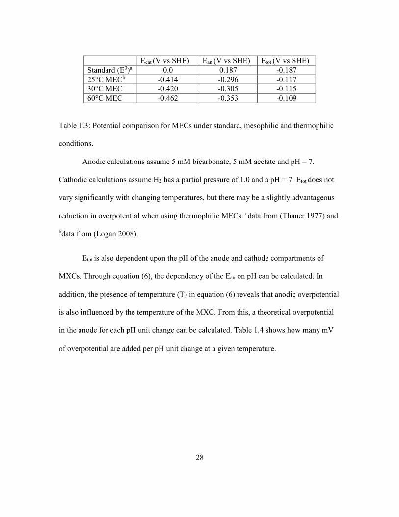
Table 1.3: Potential comparison for MECs under standard, mesophilic and thermophilic
conditions.
Anodic calculations assume 5 mM bicarbonate, 5 mM acetate and pH = 7.
Cathodic calculations assume H2 has a partial pressure of 1.0 and a pH = 7. Etot does not
vary significantly with changing temperatures, but there may be a slightly advantageous
reduction in overpotential when using thermophilic MECs. adata from (Thauer 1977) and
bdata from (Logan 2008).
Etot is also dependent upon the pH of the anode and cathode compartments of
MXCs. Through equation (6), the dependency of the Ean on pH can be calculated. In
addition, the presence of temperature (T) in equation (6) reveals that anodic overpotential
is also influenced by the temperature of the MXC. From this, a theoretical overpotential
in the anode for each pH unit change can be calculated. Table 1.4 shows how many mV
of overpotential are added per pH unit change at a given temperature.

29
T (°C) ∆mV per pH unit
0 54.2
25 59.1
30 60.1
60 66.1
Table 1.4: Temperature dependency of mV shift in overpotential per pH unit change
A decrease in pH will contribute to overpotential in Ean by shifting its value more
positive and thus decreasing Etot. An increase in pH will make Ean shift negative, creating
a more reductive anode reaction, and thus increasing Etot. Table 1.4 shows that a change
in pH in a thermophilic (60°C) MXC has a ~7.0 mV larger effect than does the same
change in pH under mesophilic (25°C) conditions- about ∆1.0 mV per 5.0°C. Table 1.4
also reveals that changes in pH are one of the most significant factors in determining the
energetics of Ean and Etot in MXCs.
Chapter 1 References:
1. Akinosho, H, Yee, K, Close, D, Ragauskas, A. 2014. The emergence of
Clostridium thermocellum as a high utility candidate for consolidated
bioprocessing applications. Frontiers in Chemistry. 2:66. doi:
10.3389/fchem.2014.00066.
2. Badalamenti, JP, Krajmalnik-Brown, R, Torres, CI. 2013. Generation of high
current densities by pure cultures of anode-respiring Geoalkalibacter spp. Under
alkaline and saline conditions in microbial electrochemical cells. Mbio. 4:e00144-
13-e00144-13. doi: 10.1128/mBio.00144-13.

30
3. Basen, M, Rhaesa, A, Kataeva, I, Prybol, C, Scott, I, Poole, F, Adams, M. 2014.
Degradation of high loads of crystalline cellulose and of unpretreated plant
biomass by the thermophilic bacterium Caldicellulosiruptor bescii. Bioresour.
Technol. 152:384-392. doi: 10.1016/j.biortech.2013.11.024.
4. Beveridge, TJ, Forsberg, CW, Doyle, RJ. 1982. Major sites of metal binding in
Bacillus licheniformis walls. J. Bacteriol. 150:1438-1448.
5. Beveridge, TJ, Murray, RG. 1980. Sites of metal deposition in the cell wall of
Bacillus subtilis. J. Bacteriol. 141:876-887.
6. Bird, LJ, Bonnefoy, V, Newman, DK. 2011. Bioenergetic challenges of microbial
iron metabolisms. Trends Microbiol. 19:330-340. doi: 10.1016/j.tim.2011.05.001.
7. Bourdakos, N, Marsili, E, Mahadevan, R. 2014. A defined co‐culture of
Geobacter sulfurreducens and Escherichia coli in a membrane‐less microbial fuel
cell. Biotechnol. Bioeng. 111:709-718. doi: 10.1002/bit.25137.
8. Brock, TD, Freeze, H. 1969. Thermus aquaticus gen. n. and sp. n., a
Nonsporulating Extreme Thermophile. J. Bacteriol. 98:289-297.
9. Brunecky, R, Alahuhta, M, Xu, Q, Donohoe, B, Crowley, M, Kataeva, I, Yang, S,
Resch, M, Adams, M, Lunin, V, Himmel, M, Bomble, Y. 2013. Revealing
Nature's Cellulase Diversity: The Digestion Mechanism of Caldicellulosiruptor
bescii CelA. Science. 342:1513-1516. doi: 10.1126/science.1244273.
10. Carere, CR, Sparling, R, Cicek, N, Levin, DB. 2008. Third generation biofuels via
direct cellulose fermentation. International Journal of Molecular Sciences.
9:1342-1360. doi: 10.3390/ijms9071342.
11. Carlson, HK, Iavarone, AT, Gorur, A, Yeo, BS, Tran, R, Melnyk, RA, Mathies,
RA, Auer, M, Coates, JD. 2012. Surface multiheme c-type cytochromes from
Thermincola potens and implications for respiratory metal reduction by Gram-
positive bacteria. Proc. Natl. Acad. Sci. U. S. A. 109:1702-1707. doi:
10.1073/pnas.1112905109.
12. Cheng, S, Logan, BE. 2007. Sustainable and Efficient Biohydrogen Production
via Electrohydrogenesis. Proc. Natl. Acad. Sci. U. S. A. 104:18871-18873. doi:
10.1073/pnas.0706379104.
13. Ciccarelli, FD, Doerks, T, von Mering, C, Creevey, CJ, Snel, B, Bork, P. 2006.
Toward Automatic Reconstruction of a Highly Resolved Tree of Life. Science.
311:1283-1287. doi: 10.1126/science.1123061.

31
14. Dalla Vecchia, E, Shao, P, Suvorova, E, Chiappe, D, Hamelin, R, Bernier-
Latmani, R. 2014. Characterization of the surfaceome of the metal-reducing
bacterium Desulfotomaculum reducens. Frontiers in Microbiology. 5:432. doi:
10.3389/fmicb.2014.00432.
15. Doney, SC, Fabry, VJ, Feely, RA, Kleypas, JA. 2009. Ocean acidification: The
other CO2 problem. Annual Review of Marine Science. 1:169-192. doi:
10.1146/annurev.marine.010908.163834.
16. Du, Z, Li, H, Gu, T. 2007. A state of the art review on microbial fuel cells: A
promising technology for wastewater treatment and bioenergy. Biotechnol. Adv.
25:464-482. doi: 10.1016/j.biotechadv.2007.05.004.
17. Ehrlich, HL. 2008. Are gram-positive bacteria capable of electron transfer across
their cell wall without an externally available electron shuttle? Geobiology.
6:220-224. doi: 10.1111/j.1472-4669.2007.00135.x.
18. Emtiazi, G, Nahvi, I. 2000. Multi-enzyme production by Cellulomonas sp. grown
on wheat straw. Biomass Bioenergy. 19:31-37. doi: 10.1016/S0961-
9534(00)00015-5.
19. Florenzano, G, Poulain, M, Goma, G. 1984. A study of acetate production from
cellulose using Clostridium thermocellum. Biomass. 4:295-303. doi:
10.1016/0144-4565(84)90042-8.
20. Franks, AE, Nevin, KP, Jia, H, Izallalen, M, Woodard, TL, Lovley, DR. 2009.
Novel strategy for three-dimensional real-time imaging of microbial fuel cell
communities: Monitoring the inhibitory effects of proton accumulation within the
anode biofilm. Energy and Environmental Science. 2:113-119. doi:
10.1039/b816445b.
21. Freier, D, Mothershed, CP, Wiegel, J. 1988. Characterization of Clostridium
thermocellum JW20. Appl. Environ. Microbiol. 54:204-211.
22. He, Q, Lokken, PM, Chen, S, Zhou, J. 2009. Characterization of the impact of
acetate and lactate on ethanolic fermentation by Thermoanaerobacter ethanolicus.
Bioresour. Technol. 100:5955-5965. doi: 10.1016/j.biortech.2009.06.084.
23. Hniman, A, Prasertsan, P, O-Thong, S. 2011. Community analysis of
thermophilic hydrogen-producing consortia enriched from Thailand hot spring
with mixed xylose and glucose. Int J Hydrogen Energy. 36:14217-14226. doi:
10.1016/j.ijhydene.2011.05.087.
32
24. Hollaus, F, Sleytr, U. 1972. On the taxonomy and fine structure of some
hyperthermophilic saccharolytic clostridia. Archiv Für Mikrobiologie. 86:129-
146. doi: 10.1007/BF00413368.
25. Hurst, LD, Merchant, AR. 2001. High guanine–cytosine content is not an
adaptation to high temperature: a comparative analysis amongst prokaryotes.
Proceedings of the Royal Society of London.Series B: Biological Sciences.
268:493-497. doi: 10.1098/rspb.2000.1397.
26. Ieropoulos, I, Melhuish, C, Greenman, J, Horsfield, I. 2005. EcoBot-II: An
artificial agent with a natural metabolism. International Journal of Advanced
Robotic Systems. 2:295-300.
27. Jong, BC, Kim, BH, Chang, IS, Liew, PWY, Choo, YF, Kang, GS. 2006.
Enrichment, performance, and microbial diversity of a thermophilic mediatorless
microbial fuel cell. Environmental Science and Technology. 40:6449-6454. doi:
10.1021/es0613512.
28. Jung, RK, Cheng, S, Oh, S, Logan, BE. 2007. Power generation using different
cation, anion, and ultrafiltration membranes in microbial fuel cells. Environmental
Science and Technology. 41:1004-1009. doi: 10.1021/es062202m.
29. Kashefi, K, Lovley, DR. 2003. Extending the Upper Temperature Limit for Life.
Science. 301:934-934. doi: 10.1126/science.1086823.
30. Kato, S, Haruta, S, Cui, ZJ, Ishii, M, Igarashi, Y. 2005. Stable Coexistence of
Five Bacterial Strains as a Cellulose-Degrading Community. Appl. Environ.
Microbiol. 71:7099-7106. doi: 10.1128/AEM.71.11.7099-7106.2005.
31. Kim, B, Kim, H, Hyun, M, Park, D. 1999. Direct electrode reaction of Fe(III)-
reducing bacterium, Shewanella putrefaciens. Journal of Microbiology and
Biotechnology. 9:127-131.
32. Knoll, AH. 2003. Life on a young planet: the first three billion years of evolution
on earth. Princeton University Press, Oxford; Princeton, N.J.
33. Leang, C, Coppi, MV, Lovley, DR. 2003. OmcB, a c-Type Polyheme
Cytochrome, Involved in Fe(III) Reduction in Geobacter sulfurreducens. J.
Bacteriol. 185:2096-2103. doi: 10.1128/JB.185.7.2096-2103.2003.
34. Lee, H, Parameswaran, P, Kato-Marcus, A, Torres, CI, Rittmann, BE. 2008.
Evaluation of energy-conversion efficiencies in microbial fuel cells (MFCs)
utilizing fermentable and non-fermentable substrates. Water Res. 42:1501-1510.
doi: 10.1016/j.watres.2007.10.036.

33
35. Lee, Y-, Jain, MK, Lee, C, Lowe, SE, Zeikus, JG. 1993. Taxonomic distinction of
saccharolytic thermophilic anaerobes. Int. J. Syst. Bacteriol. 43:41-51.
36. Letunic, I, Bork, P. 2007. Interactive Tree Of Life (iTOL): An online tool for
phylogenetic tree display and annotation. Bioinformatics. 23:127-128. doi:
10.1093/bioinformatics/btl529.
37. Liang, Y, Yesuf, J, Schmitt, S, Bender, K, Bozzola, J. 2009. Study of cellulases
from a newly isolated thermophilic and cellulolytic Brevibacillus sp. strain JXL.
Journal of Industrial Microbiology and Biotechnology. 36:961-970. doi:
10.1007/s10295-009-0575-2.
38. Liu, Y, Climent, V, Berná, A, Feliu, JM. 2011. Effect of Temperature on the
Catalytic Ability of Electrochemically Active Biofilm as Anode Catalyst in
Microbial Fuel Cells. Electroanalysis. 23:387-394. doi: 10.1002/elan.201000499.
39. Lloyd, JR, Blunt-Harris, EL, Lovley, DR. 1999. The Periplasmic 9.6-Kilodalton
c-Type Cytochrome of Geobacter sulfurreducens Is Not an Electron Shuttle to
Fe(III). J. Bacteriol. 181:7647-7649.
40. Logan, BE, Call, D, Cheng, S. 2008. Microbial Electrolysis Cells for High Yield
Hydrogen Gas Production from Organic Matter. Environmental Science &
Technology [H.W.Wilson - AST]. 42:8630.
41. Logan, BE, Hamelers, B, Rozendal, R, Schroder, U. 2006. Microbial Fuel Cells:
Methodology and Technology. Environ. Sci. Technol. 40:5181.
42. Lovley, DR. 2008. The microbe electric: conversion of organic matter to
electricity. Curr. Opin. Biotechnol. 19:564-571. doi:
10.1016/j.copbio.2008.10.005.
43. Lynd, LR, Weimer, PJ, Willem H. van Zyl, Pretorius, IS. 2002. Microbial
Cellulose Utilization: Fundamentals and Biotechnology. Microbiology and
Molecular Biology Reviews. 66:506-577. doi: 10.1128/MMBR.66.3.506-
577.2002.
44. Marcus, AK, Torres, CI, Rittmann, BE. 2011. Analysis of a microbial
electrochemical cell using the proton condition in biofilm (PCBIOFILM) model.
Bioresour. Technol. 102:253-262. doi: 10.1016/j.biortech.2010.03.100.
45. Marcus, AK, Torres, CI, Rittmann, BE. 2007. Conduction-based modeling of the
biofilm anode of a microbial fuel cell. Biotechnol. Bioeng. 98:1171-1182. doi:
10.1002/bit.21533.

34
46. Marshall, CW, May, HD. 2009. Electrochemical evidence of direct electrode
reduction by a thermophilic Gram-positive bacterium, Thermincola ferriacetica.
Energy and Environmental Science. 2:699-705. doi: 10.1039/b823237g.
47. Marsili, E, Sun, J, Bond, DR. 2010. Voltammetry and growth physiology of
Geobacter sulfurreducens biofilms as a function of growth stage and imposed
electrode potential. Electroanalysis. 22:865-874. doi: 10.1002/elan.200800007.
48. Mathis, BJ, Marshall, CW, Milliken, CE, Makkar, RS, Creager, SE, May, HD.
2008. Electricity generation by thermophilic microorganisms from marine
sediment. Appl. Microbiol. Biotechnol. 78:147-155. doi: 10.1007/s00253-007-
1266-4.
49. May, HD, Shimotori, T. U.S. Patent No. 0017512 A1. Austin, TX: U.S. Patent and
Trademark Office.
50. Miceli, JF, Parameswaran, P, Kang, D, Krajmalnik-Brown, R, Torres, CI. 2012.
Enrichment and analysis of anode-respiring bacteria from diverse anaerobic
inocula. Environmental Science and Technology. 46:10349-10355. doi:
10.1021/es301902h.
51. Mohan, S, Velvizhi, G, Krishna, K, Babu, M. 2014. Microbial catalyzed
electrochemical systems: A bio-factory with multi-facet applications. Bioresour.
Technol. 165:355-364. doi: 10.1016/j.biortech.2014.03.048.
52. Mook, WG, Koene, BKS. 1975. Chemistry of dissolved inorganic carbon in
estuarine and coastal brackish waters. Estuarine and Coastal Marine Science.
3:325-336.
53. Moon, H, Chang, IS, Kim, BH. 2006. Continuous electricity production from
artificial wastewater using a mediator-less microbial fuel cell. Bioresour. Technol.
97:621-627. doi: 10.1016/j.biortech.2005.03.027.
54. Nealson, KH, Conrad, PG. 1999. Life: past, present and future. Philosophical
Transactions of the Royal Society of London.Series B: Biological Sciences.
354:1923-1939. doi: 10.1098/rstb.1999.0532.
55. Nealson, KH, Conrad, PG. 1999. Life: past, present and future. Philosophical
Transactions of the Royal Society of London.Series B: Biological Sciences.
354:1923-1939. doi: 10.1098/rstb.1999.0532.

35
56. Niessen, J, Harnisch, F, Rosenbaum, M, Schröder, U, Scholz, F. 2006. Heat
treated soil as convenient and versatile source of bacterial communities for
microbial electricity generation. Electrochemistry Communications. 8:869-873.
doi: 10.1016/j.elecom.2006.03.025.
57. Niu, L, Song, L, Liu, X, Dong, X. 2009. Tepidimicrobium xylanilyticum sp. nov.,
an anaerobic xylanolytic bacterium, and emended description of the genus
Tepidimicrobium. Int. J. Syst. Evol. Microbiol. 59:2698-2701. doi:
10.1099/ijs.0.005124-0.
58. Olson, DG, McBride, JE, Joe Shaw, A, Lynd, LR. 2012. Recent progress in
consolidated bioprocessing. Curr. Opin. Biotechnol. 23:396-405. doi:
10.1016/j.copbio.2011.11.026.
59. Onyenwoke, RU, Kevbrin, VV, Lysenko, AM, Wiegel, J. 2007.
Thermoanaerobacter pseudethanolicus sp. nov., a thermophilic heterotrophic
anaerobe from Yellowstone National Park. Int. J. Syst. Evol. Microbiol. 57:2191-
2193. doi: 10.1099/ijs.0.65051-0.
60. Pant, D, Van Bogaert, G, Diels, L, Vanbroekhoven, K. 2010. A review of the
substrates used in microbial fuel cells (MFCs) for sustainable energy production.
Bioresour. Technol. 101:1533-1543. doi: 10.1016/j.biortech.2009.10.017.
61. Parameswaran, P, Bry, T, Popat, SC, Lusk, BG, Rittmann, BE, Torres, CI. 2013.
Kinetic, electrochemical, and microscopic characterization of the thermophilic,
anode-respiring bacterium Thermincola ferriacetica. Environmental Science and
Technology. 47:4934-4940. doi: 10.1021/es400321c.
62. Pham, TH, Boon, N, Aelterman, P, Clauwaert, P, De Schamphelaire, L,
Vanhaecke, L, De Maeyer, K, Höfte, M, Verstraete, W, Rabaey, K. 2008.
Metabolites produced by Pseudomonas sp. enable a Gram-positive bacterium to
achieve extracellular electron transfer. Appl. Microbiol. Biotechnol. 77:1119-
1129. doi: 10.1007/s00253-007-1248-6.
63. Pirbadian, S, Barchinger, SE, Leung, KM, Byun, HS, Jangir, Y, Bouhenni, RA,
Reed, SB, Romine, MF, Saffarini, DA, Shi, L, Gorby, YA, Golbeck, JH, El-
Naggar, MY. 2014. Shewanella oneidensis MR-1 nanowires are outer membrane
and periplasmic extensions of the extracellular electron transport components.
Proceedings of the National Academy of Sciences. 111:12883-12888. doi:
10.1073/pnas.1410551111.

36
64. Popat, SC, Ki, D, Rittmann, BE, Torres, CI. 2012. Importance of OH- transport
from cathodes in microbial fuel cells. Chemsuschem. 5:1071-1079. doi:
10.1002/cssc.201100777.
65. Puigb, P, Wolf, YI, Koonin, EV. 2009. Search for a 'tree of Life' in the thicket of
the phylogenetic forest. Journal of Biology. 8:59-59. doi: 10.1186/jbiol159.
66. Rabaey, K, Verstraete, W. 2005. Microbial fuel cells: novel biotechnology for
energy generation. Trends Biotechnol. 23:291-298. doi:
10.1016/j.tibtech.2005.04.008.
67. Ren, Z, Steinberg, L, Regan, J. 2008. Electricity production and microbial biofilm
characterization in cellulose-fed microbial fuel cells. Water Science and
Technology. 58:617-622. doi: 10.2166/wst.2008.431.
68. Rismani-Yazdi, H, Christy, AD, Dehority, BA, Morrison, M, Yu, Z, Tuovinen,
OH. 2007. Electricity generation from cellulose by rumen microorganisms in
microbial fuel cells. Biotechnol. Bioeng. 97:1398-1407. doi: 10.1002/bit.21366.
69. Rittmann, BE. 2008. Opportunities for renewable bioenergy using
microorganisms. Biotechnol. Bioeng. 100:203-212. doi: 10.1002/bit.21875.
70. Roh, Y, Liu, SV, Li, G, Huang, H, Phelps, TJ, Zhou, J. 2002. Isolation and
Characterization of Metal-Reducing Thermoanaerobacter Strains from Deep
Subsurface Environments of the Piceance Basin, Colorado. Appl. Environ.
Microbiol. 68:6013-6020. doi: 10.1128/AEM.68.12.6013-6020.2002.
71. Rydzak, T, Levin, DB, Cicek, N, Sparling, R. 2011. End-product induced
metabolic shifts in Clostridium thermocellum ATCC 27405. Appl. Microbiol.
Biotechnol. 92:199-209. doi: 10.1007/s00253-011-3511-0.
72. Schroder, U. 2007. Anodic electron transfer mechanisms in microbial fuel cells
and their energy efficiency. Physical Chemistry Chemical Physics. 9:2619-2629.
doi: 10.1039/003627m.
73. Seckbach, J. 2004. Origins: genesis, evolution and diversity of life. Kluwer,
Dordrecht; Boston.
74. Seckbach. 2006. Life as we know it. Springer, Dordrecht.

37
75. Slepova, TV, Sokolova, TG, Kolganova, TV, Tourova, TP, Bonch-
Osmolovskaya, EA. 2009. Carboxydothermus siderophilus sp. nov., a
thermophilic, hydrogenogenic, carboxydotrophic, dissimilatory Fe(III)-reducing
bacterium from a Kamchatka hot spring. Int. J. Syst. Evol. Microbiol. 59:213-217.
doi: 10.1099/ijs.0.000620-0.
76. Slobodkin, AI, Tourova, TP, Kostrikina, NA, Lysenko, AM, German, KE, Bonch-
Osmolovskaya, EA, Birkeland, N-. 2006. Tepidimicrobium ferriphilum gen. nov.,
sp. nov., a novel moderately thermophilic, Fe(III)-reducing bacterium of the order
Clostridiales. Int. J. Syst. Evol. Microbiol. 56:369-372. doi: 10.1099/ijs.0.63694-
0.
77. Sokolova, TG, Kostrikina, NA, Chernyh, NA, Kolganova, TV, Tourova, TP,
Bonch-Osmolovskaya, EA. 2005. Thermincola carboxydiphila gen. nov., sp. nov.,
a novel anaerobic, carboxydotrophic, hydrogenogenic bacterium from a hot spring
of the Lake Baikal area. Int. J. Syst. Evol. Microbiol. 55:2069-2073. doi:
10.1099/ijs.0.63299-0.
78. Srikanth, S, Marsili, E, Flickinger, MC, Bond, DR. 2008. Electrochemical
characterization of Geobacter sulfurreducens cells immobilized on graphite paper
electrodes. Biotechnol. Bioeng. 99:1065-1073. doi: 10.1002/bit.21671.
79. Stocker, TF. 2014; 2013. Climate change 2013: the physical science basis :
working group I contribution to the fifth assessment report of the
intergovernmental panel on climate change. Cambridge University Press, New
York, NY, USA.
80. Thauer, RK, Jungermann, K, Decker, K. 1977. Energy conservation in
chemotrophic anaerobic bacteria. Bacteriol. Rev. 41:100-180.
81. Torres, CI, Kato Marcus, A, Rittmann, BE. 2008. Proton transport inside the
biofilm limits electrical current generation by anode‐respiring bacteria.
Biotechnol. Bioeng. 100:872-881. doi: 10.1002/bit.21821.
82. Torres, CI, Marcus, AK, Lee, H, Parameswaran, P, Krajmalnik-Brown, R,
Rittmann, BE. 2010. A kinetic perspective on extracellular electron transfer by
anode-respiring bacteria. FEMS Microbiol. Rev. 34:3-17. doi: 10.1111/j.1574-
6976.2009.00191.x.
83. Torres, C. 2014. On the importance of identifying, characterizing, and predicting
fundamental phenomena towards microbial electrochemistry applications. Curr.
Opin. Biotechnol. 27:107-114. doi: 10.1016/j.copbio.2013.12.008.

38
84. U.S. Global Change Research Program. 2014. Climate change impacts in the
United States: U.S. national climate assessment.
85. Vasudeo Zambare, Archana Zambare, Kasiviswanath Muthukumarappan,Lew
P.Christopher. 2011. Biochemical characterization of thermophilic lignocellulose
degrading enzymes and their potential for biomass bioprocessing. International
Journal of Energy and Environment. 2:99-112.
86. Ventura, M, Canchaya, C, Tauch, A, Chandra, G, Fitzgerald, GF, Chater, KF,
Sinderen, Dv. 2007. Genomics of Actinobacteria: Tracing the Evolutionary
History of an Ancient Phylum. Microbiology and Molecular Biology Reviews.
71:495-548. doi: 10.1128/MMBR.00005-07.
87. Vesth, T, Ozen, A, Andersen, S, Kaas, R, Lukjancenko, O, Bohlin, J, Nookaew, I,
Wassenaar, T, Ussery, D, Department of Chemical and Biological
Engineering,Systems Biology, Chalmers University of Technology, Chalmers
tekniska högskola, Institutionen för kemi- och bioteknik,Systembiologi. 2013.
Veillonella, Firmicutes: Microbes disguised as Gram negatives. Standards in
Genomic Sciences. 9:431-448. doi: 10.4056/sigs.2981345.
88. Viljoen, JA. 1925. The Fermentation of Cellulose by Thermophilic Bacteria.
ProQuest, UMI Dissertations Publishing.
89. Wrighton, KC, Thrash, JC, Melnyk, RA, Bigi, JP, Byrne-Bailey, KG, Remis, JP,
Schichnes, D, Auer, M, Chang, CJ, Coates, JD. 2011. Evidence for Direct
Electron Transfer by a Gram-Positive Bacterium Isolated from a Microbial Fuel
Cell. Appl. Environ. Microbiol. 77:7633-7639. doi: 10.1128/AEM.05365-11.
90. Yang, Y, Xu, M, Guo, J, Sun, G. 2012. Bacterial extracellular electron transfer in
bioelectrochemical systems. Process Biochemistry. 47:1707-1714. doi:
10.1016/j.procbio.2012.07.032.
91. Yoho, R, Popat, S, Torres, C. 2014. Dynamic Potential-Dependent Electron
Transport Pathway Shifts in Anode Biofilms of Geobacter sulfurreducens.
Chemsuschem. 7:3413-3419. doi: 10.1002/cssc.201402589.
92. Zavarzina, DG, Sokolova, TG, Tourova, TP, Chernyh, NA, Kostrikina, NA,
Bonch-Osmolovskaya, EA. 2007. Thermincola ferriacetica sp. nov., a new
anaerobic, thermophilic, facultatively chemolithoautotrophic bacterium capable of
dissimilatory Fe(III) reduction. Extremophiles. 11:1-7. doi: 10.1007/s00792-006-
0004-7.
93. Zeikus, JG, Ben-Bassat, A, Hegge, PW. 1980. Microbiology of Methanogenesis
in Thermal, Volcanic Environments. J. Bacteriol. 143:432-440.

39
94. Zhang, E, Xu, W, Diao, G, Shuang, C. 2006. Electricity generation from acetate
and glucose by sedimentary bacterium attached to electrode in microbial-anode
fuel cells. J. Power Sources. 161:820-825. doi: 10.1016/j.jpowsour.2006.05.004.

40
Chapter 2: Draft Genome of the Gram-Positive Thermophilic Iron Reducer
Thermincola ferriacetica strain Z-0001T1
Overview
A 3.19-Mbp draft genome of the Gram-positive thermophilic iron-reducing
Firmicutes isolate from the Peptococcaceae family, Thermincola ferriacetica Z-0001 was
assembled at ~100x coverage from 100-bp paired-end Illumina reads. The draft genome
contains 3,274 predicted genes (3,187 protein coding genes) and putative multiheme c-
type cytochromes.
1 Chapter 2 published as: Lusk, BG, Badalamenti, JP, Parameswaran, P, Bond, DR,
Torres, CI. 2015. Draft Genome Sequence of the Gram-Positive Thermophilic Iron
Reducer Thermincola ferriacetica Strain Z-0001T. Genome Announcements. 3:.

41
Results
Thermincola ferriacetica strain Z-0001 (DSM 14005), first isolated from a
terrestrial hydrothermal spring on Kunashir Island (Kurils) (Zavarzina 2007), is a Gram-
positive, thermophilic (45-70 °C), spore-forming bacterium that is capable of
dissimilatory metal reduction and anode respiration in a microbial electrochemical cell
(MXC) (Marshall 2009, Mathis 2008, Parameswaran 2013), and it is one of only a limited
number of sequenced Gram-positive thermophilic bacteria that has been documented to
perform extracellular electron transfer (EET) to insoluble metal substrates (Byrne-Bailey
2010, Roh 2002, Wrighton 2008). Strain Z-0001 is capable of organotrophic growth with
acetate and other organic compounds while reducing extracellular electron acceptors
including amorphous Fe(III)-hydroxide, magnetite, Mn(IV), anthraquinone-2,6-
disulfonate (AQDS), and anodes in MXCs (Marhsall 2009, Parameswaran 2013,
Zavarzina 2007). Strain Z-0001 is also capable of chemolithoautotrophic growth using
molecular hydrogen as the electron donor and Fe(III) as the electron acceptor (Zavarzina
2007). In addition, strain Z-0001 is able to produce H2 and CO2 while using CO as its
electron donor and acquiring its carbon from acetate (Zavarzina 2007).
Among Gram-positive bacteria, little is known regarding the mechanism for EET
or how the peptidoglycan layer impacts this pathway (Beveridge 1982, Beveridge and
Murray 1980, Ehrlich 2008). Direct contact-dependent electron transfer has been
suggested in Thermincola potens JR (Wrighton 2011) with genetic evidence for the
presence of c-type cytochromes (Carlson 2012), proteins that are responsible for EET in
other metal-reducing bacteria (Leang 2003). In contrast to T. potens, T. ferriacetica strain

42
Z-0001 has been suggested to transfer electrons long range via an extracellular matrix
(Parameswaran 2013), suggesting it may encode additional electron transfer capabilities.
T. ferriacetica has been reported to produce current densities up to 10 A m-2 despite
having only half the cytochrome repertoire of Geobacter sulfurreducens (Methé 2003,
Parameswaran 2013). Further genetic comparison of these strains could help elucidate the
EET mechanism(s) of strain Z-0001.
The draft assembly presented here is from an axenic culture of electrode-grown T.
ferriacetica strain Z-0001 cells in order to eliminate contamination by iron or AQDS.
gDNA was collected and sequenced on an Illumina HiSeq 2000 lane, yielding > 45
million 2x100 bp reads. Raw reads were trimmed (sliding window 3 until quality > 28)
and down-sampled to provide 100x coverage for assembly using the a5 pipeline (March
26, 2013 release, Tritt 2012). The 3,196,047-bp draft genome assembly yielded 53
contigs > 500 bp with an N50 of 112112 bp, an L50 of 8, and a G+C content of 45.69%.
The draft assembly was annotated using the JGI IMG/ER pipeline, yielding 51
tRNAs, 3,274 predicted genes (3,187 predicted protein coding genes), and 35 c-type
cytochromes with three or more heme (CXXCH) binding motifs- including a 222.67 kDa
c-type cytochrome containing 58 heme binding motifs. Table 2.1 displays all 35 putative
c-type cytochromes. BLASTN sequence analysis of its 16S rRNA gene revealed 99.9%
(1436/1438 nt) identity with T. potens JR and 99.7% (1399/1403 nt) identity with
Thermincola carboxydophila (Byrne-Bailey 2010, Sokolova 2005). T. ferriacetica
contains two multiheme c-type cytochromes and 429 genes that are not present in T.
potens. However, based on an average nucleotide identity (ANI) of 98.3% between their

43
genomes, these two organisms may be members of the same species (Richter and
Rosselló-Móra 2009).
Nucleotide Sequence Accession Number
This Whole Genome Shotgun project for T. ferriacetica strain Z-0001 was
deposited at DDBJ/EMBL/GenBank under the accession LGTE00000000. The version
described in this paper is version LGTE01000000. The raw and adaptor trimmed Illumina
reads were submitted to SRA under accession SRX1100231.

44
Name CDS (within
Contig)
Heme
motifs
Size
(AA)
Size
(kDa)
Tfer_0004_putative
multiheme cytochrome c
5943�6977 10 344 39.19
Tfer_0070_putative
multiheme cytochrome c
67185�68231 10 348 38.58
Tfer_0071_NapC/NirT
cytochrome c domain
protein
68315�69538 12 407 45.15
Tfer_0072_putative
multiheme cytochrome c
69944�71470 17 508 57.33
Tfer_0075_putative
multiheme cytochrome c
73143�74204 6 353 37.54
Tfer_0076_putative
multiheme cytochrome c
74486�76330 15 614 66.86
Tfer_0077_putative
multiheme cytochrome c
76721�83047 58 2108 222.67
Tfer_0082_putative
multiheme cytochrome c
89349�91238 11 629 68.14
Tfer_0155_NapC/NirT
cytochrome c domain-
containing protein
157890�158333 4 147 16.70
Tfer_0156_ammonia-
forming nitrite reductase
158416�159660 4 414 46.74
Tfer_0401_putative
multiheme cytochrome c
63064�64845 10 593 65.21
Tfer_0712_putative
multiheme cytochrome c
89029�93423 17 1464 158.72
Tfer_0719_putative
multiheme cytochrome c
106821�110489 10 1222 131.33
Tfer_0721_putative
multiheme cytochrome c
111354�113222 8 622 68.34
Tfer_0722_putative
multiheme cytochrome c
113394�115028 6 544 58.60
Tfer_0725_NapC/NirT
cytochrome c domain-
containing protein
116864�118042 12 392 43.95
Tfer_0972_putative
multiheme cytochrome c
119742�120392 3 216 24.34
Tfer_1028_putative
multiheme cytochrome c
17876�19324 6 482 51.26

45
Tfer_1031_NapC/NirT
cytochrome c domain-
containing protein
21342�22505 12 387 43.48
Tfer_1797_methyl-
accepting chemotaxis
sensory transducer/ putative
triheme cytochrome c
49319�50038
3
239
26.52
Tfer_1887_putative
multiheme cytochrome c
49123�50214 10 363 40.63
Tfer_1988_putative
multiheme cytochrome c
33118�34446 6 442 47.85
Tfer_1990_putative
multiheme cytochrome c
35489�38845 6 1118 119.03
Tfer_1992_putative
multiheme cytochrome c
39794�44566 15 1590 171.18
Tfer_2063_putative
multiheme cytochrome c
14274�23423 45 3049 335.29
Tfer_2064_putative
multiheme cytochrome c
23444�25891 6 815 89.97
Tfer_2066_putative
multiheme cytochrome c
26970�28184 6 404 43.18
Tfer_2149_putative
multiheme cytochrome c
30081�31427 15 448 51.09
Tfer_2153_putative
multiheme cytochrome c
33759�34952 7 397 41.31
Tfer_2155_putative
multiheme cytochrome c
35822�37108 7 428 45.36
Tfer_2210_NapC/NirT
family cyctochrome c
13953�14465 5 170 18.84
Tfer_2544_chaperone
protein DnaJ/putative
triheme cytochrome c
17771�18904 3 377 41.23
Tfer_2880_putative
multiheme cytochrome c
13229�14152
6
307
32.08
Tfer_3001_putative
multiheme cytochrome c
32868�33416 5 182 20.97
Tfer_3193_putative
multiheme cytochrome c
7756�9333 9 525 56.58
Table 2.1: Putative multiheme c-type cytochromes for T. ferriacetica are listed. Included

46
are: the protein name, the coding DNA sequence (CDS) location for each protein within
its contig, and the number of heme motifs, amino acids, and kDa for each protein.
Chapter 2 References
1. Beveridge, TJ, Forsberg, CW, Doyle, RJ. 1982. Major sites of metal binding in
Bacillus licheniformis walls. J. Bacteriol. 150:1438-1448.
2. Beveridge, TJ, Murray, RG. 1980. Sites of metal deposition in the cell wall of
Bacillus subtilis. J. Bacteriol. 141:876-887.
3. Byrne-Bailey, KG, Wrighton, KC, Melnyk, RA, Agbo, P, Hazen, TC, Coates, JD.
2010. Complete Genome Sequence of the Electricity-Producing “Thermincola
potens” Strain JR. J. Bacteriol. 192:4078-4079. doi: 10.1128/JB.00044-10.
4. Carlson, HK, Iavarone, AT, Gorur, A, Yeo, BS, Tran, R, Melnyk, RA, Mathies,
RA, Auer, M, Coates, JD. 2012. Surface multiheme c-type cytochromes from
Thermincola potens and implications for respiratory metal reduction by Gram-
positive bacteria. Proc. Natl. Acad. Sci. U. S. A. 109:1702-1707. doi:
10.1073/pnas.1112905109.
5. Ehrlich, HL. 2008. Are gram-positive bacteria capable of electron transfer across
their cell wall without an externally available electron shuttle? Geobiology.
6:220-224. doi: 10.1111/j.1472-4669.2007.00135.x.
6. Holmes, DE, Mester, T, O'Neil, RA, Perpetua, LA, Larrahondo, MJ, Glaven, R,
Sharma, ML, Ward, JE, Nevin, KP, Lovley, DR. 2008. Genes for two multicopper
proteins required for Fe(III) oxide reduction in Geobacter sulfurreducens have
different expression patterns both in the subsurface and on energy-harvesting
electrodes. Microbiology. 154:1422-1435. doi: 10.1099/mic.0.2007/014365-0.
7. Leang, C, Coppi, MV, Lovley, DR. 2003. OmcB, a c-Type Polyheme
Cytochrome, Involved in Fe(III) Reduction in Geobacter sulfurreducens. J.
Bacteriol. 185:2096-2103. doi: 10.1128/JB.185.7.2096-2103.2003.
8. Leang, C, Qian, X, Mester, T, Lovley, DR. 2010. Alignment of the c-Type
Cytochrome OmcS along Pili of Geobacter sulfurreducens. Appl. Environ.
Microbiol. 76:4080-4084. doi: 10.1128/AEM.00023-10.

47
9. Lies, DP, Hernandez, ME, Kappler, A, Mielke, RE, Gralnick, JA, Newman, DK.
2005. Shewanella oneidensis MR-1 Uses Overlapping Pathways for Iron
Reduction at a Distance and by Direct Contact under Conditions Relevant for
Biofilms. Appl. Environ. Microbiol. 71:4414-4426. doi: 10.1128/AEM.71.8.4414-
4426.2005.
10. Marshall, CW, May, HD. 2009. Electrochemical evidence of direct electrode
reduction by a thermophilic Gram-positive bacterium, Thermincola ferriacetica.
Energy and Environmental Science. 2:699-705. doi: 10.1039/b823237g.
11. Parameswaran, P, Bry, T, Popat, SC, Lusk, BG, Rittmann, BE, Torres, CI. 2013.
Kinetic, electrochemical, and microscopic characterization of the thermophilic,
anode-respiring bacterium Thermincola ferriacetica. Environmental Science and
Technology. 47:4934-4940. doi: 10.1021/es400321c.
12. Reguera, G, McCarthy, KD, Mehta, T, Nicoll, JS. 2005. Extracellular electron
transfer via microbial nanowires. Nature. 435:1098.
13. Roh, Y, Liu, SV, Li, G, Huang, H, Phelps, TJ, Zhou, J. 2002. Isolation and
Characterization of Metal-Reducing Thermoanaerobacter Strains from Deep
Subsurface Environments of the Piceance Basin, Colorado. Appl. Environ.
Microbiol. 68:6013-6020. doi: 10.1128/AEM.68.12.6013-6020.2002.
14. Sokolova, TG, Kostrikina, NA, Chernyh, NA, Kolganova, TV, Tourova, TP,
Bonch-Osmolovskaya, EA. 2005. Thermincola carboxydiphila gen. nov., sp. nov.,
a novel anaerobic, carboxydotrophic, hydrogenogenic bacterium from a hot spring
of the Lake Baikal area. Int. J. Syst. Evol. Microbiol. 55:2069-2073. doi:
10.1099/ijs.0.63299-0.
15. Tritt, A, Eisen, JA, Facciotti, MT, Darling, AE. 2012. An Integrated Pipeline for
de Novo Assembly of Microbial Genomes. Plos One. 7:e42304. doi:
10.1371/journal.pone.0042304.
16. Wrighton, KC, Thrash, JC, Melnyk, RA, Bigi, JP, Byrne-Bailey, KG, Remis, JP,
Schichnes, D, Auer, M, Chang, CJ, Coates, JD. 2011. Evidence for Direct
Electron Transfer by a Gram-Positive Bacterium Isolated from a Microbial Fuel
Cell. Appl. Environ. Microbiol. 77:7633-7639. doi: 10.1128/AEM.05365-11.
17. Zavarzina, DG, Sokolova, TG, Tourova, TP, Chernyh, NA, Kostrikina, NA,
Bonch-Osmolovskaya, EA. 2007. Thermincola ferriacetica sp. nov., a new
anaerobic, thermophilic, facultatively chemolithoautotrophic bacterium capable of
dissimilatory Fe(III) reduction. Extremophiles. 11:1-7. doi: 10.1007/s00792-006-
0004-7.

48
Chapter 3: The Effect of pH and Buffer Concentration on Anode Biofilms of
Thermincola ferriacetica2
Overview
We assessed the effects of pH and buffer concentration on growth and current production
in biofilms of Thermincola ferriacetica – a thermophilic, Gram-positive, anode-respiring
bacterium (ARB) – grown on anodes poised at a potential of -0.06 V vs. SHE in
microbial electrolysis cells (MECs) at 60 °C. Maximum current density in biofilms
grown with 10 mM bicarbonate buffer was 6.8±1.1 A m-2. Increasing bicarbonate buffer
concentrations from 10 mM to 100 mM resulted in an increase in the current density by
40±6% to 11.2±2.7 A m-2. Adjusting pH between 5.2 and 8.3 resulted in an increase in j,
although with a smaller increase in j from pH 7.0 to 8.3, suggesting inhibition in current
production at low pH. Confocal laser scanning microscopy (CLSM) images indicated
that higher bicarbonate buffer concentrations resulted in larger biofilm thicknesses (Lf),
from 68±20 µm at 10 mM bicarbonate to >150 µm at 100 mM, suggesting that H+
diffusion rates have a strong influence on Lf. Thus, T. ferriacetica follows a similar
pattern to G. sulfurreducens biofilms, being inhibited by low pH and low buffer
concentrations. However in comparison, the faster transport rates at higher temperatures
and the ability to grow at lower pH values than G. sulfurreducens allows T. ferriacetica
to produce higher current densities with lower buffer concentrations, making it an
attractive alternative for low alkalinity wastewater applications.
2 Chapter 3 unpublished with Bradley G. Lusk, Prathap Parameswaran, Sudeep Popat,
Bruce E. Rittmann, Cesar I. Torres

49
Introduction
The limitation(s) to microbial respiration during growth on solid electron
acceptors or electrodes in microbial electrochemical cells (MXCs) have been studied
extensively with Gram-negative mesophilic anode-respiring bacteria (ARB) (Marcus
2007, Marcus 2011, Torres 2008a-b). However, such limitations for Gram-positive
thermophilic ARB have not been studied in detail yet (Pham 2008, Ehrlich 2008,
Wrighton 2012, Parameswaran 2013). Broadening this understanding for thermophilic
ARB can reveal the difference in kinetic limitations encountered in thermophilic biofilms
versus the mesophilic counterparts, possibly enhancing the feasibility of using MXC
technology to produce valuable products from diverse waste streams (Beveridge and
Murray 1980, Beveridge 1982, Ehrlich 2008, Mathis 2008, US Patent No. 20090017512).
In this study, our aim was to determine proton (H+) transport limitations present in
biofilms of the Gram-positive thermophilic ARB Thermincola ferriacetica, a previously
characterized (Zavarzina 2007) ARB that has been reported to perform anode respiration
using a non-shuttling, direct long-range extracellular electron transfer (EET) mechanism
(Marshall and May 2009, Parameswaran 2013).
Bacteria that perform direct, long-range EET in MXCs develop biofilms on the
anode. As these biofilms grow and mature, bacteria form layers resulting in biofilms with
thicknesses of >100 µm (Parameswaran 2013, Reguera 2006, Robuschi 2013). As
bacteria in the biofilm consume the electron donor, they produce electrons, which result
in current production, and protons (H+), which need to diffuse out of the biofilm into the

50
bulk solution (Torres 2008b). In mature biofilms, multiple layers of active bacteria
produce H+, creating a pH gradient that becomes a major limiting factor for current
production by ARB and biofilm growth (Marcus 2011, Torres 2008). This phenomenon
has been well characterized, and to limit the inhibition caused by pH gradients within the
biofilm, a buffer is be added to aid the transport of H+ out of the anode biofilm (Marcus
2011, Torres 2008). The equation below shows how H+ can be carried out of the biofilm
by transporting bicarbonate in, which protonates to produce CO2, which is then
transported out of the system. The use of a buffer is advantageous over simple proton
transport due to the low concentration of protons (and, thus, low fluxes) at neutral pH,
which is required for growth of most known ARB that produce high current densities.
HCO3- + H+ � H2CO3 � CO2 + H2O
All previous studies analyzing pH inhibition in anode biofilms have been
performed with mesophilic Gram-negative bacteria. Based on the Einstein-Stokes
equation, the rate of H+ or buffer transport via diffusion will be >2x higher at 60 oC when
compared to mesophilic systems at 30 oC. Correspondingly, previous studies have
indicated that T. ferriacetica biofilms produce higher current densities at lower buffer
concentrations when compared to mesophilic anode biofilms composed of mixed or pure
cultures (Parameswaran 2013). Previous investigations have also implied that pH and
biofilm growth play a major role in the energetics of anode respiration by ARB (Reguera
2006, Torres 2008b, Yoho 2014).

51
In this study, we used chronoamperometry experiments in microbial electrolysis
cells (MECs) to assess the limitations in current production caused by pH inhibition
resulting from H+ and buffer diffusion limitations in thermophilic biofilms. First, we
adjusted pH with the addition of either NaOH or HCl to evaluate its effect on current
production. Second, we analyzed the effect of bicarbonate buffer concentration (10, 25,
50, and 100 mM) on current production and the corresponding biofilm thickness. This
study is the first to analyze H+ transport limitations for anode respiration within a
thermophilic Gram-positive ARB. We determined that H+ diffusion is a significant
limitation in thermophilic ARM biofilms that affects current production as well as
biofilm thickness, although not to the same levels as observed in mesophilic ARB
biofilms.
Materials and Methods
Growth Media and Culture Conditions. We used a modified DSMZ Medium
962: Thermovenabulum medium to grow T. ferriacetica strain 14005 (DSMZ,
Braunshweig, Germany). The modified medium contained: NaAc*3H2O (3.4 g l-1),
NH4Cl (0.33 g l-1), K2HPO4 (0.33 g l-1), MgCl2*6H2O (0.33 g l-1), CaCl2*2H2O (0.1 g l-1),
KCl (0.33 g l-1), yeast extract (0.05 g l-1), 1 mL selenite−tungstate stock solution
(prepared by dissolving 3 mg Na2SeO3.5H2O, 4 mg Na2WO4*2H2O, and 0.5g NaOH in 1
L distilled water), ATCC vitamin solution (10 ml l-1), and trace elements solution (10 ml
l-1). The trace elements solution was composed of the following ingredients in 1 L
deionized water: 1.5 g nitrilotriacetic acid, 3 g MgSO4*7H2O, 0.5 g MnSO4*H2O, 1 g
NaCl, 0.1g FeSO4*7H2O, 0.18 g COSO4*7H2O, 0.1 g CaCl2*2H2O, 0.18 g ZnSO4*7H2O,
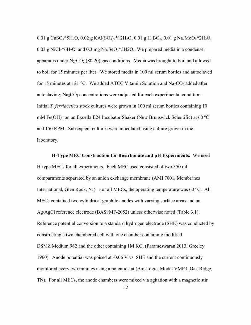
52
0.01 g CuSO4*5H2O, 0.02 g KAl(SO4)2*12H2O, 0.01 g H3BO3, 0.01 g Na2MoO4*2H2O,
0.03 g NiCl2*6H2O, and 0.3 mg Na2SeO3*5H2O. We prepared media in a condenser
apparatus under N2:CO2 (80:20) gas conditions. Media was brought to boil and allowed
to boil for 15 minutes per liter. We stored media in 100 ml serum bottles and autoclaved
for 15 minutes at 121 °C. We added ATCC Vitamin Solution and Na2CO3 added after
autoclaving; Na2CO3 concentrations were adjusted for each experimental condition.
Initial T. ferriacetica stock cultures were grown in 100 ml serum bottles containing 10
mM Fe(OH)3 on an Excella E24 Incubator Shaker (New Brunswick Scientific) at 60 ºC
and 150 RPM. Subsequent cultures were inoculated using culture grown in the
laboratory.
H-Type MEC Construction for Bicarbonate and pH Experiments. We used
H-type MECs for all experiments. Each MEC used consisted of two 350 ml
compartments separated by an anion exchange membrane (AMI 7001, Membranes
International, Glen Rock, NJ). For all MECs, the operating temperature was 60 °C. All
MECs contained two cylindrical graphite anodes with varying surface areas and an
Ag/AgCl reference electrode (BASi MF-2052) unless otherwise noted (Table 3.1).
Reference potential conversion to a standard hydrogen electrode (SHE) was conducted by
constructing a two chambered cell with one chamber containing modified
DSMZ Medium 962 and the other containing 1M KCl (Parameswaran 2013, Greeley
1960). Anode potential was poised at -0.06 V vs. SHE and the current continuously
monitored every two minutes using a potentiostat (Bio-Logic, Model VMP3, Oak Ridge,
TN). For all MECs, the anode chambers were mixed via agitation with a magnetic stir

53
bar. The cathode consisted of a single cylindrical graphite rod (0.3 cm diameter and a
total area of 6.67 cm2). Cathode pH was adjusted to 12 via addition of NaOH. Gas
collection bags were placed on the anode compartments to collect volatile products and
on the cathode to collect hydrogen.
Effect of pH on Current Density Experiments. To determine the effect of pH
on j, we used two replicated MECs with 50 mM bicarbonate buffer and 25 mM acetate as
the electron donor. Each MEC had an anode surface area of 3.89 cm2. At each pH
condition, after achieving a steady j, we altered the pH by the addition of HCl or NaOH.
Results were also used to obtain a pH range for current production for T. ferriacetica
when used during operation in MECs.
Effect of Bicarbonate Buffer on Current Density Experiments. To determine
the effect of bicarbonate buffer on j, we used four replicate MECs with varying
bicarbonate concentrations of 10, 25, 50, and 100 mM and 25 mM acetate as the electron
donor. We added new medium into each MEC by continuously flowing at a rate of ~4.5
mL s-1 until the old medium was completely replaced (~1.3 min hydraulic retention time).
Bicarbonate buffer concentration of 150 mM resulted in a decreased j. This is due to a
major limitation caused by the development of CO2 bubbles within the biofilm that
resulted in shearing (Figure 3.5). This limitation made it not possible to assess the
effect(s) or other limitation(s) resulting from increasing bicarbonate buffer beyond 100
mM bicarbonate. The anode surface areas for each MEC were: 6.78, 3.26, 3.02, and 3.64
cm2 respectively. At each buffer concentration, after achieving a steady j, bicarbonate

54
buffer was added by continuously flowing in 1 L of new medium containing 10, 25, 50,
or 100 mM bicarbonate. This experiment was conducted by either starting at 10 mM
bicarbonate and increasing to 100 mM, or by starting at 100 mM bicarbonate and
decreasing to 10 mM (Figure 3.4).
Thickness Measurements and Microscopic Analysis. Confocal laser scanning
microscopy (CSLM) was used to measure Lf per the protocol from Parameswaran (2013).
The protocal in this study differed in that all confocal images were acquired using an
upright Leica SP5 CSLM after applying LIVE/DEAD (BacLight Cell vitality kit,
Invitrogen, USA). For measuring Lf at various bicarbonate concentrations, we used an H-
type MEC with six anodes for a total surface area of 23.52 cm2. For growth MECs,
media consisted of 10 mM bicarbonate and was inoculated with scrapped biofilm from a
previous MEC. After a steady current was achieved, we replaced the medium with new
media containing 25, 50, and 100 mM bicarbonate. After reaching constant j at each
condition, cyclic voltammetry at 1 mV s-1 and 10 mV s-1 (data not shown) was performed
and an anode was sacrificed for CSLM. For imaging purposes, we place anodes in open
petri dishes containing fresh media and observed them using an HCX PL APO CS
20x/0.70NA immersion objective with a working distance of 260 µm. We also
performed scanning electron microscopy (SEM) on mature intact biofilms from two
MECs under 50 mM bicarbonate buffer and 25 mM acetate conditions. In addition, we
prepared SEM samples using scrapped biofilm on glass slides (Figure 3.8 A-D).
Preparation for SEM followed the protocol from Parameswaran (2013). We used an FEI

55
XL-30 environmental SEM (Philips) with an accelerating voltage of 5−20 kV and a
working distance of 8−10 mm.
Effect of salt and acetate on current density experiments. To confirm that j
was not influenced by ionic concentration, a mature biofilm was grown on an anode with
medium containing 50 mM bicarbonate and 25 mM acetate. The anode surface area was
1.47 cm2. Once a constant j was achieved, 10, 25, and 50 mM NaCl was added to the
media (Figure 3.6). To confirm that j was not influenced by acetate concentration, a
mature biofilm was grown on an anode with media containing 50 mM bicarbonate and 25
mM acetate. The anode surface area was 1.47 cm2. To confirm that acetate was a non-
limiting factor in all experiments, acetate was added to the reactor at 25, 50, 75, and 100
mM (Figure 3.7).
Results and Discussion
Effect of pH on Current Density Produced by T. ferriacetica Biofilms.
To determine the role that pH plays in limiting current density (j), T. ferriacetica
biofilms were grown on anodes with medium containing 50 mM bicarbonate as buffer
and 25 mM acetate as electron donor. Once the biofilms produced a constant j, bulk pH
was adjusted using either NaOH or HCl. Results were normalized vs j at ~pH 8.0 (jpH 8)
since this is the pH that indicated the highest current density (14.3 A m-2). Data from all
biofilms (n=2) is averaged in Figure 3.1 and indicates an appreciable j within pH range of
~pH 5.2 – 8.3. This range is larger than those reported for mesophilic Geobacter

56
sulfurreducens biofilms, especially showing that T. ferriacetica biofilms can produce
appreciable j at pH <6 (Torres 2008, Franks 2009).
Figure 3.1: Effect of pH on current density normalized to that produced at pH 8.0 (jpH 8
= 14.3 A m-2).
Figure 1 also indicates that j increases with increasing pH – an observation similar
to that for mesophilic Gram-negative biofilms (Torres 2008). j was the smallest at ~pH
5.2, where jpH 5.2 was ~20% (2.8 A m-2) of jpH 8. The biofilms produced ½ jpH 8 at pH ~6.0
(jpH 6 = 6.5 A m-2), and j increased consistently until ~pH 7.4, where it reached ~97% (jpH
7.4 = 13.9 A m-2) of the jpH 8 value. The impact pH had on j was less significant above pH
~7.4. Given that the pKa1 of bicarbonate is ~6.3 at 60 °C, the buffering capacity of
bicarbonate in the biofilm may be less significant once bulk pH reaches pH 7.4. Higher
than this pH, thermophilic ARB may have a limitation in j that is not directly related to

57
H+ diffusion and may be the result of pH inhibition caused by basic conditions in the
biofilm. pH inhibition may be observed in thermophilic ARB under high pH conditions
because H+ diffusion is significantly less limiting than in mesophilic biofilms due to the
increased rate of diffusion caused by higher temperature. Losses in j are likely associated
with pH inhibition of the biofilm caused by acidic conditions (Marcus 2011) in addition
to H+ diffusion limitations resulting from the buffering capacity of bicarbonate.
To monitor the impact of buffer concentrations on j, biofilms were grown on
anodes with media containing 25 mM acetate and varying bicarbonate buffer
concentrations. Biofilms were first grown with medium containing 10 mM bicarbonate
buffer and then replaced with new media containing progressively 25 mM, 50 mM, or
100 mM bicarbonate buffer. Each data point was acquired when the biofilms produced a
stable j (shown in Figure 3.2). j was normalized to j100 mM bicarbonate – the current density
of each biofilm at 100 mM bicarbonate (for j values, see Table 3.1). The j observed at 10
mM bicarbonate was ~61±6%, 25 mM was ~80±6%, and 50 mM was ~92±6% of j100 mM
bicarbonate.

58
Figure 3.2: Effect of bicarbonate buffer concentration on current density as normalized
to that produced with 100 mM bicarbonate buffer. Green box represents 10 mM
bicarbonate, blue box represents 25 mM bicarbonate, purple box represents 50 mM
bicarbonate, and red box indicates 100 mM bicarbonate buffer
To determine the statistical significance of each buffer condition, a one-way
analysis of variance (ANOVA) was conducted with a post-hoc Tukey honest significant
difference (HSD) test (Gleason 1999). ANOVA results indicate a p-value much lower
than 0.01 which indicates that one or more condition is significantly different from the
other. The Tukey HSD test results indicate that j increases with buffer (p<0.01) within the
10-25 mM bicarbonate concentration (pH = 6.9-7.1), but the increase becomes less
significant (p<0.05) in the 25 mM-50mM bicarbonate range (pH = 7.1-7.3), and
insignificant in the 50 mM- 100 mM bicarbonate range (pH = 7.3-7.8). For statistical
tables for Figure 3.2, see Appendix A.

59
Name Temp
(°C)
Potential
(V vs
SHE)
Anode
#
Surface
Area
(cm2)
j (A m-2)
10 mM
25
mM
50
mM
100
mM
RXR1 60 -0.06 2 6.78 8.8 10.9 13.2 15.5
RXR2 60 -0.06 2 3.26 5.0 7.8 9.29 9.4
RXR3 60 -0.06 2 3.02 6.1 7.2 8.31 8.7
RXR4 60 -0.06 2 3.64 6.4 8.2 8.81 10.4
Table 3.1: Results from reactors used for bicarbonate buffer experiments. Different
colored boxes correspond to color scheme from Figure 3.2.
These data show that, as reported in previous studies (Parameswaran 2013), T.
ferriacetica biofilms produce higher j at 10 mM bicarbonate buffer concentrations than
mesophilic ARB. This is extremely advantageous for dilute wastewaters that have low
alkalinities, as higher j is achievable with this ARB over G. sulfurreducens. Moreover,
the data reported in this study indicates that although H+ diffusion is a major limiting
factor at low buffer concentrations, it does not show a limitation at higher buffer
concentrations, as j in T. ferriacetica biofilms does not vary beyond 50 mM bicarbonate.
In comparison, G. sulfurreducens showed a significant increase in current densities with
increasing bicarbonate concentrations at all concentrations studied (0-100 mM, Marcus
2011).
Confocal laser scanning microscopy (CLSM) was employed to quantify the
impact of bicarbonate buffer concentrations on biofilm thickness (Lf) with results shown
in Figure 3.3. ANOVA results indicate a p-value much lower than 0.01 which indicates

60
that one or more condition is significantly different from the other. The Tukey-Kramer
HSD (Gleason 1999) test results indicate that Lf does not change with buffer within the
10-25 mM bicarbonate concentration range- indicating that Lf is not a major limitation-
with H+ diffusion likely as the primary limitation for j as shown in Figure 3.2. In addition,
increases (p<0.05) were observed in the 25 mM-50mM bicarbonate range and in the 50
mM- 100 mM bicarbonate range (p<0.05). Higher bicarbonate buffer concentrations
resulted in more varied and dynamic peaks and valleys. Lf values for live portions of the
biofilm are reported as 68±20 µm at 10 mM, 57±11 µm at 25 mM, 124±58 µm at 50 mM,
and 180±47 µm at 100 mM. This phenomenon has been predicted for mesophilic
biofilms and is attributed to the enhanced transport of H+ from the biofilm (Hunter 2005,
Marcus 2011). For raw images, see Appendix B. For statistical tables for Figure 3.3, see
Appendix C.

61
Figure 3.3: Effect of bicarbonate buffer concentration on biofilm thickness. Different
colored boxes correspond to color scheme from Figure 3.2.
To determine if Lf, rather than bicarbonate buffer, was governing the j for the
results shown in Figure 3.2, biofilms that had reached steady state at 100 mM bicarbonate
were fed new media with gradually decreasing bicarbonate buffer concentrations (100
mM, then 50 mM, 25 mM, and 10 mM) (Figure 3.4). Results indicate that Lf had no
significant impact on j at low or high buffer concentrations, but may have influenced j at
25 mM bicarbonate buffer. Similar to mesophilic G. sulfurreducens biofilms, T.
ferriacetica biofilms are not limited by Lf after reaching a steady j value (Robuschi 2013).
Results also indicate that H+ diffusion is not a major limitation in mature biofilms until
buffer concentration is as low as 10 mM bicarbonate (~pH 6.9) , compared to mesophilic

62
ARB biofilms, where 100 mM bicarbonate concentrations seem insufficient to alleviate
H+ transport limitations. This experiment also showed that limitations in j caused by H+
diffusion do not appear at high buffer concentrations in mature T. ferriacetica biofilms in
which pH > 7.3. This corroborates with the previous set of data that suggests that j in T.
ferriacetica biofilms not become limited by H+ diffusion until pH is lower than ~7.4.
Figure 3.4: Effect of buffer concentration on j as normalized to 100 mM bicarbonate for
both adding 10 mM bicarbonate to 100 mM (Up) and 100 mM bicarbonate to 10 mM
bicarbonate buffer (Down). The graph above compares the difference in j for two reactors
grown at 10 mM bicarbonate and 25 mM acetate. The two reactors were first grown at
increasing bicarbonate concentrations: 10 mM, 25 mM, 50 mM, and then 100 mM
(Figure 3.2) and then grown at decreasing bicarbonate concentrations: 100 mM, 50 mM,
25 mM, and 10 mM.

63
Bicarbonate buffer concentrations of 150 mM resulted in a decrease in j caused by
the formation of CO2 bubbles in sufficient quantities to stifle biofilm growth and cause
significant sloughing. Results are shown in Figure 3.5. For this reason, it was not possible
to measure the impact of bicarbonate buffer > 100 mM.
Figure 3.5: CO2 bubbles resulting from 150 mM bicarbonate buffer.
Effect of NaCl and Acetate Concentration on Current Density in T. ferriacetica
Biofilms.
In addition to pH and bicarbonate buffer concentration experiments, control
studies were established to assess the importance of ionic strength in the biofilm via
adding NaCl (Figure 3.6) and to determine if 25 mM acetate was limiting (Figure 3.7).
Results indicate that as NaCl was added at various concentrations from 0 mM (9.6 A m-2)
to 50 mM (9.6 A m-2) to biofilms grown with media containing 50 mM bicarbonate and
25 mM acetate, there was almost no change in j. Also, no significant change in j was
observed as acetate concentrations were increased from 25 mM (7.9 A m-2) to 100 mM

64
(7.5 A m-2) in biofilms grown with 50 mM bicarbonate. These results reveal that the
ionic strength of the media is not a major determining factor in j and that T. ferriacetica
biofilms may be tolerant to slightly halophilic conditions. This was expected because we
poised the anode potential and had placed the reference electrode adjacent to the anode.
This resulted in negligible Ohmic losses even at the lower ionic strength. Finally, these
results indicate 25 mM acetate is sufficient to be non-limiting in thermophilic biofilms of
thicknesses in the range of 124±58 µm.
Figure 3.6: NaCl concentration shows little effect on j.

65
Figure 3.7: Acetate concentration shows little effect on j.
Scanning Electron Microscopy of T. ferriacetica Biofilms
SEM was conducted to evaluate the morphological characteristics of a T.
ferriacetica biofilm grown under 50 mM bicarbonate conditions. (A) Shows a z-
directional cross section of the anode at 1000X magnification with the anode highlighted
with a white arrow- a thick biofilm can be observed. (B) Shows a biofilm sample that was
transferred onto a glass slide and magnified at 25,000X. Appendages can be observed
protruding from the cells. (C) Shows a stack of cells growing on an anode at 15,000X.
(D) Shows a broad overview of the biofilm attached to the anode taken at 38X
magnification.

66
Figure 3.8A-D: Scanning Electron Micrograms for T. ferriacetica biofilm.
Conclusion
The impacts of bicarbonate buffer concentrations and pH on j in anode biofilms of
T. ferriacetica indicate that, as with anode biofilms composed of mesophilic ARB, H+
transport and pH inhibition are significant limiting factors up to a pH 7.4 and that Lf does
not play a major role in j after steady state j is achieved. However, some important
differences from G. sulfurreducens were identified. T. ferriacetica has a wider pH range,
allowing current generation at lower bicarbonate buffer concentrations and pH values as

67
low as 5.2. This gives T. ferriacetica an advantage to produce higher j despite pH
gradients in the biofilms.
Most importantly, we observe a maximum j that is constant at 50-100 mM buffer
concentrations and pH values above ~7.4. This observation suggests that, while there is a
H+ transport limitation, this limitation is mitigated at certain conditions or does not
completely govern j. As a consequence, a second limitation has been identified, at
current densities of ~9.9 ± 2.2 A m-2. It is possible that electron transport is the main
limitation at higher j, given the extensive amount of studies that demonstrate potential
gradients in G. sulfurreducens. Nonetheless, most conditions at which G. sulfurreducens
is studied at fall under a H+ transport limitation, as shown by (Marcus 2011). Thus, T.
ferriacetica provides conditions that might be better suited for studying EET by ARB by
elucidating the limitations experienced under high turnover conditions when H+ diffusion
is no longer the major limiting factor.
Chapter 3 References:
1. Beveridge, TJ, Forsberg, CW, Doyle, RJ. 1982. Major sites of metal binding in
Bacillus licheniformis walls. J. Bacteriol. 150:1438-1448.
2. Beveridge, TJ, Murray, RG. 1980. Sites of metal deposition in the cell wall of
Bacillus subtilis. J. Bacteriol. 141:876-887.
3. Ehrlich, HL. 2008. Are gram-positive bacteria capable of electron transfer across
their cell wall without an externally available electron shuttle? Geobiology.
6:220-224. doi: 10.1111/j.1472-4669.2007.00135.x.

68
4. Franks AE, Nevin KP, Jia H, Izallalen M, Woodard TL, Lovley D R. 2009. Novel
strategy for three-dimensional real-time imaging of microbial fuel cell
communities: Monitoring the inhibitory effects of proton accumulation within the
anode biofilm. Energy and Environmental Science, 2(1), 113-119.
doi:10.1039/b816445b
5. Gleason, JR. 1999. An accurate, non-iterative approximation for studentized
range quantiles. Computational Statistics and Data Analysis. 31:147-158. doi:
10.1016/S0167-9473(99)00002-X.
6. Hunter, RC, Beveridge, TJ. 2005. Application of a pH-Sensitive Fluoroprobe (C-
SNARF-4) for pH Microenvironment Analysis in Pseudomonas aeruginosa
Biofilms. Appl. Environ. Microbiol. 71:2501-2510. doi: 10.1128/AEM.71.5.2501-
2510.2005.
7. Marcus, AK, Torres, CI, Rittmann, BE. 2011. Analysis of a microbial
electrochemical cell using the proton condition in biofilm (PCBIOFILM) model.
Bioresour. Technol. 102:253-262. doi: 10.1016/j.biortech.2010.03.100.
8. Marcus, AK, Torres, CI, Rittmann, BE. 2007. Conduction-based modeling of the
biofilm anode of a microbial fuel cell. Biotechnol. Bioeng. 98:1171-1182. doi:
10.1002/bit.21533.
9. Marshall, CW, May, HD. 2009. Electrochemical evidence of direct electrode
reduction by a thermophilic Gram-positive bacterium, Thermincola ferriacetica.
Energy and Environmental Science. 2:699-705. doi: 10.1039/b823237g.
10. Mathis, BJ, Marshall, CW, Milliken, CE, Makkar, RS, Creager, SE, May, HD.
2008. Electricity generation by thermophilic microorganisms from marine
sediment. Appl. Microbiol. Biotechnol. 78:147-155. doi: 10.1007/s00253-007-
1266-4.
11. May, HD, Shimotori T. 2009. U.S. Patent No. 0017512 A1. Austin, TX: U.S.
Patent and Trademark Office.
12. Parameswaran, P, Bry, T, Popat, SC, Lusk, BG, Rittmann, BE, Torres, CI. 2013.
Kinetic, electrochemical, and microscopic characterization of the thermophilic,
anode-respiring bacterium Thermincola ferriacetica. Environmental Science and
Technology. 47:4934-4940. doi: 10.1021/es400321c.

69
13. Pham, TH, Boon, N, Aelterman, P, Clauwaert, P, De Schamphelaire, L,
Vanhaecke, L, De Maeyer, K, Höfte, M, Verstraete, W, Rabaey, K. 2008.
Metabolites produced by Pseudomonas sp. enable a Gram-positive bacterium to
achieve extracellular electron transfer. Appl. Microbiol. Biotechnol. 77:1119-
1129. doi: 10.1007/s00253-007-1248-6.
14. Reguera, G, Nevin, KP, Nicoll, JS, Covalla, SF, Woodard, TL, Lovley, DR. 2006.
Biofilm and Nanowire Production Leads to Increased Current in Geobacter
sulfurreducens Fuel Cells. Appl. Environ. Microbiol. 72:7345-7348. doi:
10.1128/AEM.01444-06.
15. Robuschi, L, Tomba, JP, Schrott, GD, Bonanni, PS, Desimone, PM, Busalmen,
JP. 2013. Spectroscopic Slicing to Reveal Internal Redox Gradients in Electricity-
Producing Biofilms: 1. Angewandte Chemie. 52:925.
16. Torres, CI, Kato Marcus, A, Rittmann, BE. 2008. Proton transport inside the
biofilm limits electrical current generation by anode‐respiring bacteria.
Biotechnol. Bioeng. 100:872-881. doi: 10.1002/bit.21821.
17. Torres, CI, Marcus, AK, Parameswaran, P, Rittmann, BE. 2008. Kinetic
experiments for evaluating the nernst-monod model for anode-respiring bacteria
(ARB) in a biofilm
anode. Environmental Science and Technology. 42:6593-6597. doi:
10.1021/es800970w.
18. Wrighton, KC, Thrash, JC, Melnyk, RA, Bigi, JP, Byrne-Bailey, KG, Remis, JP,
Schichnes, D, Auer, M, Chang, CJ, Coates, JD. 2011. Evidence for Direct
Electron Transfer by a Gram-Positive Bacterium Isolated from a Microbial Fuel
Cell. Appl. Environ. Microbiol. 77:7633-7639. doi: 10.1128/AEM.05365-11.
19. Yoho, R, Popat, S, Torres, C. 2014. Dynamic Potential-Dependent Electron
Transport Pathway Shifts in Anode Biofilms of Geobacter sulfurreducens.
Chemsuschem. 7:3413-3419. doi: 10.1002/cssc.201402589.
20. Zavarzina, DG, Sokolova, TG, Tourova, TP, Chernyh, NA, Kostrikina, NA,
Bonch-Osmolovskaya, EA. 2007. Thermincola ferriacetica sp. nov., a new
anaerobic, thermophilic, facultatively chemolithoautotrophic bacterium capable of
dissimilatory Fe(III) reduction. Extremophiles. 11:1-7. doi: 10.1007/s00792-006-
0004-7.

70
Chapter 4: pH Shifts in the Anode Potential Response from Thermincola ferriacetica
Suggest the Presence of a Rate Limiting Proton-Coupled Electron Transfer Protein3
Overview
Thermincola ferriacetica, a thermophilic, Gram-positive, anode respiring
bacterium (ARB) was grown in biofilms in microbial electrochemical cells (MXCs) to
investigate its external electron-transport (EET) limitations. Electrochemical studies,
including cyclic voltammetry (CV), are often used to elucidate the rate limiting step of
electron transport in ARB. Previously reported CV analysis of T. ferriacetica indicated a
sigmoidal Nernst-Monod response in electrical current (j) to changes in anode potential
(V). This response suggests that a single proton (H+) coupled electron (n = 1) transport
reaction is responsible for the rate-limiting step in T. ferriacetica metabolism. The
specific protein responsible for this response is thought to be a c-type cytochrome.
Although T. ferriacetica has been shown to contain 35 c-type cytochromes, the one(s)
responsible for EET has yet to be identified. Here, we show that T. ferriacetica’s
response under certain growth conditions is composed of at least two separate n = 1
Nernst-Monod relationships; suggesting the presence of more than one pathway for
anode respiration. By altering bulk pH, it is revealed that biofilms in neutral to high pH
(6.9-8.3) show a very broad redox peak while biofilms in low pH (5.2) reveal multiple
redox peaks. In addition, bicarbonate buffer concentrations show a similar trend, with
lower bicarbonate leading to the presence of multiple redox peaks; consistent with pH
gradients developing inside the T. ferriacetica biofilm. Finally, the redox respose is
3Chapter 4 unpublished with Bradley G. Lusk, Prathap Parameswaran, Sudeep Popat,
Bruce E. Rittmann, Cesar I. Torres

71
analyzed as current density increases in T. ferriacetica biofilms- revealing the presence of
multiple redox peaks as pH gradients form within the biofilm. This chapter reveals that T.
ferriacetica contains more than one H+ coupled EET pathway and that EET pathways
within T. ferriacetica are sensitive to changes in bulk pH.
Introduction
Performance of electrode respiration by anode respiring bacteria (ARB) in
microbial electrochemical cells (MXCs) is often monitored using a potentiostat equipped
with the capability of applying an electrochemical technique known as low scan cyclic
voltammetry (LSCV). LSCV analysis occurs when a potential (V) sweep is performed
and the corresponding current (j) is measured in response to that V, referred to as a j-V
response (Badalamenti 2013, Marsili 2010, Parameswaran 2013, Srikanth 2008, Yang
2012, Yoho 2014). With ARB, respiration occurs through electron transfer from the
electron transport chain of the microorganism to the anode. A change in V at the anode
alters the energy available to ARB via anode respiration, thus allowing us to monitor the
metabolic energetics of redox active pathways in ARB (Fricke 2008, Yoho 2014). In this

72
chapter, LSCV at 1 mV s-1 was used to monitor the j-V response of T. ferriacetica
biofilms to characterize the midpoint potentials (Eka) of the redox processes occurring
within anode biofilms under various pH conditions and bicarbonate buffer
concentrations.
Many mechanisms for extracellular electron transfer (EET), including indirect
shuttling, direct contact, and direct long-range transfer via a solid conductive matrix have
been reported in Gram-negative mesophilic ARB, with little known in regard to Gram-
positive ARB (Lovley 2008, Mohan 2014, Schroder 2007, Torres 2010, Parameswaran
2013). ARB that perform EET via direct contact often give a sigmoidal Nernst-Monod
response with an n = 1 (Marcus 2007). This indicates that the mechanism through which
EET occurs in these biofilms is a single H+ coupled electron transport process (Torres
2010). T. ferriacetica has previously been indicated as an ARB capable of performing
long-range direct EET via a conductive extracellular matrix with a Nernst-Monod fit of n
= 1 (Marcus 2007).
It is well documented that H+ transport within the biofilm anode is one of the
primary limitations for anodic MXC performance (Torres 2008, Marcus 2011). Previous
studies with Geobacter sulfurreducens indicate that current production in MXCs is
enhanced by increasing either bicarbonate or phosphate buffer concentrations up to 100
mM- suggesting that pH gradients are the main kinetic limitation in these biofilms
(Torres 2008). However, as shown in Chapter 3, T. ferriacetica biofilms are only limited
at low bicarbonate buffer concentrations (< 50 mM) due to their lower pH limit for

73
growth and faster transport kinetics at high pH when compared to mesophilic G.
sulfurreducens. The lack of H+ transport limitations at high pH and buffer concentrations
in T. ferriacetica biofilms is advantageous for studying electron transport kinetics in
biofilm anodes.
Previous studies with mesophilic ARB have reported that redox active proteins,
including multiheme c-type cytochromes, are primarily responsible for direct electron
transfer to electrodes by ARB (Carlson 2012, Leang 2003). In addition, electrochemical
analysis of many ARB, including G. sulfurreducens and Geoalkalibacter ferrihydriticus,
is now revealing the presence of multiple EET pathways with Nernst-Monod responses
working simultaneously to perform anode respiration (Yoho 2014, Yoho 2015). T.
ferriacetica contains 35 multiheme c-type cytochromes (Lusk 2015) and thus may
contain multiple pathways for EET. In this chapter, we investigate the electrochemical
response of T. ferriacetica and its dependence on pH conditions, buffer concentrations,
and current density. We observe a pH dependence of the electrochemical response of T.
ferriacetica including the appearance of multiple redox peaks during LSCV analysis,
suggesting H+ coupled EET. We hypothesize that shifts in Eka and the presence of
multiple peaks during LSCV under certain conditions are the result of the expression of
multiple multiheme c-type cytochromes in either the same pathway or multiple pathways.
Materials and Methods
Growth Media and culture conditions. Please refer to Chapter 3.
H-Type MEC Construction. Please refer to Chapter 3.

74
Effect of pH on Midpoint Potential Experiments. To determine the effect of pH
Eka, two MECs were constructed and operated using 50 mM bicarbonate buffer and 25
mM acetate as the electron donor. Each MEC had an anode surface area of 3.89 cm2. At
each pH condition, after achieving a steady j, pH was altered by either the addition of
HCl or NaOH. LSCV was performed at 1.0 mV s-1 and 10 mV s-1 (Figure 4.1a-b) for
various pH values to determine the effect of pH on Eka.
Effect of Bicarbonate Buffer on Midpoint Potential Experiments. To
determine the effect of bicarbonate buffer on Eka, four MECs were constructed and
operated using 10, 25, 50, and 100 mM bicarbonate buffer with 25 mM acetate as the
electron donor. New medium was added by continuously flowing at a rate of ~4.5 mL s-1
until the old medium was completely replaced. The anode surface areas for each were:
6.78, 3.26, 3.02, and 3.64 cm2 respectively. At each buffer condition, after achieving a
steady j, LSCVs were performed at 1.0 mV s-1 (Figure 4.2a-b) and 10 mV s-1 (data not
shown). After LSCV, bicarbonate buffer was added by continuously flowing in 1 L of
new medium containing 10, 25, 50, or 100 mM bicarbonate. This experiment was
conducted by either starting at 10 mM bicarbonate and increasing to 100 mM or by
starting at 100 mM bicarbonate and decreasing to 10 mM.
Effect of Current Density on Midpoint Potential Experiments. To test
whether Lf could be a determining factor in Eka, an MEC was constructed using 25 mM
acetate, 50 mM bicarbonate, and 3 mL inoculum. This MEC had an anode surface area
of 3.89 cm2 and was monitored over 36 hours as j increased from 1 A m-2 to 12.8 A m-2.
75
LSCV was conducted at a scan rate of 1.0 mV s-1 (Figure 4.3a-b) and 10 mV s-1 (data not
shown) at each gain in j of ~1 A m-2. Samples were taken for pH before and after the
experiment.
Non-Turnover low scan cyclic voltammetry. To analyze the presence of
multiple redox peaks under non-turnover conditions, biofilms were established on anodes
with media containing 50 mM bicarbonate buffer and 25 mM acetate. Anode surface
areas were 3.02 and 3.64 cm2. After a biofilm was established, the medium was replaced
with new medium containing 50 mM bicarbonate buffer but no acetate. LSCVs were
performed at 1.0 mV s-1 (Figure 4.5) and 10 mV s-1 (data not shown).
Results and Discussion
Effect of Direct pH Manipulation on Midpoint Potential in T. ferriacetica Biofilms.
To determine the effect of pH on midpoint potential (EKA), low scan cyclic
voltammetry (LSCV) (1.0 mV s-1 and 10 mV s-1) was conducted under the 50 mM
bicarbonate, 25 mM acetate condition as pH was adjusted. Results are standardized to
the derivative value divided by the maximum derivative value (D/Dmax). Results, shown
in Figure 4.1a-b, indicate a shift in apparent EKA towards negative values with increasing
pH. Apparent EKA values from Figure 4.1a (CV at 10 mV s-1), show that an ~80 mV
negative shift in EKA was observed from pH 5.2 (-0.19 V vs SHE) to 8.3 (-0.27 V vs
SHE). This indicates that the biofilm is receiving less energy from anode respiration as
pH decreases and the theoretical potential of acetate increases (Katuri et al 2010, Logan
2006). Despite a similar pH change (~1.4 pH units) the ~60 mV EKA shift between pH

76
5.2 and pH 6.9 (-0.25 V vs SHE) is much more pronounced than the 20 mV shift between
pH 6.9 and pH 8.3.
Figure 4.1a-b: (a) Derivative LSCV (10 mV s-1) as a function of pH. Colored vertical
arrows indicate midpoint potential(s) and colored horizontal arrows indicate range of
(a)
(b)

77
midpoint potential. (b) Derivative LSCV (1.0 mV s-1) as a function of pH. The hashed
black line in (a) and (b) differentiates the forward (top) and reverse (bottom) LSCV
scans. Colors are based on pH values: green = pH 5.2, blue = pH 6.9, and purple = pH
8.3.
When comparing the EKA values for the forward (above hashed black line) and
reverse sweeps from Figure 4.1a (below hashed black line), the derivative plot reveals
similar apparent EKA values for pH 6.9 and 8.3. However, two separate peaks were
observed at pH 5.2 - with EKA values of -0.19 V vs SHE and -0.24 V vs SHE (EKA values
are indicated with colored vertical arrows). For the pH 6.9 and 8.3 conditions, a midpoint
range of ~250 mV is observed. For the pH 5.2 condition, this range is closer to ~200 mV
(midpoint ranges are represented with colored horizontal arrows). The observation of a
larger potential range may indicate that the biofilm is using a larger sweet of cytochromes
at pH values > pKa1 of bicarbonate.
In addition, the observation of multiple peaks (including the previously reported
EKA for T. ferriacetica at -0.12 V vs SHE (Parameswaran 2013)) is emphasized in the 1.0
mV s-1 CV shown in Figure 4.1b, suggesting that T. ferriacetica biofilms may be using
multiple cytochromes with non-equivalent heme groups (Katuri et al 2010) or multiple
pathways for anode respiration (Yoho 2014). The presence of multiple redox peaks
during various stages of biofilm development has been reported in mesophilic G.
sulfurreducens and Geoalkalibacter ferrihydriticus biofilms as a result of differential
expression of redox associated proteins including multiheme c-type cytochromes (Fricke

78
2008, Katuri 2010, Leang 2003, Yoho 2014, Yoho 2015). Under conditions in which pH
is non-limiting, many of these pathways may be expressed and utilized simultaneously,
leading to an apparent EKA that is an average of several cytochromes or redox pathways.
However, as pH drops, the conditions become limiting and the biofilm obtains less
energy from anode respiration. This may catalyze T. ferriacetica biofilms to channel
electrons only through pathways that are energetically favorable for these conditions.
This selective pressure may have resulted in the utilization of fewer pathways by the
biofilm for anode respiration - yielding a decreased midpoint range and the appearance of
multiple redox peaks.
This hypothesis is supported in Figures 4.1a-b. By observing the forward CV
sweep (above the black hashed line), the pH 5.2 condition reveals the presence of two
distinct redox peaks. As the V is increased during the forward sweep j is driven more
positive, resulting in the release of H+ causing acidification within the biofilm and the
development of a H+ gradient. The reverse j-V response shown in Figure 4.1a-b reveals
that one of the redox pathways is greatly inhibited by the acidic conditions created within
the biofilm during the forward V sweep. It may also be the case that T. ferriacetica
biofilms have only two pathways for electron transfer, and the shift in EKA from altering
pH makes these two pathways more distinguishable. Further studies, including the use of
electrochemical impedance spectroscopy (EIS) (Yoho2014), are needed to verify this
hypothesis.

79
Effect of Changing Bicarbonate Buffer Concentration on Midpoint Potential in T.
ferriacetica Biofilms.
LSCVs were performed as the buffer concentration was increased to analyze if
any shifts in EKA were present (Figure 4.2a-b show derivative CVs for bicarbonate
experiment reactors shown in Chapter 3, Figure 3.4). Looking at the reverse scan of
Figure 4.2a shows that the EKA exhibits a ~40 mV negative shift between 10 mM (-0.26 V
vs SHE) to 100 mM bicarbonate (-0.30 V vs SHE). The 40 mV negative shift in EKA
produced by this experiment is the result of a pH shift from ~6.9-7.8. LSCVs were also
conducted at 1 mV s-1 on each reactor as bicarbonate concentration was decreased
(Figure 4.2b). During the course of this experiment, pH shifted from ~6.7-7.8. However,
the EKA shift from 100 mM bicarbonate (-0.30 V vs SHE) to 10 mM bicarbonate (-0.285
± 0.005 V vs SHE) was ~15 ± 5 mV. These results give a clear indication that the change
in EKA from decreasing buffer concentration in a fully established biofilm is less
pronounced than when changing from a low buffer to high buffer in a new biofilm. The
decreased shift in EKA that occurs while lowering bicarbonate buffer concentration is the
result of decreased H+ diffusion from the biofilm that may be due to increased Lf (> 150
µm) caused by first growing the biofilm at 100 mM bicarbonate (See Chapter 3, Figure
3.3). Under these conditions, the pH observed in the bulk is different from the pH
experienced in the biofilm. For an overview of results, see Table 4.1.

80
Figure 4.2a-b: Derivative LSCVs (1.0 mV s-1) for increasing bicarbonate buffer
conditions. (b) Derivative LSCVs (1.0 mV s-1) for decreasing bicarbonate buffer
conditions. Different colored lines correspond to the bicarbonate concentrations: red =
(a)
(b)

81
100 mM, purple = 50 mM, blue = 25 mM, and green = 10 mM. Hashed black line
differentiates the forward and reverse LSCV scans. Colored vertical arrows indicate
midpoint potential(s) and colored horizontal arrows indicate range of midpoint potential.
10 mM
Bicarbonate 25 mM
RXR3 Eka pH Age (d) j (A m-2) Eka pH Age
(d)
j (A m-2)
Up -0.26 6.9 35 6.1 -0.28 7.2 43 7.2
Down -0.28 7.2 64 7.1 -0.29 7.3 62 8.0
RXR4
Up -0.26 6.9 5 6.4 -0.28 7.1 13 8.2
Down -0.27 6.7 31 6.8 -0.29 7.3 29 9.2
50 mM
100 mM
RXR3 Eka pH Age (d) j (A m-2) Eka pH Age
(d)
j (A m-2)
Up -0.29 7.4 46 8.3 -0.30 7.8 59 8.7
Down -0.30 7.7 61 8.1 -0.30 7.8 59 8.7
RXR4
Up -0.28 7.3 19 8.8 -0.30 7.8 26 10.4
Down -0.30 7.5 28 9.9 -0.30 7.8 26 10.4
Table 4.1: List of values for reactors used in midpoint experiments and increasing (Up)
vs decreasing (Down) bicarbonate buffer concentration experiments. Different colored
boxes correspond to color scheme from Figure 4.2a-b.
Figure 4.2a shows that multiple peaks are present at 10, 25, and 50 mM
bicarbonate buffer concentrations. These multiple peaks are present, but less pronounced
when reducing buffer concentration from 100 mM to 10 mM bicarbonate (Figure 4.2b).
The range of the EKA at 100 mM bicarbonate buffer is ~250 mV, a similar range to that

82
observed for high pH (pH ≥ 6.9) conditions in the previous experiments. The EKA range
for the 10 mM bicarbonate buffer is ~225 mV, with lower pH again corresponding to a
decreased midpoint range. The presence of multiple EKA values at low buffer
concentrations in T. ferriacetica biofilms is due to differences in pH as discussed
previously. As in the pH experiments, lower buffer is associated with lower j as a result
of pH inhibition of the biofilm and the increased potential of acetate which limits the
pathways through which the biofilm can utilize acetate for cellular metabolism.
Effect of Current Density on Midpoint Potential in Growing T. ferriacetica Biofilms.
The effect of j on the EKA during early development in T. ferriacetica biofilms
was assessed at 25 mM acetate as electron donor and 50 mM bicarbonate buffer
conditions since these concentrations were shown to be non-limiting in Chapter 3,
Figures 3.6 and 3.7. For this experiment, j was monitored over ~36 hours as it increased
from 0 A m-2 to 12.8 A m-2. LSCV analysis was conducted at a scan rate of 1.0 mV s-1
(results shown in Figure 4.3a) at each data point. The derivative results are shown in
Figure 4.3b to reveal shifts in EKA. Over the course of this experiment, there was a
modest 10 mV positive shift in EKA as pH decreased from ~7.4 (-0.25 V vs SHE) to ~7.0
(-0.24 V vs SHE). This reveals that, similar to G. sulfurreducens biofilms (Yoho 2014),
increased j results in changing EKA values. However, contrary to mesophilic biofilms, the
EKA values T. ferriacetica biofilms shift more positive with increased j, with this shift
attributed to changes in pH. Figure 4.3b shows that T. ferriacetica biofilms contain
multiple redox peaks over a range of ~250 mV during early stages of development, with
multiple peaks becoming more pronounced as pH decreases.

83
Figure 4.3a-b: (a) Effect of current density on EKA in growing biofilm. (b) Derivative
LSCV (1.0 mV s-1) for effect of current density on EKA in growing biofilm. Black vertical
(a)
10 mV (b)
84
arrows indicate midpoint potential(s) and black horizontal arrows indicate range of
midpoint potential. Black arrow on top of graph indicates the 10 mV negative shift during
the course of the experiment. Hashed black line differentiates the forward and reverse
LSCV scans. Colors in (a) and (b) are consistent and are used to delineate the current
density conditions of the LSCV scan.
Nernst-Monod Analysis of Multiple EET Pathway Hypothesis for T. ferriacetica.
In order to determine if T. ferriacetica is using two or more H+ coupled EET
pathways simultaneously, the Nernst-Monod model was eployed (Marcus 2007, Yoho
2015). Using the Nernst-Monod model, we determined that at least two EET pathways
that conduct a single electron, single H+ (n = 1) transfer are present in T. ferriacetica
biofilms (Figure 4.4). Figure 4.4 shows the D/Dmax value of LSCVs (1.0 mV s-1) for T.
ferriacetica biofilms that were producing 11 and 12 A m-2 at pH ~7 respectively.
Figure 4.4 shows that assuming only one EET pathway with a Nernst-Monod at n
= 1 (either only P1 or only P2) does not have a very good fit to the data (j = 11 A m-2 or j
= 12 A m-2) as is shown by P1 and P2. However, under the assumption that P1 and P2
(taken from the multiple peaks present in figure 4.1a-b) are working simultaneously to
perform EET (red hashed line), the Nernst-Monod model shows a good fit to the data.
This analysis suggests that T. ferriacetica is performing anode respiration with at least
two EET mechanisms simultaneously.

85
Figure 4.4: Nernst- Monod fit for D/Dmax value of LSCVs (1.0 mV s-1) for T. ferriacetica
biofilms that were producing 11 and 12 A m-2 at pH ~7 respectively. Purple line =
biofilm producing 11 A m-2, blue line = biofilm producing 12 A m-2, grey hashed line
(P1) is Nernst-Monod fit for hypothesized EET pathway 1, grey solid line (P2) is Nernst-
Monod fit for hypothesized EET pathway 2, and red hashed line (P1+ P2) is Nernst-
Monod fit for simultaneous expression of hypothesized EET pathway 1 and 2. Hashed
black line differentiates the forward and reverse LSCV scans.
Midpoint Potentials Under Non-Turnover Conditions in T. ferriacetica Biofilms.
LSCV under non-turnover conditions (Figure 4.5) allowed us to monitor the EKA
values of the redox processes occurring within a biofilm anode without the presence of
acetate at pH ~7.0. For this experiment, the j-V response revealed the potentials at which
86
the electron transfer pathway(s) within the biofilm were oxidized and reduced by the
electrode. Multiple redox peaks were observed over a ~250 mV potential range under
non-turnover conditions. This range of redox peaks correlates with the ones observed
under turnover conditions. These results indicate the possibility of either separate
pathways for anode respiration or multiple redox proteins, including c-type cytochromes,
within the same pathway (Fricke 2008, Yoho 2014). Further experiments observing
potential shifts in apparent EKA under non-turnover conditions will help to elucidate the
importance of changing pH in the energetics of the redox processes associated with the
proteins or cytochromes responsible for EET.

87
Figure 4.5: Non-Turnover LSCV (1.0 mV s-1) at 50 mM bicarbonate and 0 mM acetate.
Blue vertical arrows indicate midpoint potential(s). Hashed black line differentiates the
forward and reverse LSCV scans.
Conclusion
For all conditions studied, pH was a major contributor to altering energetics
within the biofilm anode, resulting in a shift in Eka values. This suggests that, similar to
mesophilic biofilms (Marcus 2011), the pH condition within T. ferriacetica biofilms
plays a critical role in associated EET pathway(s). This phenomenon is observed due to
the H+ coupled reaction for current production in biofilms composed of ARB, in which
the transfer of electrons is associated with the release of H+. For example, increasing pH
or bicarbonate buffer concentration indicated a negative shift in Eka, a broadening of the
88
midpoint potential range, and an increase in j. These phenomena likely occur since these
conditions enhance H+ diffusion out of the biofilm and limit pH inhibition (Marcus
2011). However, at pH conditions <~7, the midpoint range shrinks and more distinct
redox peaks appear in LSCV analysis. Although the positive shifts in Eka as pH decreases
may be accounted for by the theoretical ~66 mV shift in acetate potential for every pH
unit, the emergence of multiple Eka values and a shrinking midpoint range cannot.
Lower bicarbonate buffer concentrations (10-25 mM) and pH values (<~7)
enhance the occurrence of multiple redox peaks in T. ferriacetica biofilms. In addition,
non-turnover CV analysis indicates multiple redox peaks are present in T. ferriacetica
that correlate with Eka values observed in biofilms grown at low pH and low buffer
concentrations under turnover conditions. This data suggests that T. ferriacetica uses
multiple redox pathways or multiple redox steps for anode respiration that may be
associated with T. ferriacetica’s repertoire of 35 c-type cytochromes. Little is known
about the EET mechanism(s) in Gram-positive bacteria- the presence of a large
peptidoglycan layer make these mechanisms even more elusive. Future studies should
analyze the role peptidoglycan plays in EET and overpotential more closely to help
elucidate the limitations experienced under high turnover conditions when H+ diffusion is
no longer the major limiting factor.
89
Chapter 4 references:
1. Badalamenti, JP, Krajmalnik-Brown, R, Torres, CI. 2013. Generation of high
current densities by pure cultures of anode-respiring Geoalkalibacter spp. Under
alkaline and saline conditions in microbial electrochemical cells. Mbio. 4:e00144-
13-e00144-13. doi: 10.1128/mBio.00144-13.
2. Carlson, HK, Iavarone, AT, Gorur, A, Yeo, BS, Tran, R, Melnyk, RA, Mathies,
RA, Auer, M, Coates, JD. 2012. Surface multiheme c-type cytochromes from
Thermincola potens and implications for respiratory metal reduction by Gram-
positive bacteria. Proc. Natl. Acad. Sci. U. S. A. 109:1702-1707. doi:
10.1073/pnas.1112905109.
3. Fricke, K, Harnisch, F, Schröder, U. 2008. On the use of cyclic voltammetry for
the study of anodic electron transfer in microbial fuel cells. Energy and
Environmental Science. 1:144-147. doi: 10.1039/b802363h.
4. Katuri, KP, Kavanagh, P, Rengaraj, S, Leech, D. 2010. Geobacter sulfurreducens
biofilms developed under different growth conditions on glassy carbon electrodes:
Insights using cyclic voltammetry. Chemical Communications. 46:4758-4760.
doi: 10.1039/c003342a.
5. Leang, C, Coppi, MV, Lovley, DR. 2003. OmcB, a c-Type Polyheme
Cytochrome, Involved in Fe(III) Reduction in Geobacter sulfurreducens. J.
Bacteriol. 185:2096-2103. doi: 10.1128/JB.185.7.2096-2103.2003.
6. Logan, BE, Hamelers, B, Rozendal, R, Schroder, U. 2006. Microbial Fuel Cells:
Methodology and Technology. Environ. Sci. Technol. 40:5181.
7. Lovley, DR. 2008. The microbe electric: conversion of organic matter to
electricity. Curr. Opin. Biotechnol. 19:564-571. doi:
10.1016/j.copbio.2008.10.005.
8. Lusk, BG, Badalamenti, JP, Parameswaran, P, Bond, DR, Torres, CI. 2015. Draft
Genome Sequence of the Gram-Positive Thermophilic Iron Reducer Thermincola
ferriacetica Strain Z-0001T. Genome Announcements. 3:.

90
9. Marcus, AK, Torres, CI, Rittmann, BE. 2011. Analysis of a microbial
electrochemical cell using the proton condition in biofilm (PCBIOFILM) model.
Bioresour. Technol. 102:253-262. doi: 10.1016/j.biortech.2010.03.100.
10. Marcus, AK, Torres, CI, Rittmann, BE. 2007. Conduction-based modeling of the
biofilm anode of a microbial fuel cell. Biotechnol. Bioeng. 98:1171-1182. doi:
10.1002/bit.21533.
11. Marsili, E, Sun, J, Bond, DR. 2010. Voltammetry and growth physiology of
Geobacter sulfurreducens biofilms as a function of growth stage and imposed
electrode potential. Electroanalysis. 22:865-874. doi: 10.1002/elan.200800007.
12. Mohan, S, Velvizhi, G, Krishna, K, Babu, M. 2014. Microbial catalyzed
electrochemical systems: A bio-factory with multi-facet applications. Bioresour.
Technol. 165:355-364. doi: 10.1016/j.biortech.2014.03.048.
13. Parameswaran, P, Bry, T, Popat, SC, Lusk, BG, Rittmann, BE, Torres, CI. 2013.
Kinetic, electrochemical, and microscopic characterization of the thermophilic,
anode-respiring bacterium Thermincola ferriacetica. Environmental Science and
Technology. 47:4934-4940. doi: 10.1021/es400321c.
14. Schroder, U. 2007. Anodic electron transfer mechanisms in microbial fuel cells
and their energy efficiency. Physical Chemistry Chemical Physics. 9:2619-2629.
doi: 10.1039/003627m.
15. Srikanth, S, Marsili, E, Flickinger, MC, Bond, DR. 2008. Electrochemical
characterization of Geobacter sulfurreducens cells immobilized on graphite paper
electrodes. Biotechnol. Bioeng. 99:1065-1073. doi: 10.1002/bit.21671.
16. Torres, CI, Kato Marcus, A, Rittmann, BE. 2008. Proton transport inside the
biofilm limits electrical current generation by anode‐respiring bacteria.
Biotechnol. Bioeng. 100:872-881. doi: 10.1002/bit.21821.
17. Torres, CI, Marcus, AK, Parameswaran, P, Rittmann, BE. 2008. Kinetic
experiments for evaluating the nernst-monod model for anode-respiring bacteria
(ARB) in a biofilm anode. Environmental Science and Technology. 42:6593-
6597. doi: 10.1021/es800970w.

91
18. Torres, CI, Marcus, AK, Lee, H, Parameswaran, P, Krajmalnik-Brown, R,
Rittmann, BE. 2010. A kinetic perspective on extracellular electron transfer by
anode-respiring bacteria. FEMS Microbiol. Rev. 34:3-17. doi: 10.1111/j.1574-
6976.2009.00191.x.
19. Yang, Y, Xu, M, Guo, J, Sun, G. 2012. Bacterial extracellular electron transfer in
bioelectrochemical systems. Process Biochemistry. 47:1707-1714. doi:
10.1016/j.procbio.2012.07.032.
20. Yoho, R, Popat, S, Torres, C. 2014. Dynamic Potential-Dependent Electron
Transport Pathway Shifts in Anode Biofilms of Geobacter sulfurreducens.
Chemsuschem. 7:3413-3419. doi: 10.1002/cssc.201402589.
21. Yoho, RA, Popat, SC, Rago, L, Guisasola ,A, Torres, CI. 2015. Anode Biofilms
of Geoalkalibacter ferrihydriticus Exhibit Electrochemical Signatures of Multiple
Electron Transport Pathways. Langmuir 2015 31 (45), 12552-12559. doi:
10.1021/acs.langmuir.5b02953.
92
Chapter 5: Characterization of electrical current-generation capabilities from
thermophilic bacterium Thermoanaerobacter pseudethanolicus using xylose, glucose,
cellobiose, or acetate with fixed anode potentials4
Overview
Thermoanaerobacter pseudethanolicus 39E (ATCC 33223), a thermophilic,
Fe(III)-reducing, and fermentative bacterium, was evaluated for its ability to produce
current from four electron donors - xylose, glucose, cellobiose, and acetate -- with a fixed
anode potential (+ 0.042 V vs. SHE) in a microbial electrochemical cell (MXC). Under
thermophilic conditions (60 ºC), T. pseudethanolicus produced high current densities
from xylose (5.8 ± 2.4 A m-2), glucose (4.3 ± 1.9 A m-2), and cellobiose (5.2 ± 1.6 A m-2).
It produced insignificant current when grown with acetate, but consumed the acetate
produced from sugar fermentation to produce electrical current. Low-scan cyclic
voltammetry (LSCV) revealed a sigmoidal response with a mid-point potential of -0.17 V
vs SHE. Coulombic efficiency (CE) varied by electron donor, with xylose at 34.8% ±
0.7%, glucose at 65.3% ± 1.0%, and cellobiose at 27.7% ± 1.5%. Anode respiration was
sustained over a pH range of 5.4-8.3, with higher current densities observed at higher pH
values. Scanning electron microscopy showed a well-developed biofilm of T.
pseudethanolicus on the anode, and confocal laser scanning microscopy demonstrated a
maximum biofilm thickness (Lf) greater than ~150 µm for the glucose-fed biofilm.
4 Chapter 5 published as: Lusk, BG, Khan, QF, Parameswaran, P, Hameed, A, Ali, N, Rittmann,
BE, Torres, CI. 2015. Characterization of electrical current-generation capabilities from
thermophilic bacterium Thermoanaerobacter pseudethanolicus using xylose, glucose, cellobiose,
or acetate with fixed anode potentials. Environmental Science & Technology. Just Accepted
Manuscript. DOI: 10.1021/acs.est.5b04036

93
Introduction
Anode-respiring bacteria (ARB) are capable of catalytically converting the
chemical energy stored in organic compounds into electrical energy via extracellular
respiration at an insoluble anode. In nature, these bacteria are known to reduce Fe(III)
and Mn(IV) oxides (Badalamenti 2013) and potentially perform direct interspecies
electron transfer (DIET) (Rotaru 2015) by using simple organic compounds (acetate,
lactate) as electron donors; however, in a microbial electrochemical cell (MXC), these
oxides are replaced with an anode having a set potential. Anode respiration has been
achieved in over 30 metal-reducing bacterial isolates from various genera, including
Shewanella, Geobacter, Pseudomonas, Thermincola, and Rhodoferax, but only a select
few ARB, including Geobacter sulfurreducens (Bond and Lovley 2003), Thermincola
ferriacetica (Parameswaran 2013) Geoalkalibacter ferrihydriticus (Badalamenti 2013),
and Geoalkalibacter subterraneus (Badalamenti 2013), are able to produce high current
densities (j) through the formation of a biofilm that enables efficient extracellular electron
transport (EET) (Lovley 2008) .

94
G. sulfurreducens and S. oneidensis are the two ARB most commonly studied
over the past decade (Bond and Lovley 2003, Gorby 2006). Both microorganisms are
mesophilic, Gram-negative, and from the Proteobacteria phylum. The identification of
Thermincola ferriacetica and Thermincola potens, two ARB from the Firmicutes phylum,
was an important physiological discovery, as these microorganisms are thermophilic and
Gram-positive (Marshall 2009, Wrighton 2011). The discovery of new ARB is essential
for the technological applications envisioned for microbial electrochemistry. For
example, thermophilic ARB in the Thermincola family are known to consume only
acetate and H2 as electron donors (Parameswaran 2013, Marshall 2009, Carlson 2012),
but conversion of organic waste streams into useful products using MXCs demand
microbial communities (Mathis and May 2008) or ARB capable of producing j from
complex organic materials, such as sugars (Bond and Lovley 2005, Luo 2013, Chaudhuri
and Lovley 2003). Mesophilic Gram-negative ARB capable of converting sugars to
current production in MXCs have been reported using Geothrix fermentans (Bond and
Lovley 2005), Tolumonas osonensis (Luo 2013), and Rhodoferax ferrireducens
(Chaudhuri and Lovley 2003). However, none of these studies produced more than 0.5 A
m-2 from the sugars tested. T. pseudethanolicus is the first fermenter capable of
producing high current densities from sugars and the most versatile in terms of the
complexity of sugars utilized.
Thermophilic bacteria -- including Thermoanaerobacter pseudethanolicus,
Thermoanaerobacter ethanolicus, Clostridium thermocellum, and Clostridium
thermohydrosulfuricum -- contain thermozymes (enzymes that are stable at elevated

95
temperatures) capable of fermenting lignocellulosic materials or their hydrolysates into
ethanol, lactate, or acetate (Mathis and May 2008, Cook 1993, Demain 2005, Roh 2002,
Thomas 2014). Some of these fermentative bacteria also perform dissimilatory metal
reduction (Roh 2002); thus, they may be able to convert xylose, glucose, and cellobiose
into simple acids, including acetate, for consumption and current production in MXC
technology. T. pseudethanolicus (ATCC 33223), a thermophilic, Gram-positive, rod-
shaped bacterium, can grow with acetate in the presence of Fe(III) oxides and produces
acetate from fermentation of xylose, glucose, and cellobiose (Roh 2002, He 2009,
Hemme 2011, Hniman 2011, Onyenwoke 2007), making it an ideal candidate for use as
an ARB on an anode.
In this study, we evaluated the ability of T. pseudethanolicus to respire to an
anode while utilizing xylose, glucose, cellobiose, or acetate as an electron donor. We
carried out, for the first time, detailed chemical, electrochemical, and microscopic
evaluations on the T. pseudethanolicus biofilm anode to understand its simultaneous
fermentation and anode-respiration capabilities. Thermoanaerobacter represents only the
second thermophilic ARB family to be studied in monoculture in MXCs, and it is one of
the first ARB shown to be capable of sugar fermentation (Bond and Lovley 2005, Luo
2013, Chaudhuri and Lovley 2003). This discovery opens the possibility of using
fermentative bacteria that are also dissimilatory metal reducers as the primary ARB in
microbial electrochemistry, especially to capture the energy in the carbohydrate fraction
of biomass.
96
Materials and Methods
Growth media and culture conditions. A modified ATCC Medium 1118:
Methanobacteria medium (ATCC medium 1045) was used to grow T. pseudethanolicus
39E (ATCC 33223): K2HPO4 (0.45 g l-1), (NH4)2SO4 (0.3 g l-1), NaCl (0.6 g l-1),
MgSO4*7H2O (0.12 g l-1), CaCl2*2H2O (0.08 g l-1), yeast extract (0.2 g l-1), and Wolfe’s
Mineral Solution (10 ml l-1). Media was prepared in in a condenser apparatus under
N2:CO2 (80:20) gas conditions. Medium was brought to boil and allowed to boil for 15
minutes l-1. Medium was then stored in 100-ml serum bottle and autoclaved for 15
minutes at 121 °C. Wolfe’s Vitamin Solution (10 ml l-1) and Na2CO3 (4.2 g l-1) were
added after autoclaving. Substrate was added after autoclaving, either glucose (1.8 g l-1),
xylose (3.0 and 6.0 g l-1), cellobiose (0.34 and 3.4 g l-1), or acetate (0.82 g l-1) (Cook
1993, Onyenwoke 2007). Reducing agent, including sodium sulfide and cysteine, was
excluded from the media to minimize its background effect on electrochemical
observations. T. pseudethanolicus stock cultures were grown under fermentative
conditions with glucose as an electron donor in batch mode in 100 ml serum bottles on an
Excella E24 Incubator Shaker (New Brunswick Scientific) at 60 ºC and 150 RPM.
H-Type MEC construction. Eight H-type reactors were constructed and
operated in batch mode. Each reactor consisted of two 350-ml compartments separated
by an anion exchange membrane (AMI 7001, Membranes International, Glen Rock, NJ).
For all reactors, the operating temperature was 60 °C. Each reactor was fed with a
different substrate at the concentrations mentioned above. All reactors contained two

97
cylindrical graphite anodes (anode surface area for Glucose and xylose MXCs = 5.2 cm2;
Cellobiose and acetate MXC = 5.0 cm2) and an Ag/AgCl reference electrode (BASi MF-
2052). Reference potential conversion to a standard hydrogen electrode (SHE) was
conducted by constructing a two chambered cell with one chamber containing modified
ATCC Medium 1118 and the other containing 1M KCl (Parameswaran 2013, Greeley
1960). The anode potential was poised at 0.042 V vs SHE, and the j was monitored
continuously every two minutes using a potentiostat (Princeton Applied Research, Model
VMP3, Oak Ridge, TN). The anode chambers were kept mixed via agitation from a
magnetic stir bar at 200RPM. The cathode consisted of a single cylindrical graphite rod
(0.3 cm diameter and a total area of 6.67 cm2). Cathode pH was adjusted to 12 via
addition of NaOH. Gas collection bags were placed on the anode compartments to
collect volatile fermentation products and on the cathode to collect hydrogen.
MXC batch experimental setup. All MXC reactors were operated as microbial
electrolysis cells (MECs), inoculated with 3 or 6 ml stock culture from serum bottles and
grown under fermentative conditions in batch mode. During operation of the reactors,
the pH was measured every 2-3 days, and 1 mL of medium was collected and stored in an
amber high pressure liquid chromatography (HPLC) vial for tracking fermentation
products including: ethanol, acetate, lactate, propionate, iso-propionate, butyrate, iso-
butyrate, valerate, iso-valerate, xylose, glucose, cellobiose, and sucrose. All HPLC
samples were filtered using a 0.2-μm membrane filter (Life Sciences Acrodisc 4450T)
and stored at -20 °C until analysis was conducted using a Model LC-20AT HPLC
(Shimadzu) equipped with an AMINEX HPX/87H column (Bio-Rad Laboratories,

98
Hercules, CA,1997) as described earlier (Parameswaran 2009). Coulombic efficiency
(CE) was calculated based on initial and final TCOD and the current measurement on the
potentiostat according to previous literature (Parameswaran 2009). Yeast extract TCOD
was included in CE analysis since yeast extract utilization has been reported in previous
literature (Roh 2002). Low Scan Cyclic voltammetry (LSCV) was conducted at scan
rates of 1 mV s-1 and 10 mV s-1, when the j of the xylose reactors reached ~7.5 A m-2 (pH
= 7.58), to measure the midpoint potential (EKA).
pH-effect experiments. To determine the effect of pH on j, two separate
glucose-fed MECs were constructed and operated under the same conditions as the
previous reactors. After achieving steady state conditions, pH was altered by either the
addition of HCl or NaOH. Results are shown as a ratio of the highest j achieved vs. the j
of a given pH. Results were used to obtain a pH range for operation in MECs and also to
understand the role of pH in j.
Acetate spike experiment. A mature biofilm was grown on a xylose-fed MEC
over ~70 days. The MEC was then stopped and media was replaced with media deplete
of an electron donor. The MEC was then resumed and a stationary phase (~0.1 A m-2)
was achieved. Once the reactor reached a stationary phase, a 1mL injection containing
0.82 g l-1 acetate was added to the reactor.
Microscopic analysis. Scanning electron microscopy (SEM) and confocal laser
scanning microscopy (CSLM) were accomplished by establishing a separate H-type batch
reactor fed 20 mM xylose with the same operating conditions. After 60 days of

99
operation, the two anodes had developed mature biofilms that were sacrificed for imaging
purposes. Preparation for SEM followed the protocol from (Parameswaran 2013). An
FEI XL-30 environmental SEM (Philips) was used with an accelerating voltage of 5−20
kV and a working distance of 8−10 mm. CSLM was used to measure biofilm thickness
(Lf) per the protocol from (Parameswaran 2013). Confocal images were acquired using
an upright Leica SP5 CSLM after applying LIVE/DEAD (BacLight Cell vitality kit,
Invitrogen, USA) staining of the biofilm anode.
Results and Discussion
Initial growth shows establishment of biofilm over five days. A representative
xylose-fed electrochemical cell containing T. pseudethanolicus, with its anode potential
set at + 0.042 V vs SHE, was allowed to ferment xylose into acetate and then establish a
biofilm over five days, when it reached a stationary current density (j) of 6.5 A m-2, as
shown in Figure 5.1. The increase in current follows an exponential increase, similar to
those observed by G. sulfurreducens and T. ferriacetica (Parameswaran 2013, Marsili
2010). The rate of current increase is faster than that of G. sulfurreducens (Marsili 2010),
possibly due to the faster kinetic growth rates under thermophilic conditions and the fact
that T. pseudethanolicus can also grow under fermentative conditions.

100
Figure 5.1: Initial growth phase in the xylose-fed MXC operated in batch for five days.
After the j reached stationary phase (day 4), CVs at a scan rate of 1.0 mVs-1 and
10 mVs-1 (data not shown) were performed. The midpoint potential (EKA) was -0.17 V
vs. SHE, and the maximum current density (jmax) was ~7 A m-2 at pH 7.6, as shown in
Figure 5.2a, with the derivative shown in 5.2b. The data fit the Nernst-Monod equation
with n = 1 with only slight deviation at the highest anode potential. The good fit suggests
that, similar to G. sulfurreducens, the rate-limiting step for T. pseudethanolicus kinetics is
an enzymatic step that is involved in an electron-transfer (Torres 2008). The EKA for T.
pseudethanolicus, however, is slightly lower than that usually observed for G.
sulfurreducens, ~-0.15 V vs SHE (Marsili 2010, Srikanth 2008), and T. ferriacetica,
another Gram-positive thermophilic ARB, at -0.128 V vs SHE (Parameswaran 2013).

101
Figure 5.2a-b: (a) LSCV at 1 mV s-1 with observed data (green) for the xylose MXC and
Nernst-Monod fit at n = 1 (black dashed line). (b) Shows derivative plot.
Figure 5.3 shows how j depended on the medium pH in the range of 5.4 to 8.27.
At pH values lower than 5.40 or higher than 8.27 j dropped to ~0 A m-2. As with other
ARB, including G. sulfurreducens (Torres 2008), T. pseudethanolicus generated larger j
at higher pH values, although the trend became less pronounced above pH 7. The strong
a)
b)

102
effect of pH at lower pH likely was due to a proton-transport limitation. Although the pH
is that of the bulk, it is reasonable to assume that the pH of the outer layer of the biofilm
is similar to that of the bulk and that the pH gradually decreased throughout the biofilm
due to diffusion limitations (Marcus 2011). Proton diffusion out of the biofilm should
have been enhanced by a higher diffusion coefficient of the transporting buffer with
thermophilic conditions (Parameswaran 2013). T. pseudethanolicus generated current
over a wider pH range (pH = 5.40 - 8.27) than that observed for G. sulfurreducens (pH =
5.8 - 8.0) (Torres 2008, Franks 2009).
Figure 5.3: Effect of pH on current density normalized to the maximum value of 2.7 A
m-2 (at pH 8.27).
Xylose fermentation. At the end of the low-scan cyclic voltammetry (LSCV)
experiments, the reactor medium was replaced with new medium containing 20 mM
xylose and left in batch mode for over 75 days. The production of fermentation products
and j over time can be seen in Figure 4.5a, and the % electron equivalents for current and

103
substrate can be seen in Figure 4.5b. The mature T. pseudethanolicus biofilm did not
produce current directly from xylose, but instead fermented xylose into acetate (almost
exclusively), eventually producing j up to ~7.5 A m-2 from the fermentation products.
The sharp decrease in j from day 0 shows the loss of current production, and acetate built
up in parallel out to about day 8. Once the acetate concentration reached ~20 mM, the j
sharply increased, after which it decreased again. Around day 29, the j recovered up to
~7.5 A m-2 and remained relatively stable until it began a slow decrease as acetate
concentration declined to <10 mM. pH values remained relatively stable over the course
of the experiment, although the pH was lowered initially as a result of acetate
accumulation and increased towards the end of the experiment as a result of acetate
depletion. The anion exchange membrane (AEM) (AMI 7001, Membranes International,
Glen Rock, NJ) is thermostable up to 90 °C, and is rated for pH < 10. The pH increase
observed after j reached 0 A m-2 may be the result of OH- leakage through the membrane
from the cathode to the anode. Coulombic efficiency (CE) was calculated to be 34.8% ±
0.7%.

104
Figure 5.4a: Results from the xylose-fed MXC operated in batch for ~77 days. Orange
circles indicate pH, black line indicates j, the purple square indicates the starting xylose
(which declined to non-detectable by the next sampling point), red squares indicate
lactate, and blue diamonds indicate acetate. An LSCV was conducted at time 0, just prior
to replacing the medium.
(a)

105
Figure 5.4b: Fraction of electrons captured as current, acetate, lactate and initial
substrate are shown as a percentage of the total electrons present in the initial substrate
for 20 mM xylose-fed MXC.
Glucose fermentation. A grown T. pseudethanolicus biofilm containing no
electron donor and producing only a decay j (~0.5 A m-2) was fed with new medium
containing ~10 mM glucose and operated in batch mode for over 45 days. The
production of fermentation products and j over time can be seen in Figure 5.5a and the %
electron equivalents for current and substrate can be seen in Figure 5.5b. A mature T.
pseudethanolicus biofilm did not produce current directly from glucose, but instead
fermented glucose into lactate and acetate, producing j up to ~4.8 A m-2 from these
fermentation products. Complete glucose fermentation occurred within 5 days, and
lactate was fermented by day 12. Once the acetate concentrations reached ~25 mM, the j
(b)

106
sharply increased (up to ~4.8 A m-2), after which the j gradually decreased in parallel
with the acetate concentration for the remainder of the experiment. Lactate production
was much more significant during glucose fermentation than it was with xylose
fermentation, as was reported in a previous study (He 2009). The pH remained relatively
stable over the course of the experiment, although it was initially lowered as a result of
acetate and lactate production and increased at the end. The increase in pH at the end of
the experiment is partly a result of acetate depletion; however the increase prior to
complete acetate depletion may be the result of OH- leakage from the cathode to the
anode. The CE for glucose was 65.3% ± 1.0%, which is significantly higher than for
xylose and similar to previous observations with T. ethanolicus, a closely related
bacterium (Lacis and Lawford 1991).

107
Figure 5.5a: Results for the glucose-fed MXC operated in batch for ~48 days. Orange
circles indicate pH, black line indicates j, yellow dashes indicate glucose, red squares
indicate lactate, and blue diamonds indicate acetate.
(a)

108
Figure 5.5b: Fraction of electrons captured as current, acetate, lactate and initial
substrate are shown as a percentage of the total electrons present in the initial substrate
for 10 mM glucose-fed MXC.
Cellobiose fermentation. A grown T. pseudethanolicus biofilm at stationary
phase (~0.15 A m-2) containing no electron donor, was fed with new medium containing
~7.5 mM cellobiose and left in batch mode for over 80 days. The production of
fermentation products and j over time can be seen in Figure 5.6a, and the % electron
equivalents for current and substrate can be seen in Figure 5.6b. As with the other
substrates, T. pseudethanolicus did not produce current directly from cellobiose, but
fermented it first into acetate and other volatile acids and subsequently produced j up to
~3.5 A m-2 from these fermentation products. Complete cellobiose fermentation occurred
within 10 days of reactor operation. Once the acetate concentrations reached ~25 mM,
(b)

109
the j sharply increased, after which it gradually decreased with acetate concentration for
the remainder of the experiment. Acetate and minimal lactate production was observed
in both cellobiose-fed electrochemical cells. Consistent with the xylose- and glucose-fed
reactors, pH remained relatively stable over the course of the experiment, with a pH drop
during the initial phase resulting from the accumulation of acetate. The CE was
calculated to be 27.7% ± 1.5%.

110
Figure 5.6a: Results for the cellobiose-fed MXC operated in batch for ~82 days. Orange
circles indicate pH, black line indicates j, green boxes with Xs indicate cellobiose, red
squares indicate lactate, and blue diamonds indicate acetate.
(a)

111
Figure 5.6b: Fraction of electrons captured as current, acetate, lactate and initial
substrate are shown as a percentage of the total electrons present in the initial substrate
for 7.5 mM cellobiose-fed MXC.
Coulombic Efficiency profiles. CE values varied among substrates: glucose at
65.3% ± 1.0% > xylose at 34.8% ± 0.7% > cellobiose at 27.7% ± 1.5%. No hydrogen or
methane gas was observed in the gas phase of the anode chamber in any of the reactors.
For comparison, previous reports for T. ethanolicus, a closely related thermophile, had
22-26% of electrons from xylose and 12-18% of electrons for glucose being utilized for
cell yield (Lacis and Lawford 1991). It is likely that a significant fraction of electrons not
counted in the CE were contained in biomass and/or EPS. Previous literature using
fermentative MXCs (Lee 2008) reported the fraction of electrons used for growth by
ARB in the biofilm anode were as much as 1.5- to 2-fold higher than those used for
(b)

112
fermentative growth. Substrate-specific differences in CE also may have been caused by
differences in end-product formation as a result of the various metabolic pathways
associated with transporting and metabolizing each substrate (Hemme 2011, Lacis and
Lawford 1991, Stouthamer 1979).
Acetate consumption in T. pseudethanolicus biofilm anode. Results indicate
that T. pseudethanolicus performs fermentation (mostly to acetate), not direct anode
respiration from the fermentable substrates. This may be due to the thermodynamic
advantage of ATP production from the fermentation of xylose, glucose, and cellobiose to
acetate compared to the ATP-production capacity provided by the oxidation of acetate by
anode respiration with a fixed potential of 0.042 V vs SHE. All reactors had
accumulation of fermentation byproducts, primarily acetate, prior to consumption for
anode respiration. This accumulation likely was due to the limited size of anode surface
area (either 5.0 or 5.2 cm2 per 350 mL). Future research can investigate the effects of
anode surface area, and a larger specific surface area should minimize acetate
accumulation by being a faster acetate sink.
While the T. pseudoethanolicus biofilms consumed acetate derived from
fermentation, anode respiration was not established in an acetate-fed batch MXC.
Replicate trials yielded almost no current generation, indicating that T. pseudethanolicus
cannot develop mature biofilms in electrochemical cells without an initial fermentation
stage. To determine whether or not a mature biofilm was able to produce current from
acetate, 10 mM acetate was spiked into a xylose-fed electrochemical cell that had

113
developed a mature biofilm and was deplete of electron donors. The acetate spike
generated j of around 1.2 A m-2. This phenomenon is likely due to the limited inoculum
size coupled with a minimal anode surface area. Without a fermentable substrate, T.
pseudethanolicus may not have achieved the biomass necessary to attach to the anode.
Morphological characterization of biofilm. SEM images (Figure 5.7 a-d)
reveal biofilm morphology consistent with previous literature reports on T.
pseudethanolicus: a drumstick-shaped structure emanating from a terminal, round mother
cell (see white squares in Figure 5.7 b-d) (Lee 1993, Zeikus 1980). The bacteria appear
to be 2-3 µm in length, but some are branched or form chains of bacteria that are stacked
in a network. In concert with previous observations, not all cells form spherical spores
and cells without spores appear less elongated than those that do (Lee 1993). CLSM
images (Figure 5.8a-c) reveal a biofilm at least 150 µm thick and with many peaks and
valleys. CLSM images also show that live and dead bacteria are relatively evenly
distributed throughout the biofilm, but with a higher number of dead cells closest to the
anode. Characterization of the biofilm anode with light and electron microscopy, in
concert with electrochemical observations, showed that high j was achieved from xylose,
glucose, and cellobiose by a bacterial monoculture capable of forming thick biofilms and
transferring electrons to an anode. These findings facilitate the exploration of new EET
mechanism(s) used by an increasingly diverse set of thermophilic Gram-positive ARB.

114
Figure 5.7 a-d: SEM images of an anode biofilm grown on 40-mM xylose. White
squares indicate drumstick shaped structures which are indicative of T. pseudethanolicus
morphology (Lee 1993, Zeikus 1980). (a) A broad overview of the biofilm at 1000X. (b)
Complex biofilm morphology at 5000X magnification. (c) Multiple layers of cells
extending from the anode of the MEC taken at 5000X magnification. (d) Drumstick
structure extending from the cells at 15000X.

115
Figure 5.8 a-c: Representative CLSM images for a LIVE/DEAD assay for the anode
biofilm grown on 40 mM xylose. Shown is a cross section of the z-axis with the
cylindrical anode on the bottom black part of the image while the top part is the media.
The white lines indicate where thickness values were captured. (a) Red shows the
thickness (Lf) of DEAD cells within the biofilm. (b) Green shows the Lf of LIVE cells
within the biofilm. (c) An overlay of red and green to show a holistic representation of
LIVE/DEAD distribution within the biofilm.

116
Conclusion
Outlook of the physiological and practical implications of current production
by Thermoanaerobacter pseudethanolicus. The lag phase (~10 days) observed in
Figures 4.4a, 4.5a, and 4.6a are the result of a preference for T. pseudethanolicus to
ferment all possible substrates prior to anode respiration. Fermentation of xylose,
glucose, and cellobiose to acetate is more energetically favorable in terms of Gibbs free
energy, than is oxidation of acetate for anode respiration. Thus, it is reasonable that T.
pseudethanolicus would perform anode respiration when there are no fermentable sugars
remaining in the MXC. This phenomenon is observed under all conditions investigated in
this study. It is observed that, in all MXCs, j appears to rise and fall during operation.
This may be caused by lower pH that develops in the biofilm towards the end of the
fermentation process, which negatively affects the energetics of anode respiration. More
investigation is needed to confirm the limitations in T. pseudethanolicus biofilms.
Our results show that T. pseudethanolicus 39E was able to produce j comparable
to other ARB through the sequential fermentation of sugars and anode respiration of the
fermentation products. T. pseudethanolicus joins a cohort of fewer than 10 ARB isolates,
including Geobacter sulfurreducens (Band and Lovley 2003), Thermincola ferriacetica
(Parameswaran 2013), Geoalkalibacter ferrihydriticus (Badalamenti 2013),
Geoalkalibacter subterraneus (Badalamenti 2013), and Tolumonas osonensis (Luo 2013)
capable of high j (> 2 A m-2) Torres 2014). It is the third thermophilic ARB isolated and
the only one capable of growing by fermentation (Parameswaran 2013, Wrighton 2011).

117
The capability to simultaneously ferment sugars and convert fermentation products to
current opens up new MXC applications related to using bacterial monocultures for the
thermophilic conversion of cellulosic and lignocellulosic byproducts into energy-rich
products or electrical power. For this conversion, T. pseudethanolicus may be used alone
or combined with other efficient cellulose degraders. T. pseudethanolicus biofilms are
thick and are limited by proton diffusion; however, very little is known about the EET
mechanisms used by Gram-positive bacteria (Carlson 2012), making future studies to
elucidate these mechanisms of particular interest.
Chapter 5 references:
1. Badalamenti, JP, Krajmalnik-Brown, R, Torres, CI. 2013. Generation of high
current densities by pure cultures of anode-respiring Geoalkalibacter spp. Under
alkaline and saline conditions in microbial electrochemical cells. Mbio. 4:e00144-
13-e00144-13. doi: 10.1128/mBio.00144-13.
2. Bond, DR, Lovley, DR. 2003. Electricity Production by Geobacter sulfurreducens
Attached to Electrodes. Appl. Environ. Microbiol. 69:1548-1555. doi:
10.1128/AEM.69.3.1548-1555.2003.
3. Bond, DR, Lovley, DR. 2005. Evidence for Involvement of an Electron Shuttle in
Electricity Generation by Geothrix fermentans. Appl. Environ. Microbiol.
71:2186-2189. doi: 10.1128/AEM.71.4.2186-2189.2005.
4. Carlson, HK, Iavarone, AT, Gorur, A, Yeo, BS, Tran, R, Melnyk, RA, Mathies,
RA, Auer, M, Coates, JD. 2012. Surface multiheme c-type cytochromes from
Thermincola potens and implications for respiratory metal reduction by Gram-
positive bacteria. Proc. Natl. Acad. Sci. U. S. A. 109:1702-1707. doi:
10.1073/pnas.1112905109.
5. Chaudhuri, SK, Lovley, DR. 2003. Electricity generation by direct oxidation of
glucose in mediatorless microbial fuel cells. Nat. Biotechnol. 21:1229-1232. doi:
10.1038/nbt867.

118
6. Cook, GM, Janssen, PH, Morgan, HW. 1993. Simultaneous uptake and utilisation
of glucose and xylose by Clostridium thermohydrosulfuricum. FEMS Microbiol.
Lett. 109:55-61.
7. Demain, AL, Newcomb, M, J. H. David Wu. 2005. Cellulase, Clostridia, and
Ethanol. Microbiology and Molecular Biology Reviews. 69:124-154. doi:
10.1128/MMBR.69.1.124-154.2005.
8. Franks AE, Nevin KP, Jia H, Izallalen M, Woodard TL, Lovley D R. 2009. Novel
strategy for three-dimensional real-time imaging of microbial fuel cell
communities: Monitoring the inhibitory effects of proton accumulation within the
anode biofilm. Energy and Environmental Science, 2(1), 113-119.
doi:10.1039/b816445b
9. Gorby, YA, Yanina, S, McLean, JS, Rosso, KM, Moyles, D, Dohnalkova, A,
Beveridge, TJ, Chang, IS, Kim, KS, Kim, BH, Culley, DE, Reed, SB, Romine,
MF, Saffarini, DA, Hill, EA, Shi, L, Elias, DA, Kennedy, DW, Pinchuk, G,
Watanabe, K, Ishii, S, Logan, B, Nealson, KH, Fredrickson, JK. 2006. Electrically
Conductive Bacterial Nanowires Produced by Shewanella oneidensis Strain MR-1
and Other Microorganisms. Proc. Natl. Acad. Sci. U. S. A. 103:11358-11363. doi:
10.1073/pnas.0604517103.
10. Greeley RS, Smith WT, Stoughton RW, Lietzke M H. 1960. Electromotive for
studies in aqueous solutions at elevated temperatures. 1. The standard potential of
the silver-silver chloride electrode. J. Phys. Chem. 1960, 64 (5), 652−657.
11. He, Q, Lokken, PM, Chen, S, Zhou, J. 2009. Characterization of the impact of
acetate and lactate on ethanolic fermentation by Thermoanaerobacter ethanolicus.
Bioresour. Technol. 100:5955-5965. doi: 10.1016/j.biortech.2009.06.084.
12. Hemme, CL, Fields, MW, He, Q, Deng, Y, Lin, L, Tu, Q, Mouttaki, H, Zhou, A,
Feng, X, Zuo, Z, Ramsay, BD, He, Z, Wu, L, Nostrand, JV, Xu, J, Tang, YJ,
Wiegel, J, Phelps, TJ, Zhou, J. 2011. Correlation of Genomic and Physiological
Traits of Thermoanaerobacter Species with Biofuel Yields. Appl. Environ.
Microbiol. 77:7998-8008. doi: 10.1128/AEM.05677-11.
13. Hniman, A, Prasertsan, P, O-Thong, S. 2011. Community analysis of
thermophilic hydrogen-producing consortia enriched from Thailand hot spring
with mixed xylose and glucose. Int J Hydrogen Energy. 36:14217-14226. doi:
10.1016/j.ijhydene.2011.05.087.

119
14. Lacis, LS, Lawford, HG. 1991. Thermoanaerobacter ethanolicus Growth and
Product Yield from Elevated Levels of Xylose or Glucose in Continuous Cultures.
Appl. Environ. Microbiol. 57:579-585.
15. Lee, H, Parameswaran, P, Kato-Marcus, A, Torres, CI, Rittmann, BE. 2008.
Evaluation of energy-conversion efficiencies in microbial fuel cells (MFCs)
utilizing fermentable and non-fermentable substrates. Water Res. 42:1501-1510.
doi: 10.1016/j.watres.2007.10.036.
16. Lee Y, Jain M K, Lee C, Lowe SE, Zeikus JG. 1993. Taxonomic distinction of
saccharolytic thermophilic anaerobes. International Journal of Systematic
Bacteriology, 43(1), 41-51.
17. Lovley, DR. 2008. The microbe electric: conversion of organic matter to
electricity. Curr. Opin. Biotechnol. 19:564-571. doi:
10.1016/j.copbio.2008.10.005
18. Luo, J, Yang, J, He, H, Jin, T, Zhou, L, Wang, M, Zhou, M. 2013. A new
electrochemically active bacterium phylogenetically related to Tolumonas
osonensis and power performance in MFCs. Bioresour. Technol. 139:141. doi:
10.1016/j.biortech.2013.04.031.
19. Marcus, AK, Torres, CI, Rittmann, BE. 2011. Analysis of a microbial
electrochemical cell using the proton condition in biofilm (PCBIOFILM) model.
Bioresour. Technol. 102:253-262. doi: 10.1016/j.biortech.2010.03.100.
20. Marshall, CW, May, HD. 2009. Electrochemical evidence of direct electrode
reduction by a thermophilic Gram-positive bacterium, Thermincola ferriacetica.
Energy and Environmental Science. 2:699-705. doi: 10.1039/b823237g.
21. Marsili, E, Sun, J, Bond, DR. 2010. Voltammetry and growth physiology of
Geobacter sulfurreducens biofilms as a function of growth stage and imposed
electrode potential. Electroanalysis. 22:865-874. doi: 10.1002/elan.200800007.
22. Mathis, BJ, Marshall, CW, Milliken, CE, Makkar, RS, Creager, SE, May, HD.
2008. Electricity generation by thermophilic microorganisms from marine
sediment. Appl. Microbiol. Biotechnol. 78:147-155. doi: 10.1007/s00253-007-
1266-4.
23. Onyenwoke, RU, Kevbrin, VV, Lysenko, AM, Wiegel, J. 2007.
Thermoanaerobacter pseudethanolicus sp. nov., a thermophilic heterotrophic
anaerobe from Yellowstone National Park. Int. J. Syst. Evol. Microbiol. 57:2191-
2193. doi: 10.1099/ijs.0.65051-0.

120
24. Parameswaran, P, Bry, T, Popat, SC, Lusk, BG, Rittmann, BE, Torres, CI. 2013.
Kinetic, electrochemical, and microscopic characterization of the thermophilic,
anode-respiring bacterium Thermincola ferriacetica. Environmental Science and
Technology. 47:4934-4940. doi: 10.1021/es400321c.
25. Parameswaran, P, Torres, CI, Lee, H, Krajmalnik-Brown, R, Rittmann, BE. 2009.
Syntrophic interactions among anode respiring bacteria (ARB) and non-ARB in a
biofilm anode: Electron balances. Biotechnol. Bioeng. 103:513-523. doi:
10.1002/bit.22267.
26. Roh, Y, Liu, SV, Li, G, Huang, H, Phelps, TJ, Zhou, J. 2002. Isolation and
Characterization of Metal-Reducing Thermoanaerobacter Strains from Deep
Subsurface Environments of the Piceance Basin, Colorado. Appl. Environ.
Microbiol. 68:6013-6020. doi: 10.1128/AEM.68.12.6013-6020.2002.
27. Rotaru, A, Woodard, TL, Nevin, KP, Lovley, DR. 2015. Link between capacity
for current production and syntrophic growth in Geobacter species. Frontiers in
Microbiology. 6:744.
28. Srikanth, S, Marsili, E, Flickinger, MC, Bond, DR. 2008. Electrochemical
characterization of Geobacter sulfurreducens cells immobilized on graphite paper
electrodes. Biotechnol. Bioeng. 99:1065-1073. doi: 10.1002/bit.21671.
29. Stouthamer AH. 1979. The search for correlation between theoretical and
experimental growth yields. Int. Rev. Biochem. 21:1-47.
30. Thomas, L, Joseph, A, Gottumukkala, L. 2014. Xylanase and cellulase systems of
Clostridium sp.: An insight on molecular approaches for strain improvement.
Bioresour. Technol. 158:343-350. doi: 10.1016/j.biortech.2014.01.140.
31. Torres, C. 2014. On the importance of identifying, characterizing, and predicting
fundamental phenomena towards microbial electrochemistry applications. Curr.
Opin. Biotechnol. 27:107-114. doi: 10.1016/j.copbio.2013.12.008.
32. Torres, CI, Kato Marcus, A, Rittmann, BE. 2008. Proton transport inside the
biofilm limits electrical current generation by anode‐respiring bacteria.
Biotechnol. Bioeng. 100:872-881. doi: 10.1002/bit.21821.
33. Wrighton, KC, Thrash, JC, Melnyk, RA, Bigi, JP, Byrne-Bailey, KG, Remis, JP,
Schichnes, D, Auer, M, Chang, CJ, Coates, JD. 2011. Evidence for Direct
Electron Transfer by a Gram-Positive Bacterium Isolated from a Microbial Fuel
Cell. Appl. Environ. Microbiol. 77:7633-7639. doi: 10.1128/AEM.05365-11.

121
34. Zeikus, JG, Ben-Bassat, A, Hegge, PW. 1980. Microbiology of Methanogenesis
in Thermal, Volcanic Environments. J. Bacteriol. 143:432-440.

122
Chapter 6: Simultaneous fermentation of cellulose and current production with a
highly enriched mixed culture of thermophilic bacteria in a microbial electrolysis
cell5
Overview
A highly enriched mixed culture of thermophilic (60 °C) bacteria was assembled
for the purpose of using cellulose to produce current in thermophilic microbial
electrolysis cells (MECs). Current densities (j) were sustained at 6.54 ± 0.15 A m-2 in
duplicate reactors with a coulombic efficiency (CE) of 84 ± 0.3% and a coulombic
recovery (CR) of 54 ± 11.6%. Cellulose was fermented into sugars and acids before being
consumed by anode respiring bacteria (ARB) for current production. Low scan cyclic
voltammetry (LSCV) revealed a midpoint potential (Eka) of -0.17 V vs SHE.
LIVE/DEAD analysis using confocal laser scanning microscopy (CLSM) shows a
heterologous biofilm with peaks and valleys ranging from 40-60 µm. Scanning electron
microscopy (SEM) indicates the presence of diverse bacterial morphologies within the
biofilm. Pyrosequencing analysis presents a diverse, although highly enriched
thermophilic microbial community consisting mainly of the phylum Firmicutes with the
Thermoanaerobacteracea and Peptococcacea families occupying the anode and
Brevibacillus and Tepidmicrobium genera present in the bulk media. This study indicates
that thermophilic consortia can be used to produce high j (>2 A m-2) from cellulosic
materials in MECs with > 80% CE and > 50% CR values.
5 Chapter 6 unpublished with Bradley G. Lusk, Alexandra Colin, Prathap Parameswaran,
Bruce E. Rittmann, and Cesar I. Torres

123
Introduction
Plant biomass is the most abundant form of biomass on Earth and consists of 3-
30% lignin, 30-56% cellulose, and 10-27% hemicellulose (Carere 2008, Niessen 2005,
Emtiazi 1999). Harnessing energy from plant biomass is difficult since the glycan
polymers of which it is composed are difficult to biodegrade (Olsen 2012, Basen 2014).
For this reason, many conventional methods for extracting energy from plant biomass
consist of combustion processes that produce large amounts of ash and are highly
regulated to limit the release of volatile organic compounds (VOCs) (Badger 2002).
Unlike the combustion process, biological technologies focus on discovering new ways to
harness the energy stored in plant biomass to produce beneficial fermentation products
including electricity, ethanol, acetate, or hydrogen (Saripan 2014, Hama 2014, Li 2012,
Xia 2012, Wilson 2009, Demain 2005).
Cellulose is the most abundant polymer in plant biomass (Carere 2008, Niessen
2005). Since cellulose is a recalcitrant polymer, it is only susceptible to degradation from
organisms containing cellulolytic enzymes, or cellulases. Consolidated bioprocessing, for

124
example, is a rapidly advancing field that often uses genetically modified bacteria to
produce ethanol in high concentrations on an industrial scale from cellulosic biomass.
This is accomplished in one step and without exogenous cellulase enzymes (Olsen 2012).
Another employed biological technology is the utilization of isolated cellulolytic
enzymes to degrade cellulose into glucose that is then fermented by yeasts to produce
high concentrations of ethanol; however, the process of purifying these enzymes is costly.
In the last five years, researchers have began to consider using thermophilic bacterial
consortia, since they can degrade cellulose at higher activities than the isolated enzymes
(Bryant 2011, Olsen 2012, Zambare 2011) and can operate at slightly acidic, neutral, or
slightly basic pH conditions (Lynd 2002, Sizova 2011, Torres 2008).
Microbial electrolysis cells (MECs) utilize bacteria that are capable of consuming
fermentation products from cellulose degradation for the production of electrical current
(j) via anode respiration; they are referred to as anode respiring bacteria (ARB) (Oh 2005,
Kim 1999, Mathis 2008, Parameswaran 2013). Previous studies have reported that
cellulose fermentation products can be utilized in microbial electrochemical cells
(MXCs) for the production of j hydrogen using mixed cultures (Ren 2008, Rismani-
Yazdi 2007, Niessen 2005). Coupling ARB with cellulolytic bacteria provides the
possibility of converting cellulose directly into j in an MEC without having to collect
fermentation products. In addition, MECs may decrease inhibition caused by the
accumulation of acids from cellulose fermentation (Demain 2005, Niessen 2005), since
ARB consume these acids to produce j. Previous studies with mesophilic cellulolytic
cultures coupled with ARB in microbial fuel cells (MFCs) have been shown to produce

125
low j and low or unreported coulombic efficiency (CE) values (Ren 2008, Rismani-Yazdi
2007). However, several thermophilic bacteria have been shown to exhibit cellulolytic
activity both in pure and mixed culture studies (Sizova 2011, Demain 2005, Lynd 2002,
Kato 2005) and using them in MECs may provide enhanced eletron recovery and capture
efficiency.
Since, no bacterium is known to be capable of cellulose fermentation and
dissimilatory metal reduction, we chose to employ a thermophilic microbial consortium
for the efficient conversion of cellulosic material into j. Thermophilic MECs have the
potential to convert cellulose into j at with high coulombic recoveries (CR) and high CE
due to the increased growth kinetics, cellulase activity, stability, mixing rates, and
diffusion rates of H+ within the biofilm anode (McBee 1950, Mathis 2008, Parameswaran
2013, Sizova 2011, Taylor 2009, Torres 2008). For example, Thermincola ferriacetica, a
thermophilic ARB capable of producing j from the consumption of acetate, has a
doubling time five times faster than Geobacter sulfurreducens and can achieve high j (>2
A m-2) and CE (93%) in MECs (Parameswaran 2013, Marshall 2009). In addition, as
discussed in chapter 5, Thermoanaerobacter pseudethanolicus is capable fermenting
cellulose degradation products, including xylose, glucose, and cellobiose, into acetate and
ultimately producing j. This study employed thermophilic ARB with a cellulolytic
microbial consortium for the pupose of producing j from cellulose in MECs.

126
Materials and Methods
Growth and Media Conditions for Thermincola ferriacetica (DSMZ 14005).
The pure culture of T. ferriacetica strain 14005 was obtained from DSMZ, Braushweig,
Germany. The strain was cultivated in serum bottles a modified DSMZ Medium 962:
Thermovenabulum medium. The media consisted of the following in 1.0 L deionized
water: 0.33 g each of NH4Cl, KH2PO4, MgCl2*6H2O and KCl; 0.1 g CaCl2*2H2O; 0.05 g
yeast extract; 1 mL selenite-tungstate solution (prepared by dissolving 3 mg
Na2SeO3*5H2O, 4 mg Na2WO4*2H2O and 0.5 g NaOH in 1.0 L distilled water); 0.84 g
NaHCO3 (10 mM); 3.4 g of NaCH3COO*3H2O (25 mM); 10 mM Fe(OH)3 as electron
acceptor; 10 mL ATCC vitamin solution; and 10 mL trace element solution. The trace
elements solution consisted of the following ingredients in 1.0 L deionized water: 1.5 g
nitrilotri-acetic acid, 3.0 g MgSO4*7H2O, 0.5 g MnSO4*H2O, 1.0 g NaCl, 0.1 g
FeSO4*7H2O, 0.18 g COSO4*7H2O, 0.1 g CaCl2*2H2O, 0.18 g ZnSO4*7H2O, 0.01 g
CuSO4*5H2O, 0.02 g KAl(SO4)2*12H2O, 0.01 g H3BO3, 0.01 g Na2MoO4*2H2O, 0.03 g
NiCl2*6H2O, and 0.3 mg Na2SeO3*5H2O. The pure cultures were grown in 160 mL batch
serum bottles containing 100mL media and were incubated in an Excella E24 Incubator
Shaker (New Brunswick Scientific) at 60 ºC and 150 RPM.
Growth Conditions and Media for Enrichment of Cellulolytic Bacterial
Consortium. The strain was cultivated in serum bottles using the ATCC Medium 1190.
The media consisted of the following in 1 L deionized water: 1.36 g KH2PO4 (10mM);
4.2 g Na2HPO4.12H2O (2.5mM); 0.5 g NH4Cl; 0.18 g MgCl2.6H2O; 0.5 g yeast extract; 2
g glucose; 10 mL ATCC vitamin solution; 5 mL Wolfe’s Modified Elixir; 40 mL

127
Reducing Solution (prepared by dissolving 1 g Na2S.H2O and 2.5 g L-1 Cysteine-HCl in
200 mL 0.2 N NaOH). Cellulose was provided by adding either Whatman #0 filter paper
(Qualitative Circles, Cat no. 1001 042) with a diameter of 42.5 mm and an average
weight of 0.120g at ~2.4g l-1 or 2.40g l-1 of α-Cellulose powder (Sigma). The cultures
were grown in 160 mL batch serum bottles containing 100mL media and were incubated
in an Excella E24 Incubator Shaker (New Brunswick Scientific) at 60 ºC and 150 RPM.
Degradation of cellulose was monitored visually and the products of cellulose
fermentation were monitored via high pressure liquid chromatography. Colonies showing
the highest affinity for cellulose degradation were transferred to new serum bottles
containing cellulose. Due to increased rates of cellulose fermentation (Figure 5.2a-f),
bottles containing filter paper (Figure 5.2d-e) were added to the MECs after glucose
concentrations had diminished.
Construction, Operation, and Monitoring of Dual Chamber H-type
Microbial Electrolysis Cells. Three MECs were constructed- each contained two 350
mL chambers for the anode and the cathode for a total reactor volume of 700 mL. An
anion exchange membrane (AMI 7001, Membranes International, Glen Rock, NJ) was
used to allow ion transfer between the anode and cathode. The anode electrode was
comprised of two graphite rods with a total anode surface area of either 4.15 cm2, 2.87
cm2, or 2.40 cm2. An Ag/AgCl reference electrode (BASi MF-2052) was placed in the
anode chamber. The anode was poised at -0.06 V vs SHE using a potentiostat (Princeton
Applied Research, Model VMP3, Oak Ridge, TN). The anode chambers were kept
completely mixed via agitation from a magnetic stir bar. The cathode consisted of a

128
single cylindrical graphite rod (0.3 cm diameter and a total area of 6.67 cm2). Cathode pH
was adjusted to 12 via addition of NaOH. Gas collection bags were placed on the anode
compartments to collect volatile products and on the cathode to collect hydrogen.
Hydrogen concentration was not measured.
Reactors were inoculated with serum bottles containing an enriched cellulolytic
culture, as well as with T. ferriacetica. For the enriched cellulolytic culture, after 10 days
of growth (glucose concentration = 0 mM), 200ml of spent ATCC Medium 1190 media
with visibly unfermented filter paper was transferred in a glove box under anaerobic
conditions to the anode of an H-type MEC. In addition, under anaerobic conditions,
150ml of modified DSMZ Medium 962 media without acetate and 3ml of T. ferriacetica
from stock serum bottles was added to the anode of the MEC. The MECs were operated
in batch mode is a 60°C incubator.
EC-Lab software (version 10.31) was used to constantly monitor current in two
minute intervals for chronoamperometry (CA) and to observe the j-V response of the
biofilm anode during low-scan cyclic voltammetry (LSCV). LSCV scans were performed
1 mV s-1 and 10 mV s-1.
Monitoring of Fermentation Product Formation and Consumption with High
Pressure Liquid Chromatography, Total Chemical Oxygen Demand, and Gas
Chromatography. To monitor the fermentation of cellulose, the consumption of
fermentation products, and pH, 1 mL samples were taken from the serum bottles daily
and from the reactors every ~four days. pH was monitored using an Orion 2 Star pH
Benchtop apparatus (Thermoscientific). Liquid samples were filtered through a 0.2 µm
129
filter and stored at -20°C until they were analyzed using High Pressure Liquid
Chromatography (HPLC) (Shimadzu, USA) equipped with an Aminex HPX-87H
column. Fermentation products monitored included acetate, lactate, butyrate, ethanol,
glucose, and cellobiose.
Initial and final concentrations of total chemical oxygen demand (TCOD) were
measured using a Hach 20-1500 mg/L range TCOD kit. A biochemical methane potential
(BMP) test was used on the yeast extract to determine its potential as an electron source
since T. ferriacetica growth on yeast extract has been reported previously (Zavarzina
2007). TCOD measurements were used to calculate coulombic efficiency (CE)
(Parameswaran 2013) and coulombic recovery (CR) (Ge 2013). CE is a measurement of
the conversion efficiency of the electrons removed from the MEC utilized for current
production while CR is a measure of the total electrons that entered the MEC that were
recovered as current production.
Gas production in the headspace of serum bottles and MECs was periodically
measured using a frictionless glass syringe (Perfektum, NY). H2 and CO2 production was
quantified using a gas chromatograph (GC 2010, Shimadzu) equipped with a thermal
conductivity detector. CH4 was monitored, but not observed in any MECs or serum
bottles. H2 was not observed in the gas phase of the anode in any MECs.
Confocal Laser Scanning Microscopy (CLSM) and Scanning Electron
Microscopy (SEM). Microscopy measurements were completed by sacrificing the live
biofilms from an MEC after reaching a steady current. To ascertain the thickness of the
active and inactive biomass on the anode, we employed the LIVE/DEAD technique

130
(BacLight Cell vitality kit, Invitrogen, USA) to an intact biofilm connected to an anode.
Measurements were acquired using an upright Leica SP5 microscope. Images were taken
every 5 mm with a 40X immersion objective.
SEM was performed on an intact biofilm connected to an anode. After removal
from the reactor, the biofilm anode was fixed with 4% glutyraldehyde for 12 hours at 4°C
and then washed and stored in 10 mM PBS solution. The sample was treated with 1%
osmium tetroxide for 15 minutes, followed by graded-ethanol series dehydration (50%,
70%, 95%, and 100% for 5 minutes each). The sample was then dried by critical-point
drying and then mounted on an aluminum stub before being sputter coated with a Au/Pd
alloy with a Technics Hummer II sputter coater. Imaging was conducted using an FEI
XL-30 environmental SEM (Philips) with an accelerating voltage of 10-20 kV and a
working distance of 8-10 mm.
DNA Extraction and Pyrosequencing Community Analysis. For bacterial
community analysis of the bacteria occupying the anode, a fraction of the biofilm was
also collected in a sterilized 1.5 ml centrifuge vial. For bacterial community analysis of
the bulk, 150 ml of liquid was removed and placed in three sterilized 50 mL Falcon tubes.
The tubes were then centrifuged at 4000 RPM for 15 minutes using a centrifuge (5810 R,
Eppendorf) and the pellets were preserved. All DNA collection was conducted at the end
of the MEC batch runs. DNA extraction was performed using the Gram-positive bacteria
method from the Qiagen DNEasy Blood and Tissue Extraction Kit (Qiagen Inc.,
Mississauga, ON) following the manufacturer’s recommendations. DNA extraction was

131
confirmed and quantified using a Nanodrop ND-1000 spectrophotometer. Extracted DNA
was stored at -20 °C until ready for processing.
Bacterial community analysis was performed via pyrosequencing following the
protocol Mr. DNA Analysis Pipeline (Ontiveros-Valencia 2013). This analysis did not
probe for the presence of Archaea. Amplicon pyrosequencing was conducted at the
Research and Testing Laboratories LLC (Texas, USA), using a standard 454/GS-FLX
Titanium (Sun et al., 2011). The V6 and V7 regions of the 16S rRNA gene were targeted
with primers 939F (5′-TTGACGGGGGCCCGCAC-3′) and 1492R (5′-
TACCTTGTTACGACTT-3′) to analyze the Bacterial domain (Zhao et al., 2011). Raw
data was scrutinized using QIIME 1.4.0 suite (Caporaso et al., 2010a): sequences having
< 200 bps, homopolymers > 6 bps, primer mismatches, or an average quality score < 25
were removed. The Greengenes 16S rRNA gene database with uclust (Edgar, 2010) was
used to pick the operational taxonomic unit (OTU) based on ≥ 97% identity. OTUs that
contain < two sequences (singletons) were removed. Remaining OTUs were aligned with
the representative sequence in the Greengenes database using PyNast (DeSantis et al.,
2006; Caporaso et al., 2010b). ChimeraSlayer was used to identify chimeric sequences
(Haas et al., 2011) which were removed using a python script in QIIME. OTUs were
assigned a taxonomy using a 50% confidence threshold with the ribosomal database
project (RDP) (Wang et al., 2007).

132
Results
Establishing a highly enriched cellulolytic microbial consortium
In order to establish a culture of thermophilic cellulose degrading bacteria, serum
bottles were established containing ~2.4g l-1 cellulose powder or ~2.4g l-1 cellulose paper.
Inoculum for the serum bottles was sourced from other serum bottles in the lab that were
exhibiting cellulolytic activity. The results for the batch bottle studies with cellulolytic
cultures are shown in Figure 6.1a-f. Results indicate that the four primary products
formed from cellulose fermentation were acetate, lactate, ethanol, and H2. In all cases, the
pH of the serum bottles containing cellulolytic bacteria (Figure 6.1a-b and d-e) dropped ~
1 pH unit, while the pH in the non-cellulolytic control bottles (Figure 6.1c and f)
remained roughly the same over the course of the experiment. In addition, fermentation
rates show that cellulose paper (resctors d-e) was fermented in 3±1 days- a greater rate
than cellulose powder (reactors a-b), which took ~11 days. For this reason, serum bottles
d-e were used to inoculate additional cellulose-fed serum bottles containing ~2.4g l-1
cellulose paper.
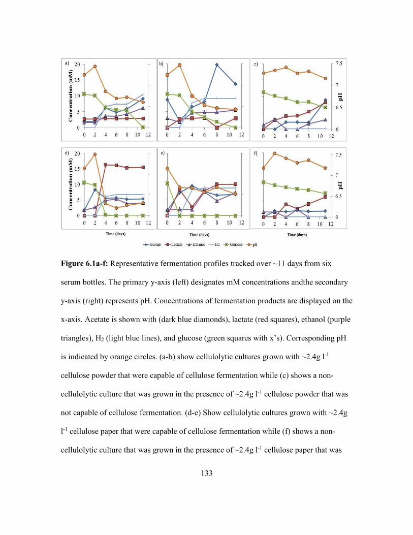
133
Figure 6.1a-f: Representative fermentation profiles tracked over ~11 days from six
serum bottles. The primary y-axis (left) designates mM concentrations andthe secondary
y-axis (right) represents pH. Concentrations of fermentation products are displayed on the
x-axis. Acetate is shown with (dark blue diamonds), lactate (red squares), ethanol (purple
triangles), H2 (light blue lines), and glucose (green squares with x’s). Corresponding pH
is indicated by orange circles. (a-b) show cellulolytic cultures grown with ~2.4g l-1
cellulose powder that were capable of cellulose fermentation while (c) shows a non-
cellulolytic culture that was grown in the presence of ~2.4g l-1 cellulose powder that was
not capable of cellulose fermentation. (d-e) Show cellulolytic cultures grown with ~2.4g
l-1 cellulose paper that were capable of cellulose fermentation while (f) shows a non-
cellulolytic culture that was grown in the presence of ~2.4g l-1 cellulose paper that was

134
not capable of cellulose fermentation. Concurrent cellulolytic serum bottles were used to
inoculate MECs with thermophilic ARB- results are shown in Figure 6.2a-b.
Initial growth and current generation from coupling cellulose fermentation
with anode respiration
Results indicate that a consortium of cellulose fermenting bacteria and ARB were
capable of producing a sustained current density (j) of ~6.54 ± 0.15 A m-2 in duplicate
MECs using cellulose as the only substrate (Figure 6.2a-b). This is higher than previously
reported cellulose-fed thermophilic MFCs (0.4 A m-2) (Mathis 2008) and cellulose-fed
mesophilic MFCs (0.05 A m-2) (Ren 2008) and (< 0.18 A m-2) (Rismani-Yazdi 2007). In
addition to operating under thermophilic conditions, the higher j observed in this study
may be the result of MEC mode of operation which limits O2 contamination and allows
for the potential of the anode to be poised at a specific voltage. For comparison, previous
cellulose-fed mesophilic MECs produced current densities up to 1.8 A m-2 (Niessen
2005).

135
Figure 6.2a: Cellulose-fed MEC 1: current generation (black line) from cellulose-fed
MEC along with concentrations of fermentation byproducts in mM are shown: acetate
(blue diamonds), lactate (red squares), and ethanol (purple triangles). Corresponding pH
is indicated by orange circles.
(a)

136
Figure 6.2b: Cellulose-fed MEC 2: current generation (black line) from duplicate
cellulose-fed MEC. Black arrow indicates when biofilm was sacrificed for SEM and
CLSM.
Figure 6.2a-b show MECs that were incoculated with 200 ml cellulolytic culture
media containing bacteria and ~2.4g l-1 cellulose, and 150 ml ARB culture media
containing thermophilic ARB. Figure 5.2a shows that acetate concentrations rose as
cellulose fermentation occurred in MEC 1, and then fell as it was consumed by ARB for
j. The initial acetate concentration (2 mM) is from fermentation of cellulose in the serum
bottles prior to transfer to the MEC. However, since the cellulose paper was not
completely degraded in the serum bottles before transferring to the MEC, an increasing
acetate concentration (~15 mM) is the result of cellulose fermentation in the anode
compartment. In addition, pH measurements indicate the acetate production correlated
(b)

137
with decreases in pH, while acetate and lactate depletion correlated with a rise in pH. A
drop in pH between days 9-20 resulted in a decrease in j. This indicates that the MEC
becomes pH inhibited as acids are produced from cellulose fermentation, but recovers as
those acids are consumed. The limited anode surface area in comparison to the bulk
volume created a scenario where fermentation products could accumulate faster than they
were consumed resulting in acidic conditions within the biofilm anode.
During MEC operation, there was a gradual decrease in ethanol concentration;
first coupled with an increase in acetate, then with a decrease in acetate concentration.
Ethanol concentrations do not rise during the fermentation process within the MEC;
therefore, it is unclear whether ethanol production occurs during batch MEC operation.
The ethanol that came into MEC 1 from the cellulolytic media bottles may have been
gradually fermented into acetate or consumed by other ARB in the reactor (Kim 2007,
Parameswaran 2011). By day 37, all acetate, lactate, and ethanol concentrations had
reached undetectable levels while TCOD analysis revealed that the media still contained
reduced electron equivalents- suggesting that biomass decay sustained current production
< 1A m-2 for nine days.
There was no presence of H2 or CH4 gas in the anode of either MEC. Previous
research indicates that thermophilic ARB are capable of H2 consumption (Zavarzina
2007) and that homoacetogens can produce acetate from H2 in MECs containing mixed
cultures. Thus, it is likely that any H2 produced during cellulose fermentation was quickly

138
consumed by homoacetogens or for anode respiration. The absence of CH4 indicates that
methanogenic Archaea were likely not present in the MECs.
TCOD analysis indicated a coulombic efficiency (CE) of 84 ± 0.3% and a
coulombic recovery (CR) of 54 ± 11.6%. The lower CR values for MEC 2 (45.9%)
compared to MEC 1 (62.3%) are the result of harvesting the anodes from MEC 2 prior to
the MEC reaching ~0 A m-2. Closing the carbon balance, and thus accounting for all
electrons from cellulose fermentation, is a common challenge in cellulolytic research
(Hogsett 2012). However, previous reports show that approximately 15-26% of electrons
lost can be attributed to biomass (Lee 2008). Therefore, it is likely that missing (~14%)
electrons in the CE calculation were contained in non-decayed biomass.
During runs for both MEC 1 (day 9) and MEC 2 (day 14), cyclic voltammetry
(CV) scans at 1 mV s-1 and 10 mV s-1 were conducted to electrochemically characterize
the anode biofilms of both MECs (Figure 6.3). At the end of the run for MEC 1, ~ day
37, biomass was collected from the anode biofilm and bulk media for the characterization
of the microbial community using pyrosequencing (Figure 6.4). Lastly, for the cellulose-
fed MEC 2, both anodes were sacrificed at day 25 (indicated by black arrow in Figure
6.2b) for the purpose of microscopic characterization of an active biofilm anode with
scanning electron microscopy (SEM) (Figure 6.5a-d) and confocal laser scanning
microscopy (CLSM) (Figure 6.6).

139
Characterization of the biofilm anode using cyclic voltammetry,
pyrosequencing, scanning electron microscopy, and confocal laser scanning
microscopy
Low-Scan Cyclic Voltammetry (LSCV) revealed a midpoint potential (EKA) of -
0.17 ± 0.003 V vs SHE for MECs 1 and 2 (Figure 6.3). This, coupled with the similar j
(~6.64 A m-2 for MEC 1 and 6.43 A m-2 for MEC 2), indicates that the ARB present in
these two MECs have similar electrochemical properties and thus the community
composition may be similar to one another. In addition, the midpoint potentials of MECs
1 and 2 are identical to the midpoint potential reported for Thermoanaerobacter
pseudethanolicus reported in Chapter 5- indicating the this ARB is likely present in both
MECs.

140
Figure 6.3: Derivative for LSCV at 1 mV s-1 for MECs 1 and 2 as normalized to D/DMax.
Black arrow indicates midpoint potential (EKA).
Sequence analysis from MEC 1 showed that both the bulk media and biofilm
anode were nearly completely inhabited by bacteria from the Firmicutes phylum (98.1%)
with Proteobacteria (1%) and unassigned bacteria (0.9%) making up a small portion of
the microbial population. The microbial community of the biofilm anode was more
concentrated with Firmicutes (99.7%) than the anode bulk media (96.5%), while the
anode bulk media contained Proteobacteria (1.9%) and the biofilm anode did not contain
Proteobacteria. The anode biofilm is suspected to contain more ARB than the anode bulk
media because ARB require the anode for respiration. Also, the bacteria responsible for
EKA
= -0.17

141
fermentation are expected to be ubiquitous throughout the MEC and occupy a higher
percentage of the anode bulk media microbiome.
Results indicate that the biofilm anode contained a highly enriched culture of
bacteria from the Peptococcaceae family (49.5%, light blue in Figure 6.4), which is likely
T. ferriacetica, and from the Thermoanaerobacteraceae family. Interestingly, presence of
the Thermoanaerobacter genus (brown in figure 6.4) is indicated in both the biofilm
anode (47.4%) and the anode bulk media (1.5%). Thermoanaerobacter species, including
Thermoanaerobacter pseudethanolicus, was implemented in Chapter 5 for its ability to
ferment cellulose fermentation products while simultaneously performing dissimilatory
metal reduction (Roh 2002). It is likely that this bacterium was selected for in the
cellulolytic fermentation bottles and functioned as both a fermenter and ARB once it was
used to inoculate the MECs.
Another major inhabitant of the biofilm anode was Tepidmicrobium (2.7%) from
the Tissierellaceae family (pink in figure 6.4) which also made up a large portion of the
anode bulk media (43.9%). Members of the Tepidmicrobium genus, including
Tepidmicrobium ferriphilum and Tepidmicrobium xylanilyticum, have been reported as
capable of oxidizing proteinaceous substrates or carbohydrates while simultaneously
reducing either 9,10-anthraquinone 2,6-disulfonate (AQDS) or Fe(III) oxides (Niu 2009,
Slobodkin 2006). The presence of these bacteria in both the anode bulk media and the
biofilm anode is indicative of the cellulose fermentation products present and bacterial
decay which was a major source of electrons during the final stages of MEC operation,
when the biomass samples were collected.

142
Figure 6.4: Overview of bacterial community for samples taken from either the anode
bulk media (“Bulk”) or the biofilm anode (“Biofilm”).

143
For the anode bulk media, the Brevibacillus genus was the most abundant (47.5%,
dark blue in figure 6.4). Brevibacillus, including Brevibacillus sp. strain JXL and
Brevibacillus laterosporus, has been reported to produce cellulosomes (Liang 2009, Kato
2005) and is likely the key microbial player for cellulolytic activity in the MECs.
Ralstonia (yellow in figure 6.4), a Gram-negative bacterial genus, was also present
(1.9%) in the bulk anode; however its role remains unclear. Members of the Ralstonia
genus, including Ralstonia paucula, have been identified in mixed thermophilic lipolytic
cultures and may have played a role in fermentation of lipids from microbial decay
(Hamid 2003). The Planococcaceae family (orange in figure 6.4) accounted for a small
portion of the anode bulk media (1.7%). Members of this family vary in Gram stain,
morphology, and fermentative ability (Shivaji 2013). Caldicoprobacter made up only a
small portion (0.2%, green in figure 6.4) of the microbiome in the anode bulk media with
members of this genus, including Caldicoprobacter oshimai and Caldicoprobacter
algeriensis, reported as thermophilic, xylanolytic, fermentative bacteria (Bouanane-
Darenfed 2011, Yokoyama 2010). Lastly, members of the Clostridium genus were found
to be present in the anode bulk media; however, its concentration in the community was <
0.1%. Members of the Clostridium genus, including Clostridium thermocellum, produce
cellulosomes and are well documented as cellulolytic thermophiles (Viljoen et al., 1926,
Akinosho 2014).
Scanning Electron Microscopy (SEM) analysis reveals a biofilm which is
composed of a diverse set of cell morphologies. Present are cocci (Figure 6.5a-b) and
bacteria with medium, rod-shaped cells (Figure 6.5c)- a similar in morphology to T.

144
ferriacetica shown in Chapter 3 (Parameswaran 2013, Zavarzina 2007). Lastly, present
are bacteria containing long, rod-shaped structures with spore like appendages (white
boxes in figure 6.5d) which may indicate the presence of cellulosomes (Freier 1988).
Figure 6.5a-d: SEM images reveal a biofilm anode with diverse bacterial morphologies.
(a) 2k X magnification shows stacks of cocci occupying the anode surface. Anode surface
is indicated by “anode”. (b) 5k X magnification reveals coccus shaped bacteria are
approximately 1-2 µm in diameter. Anode surface is indicated by “anode”. (c) 5k X
magnification reveals many bacilli occupying biofilm anode. (d) 20k X magnification

145
reveal two bacilli that have an appendage which appears to branch from one cell to
another. White squares indicate shapes that may be cellulosomes.
Confocal Laser Scanning Microscopy (CSLM) LIVE/DEAD analysis (Figure 6.6)
reveals heterogeneous biofilm morphology with peaks and valleys. Active biofilm
thickness (Lf) ranges between 40 µm to 60 µm. Similar Lf has been observed in
thermophilic biofilms under similar operating conditions in Chapter 3 and (Parameswaran
2013). This Lf is sufficient to cause a H+ gradient within the biofilm that may limit j
within the MEC (Marcus 2011) as is observed in MEC 1 when pH temporarily drops due
to the accumulation of acids from cellulose fermentation.
Figure 6.6: CLSM LIVE/DEAD analysis reveals an active biofilm layer approximately
40-60 µm thick.

146
Discussion
Many factors play a crucial role in the advancement of MEC technology as a
possible solution for producing j or H2 from cellulosic waste streams. These factors
include: the operating temperature, bacterial consortia, and dimensional properties of
thermophilic MECs. Although previously reported literature indicates that optimal
activity for thermophilic ARB is at 60 °C (Parameswaran 2013, Marshall 2009, Mathis
2008, Zavarzina 2007), other reports indicate that cellulase activity may be optimal in
other thermophilic and hyperthermophilic bacteria at higher temperatures (Johnson 1981,
Curatolo 1983, Basen 2014, Blumer-Schuette 2012). Given that there are many potential
thermophilic ARB, it may be possible to increase j or fermentation product utilization by
employing a variety of dissimilatory metal reducing bacteria (Niu 2009, Roh 2002,
Slepova 2009, Slobodkin 2006).
Many studies on cellulolytic microbial activity focus on production of
fermentation products as their ends rather than j in MECs (Johnson 1981, Curatolo 1983,
Florenzano 1984, Raman 2011, Li 2012). Future research should also focus on
optimizing j from MECs by focusing on MEC geometry and design. For example,
previous reports have indicated that anode surface area should be optimized to account
for the rate of production of acids and alcohols from cellulose fermentation (Mathis
2008) compared to the amount of surface area needed for ARB to consume those
fermentation products to produce j. Here, thermophilic cellulolytic MECs are shown to
exhibit high j, CE, and CR. However, research into other potential bacterial consortia,
temperatures, and MEC geometries is crucial to the optimization of j in cellulolytic

147
MECs. Thus, future studies should focus on analyzing the kinetics of: cellulase activity,
fermentation product formation and consumption, anode respiration, bacterial growth,
and nutrient balancing.
Chapter 6 References:
1. Akinosho, H, Yee, K, Close, D, Ragauskas, A. 2014. The emergence of
Clostridium thermocellum as a high utility candidate for consolidated
bioprocessing applications. Frontiers in Chemistry. 2:66. doi:
10.3389/fchem.2014.00066.
2. Badger, PC. 2002. Processing Cost Analysis for Biomass Feedstock. Prepared for
the US Department of Energy. Under Contract DE-AC05-00OR22725. ORNL. U.
S. Atomic Energy Commission, TM-2002(199).
3. Basen, M, Rhaesa, A, Kataeva, I, Prybol, C, Scott, I, Poole, F, Adams, M. 2014.
Degradation of high loads of crystalline cellulose and of unpretreated plant
biomass by the thermophilic bacterium Caldicellulosiruptor bescii. Bioresour.
Technol. 152:384-392. doi: 10.1016/j.biortech.2013.11.024.
4. Bouanane-Darenfed, A, Fardeau, ML, Grégoire, P, Joseph, M, Kebbouche-Gana,
S, Benayad, T, Hacene, H, Cayol, JL, Ollivier, B. 2011. Caldicoprobacter
algeriensis sp. nov. a new thermophilic anaerobic, xylanolytic bacterium isolated
from an Algerian hot spring. Curr Microbiol. 62(3):826-32. doi: 10.1007/s00284-
010-9789-9.
5. Blumer-Schuette, SE, Giannone, RJ, Zurawski, JV, Ozdemir, I, Ma, Q, Yin, Y,
Xu, Y, Kataeva, I, Farris L. Poole II, Michael W. W. Adams, Hamilton-Brehm,
SD, Elkins, JG, Larimer, FW, Land, ML, Hauser, LJ, Cottingham, RW, Hettich,
RL, Kelly, RM. 2012. Caldicellulosiruptor Core and Pangenomes Reveal
Determinants for Noncellulosomal Thermophilic Deconstruction of Plant
Biomass. J. Bacteriol. 194:4015-4028. doi: 10.1128/JB.00266-12.
6. Bryant, C. 2011. Putting the pieces together, cellulosic commercialization.
National Ethanol Conference.
7. Caporaso, JG, Bittinger, K, Bushman, FD, DeSantis, TZ, Andersen, GL, Knight,
R. 2010. PyNAST: a flexible tool for aligning sequences to a template alignment.
Bioinformatics. 26:266-267. doi: 10.1093/bioinformatics/btp636.

148
8. Carere, CR, Sparling, R, Cicek, N, Levin, DB. 2008. Third generation biofuels via
direct cellulose fermentation. International Journal of Molecular Sciences.
9:1342-1360. doi: 10.3390/ijms9071342.
9. Curatolo, W, Kanodia, S, Roberts, MF. 1983. The effect of ethanol on the phase
behavior of membrane lipids extracted from Clostridium thermocellum strains.
BBA - Biomembranes. 734:336-341. doi: 10.1016/0005-2736(83)90132-3.
10. Demain, AL, Newcomb, M, J. H. David Wu. 2005. Cellulase, Clostridia, and
Ethanol. Microbiology and Molecular Biology Reviews. 69:124-154. doi:
10.1128/MMBR.69.1.124-154.2005.
11. DeSantis, TZ, Hugenholtz, P, Larsen, N, Rojas, M, Brodie, EL, Keller, K, Huber,
T, Dalevi, D, Hu, P, Andersen, GL. 2006. Greengenes, a Chimera-Checked 16S
rRNA Gene Database and Workbench Compatible with ARB. Appl. Environ.
Microbiol. 72:5069-5072. doi: 10.1128/AEM.03006-05.
12. Edgar, RC. 2010. Search and clustering orders of magnitude faster than BLAST.
Bioinformatics. 26:2460-2461. doi: 10.1093/bioinformatics/btq461.
13. Emtiazi, G, Nahvi, I. 2000. Multi-enzyme production by Cellulomonas sp. grown
on wheat straw. Biomass Bioenergy. 19:31-37. doi: 10.1016/S0961-
9534(00)00015-5.
14. Florenzano, G, Poulain, M, Goma, G. 1984. A study of acetate production from
cellulose using Clostridium thermocellum. Biomass. 4:295-303.
15. Freier, D, Mothershed, CP, Wiegel, J. 1988. Characterization of Clostridium
thermocellum JW20. Appl. Environ. Microbiol. 54:204-211.
16. Ge, Z, Ping, Q, Xiao, L, He, Z. 2013. Reducing effluent discharge and recovering
bioenergy in an osmotic microbial fuel cell treating domestic wastewater.
Desalination. 312:52-59. doi: 10.1016/j.desal.2012.08.036.
17. Haas, BJ, Gevers, D, Earl, AM, Feldgarden, M, Ward, DV, Giannoukos, G,
Ciulla, D, Tabbaa, D, Highlander, SK, Sodergren, E, Methé, B, DeSantis, TZ,
Petrosino, JF, Knight, R, Birren, BW, Human Microbiome Consortium, The
Human Microbiome Consortium. 2011. Chimeric 16S rRNA sequence formation
and detection in Sanger and 454-pyrosequenced PCR amplicons. Genome Res.
21:494-504. doi: 10.1101/gr.112730.110.

149
18. Hama, S, Nakano, K, Onodera, K, Nakamura, M, Noda, H, Kondo, A. 2014.
Saccharification behavior of cellulose acetate during enzymatic processing for
microbial ethanol production. Bioresour. Technol. 157:1-5. doi:
10.1016/j.biortech.2014.01.002.
19. Hogsett, D, Rogers, S, Thorne, P, Tschaplinski, TJ, Lynd, L, Shao, X, Ellis, LD.
2012. Closing the Carbon Balance for Fermentation by Clostridium thermocellum
(ATCC 27405). Bioresour. Technol. 103:293-299. doi:
10.1016/j.biortech.2011.09.128.
20. Johnson, EA, Sakajoh, M, Halliwell, G, Madia, A, Demain, AL. 1982.
Saccharification of Complex Cellulosic Substrates by the Cellulase System from
Clostridium thermocellum. Appl. Environ. Microbiol. 43:1125-1132.
21. Kato, S, Haruta, S, Cui, ZJ, Ishii, M, Igarashi, Y. 2005. Stable Coexistence of
Five Bacterial Strains as a Cellulose-Degrading Community. Appl. Environ.
Microbiol. 71:7099-7106. doi: 10.1128/AEM.71.11.7099-7106.2005.
22. Kim, JR, Jung, SH, Regan, JM, Logan, BE. 2007. Electricity generation and
microbial community analysis of alcohol powered microbial fuel cells. Bioresour.
Technol. 98:2568-2577. doi: 10.1016/j.biortech.2006.09.036.
23. Lee, H, Parameswaran, P, Kato-Marcus, A, Torres, CI, Rittmann, BE. 2008.
Evaluation of energy-conversion efficiencies in microbial fuel cells (MFCs)
utilizing fermentable and non-fermentable substrates. Water Res. 42:1501-1510.
doi: 10.1016/j.watres.2007.10.036.
24. Li, H, Knutson, BL, Nokes, SE, Lynn, BC, Flythe, MD. 2012. Metabolic control
of Clostridium thermocellum via inhibition of hydrogenase activity and the
glucose transport rate. Appl. Microbiol. Biotechnol. 93:1777-1784. doi:
10.1007/s00253-011-3812-3.
25. Liang, Y, Yesuf, J, Schmitt, S, Bender, K, Bozzola, J. 2009. Study of cellulases
from a newly isolated thermophilic and cellulolytic Brevibacillus sp. strain JXL.
Journal of Industrial Microbiology and Biotechnology. 36:961-970. doi:
10.1007/s10295-009-0575-2.
26. Lynd, LR, Weimer, PJ, Willem H. van Zyl, Pretorius, IS. 2002. Microbial
Cellulose Utilization: Fundamentals and Biotechnology. Microbiology and
Molecular Biology Reviews. 66:506-577. doi: 10.1128/MMBR.66.3.506-
577.2002.

150
27. Marcus, AK, Torres, CI, Rittmann, BE. 2007. Conduction-based modeling of the
biofilm anode of a microbial fuel cell. Biotechnol. Bioeng. 98:1171-1182. doi:
10.1002/bit.21533.
28. Marshall, CW, May, HD. 2009. Electrochemical evidence of direct electrode
reduction by a thermophilic Gram-positive bacterium, Thermincola ferriacetica.
Energy and Environmental Science. 2:699-705. doi: 10.1039/b823237g.
29. Mathis, BJ, Marshall, CW, Milliken, CE, Makkar, RS, Creager, SE, May, HD.
2008. Electricity generation by thermophilic microorganisms from marine
sediment. Appl. Microbiol. Biotechnol. 78:147-155. doi: 10.1007/s00253-007-
1266-4.
30. May, HD, Shimotori T. 2009. U.S. Patent No. 0017512 A1. Austin, TX: U.S.
Patent and Trademark Office.
31. McBee, RH. 1950. The anaerobic thermophilic cellulolytic bacteria. Bacteriol.
Rev. 14:51-63.
32. Niessen, J, Schröder, U, Harnisch, F, Scholz, F. 2005. Gaining electricity from in
situ oxidation of hydrogen produced by fermentative cellulose degradation. Lett.
Appl. Microbiol. 41:286-290. doi: 10.1111/j.1472-765X.2005.01742.x.
33. Niu, L, Song, L, Liu, X, Dong, X. 2009. Tepidimicrobium xylanilyticum sp. nov.,
an anaerobic xylanolytic bacterium, and emended description of the genus
Tepidimicrobium. Int. J. Syst. Evol. Microbiol. 59:2698-2701. doi:
10.1099/ijs.0.005124-0.
34. Oh, S, Logan, BE. 2005. Hydrogen and electricity production from a food
processing wastewater using fermentation and microbial fuel cell technologies.
Water Res. 39:4673-4682. doi: 10.1016/j.watres.2005.09.019.
35. Olson, DG, McBride, JE, Joe Shaw, A, Lynd, LR. 2012. Recent progress in
consolidated bioprocessing. Curr. Opin. Biotechnol. 23:396-405. doi:
10.1016/j.copbio.2011.11.026.
36. Ontiveros‐Valencia, A, Ilhan, ZE, Kang, D, Rittmann, B, Krajmalnik‐Brown, R.
2013. Phylogenetic analysis of nitrate‐ and sulfate‐reducing bacteria in a
hydrogen‐fed biofilm. FEMS Microbiol. Ecol. 85:158-167. doi: 10.1111/1574-
6941.12107.

151
37. Parameswaran, P, Bry, T, Popat, SC, Lusk, BG, Rittmann, BE, Torres, CI. 2013.
Kinetic, electrochemical, and microscopic characterization of the thermophilic,
anode-respiring bacterium Thermincola ferriacetica. Environmental Science and
Technology. 47:4934-4940. doi: 10.1021/es400321c.
38. Raman, B, McKeown, CK, Rodriguez Jr, M, Brown, SD, Mielenz, JR. 2011.
Transcriptomic analysis of Clostridium thermocellum ATCC 27405 cellulose
fermentation. BMC Microbiology. 11:134-134. doi: 10.1186/1471-2180-11-134.
39. Ren, Z, Steinberg, LM, Regan, JM. 2008. Electricity production and microbial
biofilm characterization in cellulose-fed microbial fuel cells. Water Science and
Technology. 58:617-622. doi: 10.2166/wst.2008.431.
40. Rismani-Yazdi, H, Christy, AD, Dehority, BA, Morrison, M, Yu, Z, Tuovinen,
OH. 2007. Electricity generation from cellulose by rumen microorganisms in
microbial fuel cells. Biotechnol. Bioeng. 97:1398-1407. doi: 10.1002/bit.21366.
41. Roh, Y, Liu, SV, Li, G, Huang, H, Phelps, TJ, Zhou, J. 2002. Isolation and
Characterization of Metal-Reducing Thermoanaerobacter Strains from Deep
Subsurface Environments of the Piceance Basin, Colorado. Appl. Environ.
Microbiol. 68:6013-6020. doi: 10.1128/AEM.68.12.6013-6020.2002.
42. Saripan, A, Reungsang, A. 2014. Simultaneous saccharification and fermentation
of cellulose for bio-hydrogen production by anaerobic mixed cultures in elephant
dung. Int J Hydrogen Energy. 39:9028-9035. doi:
10.1016/j.ijhydene.2014.04.066.
43. Sheikh Abdul Hamid, N, Zen, HB, Tein, OB, Halifah, YM, Saari, N, Bakar, FA.
2003. Screening and identification of extracellular lipase-producing thermophilic
bacteria from a Malaysian hot spring. World Journal of Microbiology and
Biotechnology. 19:961-968. doi: 10.1023/B:WIBI.0000007330.84569.39.
44. Shivaji, S, Srinivas, TNR, Reddy, GSN. 2013. The Prokaryotes: Firmicutes and
Tenericutes., Edition: 4, Chapter: Family Planococcaceae., Publisher: Springer-
Verlag, Editors: Rosenberg E, DeLong EF, Lory S, Stackebrandt E, Thompson F,
pp.303-351
45. Sizova, MV, Izquierdo, JA, Panikov, NS, Lynd, LR. 2011. Cellulose- and Xylan-
Degrading Thermophilic Anaerobic Bacteria from Biocompost. Appl. Environ.
Microbiol. 77:2282-2291. doi: 10.1128/AEM.01219-10.

152
46. Slobodkin, AI, Tourova, TP, Kostrikina, NA, Lysenko, AM, German, KE, Bonch-
Osmolovskaya, EA, Birkeland, N-. 2006. Tepidimicrobium ferriphilum gen. nov.,
sp. nov., a novel moderately thermophilic, Fe(III)-reducing bacterium of the order
Clostridiales. Int. J. Syst. Evol. Microbiol. 56:369-372. doi: 10.1099/ijs.0.63694-
0.
47. Sun, Y, Wolcott, RD, Dowd, SE. 2011. Tag-encoded FLX amplicon
pyrosequencing for the elucidation of microbial and functional gene diversity in
any environment. High-Throughput Next Generation Sequencing. Methods Mol
Biol 733: 129–141.
48. Taylor, MP, Eley, KL, Martin, S, Tuffin, MI, Burton, SG, Cowan, DA. 2009.
Thermophilic ethanologenesis: future prospects for second-generation bioethanol
production. Trends Biotechnol. 27:398-405. doi: 10.1016/j.tibtech.2009.03.006.
49. Torres, CI, Kato Marcus, A, Rittmann, BE. 2008. Proton transport inside the
biofilm limits electrical current generation by anode‐respiring bacteria.
Biotechnol. Bioeng. 100:872-881. doi: 10.1002/bit.21821.
50. Vasudeo Zambare, Archana Zambare, Kasiviswanath Muthukumarappan,Lew
P.Christopher. 2011. Biochemical characterization of thermophilic lignocellulose
degrading enzymes and their potential for biomass bioprocessing. International
Journal of Energy and Environment. 2:99-112.
51. Viljoen, JA, Fred, EB, Peterson, WH. 1926. The fermentation of cellulose by
thermophilic bacteria. J. Agri. Sci., 16, 1-17.
52. Walters, WA, Pirrung, M, Peña, AG, Huttley, GA, Zaneveld, J, Kuczynski, J,
Knights, D, Bittinger, K, Costello, EK, Turnbaugh, PJ, Reeder, J, Bushman, FD,
Muegge, BD, Knight, R, Koenig, JE, Yatsunenko, T, Fierer, N, Gordon, JI,
Stombaugh, J, McDonald, D, Caporaso, JG, Sevinsky, JR, Ley, RE, Lozupone,
CA, Widmann, J, Kelley, ST, Goodrich, JK. 2010. QIIME allows analysis of
high-throughput community sequencing data. Nature Methods. 7:335-336. doi:
10.1038/nmeth.f.303.
53. Wang, Q, Garrity, GM, Tiedje, JM, Cole, JR. 2007. Naïve Bayesian Classifier for
Rapid Assignment of rRNA Sequences into the New Bacterial Taxonomy. Appl.
Environ. Microbiol. 73:5261-5267. doi: 10.1128/AEM.00062-07.
54. Wilson, DB. 2009. Cellulases and biofuels. Curr. Opin. Biotechnol. 20:295-299.
doi: 10.1016/j.copbio.2009.05.007.

153
55. Xia, Y, Zhang, T, Fang, HH. 2012. Thermophilic anaerobic degradation of
microcrystalline cellulose using mixed culture enriched from anaerobic digestion
sludge. Procedia Environmental Sciences. 12, Part A:3-8. doi:
http://dx.doi.org/10.1016/j.proenv.2012.01.239.
56. Yokoyama, H, Wagner, ID, Wiegel, J. 2010. Caldicoprobacter oshimai gen. nov.,
sp. nov., an anaerobic, xylanolytic, extremely thermophilic bacterium isolated
from sheep faeces, and proposal of Caldicoprobacteraceae fam. nov. Int J Syst
Evol Microbiol. 60(Pt 1):67-71. doi: 10.1099/ijs.0.011379-0.
57. Zavarzina, DG, Sokolova, TG, Tourova, TP, Chernyh, NA, Kostrikina, NA,
Bonch-Osmolovskaya, EA. 2007. Thermincola ferriacetica sp. nov., a new
anaerobic, thermophilic, facultatively chemolithoautotrophic bacterium capable of
dissimilatory Fe(III) reduction. Extremophiles. 11:1-7. doi: 10.1007/s00792-006-
0004-7.
58. Zhao, H, Van Ginkel, S, Tang, Y, Kang, D, Rittmann, B, Krajmalnik-Brown, R.
2011. Interactions between perchlorate and nitrate reductions in the biofilm of a
hydrogen-based membrane biofilm reactor. Environmental Science and
Technology. 45:10155-10162. doi: 10.1021/es202569b.

154
Chapter 7: Conclusion and Future Outlook
Implications of MXC Research Using Thermophilic Bacteria
Throughout this dissertation, the use of microbial electrochemical cells (MXCs)
has proven to be a powerful analytic tool for discovering fundamental properties about
anode respiring bacteria (ARB). In addition, fundamental knowledge about ARB helps in
the development of MXC technology for the purpose of enhancing energy recovery from
diverse wastes. From this perspective, the practical importance of using MXC technology
for fundamental and ‘use-inspired’ reseach is elucidated. MXCs are shown to be
beneficial for a society with perpetually changing energy demands and needs.
Researching novel ARB may lead to new discoveries and catalyze the
breakthrough needed to make MXC technologies energy neutral or positive, and thus
increase their viability in the commercial world (Torres 2014). Researching Gram-
positive bacteria, extremophiles, thermophiles, and other unique ARB is paramount to
advancing the field. For example, by researching new organisms, including Gram-
positives, new external electron transport (EET) pathways, beyond the three that are
currently well documented within the field, may be discovered (Lovley 2008, Mohan
2014, Schroder 2007, Torres 2010, Parameswaran 2013). This research may also shine a
light on the potential of ARB to use multiple redox pathways or processes to perform
anode respiration (Badalamenti 2013, Fu 2013, Yoho 2014, Yoho 2015).
As was shown in Chapters 3 and 4 of this dissertation, using thermophilic ARB
with larger pH ranges allows researchers to probe into the EET limitations of biofilm
anodes. For example, since T. ferriacetica is not limited by buffer at high bicarbonate

155
concentrations, we are better able to probe other electron transport limitations occurring
within the biofilm at non-H+ limiting conditions. In addition, as was discussed in the
introduction to this dissertation, thermophiles were likely the first inhabitants of Earth
and existed ~1 billion years prior to the accumulation of oxygen in the Earth’s
atmosphere (Seckback 2004, 2006). Respiration in these organisms likely occurred via
EET to a diverse set of metal oxides (Seckback 2004, 2006). New discoveries regarding
the functionality of thermophilic ARB may provide insight into some of the earliest
forms of respiration.
Discovering the properties and physiology of unique ARB also enables
researchers and engineers to recover more energy from an increasingly diverse range of
contaminants. As shown in Chapter 3, T. ferriacetica is more able to capture energy as
current at a lower alkalinity than is Geobacter sulfurreducens due to the increased rate of
H+ diffusion experienced at high temperatures (Torres 2008). The studies in Chapter 5
reveal that a fermentative, thermophilic ARB called Thermoanaerobacter
pseudethanolicus is capable of recovering energy as current from fermentable substrates,
including sugars (Lusk 2015). And Chapter 6 shows how we can take the information
gathered in Chapters 3-5 to develop an intelligently engineered thermophilic microbial
consortium to recover energy from solid wastes (cellulose). This discovery opens up the
possibility of using thermophilic MXCs to recover energy from a diverse array of ‘real
life’ thermophilic wastewaters including agricultural and food manufacturing wastes
(Juteau 2006, Linke 2006, Xiao 2009).

156
Future Research
The research methods in this dissertation set a good model for how to proceed
with thermophilic research in the field of MXC technology. First, unique dissimilatory
metal reducing bacteria should be discovered since they are potential candidates for
ARB. This can happen either via bio-prospecting- where a research team isolates bacteria
from samples- or by conducting in silico data searches through previously published
literature. Once potential ARB are implicated, they should be characterized in MXCs
using electrochemical techniques including cyclic voltammetry (CV),
chronoamperometry (CA), and potentially electrochemical impedance spectroscopy (EIS)
(Badalamenti 2013, Marsili 2010, Parameswaran 2013, Srikanth 2008, Yang 2012, Yoho
2014, Yoho 2015). If the bacteria are discovered to perform anode respiration, then their
genome should be sequenced to enable genetic characterization, as was shown in Chapter
2 (Lusk 2015, Badalamenti 2015). Finally, using the newly discovered electrochemical
and genetic data, these microoganisms should be used to develop ‘use-inspired’
technologies including MXCs which are capable of sustainably recovering energy from
diverse wastes, such as in Chapters 4, 5, and 6.
In addition to the research presented in this dissertation, it is also important to
acquire additional data in regards to ARB function and performance in MXCs. For
example, the data provided by mapping the genome is limited in that it only gives us a
glimpse into the potential genetic tools that ARB may be using when grown as biofilm
anodes. In order to develop a more fundamental understanding of the processes occurring
during anode respiration, it is important to recover RNA from biofilms and bulk media

157
for a transcriptomic analysis of the genes that are being transcribed by fermentative
bacteria and ARB (Qiao 2009). In addition, it is also essential to probe into the proteomic
data of these bacteria to gather a more complete understanding of which proteins are
being translated and expressed during anodic fermentation and anode respiration (Costa
2015, Vecchia 2014).
Future research directions should have two primary goals:
1. Discover new ARB
2. Optimize energy recovery from diverse wastes using MXCs.
Here, a list of potential ARB is identified using an in silico probe of previously
characterized dissimilatory metal reducing thermophilic bacteria. A comprehensive list of
other dissimilatory metal reducing thermophilic bacteria can be observed in (Amend
2001, Slobodkin 2005).

158
Tep
idim
icrob
ium
xylan
ilyticum
Tep
idim
icrob
ium
ferriph
ilum
Org
anism
25
-67
26
-62
Tem
p. (◦C
)
Ferm
entatio
n,
An
aerob
ic
Resp
iration
An
aerob
ic
Resp
iration
Metab
olism
xy
lan, x
ylo
se,
glu
cose, cello
bio
se,
pep
ton
e, tryp
ton
e,
Casam
ino
acids,
yeast
extract, b
eef extract,
casein h
yd
roly
sate,
L-cy
steine, L
-serine,
L-ly
sine, L
-gly
cine,
L-th
reon
ine,
L-m
ethio
nin
e and
py
ruv
ate
tryp
ton
e,
Casam
ino
acids,
yeast ex
tract, beef
extract, casein
hy
dro
lysate, p
rolin
e,
L-v
aline an
d n
-
pro
pan
ol
Electro
n so
urces
AQ
DS
, Fe(III),
[acetate, ethan
ol,
bu
tyrate,
hy
dro
gen
]
[fermen
tation
pro
du
cts]
AQ
DS
, Fe(III)
ox
ide, F
e (III)
citrate, Fe (III)
ED
TA
, Fe (III)
nitrilo
triacetate
Electro
n
accepto
rs
5.8
-9.3
5.5
-9.5
pH
rang
e
Niu
20
09
Slo
bo
dk
in
20
06
So
urce

159
Ba
cillus
therm
oa
mylo
vora
ns
SK
C1
Th
ermo
litho
ba
cter
ferriredu
cens
Ca
rbo
xydo
therm
us
sidero
ph
ilus
Org
anism
50
50
-75
52
-70
Tem
p. (◦C
)
Ferm
entatio
n,
An
aerob
ic
Resp
iration
An
aerob
ic
Resp
iration
An
aerob
ic
Resp
iration
Metab
olism
Yeast ex
tract,
arabin
ose
[glu
cose,
cellob
iose, x
ylo
se,
fructo
se]
[Ferm
entativ
e
gro
wth
on
ly]
H2 , fo
rmate
CO
, yeast ex
tract,
glu
cose,
Xy
lose, lactate
Electro
n so
urces
Fe (III) citrate,
Fe (III)-E
DT
A,
Cr(V
I), Te(IV
),
Se(IV
)
AQ
DS
, Fe(III),
Th
iosu
lfate,
fum
arate
AQ
DS
, Fe(III)
Electro
n
accepto
rs
6-8
6.5
-8.5
5.5
-8.5
pH
rang
e
Slo
bo
dk
ina
20
07
So
ko
lov
a
20
07
Slep
ov
a
20
09
So
urce

160
Table 7.1: List of potential thermophilic ARB for study in MXCs.
After the bacteria in Table 7.1 are characterized, then an intelligently selected
microbial consortium can be assembled for the purpose of converting diverse wastes into
electrical current or H2 (Miceli 2014). This research should coincide with ‘good’
engineering techniques for the development of optimized MXCs. This includes the
development of MXC designs that eliminate Ohmic resistance and ionic resistance by
assuring that the anode and cathode are assembled in close proximity and that ion
exchange membranes with proper temperature, pH, and salinity tolerance are employed.
In addition, the anodic surface area needs to be optimized so that the metabolism of ARB
is not inhibited by lack of surface area on the anode (Wei 2011, Zhang 2010, Zhou 2011).
Also, cathodic conditions must be optimized so that the MXC is not limited by the
cathodic section of the MXC as has been indicated as a primary source for overpotential
in MXCs (Popat 2012, Popat 2014). Keeping these goals in mind, coupled with the
discovery of new ARB, MXC technology is a valuable tool for discovering fundamental
properties of dissimilatory metal reducing bacteria and can be a viable renewable energy
option for a diverse set of waste water applications.
References
1. Amend, JP, Shock, EL. 2001. Energetics of overall metabolic reactions of
thermophilic and hyperthermophilic Archaea and Bacteria. FEMS Microbiol.
Rev. 25:175-243. doi: 10.1016/S0168-6445(00)00062-0.
2. Badalamenti, JP, Krajmalnik-Brown, R, Torres, CI. 2013. Generation of High
Current Densities by Pure Cultures of Anode-Respiring Geoalkalibacter spp.
under Alkaline and Saline Conditions in Microbial Electrochemical Cells. Mbio.
4:e00144-13-e00144-13. doi: 10.1128/mBio.00144-13.

161
3. Badalamenti, JP, Krajmalnik-Brown, R, Torres, CI, Bond, DR. 2015. Genomes of
Geoalkalibacter ferrihydriticus Z-0531T and Geoalkalibacter subterraneus Red1T,
Two Haloalkaliphilic Metal-Reducing Deltaproteobacteria. Genome
Announcements. 3:.
4. Costa, NL, Carlson, HK, Coates, JD, Louro, RO, Paquete, CM. 2015.
Heterologous expression and purification of a multiheme cytochrome from a
Gram-positive bacterium capable of performing extracellular respiration. Protein
Expr. Purif. 111:48-52. doi: 10.1016/j.pep.2015.03.007.
5. Dalla Vecchia, E, Shao, PP, Suvorova, E, Chiappe, D, Hamelin, R, Bernier-
Latmani, R. 2014. Characterization of the surfaceome of the metal-reducing
bacterium Desulfotomaculum reducens. Frontiers in Microbiology. 5:432. doi:
10.3389/fmicb.2014.00432.
6. Fu, Q, Kobayashi, H, Kawaguchi, H, Vilcaez, J, Wakayama, T, Maeda, H, Sato,
K. 2013. Electrochemical and phylogenetic analyses of current-generating
microorganisms in a thermophilic microbial fuel cell. Journal of Bioscience and
Bioengineering. 115:268. doi: 10.1016/j.jbiosc.2012.10.007.
7. Juteau, P. 2006. Review of the use of aerobic thermophilic bioprocesses for the
treatment of swine waste. Livestock Science. 102:187-196. doi:
10.1016/j.livsci.2006.03.016.
8. Linke, B. 2006. Kinetic study of thermophilic anaerobic digestion of solid wastes
from potato processing. Biomass Bioenergy. 30:892-896. doi:
10.1016/j.biombioe.2006.02.001.
9. Lovley, DR. 2008. The microbe electric: conversion of organic matter to
electricity. Curr. Opin. Biotechnol. 19:564-571. doi:
10.1016/j.copbio.2008.10.005.
10. Lusk, BG, Badalamenti, JP, Parameswaran, P, Bond, DR, Torres, CI. 2015. Draft
Genome Sequence of the Gram-Positive Thermophilic Iron Reducer Thermincola
ferriacetica Strain Z-0001T. Genome Announcements. 3:.
11. Lusk, BG, Khan, QF, Parameswaran, P, Hameed, A, Ali, N, Rittmann, BE,
Torres, CI. 2015. Characterization of electrical current-generation capabilities
from thermophilic bacterium Thermoanaerobacter pseudethanolicus using xylose,
glucose, cellobiose, or acetate with fixed anode potentials. Environmental Science
& Technology Just Accepted Manuscript. doi: 10.1021/acs.est.5b04036

162
12. Marsili, E, Sun, J, Bond, DR. 2010. Voltammetry and growth physiology of
Geobacter sulfurreducens biofilms as a function of growth stage and imposed
electrode potential. Electroanalysis. 22:865-874. doi: 10.1002/elan.200800007.
13. Miceli, 3,Joseph F., Garcia-Peña, I, Parameswaran, P, Torres, CI, Krajmalnik-
Brown, R. 2014. Combining microbial cultures for efficient production of
electricity from butyrate in a microbial electrochemical cell. Bioresour. Technol.
169:169-174. doi: 10.1016/j.biortech.2014.06.090.
14. Niu, L, Song, L, Liu, X, Dong, X. 2009. Tepidimicrobium xylanilyticum sp. nov.,
an anaerobic xylanolytic bacterium, and emended description of the genus
Tepidimicrobium. Int. J. Syst. Evol. Microbiol. 59:2698-2701. doi:
10.1099/ijs.0.005124-0.
15. Parameswaran, P, Bry, T, Popat, SC, Lusk, BG, Rittmann, BE, Torres, CI. 2013.
Kinetic, electrochemical, and microscopic characterization of the thermophilic,
anode-respiring bacterium Thermincola ferriacetica. Environ. Sci. Technol.
47:4934.
16. Popat, S. C., Ki, D., Young, M. N., Rittmann, B. E. and Torres, C. I. 2014, Buffer
pKa and Transport Govern the Concentration Overpotential in Electrochemical
Oxygen Reduction at Neutral pH. CHEMELECTROCHEM, 1: 1909–1915.
doi:10.1002/celc.201402058
17. Popat, S. C., Ki, D., Rittmann, B. E. and Torres, C. I. 2012. Importance of OH−
Transport from Cathodes in Microbial Fuel Cells. ChemSusChem, 5: 1071–1079.
doi:10.1002/cssc.201100777
18. Qiao, Y, Li, CM, Lu, Z, Ling, H, Kang, A, Chang, MW. 2009. A time-course
transcriptome analysis of Escherichia coli with direct electrochemistry behavior in
microbial fuel cells. Chemical Communications (Cambridge, England). 6183.
19. Schröder, U. 2007. Anodic electron transfer mechanisms in microbial fuel cells
and their energy efficiency. Physical Chemistry Chemical Physics : PCCP.
9:2619. doi: 10.1039/b703627m.
20. Seckbach, J. 2004. Origins: genesis, evolution and diversity of life. Kluwer,
Dordrecht; Boston.
21. Seckbach. 2006. Life as we know it. Springer, Dordrecht.
163
22. Slepova, TV, Sokolova, TG, Kolganova, TV, Tourova, TP, Bonch-
Osmolovskaya, EA. 2009. Carboxydothermus siderophilus sp. nov., a
thermophilic, hydrogenogenic, carboxydotrophic, dissimilatory Fe(III)-reducing
bacterium from a Kamchatka hot spring. Int. J. Syst. Evol. Microbiol. 59:213-217.
doi: 10.1099/ijs.0.000620-0.
23. Slobodkin, A. 2005. Thermophilic Microbial Metal Reduction. Microbiology.
74:501-501. doi: 10.1007/s11021-005-0096-6.
24. Slobodkin, AI, Tourova, TP, Kostrikina, NA, Lysenko, AM, German, KE, Bonch-
Osmolovskaya, EA, Birkeland, N-. 2006. Tepidimicrobium ferriphilum gen. nov.,
sp. nov., a novel moderately thermophilic, Fe(III)-reducing bacterium of the order
Clostridiales. Int. J. Syst. Evol. Microbiol. 56:369-372. doi: 10.1099/ijs.0.63694-
0.
25. Slobodkina, GB, Bonch-Osmolovskaya, EA, Slobodkin, AI. 2007. Reduction of
chromate, selenite, tellurite, and iron (III) by the moderately thermophilic
bacterium Bacillus thermoamylovorans SKC1. Microbiology. 76:530-534. doi:
10.1134/S0026261707050037.
26. Sokolova, T, Hanel, J, Onyenwoke, RU, Reysenbach, A-, Banta, A, Geyer, R,
González, JM, Whitman, WB, Wiegel, J. 2007. Novel chemolithotrophic,
thermophilic, anaerobic bacteria Thermolithobacter ferrireducens gen. nov., sp.
nov. and Thermolithobacter carboxydivorans sp. nov. Extremophiles. 11:145-157.
doi: 10.1007/s00792-006-0022-5.
27. Srikanth, S, Marsili, E, Flickinger, MC, Bond, DR. 2008. Electrochemical
characterization of Geobacter sulfurreducens cells immobilized on graphite paper
electrodes. Biotechnol. Bioeng. 99:1065-1073. doi: 10.1002/bit.21671.
28. Torres, CI, Kato Marcus, A, Rittmann, BE. 2008. Proton transport inside the
biofilm limits electrical current generation by anode‐respiring bacteria.
Biotechnol. Bioeng. 100:872-881. doi: 10.1002/bit.21821.
29. Torres, CI, Marcus, AK, Lee, H, Parameswaran, P, Krajmalnik-Brown, R,
Rittmann, BE. 2010. A kinetic perspective on extracellular electron transfer by
anode-respiring bacteria. FEMS Microbiol. Rev. 34:3-17. doi: 10.1111/j.1574-
6976.2009.00191.x.
30. Torres, CI. 2014. On the importance of identifying, characterizing, and predicting
fundamental phenomena towards microbial electrochemistry applications. Curr.
Opin. Biotechnol. 27:107-114. doi: 10.1016/j.copbio.2013.12.008.

164
31. Venkata Mohan, S, Velvizhi, G, Vamshi Krishna, K, Lenin Babu, M. 2014.
Microbial catalyzed electrochemical systems: a bio-factory with multi-facet
applications. Bioresour. Technol. 165:355-364. doi:
10.1016/j.biortech.2014.03.048.
32. Wei, J, Liang, P, Huang, X. 2011. Recent progress in electrodes for microbial fuel
cells. Bioresour. Technol. 102:9335-9344. doi: 10.1016/j.biortech.2011.07.019.
33. Xiao, Y, Zeng, G, Yang, Z, Shi, W, Huang, C, Fan, C, Xu, Z. 2009. Continuous
thermophilic composting (CTC) for rapid biodegradation and maturation of
organic municipal solid waste. Bioresour. Technol. 100:4807-4813. doi:
10.1016/j.biortech.2009.05.013.
34. Yang, Y, Xu, M, Guo, J, Sun, G. 2012. Bacterial extracellular electron transfer in
bioelectrochemical systems. Process Biochemistry. 47:1707-1714. doi:
10.1016/j.procbio.2012.07.032.
35. Yoho, R, Popat, S, Torres, C. 2014. Dynamic Potential-Dependent Electron
Transport Pathway Shifts in Anode Biofilms of Geobacter sulfurreducens.
Chemsuschem. 7:3413-3419. doi: 10.1002/cssc.201402589.
36. Yoho, RA, Popat, SC, Rago, L, Guisasola ,A, Torres, CI. 2015. Anode Biofilms
of Geoalkalibacter ferrihydriticus Exhibit Electrochemical Signatures of Multiple
Electron Transport Pathways. Langmuir 2015 31 (45), 12552-12559. doi:
10.1021/acs.langmuir.5b02953.
37. Zhang, P, Liu, Z. 2010. Experimental study of the microbial fuel cell internal
resistance. J. Power Sources. 195:8013-8018. doi:
10.1016/j.jpowsour.2010.06.062.
38. Zhou, M, Chi, M, Luo, J, He, H, Jin, T. 2011. An overview of electrode materials
in microbial fuel cells. J. Power Sources. 196:4427-4435. doi:
10.1016/j.jpowsour.2011.01.012.

165
REFERENCES
Chapter 1 References:
1. Akinosho, H, Yee, K, Close, D, Ragauskas, A. 2014. The emergence of
Clostridium thermocellum as a high utility candidate for consolidated
bioprocessing applications. Frontiers in Chemistry. 2:66. doi:
10.3389/fchem.2014.00066.
2. Badalamenti, JP, Krajmalnik-Brown, R, Torres, CI. 2013. Generation of high
current densities by pure cultures of anode-respiring Geoalkalibacter spp. Under
alkaline and saline conditions in microbial electrochemical cells. Mbio. 4:e00144-
13-e00144-13. doi: 10.1128/mBio.00144-13.
3. Basen, M, Rhaesa, A, Kataeva, I, Prybol, C, Scott, I, Poole, F, Adams, M. 2014.
Degradation of high loads of crystalline cellulose and of unpretreated plant
biomass by the thermophilic bacterium Caldicellulosiruptor bescii. Bioresour.
Technol. 152:384-392. doi: 10.1016/j.biortech.2013.11.024.
4. Beveridge, TJ, Forsberg, CW, Doyle, RJ. 1982. Major sites of metal binding in
Bacillus licheniformis walls. J. Bacteriol. 150:1438-1448.
5. Beveridge, TJ, Murray, RG. 1980. Sites of metal deposition in the cell wall of
Bacillus subtilis. J. Bacteriol. 141:876-887.
6. Bird, LJ, Bonnefoy, V, Newman, DK. 2011. Bioenergetic challenges of microbial
iron metabolisms. Trends Microbiol. 19:330-340. doi: 10.1016/j.tim.2011.05.001.
7. Bourdakos, N, Marsili, E, Mahadevan, R. 2014. A defined co‐culture of
Geobacter sulfurreducens and Escherichia coli in a membrane‐less microbial fuel
cell. Biotechnol. Bioeng. 111:709-718. doi: 10.1002/bit.25137.
8. Brock, TD, Freeze, H. 1969. Thermus aquaticus gen. n. and sp. n., a
Nonsporulating Extreme Thermophile. J. Bacteriol. 98:289-297.
9. Brunecky, R, Alahuhta, M, Xu, Q, Donohoe, B, Crowley, M, Kataeva, I, Yang, S,
Resch, M, Adams, M, Lunin, V, Himmel, M, Bomble, Y. 2013. Revealing
Nature's Cellulase Diversity: The Digestion Mechanism of Caldicellulosiruptor
bescii CelA. Science. 342:1513-1516. doi: 10.1126/science.1244273.
10. Carere, CR, Sparling, R, Cicek, N, Levin, DB. 2008. Third generation biofuels via
direct cellulose fermentation. International Journal of Molecular Sciences.
9:1342-1360. doi: 10.3390/ijms9071342.

166
11. Carlson, HK, Iavarone, AT, Gorur, A, Yeo, BS, Tran, R, Melnyk, RA, Mathies,
RA, Auer, M, Coates, JD. 2012. Surface multiheme c-type cytochromes from
Thermincola potens and implications for respiratory metal reduction by Gram-
positive bacteria. Proc. Natl. Acad. Sci. U. S. A. 109:1702-1707. doi:
10.1073/pnas.1112905109.
12. Cheng, S, Logan, BE. 2007. Sustainable and Efficient Biohydrogen Production
via Electrohydrogenesis. Proc. Natl. Acad. Sci. U. S. A. 104:18871-18873. doi:
10.1073/pnas.0706379104.
13. Ciccarelli, FD, Doerks, T, von Mering, C, Creevey, CJ, Snel, B, Bork, P. 2006.
Toward Automatic Reconstruction of a Highly Resolved Tree of Life. Science.
311:1283-1287. doi: 10.1126/science.1123061.
14. Dalla Vecchia, E, Shao, P, Suvorova, E, Chiappe, D, Hamelin, R, Bernier-
Latmani, R. 2014. Characterization of the surfaceome of the metal-reducing
bacterium Desulfotomaculum reducens. Frontiers in Microbiology. 5:432. doi:
10.3389/fmicb.2014.00432.
15. Doney, SC, Fabry, VJ, Feely, RA, Kleypas, JA. 2009. Ocean acidification: The
other CO2 problem. Annual Review of Marine Science. 1:169-192. doi:
10.1146/annurev.marine.010908.163834.
16. Du, Z, Li, H, Gu, T. 2007. A state of the art review on microbial fuel cells: A
promising technology for wastewater treatment and bioenergy. Biotechnol. Adv.
25:464-482. doi: 10.1016/j.biotechadv.2007.05.004.
17. Ehrlich, HL. 2008. Are gram-positive bacteria capable of electron transfer across
their cell wall without an externally available electron shuttle? Geobiology.
6:220-224. doi: 10.1111/j.1472-4669.2007.00135.x.
18. Emtiazi, G, Nahvi, I. 2000. Multi-enzyme production by Cellulomonas sp. grown
on wheat straw. Biomass Bioenergy. 19:31-37. doi: 10.1016/S0961-
9534(00)00015-5.
19. Florenzano, G, Poulain, M, Goma, G. 1984. A study of acetate production from
cellulose using Clostridium thermocellum. Biomass. 4:295-303. doi:
10.1016/0144-4565(84)90042-8.

167
20. Franks, AE, Nevin, KP, Jia, H, Izallalen, M, Woodard, TL, Lovley, DR. 2009.
Novel strategy for three-dimensional real-time imaging of microbial fuel cell
communities: Monitoring the inhibitory effects of proton accumulation within the
anode biofilm. Energy and Environmental Science. 2:113-119. doi:
10.1039/b816445b.
21. Freier, D, Mothershed, CP, Wiegel, J. 1988. Characterization of Clostridium
thermocellum JW20. Appl. Environ. Microbiol. 54:204-211.
22. He, Q, Lokken, PM, Chen, S, Zhou, J. 2009. Characterization of the impact of
acetate and lactate on ethanolic fermentation by Thermoanaerobacter ethanolicus.
Bioresour. Technol. 100:5955-5965. doi: 10.1016/j.biortech.2009.06.084.
23. Hniman, A, Prasertsan, P, O-Thong, S. 2011. Community analysis of
thermophilic hydrogen-producing consortia enriched from Thailand hot spring
with mixed xylose and glucose. Int J Hydrogen Energy. 36:14217-14226. doi:
10.1016/j.ijhydene.2011.05.087.
24. Hollaus, F, Sleytr, U. 1972. On the taxonomy and fine structure of some
hyperthermophilic saccharolytic clostridia. Archiv Für Mikrobiologie. 86:129-
146. doi: 10.1007/BF00413368.
25. Hurst, LD, Merchant, AR. 2001. High guanine–cytosine content is not an
adaptation to high temperature: a comparative analysis amongst prokaryotes.
Proceedings of the Royal Society of London.Series B: Biological Sciences.
268:493-497. doi: 10.1098/rspb.2000.1397.
26. Ieropoulos, I, Melhuish, C, Greenman, J, Horsfield, I. 2005. EcoBot-II: An
artificial agent with a natural metabolism. International Journal of Advanced
Robotic Systems. 2:295-300.
27. Jong, BC, Kim, BH, Chang, IS, Liew, PWY, Choo, YF, Kang, GS. 2006.
Enrichment, performance, and microbial diversity of a thermophilic mediatorless
microbial fuel cell. Environmental Science and Technology. 40:6449-6454. doi:
10.1021/es0613512.
28. Jung, RK, Cheng, S, Oh, S, Logan, BE. 2007. Power generation using different
cation, anion, and ultrafiltration membranes in microbial fuel cells. Environmental
Science and Technology. 41:1004-1009. doi: 10.1021/es062202m.
29. Kashefi, K, Lovley, DR. 2003. Extending the Upper Temperature Limit for Life.
Science. 301:934-934. doi: 10.1126/science.1086823.

168
30. Kato, S, Haruta, S, Cui, ZJ, Ishii, M, Igarashi, Y. 2005. Stable Coexistence of
Five Bacterial Strains as a Cellulose-Degrading Community. Appl. Environ.
Microbiol. 71:7099-7106. doi: 10.1128/AEM.71.11.7099-7106.2005.
31. Kim, B, Kim, H, Hyun, M, Park, D. 1999. Direct electrode reaction of Fe(III)-
reducing bacterium, Shewanella putrefaciens. Journal of Microbiology and
Biotechnology. 9:127-131.
32. Knoll, AH. 2003. Life on a young planet: the first three billion years of evolution
on earth. Princeton University Press, Oxford; Princeton, N.J.
33. Leang, C, Coppi, MV, Lovley, DR. 2003. OmcB, a c-Type Polyheme
Cytochrome, Involved in Fe(III) Reduction in Geobacter sulfurreducens. J.
Bacteriol. 185:2096-2103. doi: 10.1128/JB.185.7.2096-2103.2003.
34. Lee, H, Parameswaran, P, Kato-Marcus, A, Torres, CI, Rittmann, BE. 2008.
Evaluation of energy-conversion efficiencies in microbial fuel cells (MFCs)
utilizing fermentable and non-fermentable substrates. Water Res. 42:1501-1510.
doi: 10.1016/j.watres.2007.10.036.
35. Lee, Y-, Jain, MK, Lee, C, Lowe, SE, Zeikus, JG. 1993. Taxonomic distinction of
saccharolytic thermophilic anaerobes. Int. J. Syst. Bacteriol. 43:41-51.
36. Letunic, I, Bork, P. 2007. Interactive Tree Of Life (iTOL): An online tool for
phylogenetic tree display and annotation. Bioinformatics. 23:127-128. doi:
10.1093/bioinformatics/btl529.
37. Liang, Y, Yesuf, J, Schmitt, S, Bender, K, Bozzola, J. 2009. Study of cellulases
from a newly isolated thermophilic and cellulolytic Brevibacillus sp. strain JXL.
Journal of Industrial Microbiology and Biotechnology. 36:961-970. doi:
10.1007/s10295-009-0575-2.
38. Liu, Y, Climent, V, Berná, A, Feliu, JM. 2011. Effect of Temperature on the
Catalytic Ability of Electrochemically Active Biofilm as Anode Catalyst in
Microbial Fuel Cells. Electroanalysis. 23:387-394. doi: 10.1002/elan.201000499.
39. Lloyd, JR, Blunt-Harris, EL, Lovley, DR. 1999. The Periplasmic 9.6-Kilodalton
c-Type Cytochrome of Geobacter sulfurreducens Is Not an Electron Shuttle to
Fe(III). J. Bacteriol. 181:7647-7649.
40. Logan, BE, Call, D, Cheng, S. 2008. Microbial Electrolysis Cells for High Yield
Hydrogen Gas Production from Organic Matter. Environmental Science &
Technology [H.W.Wilson - AST]. 42:8630.

169
41. Logan, BE, Hamelers, B, Rozendal, R, Schroder, U. 2006. Microbial Fuel Cells:
Methodology and Technology. Environ. Sci. Technol. 40:5181.
42. Lovley, DR. 2008. The microbe electric: conversion of organic matter to
electricity. Curr. Opin. Biotechnol. 19:564-571. doi:
10.1016/j.copbio.2008.10.005.
43. Lynd, LR, Weimer, PJ, Willem H. van Zyl, Pretorius, IS. 2002. Microbial
Cellulose Utilization: Fundamentals and Biotechnology. Microbiology and
Molecular Biology Reviews. 66:506-577. doi: 10.1128/MMBR.66.3.506-
577.2002.
44. Marcus, AK, Torres, CI, Rittmann, BE. 2011. Analysis of a microbial
electrochemical cell using the proton condition in biofilm (PCBIOFILM) model.
Bioresour. Technol. 102:253-262. doi: 10.1016/j.biortech.2010.03.100.
45. Marcus, AK, Torres, CI, Rittmann, BE. 2007. Conduction-based modeling of the
biofilm anode of a microbial fuel cell. Biotechnol. Bioeng. 98:1171-1182. doi:
10.1002/bit.21533.
46. Marshall, CW, May, HD. 2009. Electrochemical evidence of direct electrode
reduction by a thermophilic Gram-positive bacterium, Thermincola ferriacetica.
Energy and Environmental Science. 2:699-705. doi: 10.1039/b823237g.
47. Marsili, E, Sun, J, Bond, DR. 2010. Voltammetry and growth physiology of
Geobacter sulfurreducens biofilms as a function of growth stage and imposed
electrode potential. Electroanalysis. 22:865-874. doi: 10.1002/elan.200800007.
48. Mathis, BJ, Marshall, CW, Milliken, CE, Makkar, RS, Creager, SE, May, HD.
2008. Electricity generation by thermophilic microorganisms from marine
sediment. Appl. Microbiol. Biotechnol. 78:147-155. doi: 10.1007/s00253-007-
1266-4.
49. May, HD, Shimotori, T. U.S. Patent No. 0017512 A1. Austin, TX: U.S. Patent and
Trademark Office.
50. Miceli, JF, Parameswaran, P, Kang, D, Krajmalnik-Brown, R, Torres, CI. 2012.
Enrichment and analysis of anode-respiring bacteria from diverse anaerobic
inocula. Environmental Science and Technology. 46:10349-10355. doi:
10.1021/es301902h.

170
51. Mohan, S, Velvizhi, G, Krishna, K, Babu, M. 2014. Microbial catalyzed
electrochemical systems: A bio-factory with multi-facet applications. Bioresour.
Technol. 165:355-364. doi: 10.1016/j.biortech.2014.03.048.
52. Mook, WG, Koene, BKS. 1975. Chemistry of dissolved inorganic carbon in
estuarine and coastal brackish waters. Estuarine and Coastal Marine Science.
3:325-336.
53. Moon, H, Chang, IS, Kim, BH. 2006. Continuous electricity production from
artificial wastewater using a mediator-less microbial fuel cell. Bioresour. Technol.
97:621-627. doi: 10.1016/j.biortech.2005.03.027.
54. Nealson, KH, Conrad, PG. 1999. Life: past, present and future. Philosophical
Transactions of the Royal Society of London.Series B: Biological Sciences.
354:1923-1939. doi: 10.1098/rstb.1999.0532.
55. Nealson, KH, Conrad, PG. 1999. Life: past, present and future. Philosophical
Transactions of the Royal Society of London.Series B: Biological Sciences.
354:1923-1939. doi: 10.1098/rstb.1999.0532.
56. Niessen, J, Harnisch, F, Rosenbaum, M, Schröder, U, Scholz, F. 2006. Heat
treated soil as convenient and versatile source of bacterial communities for
microbial electricity generation. Electrochemistry Communications. 8:869-873.
doi: 10.1016/j.elecom.2006.03.025.
57. Niu, L, Song, L, Liu, X, Dong, X. 2009. Tepidimicrobium xylanilyticum sp. nov.,
an anaerobic xylanolytic bacterium, and emended description of the genus
Tepidimicrobium. Int. J. Syst. Evol. Microbiol. 59:2698-2701. doi:
10.1099/ijs.0.005124-0.
58. Olson, DG, McBride, JE, Joe Shaw, A, Lynd, LR. 2012. Recent progress in
consolidated bioprocessing. Curr. Opin. Biotechnol. 23:396-405. doi:
10.1016/j.copbio.2011.11.026.
59. Onyenwoke, RU, Kevbrin, VV, Lysenko, AM, Wiegel, J. 2007.
Thermoanaerobacter pseudethanolicus sp. nov., a thermophilic heterotrophic
anaerobe from Yellowstone National Park. Int. J. Syst. Evol. Microbiol. 57:2191-
2193. doi: 10.1099/ijs.0.65051-0.
60. Pant, D, Van Bogaert, G, Diels, L, Vanbroekhoven, K. 2010. A review of the
substrates used in microbial fuel cells (MFCs) for sustainable energy production.
Bioresour. Technol. 101:1533-1543. doi: 10.1016/j.biortech.2009.10.017.

171
61. Parameswaran, P, Bry, T, Popat, SC, Lusk, BG, Rittmann, BE, Torres, CI. 2013.
Kinetic, electrochemical, and microscopic characterization of the thermophilic,
anode-respiring bacterium Thermincola ferriacetica. Environmental Science and
Technology. 47:4934-4940. doi: 10.1021/es400321c.
62. Pham, TH, Boon, N, Aelterman, P, Clauwaert, P, De Schamphelaire, L,
Vanhaecke, L, De Maeyer, K, Höfte, M, Verstraete, W, Rabaey, K. 2008.
Metabolites produced by Pseudomonas sp. enable a Gram-positive bacterium to
achieve extracellular electron transfer. Appl. Microbiol. Biotechnol. 77:1119-
1129. doi: 10.1007/s00253-007-1248-6.
63. Pirbadian, S, Barchinger, SE, Leung, KM, Byun, HS, Jangir, Y, Bouhenni, RA,
Reed, SB, Romine, MF, Saffarini, DA, Shi, L, Gorby, YA, Golbeck, JH, El-
Naggar, MY. 2014. Shewanella oneidensis MR-1 nanowires are outer membrane
and periplasmic extensions of the extracellular electron transport components.
Proceedings of the National Academy of Sciences. 111:12883-12888. doi:
10.1073/pnas.1410551111.
64. Popat, SC, Ki, D, Rittmann, BE, Torres, CI. 2012. Importance of OH- transport
from cathodes in microbial fuel cells. Chemsuschem. 5:1071-1079. doi:
10.1002/cssc.201100777.
65. Puigb, P, Wolf, YI, Koonin, EV. 2009. Search for a 'tree of Life' in the thicket of
the phylogenetic forest. Journal of Biology. 8:59-59. doi: 10.1186/jbiol159.
66. Rabaey, K, Verstraete, W. 2005. Microbial fuel cells: novel biotechnology for
energy generation. Trends Biotechnol. 23:291-298. doi:
10.1016/j.tibtech.2005.04.008.
67. Ren, Z, Steinberg, L, Regan, J. 2008. Electricity production and microbial biofilm
characterization in cellulose-fed microbial fuel cells. Water Science and
Technology. 58:617-622. doi: 10.2166/wst.2008.431.
68. Rismani-Yazdi, H, Christy, AD, Dehority, BA, Morrison, M, Yu, Z, Tuovinen,
OH. 2007. Electricity generation from cellulose by rumen microorganisms in
microbial fuel cells. Biotechnol. Bioeng. 97:1398-1407. doi: 10.1002/bit.21366.
69. Rittmann, BE. 2008. Opportunities for renewable bioenergy using
microorganisms. Biotechnol. Bioeng. 100:203-212. doi: 10.1002/bit.21875.

172
70. Roh, Y, Liu, SV, Li, G, Huang, H, Phelps, TJ, Zhou, J. 2002. Isolation and
Characterization of Metal-Reducing Thermoanaerobacter Strains from Deep
Subsurface Environments of the Piceance Basin, Colorado. Appl. Environ.
Microbiol. 68:6013-6020. doi: 10.1128/AEM.68.12.6013-6020.2002.
71. Rydzak, T, Levin, DB, Cicek, N, Sparling, R. 2011. End-product induced
metabolic shifts in Clostridium thermocellum ATCC 27405. Appl. Microbiol.
Biotechnol. 92:199-209. doi: 10.1007/s00253-011-3511-0.
72. Schroder, U. 2007. Anodic electron transfer mechanisms in microbial fuel cells
and their energy efficiency. Physical Chemistry Chemical Physics. 9:2619-2629.
doi: 10.1039/003627m.
73. Seckbach, J. 2004. Origins: genesis, evolution and diversity of life. Kluwer,
Dordrecht; Boston.
74. Seckbach. 2006. Life as we know it. Springer, Dordrecht.
75. Slepova, TV, Sokolova, TG, Kolganova, TV, Tourova, TP, Bonch-
Osmolovskaya, EA. 2009. Carboxydothermus siderophilus sp. nov., a
thermophilic, hydrogenogenic, carboxydotrophic, dissimilatory Fe(III)-reducing
bacterium from a Kamchatka hot spring. Int. J. Syst. Evol. Microbiol. 59:213-217.
doi: 10.1099/ijs.0.000620-0.
76. Slobodkin, AI, Tourova, TP, Kostrikina, NA, Lysenko, AM, German, KE, Bonch-
Osmolovskaya, EA, Birkeland, N-. 2006. Tepidimicrobium ferriphilum gen. nov.,
sp. nov., a novel moderately thermophilic, Fe(III)-reducing bacterium of the order
Clostridiales. Int. J. Syst. Evol. Microbiol. 56:369-372. doi: 10.1099/ijs.0.63694-
0.
77. Sokolova, TG, Kostrikina, NA, Chernyh, NA, Kolganova, TV, Tourova, TP,
Bonch-Osmolovskaya, EA. 2005. Thermincola carboxydiphila gen. nov., sp. nov.,
a novel anaerobic, carboxydotrophic, hydrogenogenic bacterium from a hot spring
of the Lake Baikal area. Int. J. Syst. Evol. Microbiol. 55:2069-2073. doi:
10.1099/ijs.0.63299-0.
78. Srikanth, S, Marsili, E, Flickinger, MC, Bond, DR. 2008. Electrochemical
characterization of Geobacter sulfurreducens cells immobilized on graphite paper
electrodes. Biotechnol. Bioeng. 99:1065-1073. doi: 10.1002/bit.21671.

173
79. Stocker, TF. 2014; 2013. Climate change 2013: the physical science basis :
working group I contribution to the fifth assessment report of the
intergovernmental panel on climate change. Cambridge University Press, New
York, NY, USA.
80. Thauer, RK, Jungermann, K, Decker, K. 1977. Energy conservation in
chemotrophic anaerobic bacteria. Bacteriol. Rev. 41:100-180.
81. Torres, CI, Kato Marcus, A, Rittmann, BE. 2008. Proton transport inside the
biofilm limits electrical current generation by anode‐respiring bacteria.
Biotechnol. Bioeng. 100:872-881. doi: 10.1002/bit.21821.
82. Torres, CI, Marcus, AK, Lee, H, Parameswaran, P, Krajmalnik-Brown, R,
Rittmann, BE. 2010. A kinetic perspective on extracellular electron transfer by
anode-respiring bacteria. FEMS Microbiol. Rev. 34:3-17. doi: 10.1111/j.1574-
6976.2009.00191.x.
83. Torres, C. 2014. On the importance of identifying, characterizing, and predicting
fundamental phenomena towards microbial electrochemistry applications. Curr.
Opin. Biotechnol. 27:107-114. doi: 10.1016/j.copbio.2013.12.008.
84. U.S. Global Change Research Program. 2014. Climate change impacts in the
United States: U.S. national climate assessment.
85. Vasudeo Zambare, Archana Zambare, Kasiviswanath Muthukumarappan,Lew
P.Christopher. 2011. Biochemical characterization of thermophilic lignocellulose
degrading enzymes and their potential for biomass bioprocessing. International
Journal of Energy and Environment. 2:99-112.
86. Ventura, M, Canchaya, C, Tauch, A, Chandra, G, Fitzgerald, GF, Chater, KF,
Sinderen, Dv. 2007. Genomics of Actinobacteria: Tracing the Evolutionary
History of an Ancient Phylum. Microbiology and Molecular Biology Reviews.
71:495-548. doi: 10.1128/MMBR.00005-07.
87. Vesth, T, Ozen, A, Andersen, S, Kaas, R, Lukjancenko, O, Bohlin, J, Nookaew, I,
Wassenaar, T, Ussery, D, Department of Chemical and Biological
Engineering,Systems Biology, Chalmers University of Technology, Chalmers
tekniska högskola, Institutionen för kemi- och bioteknik,Systembiologi. 2013.
Veillonella, Firmicutes: Microbes disguised as Gram negatives. Standards in
Genomic Sciences. 9:431-448. doi: 10.4056/sigs.2981345.
88. Viljoen, JA. 1925. The Fermentation of Cellulose by Thermophilic Bacteria.
ProQuest, UMI Dissertations Publishing.

174
89. Wrighton, KC, Thrash, JC, Melnyk, RA, Bigi, JP, Byrne-Bailey, KG, Remis, JP,
Schichnes, D, Auer, M, Chang, CJ, Coates, JD. 2011. Evidence for Direct
Electron Transfer by a Gram-Positive Bacterium Isolated from a Microbial Fuel
Cell. Appl. Environ. Microbiol. 77:7633-7639. doi: 10.1128/AEM.05365-11.
90. Yang, Y, Xu, M, Guo, J, Sun, G. 2012. Bacterial extracellular electron transfer in
bioelectrochemical systems. Process Biochemistry. 47:1707-1714. doi:
10.1016/j.procbio.2012.07.032.
91. Yoho, R, Popat, S, Torres, C. 2014. Dynamic Potential-Dependent Electron
Transport Pathway Shifts in Anode Biofilms of Geobacter sulfurreducens.
Chemsuschem. 7:3413-3419. doi: 10.1002/cssc.201402589.
92. Zavarzina, DG, Sokolova, TG, Tourova, TP, Chernyh, NA, Kostrikina, NA,
Bonch-Osmolovskaya, EA. 2007. Thermincola ferriacetica sp. nov., a new
anaerobic, thermophilic, facultatively chemolithoautotrophic bacterium capable of
dissimilatory Fe(III) reduction. Extremophiles. 11:1-7. doi: 10.1007/s00792-006-
0004-7.
93. Zeikus, JG, Ben-Bassat, A, Hegge, PW. 1980. Microbiology of Methanogenesis
in Thermal, Volcanic Environments. J. Bacteriol. 143:432-440.
94. Zhang, E, Xu, W, Diao, G, Shuang, C. 2006. Electricity generation from acetate
and glucose by sedimentary bacterium attached to electrode in microbial-anode
fuel cells. J. Power Sources. 161:820-825. doi: 10.1016/j.jpowsour.2006.05.004.
Chapter 2 References:
1. Beveridge, TJ, Forsberg, CW, Doyle, RJ. 1982. Major sites of metal binding in
Bacillus licheniformis walls. J. Bacteriol. 150:1438-1448.
2. Beveridge, TJ, Murray, RG. 1980. Sites of metal deposition in the cell wall of
Bacillus subtilis. J. Bacteriol. 141:876-887.
3. Byrne-Bailey, KG, Wrighton, KC, Melnyk, RA, Agbo, P, Hazen, TC, Coates, JD.
2010. Complete Genome Sequence of the Electricity-Producing “Thermincola
potens” Strain JR. J. Bacteriol. 192:4078-4079. doi: 10.1128/JB.00044-10.

175
4. Carlson, HK, Iavarone, AT, Gorur, A, Yeo, BS, Tran, R, Melnyk, RA, Mathies,
RA, Auer, M, Coates, JD. 2012. Surface multiheme c-type cytochromes from
Thermincola potens and implications for respiratory metal reduction by Gram-
positive bacteria. Proc. Natl. Acad. Sci. U. S. A. 109:1702-1707. doi:
10.1073/pnas.1112905109.
5. Ehrlich, HL. 2008. Are gram-positive bacteria capable of electron transfer across
their cell wall without an externally available electron shuttle? Geobiology.
6:220-224. doi: 10.1111/j.1472-4669.2007.00135.x.
6. Holmes, DE, Mester, T, O'Neil, RA, Perpetua, LA, Larrahondo, MJ, Glaven, R,
Sharma, ML, Ward, JE, Nevin, KP, Lovley, DR. 2008. Genes for two multicopper
proteins required for Fe(III) oxide reduction in Geobacter sulfurreducens have
different expression patterns both in the subsurface and on energy-harvesting
electrodes. Microbiology. 154:1422-1435. doi: 10.1099/mic.0.2007/014365-0.
7. Leang, C, Coppi, MV, Lovley, DR. 2003. OmcB, a c-Type Polyheme
Cytochrome, Involved in Fe(III) Reduction in Geobacter sulfurreducens. J.
Bacteriol. 185:2096-2103. doi: 10.1128/JB.185.7.2096-2103.2003.
8. Leang, C, Qian, X, Mester, T, Lovley, DR. 2010. Alignment of the c-Type
Cytochrome OmcS along Pili of Geobacter sulfurreducens. Appl. Environ.
Microbiol. 76:4080-4084. doi: 10.1128/AEM.00023-10.
9. Lies, DP, Hernandez, ME, Kappler, A, Mielke, RE, Gralnick, JA, Newman, DK.
2005. Shewanella oneidensis MR-1 Uses Overlapping Pathways for Iron
Reduction at a Distance and by Direct Contact under Conditions Relevant for
Biofilms. Appl. Environ. Microbiol. 71:4414-4426. doi: 10.1128/AEM.71.8.4414-
4426.2005.
10. Marshall, CW, May, HD. 2009. Electrochemical evidence of direct electrode
reduction by a thermophilic Gram-positive bacterium, Thermincola ferriacetica.
Energy and Environmental Science. 2:699-705. doi: 10.1039/b823237g.
11. Parameswaran, P, Bry, T, Popat, SC, Lusk, BG, Rittmann, BE, Torres, CI. 2013.
Kinetic, electrochemical, and microscopic characterization of the thermophilic,
anode-respiring bacterium Thermincola ferriacetica. Environmental Science and
Technology. 47:4934-4940. doi: 10.1021/es400321c.
12. Reguera, G, McCarthy, KD, Mehta, T, Nicoll, JS. 2005. Extracellular electron
transfer via microbial nanowires. Nature. 435:1098.

176
13. Roh, Y, Liu, SV, Li, G, Huang, H, Phelps, TJ, Zhou, J. 2002. Isolation and
Characterization of Metal-Reducing Thermoanaerobacter Strains from Deep
Subsurface Environments of the Piceance Basin, Colorado. Appl. Environ.
Microbiol. 68:6013-6020. doi: 10.1128/AEM.68.12.6013-6020.2002.
14. Sokolova, TG, Kostrikina, NA, Chernyh, NA, Kolganova, TV, Tourova, TP,
Bonch-Osmolovskaya, EA. 2005. Thermincola carboxydiphila gen. nov., sp. nov.,
a novel anaerobic, carboxydotrophic, hydrogenogenic bacterium from a hot spring
of the Lake Baikal area. Int. J. Syst. Evol. Microbiol. 55:2069-2073. doi:
10.1099/ijs.0.63299-0.
15. Tritt, A, Eisen, JA, Facciotti, MT, Darling, AE. 2012. An Integrated Pipeline for
de Novo Assembly of Microbial Genomes. Plos One. 7:e42304. doi:
10.1371/journal.pone.0042304.
16. Wrighton, KC, Thrash, JC, Melnyk, RA, Bigi, JP, Byrne-Bailey, KG, Remis, JP,
Schichnes, D, Auer, M, Chang, CJ, Coates, JD. 2011. Evidence for Direct
Electron Transfer by a Gram-Positive Bacterium Isolated from a Microbial Fuel
Cell. Appl. Environ. Microbiol. 77:7633-7639. doi: 10.1128/AEM.05365-11.
17. Zavarzina, DG, Sokolova, TG, Tourova, TP, Chernyh, NA, Kostrikina, NA,
Bonch-Osmolovskaya, EA. 2007. Thermincola ferriacetica sp. nov., a new
anaerobic, thermophilic, facultatively chemolithoautotrophic bacterium capable of
dissimilatory Fe(III) reduction. Extremophiles. 11:1-7. doi: 10.1007/s00792-006-
0004-7.
Chapter 3 References:
1. Beveridge, TJ, Forsberg, CW, Doyle, RJ. 1982. Major sites of metal binding in
Bacillus licheniformis walls. J. Bacteriol. 150:1438-1448.
2. Beveridge, TJ, Murray, RG. 1980. Sites of metal deposition in the cell wall of
Bacillus subtilis. J. Bacteriol. 141:876-887.
3. Ehrlich, HL. 2008. Are gram-positive bacteria capable of electron transfer across
their cell wall without an externally available electron shuttle? Geobiology.
6:220-224. doi: 10.1111/j.1472-4669.2007.00135.x.

177
4. Franks AE, Nevin KP, Jia H, Izallalen M, Woodard TL, Lovley D R. 2009. Novel
strategy for three-dimensional real-time imaging of microbial fuel cell
communities: Monitoring the inhibitory effects of proton accumulation within the
anode biofilm. Energy and Environmental Science, 2(1), 113-119.
doi:10.1039/b816445b
5. Gleason, JR. 1999. An accurate, non-iterative approximation for studentized
range quantiles. Computational Statistics and Data Analysis. 31:147-158. doi:
10.1016/S0167-9473(99)00002-X.
6. Hunter, RC, Beveridge, TJ. 2005. Application of a pH-Sensitive Fluoroprobe (C-
SNARF-4) for pH Microenvironment Analysis in Pseudomonas aeruginosa
Biofilms. Appl. Environ. Microbiol. 71:2501-2510. doi: 10.1128/AEM.71.5.2501-
2510.2005.
7. Marcus, AK, Torres, CI, Rittmann, BE. 2011. Analysis of a microbial
electrochemical cell using the proton condition in biofilm (PCBIOFILM) model.
Bioresour. Technol. 102:253-262. doi: 10.1016/j.biortech.2010.03.100.
8. Marcus, AK, Torres, CI, Rittmann, BE. 2007. Conduction-based modeling of the
biofilm anode of a microbial fuel cell. Biotechnol. Bioeng. 98:1171-1182. doi:
10.1002/bit.21533.
9. Marshall, CW, May, HD. 2009. Electrochemical evidence of direct electrode
reduction by a thermophilic Gram-positive bacterium, Thermincola ferriacetica.
Energy and Environmental Science. 2:699-705. doi: 10.1039/b823237g.
10. Mathis, BJ, Marshall, CW, Milliken, CE, Makkar, RS, Creager, SE, May, HD.
2008. Electricity generation by thermophilic microorganisms from marine
sediment. Appl. Microbiol. Biotechnol. 78:147-155. doi: 10.1007/s00253-007-
1266-4.
11. May, HD, Shimotori T. 2009. U.S. Patent No. 0017512 A1. Austin, TX: U.S.
Patent and Trademark Office.
12. Parameswaran, P, Bry, T, Popat, SC, Lusk, BG, Rittmann, BE, Torres, CI. 2013.
Kinetic, electrochemical, and microscopic characterization of the thermophilic,
anode-respiring bacterium Thermincola ferriacetica. Environmental Science and
Technology. 47:4934-4940. doi: 10.1021/es400321c.

178
13. Pham, TH, Boon, N, Aelterman, P, Clauwaert, P, De Schamphelaire, L,
Vanhaecke, L, De Maeyer, K, Höfte, M, Verstraete, W, Rabaey, K. 2008.
Metabolites produced by Pseudomonas sp. enable a Gram-positive bacterium to
achieve extracellular electron transfer. Appl. Microbiol. Biotechnol. 77:1119-
1129. doi: 10.1007/s00253-007-1248-6.
14. Reguera, G, Nevin, KP, Nicoll, JS, Covalla, SF, Woodard, TL, Lovley, DR. 2006.
Biofilm and Nanowire Production Leads to Increased Current in Geobacter
sulfurreducens Fuel Cells. Appl. Environ. Microbiol. 72:7345-7348. doi:
10.1128/AEM.01444-06.
15. Robuschi, L, Tomba, JP, Schrott, GD, Bonanni, PS, Desimone, PM, Busalmen,
JP. 2013. Spectroscopic Slicing to Reveal Internal Redox Gradients in Electricity-
Producing Biofilms: 1. Angewandte Chemie. 52:925.
16. Torres, CI, Kato Marcus, A, Rittmann, BE. 2008. Proton transport inside the
biofilm limits electrical current generation by anode‐respiring bacteria.
Biotechnol. Bioeng. 100:872-881. doi: 10.1002/bit.21821.
17. Torres, CI, Marcus, AK, Parameswaran, P, Rittmann, BE. 2008. Kinetic
experiments for evaluating the nernst-monod model for anode-respiring bacteria
(ARB) in a biofilm
anode. Environmental Science and Technology. 42:6593-6597. doi:
10.1021/es800970w.
18. Wrighton, KC, Thrash, JC, Melnyk, RA, Bigi, JP, Byrne-Bailey, KG, Remis, JP,
Schichnes, D, Auer, M, Chang, CJ, Coates, JD. 2011. Evidence for Direct
Electron Transfer by a Gram-Positive Bacterium Isolated from a Microbial Fuel
Cell. Appl. Environ. Microbiol. 77:7633-7639. doi: 10.1128/AEM.05365-11.
19. Yoho, R, Popat, S, Torres, C. 2014. Dynamic Potential-Dependent Electron
Transport Pathway Shifts in Anode Biofilms of Geobacter sulfurreducens.
Chemsuschem. 7:3413-3419. doi: 10.1002/cssc.201402589.
20. Zavarzina, DG, Sokolova, TG, Tourova, TP, Chernyh, NA, Kostrikina, NA,
Bonch-Osmolovskaya, EA. 2007. Thermincola ferriacetica sp. nov., a new
anaerobic, thermophilic, facultatively chemolithoautotrophic bacterium capable of
dissimilatory Fe(III) reduction. Extremophiles. 11:1-7. doi: 10.1007/s00792-006-
0004-7.

179
Chapter 4 references:
1. Badalamenti, JP, Krajmalnik-Brown, R, Torres, CI. 2013. Generation of high
current densities by pure cultures of anode-respiring Geoalkalibacter spp. Under
alkaline and saline conditions in microbial electrochemical cells. Mbio. 4:e00144-
13-e00144-13. doi: 10.1128/mBio.00144-13.
2. Carlson, HK, Iavarone, AT, Gorur, A, Yeo, BS, Tran, R, Melnyk, RA, Mathies,
RA, Auer, M, Coates, JD. 2012. Surface multiheme c-type cytochromes from
Thermincola potens and implications for respiratory metal reduction by Gram-
positive bacteria. Proc. Natl. Acad. Sci. U. S. A. 109:1702-1707. doi:
10.1073/pnas.1112905109.
3. Fricke, K, Harnisch, F, Schröder, U. 2008. On the use of cyclic voltammetry for
the study of anodic electron transfer in microbial fuel cells. Energy and
Environmental Science. 1:144-147. doi: 10.1039/b802363h.
4. Katuri, KP, Kavanagh, P, Rengaraj, S, Leech, D. 2010. Geobacter sulfurreducens
biofilms developed under different growth conditions on glassy carbon electrodes:
Insights using cyclic voltammetry. Chemical Communications. 46:4758-4760.
doi: 10.1039/c003342a.
5. Leang, C, Coppi, MV, Lovley, DR. 2003. OmcB, a c-Type Polyheme
Cytochrome, Involved in Fe(III) Reduction in Geobacter sulfurreducens. J.
Bacteriol. 185:2096-2103. doi: 10.1128/JB.185.7.2096-2103.2003.
6. Logan, BE, Hamelers, B, Rozendal, R, Schroder, U. 2006. Microbial Fuel Cells:
Methodology and Technology. Environ. Sci. Technol. 40:5181.
7. Lovley, DR. 2008. The microbe electric: conversion of organic matter to
electricity. Curr. Opin. Biotechnol. 19:564-571. doi:
10.1016/j.copbio.2008.10.005.
8. Lusk, BG, Badalamenti, JP, Parameswaran, P, Bond, DR, Torres, CI. 2015. Draft
Genome Sequence of the Gram-Positive Thermophilic Iron Reducer Thermincola
ferriacetica Strain Z-0001T. Genome Announcements. 3:.

180
9. Marcus, AK, Torres, CI, Rittmann, BE. 2011. Analysis of a microbial
electrochemical cell using the proton condition in biofilm (PCBIOFILM) model.
Bioresour. Technol. 102:253-262. doi: 10.1016/j.biortech.2010.03.100.
10. Marcus, AK, Torres, CI, Rittmann, BE. 2007. Conduction-based modeling of the
biofilm anode of a microbial fuel cell. Biotechnol. Bioeng. 98:1171-1182. doi:
10.1002/bit.21533.
11. Marsili, E, Sun, J, Bond, DR. 2010. Voltammetry and growth physiology of
Geobacter sulfurreducens biofilms as a function of growth stage and imposed
electrode potential. Electroanalysis. 22:865-874. doi: 10.1002/elan.200800007.
12. Mohan, S, Velvizhi, G, Krishna, K, Babu, M. 2014. Microbial catalyzed
electrochemical systems: A bio-factory with multi-facet applications. Bioresour.
Technol. 165:355-364. doi: 10.1016/j.biortech.2014.03.048.
13. Parameswaran, P, Bry, T, Popat, SC, Lusk, BG, Rittmann, BE, Torres, CI. 2013.
Kinetic, electrochemical, and microscopic characterization of the thermophilic,
anode-respiring bacterium Thermincola ferriacetica. Environmental Science and
Technology. 47:4934-4940. doi: 10.1021/es400321c.
14. Schroder, U. 2007. Anodic electron transfer mechanisms in microbial fuel cells
and their energy efficiency. Physical Chemistry Chemical Physics. 9:2619-2629.
doi: 10.1039/003627m.
15. Srikanth, S, Marsili, E, Flickinger, MC, Bond, DR. 2008. Electrochemical
characterization of Geobacter sulfurreducens cells immobilized on graphite paper
electrodes. Biotechnol. Bioeng. 99:1065-1073. doi: 10.1002/bit.21671.
16. Torres, CI, Kato Marcus, A, Rittmann, BE. 2008. Proton transport inside the
biofilm limits electrical current generation by anode‐respiring bacteria.
Biotechnol. Bioeng. 100:872-881. doi: 10.1002/bit.21821.
17. Torres, CI, Marcus, AK, Parameswaran, P, Rittmann, BE. 2008. Kinetic
experiments for evaluating the nernst-monod model for anode-respiring bacteria
(ARB) in a biofilm anode. Environmental Science and Technology. 42:6593-
6597. doi: 10.1021/es800970w.

181
18. Torres, CI, Marcus, AK, Lee, H, Parameswaran, P, Krajmalnik-Brown, R,
Rittmann, BE. 2010. A kinetic perspective on extracellular electron transfer by
anode-respiring bacteria. FEMS Microbiol. Rev. 34:3-17. doi: 10.1111/j.1574-
6976.2009.00191.x.
19. Yang, Y, Xu, M, Guo, J, Sun, G. 2012. Bacterial extracellular electron transfer in
bioelectrochemical systems. Process Biochemistry. 47:1707-1714. doi:
10.1016/j.procbio.2012.07.032.
20. Yoho, R, Popat, S, Torres, C. 2014. Dynamic Potential-Dependent Electron
Transport Pathway Shifts in Anode Biofilms of Geobacter sulfurreducens.
Chemsuschem. 7:3413-3419. doi: 10.1002/cssc.201402589.
21. Yoho, RA, Popat, SC, Rago, L, Guisasola ,A, Torres, CI. 2015. Anode Biofilms
of Geoalkalibacter ferrihydriticus Exhibit Electrochemical Signatures of Multiple
Electron Transport Pathways. Langmuir 2015 31 (45), 12552-12559. doi:
10.1021/acs.langmuir.5b02953.
Chapter 5 references:
1. Badalamenti, JP, Krajmalnik-Brown, R, Torres, CI. 2013. Generation of high
current densities by pure cultures of anode-respiring Geoalkalibacter spp. Under
alkaline and saline conditions in microbial electrochemical cells. Mbio. 4:e00144-
13-e00144-13. doi: 10.1128/mBio.00144-13.
2. Bond, DR, Lovley, DR. 2003. Electricity Production by Geobacter sulfurreducens
Attached to Electrodes. Appl. Environ. Microbiol. 69:1548-1555. doi:
10.1128/AEM.69.3.1548-1555.2003.
3. Bond, DR, Lovley, DR. 2005. Evidence for Involvement of an Electron Shuttle in
Electricity Generation by Geothrix fermentans. Appl. Environ. Microbiol.
71:2186-2189. doi: 10.1128/AEM.71.4.2186-2189.2005.
4. Carlson, HK, Iavarone, AT, Gorur, A, Yeo, BS, Tran, R, Melnyk, RA, Mathies,
RA, Auer, M, Coates, JD. 2012. Surface multiheme c-type cytochromes from
Thermincola potens and implications for respiratory metal reduction by Gram-
positive bacteria. Proc. Natl. Acad. Sci. U. S. A. 109:1702-1707. doi:
10.1073/pnas.1112905109.

182
5. Chaudhuri, SK, Lovley, DR. 2003. Electricity generation by direct oxidation of
glucose in mediatorless microbial fuel cells. Nat. Biotechnol. 21:1229-1232. doi:
10.1038/nbt867.
6. Cook, GM, Janssen, PH, Morgan, HW. 1993. Simultaneous uptake and utilisation
of glucose and xylose by Clostridium thermohydrosulfuricum. FEMS Microbiol.
Lett. 109:55-61.
7. Demain, AL, Newcomb, M, J. H. David Wu. 2005. Cellulase, Clostridia, and
Ethanol. Microbiology and Molecular Biology Reviews. 69:124-154. doi:
10.1128/MMBR.69.1.124-154.2005.
8. Franks AE, Nevin KP, Jia H, Izallalen M, Woodard TL, Lovley D R. 2009. Novel
strategy for three-dimensional real-time imaging of microbial fuel cell
communities: Monitoring the inhibitory effects of proton accumulation within the
anode biofilm. Energy and Environmental Science, 2(1), 113-119.
doi:10.1039/b816445b
9. Gorby, YA, Yanina, S, McLean, JS, Rosso, KM, Moyles, D, Dohnalkova, A,
Beveridge, TJ, Chang, IS, Kim, KS, Kim, BH, Culley, DE, Reed, SB, Romine,
MF, Saffarini, DA, Hill, EA, Shi, L, Elias, DA, Kennedy, DW, Pinchuk, G,
Watanabe, K, Ishii, S, Logan, B, Nealson, KH, Fredrickson, JK. 2006. Electrically
Conductive Bacterial Nanowires Produced by Shewanella oneidensis Strain MR-1
and Other Microorganisms. Proc. Natl. Acad. Sci. U. S. A. 103:11358-11363. doi:
10.1073/pnas.0604517103.
10. Greeley RS, Smith WT, Stoughton RW, Lietzke M H. 1960. Electromotive for
studies in aqueous solutions at elevated temperatures. 1. The standard potential of
the silver-silver chloride electrode. J. Phys. Chem. 1960, 64 (5), 652−657.
11. He, Q, Lokken, PM, Chen, S, Zhou, J. 2009. Characterization of the impact of
acetate and lactate on ethanolic fermentation by Thermoanaerobacter ethanolicus.
Bioresour. Technol. 100:5955-5965. doi: 10.1016/j.biortech.2009.06.084.
12. Hemme, CL, Fields, MW, He, Q, Deng, Y, Lin, L, Tu, Q, Mouttaki, H, Zhou, A,
Feng, X, Zuo, Z, Ramsay, BD, He, Z, Wu, L, Nostrand, JV, Xu, J, Tang, YJ,
Wiegel, J, Phelps, TJ, Zhou, J. 2011. Correlation of Genomic and Physiological
Traits of Thermoanaerobacter Species with Biofuel Yields. Appl. Environ.
Microbiol. 77:7998-8008. doi: 10.1128/AEM.05677-11.

183
13. Hniman, A, Prasertsan, P, O-Thong, S. 2011. Community analysis of
thermophilic hydrogen-producing consortia enriched from Thailand hot spring
with mixed xylose and glucose. Int J Hydrogen Energy. 36:14217-14226. doi:
10.1016/j.ijhydene.2011.05.087.
14. Lacis, LS, Lawford, HG. 1991. Thermoanaerobacter ethanolicus Growth and
Product Yield from Elevated Levels of Xylose or Glucose in Continuous Cultures.
Appl. Environ. Microbiol. 57:579-585.
15. Lee, H, Parameswaran, P, Kato-Marcus, A, Torres, CI, Rittmann, BE. 2008.
Evaluation of energy-conversion efficiencies in microbial fuel cells (MFCs)
utilizing fermentable and non-fermentable substrates. Water Res. 42:1501-1510.
doi: 10.1016/j.watres.2007.10.036.
16. Lee Y, Jain M K, Lee C, Lowe SE, Zeikus JG. 1993. Taxonomic distinction of
saccharolytic thermophilic anaerobes. International Journal of Systematic
Bacteriology, 43(1), 41-51.
17. Lovley, DR. 2008. The microbe electric: conversion of organic matter to
electricity. Curr. Opin. Biotechnol. 19:564-571. doi:
10.1016/j.copbio.2008.10.005
18. Luo, J, Yang, J, He, H, Jin, T, Zhou, L, Wang, M, Zhou, M. 2013. A new
electrochemically active bacterium phylogenetically related to Tolumonas
osonensis and power performance in MFCs. Bioresour. Technol. 139:141. doi:
10.1016/j.biortech.2013.04.031.
19. Marcus, AK, Torres, CI, Rittmann, BE. 2011. Analysis of a microbial
electrochemical cell using the proton condition in biofilm (PCBIOFILM) model.
Bioresour. Technol. 102:253-262. doi: 10.1016/j.biortech.2010.03.100.
20. Marshall, CW, May, HD. 2009. Electrochemical evidence of direct electrode
reduction by a thermophilic Gram-positive bacterium, Thermincola ferriacetica.
Energy and Environmental Science. 2:699-705. doi: 10.1039/b823237g.
21. Marsili, E, Sun, J, Bond, DR. 2010. Voltammetry and growth physiology of
Geobacter sulfurreducens biofilms as a function of growth stage and imposed
electrode potential. Electroanalysis. 22:865-874. doi: 10.1002/elan.200800007.
184
22. Mathis, BJ, Marshall, CW, Milliken, CE, Makkar, RS, Creager, SE, May, HD.
2008. Electricity generation by thermophilic microorganisms from marine
sediment. Appl. Microbiol. Biotechnol. 78:147-155. doi: 10.1007/s00253-007-
1266-4.
23. Onyenwoke, RU, Kevbrin, VV, Lysenko, AM, Wiegel, J. 2007.
Thermoanaerobacter pseudethanolicus sp. nov., a thermophilic heterotrophic
anaerobe from Yellowstone National Park. Int. J. Syst. Evol. Microbiol. 57:2191-
2193. doi: 10.1099/ijs.0.65051-0.
24. Parameswaran, P, Bry, T, Popat, SC, Lusk, BG, Rittmann, BE, Torres, CI. 2013.
Kinetic, electrochemical, and microscopic characterization of the thermophilic,
anode-respiring bacterium Thermincola ferriacetica. Environmental Science and
Technology. 47:4934-4940. doi: 10.1021/es400321c.
25. Parameswaran, P, Torres, CI, Lee, H, Krajmalnik-Brown, R, Rittmann, BE. 2009.
Syntrophic interactions among anode respiring bacteria (ARB) and non-ARB in a
biofilm anode: Electron balances. Biotechnol. Bioeng. 103:513-523. doi:
10.1002/bit.22267.
26. Roh, Y, Liu, SV, Li, G, Huang, H, Phelps, TJ, Zhou, J. 2002. Isolation and
Characterization of Metal-Reducing Thermoanaerobacter Strains from Deep
Subsurface Environments of the Piceance Basin, Colorado. Appl. Environ.
Microbiol. 68:6013-6020. doi: 10.1128/AEM.68.12.6013-6020.2002.
27. Rotaru, A, Woodard, TL, Nevin, KP, Lovley, DR. 2015. Link between capacity
for current production and syntrophic growth in Geobacter species. Frontiers in
Microbiology. 6:744.
28. Srikanth, S, Marsili, E, Flickinger, MC, Bond, DR. 2008. Electrochemical
characterization of Geobacter sulfurreducens cells immobilized on graphite paper
electrodes. Biotechnol. Bioeng. 99:1065-1073. doi: 10.1002/bit.21671.
29. Stouthamer AH. 1979. The search for correlation between theoretical and
experimental growth yields. Int. Rev. Biochem. 21:1-47.
30. Thomas, L, Joseph, A, Gottumukkala, L. 2014. Xylanase and cellulase systems of
Clostridium sp.: An insight on molecular approaches for strain improvement.
Bioresour. Technol. 158:343-350. doi: 10.1016/j.biortech.2014.01.140.
31. Torres, C. 2014. On the importance of identifying, characterizing, and predicting
fundamental phenomena towards microbial electrochemistry applications. Curr.
Opin. Biotechnol. 27:107-114. doi: 10.1016/j.copbio.2013.12.008.

185
32. Torres, CI, Kato Marcus, A, Rittmann, BE. 2008. Proton transport inside the
biofilm limits electrical current generation by anode‐respiring bacteria.
Biotechnol. Bioeng. 100:872-881. doi: 10.1002/bit.21821.
33. Wrighton, KC, Thrash, JC, Melnyk, RA, Bigi, JP, Byrne-Bailey, KG, Remis, JP,
Schichnes, D, Auer, M, Chang, CJ, Coates, JD. 2011. Evidence for Direct
Electron Transfer by a Gram-Positive Bacterium Isolated from a Microbial Fuel
Cell. Appl. Environ. Microbiol. 77:7633-7639. doi: 10.1128/AEM.05365-11.
34. Zeikus, JG, Ben-Bassat, A, Hegge, PW. 1980. Microbiology of Methanogenesis
in Thermal, Volcanic Environments. J. Bacteriol. 143:432-440.
Chapter 6 References:
1. Akinosho, H, Yee, K, Close, D, Ragauskas, A. 2014. The emergence of
Clostridium thermocellum as a high utility candidate for consolidated
bioprocessing applications. Frontiers in Chemistry. 2:66. doi:
10.3389/fchem.2014.00066.
2. Badger, PC. 2002. Processing Cost Analysis for Biomass Feedstock. Prepared for
the US Department of Energy. Under Contract DE-AC05-00OR22725. ORNL. U.
S. Atomic Energy Commission, TM-2002(199).
3. Basen, M, Rhaesa, A, Kataeva, I, Prybol, C, Scott, I, Poole, F, Adams, M. 2014.
Degradation of high loads of crystalline cellulose and of unpretreated plant
biomass by the thermophilic bacterium Caldicellulosiruptor bescii. Bioresour.
Technol. 152:384-392. doi: 10.1016/j.biortech.2013.11.024.
4. Bouanane-Darenfed, A, Fardeau, ML, Grégoire, P, Joseph, M, Kebbouche-Gana,
S, Benayad, T, Hacene, H, Cayol, JL, Ollivier, B. 2011. Caldicoprobacter
algeriensis sp. nov. a new thermophilic anaerobic, xylanolytic bacterium isolated
from an Algerian hot spring. Curr Microbiol. 62(3):826-32. doi: 10.1007/s00284-
010-9789-9.
5. Blumer-Schuette, SE, Giannone, RJ, Zurawski, JV, Ozdemir, I, Ma, Q, Yin, Y,
Xu, Y, Kataeva, I, Farris L. Poole II, Michael W. W. Adams, Hamilton-Brehm,
SD, Elkins, JG, Larimer, FW, Land, ML, Hauser, LJ, Cottingham, RW, Hettich,
RL, Kelly, RM. 2012. Caldicellulosiruptor Core and Pangenomes Reveal
Determinants for Noncellulosomal Thermophilic Deconstruction of Plant
Biomass. J. Bacteriol. 194:4015-4028. doi: 10.1128/JB.00266-12.
186
6. Bryant, C. 2011. Putting the pieces together, cellulosic commercialization.
National Ethanol Conference.
7. Caporaso, JG, Bittinger, K, Bushman, FD, DeSantis, TZ, Andersen, GL, Knight,
R. 2010. PyNAST: a flexible tool for aligning sequences to a template alignment.
Bioinformatics. 26:266-267. doi: 10.1093/bioinformatics/btp636.
8. Carere, CR, Sparling, R, Cicek, N, Levin, DB. 2008. Third generation biofuels via
direct cellulose fermentation. International Journal of Molecular Sciences.
9:1342-1360. doi: 10.3390/ijms9071342.
9. Curatolo, W, Kanodia, S, Roberts, MF. 1983. The effect of ethanol on the phase
behavior of membrane lipids extracted from Clostridium thermocellum strains.
BBA - Biomembranes. 734:336-341. doi: 10.1016/0005-2736(83)90132-3.
10. Demain, AL, Newcomb, M, J. H. David Wu. 2005. Cellulase, Clostridia, and
Ethanol. Microbiology and Molecular Biology Reviews. 69:124-154. doi:
10.1128/MMBR.69.1.124-154.2005.
11. DeSantis, TZ, Hugenholtz, P, Larsen, N, Rojas, M, Brodie, EL, Keller, K, Huber,
T, Dalevi, D, Hu, P, Andersen, GL. 2006. Greengenes, a Chimera-Checked 16S
rRNA Gene Database and Workbench Compatible with ARB. Appl. Environ.
Microbiol. 72:5069-5072. doi: 10.1128/AEM.03006-05.
12. Edgar, RC. 2010. Search and clustering orders of magnitude faster than BLAST.
Bioinformatics. 26:2460-2461. doi: 10.1093/bioinformatics/btq461.
13. Emtiazi, G, Nahvi, I. 2000. Multi-enzyme production by Cellulomonas sp. grown
on wheat straw. Biomass Bioenergy. 19:31-37. doi: 10.1016/S0961-
9534(00)00015-5.
14. Florenzano, G, Poulain, M, Goma, G. 1984. A study of acetate production from
cellulose using Clostridium thermocellum. Biomass. 4:295-303.
15. Freier, D, Mothershed, CP, Wiegel, J. 1988. Characterization of Clostridium
thermocellum JW20. Appl. Environ. Microbiol. 54:204-211.
16. Ge, Z, Ping, Q, Xiao, L, He, Z. 2013. Reducing effluent discharge and recovering
bioenergy in an osmotic microbial fuel cell treating domestic wastewater.
Desalination. 312:52-59. doi: 10.1016/j.desal.2012.08.036.

187
17. Haas, BJ, Gevers, D, Earl, AM, Feldgarden, M, Ward, DV, Giannoukos, G,
Ciulla, D, Tabbaa, D, Highlander, SK, Sodergren, E, Methé, B, DeSantis, TZ,
Petrosino, JF, Knight, R, Birren, BW, Human Microbiome Consortium, The
Human Microbiome Consortium. 2011. Chimeric 16S rRNA sequence formation
and detection in Sanger and 454-pyrosequenced PCR amplicons. Genome Res.
21:494-504. doi: 10.1101/gr.112730.110.
18. Hama, S, Nakano, K, Onodera, K, Nakamura, M, Noda, H, Kondo, A. 2014.
Saccharification behavior of cellulose acetate during enzymatic processing for
microbial ethanol production. Bioresour. Technol. 157:1-5. doi:
10.1016/j.biortech.2014.01.002.
19. Hogsett, D, Rogers, S, Thorne, P, Tschaplinski, TJ, Lynd, L, Shao, X, Ellis, LD.
2012. Closing the Carbon Balance for Fermentation by Clostridium thermocellum
(ATCC 27405). Bioresour. Technol. 103:293-299. doi:
10.1016/j.biortech.2011.09.128.
20. Johnson, EA, Sakajoh, M, Halliwell, G, Madia, A, Demain, AL. 1982.
Saccharification of Complex Cellulosic Substrates by the Cellulase System from
Clostridium thermocellum. Appl. Environ. Microbiol. 43:1125-1132.
21. Kato, S, Haruta, S, Cui, ZJ, Ishii, M, Igarashi, Y. 2005. Stable Coexistence of
Five Bacterial Strains as a Cellulose-Degrading Community. Appl. Environ.
Microbiol. 71:7099-7106. doi: 10.1128/AEM.71.11.7099-7106.2005.
22. Kim, JR, Jung, SH, Regan, JM, Logan, BE. 2007. Electricity generation and
microbial community analysis of alcohol powered microbial fuel cells. Bioresour.
Technol. 98:2568-2577. doi: 10.1016/j.biortech.2006.09.036.
23. Lee, H, Parameswaran, P, Kato-Marcus, A, Torres, CI, Rittmann, BE. 2008.
Evaluation of energy-conversion efficiencies in microbial fuel cells (MFCs)
utilizing fermentable and non-fermentable substrates. Water Res. 42:1501-1510.
doi: 10.1016/j.watres.2007.10.036.
24. Li, H, Knutson, BL, Nokes, SE, Lynn, BC, Flythe, MD. 2012. Metabolic control
of Clostridium thermocellum via inhibition of hydrogenase activity and the
glucose transport rate. Appl. Microbiol. Biotechnol. 93:1777-1784. doi:
10.1007/s00253-011-3812-3.

188
25. Liang, Y, Yesuf, J, Schmitt, S, Bender, K, Bozzola, J. 2009. Study of cellulases
from a newly isolated thermophilic and cellulolytic Brevibacillus sp. strain JXL.
Journal of Industrial Microbiology and Biotechnology. 36:961-970. doi:
10.1007/s10295-009-0575-2.
26. Lynd, LR, Weimer, PJ, Willem H. van Zyl, Pretorius, IS. 2002. Microbial
Cellulose Utilization: Fundamentals and Biotechnology. Microbiology and
Molecular Biology Reviews. 66:506-577. doi: 10.1128/MMBR.66.3.506-
577.2002.
27. Marcus, AK, Torres, CI, Rittmann, BE. 2007. Conduction-based modeling of the
biofilm anode of a microbial fuel cell. Biotechnol. Bioeng. 98:1171-1182. doi:
10.1002/bit.21533.
28. Marshall, CW, May, HD. 2009. Electrochemical evidence of direct electrode
reduction by a thermophilic Gram-positive bacterium, Thermincola ferriacetica.
Energy and Environmental Science. 2:699-705. doi: 10.1039/b823237g.
29. Mathis, BJ, Marshall, CW, Milliken, CE, Makkar, RS, Creager, SE, May, HD.
2008. Electricity generation by thermophilic microorganisms from marine
sediment. Appl. Microbiol. Biotechnol. 78:147-155. doi: 10.1007/s00253-007-
1266-4.
30. May, HD, Shimotori T. 2009. U.S. Patent No. 0017512 A1. Austin, TX: U.S.
Patent and Trademark Office.
31. McBee, RH. 1950. The anaerobic thermophilic cellulolytic bacteria. Bacteriol.
Rev. 14:51-63.
32. Niessen, J, Schröder, U, Harnisch, F, Scholz, F. 2005. Gaining electricity from in
situ oxidation of hydrogen produced by fermentative cellulose degradation. Lett.
Appl. Microbiol. 41:286-290. doi: 10.1111/j.1472-765X.2005.01742.x.
33. Niu, L, Song, L, Liu, X, Dong, X. 2009. Tepidimicrobium xylanilyticum sp. nov.,
an anaerobic xylanolytic bacterium, and emended description of the genus
Tepidimicrobium. Int. J. Syst. Evol. Microbiol. 59:2698-2701. doi:
10.1099/ijs.0.005124-0.
34. Oh, S, Logan, BE. 2005. Hydrogen and electricity production from a food
processing wastewater using fermentation and microbial fuel cell technologies.
Water Res. 39:4673-4682. doi: 10.1016/j.watres.2005.09.019.

189
35. Olson, DG, McBride, JE, Joe Shaw, A, Lynd, LR. 2012. Recent progress in
consolidated bioprocessing. Curr. Opin. Biotechnol. 23:396-405. doi:
10.1016/j.copbio.2011.11.026.
36. Ontiveros‐Valencia, A, Ilhan, ZE, Kang, D, Rittmann, B, Krajmalnik‐Brown, R.
2013. Phylogenetic analysis of nitrate‐ and sulfate‐reducing bacteria in a
hydrogen‐fed biofilm. FEMS Microbiol. Ecol. 85:158-167. doi: 10.1111/1574-
6941.12107.
37. Parameswaran, P, Bry, T, Popat, SC, Lusk, BG, Rittmann, BE, Torres, CI. 2013.
Kinetic, electrochemical, and microscopic characterization of the thermophilic,
anode-respiring bacterium Thermincola ferriacetica. Environmental Science and
Technology. 47:4934-4940. doi: 10.1021/es400321c.
38. Raman, B, McKeown, CK, Rodriguez Jr, M, Brown, SD, Mielenz, JR. 2011.
Transcriptomic analysis of Clostridium thermocellum ATCC 27405 cellulose
fermentation. BMC Microbiology. 11:134-134. doi: 10.1186/1471-2180-11-134.
39. Ren, Z, Steinberg, LM, Regan, JM. 2008. Electricity production and microbial
biofilm characterization in cellulose-fed microbial fuel cells. Water Science and
Technology. 58:617-622. doi: 10.2166/wst.2008.431.
40. Rismani-Yazdi, H, Christy, AD, Dehority, BA, Morrison, M, Yu, Z, Tuovinen,
OH. 2007. Electricity generation from cellulose by rumen microorganisms in
microbial fuel cells. Biotechnol. Bioeng. 97:1398-1407. doi: 10.1002/bit.21366.
41. Roh, Y, Liu, SV, Li, G, Huang, H, Phelps, TJ, Zhou, J. 2002. Isolation and
Characterization of Metal-Reducing Thermoanaerobacter Strains from Deep
Subsurface Environments of the Piceance Basin, Colorado. Appl. Environ.
Microbiol. 68:6013-6020. doi: 10.1128/AEM.68.12.6013-6020.2002.
42. Saripan, A, Reungsang, A. 2014. Simultaneous saccharification and fermentation
of cellulose for bio-hydrogen production by anaerobic mixed cultures in elephant
dung. Int J Hydrogen Energy. 39:9028-9035. doi:
10.1016/j.ijhydene.2014.04.066.
43. Sheikh Abdul Hamid, N, Zen, HB, Tein, OB, Halifah, YM, Saari, N, Bakar, FA.
2003. Screening and identification of extracellular lipase-producing thermophilic
bacteria from a Malaysian hot spring. World Journal of Microbiology and
Biotechnology. 19:961-968. doi: 10.1023/B:WIBI.0000007330.84569.39.

190
44. Shivaji, S, Srinivas, TNR, Reddy, GSN. 2013. The Prokaryotes: Firmicutes and
Tenericutes., Edition: 4, Chapter: Family Planococcaceae., Publisher: Springer-
Verlag, Editors: Rosenberg E, DeLong EF, Lory S, Stackebrandt E, Thompson F,
pp.303-351
45. Sizova, MV, Izquierdo, JA, Panikov, NS, Lynd, LR. 2011. Cellulose- and Xylan-
Degrading Thermophilic Anaerobic Bacteria from Biocompost. Appl. Environ.
Microbiol. 77:2282-2291. doi: 10.1128/AEM.01219-10.
46. Slobodkin, AI, Tourova, TP, Kostrikina, NA, Lysenko, AM, German, KE, Bonch-
Osmolovskaya, EA, Birkeland, N-. 2006. Tepidimicrobium ferriphilum gen. nov.,
sp. nov., a novel moderately thermophilic, Fe(III)-reducing bacterium of the order
Clostridiales. Int. J. Syst. Evol. Microbiol. 56:369-372. doi: 10.1099/ijs.0.63694-
0.
47. Sun, Y, Wolcott, RD, Dowd, SE. 2011. Tag-encoded FLX amplicon
pyrosequencing for the elucidation of microbial and functional gene diversity in
any environment. High-Throughput Next Generation Sequencing. Methods Mol
Biol 733: 129–141.
48. Taylor, MP, Eley, KL, Martin, S, Tuffin, MI, Burton, SG, Cowan, DA. 2009.
Thermophilic ethanologenesis: future prospects for second-generation bioethanol
production. Trends Biotechnol. 27:398-405. doi: 10.1016/j.tibtech.2009.03.006.
49. Torres, CI, Kato Marcus, A, Rittmann, BE. 2008. Proton transport inside the
biofilm limits electrical current generation by anode‐respiring bacteria.
Biotechnol. Bioeng. 100:872-881. doi: 10.1002/bit.21821.
50. Vasudeo Zambare, Archana Zambare, Kasiviswanath Muthukumarappan,Lew
P.Christopher. 2011. Biochemical characterization of thermophilic lignocellulose
degrading enzymes and their potential for biomass bioprocessing. International
Journal of Energy and Environment. 2:99-112.
51. Viljoen, JA, Fred, EB, Peterson, WH. 1926. The fermentation of cellulose by
thermophilic bacteria. J. Agri. Sci., 16, 1-17.
52. Walters, WA, Pirrung, M, Peña, AG, Huttley, GA, Zaneveld, J, Kuczynski, J,
Knights, D, Bittinger, K, Costello, EK, Turnbaugh, PJ, Reeder, J, Bushman, FD,
Muegge, BD, Knight, R, Koenig, JE, Yatsunenko, T, Fierer, N, Gordon, JI,
Stombaugh, J, McDonald, D, Caporaso, JG, Sevinsky, JR, Ley, RE, Lozupone,
CA, Widmann, J, Kelley, ST, Goodrich, JK. 2010. QIIME allows analysis of
high-throughput community sequencing data. Nature Methods. 7:335-336. doi:
10.1038/nmeth.f.303.

191
53. Wang, Q, Garrity, GM, Tiedje, JM, Cole, JR. 2007. Naïve Bayesian Classifier for
Rapid Assignment of rRNA Sequences into the New Bacterial Taxonomy. Appl.
Environ. Microbiol. 73:5261-5267. doi: 10.1128/AEM.00062-07.
54. Wilson, DB. 2009. Cellulases and biofuels. Curr. Opin. Biotechnol. 20:295-299.
doi: 10.1016/j.copbio.2009.05.007.
55. Xia, Y, Zhang, T, Fang, HH. 2012. Thermophilic anaerobic degradation of
microcrystalline cellulose using mixed culture enriched from anaerobic digestion
sludge. Procedia Environmental Sciences. 12, Part A:3-8. doi:
http://dx.doi.org/10.1016/j.proenv.2012.01.239.
56. Yokoyama, H, Wagner, ID, Wiegel, J. 2010. Caldicoprobacter oshimai gen. nov.,
sp. nov., an anaerobic, xylanolytic, extremely thermophilic bacterium isolated
from sheep faeces, and proposal of Caldicoprobacteraceae fam. nov. Int J Syst
Evol Microbiol. 60(Pt 1):67-71. doi: 10.1099/ijs.0.011379-0.
57. Zavarzina, DG, Sokolova, TG, Tourova, TP, Chernyh, NA, Kostrikina, NA,
Bonch-Osmolovskaya, EA. 2007. Thermincola ferriacetica sp. nov., a new
anaerobic, thermophilic, facultatively chemolithoautotrophic bacterium capable of
dissimilatory Fe(III) reduction. Extremophiles. 11:1-7. doi: 10.1007/s00792-006-
0004-7.
58. Zhao, H, Van Ginkel, S, Tang, Y, Kang, D, Rittmann, B, Krajmalnik-Brown, R.
2011. Interactions between perchlorate and nitrate reductions in the biofilm of a
hydrogen-based membrane biofilm reactor. Environmental Science and
Technology. 45:10155-10162. doi: 10.1021/es202569b.
Chapter 7 references:
1. Amend, JP, Shock, EL. 2001. Energetics of overall metabolic reactions of
thermophilic and hyperthermophilic Archaea and Bacteria. FEMS Microbiol.
Rev. 25:175-243. doi: 10.1016/S0168-6445(00)00062-0.
2. Badalamenti, JP, Krajmalnik-Brown, R, Torres, CI. 2013. Generation of High
Current Densities by Pure Cultures of Anode-Respiring Geoalkalibacter spp.
under Alkaline and Saline Conditions in Microbial Electrochemical Cells. Mbio.
4:e00144-13-e00144-13. doi: 10.1128/mBio.00144-13.

192
3. Badalamenti, JP, Krajmalnik-Brown, R, Torres, CI, Bond, DR. 2015. Genomes of
Geoalkalibacter ferrihydriticus Z-0531T and Geoalkalibacter subterraneus Red1T,
Two Haloalkaliphilic Metal-Reducing Deltaproteobacteria. Genome
Announcements. 3:.
4. Costa, NL, Carlson, HK, Coates, JD, Louro, RO, Paquete, CM. 2015.
Heterologous expression and purification of a multiheme cytochrome from a
Gram-positive bacterium capable of performing extracellular respiration. Protein
Expr. Purif. 111:48-52. doi: 10.1016/j.pep.2015.03.007.
5. Dalla Vecchia, E, Shao, PP, Suvorova, E, Chiappe, D, Hamelin, R, Bernier-
Latmani, R. 2014. Characterization of the surfaceome of the metal-reducing
bacterium Desulfotomaculum reducens. Frontiers in Microbiology. 5:432. doi:
10.3389/fmicb.2014.00432.
6. Fu, Q, Kobayashi, H, Kawaguchi, H, Vilcaez, J, Wakayama, T, Maeda, H, Sato,
K. 2013. Electrochemical and phylogenetic analyses of current-generating
microorganisms in a thermophilic microbial fuel cell. Journal of Bioscience and
Bioengineering. 115:268. doi: 10.1016/j.jbiosc.2012.10.007.
7. Juteau, P. 2006. Review of the use of aerobic thermophilic bioprocesses for the
treatment of swine waste. Livestock Science. 102:187-196. doi:
10.1016/j.livsci.2006.03.016.
8. Linke, B. 2006. Kinetic study of thermophilic anaerobic digestion of solid wastes
from potato processing. Biomass Bioenergy. 30:892-896. doi:
10.1016/j.biombioe.2006.02.001.
9. Lovley, DR. 2008. The microbe electric: conversion of organic matter to
electricity. Curr. Opin. Biotechnol. 19:564-571. doi:
10.1016/j.copbio.2008.10.005.
10. Lusk, BG, Badalamenti, JP, Parameswaran, P, Bond, DR, Torres, CI. 2015. Draft
Genome Sequence of the Gram-Positive Thermophilic Iron Reducer Thermincola
ferriacetica Strain Z-0001T. Genome Announcements. 3:.
11. Lusk, BG, Khan, QF, Parameswaran, P, Hameed, A, Ali, N, Rittmann, BE,
Torres, CI. 2015. Characterization of electrical current-generation capabilities
from thermophilic bacterium Thermoanaerobacter pseudethanolicus using xylose,
glucose, cellobiose, or acetate with fixed anode potentials. Environmental Science
& Technology Just Accepted Manuscript. doi: 10.1021/acs.est.5b04036

193
12. Marsili, E, Sun, J, Bond, DR. 2010. Voltammetry and growth physiology of
Geobacter sulfurreducens biofilms as a function of growth stage and imposed
electrode potential. Electroanalysis. 22:865-874. doi: 10.1002/elan.200800007.
13. Miceli, 3,Joseph F., Garcia-Peña, I, Parameswaran, P, Torres, CI, Krajmalnik-
Brown, R. 2014. Combining microbial cultures for efficient production of
electricity from butyrate in a microbial electrochemical cell. Bioresour. Technol.
169:169-174. doi: 10.1016/j.biortech.2014.06.090.
14. Niu, L, Song, L, Liu, X, Dong, X. 2009. Tepidimicrobium xylanilyticum sp. nov.,
an anaerobic xylanolytic bacterium, and emended description of the genus
Tepidimicrobium. Int. J. Syst. Evol. Microbiol. 59:2698-2701. doi:
10.1099/ijs.0.005124-0.
15. Parameswaran, P, Bry, T, Popat, SC, Lusk, BG, Rittmann, BE, Torres, CI. 2013.
Kinetic, electrochemical, and microscopic characterization of the thermophilic,
anode-respiring bacterium Thermincola ferriacetica. Environ. Sci. Technol.
47:4934.
16. Popat, S. C., Ki, D., Young, M. N., Rittmann, B. E. and Torres, C. I. 2014, Buffer
pKa and Transport Govern the Concentration Overpotential in Electrochemical
Oxygen Reduction at Neutral pH. CHEMELECTROCHEM, 1: 1909–1915.
doi:10.1002/celc.201402058
17. Popat, S. C., Ki, D., Rittmann, B. E. and Torres, C. I. 2012. Importance of OH−
Transport from Cathodes in Microbial Fuel Cells. ChemSusChem, 5: 1071–1079.
doi:10.1002/cssc.201100777
18. Qiao, Y, Li, CM, Lu, Z, Ling, H, Kang, A, Chang, MW. 2009. A time-course
transcriptome analysis of Escherichia coli with direct electrochemistry behavior in
microbial fuel cells. Chemical Communications (Cambridge, England). 6183.
19. Schröder, U. 2007. Anodic electron transfer mechanisms in microbial fuel cells
and their energy efficiency. Physical Chemistry Chemical Physics : PCCP.
9:2619. doi: 10.1039/b703627m.
20. Seckbach, J. 2004. Origins: genesis, evolution and diversity of life. Kluwer,
Dordrecht; Boston.
21. Seckbach. 2006. Life as we know it. Springer, Dordrecht.

194
22. Slepova, TV, Sokolova, TG, Kolganova, TV, Tourova, TP, Bonch-
Osmolovskaya, EA. 2009. Carboxydothermus siderophilus sp. nov., a
thermophilic, hydrogenogenic, carboxydotrophic, dissimilatory Fe(III)-reducing
bacterium from a Kamchatka hot spring. Int. J. Syst. Evol. Microbiol. 59:213-217.
doi: 10.1099/ijs.0.000620-0.
23. Slobodkin, A. 2005. Thermophilic Microbial Metal Reduction. Microbiology.
74:501-501. doi: 10.1007/s11021-005-0096-6.
24. Slobodkin, AI, Tourova, TP, Kostrikina, NA, Lysenko, AM, German, KE, Bonch-
Osmolovskaya, EA, Birkeland, N-. 2006. Tepidimicrobium ferriphilum gen. nov.,
sp. nov., a novel moderately thermophilic, Fe(III)-reducing bacterium of the order
Clostridiales. Int. J. Syst. Evol. Microbiol. 56:369-372. doi: 10.1099/ijs.0.63694-
0.
25. Slobodkina, GB, Bonch-Osmolovskaya, EA, Slobodkin, AI. 2007. Reduction of
chromate, selenite, tellurite, and iron (III) by the moderately thermophilic
bacterium Bacillus thermoamylovorans SKC1. Microbiology. 76:530-534. doi:
10.1134/S0026261707050037.
26. Sokolova, T, Hanel, J, Onyenwoke, RU, Reysenbach, A-, Banta, A, Geyer, R,
González, JM, Whitman, WB, Wiegel, J. 2007. Novel chemolithotrophic,
thermophilic, anaerobic bacteria Thermolithobacter ferrireducens gen. nov., sp.
nov. and Thermolithobacter carboxydivorans sp. nov. Extremophiles. 11:145-157.
doi: 10.1007/s00792-006-0022-5.
27. Srikanth, S, Marsili, E, Flickinger, MC, Bond, DR. 2008. Electrochemical
characterization of Geobacter sulfurreducens cells immobilized on graphite paper
electrodes. Biotechnol. Bioeng. 99:1065-1073. doi: 10.1002/bit.21671.
28. Torres, CI, Kato Marcus, A, Rittmann, BE. 2008. Proton transport inside the
biofilm limits electrical current generation by anode‐respiring bacteria.
Biotechnol. Bioeng. 100:872-881. doi: 10.1002/bit.21821.
29. Torres, CI, Marcus, AK, Lee, H, Parameswaran, P, Krajmalnik-Brown, R,
Rittmann, BE. 2010. A kinetic perspective on extracellular electron transfer by
anode-respiring bacteria. FEMS Microbiol. Rev. 34:3-17. doi: 10.1111/j.1574-
6976.2009.00191.x.
30. Torres, CI. 2014. On the importance of identifying, characterizing, and predicting
fundamental phenomena towards microbial electrochemistry applications. Curr.
Opin. Biotechnol. 27:107-114. doi: 10.1016/j.copbio.2013.12.008.

195
31. Venkata Mohan, S, Velvizhi, G, Vamshi Krishna, K, Lenin Babu, M. 2014.
Microbial catalyzed electrochemical systems: a bio-factory with multi-facet
applications. Bioresour. Technol. 165:355-364. doi:
10.1016/j.biortech.2014.03.048.
32. Wei, J, Liang, P, Huang, X. 2011. Recent progress in electrodes for microbial fuel
cells. Bioresour. Technol. 102:9335-9344. doi: 10.1016/j.biortech.2011.07.019.
33. Xiao, Y, Zeng, G, Yang, Z, Shi, W, Huang, C, Fan, C, Xu, Z. 2009. Continuous
thermophilic composting (CTC) for rapid biodegradation and maturation of
organic municipal solid waste. Bioresour. Technol. 100:4807-4813. doi:
10.1016/j.biortech.2009.05.013.
34. Yang, Y, Xu, M, Guo, J, Sun, G. 2012. Bacterial extracellular electron transfer in
bioelectrochemical systems. Process Biochemistry. 47:1707-1714. doi:
10.1016/j.procbio.2012.07.032.
35. Yoho, R, Popat, S, Torres, C. 2014. Dynamic Potential-Dependent Electron
Transport Pathway Shifts in Anode Biofilms of Geobacter sulfurreducens.
Chemsuschem. 7:3413-3419. doi: 10.1002/cssc.201402589.
36. Yoho, RA, Popat, SC, Rago, L, Guisasola ,A, Torres, CI. 2015. Anode Biofilms
of Geoalkalibacter ferrihydriticus Exhibit Electrochemical Signatures of Multiple
Electron Transport Pathways. Langmuir 2015 31 (45), 12552-12559. doi:
10.1021/acs.langmuir.5b02953.
37. Zhang, P, Liu, Z. 2010. Experimental study of the microbial fuel cell internal
resistance. J. Power Sources. 195:8013-8018. doi:
10.1016/j.jpowsour.2010.06.062.
38. Zhou, M, Chi, M, Luo, J, He, H, Jin, T. 2011. An overview of electrode materials
in microbial fuel cells. J. Power Sources. 196:4427-4435. doi:
10.1016/j.jpowsour.2011.01.012.

196
APPENDIX A
ANOVA AND POST-HOC RESULTS FOR BICARBONATE BUFFER
EXPERIMENTS SHOWN IN FIGURE 3.2 FROM CHAPTER 3
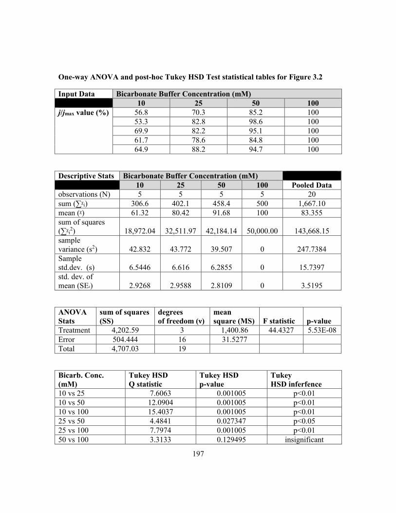
197
One-way ANOVA and post-hoc Tukey HSD Test statistical tables for Figure 3.2
Input Data Bicarbonate Buffer Concentration (mM)
10 25 50 100
j/jmax value (%) 56.8 70.3 85.2 100
53.3 82.8 98.6 100
69.9 82.2 95.1 100
61.7 78.6 84.8 100
64.9 88.2 94.7 100
Descriptive Stats Bicarbonate Buffer Concentration (mM)
10 25 50 100 Pooled Data
observations (N) 5 5 5 5 20
sum (∑ᵡi) 306.6 402.1 458.4 500 1,667.10
mean (ᵡ) 61.32 80.42 91.68 100 83.355
sum of squares
(∑ᵡi2) 18,972.04 32,511.97 42,184.14 50,000.00 143,668.15
sample
variance (s2) 42.832 43.772 39.507 0 247.7384
Sample
std.dev. (s) 6.5446 6.616 6.2855 0 15.7397
std. dev. of
mean (SEᵡ) 2.9268 2.9588 2.8109 0 3.5195
ANOVA
Stats
sum of squares
(SS)
degrees
of freedom (ν)
mean
square (MS) F statistic p-value
Treatment 4,202.59 3 1,400.86 44.4327 5.53E-08
Error 504.444 16 31.5277
Total 4,707.03 19
Bicarb. Conc.
(mM)
Tukey HSD
Q statistic
Tukey HSD
p-value
Tukey
HSD inferfence
10 vs 25 7.6063 0.001005 p<0.01
10 vs 50 12.0904 0.001005 p<0.01
10 vs 100 15.4037 0.001005 p<0.01
25 vs 50 4.4841 0.027347 p<0.05
25 vs 100 7.7974 0.001005 p<0.01
50 vs 100 3.3133 0.129495 insignificant

198
APPENDIX B
CONFOCAL LASER SCANNING MICROSCOPY RAW IMAGES FROM
CHAPTER 3

199
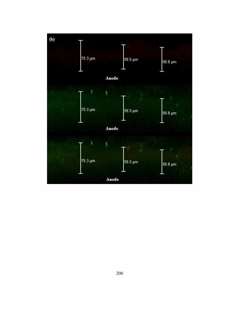
200

201

202

203
APPENDIX B: Confocal Laser Scanning Micrographs for T. ferriacetica biofilm at
corresponding buffer concentrations. All pictures are shown as a cross section of the z-
dimension with red = dead/ inactive biomass and green = live/ active biomass. The
location of the anode is marked with “Anode”. Displayed for all conditions are the dead
segment, the live segment, and/or an overlay of the two. All measurements (in µm)
displayed on graphs are relative to the thickness of the active layer of the biofilm. (a) at
10mM bicarbonate, (b) at 25mM bicarbonate, (c) at 50mM bicarbonate, and (d) at
100mM bicarbonate buffer.

204
APPENDIX C
ANOVA AND POST-HOC RESULTS FOR CONFOCAL LASER SCANNING
MICROSCOPY RESULTS SHOWN IN FIGURE 3.3 FROM CHAPTER 3

205
One-way ANOVA and post-hoc Tukey-Kramer HSD Test statistical tables for
Figure 3.3
Input Data Bicarbonate Buffer Concentration (mM)
10 25 50 100
Lf value (µm) 70 75 131 153
97 60 105 138
68 59 80 152
56 50 99 173
42 43 103 211
95 53 226 276
67 206 203
51 188 235
161 162
53 137
71 121
64 224
Descriptive Stats Bicarbonate Buffer Concentration (mM)
10 25 50 100 Pooled Data
observations (N) 8 6 12 12 38
sum (∑ᵡi) 546 340 1,487.00 2,185.00 4,558.00
mean (ᵡ) 68.25 56.6667 123.9167 182.0833 119.9474
sum of squares
(∑ᵡi2)
39,948.00 19,864.00 221,719.00 422,447.00 703,978.00
sample
variance (s2)
383.3571 119.4667 3,404.99 2,235.90 4,250.21
Sample
std.dev. (s)
19.5795 10.9301 58.3523 47.2853 65.1937
std. dev. of
mean (SEᵡ)
6.9224 4.4622 16.8449 13.6501 10.5758
ANOVA
Stats
sum of squares
(SS)
degrees
of freedom (ν)
mean
square (MS) F statistic p-value
Treatment 91,927.23 3 30,642.41 15.9472 1.21E-06
Error 65,330.67 34 1,921.49
Total 157,257.89 37

206
Bicarb. Conc.
(mM)
Tukey HSD
Q statistic
Tukey HSD
p-value
Tukey
HSD inferfence
10 vs 25 0.692 0.8999947 insignificant
10 vs 50 3.9347 0.0414836 p<0.05
10 vs 100 8.0461 0.0010053 p<0.01
25 vs 50 4.3393 0.0208938 p<0.05
25 vs 100 8.0925 0.0010053 p<0.01
50 vs 100 4.5967 0.0132229 p<0.05

207
BIOGRAPHICAL SKETCH
Scientist, avid runner, businessman, and social entrepreneur, Brad Lusk spent 6.5
of his 10.5 years at Arizona State University (ASU) as a graduate student in the Swette
Center for Environmental Biotechnology researching thermophilic bacteria in microbial
electrochemical cells. During his time as a Ph.D. student, he has hosted and attended
scientific conferences throughout the United States including the North American
meeting for the International Society for Microbial Electrochemical Technology
(ISMET) at Penn State University, the International meeting for ISMET at ASU, the
Association of Environmental Engineering and Science Professors meeting at the
Colorado School of Mines, and the general meeting for the American Society for
Microbiology in Boston, Massachusetts. His experiences and friendships formed during
his graduate studies have enabled him to travel to exotic locations in India and Pakistan,
where he has given presentations about his research to a broad, global audience. He has
also shared his love for science to live public audiences at local Phoenix events including
IGNITE, Night of the Open Door, and COMICON. In his free time, he volunteers with a
local non-profit called Camp Sparky -where he served as Chair from 2014-2015- to visit
local Title 1 elementary schools through the Phoenix metropolitan area. This volunteer
work allows him to spread a message of scientific awareness and emphasize the positive
impacts of education to local ‘at-risk’ youth. In 2012, he turned his interest and longtime
knack for vintage collectibles into a lucrative online business. He also uses his passion
for nutrition and running to conquer 5Ks, 10Ks, and half marathons. With his degree,
Brad is looking to travel the world to visit laboratories and industries that are interested in
microbial electrochemical research in order to gain a global perspective on his field of
interest and to share his experiences to spread cultural awareness.


![Microbial Degradation of Plastic Waste: A Review · PDF fileused in transportation, food, clothing, shelter ... bioplastic degrading bacteria[37]. In addition to these strains, a thermophilic](https://img.pdfslide.us/doc/110x75/5aabc7fc7f8b9ac55c8c5015/microbial-degradation-of-plastic-waste-a-review-in-transportation-food-clothing.jpg)