Embed Size (px)
Citation preview

1
•First, a bit of information relating to receptors: •From sensory organ to sensory organ, receptors work in fundamentally the same
way •A receptor is always the dendrite of a sensory neuron or a specialized structure that collects information and stimulates a dendrite to send a message to the brain. •The sensory receptor is particularly sensitive to some type of “environmental change” and insensitive to everything else. •The sensory receptor does not “interpret” the change….that is the job of your brain.
There Are 5 Different Types Of Sensory Receptors* *Note: Not all are touch (somatic) receptors.
(1) Chemoreceptors – these are receptors that are designed to sense changes in chemical concentrations in the environment surrounding the receptor. •Smell and taste receptors are chemoreceptors.
Olfactory (smell) receptor Taste receptor •We also possess internal chemoreceptors that are sensitive to changes in the concentrations of substance transported by blood – sugar, oxygen, hydrogen ions (pH), carbon dioxide, hormones……

2
(2) Nociceptors – free nerve endings (dendrites) that are sensitive to tissue damage. These are also called pain receptors because the response triggered by their stimulation is pain. •Nociceptors can be very long neurons. With their sensory endings in the skin, in joints or muscles, they can measure more than a meter up to their synaptic ending in dorsal horn of the spinal cord. •The sensory endings of nociceptors usually respond to very strong stimuli: temperatures in excess of 40o C, acid solutions, cuts, bruises, etc. In contrast to subtle sensory stimuli like dim light or weak odors, such intense stimulation does not require amplifying mechanisms in the sensory cell.
(3) Thermoreceptors – Thermoreceptors are sensory receptors that are sensitive to temperature change. They are located in the dermis, just beneath the epidermis. Like Nociceptors, they are raw nerve endings. •There are two types of thermoreceptors, warm and cold.

3
(4) Mechanoreceptors – sense mechanical changes (slight touch, pushing, squeezing). The three categories of mechanoreceptors are: (1) Proprioceptors – receptors embedded within muscles and tendons that sense changes in tension and stretching. The muscle spindle is the most common type of proprioceptor. (2) Baroreceptors – receptors in the walls of vessels (principally the carotid arteries and the aortic arch) that detect changes in blood pressure.

4
(3) Stretch receptors – sense “stretching” in tissues and walls. Ex. stretch receptors in the colon and bladder, stretch receptors in lung tissue to detect the degree of inflation.
Stretch Receptors
(5) Photoreceptors – Vision is possible due to the absorption of light by photoreceptors on the retina of the eye. Humans have two kinds of photoreceptor cells called rods and cones due to their distinctive shapes. Rods and cones account for 70% of all sensory receptors in the body thus showing the importance of the eyes. Cones require a relatively high level of light to be stimulated so therefore only function in bright (during the day) and are responsible for color vision. Rods are more sensitive to light so will function in dim light but do not distinguish color. They enable us to see at night but only in black and white. A human retina contains about 125 million rod cells and 6 million cone cells.
Rods
Cone

5
•Sensory Receptors are either: •the raw ends (dendrites) of nerve fibers or •specialized cells (or organs) next to the ends (dendrites) of nerve fibers.
Specialized Organ Raw Ends of
Nerve fibers
•A sensation occurs when the brain interprets or “figures out” a sensory impulse. •”How” the brain interprets the impulse depends upon where in the brain the impulse “ends up” (because all impulses are basically the same). •The brain can be mapped to show the different “sensory areas”. As an example, sensory impulses reaching the temporal lobe of the brain are interpreted as “noises” or “sounds”.

6
•Even though “sensations” are formed in the brain, the cerebral cortex
interprets the sensations as coming from the part of the body where the receptors are being stimulated. This process is termed projection. If projection did not occur, a person could not tell where the source of
stimulation occurred. For instance, you would not be able to identify the “source” of a sliver. You would still experience a sharp pain, but you couldn't tell, with specificity, where the pain was coming from.
Projection
•If a sensory receptor is stimulated repeatedly, the receptor undergoes a
period of adjustment during which the receptor increasingly “ignores” the stimuli. The stimuli must increase in order for the sensory receptor to detect the stimulus. If the stimulus stays at the same level, the receptor may eventually cease sending signals to the brain. This is sensory adaptation. Sensory adaptation occurs with touch, smell, taste, vision, and hearing. Give an example of each.
Sensory Adaptation
Annoyingly, there is no sensory adaptation for pain.

7
Touch and Pressure On External Body Surfaces
•Humans have four types of receptors that detect touch and pressure. These receptors work by sensing shifts in the positions of cells or changes in the shapes of specialized receptors. They are, of course, all mechanoreceptors.
(1) Sensory nerve fibers – raw nerve endings (dendrites) that lie between epithelial cells and detect pain, temperature (hot & cold), itch, and certain types of movement.
(2) Meissner’s Corpuscles – small oval masses of connective tissue that lie just beneath the epidermis near the epidermal-dermal border. At least two nerve fibers are embedded in the corpuscles. The Meissner’s Corpuscles are located in the most “sensitive” parts of the body (lips, fingertips, palms, soles, nipples, and genitals. They are designed to detect the slightest of movements in the shiftings that result from light touch. You use them when you are touching something to judge its texture. Fast-adapting.
(3) Pacinian Corpuscles – larger, elliptoid bodies that lie deeper in the dermis and are designed to detect heavy pressure or vibrations. Looks like an onion. (4) Merkel's Disks - slowly-adapting light touch receptors that respond to light touch and superficial pressure maintained over time.

8
Temperature Detection On External Body Surfaces
•Humans have two types of temperature receptors located in the dermis: •Both are free nerve endings. •Both are thermoreceptors.
(1) Heat receptors – most sensitive to temperatures above 77o F, unresponsive above 113o F. Beyond 113o F., pain receptors are stimulated.
(2) Cold receptors – most sensitive to temperatures between 50o F. and 68o F. Below 50o F., pain receptors are stimulated and it becomes increasingly difficult to distinguish between hot and cold….they both elicit searing pain.
•At intermediate temperatures, the brain is interpreting impulses from both heat and cold receptors. Sensory adaptation occurs rapidly in thermoreceptors.

9
Pain Detection On External Body Surfaces
•Free nerve endings detect pain. •Pain receptors are found in many sites in the body, but not in the nervous tissue of the brain. •Pain receptor stimulation results from tissue damage. •Unfortunately, pain receptors do not demonstrate sensory adaptation. •Pain receptor stimulation is designed to direct the body’s attention to the source
of pain. •Pain receptors trigger the reflex arc.
Referred Pain
•Sometimes, pain in one part of the body is detected as coming from some “other” part of the body. This is termed “referred pain”. Referred pain is thought to result from pain passing through a nerve tract that is also connected to another organ. The body mistakes the source of the pain during projection. As a specific example, a person suffering a heart attack grabs the left arm because the medial surface of the left arm and the heart share a common nerve pathway to sensory interpretive areas in the brain. •”Brain freeze” is a common example of referred pain. The pain-sensitive cells in the mouth and the throat share a nerve pathway with the sinuses. When you eat flavored shaved ice (a "Slurpee"), the pain is projected back to your sinuses and you enjoy “brain freeze”.
10
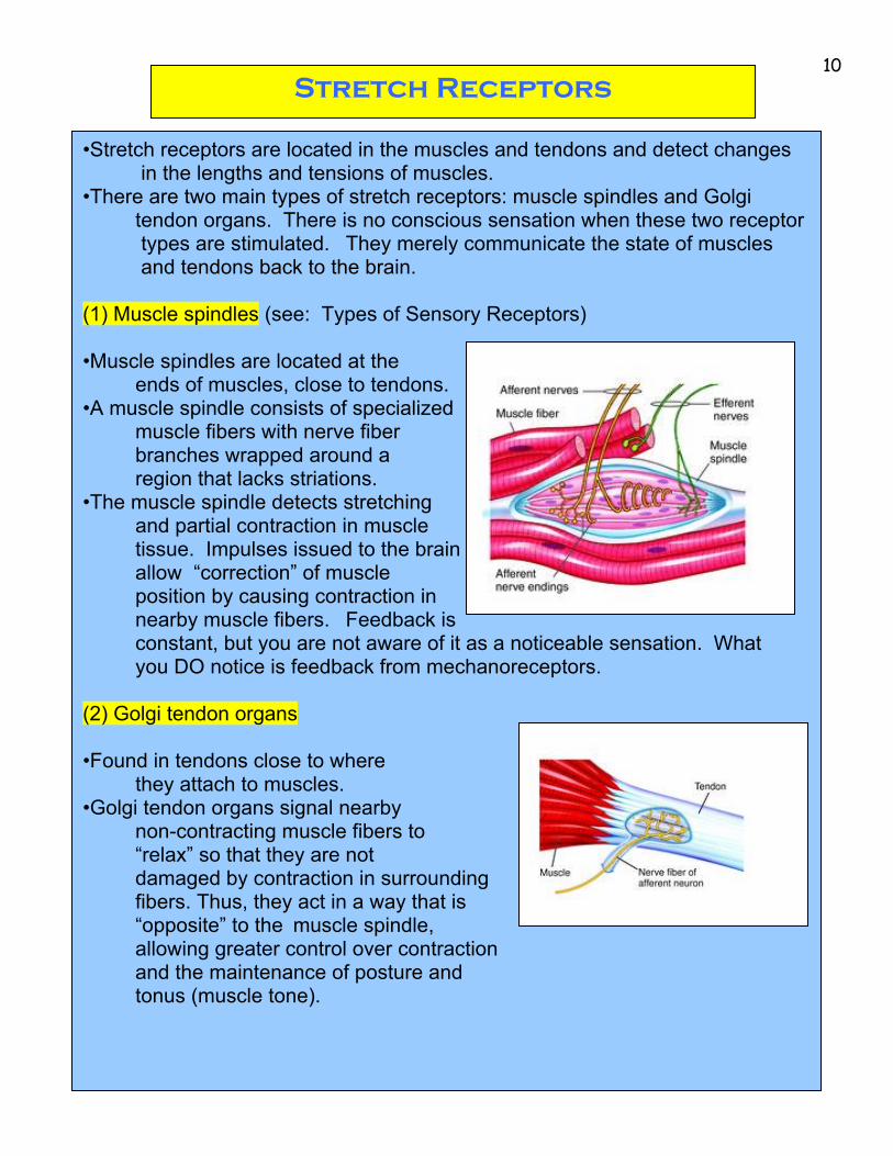
Stretch Receptors
•Stretch receptors are located in the muscles and tendons and detect changes in the lengths and tensions of muscles. •There are two main types of stretch receptors: muscle spindles and Golgi tendon organs. There is no conscious sensation when these two receptor types are stimulated. They merely communicate the state of muscles and tendons back to the brain. (1) Muscle spindles (see: Types of Sensory Receptors) •Muscle spindles are located at the
ends of muscles, close to tendons. •A muscle spindle consists of specialized
muscle fibers with nerve fiber branches wrapped around a region that lacks striations.
•The muscle spindle detects stretching and partial contraction in muscle tissue. Impulses issued to the brain allow “correction” of muscle position by causing contraction in nearby muscle fibers. Feedback is constant, but you are not aware of it as a noticeable sensation. What
you DO notice is feedback from mechanoreceptors. (2) Golgi tendon organs •Found in tendons close to where
they attach to muscles. •Golgi tendon organs signal nearby
non-contracting muscle fibers to “relax” so that they are not damaged by contraction in surrounding fibers. Thus, they act in a way that is “opposite” to the muscle spindle, allowing greater control over contraction and the maintenance of posture and tonus (muscle tone).