Embed Size (px)
DESCRIPTION
SENSORY RECEPTORS. SENSORY RECEPTORS. Objectives: E - label the parts of the eye D – Describe their functions C – explain how parts function to adjust focus and the amount of light entering the eye. The eye as a receptor. vitreous humour. choroid. choroid. iris. retina. cornea. pupil. - PowerPoint PPT Presentation
Citation preview
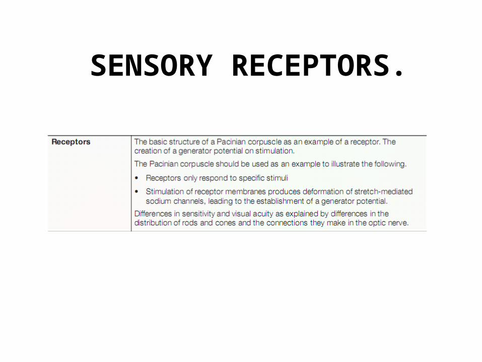
SENSORY RECEPTORS.

SENSORY RECEPTORS.
Objectives:E - label the parts of the eyeD – Describe their functions
C – explain how parts function to adjust focus and the amount of light entering
the eye

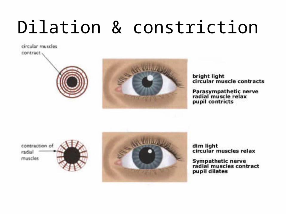
The eye as a receptor

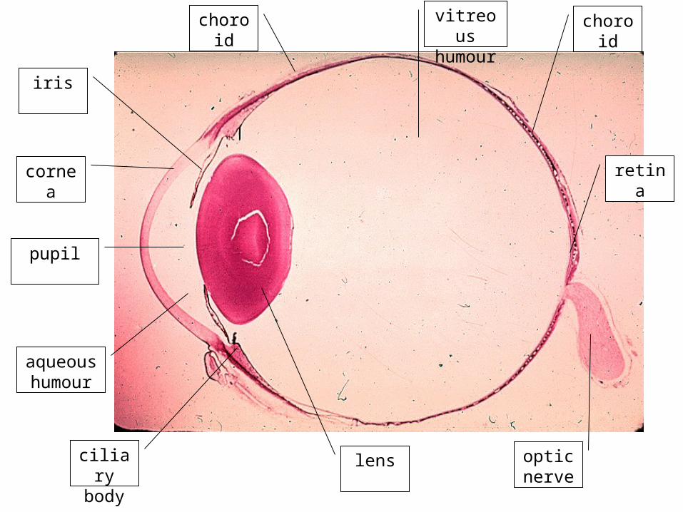
cornea
iris
pupil
aqueous humour
ciliary body
lens
choroid choroid
retina
vitreous humour
optic nerve

Soper 2 p593
• Draw eye diagram and label functions

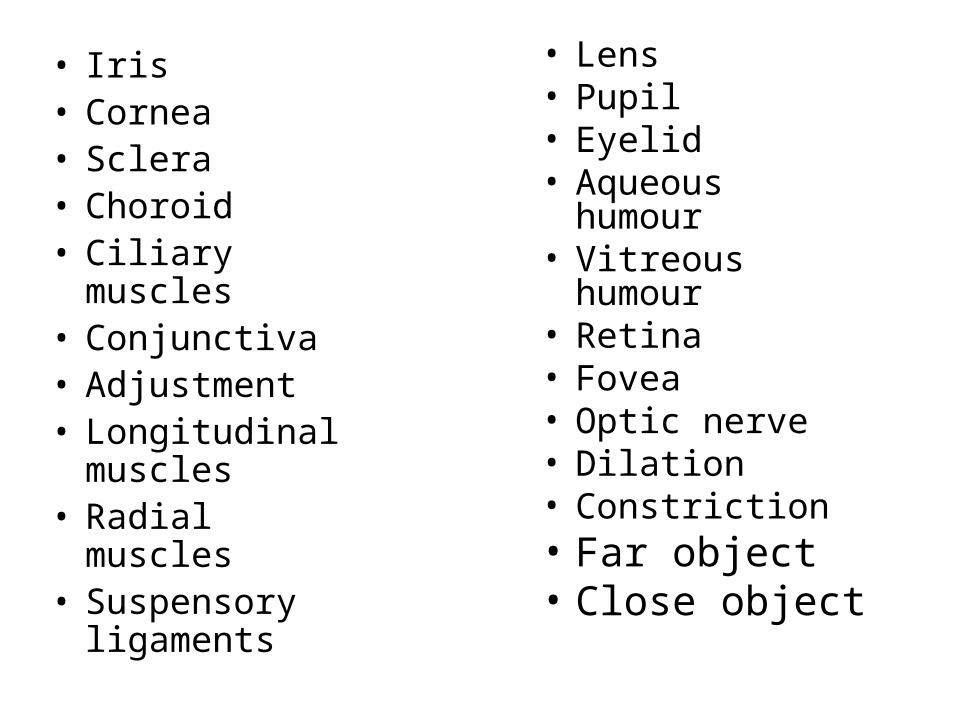
• Iris• Cornea• Sclera• Choroid• Ciliary muscles• Conjunctiva• Adjustment• Longitudinal
muscles• Radial muscles• Suspensory
ligaments
• Lens• Pupil• Eyelid• Aqueous
humour• Vitreous
humour• Retina• Fovea• Optic nerve• Dilation• Constriction• Far object• Close object

Accomodation
• This is the focusing of light by the lens

• Iris• Cornea• Sclera• Choroid• Ciliary muscles• Conjunctiva• Rhodopsin • Longitudinal
muscles• Radial muscles• Suspensory
ligaments• cones
• Lens• Pupil• Eyelid• Aqueous
humour• Vitreous
humour• Retina• Fovea• Optic nerve• Visual accuity• Constriction• Far object• Close object• Rods
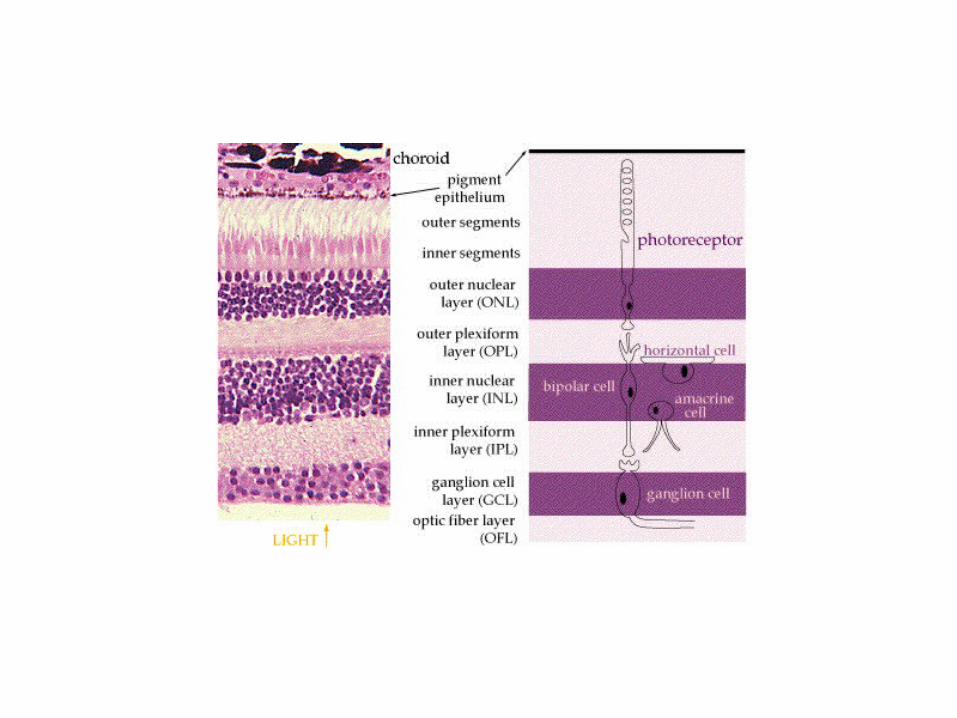
The retina


Blood vessel in choroid
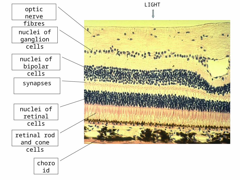
choroid
retinal rod and cone cells
optic nerve fibres
synapses
nuclei of retinal cells
nuclei of bipolar cells
nuclei of ganglion cells
LIGHT
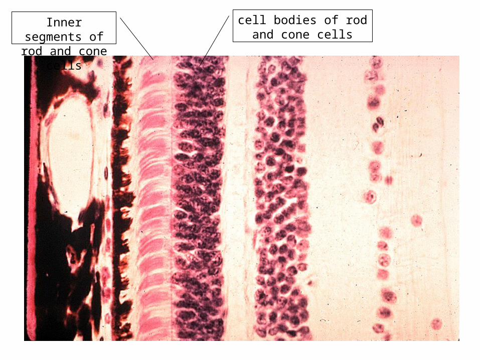
Inner segments of rod and cone cells
cell bodies of rod and cone cells
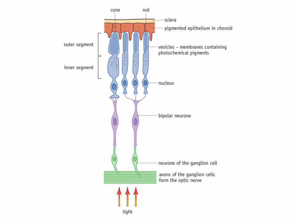

rod cell
cone cell

rod cellcone cell
cell body
bipolar cell
ganglion cell
optic nerve fibres
synapse

Structure of the retina
Axons of ganglion cells optic nerve visual area of the brain
Photoreceptor cells(Rods and cones)
Bipolar neurones
connect photoreceptors to optic nerve
•rods connected in groups
•cones connected singly
Melanin – absorbs light to prevent internal reflection

fovea
Approx 1mm diameter on visual axis of eyeCones only
Point of maximum intensity of visionMain point of interest in visual field focused
here
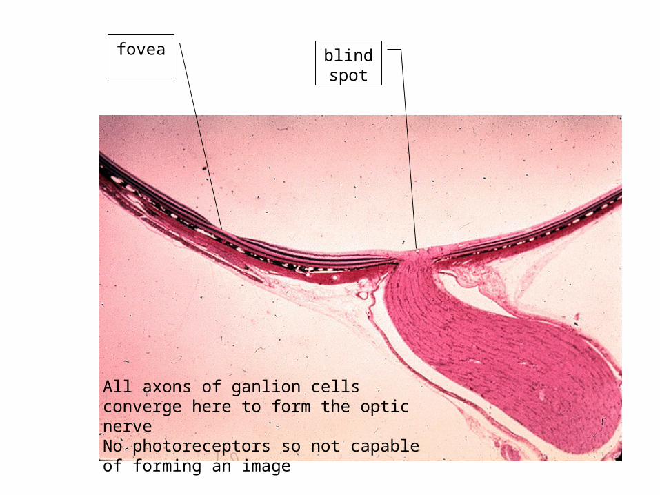
blind spot
fovea
All axons of ganlion cells converge here to form the optic nerveNo photoreceptors so not capable of forming an image
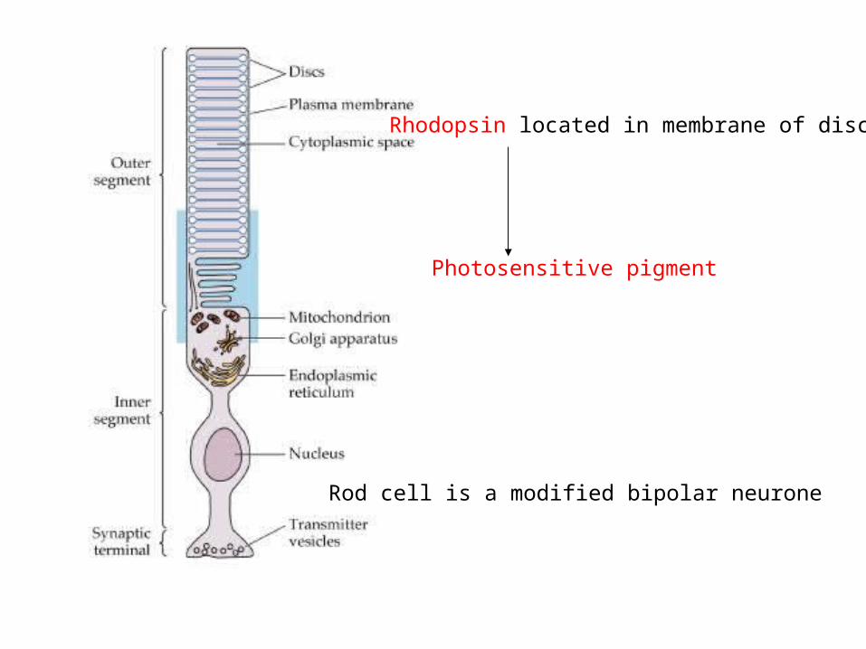
Rhodopsin located in membrane of discs
Photosensitive pigment
Rod cell is a modified bipolar neurone

How cone cells work; colour vision
• Cone cells work in basically the same way as rod cells.
• However bleaching requires a much higher light intensity (so cone cells cannot function in dim light).
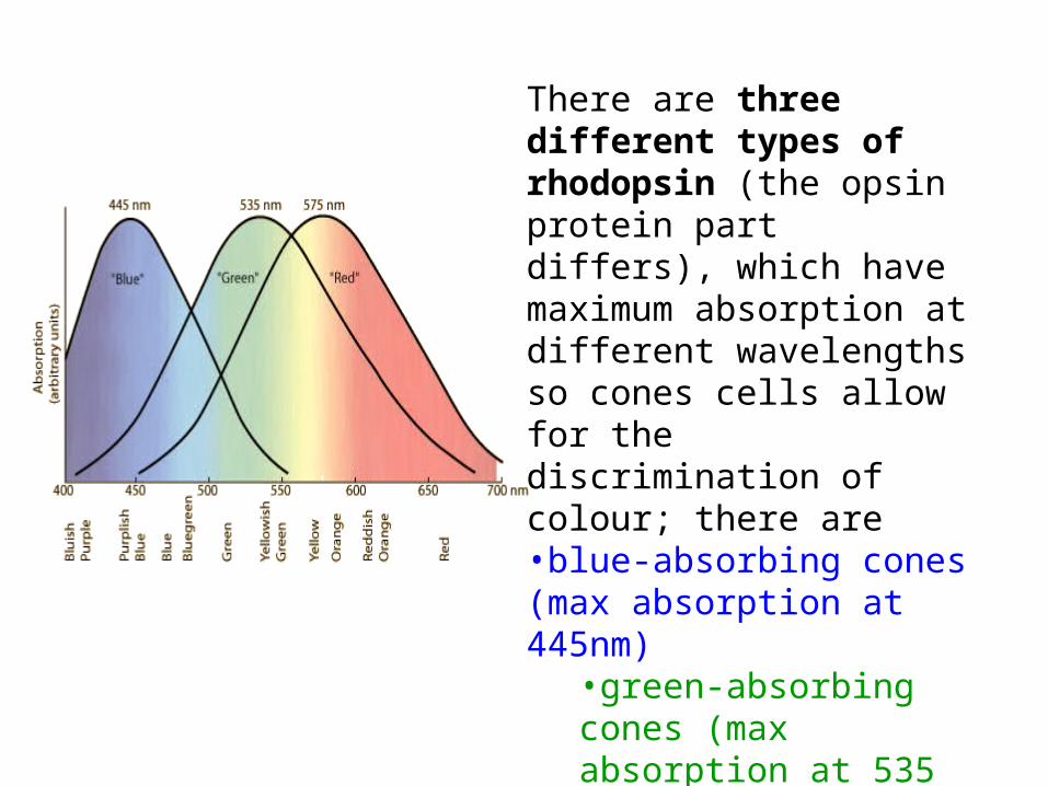
There are three different types of rhodopsin (the opsin protein part differs), which have maximum absorption at different wavelengths so cones cells allow for the discrimination of colour; there are•blue-absorbing cones (max absorption at 445nm)
•green-absorbing cones (max absorption at 535 nm)•red-absorbing cones (max absorption at 570 nm)
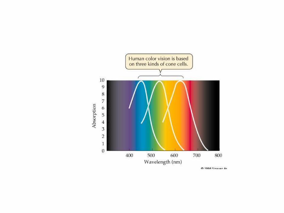


• this is the trichromatic theory of colour vision
• different colours are perceived as a result of the degree of stimulation of the blue + green + red cones (i.e. colours are the result of the mixture of inputs from all three cone types)
• By population, about 64% of the cones are red-sensitive, about 32% green sensitive, and about 2% are blue sensitive
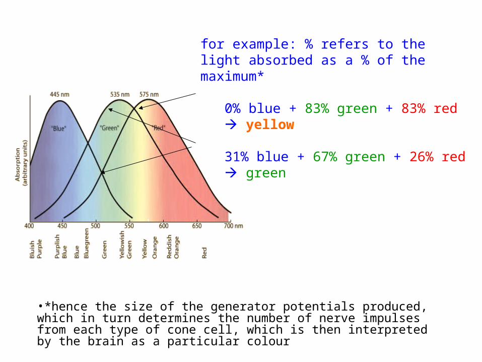
for example: % refers to the light absorbed as a % of the maximum*
0% blue + 83% green + 83% red yellow
31% blue + 67% green + 26% red green
•*hence the size of the generator potentials produced, which in turn determines the number of nerve impulses from each type of cone cell, which is then interpreted by the brain as a particular colour