Embed Size (px)
Citation preview
RECEPTORS and SENSATION
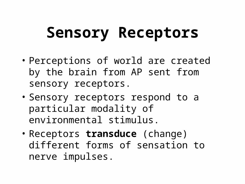
Sensory Receptors
• Perceptions of world are created by the brain from AP sent from sensory receptors.
• Sensory receptors respond to a particular modality of environmental stimulus.
• Receptors transduce (change) different forms of sensation to nerve impulses.
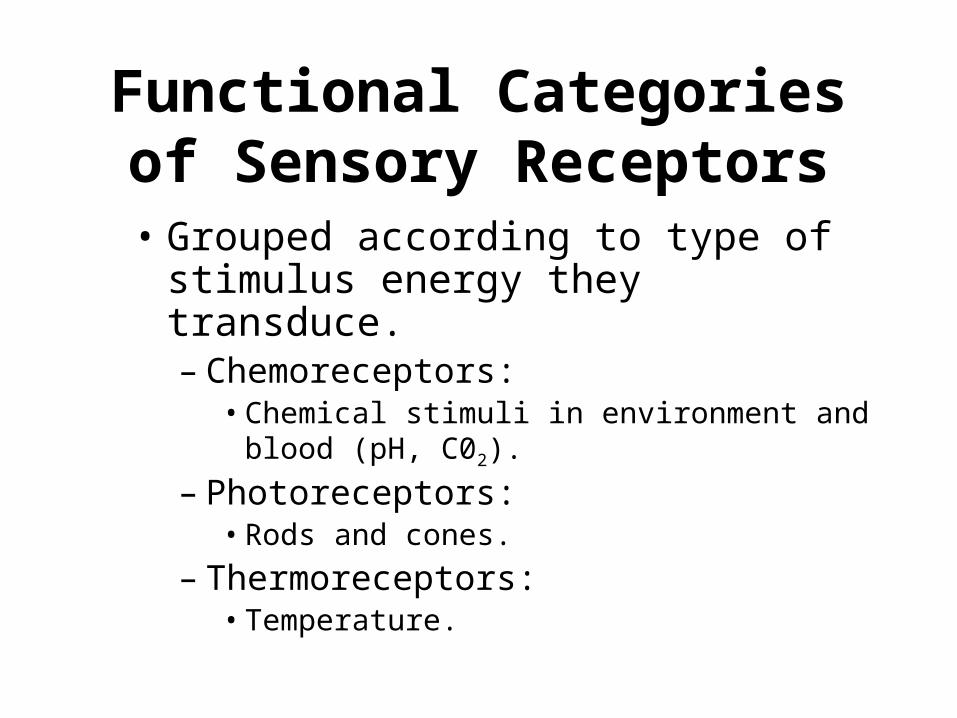
Functional Categories of Sensory Receptors
• Grouped according to type of stimulus energy they transduce.– Chemoreceptors:
• Chemical stimuli in environment and blood (pH, C02).
– Photoreceptors:• Rods and cones.
– Thermoreceptors:• Temperature.
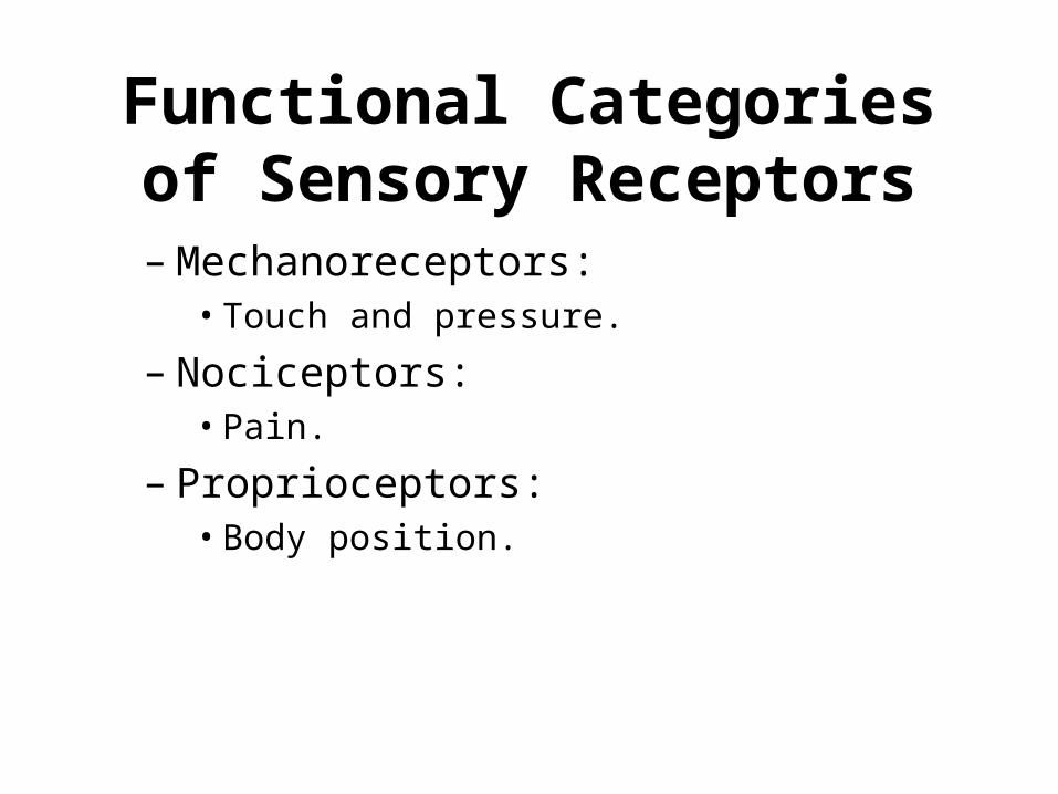
Functional Categories of Sensory Receptors
– Mechanoreceptors:• Touch and pressure.
– Nociceptors:• Pain.
– Proprioceptors:• Body position.
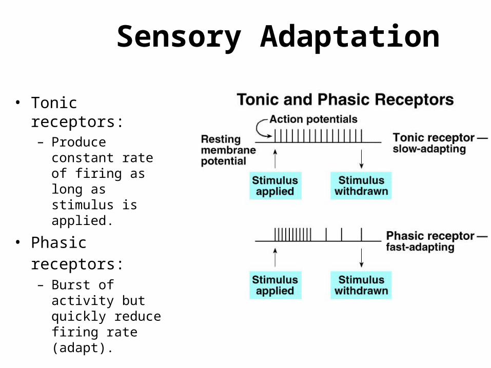
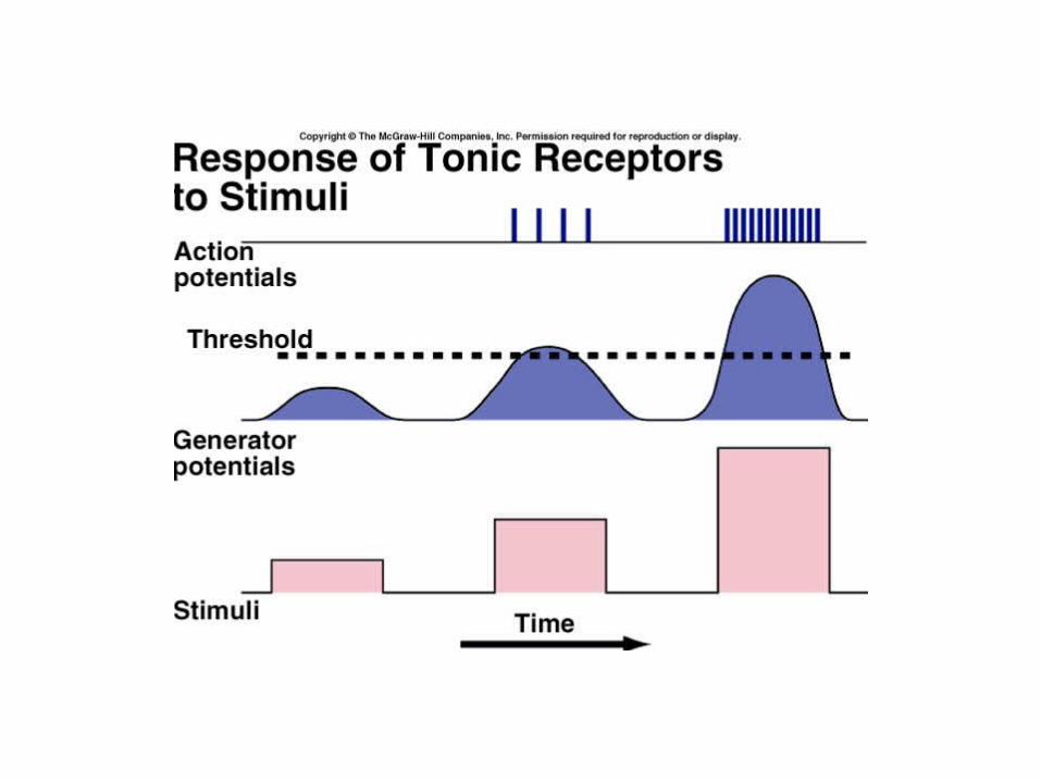
Sensory Adaptation
• Tonic receptors:– Produce constant rate
of firing as long as stimulus is applied.
• Phasic receptors: – Burst of activity but
quickly reduce firing rate (adapt).
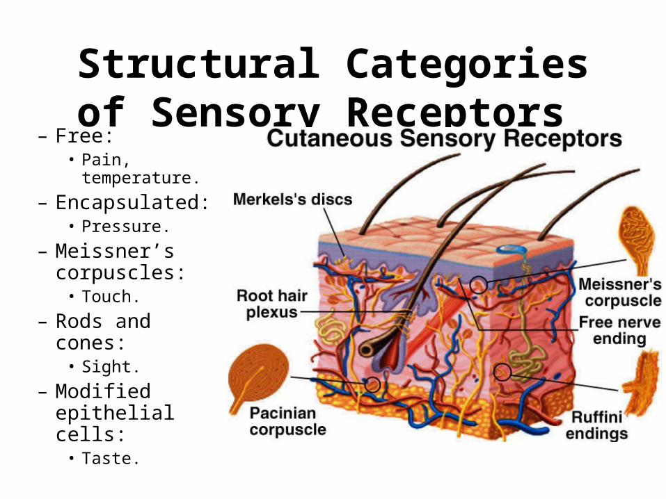
Structural Categories of Sensory Receptors
– Free:• Pain,
temperature.
– Encapsulated:• Pressure.
– Meissner’s corpuscles:
• Touch.
– Rods and cones:• Sight.
– Modified epithelial cells:
• Taste.
Special Senses
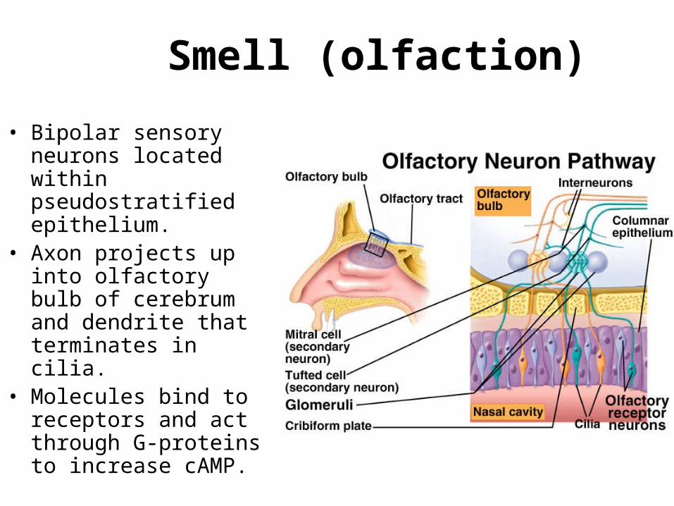
Smell (olfaction)
• Bipolar sensory neurons located within pseudostratified epithelium.
• Axon projects up into olfactory bulb of cerebrum and dendrite that terminates in cilia.
• Molecules bind to receptors and act through G-proteins to increase cAMP.
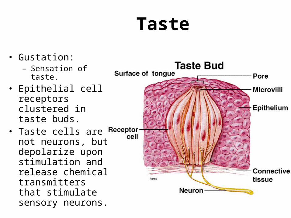
Taste
• Gustation:– Sensation of taste.
• Epithelial cell receptors clustered in taste buds.
• Taste cells are not neurons, but depolarize upon stimulation and release chemical transmitters that stimulate sensory neurons.
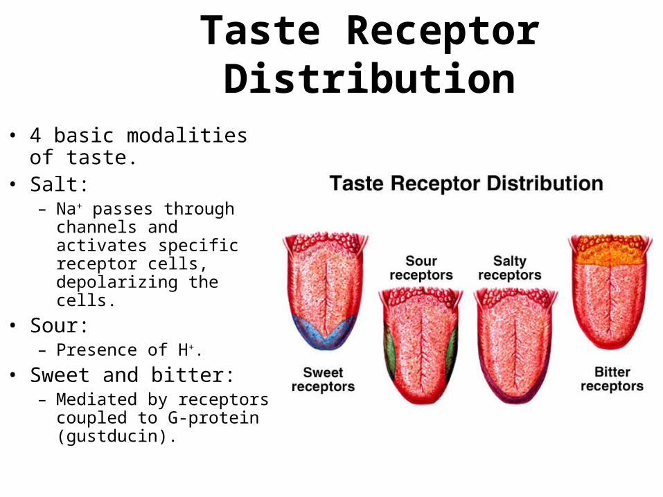
Taste Receptor Distribution
• 4 basic modalities of taste.• Salt:
– Na+ passes through channels and activates specific receptor cells, depolarizing the cells.
• Sour:– Presence of H+.
• Sweet and bitter:– Mediated by receptors
coupled to G-protein (gustducin).
Ears and Hearing
• Sound waves travel in all directions from their source.
• Waves are characterized by frequency and intensity.– Frequency:
• Measured in hertz (cycles per second).• Greater the frequency the higher the pitch.
– Intensity:• Directly related to amplitude of sound waves.• Measured in decibels.
Outer Ear
• Sound waves are funneled by the auricle into the external auditory meatus.
• External auditory meatus channels sound waves to the tympanic membrane.– Increases sound wave intensity.
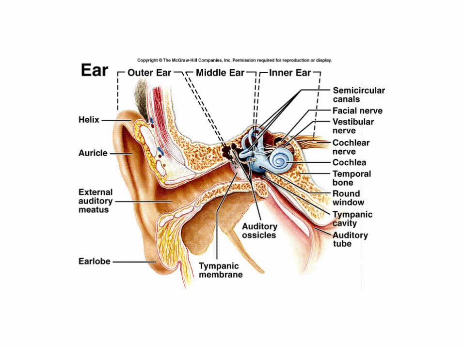
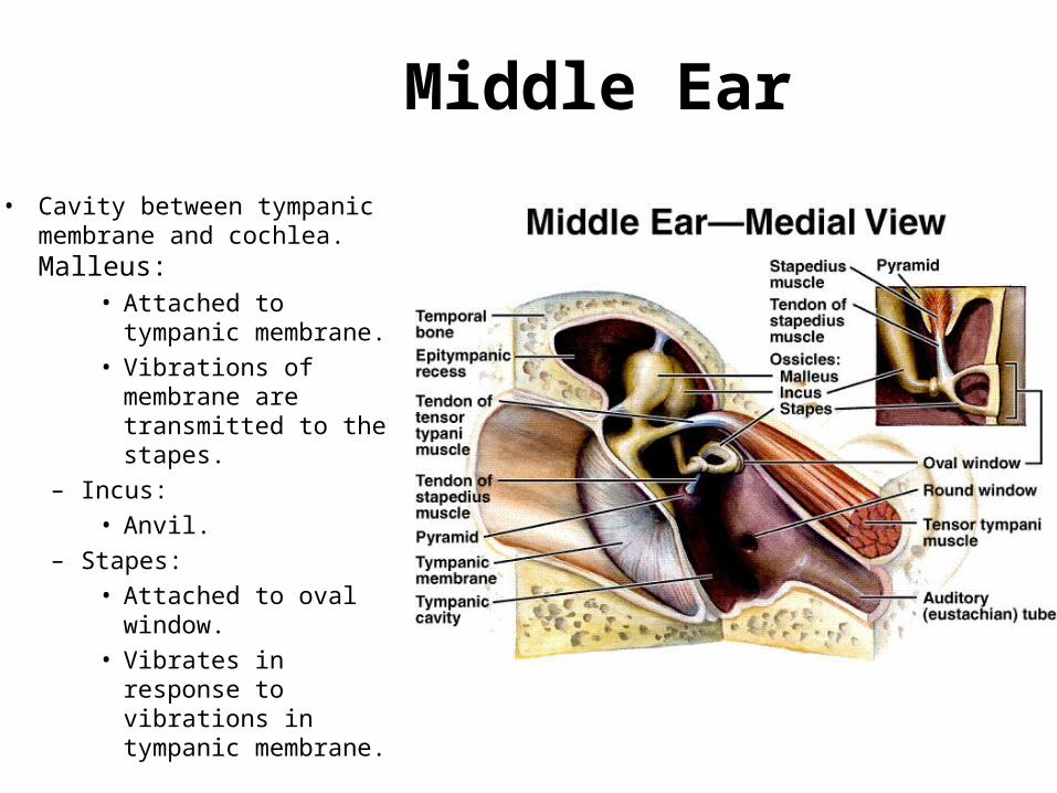
Middle Ear
• Cavity between tympanic membrane and cochlea. Malleus:
• Attached to tympanic membrane.
• Vibrations of membrane are transmitted to the stapes.
– Incus:
• Anvil.
– Stapes:
• Attached to oval window.
• Vibrates in response to vibrations in tympanic membrane.
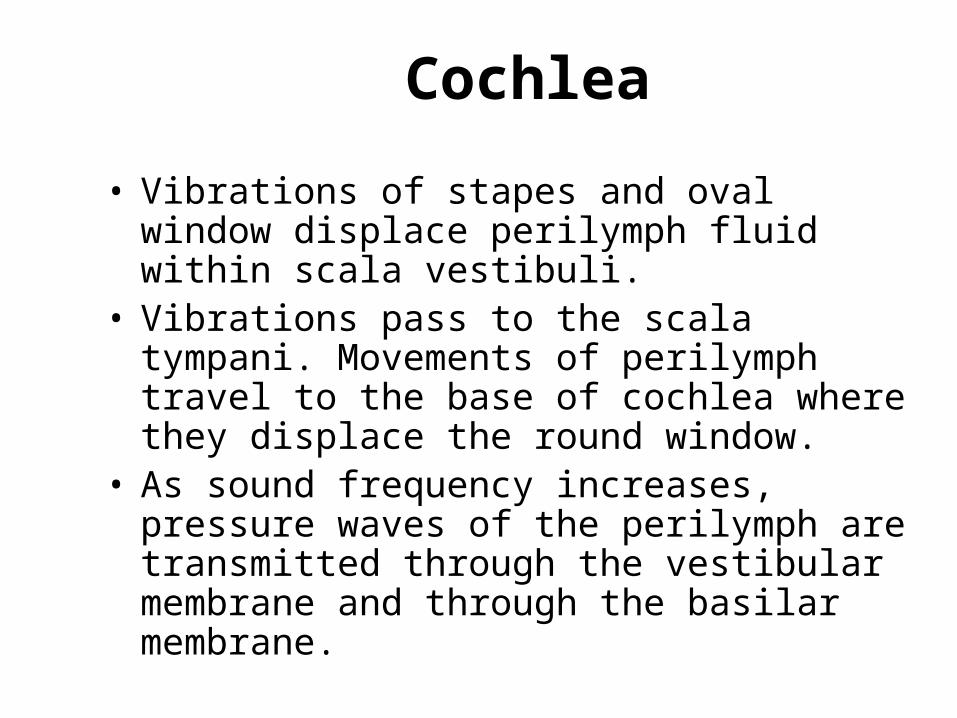
Cochlea
• Vibrations of stapes and oval window displace perilymph fluid within scala vestibuli.
• Vibrations pass to the scala tympani. Movements of perilymph travel to the base of cochlea where they displace the round window.
• As sound frequency increases, pressure waves of the perilymph are transmitted through the vestibular membrane and through the basilar membrane.
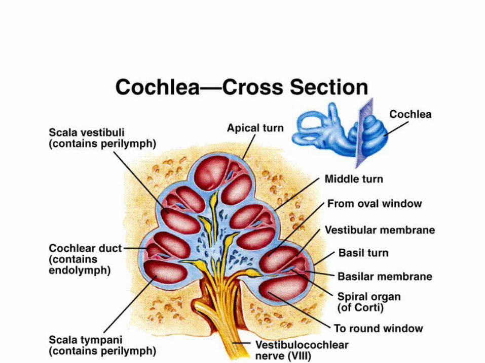
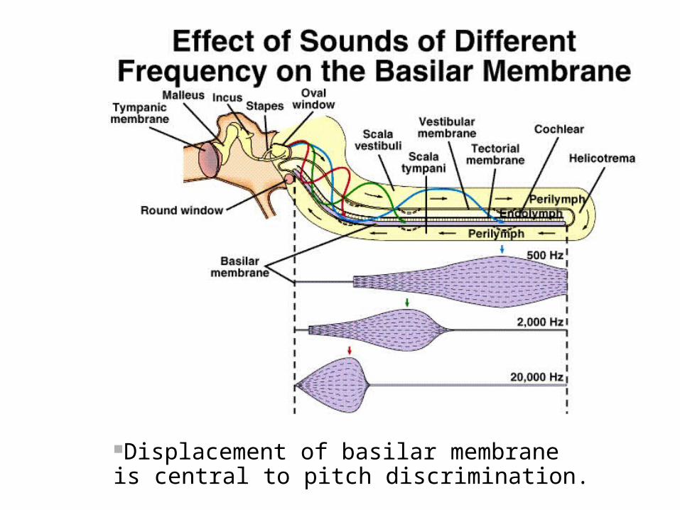
Displacement of basilar membrane is central to pitch discrimination.
Organ of Corti
• Sensory hair cells located on the basilar membrane.
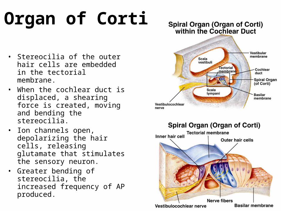
Organ of Corti
• Stereocilia of the outer hair cells are embedded in the tectorial membrane.
• When the cochlear duct is displaced, a shearing force is created, moving and bending the stereocilia.
• Ion channels open, depolarizing the hair cells, releasing glutamate that stimulates the sensory neuron.
• Greater bending of stereocilia, the increased frequency of AP produced.
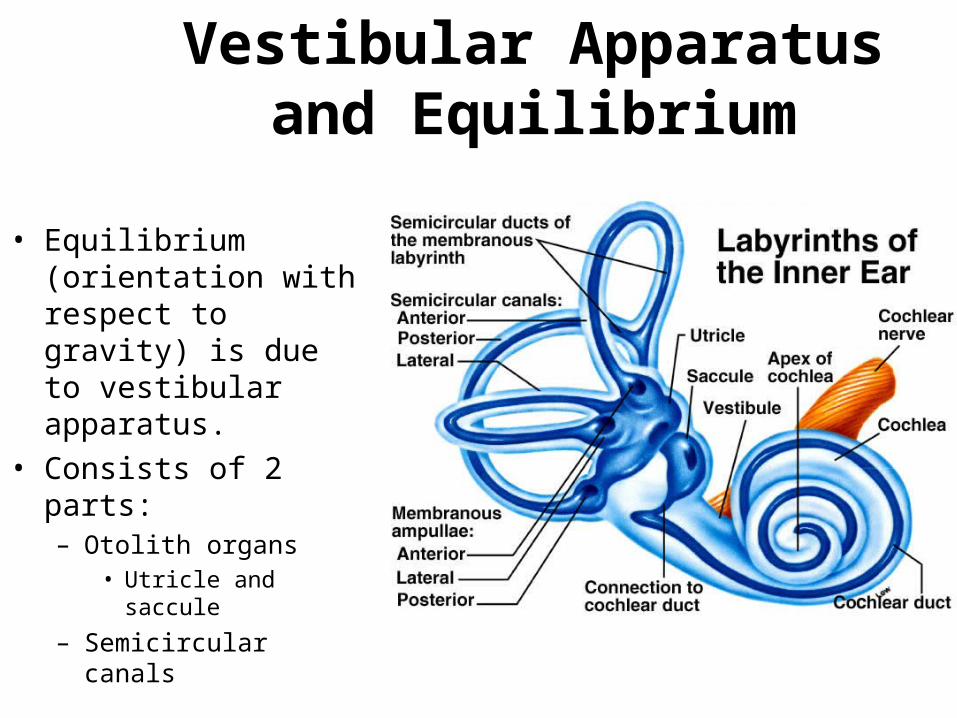
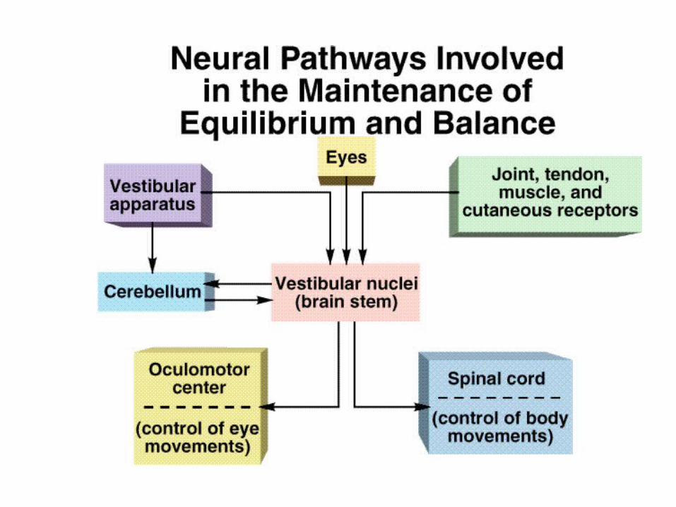
Vestibular Apparatus and Equilibrium
• Equilibrium (orientation with respect to gravity) is due to vestibular apparatus.
• Consists of 2 parts:– Otolith organs
• Utricle and saccule
– Semicircular canals
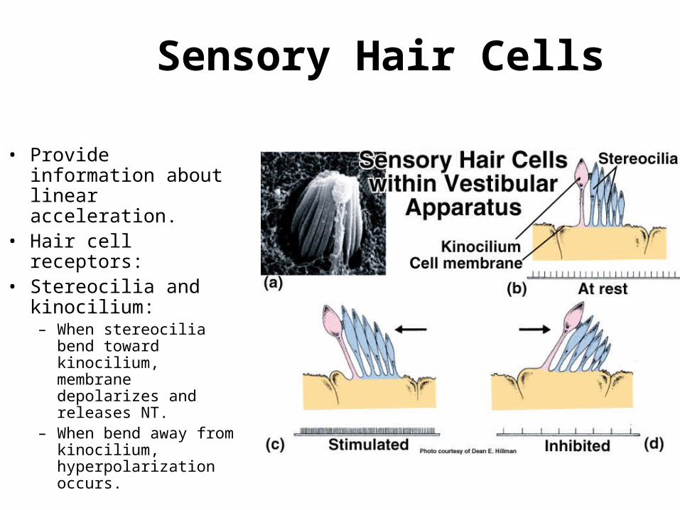
Sensory Hair Cells
• Provide information about linear acceleration.
• Hair cell receptors:• Stereocilia and
kinocilium:– When stereocilia bend
toward kinocilium, membrane depolarizes and releases NT.
– When bend away from kinocilium, hyperpolarization occurs.
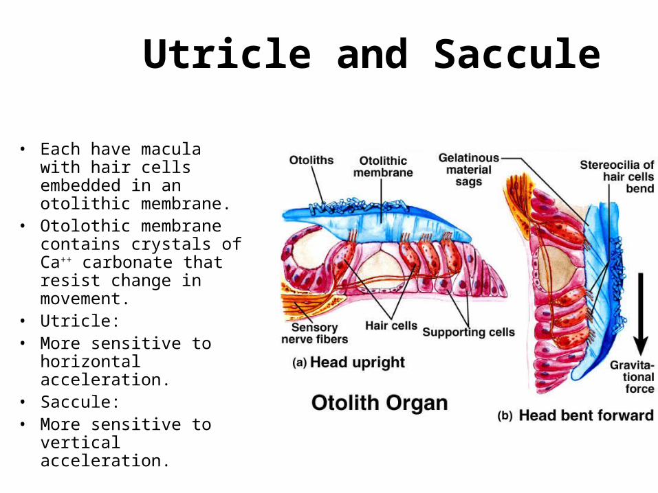
Utricle and Saccule
• Each have macula with hair cells embedded in an otolithic membrane.
• Otolothic membrane contains crystals of Ca++ carbonate that resist change in movement.
• Utricle:• More sensitive to
horizontal acceleration.• Saccule:• More sensitive to vertical
acceleration.
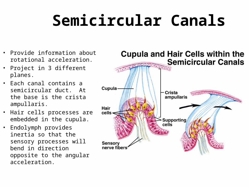
Semicircular Canals
• Provide information about rotational acceleration.
• Project in 3 different planes.
• Each canal contains a semicircular duct. At the base is the crista ampullaris.
• Hair cells processes are embedded in the cupula.
• Endolymph provides inertia so that the sensory processes will bend in direction opposite to the angular acceleration.
Vision
• Eyes transduce energy in the electrmagnetic spectrum into APs.
• Only wavelengths of 400 – 700 nm constitute visible light.
• Neurons in the retina contribute fibers that are gathered together at the optic disc, where they exit as the optic nerve.
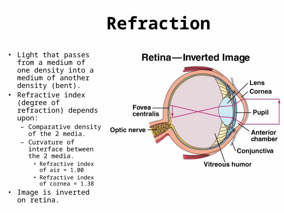
Refraction
• Light that passes from a medium of one density into a medium of another density (bent).
• Refractive index (degree of refraction) depends upon:– Comparative density of
the 2 media.– Curvature of interface
between the 2 media.• Refractive index of air
= 1.00• Refractive index of
cornea = 1.38
• Image is inverted on retina.
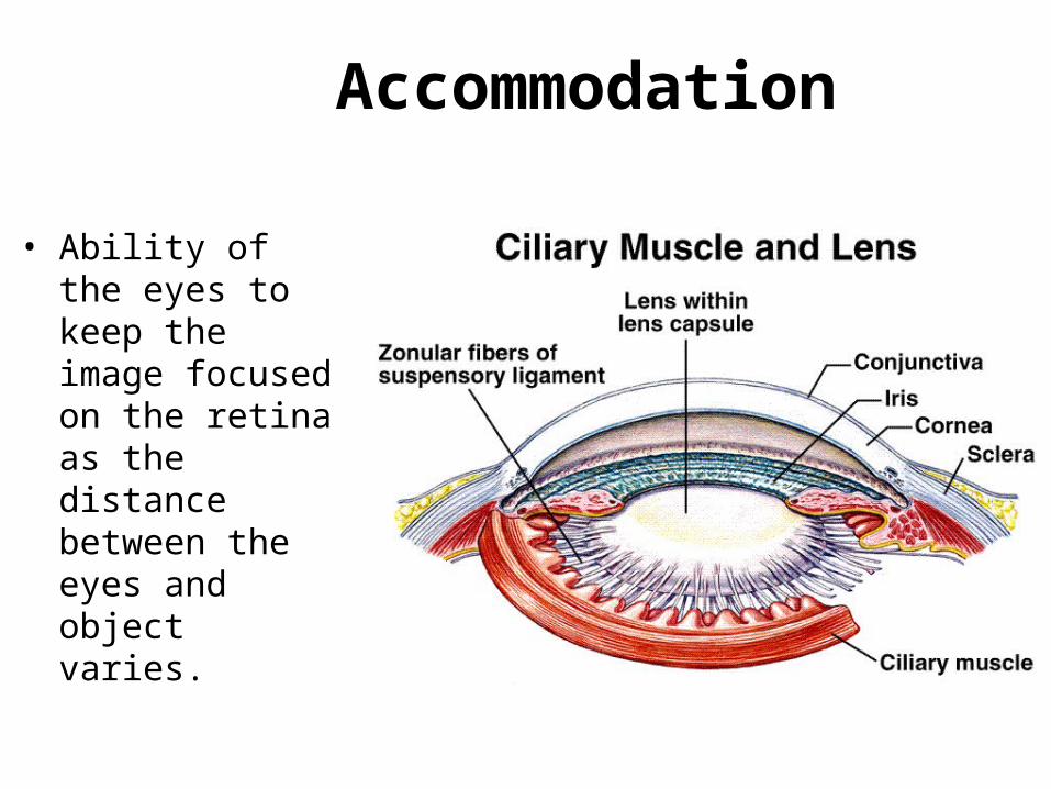
Accommodation
• Ability of the eyes to keep the image focused on the retina as the distance between the eyes and object varies.
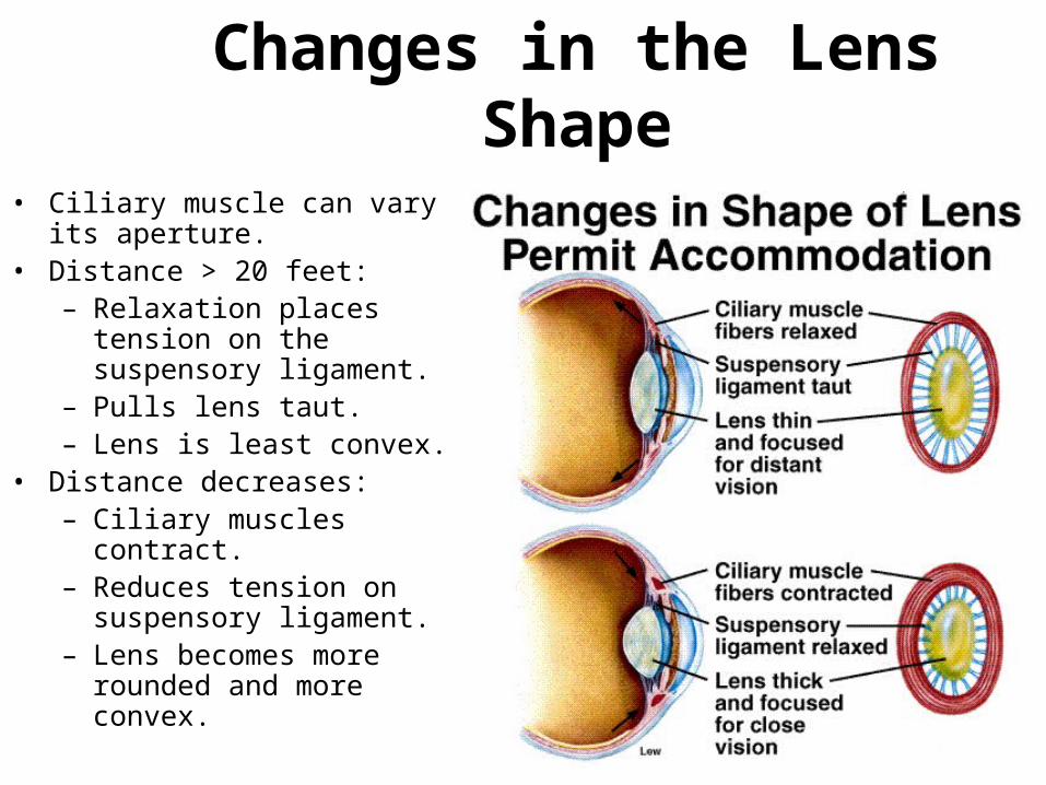
Changes in the Lens Shape
• Ciliary muscle can vary its aperture.
• Distance > 20 feet:– Relaxation places tension on
the suspensory ligament.– Pulls lens taut. – Lens is least convex.
• Distance decreases:– Ciliary muscles contract.– Reduces tension on
suspensory ligament.– Lens becomes more rounded
and more convex.
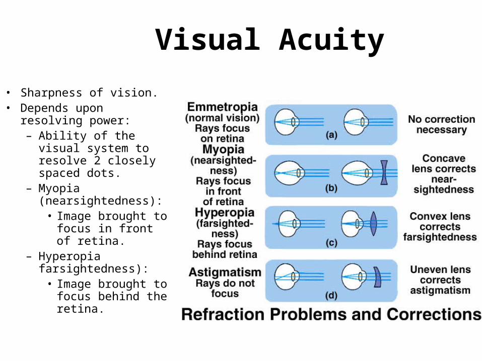
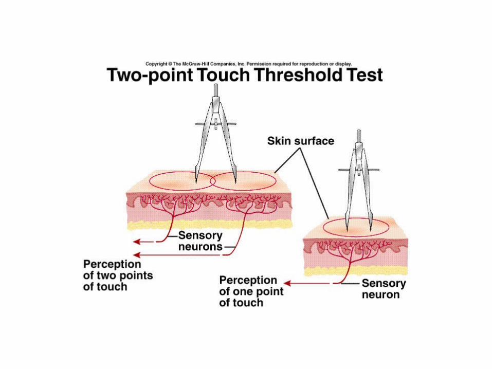
Visual Acuity
• Sharpness of vision.• Depends upon resolving
power:– Ability of the visual
system to resolve 2 closely spaced dots.
– Myopia (nearsightedness):
• Image brought to focus in front of retina.
– Hyperopia farsightedness):
• Image brought to focus behind the retina.
Visual Acuity
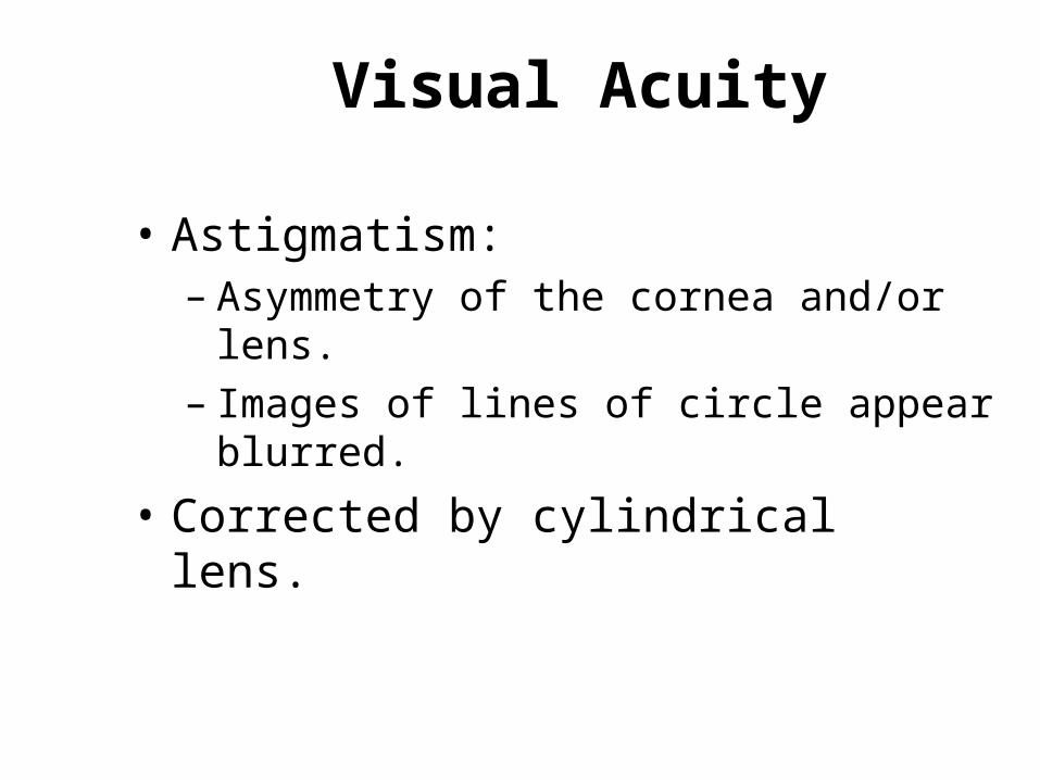
• Astigmatism:– Asymmetry of the cornea and/or lens.– Images of lines of circle appear blurred.
• Corrected by cylindrical lens.
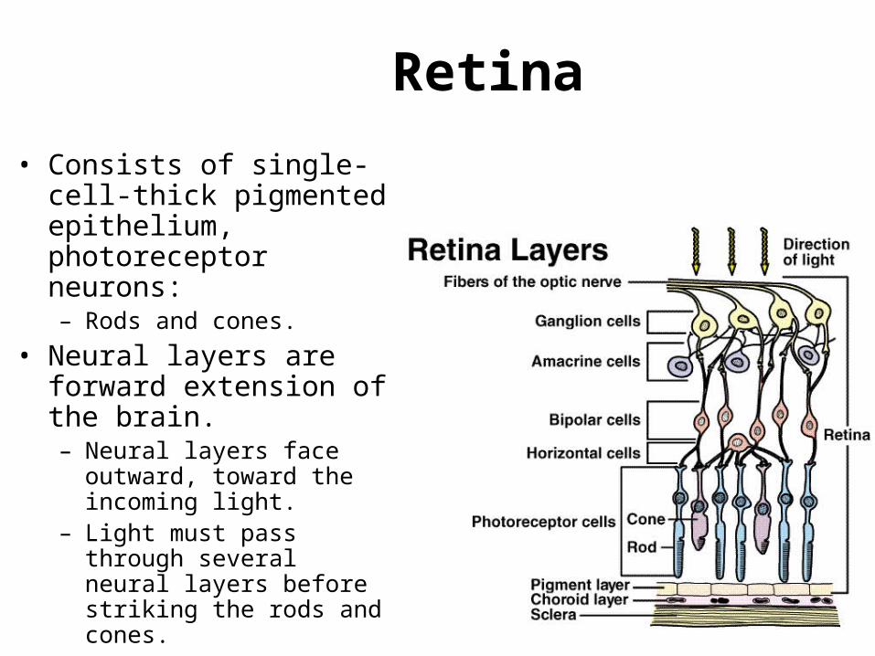
Retina
• Consists of single-cell-thick pigmented epithelium, photoreceptor neurons:– Rods and cones.
• Neural layers are forward extension of the brain.– Neural layers face outward,
toward the incoming light.– Light must pass through
several neural layers before striking the rods and cones.
Dark Adaptation• Gradual increase in photoreceptor
sensitivity when entering a dark room.• Maximal sensitivity reached in 20 min.• Increased amounts of visual pigments
produced.– Slight increased pigment in cones.– Greater increased rhodopsin in rods.
• 100,00-fold increase in light sensitivity in rods.

Cones and Color Vision
• Cones less sensitive than rods to light.
• Cones provide color vision and greater visual acuity.
• High light intensity bleaches out the rods, and color vision with high acuity produced by cones.
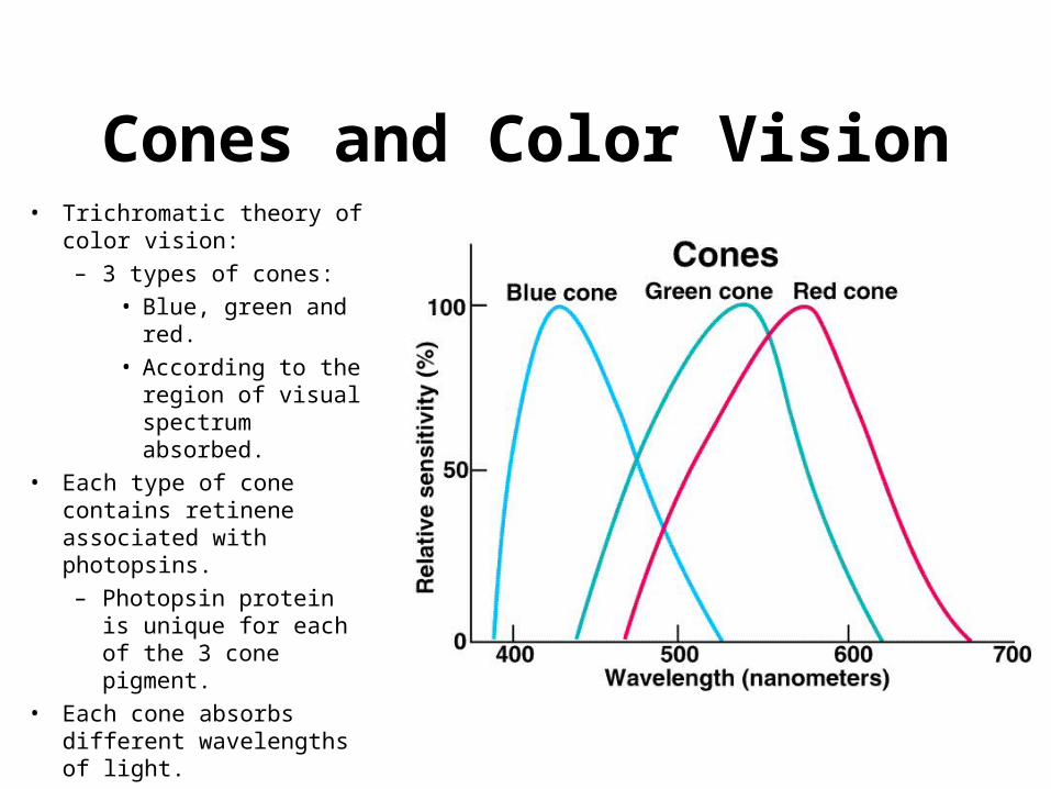
Cones and Color Vision• Trichromatic theory of color
vision:
– 3 types of cones:
• Blue, green and red.
• According to the region of visual spectrum absorbed.
• Each type of cone contains retinene associated with photopsins.
– Photopsin protein is unique for each of the 3 cone pigment.
• Each cone absorbs different wavelengths of light.
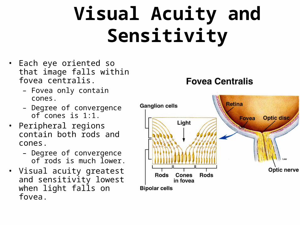
Visual Acuity and Sensitivity
• Each eye oriented so that image falls within fovea centralis.– Fovea only contain cones.– Degree of convergence of cones
is 1:1.
• Peripheral regions contain both rods and cones.– Degree of convergence of rods
is much lower.
• Visual acuity greatest and sensitivity lowest when light falls on fovea.
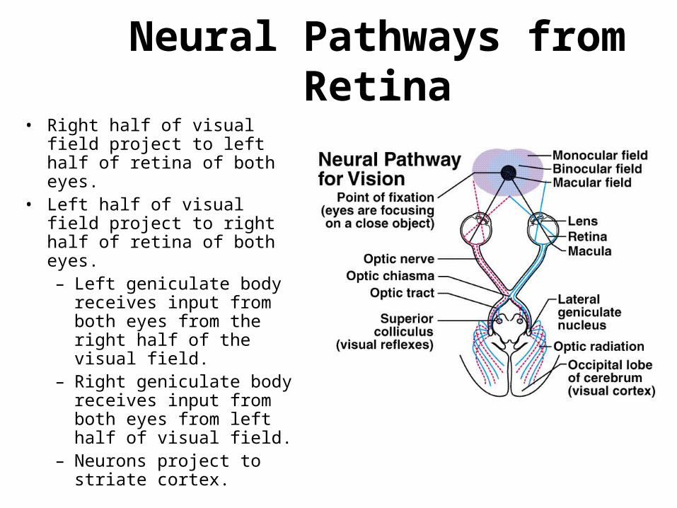
Neural Pathways from Retina• Right half of visual field project
to left half of retina of both eyes.
• Left half of visual field project to right half of retina of both eyes.– Left geniculate body
receives input from both eyes from the right half of the visual field.
– Right geniculate body receives input from both eyes from left half of visual field.
– Neurons project to striate cortex.