Embed Size (px)
Citation preview

© 2012 Pearson Education, Inc.
PowerPoint® Lecture Presentations prepared by
Jason LaPres
Lone Star College—North Harris
4 The Tissue Level of
Organization

© 2012 Pearson Education, Inc.
4-1 Four Types of Tissue
• Tissue
• Are collections of cells and cell products that
perform specific, limited functions
• Four types of tissue
1. Epithelial tissue
2. Connective tissue
3. Muscle tissue
4. Neural tissue

© 2012 Pearson Education, Inc.
4-1 Four Types of Tissue
• Epithelial Tissue
• Covers exposed surfaces
• Lines internal passageways
• Forms glands
• Connective Tissue
• Fills internal spaces
• Supports other tissues
• Transports materials
• Stores energy

© 2012 Pearson Education, Inc.
4-1 Four Types of Tissue
• Muscle Tissue
• Specialized for contraction
• Skeletal muscle, heart muscle, and walls of hollow
organs
• Neural Tissue
• Carries electrical signals from one part of the body
to another

© 2012 Pearson Education, Inc.
4-2 Epithelial Tissue
• Epithelia
• Layers of cells covering internal or external surfaces
• Glands
• Structures that produce secretions

© 2012 Pearson Education, Inc.
4-2 Epithelial Tissue
• Characteristics of Epithelia
• Cellularity (cell junctions)
• Polarity (apical and basal surfaces)
• Attachment (basement membrane or basal
lamina)
• Avascularity
• Regeneration

© 2012 Pearson Education, Inc.
Figure 4-1 The Polarity of Epithelial Cells
Cilia
Microvilli
Apical
surface
Golgi
apparatus
Nucleus
Mitochondria
Basement membrane
Basolateral
surfaces
© 2012 Pearson Education, Inc.
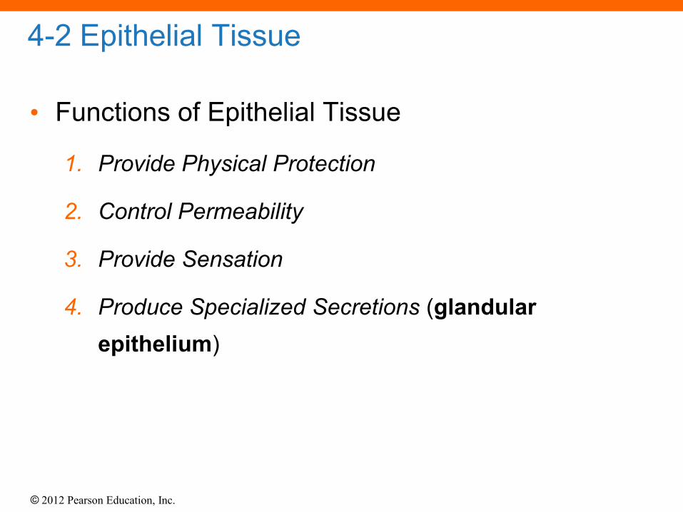
4-2 Epithelial Tissue
• Functions of Epithelial Tissue
1. Provide Physical Protection
2. Control Permeability
3. Provide Sensation
4. Produce Specialized Secretions (glandular
epithelium)

© 2012 Pearson Education, Inc.
4-2 Epithelial Tissue
• Maintaining the Integrity of Epithelia
1. Intercellular connections
2. Attachment to the basement membrane
3. Epithelial maintenance and repair

© 2012 Pearson Education, Inc.
4-2 Epithelial Tissue
• Attachment to the Basement Membrane
• Clear layer (lamina lucida)
• Thin layer
• Secreted by epithelia
• Barrier to proteins
• Dense layer (lamina densa)
• Thick fibers
• Produced by connective tissue
• Strength and filtration

© 2012 Pearson Education, Inc.
4-2 Epithelial Tissue
• Epithelial Maintenance and Repair
• Epithelia are replaced by division of germinative
cells (stem cells)
• Near basement membrane

© 2012 Pearson Education, Inc.
4-3 Classification of Epithelia
• Singular = Epithelium; Plural = Epithelia
• Classes of Epithelia
1. Based on shape
• Squamous epithelia — thin and flat
• Cuboidal epithelia — square shaped
• Columnar epithelia — tall, slender rectangles
2. Based on layers
• Simple epithelium — single layer of cells
• Stratified epithelium — several layers of cells

© 2012 Pearson Education, Inc.
Table 4-1 Classifying Epithelia

© 2012 Pearson Education, Inc.
Table 4-1 Classifying Epithelia

© 2012 Pearson Education, Inc.
4-3 Classification of Epithelia
• Squamous Epithelia
• Simple squamous epithelium
• Absorption and diffusion
• Mesothelium
• Lines body cavities
• Endothelium
• Lines heart and blood vessels

© 2012 Pearson Education, Inc.
Figure 4-3a Squamous Epithelia
Simple Squamous Epithelium
Lining of peritoneal cavity
Connective tissue
Nucleus
Cytoplasm
LOCATIONS: Mesothelia lining ventral body cavities; endothelia lining heart
and blood vessels; portions of kidney tubules (thin sections of nephron loops);
inner lining of cornea; alveoli of lungs
FUNCTIONS: Reduces friction; controls vessel permeability; performs
absorption and secretion
LM 238

© 2012 Pearson Education, Inc.
4-3 Classification of Epithelia
• Squamous Epithelia
• Stratified squamous epithelium
• Protects against attacks
• Keratin protein adds strength and water resistance

© 2012 Pearson Education, Inc.
Figure 4-3b Squamous Epithelia
LOCATIONS: Surface of skin; lining of mouth, throat, esophagus, rectum,
anus, and vagina
FUNCTIONS: Provides physical protection against abrasion, pathogens,
and chemical attack
Surface of tongue
Squamous
superficial cells
Stem cells
Basement membrane
Connective tissue
LM 310
Stratified Squamous Epithelium

© 2012 Pearson Education, Inc.
4-3 Classification of Epithelia
• Cuboidal Epithelia
• Simple cuboidal epithelium
• Secretion and absorption
• Stratified cuboidal epithelia
• Sweat ducts and mammary ducts
© 2012 Pearson Education, Inc.
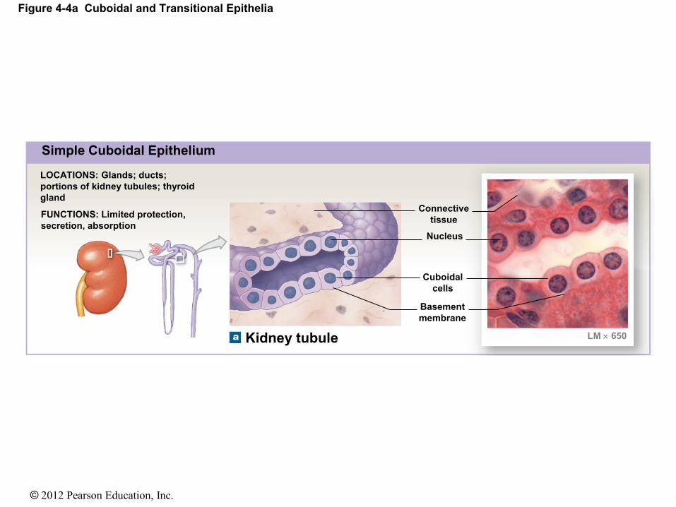
Figure 4-4a Cuboidal and Transitional Epithelia
Simple Cuboidal Epithelium
LOCATIONS: Glands; ducts;
portions of kidney tubules; thyroid
gland
FUNCTIONS: Limited protection,
secretion, absorption
Kidney tubule
Connective
tissue
Nucleus
Cuboidal
cells
Basement
membrane
LM 650

© 2012 Pearson Education, Inc.
Figure 4-4b Cuboidal and Transitional Epithelia
LOCATIONS: Lining of some ducts
(rare)
FUNCTIONS: Protection, secretion,
absorption
LM 500
Lumen
of duct
Sweat gland duct
Stratified
cuboidal
cells Basement
membrane
Nuclei
Connective
tissue
Stratified Cuboidal Epithelium
© 2012 Pearson Education, Inc.
4-3 Classification of Epithelia
• Transitional Epithelium
• Tolerates repeated cycles of stretching and recoiling
and returns to its previous shape without damage
• Appearance changes as stretching occurs
• Situated in regions of the urinary system (e.g., urinary
bladder)

© 2012 Pearson Education, Inc.
Figure 4-4c Cuboidal and Transitional Epithelia
Transitional Epithelium
FUNCTIONS: Permits
expansion and recoil
after stretching
LOCATIONS: Urinary
bladder; renal pelvis;
ureters
LM 400
Urinary bladder
LM 400
Empty bladder
Epithelium
(relaxed)
Connective tissue and
smooth muscle layers
Epithelium
(stretched)
Connective tissue and
smooth muscle layers
Basement membrane
Full bladder
Basement membrane
LM 400

© 2012 Pearson Education, Inc.
4-3 Classification of Epithelia
• Columnar Epithelia
• Simple columnar epithelium
• Absorption and secretion
• Pseudostratified columnar epithelium
• Cilia movement
• Stratified columnar epithelium
• Protection
© 2012 Pearson Education, Inc.
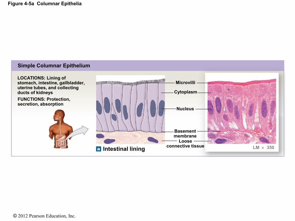
Figure 4-5a Columnar Epithelia
LOCATIONS: Lining of stomach, intestine, gallbladder, uterine tubes, and collecting ducts of kidneys
FUNCTIONS: Protection, secretion, absorption
Simple Columnar Epithelium
Microvilli
Cytoplasm
Intestinal lining
Basement membrane
Loose connective tissue LM 350
Nucleus
© 2012 Pearson Education, Inc.
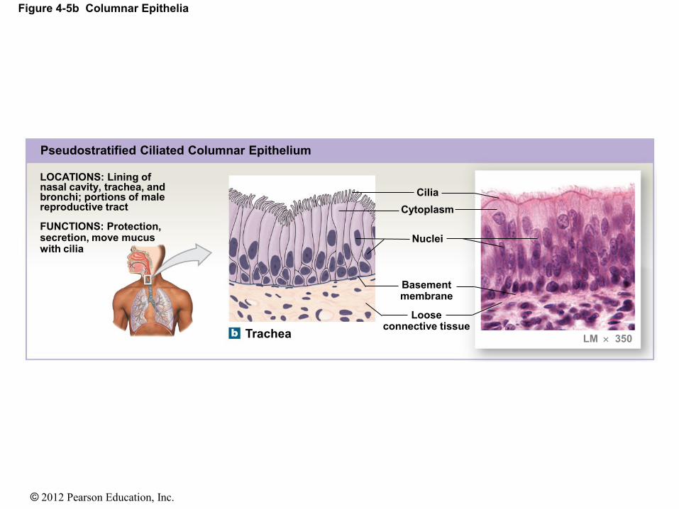
Figure 4-5b Columnar Epithelia
LOCATIONS: Lining of nasal cavity, trachea, and bronchi; portions of male reproductive tract
FUNCTIONS: Protection, secretion, move mucus with cilia
Pseudostratified Ciliated Columnar Epithelium
Cilia
Trachea
Cytoplasm
Nuclei
Basement membrane
Loose connective tissue
LM 350
© 2012 Pearson Education, Inc.
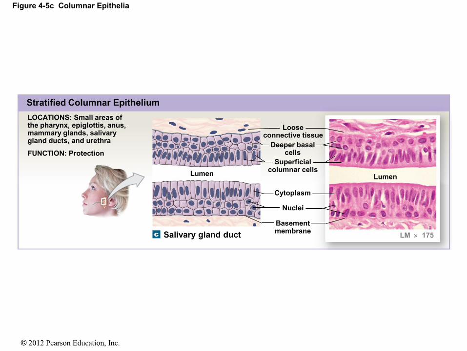
Figure 4-5c Columnar Epithelia
LOCATIONS: Small areas of the pharynx, epiglottis, anus, mammary glands, salivary gland ducts, and urethra
FUNCTION: Protection
Stratified Columnar Epithelium
Salivary gland duct
Deeper basal cells
Loose connective tissue
Superficial columnar cells
Cytoplasm
Nuclei
Basement membrane
Lumen Lumen
LM 175

© 2012 Pearson Education, Inc.
4-3 Classification of Epithelia
• Glandular Epithelia
• Endocrine glands
• Release hormones
• Into interstitial fluid
• No ducts
• Exocrine glands
• Produce secretions
• Onto epithelial surfaces
• Through ducts

© 2012 Pearson Education, Inc.
4-4 Connective Tissue
• Characteristics of Connective Tissue
1. Specialized cells
2. Solid extracellular protein fibers
3. Fluid extracellular ground substance
• The Extracellular Components of Connective
Tissue (Fibers and Ground Substance)
• Make up the matrix

© 2012 Pearson Education, Inc.
4-4 Connective Tissue
• Functions of Connective Tissue
• Establishing a structural framework for the body
• Transporting fluids and dissolved materials
• Protecting delicate organs
• Supporting, surrounding, and interconnecting other
types of tissue
• Storing energy reserves, especially in the form of
triglycerides
• Defending the body from invading microorganisms

© 2012 Pearson Education, Inc.
4-4 Connective Tissue
• Classification of Connective Tissues
1. Connective tissue proper
• Connect and protect
2. Fluid connective tissues
• Transport
3. Supporting connective tissues
• Structural strength

© 2012 Pearson Education, Inc.
4-4 Connective Tissue
• Categories of Connective Tissue Proper
• Loose connective tissue
• More ground substance, fewer fibers
• For example, fat (adipose tissue)
• Dense connective tissue
• More fibers, less ground substance
• For example, tendons

© 2012 Pearson Education, Inc.
4-4 Connective Tissue
• Fibroblasts
• Fibrocytes
• Adipocytes
• Mesenchymal cells
• Macrophages
• Mast cells
• Lymphocytes
• Microphages
• Melanocytes
Connective Tissue Proper Cell Populations

© 2012 Pearson Education, Inc.
4-4 Connective Tissue
• Connective Tissue Fibers
1. Collagen fibers
2. Reticular fibers
3. Elastic fibers
© 2012 Pearson Education, Inc.
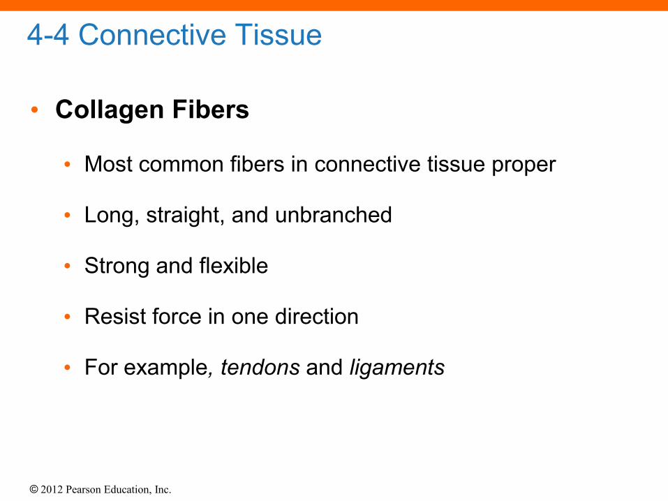
4-4 Connective Tissue
• Collagen Fibers
• Most common fibers in connective tissue proper
• Long, straight, and unbranched
• Strong and flexible
• Resist force in one direction
• For example, tendons and ligaments

© 2012 Pearson Education, Inc.
4-4 Connective Tissue
• Reticular Fibers
• Network of interwoven fibers (stroma)
• Strong and flexible
• Resist force in many directions
• Stabilize functional cells and structures
• For example, Bone marrow

© 2012 Pearson Education, Inc.
4-4 Connective Tissue
• Elastic Fibers
• Contain elastin
• Branched and wavy
• Return to original length after stretching
• For example, elastic ligaments of vertebrae

© 2012 Pearson Education, Inc.
4-4 Connective Tissue
• Ground Substance
• Is clear, colorless, and viscous
• Fills spaces between cells and slows pathogen
movement

© 2012 Pearson Education, Inc.
Figure 4-8 The Cells and Fibers of Connective Tissue Proper
Reticular
fibers
Melanocyte
Fixed
macrophage
Plasma cell
Blood
in vessel
Adipocytes
(fat cells)
Ground
substance
Mast cell
Elastic
fibers
Free
macrophage
Collagen
fibers
Fibroblast
Mesenchymal
cell
Lymphocyte

© 2012 Pearson Education, Inc.
4-4 Connective Tissue
• Loose Connective Tissues
• The “packing materials” of the body
• Three types in adults
1. Areolar
2. Adipose
3. Reticular
© 2012 Pearson Education, Inc.
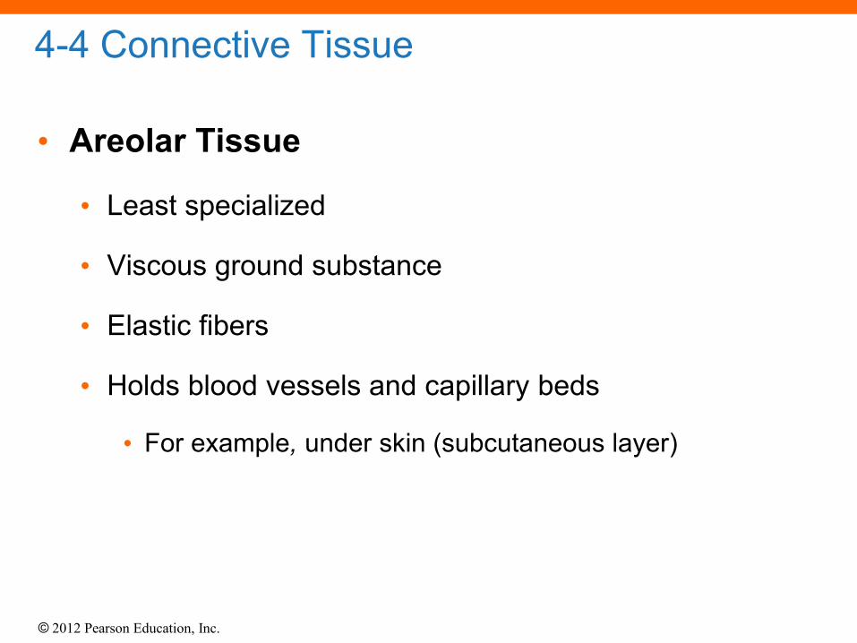
4-4 Connective Tissue
• Areolar Tissue
• Least specialized
• Viscous ground substance
• Elastic fibers
• Holds blood vessels and capillary beds
• For example, under skin (subcutaneous layer)

© 2012 Pearson Education, Inc.
4-4 Connective Tissue
• Adipose Tissue
• Adipose cells
• Adipocytes in adults do not divide
• Expand to store fat
• Shrink as fats are released
• Mesenchymal cells divide and differentiate
• To produce more fat cells
• When more storage is needed

© 2012 Pearson Education, Inc.
4-4 Connective Tissue
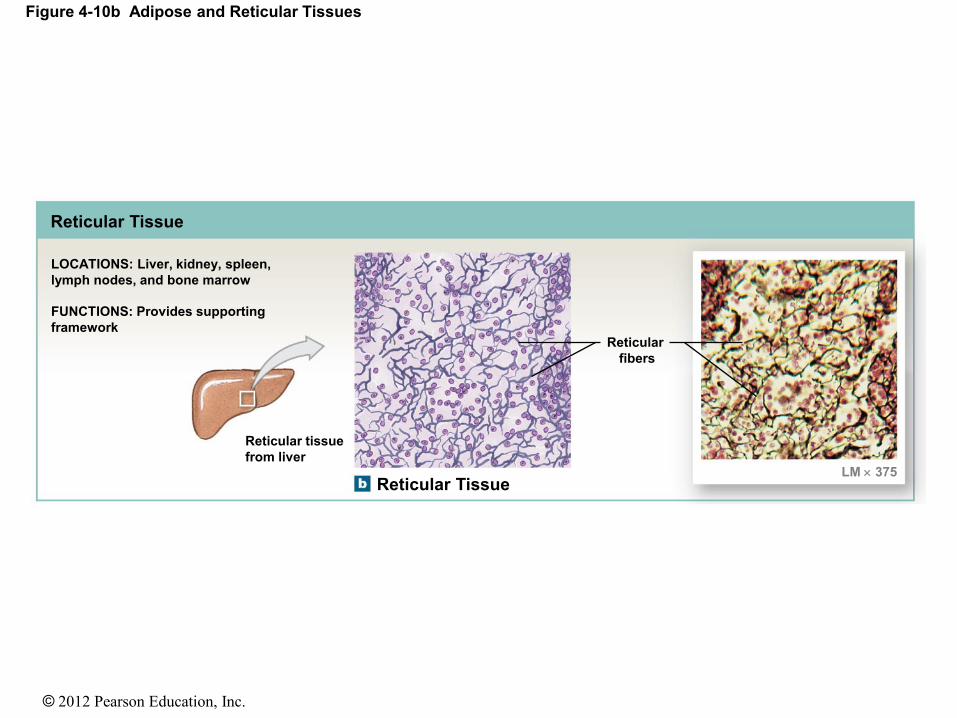
• Reticular Tissue
• Provides support
• Complex, three-dimensional network
• Supportive fibers (stroma)
• Support functional cells
• Reticular organs
• Spleen, liver, lymph nodes, and bone marrow

© 2012 Pearson Education, Inc.
Figure 4-10a Adipose and Reticular Tissues
Adipose Tissue
LOCATIONS: Deep to the skin,
especially at sides, buttocks,
breasts; padding around eyes
and kidneys
FUNCTIONS: Provides
padding and cushions
shocks; insulates
(reduces heat loss);
stores energy
Adipose tissue
Adipocytes
(white adipose
cells)
LM 300
© 2012 Pearson Education, Inc.
Figure 4-10b Adipose and Reticular Tissues
Reticular Tissue
FUNCTIONS: Provides supporting
framework
LOCATIONS: Liver, kidney, spleen,
lymph nodes, and bone marrow
Reticular tissue
from liver
Reticular Tissue
Reticular
fibers
LM 375

© 2012 Pearson Education, Inc.
4-4 Connective Tissue
• Dense Connective Tissues
• Connective tissues proper, tightly packed with high
numbers of collagen or elastic fibers
• Dense regular connective tissue
• Dense irregular connective tissue
• Elastic tissue

© 2012 Pearson Education, Inc.
4-4 Connective Tissue
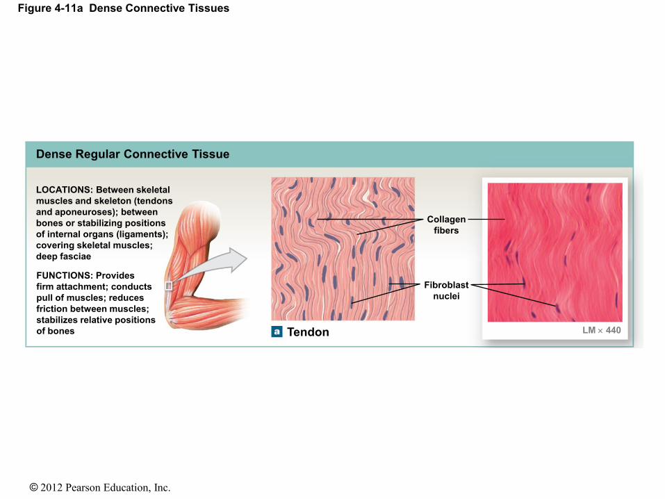
• Dense Regular Connective Tissue
• Tightly packed, parallel collagen fibers
• Tendons attach muscles to bones
• Ligaments connect bone to bone and stabilize organs
© 2012 Pearson Education, Inc.
Figure 4-11a Dense Connective Tissues
Dense Regular Connective Tissue
Collagen
fibers
Fibroblast
nuclei
Tendon LM 440
LOCATIONS: Between skeletal
muscles and skeleton (tendons
and aponeuroses); between
bones or stabilizing positions
of internal organs (ligaments);
covering skeletal muscles;
deep fasciae
FUNCTIONS: Provides
firm attachment; conducts
pull of muscles; reduces
friction between muscles;
stabilizes relative positions
of bones

© 2012 Pearson Education, Inc.
4-4 Connective Tissue
• Dense Irregular Connective Tissue
• Interwoven networks of collagen fibers
• Layered in skin
• Around cartilages (perichondrium)
• Around bones (periosteum)
• Form capsules around some organs (e.g., liver,
kidneys)

© 2012 Pearson Education, Inc.
Figure 4-11b Dense Connective Tissues
Collagen
fiber
bundles
Deep dermis LM 111
Dense Irregular Connective Tissue
LOCATIONS: Capsules of
visceral organs; periostea
and perichondria; nerve
and muscle sheaths; dermis
FUNCTIONS: Provides
strength to resist forces
applied from many
directions; helps
prevent overexpansion
of organs such as
the urinary bladder
© 2012 Pearson Education, Inc.
4-4 Connective Tissue
• Elastic Tissue
• Made of elastic fibers
• For example, elastic ligaments of spinal vertebrae

© 2012 Pearson Education, Inc.
Figure 4-11c Dense Connective Tissues
Elastic
fibers
Fibroblast
nuclei
Elastic ligament
Elastic Tissue
LOCATIONS: Between vertebrae
of the spinal column (ligamentum
flavum and ligamentum nuchae);
ligaments supporting penis;
ligaments supporting transitional
epithelia; in blood vessel walls
FUNCTIONS: Stabilizes
positions of vertebrae and
penis; cushions shocks;
permits expansion and
contraction of organs
LM 887

© 2012 Pearson Education, Inc.
4-4 Connective Tissue
• Fluid Connective Tissues
• Blood and lymph
• Watery matrix of dissolved proteins
• Carry specific cell types (formed elements)
• Formed elements of blood
• Red blood cells (erythrocytes)
• White blood cells (leukocytes)
• Platelets

© 2012 Pearson Education, Inc.
4-4 Connective Tissue
• Fluid Elements of Connective Tissues
• Extracellular
• Plasma
• Interstitial fluid
• Lymph

© 2012 Pearson Education, Inc.
Figure 4-12 Formed Elements of the Blood
Red blood cells
Red blood cells
account for roughly
half the volume of
whole blood and
give blood its color.
Red blood cells, or
erythrocytes
(e-RITH-ro-sıts), are
responsible for the
transport of oxygen
(and, to a lesser degree,
of carbon dioxide) in
the blood.
¯ ¯

© 2012 Pearson Education, Inc.
Figure 4-12 Formed Elements of the Blood
White blood cells
Eosinophil
Neutrophil
Basophil
White blood cells, or leukocytes
(LOO-ko-sıts; leuko-, white), help defend
the body from infection and disease.
¯ ¯
Eosinophils and neutro-
phils are phagocytes. Ba-
sophils promote inflamma-
tion much like mast cells in
other connective tissues.
Lymphocytes are un-
common in the blood
but they are the domi-
nant cell type in lymph,
the second type of
fluid connective tissue.
Monocytes
are phagocytes
similar to the
free macro-
phages in
other tissues.

© 2012 Pearson Education, Inc.
Figure 4-12 Formed Elements of the Blood
Platelets
Platelets are
membrane-enclosed
packets of cytoplasm
that function in blood
clotting.
These cell fragments
are involved in the
clotting response
that seals leaks in
damaged or broken
blood vessels.

© 2012 Pearson Education, Inc.
4-4 Connective Tissue
• Lymph
• Extracellular fluid
• Collected from interstitial space
• Monitored by immune system
• Transported by lymphatic (lymphoid) system
• Returned to venous system

© 2012 Pearson Education, Inc.
4-5 Supporting Connective Tissues
• Support Soft Tissues and Body Weight
• Cartilage
• Gel-type ground substance
• For shock absorption and protection
• Bone
• Calcified (made rigid by calcium salts, minerals)
• For weight support

© 2012 Pearson Education, Inc.
4-5 Supporting Connective Tissues
• Cartilage Matrix
• Proteoglycans derived from chondroitin sulfates
• Ground substance proteins
• Chondrocytes (cartilage cells) surrounded by
lacunae (chambers)

© 2012 Pearson Education, Inc.
4-5 Supporting Connective Tissues
• Cartilage Structure
• No blood vessels
• Perichondrium
• Outer, fibrous layer (for strength)
• Inner, cellular layer (for growth and maintenance)

© 2012 Pearson Education, Inc.
4-5 Supporting Connective Tissues
• Types of Cartilage
1. Hyaline cartilage
2. Elastic cartilage
3. Fibrocartilage (fibrous cartilage)
© 2012 Pearson Education, Inc.
4-5 Supporting Connective Tissues
• Hyaline Cartilage
• Stiff, flexible support
• Reduces friction between bones
• Found in synovial joints, rib tips, sternum, and
trachea
• Elastic Cartilage
• Supportive but bends easily
• Found in external ear and epiglottis

© 2012 Pearson Education, Inc.
4-5 Supporting Connective Tissues
• Fibrocartilage (Fibrous Cartilage)
• Limits movement
• Prevents bone-to-bone contact
• Pads knee joints
• Found between pubic bones and
intervertebral discs

© 2012 Pearson Education, Inc.
Figure 4-14a Types of Cartilage
Hyaline Cartilage
LOCATIONS: Between tips of ribs and bones of sternum; covering bone surfaces at synovial joints; supporting larynx (voice box), trachea, and bronchi; forming part of nasal septum FUNCTIONS: Provides stiff but somewhat flexible support; reduces friction between bony surfaces
Hyaline cartilage
LM 500
Matrix
Chondrocytes in lacunae

© 2012 Pearson Education, Inc.
Figure 4-14b Types of Cartilage
Elastic Cartilage
LOCATIONS: Auricle of external ear; epiglottis; auditory canal; cuneiform cartilages of larynx
FUNCTIONS: Provides support, but tolerates distortion without damage and returns to original shape
Elastic cartilage
Elastic fibers
in matrix
Chondrocyte
in lacuna
LM 358

© 2012 Pearson Education, Inc.
Figure 4-14c Types of Cartilage
Fibrocartilage
LOCATIONS: Pads within knee joint; between pubic bones of pelvis; intervertebral discs
FUNCTIONS: Resists compression; prevents bone- to-bone contact; limits movement
Fibrocartilage
Fibrous
matrix
Chondrocytes
in lacunae
LM 400

© 2012 Pearson Education, Inc.
4-5 Supporting Connective Tissues
• Bone or Osseous Tissue
• Strong (calcified calcium salt deposits)
• Resists shattering (flexible collagen fibers)
• Bone Cells or Osteocytes
• Arranged around central canals within matrix
• Small channels through matrix (canaliculi) access blood supply
• Periosteum
• Covers bone surfaces
• Fibrous layer
• Cellular layer

© 2012 Pearson Education, Inc.
Figure 4-15 Bone
Canaliculi
Osteocytes in lacunae
Matrix
Central canal
Blood vessels
LM 375
Osteon
Osteon
Fibrous
layer
Cellular
layer
Periosteum

© 2012 Pearson Education, Inc.
4-8 Muscle Tissue
• Muscle Tissue
• Specialized for contraction
• Produces all body movement
• Three types of muscle tissue
1. Skeletal muscle tissue
• Large body muscles responsible for movement
2. Cardiac muscle tissue
• Found only in the heart
3. Smooth muscle tissue
• Found in walls of hollow, contracting organs (blood
vessels; urinary bladder; respiratory, digestive, and
reproductive tracts)
© 2012 Pearson Education, Inc.
4-8 Muscle Tissue
• Classification of Muscle Cells
• Striated (muscle cells with a banded appearance)
• Nonstriated (not banded; smooth)
• Muscle cells can have a single nucleus
• Muscle cells can be multinucleate
• Muscle cells can be controlled voluntarily (consciously)
• Muscle cells can be controlled involuntarily
(automatically)

© 2012 Pearson Education, Inc.
4-8 Muscle Tissue
• Skeletal Muscle Cells
• Long and thin
• Usually called muscle fibers
• Do not divide
• New fibers are produced by stem cells (myosatellite
cells)

© 2012 Pearson Education, Inc.
Figure 4-18a Muscle Tissue
Skeletal Muscle Tissue
Skeletal muscle
Cells are long, cylindrical,
striated, and multinucleate.
LOCATIONS: Combined
with connective tissues
and neural tissue in
skeletal muscles
FUNCTIONS: Moves or
stabilizes the position of
the skeleton; guards
entrances and exits to
the digestive,
respiratory, and urinary
tracts; generates heat;
protects internal organs
Nuclei
Muscle
fiber
Striations
LM 180
© 2012 Pearson Education, Inc.
4-8 Muscle Tissue
• Cardiac Muscle Cells
• Called cardiocytes
• Form branching networks connected at intercalated
discs
• Regulated by pacemaker cells
• Smooth Muscle Cells
• Small and tapered
• Can divide and regenerate

© 2012 Pearson Education, Inc.
Figure 4-18b Muscle Tissue
Cardiac Muscle Tissue
Cardiac muscle
Cells are short, branched,
and striated, usually with a
single nucleus; cells are
interconnected by
intercalated discs.
FUNCTIONS:
Circulates blood;
maintains blood
(hydrostatic) pressure
LOCATION: Heart
LM 450
Nucleus
Cardiac
muscle
cells
Intercalated
discs
Striations
© 2012 Pearson Education, Inc.
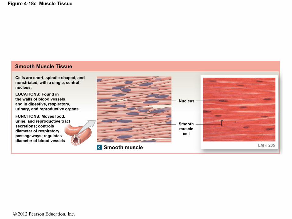
Figure 4-18c Muscle Tissue
Smooth muscle
Cells are short, spindle-shaped, and
nonstriated, with a single, central
nucleus.
LOCATIONS: Found in
the walls of blood vessels
and in digestive, respiratory,
urinary, and reproductive organs
FUNCTIONS: Moves food,
urine, and reproductive tract
secretions; controls
diameter of respiratory
passageways; regulates
diameter of blood vessels LM 235
Nucleus
Smooth
muscle
cell
Smooth Muscle Tissue

© 2012 Pearson Education, Inc.
4-9 Neural Tissue
• Neural Tissue
• Also called nervous or nerve tissue
• Specialized for conducting electrical impulses
• Rapidly senses internal or external environment
• Processes information and controls responses
• Neural tissue is concentrated in the central nervous
system
• Brain
• Spinal cord
© 2012 Pearson Education, Inc.
4-9 Neural Tissue
• Two Types of Neural Cells
1. Neurons
• Nerve cells
• Perform electrical communication
2. Neuroglia
• Supporting cells
• Repair and supply nutrients to neurons

© 2012 Pearson Education, Inc.
4-9 Neural Tissue
• Cell Parts of a Neuron
• Cell body
• Contains the nucleus and nucleolus
• Dendrites
• Short branches extending from the cell body
• Receive incoming signals
• Axon (nerve fiber)
• Long, thin extension of the cell body
• Carries outgoing electrical signals to their destination

© 2012 Pearson Education, Inc.
Figure 4-19 Neural Tissue
NEURONS NEUROGLIA (supporting cells)
• Maintain physical structure
• Repair tissue framework
• Perform phagocytosis
• Regulate the composition of the interstitial fluid surrounding neurons
of tissues
after injury
• Provide nutrients to neurons
Axon
Nucleolus Nucleus
of neuron
Dendrites
(contacted by
other neurons) Axon (conducts
information to
other cells)
Cell body
Dendrites
Mitochondrion Nucleus
Microfibrils and
microtubules Nucleolus
Cell body (contains nucleus
and major organelles) A representative neuron
(sizes and shapes vary widely)
Contact with
other cells
Nuclei of neuroglia
LM 600