Embed Size (px)
Citation preview

- 1 -
The study of cationic amphiphilic peptides with anti-cancer selective toxicity
Pfariso Maumela
0706720g
A Dissertation submitted to the Faculty of Science, University of the Witwatersrand,
Johannesburg, in partial fulfilment of requirements for the Degree of Masters of Science.
Johannesburg, 2014.

I
Declaration
I declare that this dissertation is my own, unaided work. It is being submitted for the Degree
of Master of Science at the University of the Witwatersrand, Johannesburg. It has not been
submitted before for any degree or examination at any other University.
10th
of July 2014

II
Abstract
The exposure of organisms to environmental stresses and pathogens results in rapid activation
of a range of defensive pathways that act as part of the innate immune system. The most
common innate immunity response is the activation of cationic amphiphilic peptides in
response to microbial infection. Moreover, cationic amphiphilic peptides possess desirable
attributes for the pharmaceutical development of cancer-selective drugs. They selectively and
rapidly kill cancer cells without killing normal mammalian cells and have a broad spectrum
of mechanisms of action. The aim of this exploratory study was to screen for cationic
amphiphilic peptides with anti-proliferative activity that is induced by genotoxicity.
GeneFishing® technology, 2-D gel analysis and bioassays were used to identify and analyse
molecules induced in response to genotoxic stress in an embryonic cell line originating from
the dung beetle Euoniticellus intermedius. Bioassay results revealed that the cell line has
constitutive expression of probable cationic amphiphilic proteins that are further induced by
camptothecin treatment. GeneFishing® and 2-D gel analysis showed changes in gene
expression at both transcriptional and translational levels, respectively. Overall, the study
failed to identify the involvement or induction of cationic amphiphilic peptides in response to
genotoxic stress. However, gene expression analyses revealed changes in the expression of
classes of proteins involved in stress response, oxidative phosphorylation, mitochondrial
maintenance, protein translation, cytoskeletal proteins and immunophilins. The results show
that the cell line constitutively expresses probable cationic amphiphilic peptides which are
further induced by camptothecin.

III
Acknowledgements
I would like to express my sincere gratitude to the following awesome individuals:
My supervisor, Dr. M. M. Ntwasa for believing in me, his support and patience
Dr. Rodney Hull for his technical assistance and assistance with my write-up
Ms Keneilwe Sebola for her technical assistance and encouragement
The flylab members for their great company, input and advice
Mum Ziphorah and Charlotte for their assistance in keeping the flies going and making the
lab a conducive environment to work in
Dr. Stoyan Stoychev for the mass spectroscopy
Postgraduate merit award, NRF and GDARD for the financial assistance
My grandfather, the Late Dr. Moyo for believing in me and his continuous support

IV
Research output
1. Conference contribution
Poster Presentation:
Maumela, P., Hull, R., Mnisi, N., Ntwasa, M. and Cajee, U. The study of cationic
amphiphilic peptides with anti-cancer selective toxicity and antimicrobial activity. EMBO
global exchange lecture course. Innate immunity: Evolution and advances in clinical
medicine. Stay City, Johannesburg, 4 September 2012.

V
Table of Contents
Declaration ................................................................................................................................. I
Abstract ..................................................................................................................................... II
Acknowledgements .................................................................................................................. III
Research output ........................................................................................................................ IV
List of figures ......................................................................................................................... VII
List of tables .......................................................................................................................... VIII
List of abbreviations ................................................................................................................ IX
Chapter 1: Introduction ......................................................................................................... - 1 -
1.1 Introduction ................................................................................................................. - 1 -
1.2 Cationic amphiphilic peptides overview ..................................................................... - 1 -
1.3 Structure of CAPs........................................................................................................ - 2 -
1.4 Cationic amphiphilic peptides structural-functional relationships .............................. - 3 -
1.5 Mechanisms of action of CAPs ................................................................................... - 3 -
1.5.1 Overview .............................................................................................................. - 3 -
1.5.2 The carpet model .................................................................................................. - 5 -
1.5.3 The toroidal-pore model ....................................................................................... - 5 -
1.5.4 The barrel starve model ........................................................................................ - 5 -
1.5.5 The aggregate or channel-forming model ............................................................ - 6 -
1.6 CAPs interaction with subcellular targets ................................................................... - 6 -
1.7 The role of CAPs in apoptosis .................................................................................... - 6 -
1.8 The expression of CAPs in response to stress and roles in immunity ........................ - 7 -
1.8.1 CAPs synthesis and modification ......................................................................... - 7 -
1.8.2 Signalling pathways involved in CAPs response and action ................................ - 8 -
1.9 Roles in innate immunity .......................................................................................... - 10 -
1.10 Immunomodulatory roles of CAPs ......................................................................... - 10 -
1.11 Anti- cancer activity of CAPs ................................................................................. - 11 -
1.12 Development of CAPs as therapeutic agents .......................................................... - 14 -
1.13 Review of the SAEIE08 cell line ............................................................................ - 14 -
1.14 Camptothecin as a DNA damaging agent……………….…………………………..-15-
1.15 Objectives of the study ............................................................................................ - 15 -
Chapter 2: Materials and methods ...................................................................................... - 17 -
2.1 Materials .................................................................................................................... - 17 -

VI
2.2 Tissue culture ............................................................................................................ - 18 -
2.3 RNA extraction ......................................................................................................... - 18 -
2.4 Protein extraction from lysed cells and concentration from the supernatant ............ - 19 -
2.5 Isolation of the cationic amphiphilic proteins ........................................................... - 21 -
2.6 Antimicrobial activity assays .................................................................................... - 21 -
2.7 Chemical characterisation of the antimicrobial active agent using the Protein kinase
assay ................................................................................................................................ - 22 -
2.8 Differential expression of genes after camptothecin treatment ................................. - 22 -
2.8.1 Genefishing Technology..................................................................................... - 22 -
2.8.2 PCR products recovery and purification from electrophoretic gels ................... - 24 -
2.9 Cloning and sequencing of the GeneFishing® PCR product .................................... - 24 -
2.9.1 Cloning ............................................................................................................... - 24 -
2.9.2 Sequencing.......................................................................................................... - 24 -
2.10 3’ Rapid amplification of cDNA Ends .................................................................... - 24 -
2.11 Proteomic analysis of molecules involved in genotoxic response of E. intermedius
embryo cell line ............................................................................................................... - 26 -
2.11.1 2-D electrophoresis ........................................................................................... - 26 -
2.11.2 Image analysis .................................................................................................. - 26 -
2.11.3 Mass spectroscopy ............................................................................................ - 27 -
2.11.4 Statistical and bioinformatics analysis ............................................................. - 27 -
Chapter 3: Results ............................................................................................................... - 28 -
3 Results .......................................................................................................................... - 28 -
3.2 Antimicrobial activity assays .................................................................................... - 28 -
3.3 Differential transcription of genes induced by camptothecin ................................... - 31 -
3.4 2-D analysis of molecules involved in genotoxic response of E. intermedius embryo
cell line ............................................................................................................................ - 34 -
Chapter 4: Discussion ......................................................................................................... - 38 -
4.1 Discussion ................................................................................................................. - 38 -
4.2 Cationic peptides detection ....................................................................................... - 38 -
4.3 Changes in gene expression induced by genotoxic stress ......................................... - 39 -
4.4 Camptothecin treatment and differential protein expression summary .................... - 42 -
4.5 Conclusion……………………………………………………………………………-43-
4.6 Future prospect .......................................................................................................... - 43 -
6 References ........................................................................................................................ - 44 -

VII
List of figures
Figure 1: Structures of peptides representing the four major classes of CAPs 2
Figure 2: Models for CAPs membrane permeabilisation 4
Figure 3: Total RNA extracted from the beetle cell culture 19
Figure 4: Standard curve for protein concentration using the Bradford assay 20
Figure 5: Flow chart of cDNA synthesis and GeneFishing® PCR 23
Figure 6: Overview of 3’ RACE 25
Figure 7: Radial diffusion assays 30
Figure 8: Characterisation of the inhibitory or toxic molecule 30
Figure 9: GeneFishing® for differential transcribed genes in response to camptothecin 32
Figure 10: Chromatogram showing the 3’ RACE forward primer 33
Figure 11: Rapid Amplification of cDNA Ends (3’RACE) of the ACP 17 fragment 33
Figure 12: 2-D gel analysis 35

VIII
List of tables
Table 1: Media 17
Table 2: Biological buffers and solutions 17
Table 3: Primer 18
Table 4: Proteins identified with mass spectroscopy 36
Table 5: Comparison of MS database identified proteins with the NCBI and flylab base
genomebase database 37
IX
List of abbreviations
ABP-CM4 antibacterial peptide-CM4
CAPs Cationic amphiphilic peptides
DNA Deoxyribonucleic acid
ERK extracellular signal-regulated kinase
FPRL formyl peptide-like receptor
hBD human -defensin
HEK-293 human embryonic kidney cell line
HeLa epithelial carcinoma cell lines
HIV human immunodeficiency virus
HT-1080 fibrosarcoma cell line
RNA Ribonucleic acid
TFE 2,2,2-trifluoroethanol
MAPK mitogen-activated protein kinase
MTT 3-(4,5-dimethyl-2-thiazolyl)-2,5-diphenyl-2H-tetrazolium bromide
PBMCs mononuclear cells

- 1 -
Chapter 1: Introduction
1.1 Introduction
The exposure of organisms to environmental stresses, toxins and pathogens results in rapid
activation of a range of energy efficient defensive pathways that act as part of the innate
immune system. The most common innate immunity response is the activation of peptides in
response to microbial infection. This response is conserved from microorganisms to humans
(Papo and Shai, 2003). These peptides are widely synthesised in areas of the organism that
are regularly exposed to pathogens. For instance, dermicidin, a natural antibiotic is secreted
with sweat and found on the surface of human skin (Paulmann et al., 2012). Most
antimicrobial peptides are amphipathic and positively charged and are widely known as
cationic amphiphilic peptides (CAPs) (Hancock, 2001). The above properties contribute to
their ability to interact with and rapidly disrupt the negatively charged phospholipid
membranes of microorganisms (Hancock, 2001; Leuschner and Hansel, 2004). Consequently,
CAPs have antibiotic activity against a range of pathogenic bacteria, viruses, fungi and
protozoa. Furthermore, they have selective anti-cancer toxicity (Lehmann et al., 2006).
1.2 Cationic amphiphilic peptides overview
CAPs range between 5–40 amino acids in length and are in most cases membrane active
(Hoskin and Ramamoorthy, 2008; Hancock, 2001). Moreover, they can be classified into
peptides that have toxicity against bacteria and viruses but not against mammalian cells and
fungi; and those with toxicity against bacteria, viruses, fungi and mammalian cells (Papo and
Shai, 2003). CAPs play a significant role in the innate immune system of a range of plants
and animal species by acting as natural antibiotics against bacterial, enveloped viruses and
fungal pathogens (Papo et al., 2003). They are also capable of acting synergistically with
each other or other host immune molecules. For instance, magainin II and peptide PLGa have
been shown to have synergistic toxicity in frogs, while lysozyme showed synergistic
antibacterial toxicity with a range of conventional antibiotics (Hancock, 2001). Furthermore,
they have the capacity to act as immune modulators for adaptive immunity in vertebrates
(Hoskin and Ramamoorthy, 2008; Giuliani et al., 2007). For instance, human - and -
defensins have been established to exclusively chemo attract different groups of T
lymphocytes and immature dendritic cells (Wu et al., 2003). CAPs have been proposed to be

- 2 -
involved in infection clearance and wound healing by acting as chemokines or by inducing
chemokine production (Giuliani et al., 2007). Interestingly CAPs have cancer-selective
toxicity linked to the electrostatic interactions between their net positive charge and the
highly negative charge on cancer cells (Hoskin and Ramamoorthy, 2008). They are also
capable of inducing apoptosis through interaction with the mitochondrion (Papo et al., 2003).
Thus CAPs are potential candidates for anticancer drugs development since they have been
reported to possess a novel mode of action, less cytotoxicity to normal cells and have a low
likelihood of resistance development (Hsu et al., 2011). CAPs are synthesised in a range of
cells such as epidermal, epithelial cells and neutrophils (Scot et al., 2008; Lohner and
Prossnigg, 2009).
1.3 Structure of CAPs
CAPs are divided into four major secondary structural classes; α-helices, β-sheets connected
by atleast one disulphide bridge, extended polyproline-like helices and loop structures
(Ntwasa, 2012; Powers and Hancock, 2003) (Figure 1). The formation of secondary
structures enables peptides residues to aggregate into an amphipathic structure that enables
solubility in phospholipid membranes (Ntwasa, 2012).
Figure 1: Structures and examples of peptides representing the four major classes of CAPs: (A) α-helical,
magainin 2 (PDB ID: 2MAG); (B) β-sheet, lactoferricin B (PDB ID: 1LFC); (C) Loop, thanatin (PDB ID:
8TFU); (D) Extended, indolucidin (PDB ID: 1G89). The 2-D images were generated with swis PDB viewer.
A B
C D

- 3 -
1.4 Cationic amphiphilic peptides structural-functional relationships
Despite a variety of sequences and structures, CAPs share common features such as
biochemical properties, antimicrobial and anticancer activities (Hoskin and Ramamoorthy,
2008).
They are linear peptides, generally unstructured in solution, amphipathic and positively
charged due to a high content of basic lysine and arginine residues. As previously mentioned
these properties result in the rapid disintegration of negatively charged phospholipid
membranes (Hancock, 2001; Leuschner and Hansel, 2004). The overall positive charge of
CAPs is believed to be crucial in facilitating electrostatic interaction between the peptide and
anionic lipopolysaccharides of the membrane during the initial binding (Powers and
Hancock, 2003). Furthermore, they are capable of integrating into the phospholipid
membrane due to their amphiphilicity, resulting in rapid disruption of the membrane
(Paulmann et al., 2012). In addition, the induction of α-helices, β-sheets, extended
polyproline-like helices and loop secondary structures in a hydrophobic environment
enhances the amphipathicity of CAPs (Hoskin and Ramamoorthy, 2008; Powers and
Hancock, 2003). The carpet model, barrel-stave model, toroidal-pore worm model and
detergent-like membrane lytic mechanism have been used to describe the mode of action of
CAPs’ membrane disruption. The membrane disruption property of CAPs has been
established to be dependent on a range of physicochemical properties such as the amino acid
sequence, net charge, amphipathicity and structural folding in membranes (Hoskin and
Ramamoorthy, 2008; Hsu et al., 2011).
1.5 Mechanisms of action of CAPs
1.5.1 Overview
The membrane-selective toxicity of CAPs has been linked to the electrostatic interactions
between their net positive charge and the negative charge of membranes, secondary structure
conformation and the amphipathic nature of the peptides (Jang et al., 2011; Lohner and
Prossnigg, 2009).
The mechanisms of cell necrosis are localised on the cell membrane and involve either pore
formation or disruption of membrane integrity (Maher and Mclean, 2004). Disruption of the
membrane integrity is a result of alterations in the phospholipid bilayer architecture and thus

- 4 -
leads to the leakage of cytoplasmic contents (Chen et al., 2010). Interestingly the peptides are
able to discriminate between different lipid components of cell membranes which ultimately
enhance selectivity (Lohner and Prossnigg, 2009). Furthermore, electrostatic interactions
facilitate the adsorption of the peptide to the membrane which is followed by conformational
changes of the unstructured peptide into a more amphipathic molecule with a defined
secondary structure. The active peptide interacts with the hydrophobic core leading to
membrane disruption and/or membrane potential disturbance (Drechsler and Andre, 2011;
Lohner and Prossnigg, 2009). Javadpour et al., (1999) showed that a leucine/alanine, 21-mer
peptide had an increased propensity to form helical conformations in amphipathic
environments and ultimately increased toxicity against immortalised mouse fibroblast cells,
compared to 14-mer peptides and glycine rich 21-mer peptides. The above study
demonstrates the significance of the secondary structure conformation and amphipathicity in
membrane disruption (Drechsler and Andre, 2011; Javadpour et al., 1999). Moreover,
displacement of divalent ions such as Mg2+
and Ca2+
from membrane surfaces destabilises the
membrane and facilitates binding regions for the peptides (Giuliani et al., 2007)
Several models have been proposed to explain the membrane-localised selective activity of
CAPs (Figure 2). Membrane lysis is a result of peptide aggregation on the membrane and
formation of ion-channel pores. Furthermore, membrane disintegration leads to
depolarisation of the membrane potential (Papo and Shai, 2003).
(Hoskins and Ramamoorthy, 2008)
Figure 2: Models for CAPs membrane permeabilisation. Depending on the mechanism, peptides can either
oligomerise before interacting with the membrane or oligomerise in the membrane.

- 5 -
1.5.2 The carpet model
The carpet model proposes that peptides accumulate and bind on the surface of the
phospholipid membrane and then integrate into the membrane after reaching a given
threshold level. The permeation varies depending on the type of peptide, with mechanisms
such as detergent-like disintegration and channel aggregate formation. The interaction
between the membrane and peptides is predetermined by hydrophobic charges (Hancock,
2001; Papo and Shai, 2003). The disturbance of membrane integrity results in the leakage of
cytoplasmic contents, disturbance of the membrane potential and consequently the
disintegration of the membrane. Furthermore, the rapid depolarisation of the target cells leads
to rapid death (Powers and Hancock, 2003).
In the detergent-like mechanism, peptides spread on the surface of the membrane in an
orientation parallel to the phospholipid bilayer head region. The peptides aggregate to a
threshold concentration giving the peptide aggregate a highly amphipathic character that
enables the peptides to act like a detergent thereby breaking the phospholipid membrane into
micelles or bicelles-like small fragments (Hoskin and Ramamoorthy, 2008).
1.5.3 The toroidal-pore model
The toroidal-pore mechanism involves the orientation of peptides parallel to the phospholipid
bilayer surface before they are eventually located in proximity to the head region of the
phospholipid bilayer. The latter positioning facilitates the interaction between the hydrophilic
side of the helix with the hydrophilic lipid head groups and the water phase outside bilayer,
while the hydrophobic phase of the helix is embedded in the hydrophobic region of the lipid
layer. Local aggregation of the peptides on the membrane surface increases until a threshold
level is reached; and this increases the potential of toroidal-pore formation (Hoskin and
Ramamoorthy, 2008).
1.5.4 The barrel starve model
Peptides aggregate in the membrane by aligning parallel to the phospholipids ultimately
forming an ion-channel pore (Hoskin and Ramamoorthy, 2008). The membrane bound
peptides are capable of recognising each other and then oligomerise to form the ion-channel
pore. Moreover, the amphipathicity and secondary structure conformation of the peptides

- 6 -
play a significant role in pore formation. Peptides that use this mechanism are capable of
interacting strongly with both zwitterionic and negatively-charged membranes and are
consequently non-selective (Giuliani et al., 2007; Papo and Shai, 2003).
1.5.5 The aggregate or channel-forming model
The aggregate model proposes that membrane permeabilisation is dependent on the
oligemerisation of the peptides within the membrane without a fixed stoichemistry. The
oligemerisation is concentration and voltage dependant, and ultimately follows a sigmoidal
curve. Electrostatic interactions have been established to play a significant role in the initial
interaction (Ntwasa, 2012).
1.6 CAPs interaction with subcellular targets
Although the primary target of CAPs toxicity is the cell membrane, they have been described
to translocate across the membrane and cause mitochondria morphological distortions and
inhibit energised mitochondria in fungi. Some peptides have been proposed to result in the
blockage of virus-cell fusion and the activity of HIV long terminal repeats. In some instance,
peptides are translocated into the cytoplasm where they have enzyme targets (Hancock,
2001). An antibacterial peptide, ABP-CM4 has been reported to have significant antifungal
activity through the disruption of cell membrane architecture, the cytoskeleton, and
interaction with the mitochondrion and deoxyribonucleic acid (DNA) (Chen et al., 2010).
CAPs have also been reported to interact and interfere with the synthesis of DNA and
ribonucleic acid (RNA) in bacteria (Powers and Hancock, 2003). Furthermore, nisin A and
gallidermin from Lactococcus lactis and Streptococcus gallinarium, respectively, have been
reported to inhibit peptidoglycan synthesis (Maher and McClean, 2006).
1.7 The role of CAPs in apoptosis
Some CAPs have been reported to induce apoptosis through interaction with the host
mitochondrion (Maher and McClean, 2006). This is directly linked to their ability to
translocate across the lipid bilayer into the cytoplasm and other organelles (Terrone et al.,
2003).

- 7 -
It has been established that CAPs translocate across the lipid bilayer via a gradient and
composition dependant mechanism with minimal disruption to the integrity and permeability
of the membrane (Terrone et al., 2003). The CAP, (KLAKLAK)2 has been demonstrated to
disrupt plasma and mitochondrial membranes, and subsequently induce caspase-independent
and caspase-dependent cell death, respectively. The -helical peptide nanostructure has been
established to be highly stable, membrane permeable and capable of inducing caspase-
independent and Bax/Bak-independent apoptosis in breast cancer cells with minimal potency
to normal cells (Standley et al., 2010). Human antimicrobial peptide, cathelicidin LL-37 has
been reported to induce apoptosis in vitro in human lung epithelial cell line A549. The
peptide induced caspase-dependant cell apoptosis in a dose-dependent manner. Furthermore
disruption of cell membrane integrity and cell death was limited to tumour cells only (Lau et
al., 2010).
1.8 The expression of CAPs in response to stress and roles in immunity
1.8.1 CAPs synthesis and modification
CAPs are principally synthesized in the skin, eyes and lungs since these tissues are
significantly exposed to pathogens (Papo and Shai, 2006). They are either synthesised and
stored in these specialised cells and released as a result of infection or their synthesis and
secretion is triggered by infection (Martin et al., 1995). Human -defensin-1 is constitutively
expressed in intestinal epithelial cells while the expression of -defensin-3 is induced as a
result of wounding or injury (Yeung et al., 2011).
Mammals synthesize and secrete CAPs in the skin, mucosal surfaces and neutrophils and
these facilitate local response to infection (Scot et al., 2008; Yeung et al., 2011). For instance,
dermicidin is a human antibacterial peptide with a broad spectrum of toxicity, which is
synthesised in the eccrine sweat glands, secreted into sweat and is transported to the skin
surface during sweating. Dermcidin is constitutively expressed as a full length 110 amino
acid peptide with an N-terminal 19 amino acid signal peptide. The peptide is further
differentially processed into C-terminal peptides of 47 amino acid residues or less. The
peptide has been reported to have in vitro toxicity against pathogens such as, Staphylococcus
aureus, Escherichia coli, Enterococcus faecalis and Candida albicans. Human cathelicidin
LL-37 and human -defensin (hBD-2) are expressed in neutrophils and keratinocytes,
respectively, in response to injury and inflammation (Reig, et al., 2004), while human β-

- 8 -
defensin-1 (hBD-1) is constitutively expressed in the epithelial cells of the intestines (Yeung
et al., 2011).
In amphibians, such as the South African clawed frog, Xenopus laevis, the antimicrobial
peptide magainin II is synthesized and secreted in the granular skin glands and
gastrointestinal tract (granular cells) (Lehmann et al., 2006; Lohner and Prossnigg, 2009).
Both magainin I and II have been reported to be synthesised and stored in granular skin
glands in frogs and consequently released to the skin surface in response to skin injury or
exposure to pathogens or toxins. Furthermore, isolation of magainin cDNA has led to the
conclusion that both magainin I and II are encoded by a single precursor (Reilly et al., 1994).
CAPs are synthesized as protein precursors with a generally conserved precursor region,
highly diverse biologically active region and an endoplasmic reticulum targeting signalling
peptide (Martin, et al., 1995; Patrzykat and Douglas, 2003). Moreover, CAPs are single gene-
encoded peptides (Patrzykat and Douglas, 2003), and can be either ribosomally or non-
ribosomally synthesized (Scot et al., 2008).
Cathelicidin-associated antimicrobial peptides are ribosomally synthesised peptides with a
conserved precursor region of approximately 100 amino acid residues, and with homology to
cysteine protease inhibitor, cathelicidin. Moreover, the highly diverse C-terminal cationic
antimicrobial domain is cleaved off during formation of the mature antimicrobial peptide
known as cathelicidin (Nissen-Meyer and Nes, 1997). Post-translational modifications of
CAPs include the formation of disulphide bonds, C-terminal amidation, N-terminal
pyroglutamic acid formation and, in a few cases, glycosylation. These modifications
contribute to the stability and activity of some CAPs. Furthermore, multiple isoforms of a
peptide can be derived by N-terminal truncation. CAPs may also be cleavage products of
larger molecules, such as histones and ribosomal proteins (Patrzykat and Douglas, 2003).
1.8.2 Signalling pathways involved in CAPs response and action
Injury, inflammation and microbial pathogenicity determinants induce the expression of
CAPs as a first line of defence (Leuschner and Hansel, 2004; Yeung et al., 2011).
Furthermore, local CAP synthesis can be increased rapidly through degranulation of
phagocytes or via Toll receptor-mediated pathways (Patrzykat and Douglas, 2003).

- 9 -
Wounding and exposure to pathogen-associated molecular patterns such as
lipopolysaccharides trigger innate immune pathways that induce the synthesis and secretion
of CAPs. Injury or exposure to bacterial endotoxins induces the production and secretion into
blood of a range of antibacterial peptides from insect blood cells and fat cells. This immune
response resembles the mammalian immune response and acute-phase response since gene
expression is regulated by a kB-related cis-regulatory motif and by an NF-kB-related
transcription factor (Nissen-Meyer and Nes, 1997).
Bacterial endotoxins, lipopolysaccharides, lipoteichoic acids and unmethylated CpG DNA
are capable of interacting with Toll-like receptors triggering a range of host signalling
pathways. Such an interaction has been linked to the up-regulation of apoptosis inducing
factors (tumour necrosis factors), pro-inflammatory and anti-inflammatory (interleukin 6)
cytokines, and chemokine-like inflammatory proteins. CAPs are capable of suppressing the
up-regulation of these pathways and prevent endotoxaemia by binding directly to the
endotoxins or receptors (Hancock, 2001). For instance, the peptide ABP-CM4 from the
Chinese silk worm, Bombyx mori has been demonstrated to be capable of preventing
lipopolysaccharides from binding to CD14+ and consequently preventing the production of
cytokines and nitric oxide (Chen et al., 2010). Furthermore, the immunomodulation of human
peptide, cathelicidin LL-37’s is mediated by inducing the phosphorylation of mitogen-
activated protein kinase (MAPK), extracellular signal-regulated kinase ½ (ERK ½) and p38
kinase. This peptide is also able to stimulate innate immunity effector cells through a formyl
peptide-like receptor 1 (FPRL-1). FPRL-1 is a pertussis toxin sensitive, G protein-coupled
receptor that has been reported to be the chemotactic receptor on neutrophils, T-cells subsets
and monocytes (Bowdish, 2004).
Microbial lipopolysaccharides and lipotechoic acid induce immune response in airway
epithelial lining via interaction with Toll-like receptors. Rhinovirus double stranded
ribonucleic acid has been reported to be a ligand for Toll-like receptor 3. Rhinovirus infection
induces increased expression of human β-defensins 2 and 3, and chemokines. Moreover,
Toll-like receptor 9 facilitates the expression of interleukin-8 from colony epithelial cells in
response to bacterial infection. Distinct types of Toll-like receptors from a range of cells
induce unique signalling pathways that are specific to both cell type and microbe. For
instance, Toll-like receptors and interleukin-1 receptors bind to the MyD88 adaptor molecule
through the Toll-interleukin-1 domain. MyD88 recruits and results in the phosphorylation of
serine/threonine kinase interleukin-1 receptor associated kinase and the latter consequently

- 10 -
interacts with TRAF6. TRAF6 mediates downstream signalling to mitogen activated protein
kinases and transcription factors (Bals and Hiemstra, 2004).
1.9 Roles in innate immunity
As previously stated CAPs contribute to the first line of defence against bacterial, viral,
protozoa and fungal infections and play a significant role in innate immunity (Hoskins and
Ramamoorthy, 2008; Hsu et al., 2011; Torrent et al., 2007). Moreover, they provide the
defence mechanism against infection in insects and plants which lack the T-cell and antibody
based adaptive immunity (Scot et al., 2008). These peptides are toxic against a range of
bacterial pathogens including drug resistant strains such as Pseudomonas aeruginosa,
Staphylococcus aureus and Stenotrophonomas maltophilia, with minimum inhibitory
concentrations in the range of 1-4µg\ml, but many are not toxic to mammalian cell
membranes. Moreover, they have been reported to have antifungal activity via morphology
integrity distortion and mitochondrion induced apoptosis (Hancock, 2001; Yeung et al.,
2011). For instance, rabbit defensin NP-2 has been reported to cause permeabilisation in
yeast Candida albicans (Hancok, 2001). Magainin has been reported to have a broad
spectrum of toxicity and to be active against both Gram-positive and Gram-negative bacteria,
fungi and protozoa (Matsuzaki, 1998). CAPs such as defensins, melittin and polyphemusin,
have been reported to have antiviral activity (Hancok, 2001). Dermaseptin has been shown to
be directly active against the human immunodeficiency virus (HIV) through interaction with
and destabilisation of the viral envelope while the polyphemusin analogue T22 blocks the
entry of chemokine receptor-dependant HIV strains into cells by interaction with the
chemokine receptor CXCR4 on T cells (Yeung et al., 2011). Other peptides have been
reported to be capable of blocking virus cell fusion (Hancok, 2001).
1.10 Immunomodulatory roles of CAPs
Cationic peptides are also involved in innate immunity through stimulating the chemo-
attraction of monocytes and neutrophils, promotion of histamine release from mast cells,
inhibition of tissue proteases and stimulation of wound healing (Hancock, 2001).
Interestingly CAPs have been found to be capable of modulating the host immune response
and inducing the adaptive immune response through stimulating the production of immune
mediators and signalling molecules (Bechinger, 2010; Hoskins and Ramamoorthy, 2008;
- 11 -
Patrzykat and Douglas, 2003; Torrent et al., 2007). Human defence peptides, such as
cathelicidin LL-37, have been established to selectively enhance and modulate the host
immune system through a range of activities including direct stimulation of chemotaxis
and/or through chemokine production, suppression of the synthesis of bacterial induced pro-
inflammatory cytokines, regulation of neutrophil and epithelial cell apoptosis, modulation of
cellular differentiation pathways, modulation of dendritic cell activation and differentiation,
and promotion of angiogenesis and wound healing (Lau et al., 2004; Yeung et al., 2011).
Moreover, peptides such as melittin and defensins participate in acute and chronic
inflammation in humans (Patrzykat and Douglas, 2003). For instance, hBD-3 is expressed in
response to inflammatory disorders such as Crohn’s disease (Yeung et al., 2011).
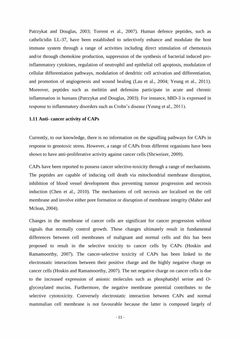
1.11 Anti- cancer activity of CAPs
Currently, to our knowledge, there is no information on the signalling pathways for CAPs in
response to genotoxic stress. However, a range of CAPs from different organisms have been
shown to have anti-proliferative activity against cancer cells (Shcweizer, 2009).
CAPs have been reported to possess cancer selective-toxicity through a range of mechanisms.
The peptides are capable of inducing cell death via mitochondrial membrane disruption,
inhibition of blood vessel development thus preventing tumour progression and necrosis
induction (Chen et al., 2010). The mechanisms of cell necrosis are localised on the cell
membrane and involve either pore formation or disruption of membrane integrity (Maher and
Mclean, 2004).
Changes in the membrane of cancer cells are significant for cancer progression without
signals that normally control growth. These changes ultimately result in fundamental
differences between cell membranes of malignant and normal cells and this has been
proposed to result in the selective toxicity to cancer cells by CAPs (Hoskin and
Ramamoorthy, 2007). The cancer-selective toxicity of CAPs has been linked to the
electrostatic interactions between their positive charge and the highly negative charge on
cancer cells (Hoskin and Ramamoorthy, 2007). The net negative charge on cancer cells is due
to the increased expression of anionic molecules such as phosphatidyl serine and O-
glycosylated mucins. Furthermore, the negative membrane potential contributes to the
selective cytotoxicity. Conversely electrostatic interaction between CAPs and normal
mammalian cell membrane is not favourable because the latter is composed largely of

- 12 -
zwitterionic phospholipids such as phosphatidylethanolamine, phoshatidylcholine and
sphingomyelin resulting in a net neutral charge. Moreover, the cholesterol on the membrane
interferes with the CAPs permeabilisation and disruption of the lipid bilayer (Hoskin and
Ramamoorthy, 2007).
Chen et al., (2010) studied the in vitro anticancer activity of the cationic amphiphilic
antibacterial peptide-CM4 (ABP-CM4) against human leukemia cells compared to normal
mammalian cells; human embryonic kidney cell line (HEK-293) and mononuclear cells
(PBMCs). The peptide was isolated from the haemolymph of the Chinese silkworm Bombyx
mori. Conformational studies using circular dichroism showed that ABP-CM4 had a
significant random coil conformation in water. Inversely, the peptide showed an increasingly
characteristic α-helical secondary structure in 20% v/v 2,2,2-trifluoroethanol (TFE) in water
and a well-defined α-helical conformation in 30 % and 50% v/v TFE in water. Furthermore,
the 3-(4,5-dimethyl-2-thiazolyl)-2,5-diphenyl-2H-tetrazolium bromide (MTT) colometric
assay results indicated dose-dependent cytotoxicity of ABP-CM4 against leukemia cells. The
standard MTT assay measures cell viability after exposure to a growth inhibitor. The IC50
values of ABP-CM4 in inhibiting THP-1, K562 and U937 cells were about 14.2, 15.8 and
17.5 µM, respectively. Interestingly, the peptide showed no significant cytotoxicity against
HEK-293 cell line and PBMCs even at the highest concentration evaluated, 80 µM. Trypan
blue exclusion assays also confirmed that ABP-CM4 showed no significant cytotoxic activity
against PMBCs at a concentration more than tenfold the IC50 obtained against leukemia cells.
The results demonstrated selective cytotoxicity against leukemia cells (Chen et al., 2010).
A flow cytometry analysis showed that FITC-labelled ABP-CM4 had a high affinity for the
cell membrane of leukemia cells as indicated by higher fluorescence compared to normal cell
lines. Moreover, the increase in fluorescence was proportional to the peptide concentration
(Chen et al., 2010). Confocal laser scanning microscopy examination further confirmed that
ABP-CM4 interacts with and binds to the cytoplasmic membrane since the FITC-labelled
ABP-CM4 was localized on cell surfaces (Chen et al., 2010). LDH release assay and
fluorescent propidium iodide (PI) uptake assay indicated a cell membrane damage
mechanism of ABP-CM4 against leukemia cells. The levels of LDH release and fluorescent
PI uptake where proportional to the concentration of the peptide used to treat the cells (Chen
et al., 2010).

- 13 -
Hsu et al., (2011) demonstrated that synthetic Pardaxin induced apoptosis enhances its
antitumor activity in human fibrosarcoma cell line (HT-1080) and epithelial carcinoma cell
lines (HeLa). Pardaxin is a 33-amino acid pore-forming toxin from the Red Sea Moses sole,
Pardachirus marmoratus. The study showed that Pardaxin inhibited the proliferation of HT-
1080 and HeLa cells in a dose-dependent manner after a 6-24 hour exposure at concentrations
between 6-50µg/ml. The anti-proliferative effect of Paradaxin on HT-1080 cells was at the
highest levels 6 hours after treatment. However, HeLa proliferation was inhibited after 24
hours. DNA fragmentation studies showed DNA fragmentation in HeLa cells and not in HT-
1080 after exposure to 15µg/ml of pardaxin. LDH release assay showed that LDH release
from HeLa and HT-1080 cells was dose dependent. This showed that pardaxin disrupted the
cytoplasmic membrane integrity of HeLa and HT-1080 cells. Pardaxin had no effect on the
cytoplasmic membranes of human red blood cells (HRBCs). Moreover, flow cytometry
indicated an increased proportion of HeLa cells in the subG1 phase. The above results imply
that pardaxin induces apoptosis in HeLa cells and not in HT-1080. Furthermore, Real-time
reverse-transcription polymerase chain reaction analysis showed that pardaxin induces the
synthesis of cytokines, down regulates the expression of invasion-related genes and probably
has pleiotropic effects on different cells (Hsu et al., 2011).
Several other CAPs have been found to exhibit anti-proliferative activity against different
cancer cell lines. Lehmann et al., (2006) established the antitumor activity of the
antimicrobial peptide magainin II against bladder cancer cell lines (RT4, 647V and 486P)
with a mean LC50 value of 198.1µM. Cecropins are a class of α-helical antimicrobial peptides
derived from insects and have also been established to have cytotoxic effects on HL-60
human promyelocytic leukemia cell lines, in particular cecropin B1 (Hoskin and
Ramamoorthy, 2008). Papo et al., (2003) designed a short cationic diastereomeric peptide
composed of D- and L-leucines, lysines, and arginines that showed selective toxicity towards
mouse melanoma, fibroblast and lung carcinoma cells and significantly inhibited lung
metastasis in about 86% of mice, with no detectable side effects. The peptides displayed the
ability to depolarize the membrane potential rapidly at a concentration of about 3 µM.
Moreover, confocal microscopy studies verified that the cells died as a result of acute injury,
swelling, and bursting thus, suggesting necrosis.
- 14 -
1.12 Development of CAPs as therapeutic agents
CAPs possess desirable attributes for the pharmaceutical development of cancer-selective
drugs. They selectively and rapidly kill cancer cells without killing normal mammalian cells
and have a broad spectrum of mechanisms of action (Ntwasa, 2012). However, most of the
well-studied and documented CAPs for cancer-selective toxicity have been isolated from
amphibians and marine organisms (Hoskin and Ramamoorthy, 2007). Moreover, they are
secreted in response to microbial infection and injury (Leuschner and Hansel, 2004). Several
challenges have been established to impede the therapeutic development of commercially
viable peptide-based drugs (Ntwasa et al., 2012). Foreign CAPs may have great potential to
elicit treatment-neutralizing antibodies and/or potentially dangerous allergic responses in
cancer patients. They are also prone to proteolysis and inactivation in blood serum resulting
in a short half-life in vivo thus hampering their systemic use. For instance, blood serum
strongly inhibits HNP-mediated cytotoxicity and this is an obstacle to the systemic
administration of these human α-defensins. Synthetic CAPs such as DP-1 and r7-kla that
have been engineered for rapid cellular uptake and resistance to proteolysis require selective
targeting to tumour sites to enhance cancer-selective toxicity following systemic
administration. In addition, the exorbitant production cost of synthetic CAPs is a huge
obstacle to their development as anticancer agents. It has been established that magainin II,
isolated from a frog species, showed reduced cytotoxicity against a differentiated bladder
cancer cell line in comparison to the undifferentiated cell line (Lehmann et al., 2006). The
low toxicity to certain neoplastic cells observed in some studies may require frequent
administration to keep doses high if those peptides are to be used as anticancer agents.
Moreover, the low cytotoxicity observed against normal cells and the hemolysis of
erythrocytes need to be taken into consideration if CAPs are to be further studied and
developed for cancer-selective toxicity.
1.13 Review of the SAEIE08 cell line
SAEIE08 is a dung beetle E. intermedius embryonic cell line developed by Rodney Hull from
the Flylab, University of the Witwatersrand. The karyotype of the beetle was established to
be 2n = 23 + XY, resembling the majority of the Coleoptera species. The cell was reported to
divide asymmetrically into two daughter cells of different sizes, with a doubling time of
approximately 5 hours. The cell line rapid growth at 26oC may be a result of the early
- 15 -
expression of cytokine, Unpaired-3, leading to the early induction of the JAK-STAT pathway
in embryonic cell line (Alouna, 2012).
1.14 Camptothecin as a DNA damaging agent
Camptothecin (CPT), is an alkaloid that is often used as an anticancer agent as it is an
inhibitor of DNA topoisomerase I and thus disrupts the process of DNA replication.
Camptothecin is, therefore a genotoxic stressor (Seong et al., 2012). Topoisomerases are
enzymes that regulate the level of DNA supercoiling, to facilitate interaction with proteins
during replication and transcription. These enzymes bind to either single stranded or double
stranded DNA and their actions result in temporary breaks to DNA during replication,
transcription, recombination and chromatin remodelling (Champoux, 2001). Seong et al.,
(2012) reported that CPT binds to the topoisomeraseI-DNA complex introducing irreversible
covalent linkage that prevents re-sealing of the topoisomerase 1 introduced DNA breaks. This
causes replication-dependent double-strand breakages in the DNA of replicating cells. Thus
the CPT-DNA-topoisomerase complex disrupts replication and causes unfavourable DNA
topology leading to homologous and non-homologous recombination. DNA damage and/or
the failure to repair the damage induce DNA repair pathways which are controlled by a range
of genes including tumour response genes (Davis and Lin, 2011).
Drosophila melanogaster (Canton S) has been used as model system to study the effects of
CPT on normal cells. In a previous study, CPT treatment was established to cause an increase
in the transcription of the Drosophila homologue of p53 (Dmp53) revealing the activation of
the p53 pathway. In this study, the transcription of a Drosophila member of the
retinoblastoma binding protein 6 family, Snama, was reduced during CPT treatment, but up-
regulated when the flies recovered from treatment. This up-regulation may be associated with
glycolytic flux that was induced in recovering flies (Hull and Ntwasa, 2010). In another
study, Dmp53 was reported to have roles in DNA damage-induced cell cycle arrest under
specific circumstances and tissue regeneration in response to imaginal discs damage (Monk et
al., 2012). Dmp53 is known to induce apoptosis by up-regulating the transcription of
downstream effectors such as reaper, head involution defective and sickle. In this study CPT
is therefore considered a suitable model for DNA damage since there is background
information about its effect on insects. CPT does, however, have some disadvantages; it is
insoluble in water and is solubilised here in DMSO which may also have adverse effects on
cells.

- 16 -
1.15 Objectives of the study
The current study is exploratory and aims to screen for genotoxic induced CAPs of insect
origin with increased anti-proliferative activity against cancer cells, no toxicity to normal
mammalian cells and resistant to proteolysis.
The current study focused on the identification and characterisation of molecules induced in
response to genotoxic stress from an embryonic cell line originating from the dung beetle
Euoniticellus intermedius. Moreover, it is important to establish if the induction of CAPs
expression is part of the response. E intermedius (Coleoptera: Scarabaeidae) is a well-known
dung beetle found largely in the Afrotropical region. E intermedius has a wide ecological
tolerance and survives under extreme environments and has been reported to be tolerant to a
range of conventional pesticides (Kruger et al., 1999). The beetle is being studied in the
Flylab as a model of innate immunity in coleopterans.
The objectives of the study are:
To isolate camptothecin induced cationic amphiphilic peptides using a C18 column
To assay for potential anti-proliferative activity of induced CAPs using radial
diffusion assay as a model
To evaluate differential gene expression following camptothecin treatment using the
GeneFishing® technology
To evaluate differential protein expression using 2-D gel electrophoresis
To identify differential expressed proteins using mass spectroscopy

- 17 -
Chapter 2: Materials and methods
2.1 Materials
The materials used in this study are listed in the tables below. The materials were grouped
into three categories: media, biological buffers and solutions, and primer.
Table 1: Media
Media Composition/ supplier
Grace’s Insect media Highveld Biological (catalogue # L16)
Luria broth 1% Sodium chloride (Merck, Germany)
1% Tryptone ( Biolab)
0.5% Yeast extract (Biolab)
Solid agar 1% Sodium chloride (Biolab)
1% Tryptone (Biolab)
0.5% Yeast extract (Biolab)
1.5% Agar (Biolab)
Tryptic soy broth 3% Tryptic soy (Biolab)
Underlay agar 0.03% Tryptic Soy Broth (Biolab)
1% Agarose (Biolab)
0.02% Tween (Merck Germany)
Overlay agar 6% Tryptic soy (Biolab)
1% Agarose (Biolab)
Table 2: Biological buffers and solutions
Buffer/solution Composition
50X TAE electrophoresis buffer pH 8 24.2 w/v % Trisbase (Merck, Germany)
5.71 v/v % Glacial acetic acid (Merck)
3.72 w/v % Na2EDTA.2H2O (Merck)
Buffer I 50mM glucose (Merck, Germany)
10mM EDTA (Merck, Germany)
0.25M TrisHCl pH 8 (Merck, Germany)

- 18 -
Buffer II 1% SDS (Prolabo catalogue #27926.238)
0.2N NaOH (Merck, Germany)
Buffer III 5M Potassium acetate (Merck, Germany)
Ethanol : Acetate 25 : 1 (Merck, Germany)
Camptothecin 50mM in DMSO (Merck, Germany)
Rehydration buffer 8mM Urea (Bio-rad, USA)
2% CHAPS (Bio-rad, Canada)
50mM DTT (Fermentas, Canada)
0.2% Biolyte (Bio-rad, Canada)
0.5% Bromophenol blue (Sigma, USA)
Table 3: Primer
Primer name Sequence 5’- 3’
3’ RACE forward specific primer CGAGATCTTCTAGAAG
2.2 Tissue culture
The embryonic cell line (SAEIE08) from E. intermedius was cultured in 30ml Grace’s insect
media to give a concentration of 100cells\ml in the final volume. The cells were incubated at
25oC for 12 hours to reach an exponential growth phase. The cells were treated with
camptothecin dissolved in DMSO to give a final concentration of 50mM in the insect media.
DMSO was used as a control treatment. The cells were incubated for a further 4 hours, and
the cell concentration was estimated to be approximately 5 x 10⁶
cells\ml.
2.3 RNA extraction
Total RNA for GeneFishing® and RACE was extracted using the ZR RNA MiniPrep kit
from Zymo Research (catalogue number: R1064) according to the manufacturer’s protocol. A
cell concentration of approximately 5 x 10⁶
cells\ml was used during the extraction.
Extraction was performed for both camptothecin and DMSO treated cell cultures. The
concentration and purity of total RNA was determined through standard spectroscopy and
formaldehyde RNA gel (1% agarose) (Figure 3). The 260/280 ratio was used to determine the

- 19 -
purity of RNA. This ratio was measured using a Nanodrop and measures the level of protein
contamination. A ratio of greater than 1.8 is acceptable.
Figure 3: Total RNA extracted from the beetle cell culture. The integrity of RNA was determined using
formaldehyde RNA gel (1% agarose). The gel shows both the 18S and 28S ribosomal RNA.
2.4 Protein extraction from lysed cells and concentration from the supernatant
Crude proteins from lysed cells were extracted from both the camptothecin treated and
DMSO treated cell cultures using Tri-reagent from Sigma-Aldrich (catalogue number:
T9424) following the manufacturer’s protocol. The protein was however, washed in 100%
ethanol by spinning for 5 minutes at 7500rpm after precipitation and dissolved in rehydration
buffer for 2-D gel analysis and 1% SDS for C18 column purification. A cell concentration of
approximately 5 x 10⁶
cells\ml was used for the extraction. The protein concentration was
determined using the Bradford reagent from Bio-rad (Cat # 10563) and a UV VIS
spectrophotometer. The Bradford assay was performed according to the manufacturer’s
protocol. The standard curve (Figure 4) used was determined by Hull, (2012) and constructed
in the spectrophotometer. The Bradford assay is a method used to determine protein
concentration. The procedure involves binding of Coomassie Brilliant Blue G-250 dye to the
proteins. The dye exists in three forms: cationic (red), neutral (green) and anionic (blue) and
subsequently changes into a stable unprotonated blue form upon binding to proteins. The
Ca
mp
toth
ecin
tre
ate
d
DM
SO
tre
ate
d
28S
18S

- 20 -
unprotonated from has a wavelength of 592nm which is the wavelength used to determine
protein concentration using a spectrophotometer.
Figure 4: Standard curve for protein concentration using the Bradford assay. The curve was done in technical
replicates and mean values used to construct the curve in the spectrophotometer. The equation of the line is y =
0.29564x -0.00004 and R2 = 0.9994 (Hull, 2012).
Secreted proteins were concentrated from the supernatant. Camptothecin treated and
untreated cell cultures were spun in 15ml centrifuge tubes for 5 minutes at 3.5rpm. The
supernatant was used to concentrate the secreted proteins using Millipore’s Amicon Ultra-4
centrifugal filter (Cat # UFC4 LCC 25). The filter enables retention of proteins from dilute
samples, serum and cell culture samples through the nominal molecular weight limit
membrane. The proteins were recovered from the bottom of the filter by sucking with a
pipette. The lysate ~ 250µl was re-suspended in 250µl of 1% SDS. One volume of 20%
trichloroacetic acid was added and the mixture incubated on ice for 30 minutes. The sample
was spun at 12.5rpm for 10 minutes. The supernatant was discarded and pellet washed in
500µl cold acetone by spinning at 12.5rpm for 5 minutes. Acetone was discarded and pellet
air dried. The pellet was re-suspended in rehydration buffer by pipetting and used in 2-D
electrophoresis.

- 21 -
2.5 Isolation of the cationic amphiphilic proteins
The crude protein extract from the SAEIE08 cell line contains a complex mixture of proteins
which needs further and specific purification to enable isolation of a specific group of
proteins. In this study the Chromabond® C18 column from Macherey-Nagel (Cat # 0206/8)
with a void volume of 0.8ml was used to isolate cationic amphiphilic peptides for radial
diffusion assays. The column contains a hydrophobic, reverse phase, silica-based bonded
phase that is capable of adsorbing cationic hydrophobic molecules from aqueous solutions.
The column was activated with 2 volumes (0.8ml) of methanol. 0.8ml of the crude protein
extract solubilised in 1% SDS was added to the column using a 10ml syringe and the flow
through was collected. The column was washed with two volumes (0.8ml) of 5mM sodium
phosphate buffer pH 8. The column was finally washed with 1ml of 80% acetonitrile in 5mM
sodium phosphate buffer pH 8. Acetonitrile was removed from the samples through exposure
to a stream of nitrogen.
2.6 Antimicrobial activity assays
The radial diffusion assays were based on the technique of Leher et al., (1991). Tryptic soy
broth was inoculated with a single Escherichia coli colony and allowed to grow for 18 hours
at 37oC. 50µl of the culture was used to inoculate 50ml of fresh Tryptic soy broth and this
was grown further for 3 hours at 37oC. The culture was centrifuged and re-suspended in cold
10mM sodium phosphate buffer. The optical density of the culture was determined at 620nm
and this was used to calculate the volume required to make a culture with 4 x 10⁶ colony
forming units per ml in a final volume of 15 ml underlay agar using the formula: 0.2 O.D620nm
= 5x 10⁷ CFU/ml. 15ml of the underlay agar with bacterial culture was prepared and plates
were allowed to set. Holes were punched in the agar and the protein samples, tetracycline,
phosphate buffer, acetonitrile and water controls were added to the holes. Tetracycline is a
known antimicrobial and was used as the positive control while water was used as a negative
control. Acetonitrile and phosphate buffer controls were used to verify that the solvents used
in eluting proteins did not contribute to the antimicrobial activity of the proteins under
investigation. Protein eluted with acetonitrile and sodium phosphate buffer were used as test
samples. The plates were incubated for 3 hours at 37oC. 15ml of overlay agar was then added

- 22 -
and plates incubated for 18 hours at 37oC. The diameter of the clearings was measured. The
assays were performed in triplicates for each test sample and control.
2.7 Chemical characterisation of the antimicrobial active agent using the Protein kinase
assay
The inhibitory samples were treated with Proteinase K to determine the nature of the
antimicrobial active molecules. Proteinase K was added to the samples to the final
concentration of 100µg/ml. 100µg/ml Proteinase K and water were also included as controls.
The reactions were incubated at 37oC for 15 minutes and Proteinase K was inactivated by
heating at 70oC for 15 minutes.
The inhibition assay was repeated with the Proteinase K treated samples. Three technical
replicates were used for each sample in the assays.
2.8 Differential expression of genes after camptothecin treatment
2.8.1 Genefishing Technology
The cDNA of differential expressed genes was synthesised using a GeneFishing® kit from
SeeGene® (Catalogue number: K1026). The kit enables the identification of differentially
expressed genes in 2 or more RNA samples. The kit works in three steps, which consist of the
first strand cDNA synthesis step and a two-stage PCR. These steps use a set of three unique
annealing controlled primers (Figure 5). The first step applies reverse transcription using dT-
ACP 1 to synthesize cDNA from the mRNA. The 3΄ end hybridizing sequence of dT-ACP 1
is complementary to the mRNA poly A tail and this results in a first strand cDNA transcript
with the universal sequence of dT-ACP 1 at its 5΄ end. The first strand cDNA is then diluted
and placed in tube with an arbitrary ACP and dT-ACP 2 for the PCR step. The first PCR
stage is a single PCR cycle under conditions that enable only the annealing of the arbitrary
ACP with its 3΄ end core to the first strand cDNA. This results in a second-strand cDNA with
a complementary sequence of the universal sequence of dT-ACP 1 on its 3΄ end and the
universal sequence of the arbitrary ACP on its 5΄ end. The second PCR stage enables the
amplification of the targeted PCR product only. The conditions are set to exclusively allow
both dT-ACP 2 and the arbitrary ACP to anneal to the 3΄ and 5΄ ends of the second strand

- 23 -
cDNA, respectively. The conditions also prevent the annealing of both primers to the first
strand cDNA. The gene fishing experiment was performed following the manufacturer’s
protocol. The PCR products were electrophoresed in 1% agarose gel with ethidium bromide
in 2X TAE electrophoresis buffer at 100Volts. A 1Kbp plus marker was used.The gel was
kept for the recovery and purification of differentially expressed genes/bands.
Figure 5: Flow chart of cDNA synthesis and GeneFishing® PCR (SeeGene® GeneFishingTM
DEG premix kit
user manual 2005, Catalogue number: K1026).
First strand cDNA
synthesis
First stage PCR for second
strand cDNA synthesis.
Only the arbitrary ACP
binds to the first strand
cDNA
Second stage PCR to amplify target
PCR products. The conditions are
set to enable exclusive annealing of
both primers to the second strand
cDNA only

- 24 -
2.8.2 PCR products recovery and purification from electrophoretic gels
DNA purification from the gel was performed using with the Zymogen DNA recovery kit
(Cat # D 4001). The kit provides an efficient method for high quality DNA purification and
concentration from agarose gels without the use of organic denaturants. The kit purifies and
concentrates DNA in a fast spin column. The bands of differentially expressed genes from
gene fishing were excised from the gel under UV-light illumination. The purification was
carried out following the manufacturer’s protocol.
2.9 Cloning and sequencing of the GeneFishing® PCR product
2.9.1 Cloning
The recovered DNA was eluted with RNAase free water and cloned into a pGEM-T Easy
Vector since gene fishing PCR products have poly-A tails. The ligation reaction was set-up
according to the manufacturer’s protocol. The total amount of insert DNA used was 25ng for
each reaction. The tubes were sealed with parafilm and placed under water to prevent
evaporation in the tube. The reaction was incubated for 12 hours at 4oC and inactivated by
incubating at -70oC for 15 minutes
10µl of the ligation reaction was added to 40µl competent cells on ice, and was left to stand
for 30 minutes. The cells were heat shocked for 45 seconds at 45oC and cooled on ice for 2
minutes. 960µl of the recovery medium was added and the mixture was incubated at 37oC
with shaking at 250rpm for 90 minutes. 100µl of the transformation reaction was plated on
pre-warmed agar with ampicilin/X-gal and IPTG. X-gal/IPTG was spread on the agar plate
30 minutes before plating and plates pre-warmed at 37oC.
2.9.2 Sequencing
Gene fishing PCR products cloned in bacterial cells were sent to Inqaba Biotech® for
sequencing.
2.10 3’ Rapid amplification of cDNA Ends
Sequencing produced a partial sequence of a differentially expressed transcript which was
used in 3’ RACE to synthesise a full-length transcript of the gene (Figure 4). This technique

- 25 -
enables the generation of a full length sequence of RNA from cultured cells. The cDNA is
generated using an Oligo-dT primer complementary to the natural polyA tail on 3′end of an
mRNA transcript. The mRNA is then degraded before the PCR step. This technique was used
because the PCR step requires only one specific primer (forward primer) and an anchor
primer. A forward primer was designed from the partial sequence (Figure 10). The 3` RACE
was performed using the 5΄\3΄ RACE kit, 2nd
generation from Roche® (Cat # 03 353 621
001) according to manufacturer’s protocol. The full-length sequence of the gene fishing
transcript was amplified from total RNA from camptothecin treated cells. The PCR
amplification program was performed as follows: Initial denaturation at 94oC for 3 minutes,
denaturation at 94oC for 30 seconds, annealing at 60
oC for 30 seconds, 40 cycles of
elongation at 72oC for 45 seconds, and a final elongation at 72
oC for 5 minutes. PCR products
were separated with electrophoresis. The whole PCR reaction was sent for sequencing by
Inqaba biotech®.
Figure 6: Overview of 3’ RACE (Roche® Applied Science 5’\3’ RACE kit, 2nd
generation manual, Version
October 2005, Cat # 03 353 621 001).

- 26 -
2.11 Proteomic analysis of molecules involved in genotoxic response of E. intermedius
embryo cell line
2.11.1 2-D electrophoresis
A protein concentration of 100µg/ml was used for 2-D analysis. The protein sample was
pipetted along the length of the rehydration tray and an 11cm IPG strip (pH 3-10) was laid
carefully facing down avoiding the formation of bubbles. The strip was overlaid with mineral
oil, sealed with parafilm and left to stand for 20 hours at room temperature. The wire
electrodes of the isoelectric focusing tray were each covered with a paper wick and moistened
with ~10µl of nanopure water. The IPG strip was removed from the rehydration tray and held
for about 20 seconds to allow the mineral oil to drip off. The strip was placed facing down in
the channel of the isoelectric focusing tray with its positive side on the anode left side of the
tray. The strips were covered with mineral oil. The isoelectric focusing was performed in
three steps. The first step was done at 250 Volts for 20 minutes with linear ramping, the
second step at 8000 Volts for 2.5 hours with linear ramping and the last step was rapid
ramping for 20000 Volts-hour. The IPG strip was removed from the isoelectric focusing tray
and placed in a rehydration tray. 2 ml of equilibrating buffer I was added to the channel with
the strip and shaken gently for 5 minutes. The buffer was discarded and replaced with
equilibrating buffer II. The tray was gently shaken for 5 minutes and the buffer discarded.
The strip was removed and dipped for about 30 seconds in 1X SDS running buffer in a
measuring cylinder. The strip was over-laid on a precast gel. A paper wick loaded with 2µl
pre-stained protein marker was placed next to the IPG strip. Overlay agarose was added and
allowed to solidify. The PAGE was run at 100 Volts in 1X SDS running buffer. The gel was
stained in Coomassie brilliant blue for 14 hours. The dye was de-stained for 4 hours and
washed for 5 hours with several changes of distilled water.
2.11.2 Image analysis
A PDQuest 2-D analysis software version 6.2 (Bio Rad Cat #170-9630) was used to scan and
upload the gel images. The camptothecin treated gel was used as the master gel. The spots
from the camptothecin and DMSO treated gels were initially matched using the automated
matching function. The spots were further matched manually with manual matching functions
that enable deletion and addition of spots not detected or improperly detected during
automated matching. Normalization was done automatically by the program based on the

- 27 -
given normalization formula: Normalized spot quantity = Raw spot quantity x scaling factor /
Normalization factor (total quantity in all valid spots). The scaling factor was 106 parts per
million
2.11.3 Mass spectroscopy
Spots representing differential expression were excised from the gels and sent for LC MS
mass spectroscopy analysis at the Council for Scientific and Industrial Research, Pretoria,
South Africa. The trypsin digested peptides were analysed using a Dionex Ultimate 3000
RSLC system coupled to a QSTAR ELITE mass spectrometer. Peaks v6.0 search engine
(Bioinformatics Solutions Inc.) was used for comparison of the obtained MS/MS spectra with
the MS protein sequence DataBase (MSDB) as well as the Flylab genome base. A False
Discovery Rate (FDR) of 0.1% was used and proteins with more than one unique peptide
reported.
2.11.4 Statistical and bioinformatics analysis
The mean OD values from the 2-D gels were calculated and changes in the expression of
individual peptides were analysed using a one sided Wilcoxon Signed-Rank test at 10%
significance level to compare differences in the mean since the data distribution is not
normal. The Wilcoxon Signed-Rank t-test was also used to compare the differences in the
mean inhibition in the activity assay. Peptide fingerprints from MS/MS were identified
through manual search from the flylab genome base database
(http://flylab.wits.ac.za/EI/est2uni/blast.php) (Khanyile et al., 2008) consisting of EST
sequences of the adult transcriptome using TBLASTN. A BLAST was also performed from
the National Centre for Biotechnology Information (NCBI) database (www.ncbi.nlm.nih.gov)
using the UnitProtKB/Swiss-Prot(swissprot) database to identify proteins that had similar
sequences to those identified by mass spectroscopy.

- 28 -
Chapter 3: Results
3.1 Results
The primary objective of the study is to investigate whether CAPs are activated by
camptothecin and the molecular pathways involved in the process. GeneFishing® and 2-D
analysis were used to determine the changes in gene expression at transcription and
translation levels, respectively.
CAPs are an interesting group of molecules that are reported to play roles in apoptosis, stress
response and to possess cancer selective toxicity as reviewed in this study. Moreover, it is in
the view of this study that an understanding of the molecules and/or pathways involved in
DNA damage paves a way for their exploitation to design novel cancer selective therapeutics.
The objective of this study was to use GeneFishing® and 2-D gel analysis to analyse
molecules involved in genotoxic stress response in an E. intermedius embryo cell line and to
establish if the induction of CAPs is part of the response.
3.2 Antimicrobial activity assays
Genotoxic stress induces a series of response pathways in cells. The molecules induced in
these response mechanisms are activated to either reverse the damage caused by the stress or
cause cell apoptosis. This experiment aims to establish whether CAPs are specifically
induced in response to genotoxic stress. In order to determine whether CAPs were expressed
by the cell line, the C18 elutes of crude extracts from induced and un-induced cell cultures
were assayed for anti-bacterial activity. This approach was used because CAPs are expected
to have anti-bacterial activity.
The SAEIE08 cell line was used as a model in this study. The cell line was treated with
camptothecin. The characteristic amphiphilic and cationic nature of CAPs was exploited to
exclusively separate them from the crude protein extract from cell cultures using the C18
column. The column is designed to adsorb cationic hydrophobic molecules from an aqueous
solution. Radial diffusion assays were subsequently used to determine whether camptothecin
treatment induced molecules that possess anti-bacterial toxicity. The Gram negative, E.coli
was used as a model for the toxicity assay because CAPs anti-bacterial activity employs the
same mechanisms of action as during anti-proliferative activity against cancer cells. The

- 29 -
above is attributed to similarity in the overall characteristic of the membrane of cancer and
bacterial cells.
Microbial inhibition was present in the samples eluted with acetonitrile (Figure 7a). This
shows that the inhibitory molecules are cationic and amphiphilic which is consistent with the
character of most CAPs. Interestingly acetonitrile purified samples from both camptothecin
treated and untreated crude extracts showed microbial inhibition. This indicated constitutive
expression of antimicrobial activity. However, inhibition by the former was significantly
higher since the same concentration of the protein was used in the assays (T value = 0 <
Tcritical=2, for n = 3) (Figure 7a). This indicated that more activity was induced by
camptothecin. Water was included as a standard negative control to enable comparison
between microbial inhibition and non-inhibition (Figure 7b). No inhibition was shown in the
water, sodium phosphate and acetonitrile control. Sodium phosphate and acetonitrile were
included as controls since they were essential components of the elution buffers used to elute
the proteins from the C18 column (Figure 7c). The positive control, tetracycline showed
significant microbial inhibition (Figure 7c). Overall, the results show that the inhibitory
molecules are constitutively active in the cells and that further expression is induced in
response to camptothecin treatment. The constitutive activity may be attributed to the
microbe-rich environment in which the beetles live.
To investigate the nature of the antimicrobial active molecules, the samples showing
microbial inhibition were treated with Proteinase K. No antimicrobial inhibition was shown
following the treatment of the inhibitory sample with Proteinase K (Figure 8a). This implies
that the antimicrobial inhibition is a consequence of a protein since Proteinase K degrades
proteins. Proteinase K alone also showed no inhibition (Figure 8c). Microbial inhibition was
however, observed from untreated sample and from the tetracycline control.
Given these results, it may be proposed that probable cationic amphiphilic proteins are
constitutively expressed by the cells and that camptothecin induces the production of more
proteins.
The above observations pave the way to further study the pathways involved in the up-
regulation of the anti-bacterial activity and to determine whether the induction of CAPs is
part of the pathways and if they also play a role in genotoxic stress.

- 30 -
Figure 7: Radial diffusion assays. The crude protein extracts from E intermedius cells treated with
camptothecin and untreated cells were purified with a C18 Sep pak vac column eluting with sodium phosphate
buffer and acetonitrile. Figure 7 A shows inhibition by the acetonitrile purified samples. However, the sample
from proteins extracted from camptothecin treated cells shows enhanced inhibition compared to the sample from
the proteins extracted from untreated cells. T value = 0 < Tcritical=2, for n = 3. The negative controls and
positive control (tetracyclin) are represented in Figure 7 B and C.
Figure 8: Characterisation of the inhibitory or toxic molecule. The sample that showed inhibition in the radial
diffusion assay was treated with Proteinase K to verify that a peptide was responsible for the antimicrobial
activity. Radial diffusion assays were used to test for microbial inhibition after treatment with Proteinase K.
Figure 8A shows that no inhibition was seen after treatment with Proteinase K for both camptothecin treated and
untreated samples. Similarly Figure 8 B and C show no inhibition by the controls.

- 31 -
3.3 Differential transcription of genes induced by camptothecin
Following the confirmation that the SAEIE08 cell line up-regulates the expression of
probable antimicrobial proteins, the gene fishing technique in combination with 3’ RACE
was used to determine the identity of differentially transcribed genes following camptothecin
exposure. The principle of gene fishing is based on the use of an annealing control primer
(ACP) that anneals with high specificity to the intended template to allow amplification of the
target product. The ACP is a tripartite structure with distinct 3΄- and 5΄-end portions
separated by a regulator. The 3΄-end core is designed to target the template via the
hybridising sequence and is significantly complementary to a portion on the target nucleic
acid template. The 5΄-end core has a universal sequence and together with the regulator plays
an important role in the annealing of the ACP to the template. RNA samples from
camptothecin treated cells and from untreated cells were used in the gene fishing to identify
differential expression between the two RNA samples (Figure 5). Electrophoresis was used
to separate PCR products generated in the gene fishing experiment. The partial sequence of
the up-regulated transcript generated by ACP 17 was used to design a forward primer to
synthesise a full-length transcript of the gene using 3΄ RACE (Figure 6). The full length
sequence of the gene was generated from the RNA of camptothecin treated cells. The PCR
product was separated by electrophoresis. The whole PCR reaction containing two bands was
sent for sequencing by Inqaba Biotech®.
Camptothecin exposure also resulted in differential transcription of certain genes. Arbitrary
complementary primers (ACP) 16- and 19-amplified fragments (300bp and 200bp,
respectively) show down-regulation in the transcription of genes following campthotecin
treatment (Figure 9b). Furthermore, ACP 17-amplified fragment (100bp) showing up-
regulation in gene transcription (Figure 9b). ACP 18 also produced an up-regulated gene of ~
250bp (Figure 9b).
The ACP 17-amplified transcript was cloned in pGem T-easy vector. Sequencing of the
transformed clones produced a partial sequence of the gene. The sequence was subsequently
used to design a forward primer (Figure 10) that was then used to perform 3’ RACE to
amplify the full sequence of the gene. Sequencing of the RACE PCR product produced two
sequences and this is consistent with the two products shown in the electrophoretic gels
(Figure 11). Only one of the sequences gave a likely match following a BLAST search in

- 32 -
NCBI nucleotide database. The sequence was highly similar to a yeast Yph1p protein
homologue. Gene fishing coupled with 3’RACE results suggest that the gene for the Yph1p
protein is constitutively expressed and also induced by camptothecin.
A
B
Figure 9: Gene fishing to identify differentially transcribed genes between camptothecin treated (T) and
untreated/normal cells (U). Figure 9 A shows the arbitary complementary primers that amplified transcripts
produced in response to camptothecin treatment in comparison to untreated\normal cells. B is a composite figure
of figure A. The solid arrow shows the down-regulated transcript amplified with ACP 16. The dotted arrows
show up-regulated transcripts amplified by ACP 18 and ACP 19.
400bp
500bp
300bp 200bp
75bp
AC
P
18
tre
ate
d
AC
P
18
un
trea
ted
AC
P
19
tre
ate
d
AC
P
19
un
trea
ted
AC
P
16
tre
ate
d
AC
P
16
un
trea
ted
AC
P
17
un
trea
ted
AC
P
17
tre
ate
d
AC
P
16
un
trea
ted
AC
P
16
tre
ate
d
AC
P
17
un
trea
ted
AC
P
17
tre
ate
d
AC
P
18
un
trea
ted
AC
P
18
tre
ate
d
AC
P
19
un
trea
ted
AC
P
19
tre
ate
d
700 bp
500 bp
400 bp
300 bp
200 bp
75 bp

- 33 -
5’ CGAGATCTTCTAGAAG 3’
Figure 10: Chromatogram showing the ACP 17 generated partial transcript that was used to design the forward
primer for 3’ RACE.
Figure 11: Rapid Amplification of cDNA Ends (3’ RACE) of the partial sequence obtained from differentially
transcribed transcripts using the ACP17 GeneFishing® primer. The cDNA was amplified with a forward
specific primer and PCR anchor primer. The gel shows two significant bands, one ~460bp and the other ~250bp.
3’
RA
CE
PC
R P
RO
DU
CT
S
460 bp
250 bp
1500 bp
700 bp
500 bp
400 bp
300 bp
200 bp
75 bp
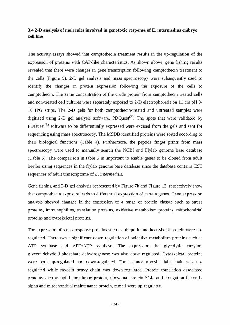
- 34 -
3.4 2-D analysis of molecules involved in genotoxic response of E. intermedius embryo
cell line
The activity assays showed that camptothecin treatment results in the up-regulation of the
expression of proteins with CAP-like characteristics. As shown above, gene fishing results
revealed that there were changes in gene transcription following camptothecin treatment to
the cells (Figure 9). 2-D gel analysis and mass spectroscopy were subsequently used to
identify the changes in protein expression following the exposure of the cells to
camptothecin. The same concentration of the crude protein from camptothecin treated cells
and non-treated cell cultures were separately exposed to 2-D electrophoresis on 11 cm pH 3-
10 IPG strips. The 2-D gels for both camptothecin-treated and untreated samples were
digitised using 2-D gel analysis software, PDQuest(R)
. The spots that were validated by
PDQuest(R)
software to be differentially expressed were excised from the gels and sent for
sequencing using mass spectroscopy. The MSDB identified proteins were sorted according to
their biological functions (Table 4). Furthermore, the peptide finger prints from mass
spectroscopy were used to manually search the NCBI and Flylab genome base database
(Table 5). The comparison in table 5 is important to enable genes to be cloned from adult
beetles using sequences in the flylab genome base database since the database contains EST
sequences of adult transcriptome of E. intermedius.
Gene fishing and 2-D gel analysis represented by Figure 7b and Figure 12, respectively show
that camptothecin exposure leads to differential expression of certain genes. Gene expression
analysis showed changes in the expression of a range of protein classes such as stress
proteins, immunophilins, translation proteins, oxidative metabolism proteins, mitochondrial
proteins and cytoskeletal proteins.
The expression of stress response proteins such as ubiquitin and heat-shock protein were up-
regulated. There was a significant down-regulation of oxidative metabolism proteins such as
ATP synthase and ADP/ATP synthase. The expression the glycolytic enzyme,
glyceraldehyde-3-phosphate dehydrogenase was also down-regulated. Cytoskeletal proteins
were both up-regulated and down-regulated. For instance myosin light chain was up-
regulated while myosin heavy chain was down-regulated. Protein translation associated
proteins such as upf 1 membrane protein, ribosomal protein S14e and elongation factor 1-
alpha and mitochondrial maintenance protein, mmf 1 were up-regulated.

- 35 -
Figure 12: 2-D electrophoresis analysis of differential expression of proteins between untreated (A) and
Camptothecin-treated (B) in South African Euoniticellus intermedius embryonic cell line. Exponentially
growing cells were treated with 50mM camptothecin and the crude protein extracted from cells after 4 hours
using Trizol reagent. The isoelectric focusing was done with an 11 cm IPG strip and the 2nd
dimension was run
using a precast TGX gel from Biorad®. Spots numbered in Red are only present in the untreated (A) gel, spots
marked in dark blue with a rectangle are found in treated only (B) and spots marked in blue are found in both
gels.
1203
1107
1215
3002
5202
6207 7202
3910
1306
4203
7201
7307
8301
8303
B (treated) pH 3-10
5205 5205
5202 3204
3207
3207
3204
1107
1203 7202 6207 3002
1215
A (untreated) pH 3-10
250kDa 130kDa
70kDa 55kDa 35kDa
25kDa
15kDa
10kDa

- 36 -
Table 4. Proteins identified with MSDB and BLAST searches in NCBI database and Flylab base genomebase.
Camptothecin exposure resulted in both up-regulation and down-regulation of protein expression. A one sided
Wilcoxon Signed-Rank test at 10% significance level to verify if there was a significant change in the levels of
protein expression between camptothecin treated cell line and untreated cell line. A T-value less than Tcritical
indicates a significant difference in protein expression. Tcritical = 2.
Spot MSDB predicted protein
Spot
MR
(Kda) Flylab genomebase matching peptide
Mean
OD
before
Mean
OD
after T-value
Stress proteins
2309 Yeast heat shock protein 20 4.8 75.2 0
5202 Ubiquitin 8 109.1 131.1 1
4203 Un-identified 11 Tribolium mrj (100%) 62.3 0.2 0
4203 Ubiquitin-60S ribosomal protein 11.5 62.3 0.2 0
Oxidative
metabolism
and
mitochondrial
7307 Mitochondrial peroxiredoxin 13 119.6 0.2 0
8303 Protein mmf1 mitochondrial 14.5 154.1 0.2 0
8303 Glyceraldehyde-3-phosphate deyhdrogenase 14.5 154.1 0.2 0
7202 ATP synthase 7 158.5 3.1 0
7202 ADP/ATP translocase 7 158.5 3.1 0
Protein
translation
1203 ribosomal rna small subunit methyltransferase 7.5 Glycosyl hydrolase family (100%) 44.2 121.8 0
5205 Un-identified 12.5 Elongation factor 1-alpha (100%) 13.8 57.9 1
5206 40S ribosomalprotein S14 8 Putative ribosomal protein S14e (100%) 0.3 88.1 0
9301 upf 1 membrane protein 17 Myosin heavy chain isoform 2 (100%) 0.3 124.3 0
3207 eukaryotic translation initiation factor 3 subunit 11 57.5 114.4 0
Cytoskeletal
protein
3207 Un-identified 11 Similar to Tropomyosin 1 (100%) 57.5 114.4 0
1215 myosin light chain 12 Myosin alkali light chain 1(100%) 10.4 37.6 1
7202 myosin heavy chain 7 158.5 3.1 0
6207 actin fragment 6.5 86.1 125.1 1
Immunophilins
3204 FK506 Binding protein 7 FK506 Bingding protein (100%) 58.6 116.7 0
7201 Un-identified 7.5 Cyclophilin 1 (71%) 392.7 0.2 0

- 37 -
Table 5: Comparison of MS database identified proteins with the NCBI and Flylab base genomebase databases
identified proteins.
Spot NCBI predicted protein
Accession
number Flylab genomebase matching peptide Flylab genome DB ID
2309 Heat shock 70 kDa protein P12795.1
5202 Ubiquitin P19848.1
4203 Tribolium mrj (100)
4203 Ubiquitin -60S ribosomal protein L40 P0CH08.1
7307 Mitochondrial peroxoredoxin PRX1 P34227.1
8303 Protein mmf1 mitochondrial P40185.1
8303 Glyceraldehyde-3-phosphate dehydrogenase P17819.1
7202 ATP synthase F1 sector subunit beta B0THN2.1
7202 ADP/ATP translocase P05141.7
1203 ribosomal rna small subunit methyltransferase Q4FPN5.1 Glycosyl hydrolase family (100%) 005275_1537_3307_c_s
5205 Elongation factor 1-alpha Q07051.1 Elongation factor 1-alpha (100%) CL17Contig1
5206 40S ribosomalprotein S14 P27069.1 Putative ribosomal protein S14e (100%) CL171Contig1
9301 Buforin 1 P55897.1 Muscle myosin heavy chain isoform 2 (100%) CL448Contig1
3207 Eurkaryotic translation initiation factor 3 subunit P23301.3
3207 Tropomyosin P42637.1 Similar to Tropomyosin 1 (100%) CL137Contig1
1215 Myosin-2 light chain P53141.1 (Tribolium) Myosin alkali light chain 1(100%) CL25Contig1
7202 myosin heavy chain D P02567.3
6207 actin 1 P85911.1
3204 FK506 Binding protein Q754K8.1 FK506 Bingding protein (100%) 009937_1646_3733_c_s
7201 Cyclophilin P14832.3 Cyclophilin 1 (71%) 005522_1647_2340_c_s

- 38 -
Chapter 4: Discussion
4.1 Discussion
This study shows that the SAEIE 08 cell line constitutively expresses antimicrobial proteins.
Moreover, more anti-bacterial proteins are induced upon challenge by camptothecin. Analysis
of gene expression has not revealed any cationic peptides as expected but showed classes of
biological molecules involved in a range of pathways. Gene fishing and 2-D gel analysis
showed changes in the expression of proteins involved in stress response, oxidative
metabolism, mitochondrial maintenance, cellular regulation and protein translation, following
camptothecin exposure of the E intermedius embryonic cell line.
4.2 Cationic peptides detection
The C18 column was used to isolate cationic amphiphilic molecules expressed in the cell line
before and after treatment with camptothecin. Radial diffusion assay and Proteinase K assay
were then used to determine if the molecules have any antimicrobial activity and the nature of
the molecules, respectively.
The microbial inhibition in the bioassays from samples eluted with acetonitrile demonstrated
the presence of an antimicrobial molecule which is cationic and amphipathic. The C18
column contains a hydrophobic, silica-based bonded phase that is capable of adsorbing
cationic hydrophobic molecules from aqueous solutions, and hence it requires a strong polar
aprotic solvent to elute them. Furthermore, the Proteinase K assay verified that this inhibitory
molecule is a protein. Bacterial cells were used as a model for the toxicity assays because
cancer cells, like bacterial cells, have negatively charged phospholipid membranes which are
targets of the cationic peptides (Hancock, 2001; Leuschner and Hansel, 2004). The
electrostatic attraction between the negatively charged membranes and the cationic peptides,
and the amphipathicity of the peptides play a major role in the membrane-localised mode of
action of the cationic amphiphilic peptides (Jang et al., 2011; Lohner and Prossnigg, 2009).
Although the study showed the induction of probable cationic amphiphilic proteins with
antimicrobial activity, cationic amphiphilic peptides were not identified.

- 39 -
4.3 Changes in gene expression induced by genotoxic stress
Gene fishing and 2-D gel analysis were used to analyse the changes in gene expression
following the exposure of the cell line to camptothecin. RNA samples from camptothecin-
treated and untreated cell culture were used in GeneFishing® to identify changes in
expression from a transcriptional level. Protein samples from camptothecin treated and from
untreated cells were used in 2-D analysis to identity changes in expression at a translational
level. The protein identities from mass spectroscopy were analysed and grouped according to
their biological functions.
Overall gene expression analysis showed that there were significant changes in protein
expression following camptothecin treatment. These changes in protein expression occurred
particularly in the expression of proteins involved in stress response, oxidative metabolism,
protein translation and cytoskeletal proteins.
Cell cycle regulator
Gene fishing and 3′ RACE resulted in the identification of a gene encoding a yeast Yph1p
protein homologue. The expression of this gene was shown to be up-regulated. Yph1p is an
88 kDa protein and is highly conserved across species. Yph1p has been established to form
complexes with proteins involved in cell cycle regulation, 60S ribosomal subunit biogenesis,
DNA replication and regulation of chromatin structure, hence suggesting that the protein
plays a critical role in these respective cellular processes. Interestingly, the mammalian
ortholog of the protein has been shown to be up-regulated in metastatic cancer tissues giving
further evidence to support its role in DNA replication, cell cycle regulation and checkpoint
control (Du and Stillman, 2002).
Yph1p has been established to have two domains, a coiled-coil BRCT domain and a putative
bipartite nuclear localisation signal domain (Du and Stillman, 2002). The former was first
discovered at the C-terminus of the breast cancer protein BRCA1. A significant number of
proteins have been found to have at least one BRCT motif and most of them have been
suggested to have roles in cell cycle regulation, checkpoint control, and DNA damage repair.
The BRCA1 BRCT domain interacts directly with a phosphorylated BRCA1 associated
carboxyl terminal helicase, and this interaction is specifically cell cycle regulated and critical
for DNA damage induced checkpoint control (Yu et al., 2003).

- 40 -
Since Yph1p contains a BCRT domain which is also found in most proteins with roles in cell
cycle regulation, and DNA repair and regulation (Yu et al., 2003), the up-regulation of Yph1p
in this study may be induced by the reported DNA damage caused by camptothecin treatment
to normal cells (Hull and Ntwasa, 2010).
Stress response proteins
Camptothecin exposure resulted in significant changes in the expression of stress proteins.
The yeast heat-shock protein up-regulated in cells following exposure to camptothecin is a
general stress protein induced when cells are exposed to temperatures slightly higher than
their optimum growth temperature, or by a range of other environmental stresses such as
genotoxic stress, inflammation and oxidative stress. For this reason they are also known as
stress proteins (Verghese, 2012; Lindquist, 1984). The expression of ubiquitin was up-
regulated following exposure to camptothecin. Ubiquitylation regulates many cellular
processes including DNA repair pathways, damage avoidance mechanisms, cell cycle
checkpoints, apoptosis (Bergink and Jentsch, 2009) and may be associated with genotoxic
stress since it is up-regulated in this study.
An interesting observation was the down-regulation of MRJ protein. This protein is a member
of the DNAJ/heat shock protein 40 family of chaperones (Hunter et al., 1999). Watson et al.,
(2007) showed that MRJ deficiency inhibits normal keratin turnover promoting the formation
of large toxic keratin aggregates thus inhibiting the normal function of the trophoblast. This
suggests a role of MRJ in preventing the formation of toxic inclusion bodies. Moreover,
Andrews et al., (2012) showed that an MRJ small variant that is localised in the cytoplasm is
capable of translocating to the nucleus in response to heat shock and hypoxia. Thus, MRJ is
likely to be a heat shock protein that specifically responds to cellular stress and plays a
limited role in genotoxic stress since it was down-regulated in this study. Furthermore, it is
likely down-regulated to promote cell death in cells that have been damaged by
camptothecin.
Immunophilins
Camptothecin exposure resulted in changes in the expression of FK506 binding protein and
Cyclophilin A belonging to the immunophilin family. These proteins have similar roles such
as receptor signalling, protein trafficking, transcription and regulation of other protein
activity (Bang et al., 2008; Obchoei et al., 2011).

- 41 -
The expression of an FK506 binding protein was up-regulated in response to camptohecin
treatment. FK506 binding proteins are a family of proteins that bind to FK506, an
immunosuppressant drug. They act as immunomodulators when in complex with specific
ligands. Recently they have been established to have roles in apoptosis through molecular
interactions with receptors (Bang et al., 2008). Moreover, Aghdasi et al., (2001) established
that the FK506 binding protein 12 is a physiological regulator of the cell cycle. They showed
that the protein was a subunit of transforming growth factor beta and that the deletion of this
protein resulted in cell cycle arrest in the G1 phase in mice fibroblasts.
In contrast to FK506 binding protein up-regulation, cylophilin A, another major
immunophilin was down-regulated following camptothecin exposure. Cyclophilin A is
multifunctional protein with similar roles to FK506 binding protein (Obchoei et al., 2011).
The protein is also up-regulated in response to a range of inflammatory conditions.
Furthermore, cyclophilin A secretion was established to be induced by oxidative stress via the
activation of extracellular responsive kinase1/2 (Satoh et al., 2010). Cyclophilin A has also
been reported to protect cells from hypoxia and infection (Obchoei et al., 2011).
In this study, the up-regulation of an FK506 binding protein may be associated with the
increase in gene expression since some FK506 binding proteins have been reported to have
roles as chaperones (Kang et al., 2008).
Oxidative metabolism and mitochondrial proteins
Treatment of the cell line with camptothecin resulted in the down-regulation proteins
involved in oxidative metabolism and mitochondrial maintenance. ATP synthase, ADP/ATP
translocase, glyceraldehyde-3-phosphaste dehydrogenase, mitochondrial peroxiredoxin and
mitochondrial matrix factor-1 protein were shown to be down-regulated. The apparent down-
regulation of certain proteins involved in oxidative metabolism observed in this study is
different from the increase in oxidative metabolism observed in the Drosophila system after
camptothecin treatment.
The down-regulation of mitochondrial peroxiredoxin, an H2O2 scavenger (Castro et al., 2011)
may be a genotoxic stress response mechanism. High levels of H2O2 have been reported to
induce cell death (Nonn et al., 2003). Moreover, Sung et al., (2011) reported that
mitochondrial peroxiredoxin-3 is a potential target for novel cancer therapeutics since
deletion (RNA silencing or expression knockdown) of peroxiredoxin-3 in HeLa cells

- 42 -
increased H2O2 concentration and ultimately increased sensitivity to cell death. Since
camptothecin is known to increase the production of H2O2 (Hull and Ntwasa, 2010), the
down-regulation of peroxiredoxin observed in this study may be related to H2O2 sensitive cell
death. In this study, however, cell death was not investigated but inferred from studies in
other organisms.
Protein translation
Camptothecin exposure resulted in up-regulation of ribosomal RNA small subunit
methyltransferase, Upf 1 membrane protein, the eukaryotic translation initiation factor 3
subunit, translation elongation factor-1-alpha and ribosomal protein s14. The up-regulation of
proteins involved in protein translation is consistent with the new expression of genes.
Cytoskeletal proteins
Camptohecin exposure induced higher expression of cytoskeletal protein such as
tropomyosin, myosin light chain and actin in the cell line. Camptothecin treatment may be
leading to increased tropomyosin expression in order for the cell to remain viable (Polevoda
et al., 2003). Furthermore, these cells are not cancer cells even though they are a rapidly
dividing immortal cell line, hence the increased expression of cytoskeletal proteins observed
in this study may be important to maintain membrane integrity and cell viability following
camptothecin treatment.
4.4 Camptothecin treatment and differential protein expression summary
Gene fishing and 2-D gel analysis showed changes in gene expression at transcription and
translation levels, respectively following camptothecin treatment. Gene expression analysis
has shown that E. intermedius cell line alters the expression of stress proteins, oxidative
phosphorylation proteins, mitochondrial maintenance proteins, translation proteins,
cytoskeletal proteins and immunophilins to deal with the effects of camptothecin treatment.
However, the expression of probable cationic amphiphilic peptides was not revealed by this
analysis. Moreover, the induction of stress response proteins can be related to changes in the
cell metabolism and morphology following camptothecin exposure.

- 43 -
4.5 Conclusion
Although no novel CAPs were directly identified, the results showed that the cell line
constitutively expressed probable cationic amphiphilic peptides which are further induced by
camptothecin. Furthermore, camptothecin induced the differential expression of a well-
known antimicrobial peptide, Buforin 1.
4.6 Future prospect
Since CAPs were not identified in the lysed cells protein extract, attempts were made to
isolate and determine the activity of secreted CAPs. Secreted proteins were concentrated
from the supernatant as describes in the materials and methods chapter. However, activity
assays could not be performed since the protein yield from the cell culture media was
significantly low. Furthermore, 2-D gel analysis resulted in low sensitivity to staining and
poor resolution of the gels as result of low protein yields and high salt concentration in the
protein samples. The use of an ultra-filtration protein isolation technique that combines
desalting and concentration in a single step may reduce the salt concentration and increase the
protein yield. Thus a high protein yield of secreted proteins may enable the detection and
isolation of CAPs.
The use of fluorescence staining techniques instead of Coomassie and silver staining may
increase sensitivity in detecting CAPs from 2-D gels.
- 44 -
6 References
Aghdasi, B., Ye, K., Resnick, A., Huang, A., Ha, H. C., Guo, X., Dawson, T. M., Dawson, V.
L. and Snyder, S. H. (2001) FKBP12, the 12kDa FK506-binding protein, is a physiologic
regulator of the cell cycle. Proceedings of the National Academy of Sciences. 98, 2425-2430.
Alouna, M. (2012) Expression patterns of immune associated genes in Euoniticellus
intermedius and characterization of the embryonic cell line. Masters dissertation, University
of the Witwatersrand.
Andrews, J. F., Sykora, L. J., Letostak, T. B., Menezes, M. E., Mitra, A., Barik, S., Shevde,
L. A. and Samant, R. S. (2012) Cellular stress stimulates nuclear localisation signal (NLS)
independent nuckear transport. Experimental Cell Research. 318, 1086-1093.
Bals, R. and Hiemstra, P. S. (2004) Innate immunity in the lung: How epithelial cells fight
against respiratory pathogens. European Respiratory Journal. 23, 327-333.
Bang, C. B., Ye, H., Dhe-Paganon, S. and Yoon, H. S. (2008) FKPB family proteins:
Immunophilins with versatile biological functions. Neurosignals. 16, 218-325.
Bechinger, B (2010) Insights into the mechanisms of action of host defense peptides from
biophysical and structural investigations. Journal of Peptide Science. 17, 306-314.
Bergink, S. and Jentsch, S. (2009) Principles of ubiquitin and SUMO modifications in DNA
repair. Nature. 458, 461-467.
Bowdish, D. M. E., Davidson, D. J., Speert, D. P. and Hancock, R. E. W. (2004) The human
cationic peptide LL-37 induces activation of the extracellular regulated-signal kinase and p38
kinase pathways in human monocytes. The Journal of Immunology. 172, 3758-3765.
Castro, H., Teixeira, F., Romao, S., Santos, M., Cruz, T., Florido, M., Appelberg, R.,
Oliveira, P., Ferreira da silva, F. and Tomas, A. M. (2011) Leishmania mitochondrial
peroxiredoxin plays a crucial peroxidise-unrelated role during infection: Insight into its novel
chaperone activity. PLOS Pathogens. 7, 1-11.
Champoux, J. J. (2001) DNA Topoisomerases: Structure, function and mechanisms. Annual
Reviews of Biochemistry. 70, 369-403.

- 45 -
Chen, Y. Q., Min, C., Sang, M.,Han, Y. Y., Ma, X., Xue, X, Q. and Zhang, Q. S. (2010) A
cationic amphiphilic peptide ABP-CM4 exhibits selective cytotoxicity against leukemia cells.
Peptides. 31, 1504-1510.
Davis, J. D. and Lin, S. (2011) DNA damage and breast cancer. World Journal of Clinical
Oncology. 9, 329-338.
Drechsler, S. and Andra, J. (2011) Online monitoring of metabolism and morphology of
peptide-treated neuroblastoma cancer cells and keratinocytes. Journal of Bioenergetics and
Biomembranes. DOI 10.1007/s10863-011-9350-y.
Du, N. Y. and Stillman, B. (2002) Yph1p, an ORC-interacting protein: Potential links
between cell proliferation control, DNA replication, and ribosome biogenesis. Cell. 109, 835-
848.
Giuliani, A., Pirri, G. and Nicoletto, S. F. (2007) Antimicrobial peptides: An overview of a
promising class of therapeutics. Central Journal of European biology. 2, 1-33.
Hancock, R. E. W. (2001) Cationic peptides: effectors in innate immunity and novel
antimicrobials. The Lancet Infectious Diseases. 1, 1-9.
Hoskin, D. W. and Ramamoorthy, A. (2008) Studies on anticancer activities of antimicrobial
peptides. Biochimica et Biophysica Acta. 1778, 357-375.
Hsu, J., Lin, J., Tzen, J. T. C. and Chen, J. (2011) Pardaxin-induced apoptosis enhances
antitumor activity HeLa cells. Peptides. 32, 1110-1116.
Hull R. (2012) Stress response to genotoxic agents and to infection. PhD Thesis, University
of the Witwatersrand.
Hull R. and Ntwasa, M. M. (2010) Glycolytic flux occurs in Drosophilia melangoster
recovering from camptothecin treatment. Anti-cancer Drugs. 21, 945-957.
Hunter, P. J., Swanson, B. J., Haendel, M. A., Lyons, G. E. and Cross J. C. (1999) Mrj
encodes a DnaJ-related co-chaperone that is essential for murine placental development.
Development. 126. 1247-1258.

- 46 -
Jang, J. H., Kim, M. Y., Lee, J., Kim, S. C. and Cho, J. H. (2011) Enhancement of the cancer
targeting specificity of buforin IIb by fusion with an anionic peptide via a matrix
metalloproteinases-cleavable linker. Peptides. 32. 985-899.
Javadpour, M. M., Juban, M. M., Lo, W. J., Bishop, S. M., Alberty, J. B., Cowell, S. M.,
Becker, C. L., McLaughlin, M. L. (1996) De novo antimicrobial peptides with low
mammalian cell toxicity. Journal of Medicinal Chemistry. 39, 3107-3113.
Kang, C. O., Ye, H., Dhe-Peganon, S. and Yoon, H. S. (2008) FKBP family proteins:
Immunophilins with versatile biological functions. Neurosignals. 16, 318-325.
Khanyile L. M., Hull, R. and Ntwasa, M. (2008) Dung beetle database: comparison with
other invertebrate transcriptomes. Bioinformation. 3, 159-161.
Kruger, K., Lukhele, O. M. and Scholtz, C. H. (1999) Survival and reproduction of
Euoniticellus intermedius (Coleoptera: Scarabaeidae) in dung following application of
cypermethrin and flumethrin pour-ons to cattle. Bulletin of Entomological Research. 89, 543-
548.
Lau, Y. E., Bowdish, D. M. E, Cosseau, E., Hancock, R. E. W. and Davidson, D. J. (2010)
Apoptosis of airway epithelial cells: Human Serum sensitive induction by the cathelicidin
LL-37. American Journal of Respiratory Cell and Molecular Biology. 34, 339-409.
Leher, R. I., Rosenman, M., Harwig, S. S. S. L., Jackson, R. and Eisenhauer, P. (1991)
Ultrasensitive assays for endogenous antimicrobial polypeptides. Journal of Immunological
Methods. 137, 167-173.
Lehmann, J., Retz, M., Sidhu, S. S., Suttmann, H., Sell, M., Paulsen, F., Harder, J.,
Unteregger, G. and Stockle, M. (2006) Antitumor Activity of the Antimicrobial Peptide
Magainin II against Bladder Cancer Cell Lines. European Urology. 50, 141-146.
Leuschner, C. and Hansel, W. (2004) Membrane Disrupting Lytic Peptides for Cancer
Treatments. Current Pharmaceutical Design. 10, 2299-2310.
Lindquist, S. (1984) Heat shock-a comparison of Drosophilia and yeast. Journal of
Embryology and Experimental Morphology. 83, 147-161.

- 47 -
Lohner, K. and Prossnigg, F. (2009) Biological activity and structural aspects of PGLa
interaction with membrane mimetic systems. Biochimica et Biophysica Acta. 1788, 1656-
1666.
Maher, S. and McClean, S. (2006) Investigation of the cytotoxicity of eukaryotic and
prokaryotic antimicrobial peptides in intestinal epithelial cells in vitro. Biochemical
Pharmacology. 71, 1289-1298.
Martin, E., Ganz, T. and Lehrer, R. I. (1995) Defensins and other endogenous peptide
antibiotics of vertebrates. Journal of Leukocytes Biology. 58, 128-136.
Matsuzaki, K. (1998) Magainin is a paradigme for the mode of action of pore forming
polypeptides. Biochimica et Biophysica Acta. 1376, 391-400.
Ntwasa, M. (2012) Cationic Peptide Interactions with Biological Macromolecules, Binding
Protein, PhD. Kotb Abdelmohsen (Ed.), ISBN: 978-953-51-0758-3, InTech, DOI:
10.5772/48492. Available from: http://www.intechopen.com/books/binding-protein/cationic-
peptide-interactions-with-biological-macromolecules
Ntwasa, M., Goto, A. and Kurata, S. (2012) Coleopteran ntimicrobial Peptides: rospects for
clinical applications. International Journal of Microbiology. 2012, 1-8.
Nissen-Meyer, J. and Nes, I. F. (1997) Ribosomally synthesised antimicrobial peptides: The
function, structure, biogenesis and mechanism of action. Archives of Microbiology. 167, 67-
77.
Nonn, L., Berggren, M., and Powis, G. (2003) Increased expression of mitochondrial
peroxiredoxin-3 (Thioredoxin Peroxidase-2) Protects cancer cells against hypoxia and drug-
induced hydrogen peroxide-dependant apoptosis. Molecular Cancer Research. 1, 682-689.
Obchoei, S., Weakley, S. M., Wongkham, S., Wongkham, C., Sawanyawisuth, Q. Y. and
Chen, C. (2011) Cyclophilin A enhances cell proliferation and tumour growth of liver fluke-
associated cholangiocarcinoma. Molecular Cancer. 10, 102-117.
Papo, N., Shahar, M., Eisenbach, L. and Shai, Y. (2003) A novel lytic peptide composed of
DL-Amino acids selectively kills cancer cells in culture and mice. The American Society for
Biochemistry and Cell biology. 278, 21019-21023.

- 48 -
Papo, N. and Shai, Y. (2003) New lytic peptides based on the D,L-amphipathic helix motif
preferentially kill tumor cells compared to normal cells. Biochemistry. 42 9346-9354.
Patrzykat, A. and Douglas, S. E. (2003) Gone gene fishing: how to catch novel marine
antimicrobials. Trends in Biotechnology. 21, 362-369.
Paulmann, M., Arnold, T., Linke, D., Ozdirekcan, S., Kopp, A., Gutsmann, T., Kalbacher, H.,
Wanke, I., Schuenemann, V. J., Habeck, M., Burck, J., Ulrich, A. S. and Schittek, B. (2012)
Structure-activity analysis of the dermcidin-derived peptide DCD-1L, an anionic
antimicrobial peptide present in human sweat. Journal of Biological Chemistry. 287, 8434-
8443.
Polevoda, B., Cardillo, T.S., Doyle, T.C., Bedi, G.S. and Sherman, F. (2003) Nat3p and
Mdm20p are required for function of yeast NatB Nalpha-terminal acetyltransferase and of
actin and tropomyosin. Journal of Biological Chemistry. 278, 30686-30697.
Powers, S. J. and Hancock, R. E. W. (2003) The relationship between peptide structure and
antibacterial activity. Peptides. 24, 1681-1691.
Scott, R. W., DeGrado, W. F., Tew, G. N. (2008) De novo designed synthetic mimics of
antimicrobial peptides. Current Trends in Biotechnology. 19, 620-627.
Reig, S, Carbe, C., Saucer, B., Kalbacher, H. and Schittek, B. (2004) Dermicidin is
constitutively produced by eccrine sweat glands and is not induced in epidermal cells under
inflammatory skin conditions. British Journal of Dermatology. 151, 534-539.
Reilly, D. S., Tomassini, N. and Zasloff, M. (1994) Expression of magainin antimicrobial
peptide gene in the developing granular glands of Xenopus skin and induction by thyroid
hormone. Developmental Biology. 162, 123-133.
Satoh, K., Shimokawa, H. and Bradford, C. B. (2010) Cyclophilin A: Promising new target in
cardiovascular therapy. Circulation Journal. 74, 2249-2256.
Seong, K. M., Nam, S. Y., Kim, J., Yang, K. H., An, S., Jin, Y. and Kim, C. S. (2012)
TOPORS modulates H2AX discriminating genotoxic stress. Journal of Biochemical and
Molecular Toxicology. 26, 429-438.

- 49 -
Shcweizer, D. (2009) Cationic amphiphilic peptides with cancer-selective toxicity. European
Journal of Pharmacology. 625, 190-194.
Standley, S. M., Toft, D. J. and Cheng, H. (2010) Induction of cancer cell death by self-
assembling nanostructures incorporating a cytotoxic peptide. Cancer Research. 70, 3020-
3026.
Sung, i., Kim, H., Jeong, S., Lee, S., Kim, N., Rhee, B. D., Ko, K. S. and Han, J. (2011)
Mitochondrial peroxiredoxin III is a potential target for cancer therapy. International Journal
of Molecular Sciences. 12, 7163-7185.
Terrone, D., Sang, S. L. W., Roudaia, L. and Silvius, J. R. (2003) Penetratin and related cell-
penetrating cationic peptides can translocate across lipid bilayers in the presence of a
transbilayer potential. Biochemistry. 42, 13788-13799.
Torrent, M., Cuyas, E., Carreras, E., Navarro, S., Lopez, O., de la Maza, A., Nogues, M. V.,
Reshetnyak, Y. K. and Boix, E. (2007) Topography studies on the membrane interaction
mechanism of the eosinophil cationic protein. Biochemistry. 46, 720-733.
Verghese, J., Abrams, J., Wang, Y. and Morano, K. A. (2012) Biology of the heat shock
response and protein chaperones: Budding yeast (Saccharomyces cerevisiae) as a model
system. Microbiology and Molecular Biology Reviews. 76(20, 115-158.
Watson, E. D., Geary-Joo, C., Hughes, M. and Cross, J. C. (2007) The mrj co-chaperone
mediates keratin turnover and prevents the formation of toxic inclusion bodies in trophoblast
cells of the placenta. Development. 134, 1809-1817.
Wu, Z., Woover, D. M., Yang, D., Boulegue, C., Santamaria, F., Oppenheim, J. J.,
Lubkowski, J. and Lu, W. (2003) Engineering disulfide bridges to dissect antimicrobial and
chemotactic activities of human -d 3. Proceeding of the National Academy of Sciences .
100, 8880-8885.
Yeung, A. T. Y., Gellatly, S. L., and Hancock, R. E. W. (2011) Multifunctional cationic host
defence peptides and their clinical applications. Cellular and Molecular Life Sciences. 68,
2161-2176.
Yu, X., Chini, C. C. S., He, M., Mer, G. and Chen, J. (2003) The BRCT domain is a
phosphor-protein binding domain. Science. 302, 639-642.


![A dual targeting dendrimer-mediated siRNA delivery system …nanosized volume.[16-20] We have recently developed a series of cationic amphiphilic dendrimers[21-24] which couple the](https://img.pdfslide.us/doc/110x75/5feda2b5c8964e53213a54f4/a-dual-targeting-dendrimer-mediated-sirna-delivery-system-nanosized-volume16-20.jpg)