Embed Size (px)
Citation preview

J Pharm Pharm Sci (www.cspsCanada.org) 21, 60 - 72, 2018
60
The Remarkable Cationic Peptides: A Boon to Pharmaceutical Sciences? Ruchi Omar and Arpita Yadav University Institute of Engineering and Technology CSJM University Kanpur. November 16, 2017; Revised, January 27; 2018, Accepted, February 16, 2018; Published, February 18, 2018. ABSTRACT - In this opinion article, the authors discuss a number of interesting, beneficial properties of naturally occurring and synthetic cationic antimicrobial peptides (AMPs) with the prospective aim of bringing these compounds into therapeutic use to avoid antibiotic resistance and utilize their numerous properties. The structural diversity and the conformational freedom of these compounds adversely affects their mechanistic elucidation. Our molecular level mechanistic exploration of these peptides has shown their ion carriage properties and systematically explains their antibiotic activity through disruption of bacterial cell homeostasis and inhibition of 14-α demethylase enzyme. We have also shown self-assemblage in AMPs in different nanoparticulate and tubular forms. Some AMPs possess cell penetration capability and their co-administration with drug enhances antibacterial activity through a non-disruptive mechanism. The anti-HIV activity of AMPs has been explained based on their non-covalent, non-base-pair base-pair type interactions with HIV viral ssRNA template. Design of peptidomimetic compounds with enhanced druggability based on our mechanistic explorations will definitely lead to better non-toxic drugs with antibacterial, anti-HIV activity and may contribute towards development of efficient drug delivery systems. This article is open to POST-PUBLICATION REVIEW. Registered readers (see “For Readers”) may comment by clicking on ABSTRACT on the issue’s contents page. ____________________________________________________________________ INTRODUCTION Antimicrobial peptides (AMPs) are small peptides upto 50 amino acids in length with broad spectrum antimicrobial activity against bacteria, yeasts, fungi, viruses (1). These are naturally occurring peptides produced by all multicellular organisms to protect themselves against pathogens. Some data of flora and fauna produced such AMPs is collected in Table 1 (2-30) which is by no means exhaustive (31). The obvious therapeutic usage of these peptides is in the form of antibiotics. But, the advent of penicillin family and later many semi-synthetic and synthetic antibiotics masked the growth of AMPs as antibiotics. Nevertheless, AMPs being linked to an organism’s defense mechanism play an important role in immune homeostasis (32). Ever increasing cases of antibiotic resistance and immunodeficiency diseases in humans have once again rejuvenated interest in AMPs (33).Despite being revisited a number of times, AMPs have not taken over drug market due to their subtle barriers in pharmacodynamic properties like high molecular weight, slow absorption, proteolysis before reaching target etc. (34). Structural tuning of AMPs for the therapeutic usage of mankind requires extensive knowledge of structure-function correlation and mechanistic aspects.
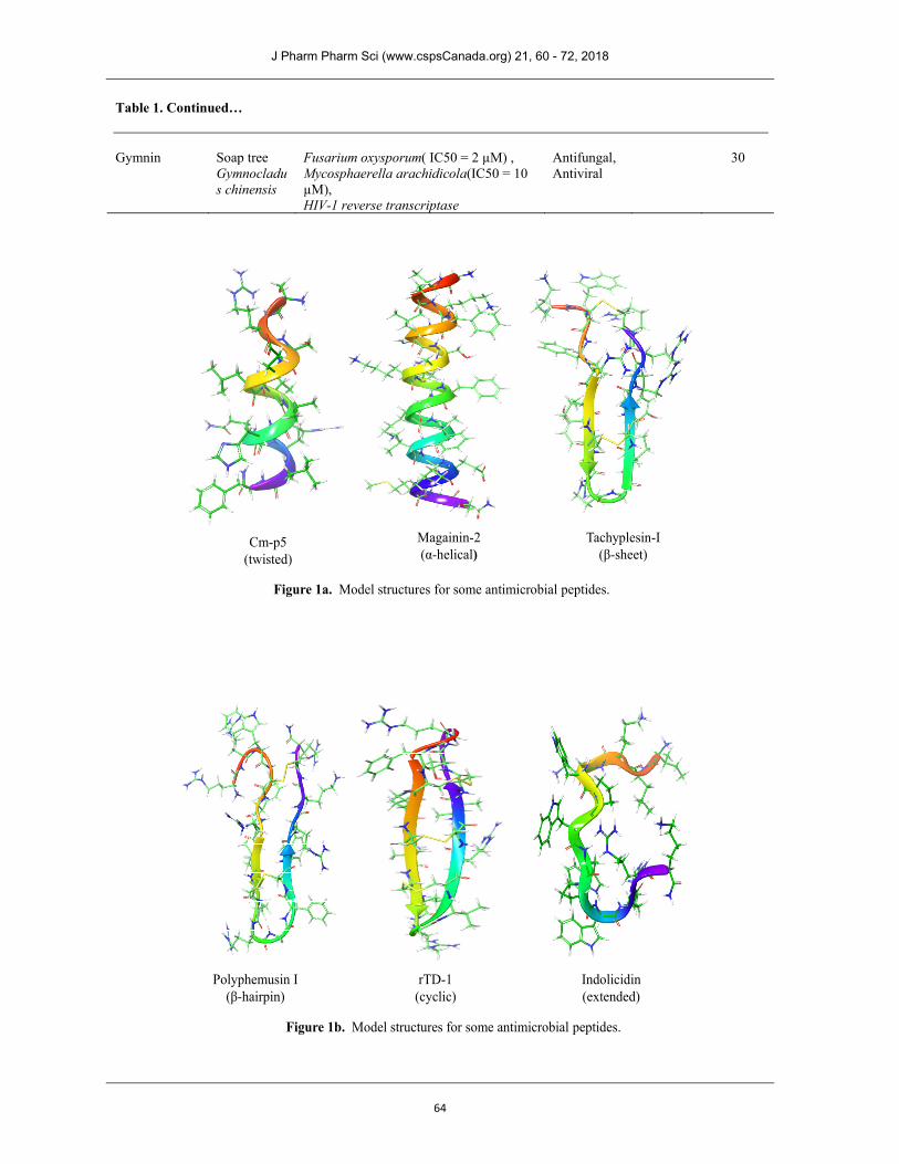
STRUCTURAL ASPECTS OF AMPs Several AMPs have been isolated, characterized and synthesized. Typically AMPs are cationic containing more often basic and hydrophobic amino acids that align on opposing faces facilitating their water solubility. Depending upon the length which is typically below 50 amino acid residues these peptides may possess α-helical, -sheet type or more complicated secondary structure (35). Examples of modelled structures for some of these peptides based largely on solution NMR data are given in Fig. 1. However, they may adopt variable structure at lipid interface (36-38) which allows them to penetrate through cell membrane. Wimley (39) has proposed an interfacial activity model based on interaction of AMPs with lipid membrane. Many AMPs possess disulfide linkages that stabilize their secondary structure (40). This structural diversity does not correlate with their antimicrobial activity. In fact few plausible mechanistic explanations have been put forward for their antimicrobial activity which are discussed in the following section. ________________________________________ Corresponding Author: Ruchi Omar and Arpita Yadav, Department of Chemistry, University Institute of Engineering and Technology, Chhatrapati Shahu Ji Maharaj University, Kanpur 208024, India. E-Mail: [email protected] & [email protected].

J Pharm Pharm Sci (www.cspsCanada.org) 21, 60 - 72, 2018
61
Table 1. Naturally occurring antimicrobial peptides AMP Source Antimicrobial activity Gram
nature Ref.
Target species Other properties
Aurein 1.2 Southern bell frog, Litoria raniformis
C. albicans, Candida glabrata, Candida krusei, Candida parapsilosis, Candida tropicalis (MICs: 16–128 µg/ml), Candida kefyr (MIC: 32–256 µg/ml )
Antifungal, Anticancer
2,3
Tigerinin 1 Indian bullfrog, Rana tigrina
Saccharomyces cerevisiae (MIC: 80 µg/ml), B. subtilis ( MIC = 30 µg/ml ), E. coli (MIC = 40 µg/ml )
Antibacterial, Antifungal
Gram+ve, Gram-ve
4
BMAP-27 Cow, Bos taurus
C. albicans, C. neoformans( MICs: 4–16 µM)
Antifungal 5
RTD-1 Rhesus macaque, Macaca mulatta
Candida albicans (MICs: 1μg/ml) Antifungal 6
Hepcidin-20
Human (liver), H. sapiens
Aspergillus niger (spores), A. fumigatus (spores)(MIC: complete inhibition at 20-40 μM)
Antibacterial, Antifungal
Gram+ve, Gram-ve
7,8
Tachyplesin -2
Southeast Asian horseshoe crab, Tachypleus tridentatus
Candida albicans M9 (MIC: 3.1μg/ml)
Antibacterial, Antifungal
Gram+ve, Gram-ve
9,10
Polyphemusin-1
American horseshoecrab, Limulus polyphemus
C. albicans (MIC: 1.26 µM) Antibacterial, Antifungal
Gram+ve, Gram-ve
10
Protegrin-1 Sus scrofa C. albicans In vitro MIC (3.0–60.0 μg/ml)
Antifungal, Antibacterial
11
Penetratin Fruit fly, Drosophila melanogaster
C. albicans (MIC: 100 µM; 95% growth inhibition at 50 µM) C. neoformans(MICs and MFCs: 25-125µM)
Antifungal 12
Vespid chemotactic peptide-5g
Vespine wasp(venom), Vespa magnifica
C. albicans ( MIC: 12.5 µg/ml)
Antibacterial, Antifungal
Gram+ve, Gram-ve
13
Ascaphin-8 American coastal frog, Ascaphus truei
C. albicans( MICs: 12–25 µM) Antibacterial, Antifungal
Gram+ve, Gram-ve
14
J Pharm Pharm Sci (www.cspsCanada.org) 21, 60 - 72, 2018
62
Table 1 Continued… Latarcin 3a Spider,
Lachesana tarabaevi
P. pastoris, S. cerevisiae (MIC: 20 µM)
Antibacterial, Antifungal
Gram+ve, Gram-ve
15
Gomesin Tarantula spider Acanthoscurria gomesiana
Alternaria brassicola, Aspergillus fumigatus, C. albicans, C. tropicalis, Cryptococcus neoformans, Fusarium culmorum, Fusarium oxysporum, Nectria haematococca, Neurospora crassa, S. cerevisiae, Tricoderma viridae, Tricophvton mentagrophytes (MICs: 0.15–6.25 µM) Beauveria bassina, C. glabrata (MICs: 12.5–25 µM)
Antibacterial, Antifungal, Antiparasitic
Gram+ve, Gram-ve
16
Jelleine-I Honeybee (royal jelly), Apis melliferia
Candida albicans (MIC: 2.5 µg/ml) Antibacterial, Antifungal
Gram+ve, Gram-ve
17
Decoralin Solitary eumenine wasp, Oreumenes decoratus (poison)
Candida albicans (MIC: 40 µM), S.saprophyticus ( MIC = 40 µM), B.thuringiensis ( MIC = 40 µM)
Antibacterial, Antifungal
Gram+ve, Gram-ve
18
Hylin-a1 Spotted tree frog Hypsiboas albopunctatus
S.aureus ATCC 25926 (MIC=8 µM), E.faecalis ATCC 29212 (MIC=16 µM), B.subtilis ATCC 19659 (MIC=8 µM. C.albicans ATCC 90028 (MIC=16.7 µM), C.krusei ATCC 6258 (MIC=16.7 µM), C.parapsilosis ATCC 22019 (MIC=67 µM), C.neoformans ATCC 90012 (MIC=33.5 µM).
Antibacterial, Antifungal
Gram+ve, Gram-ve
19
Ascalin Shallot Allium cepa var. aggregatum
B.cinerea, HIV-1 reverse transcriptase ( IC50 = 10 µM)
Antifungal, Antiviral
20
Sesquin Cowpea Vigna unguiculata subsp. sesquipedalis
Botrytis cinerea ( IC50 = 2.5 µM ), Fusarium oxysporum ( IC50 = 1.4 µM ), Mycosphaerella arachidicola ( IC50 = 0.15 µM ), Proteus vulgaris, Mycobacterium phlei , Bacillus megaterium, B. subtilis, HIV-1reverse transcriptase
Antibacterial, Antifungal, Antiviral
Gram+ve, Gram-ve
21
J Pharm Pharm Sci (www.cspsCanada.org) 21, 60 - 72, 2018
63
Table 1. Continued… Rondonin Spider
Acanthoscurria rondoniae
Trichosporon sp IOC 4569 ( MIC= 1.1 µM ), Candida albicans MDM8 ( MIC= 16.75 µM ), Candida krusei IOC 4559 ( MIC= 16.75 µM ), Candida glabrata IOC 4565 ( MIC= 8.37 µM ), Candida albicans IOC 4558 ( MIC= 8.37 µM ), Candida parapsilosis
Antifungal 22
OdG1 Yunnanfu frog Odorrana grahami
Escherichia Coli ( MIC = 4.68 µg/ml ), Staphylococcus aureus ( MIC = 9.37 µg/ml ), Bacillus subtilis ( MIC = 37.5 µg/ml ), Candida albicans ( MIC = 1.10 µg/ml )
Antibacterial, Antifungal
Gram+ve, Gram-ve
23
Alpha-MSH Human Homo sapiens
Staphylococcus aureus, Candida albicans
Antibacterial, Antifungal
Gram+ve 24
Mastoparan-S Giant African praying mantis Sphodromantis viridis
Escherichia coli ( MIC = 28.3 µg/ml ), Klebsiella pneumoniae ( MIC = 26.7 µg/ml ), Pseudomonas aeroginosa ( MIC = 24.2 µg/ml ), Bacillus subtilis ( MIC = 17.6 µg/ml ), Leuconostoc mesenteroides ( MIC = 19.8 µg/ml ), Bacillus cereus ( MIC = 15.1 µg/ml ), Aspergillus niger ( MIC = 24.6 µg/ml ), Aspergillus fumigates ( MIC = 19.3 µg/ml ), Candida albicans ( MIC = 20.4 µg/ml )
Antibacterial, Antifungal
Gram+ve, Gram-ve
25
Pantinin-1 Emperor scorpion Pandinus imperator
S.aureus(MIC=8 µM), B.magaterium(MIC=32µM), M.luteus(MIC=32µM), vancomycin-resistantEnterococci(MIC=14µM), E.cloacae(MIC=76µM), S.enterica(MIC=72µM) C.tropicalis(MIC=16µM)
Antibacterial, Antifungal
Gram+ve, Gram-ve
26
Chitinase Streptomyces venezuelae Streptomyces violaceus
Aspergillus niger, Alternaria alternata, H. sativum
Antifungal 27
Ranacyclin-E Edible frog Rana esculenta
S.lentus ( MIC = 7 µM), M.luteus ( MIC = 5 µM), C.tropicalis ( MIC = 7.4 µM), C.guillermondii ( MIC = 3.4 µM),
Antibacterial, Antifungal
Gram+ve, Gram-ve
28
Maximin H3 Giant fire-bellied toad Bombina maxima
Escherichia coli ATCC25922 ( MIC = 20 µg/ml ), Staphylococcus aureus ATCC2592 ( MIC = 10 µg/ml ), Bacillus pyocyaneus CMCCB1010 ( MIC = 20 µg/ml ), Candida albicans ATCC2002 ( MIC = 5 µg/ml )
Antibacterial, Antifungal
Gram+ve, Gram-ve
29
J Pharm Pharm Sci (www.cspsCanada.org) 21, 60 - 72, 2018
64
Magainin-2(α-helical)
Tachyplesin-I(β-sheet)
Cm-p5 (twisted)
Figure 1a. Model structures for some antimicrobial peptides.
Polyphemusin I(β-hairpin)
rTD-1(cyclic)
Indolicidin(extended)
Figure 1b. Model structures for some antimicrobial peptides.
Table 1. Continued… Gymnin Soap tree
Gymnocladus chinensis
Fusarium oxysporum( IC50 = 2 µM) , Mycosphaerella arachidicola(IC50 = 10 µM), HIV-1 reverse transcriptase
Antifungal, Antiviral
30

J Pharm Pharm Sci (www.cspsCanada.org) 21, 60 - 72, 2018
65
MECHANISTIC ASPECTS OF AMPs It is believed that being cationic in nature they interact with the microbial cell wall weakening it to allow seepage of extracellular ions resulting in bloating and eventual death of microbe (c.f. Fig. 2). In the last five years there have been continued efforts to understand and utilize the bacterial membrane disruptive ability of AMPS (41). Sharma et al (42) have described the formation of AMP-lined ion channel which modulate the membrane potential. An interesting computational investigation utilizing molecular dynamics simulation of pore formation has recently been reported (43). A flora derived AMP Snakin-2 was recently studied for its broad spectrum antimicrobial activity and its interaction with cell membrane was investigated by microscopy (44). Lee et al (41) have summarized the usage of biophysical techniques to probe interactions between AMPs and cell membrane.
Our lab has explored mechanistic aspects of properties of these AMPs at the molecular level. We have shown ion carriage characteristics of cyclic counterparts of these peptides (c.f. Fig. 3) (45). For the antifungal peptides another mode of action may be proposed based on the interaction of azole antifungals with 14α-demethylase enzyme active site (c.f. Fig. 4). Since, the antifungal peptides also possess ion affinity they may also interact with heme Fe required for 14α-demethylase activity thus inhibiting the formation of ergosterol an essential component of microbial cell wall. Complete mechanism of AMPs destroying the bacterial or fungal cell is still not known at the molecular level and research along these lines is currently being pursued in our lab. In our opinion the non membrane permeabilizing AMPs (46) may follow a mechanism utilizing ion interaction.
Cell membraneInteraction with membrane
Pore formationCell death
Antimicrobial peptides
Figure 2. Interaction of cationic AMPs with microbial cell wall.

J Pharm Pharm Sci (www.cspsCanada.org) 21, 60 - 72, 2018
66
Ion carriage through membrane
Cyclic peptide
Non cyclic peptide
Cell Membrane
Mg2+
Burst cell
Influx of ion resulting and bloating
Figure 3. Ion carriage characteristics of AMPs.
Azole antifungals
ErgosterolSynthetic pathway
Lanosterol 14-alpha demethylase
Ergosterol
Inhibition of 14-alpha demethylase
Toxic sterols
Figure 4. Inhibition of 14-alpha demethylase enzyme by AMPs.

J Pharm Pharm Sci (www.cspsCanada.org) 21, 60 - 72, 2018
67
Figure 5. Different self aggregated forms of synthetic AMPs.
[Ala]12
Tubular Aggregation
TEM images of
[WR]4
Nanoparticulate formed by a cyclic peptide
Film formation by [WR]4

J Pharm Pharm Sci (www.cspsCanada.org) 21, 60 - 72, 2018
68
Figure 5. Continued...
Figure 5. Different self aggregated forms of synthetic AMPs.
OTHER IMPORTANT PROPERTIES OF AMPs Self assemblage Some AMPs have the ability to self assemble in different forms either in presence of counterions or in absence of counterions (47). The self assembled forms are highly significant in different ways, for example, in drug delivery systems, as structural materials for different body implants and also in the bottom-up approach to understanding evolution of mankind (48). Different self assembled forms of some peptides are shown in Fig. 5.We have shown tubular structure formation by a completely hydrophobic cyclic peptide [Ala]12 (49, Fig. 5). Cyclic peptide [WR]4 and [WR]5 form nanoparticulates upto 50 nm in dimension (50, Fig. 5) which have been captured by TEM images and used to enhance delivery of small anticancer agents. A good hydrophilic lipophilic balance (HLB) is needed for nanoparticulates suitable for drug delivery. The same conclusion has been drawn by another recent study (51) to explain antimicrobial property of these peptides. Some AMPs are under pre-clinical studies for cancer treatment (52).
The magnitude of importance of understanding mechanistic issues of AMPs can only be glimpsed by noticing that cancer cells have recently been
shown to develop resistance to chemotherapeutic agents due to the presence of membrane associated multi drug resistant proteins such as P-glycoprotein (53). Cell penetration capability Cationic peptides have shown rapid cellular uptake. Researchers have made efforts to utilize this property by conjugating them with nuclear targeted drugs to enhance the cellular uptake of the drug. Covalent linkage is not always a necessity as even co-administration of a cell penetrating peptide (CPP) has shown increased efficacy of anticancer agents (54). Some work along these lines has been done in our lab to show the drug delivery properties of these peptides at molecular level utilizing computational techniques (55). A recent review article (56) discusses covalently linked different conjugates of AMPs for target selectivity or enhanced therapeutic usage. Many pre-clinical and clinical trials are being performed to evaluate the performance of CPPs in drug delivery. Bolhassani et al (57) have discussed the in vitro and in vivo delivery efficiencies of CPPs. CPPs are the non-membrane permeabilizing category of AMPs which translocate through the membrane without disrupting its integrity (58).
Self aggregation of non cyclic RADA peptide

J Pharm Pharm Sci (www.cspsCanada.org) 21, 60 - 72, 2018
69
Tashima (59) has recently discussed the non-invasive intracellular substance delivery (including macromolecules which are otherwise impermeable) by CPPs. Anti HIV activity Recent rise in HIV patients has compelled researchers to revisit AMPs for two important reasons: to overcome ever-increasing cases of antibiotic resistance and to overcome the risk of internal fungal infection in HIV patients due to their reduced immunity. It is fascinating to understand how nature has devised these systems to protect flora and fauna from microbial activity. Apart from antimicrobial activity recent studies have shown anti HIV activity of these peptides (60). It is not clear from the literature whether the peptides themselves show anti HIV activity or simply enhance the delivery of anti HIV agents. Recent computer aided modelling and docking studies from our lab have shown interactions between AMPs and HIV ssRNA primer binding site through interactions other than base pair-base pair type (c.f. Fig. 6). Our studies have shown HIV inhibitory capability of these peptides (61) that can be exploited to design a non-toxic drug with anti HIV and antimicrobial activity.
The question now clearly is that with many interesting and pharmacologically important properties at hand, AMPs to date have not been able to capture the drug market though the researchers have visited and revisited these systems a number of times in search of safe
pharmaceuticals. We must therefore put in our best efforts to overcome pharmacodynamic and related ADME issues by design of peptidomimetic compounds. To be able to design peptidomimetic compounds complete structure-function elucidation along with mechanistic details are required. Thimmegowda and Yeldur (62) have correlated the structure function of arginine-rich proteins with their biological significance. In recent years, we have designed peptidomimetic compounds with complete or partial artificial backbone for different pharmaceutical applications (61,63-66). We hope that synthetic organic chemists and pharmaceutical scientists shall come together to synthesize and test these and similar compounds for their ADME propertiesand prospective druggability features.
Some recent synthetic efforts in this direction are summarized here. Molchanova et al (67) describe α-peptoids, β-peptoids and hybrid peptidomimetic compounds for their antibacterial activity and enhanced stability in body. Cheap synthesis on large scale and bioavailabity have been the biggest hurdle in development of these compounds as commercial drugs. Sgolastra et al have described their efforts towards building synthetic mimics of AMPs (68) using unique molecular scaffolds and guanidinium-rich side chains.
However, much remains to be done at the molecular level for significant advancement in the area of AMPs and their druggable future.
Figure 6. Single stranded viral RNA template inhibition by antimicrobial peptide through interactions other than base pair-base pair type.

J Pharm Pharm Sci (www.cspsCanada.org) 21, 60 - 72, 2018
70
FUTURE DIRECTIVES This article reiterates the multiple beneficial properties of naturally occurring antimicrobial peptides that can be harnessed and tuned as per our needs. Proper utilization in pharmaceutical industry is only possible with mechanistic understanding of their mode of action at the molecular level. Design of peptidomimetic compounds based on molecular level understanding is desired. Such efforts should be welcomed by researchers in the field to eventually lead to nontoxic drugs for fatal diseases and drug delivery systems for enhanced bioavailability. ACKNOWLEDGEMENTS The authors gratefully acknowledge financial support from Science and Engineering Research Board (SERB), New Delhi (Project no. EMR/2016/000769). Ms. Ruchi Omar is thankful to SERB for Junior research fellowship. The authors are also grateful to Chhatrapati Shahu Ji Maharaj University, Kanpur, India for infrastructural support. REFERENCES 1. Pushpanathan M, Gunasekaran P, Rajendhran
J.Antimicrobial peptides: Versatile biological properties. Intl. J. Peptides. 2013; article ID 675391. DOI: 10.1155/2013/675391.
2. Rozek T, Wegener KL, Bowie JH, Olver IN, Carver JA, Wallace JC, Tyler MJ. The antibiotic and anticancer active aurein peptides from the Australian Bell Frogs Litoria aurea and Litoria raniformis the solution structure of aurein 1.2. Eur. J. Biochem. 2000; 267:5330-5341.DOI: 10.1046/j.1432-1327.2000.01536.x.
3. Kamysz W, Nadolski P, Kedzia A, Cirioni O, Barchiesi F, Giacometti A, Scalise G, Lukasiak J, Okroj M. In vitro activity of synthetic antimicrobial peptides against Candida. Pol. J. Microbiol. 2006 ; 55:303-307. PMID :17416067.
4. Sai KP, Jagannadham MV, Vairamani V, Raju NP, Devi AS, Nagaraj R, Sitaram N. Tigerinins: novel antimicrobial peptides from the Indian frog Rana tigerina. J. Biol. Chem. 2001; 276:2701-2707. DOI: 10.1128/AAC.46.7.2279-2283.2002.
5. Skerlavaj B, Gennaro R, Bagella L, Merluzzi L, Risso A, Zanetti M. Biological characterisation of two novel cathelicidin-derived peptides and identification of structural requirements for their antimicrobial and cell lytic activities. J. Biol. Chem. 1996; 271:28375-28381.DOI: 10.1074/jbc.271.45.28375.
6. Matejuk A, Leng Q, Bequm MD, Woodle MC, Scaira P, Chou ST, Mixson AJ. Peptide based
antifungal therapies against emerging infections. Drugs future. 2010; 35, 197. PMID:20495663.
7. Hunter HN, Fulton DB, Ganz T, Vogel HJ. The solution structure of human Hepcidin, a peptide hormone with antimicrobial activity that is involved in iron uptake and hereditary Hemochromatosis. J. Biol. Chem. 2002; 277:37597-37603. DOI: 10.1074/jbc.M205305200.
8. Park CH, Valore EV, Waring AJ, Ganz T. Hepcidin, a urinary antimicrobial peptide synthesized in the liver. J. Biol. Chem. 2001;276:7806-7810. DOI: 10.1074/jbc.M008922200.
9. Katsu, T, Nakao S, Iwanaga S. Mode of action of an antimicrobial peptide, tachyplesin I, on biomembranes. Biol. Pharm. Bull. 1993; 16:178-181. DOI: 10.1248/bpb.16.178.
10. Miyata T, Tokunaga F, Yoneya T, Yoshikawa K., Iwanag, S, Niwa M, Takao T, Shimonishi Y. Antimicrobial peptides, isolated from Horseshoe crab hemocytes, Tachyplesin II, and Polyphemusins I and II: chemical structures and biological activity. J. Biochem. 1989; 106:663-668. DOI:10.1093/oxfordjournals.jbchem.a122913.
11. De Lucca AJ.; Walsh TJ.; Antifungal Peptides: Novel Therapeutic Compounds against Emerging Pathogens. Antimicrob Agents Chemother. 1999; 43:1–11. PMID:9869556.
12. Masma MF, Rodriguez AM, Raimondi M, Zacchino SA, Luiten PG, Somlai C, Kortvelyesi T, Penke B, Enriz RD. Penetratin and derivatives acting as antifungal agents. Eur. J. Med. Chem. 2009; 44:212-228. DOI: 10.1016/j.ejmech.2008.02.019.
13. Xu X, Li J, Lu Q, Yang H, Zhang Y, Lai R. Two families of antimicrobialpeptides from wasp (Vespa magnifica) venom.Toxicon. 2006; 47:249-253. DOI:10.1016/j.toxicon.2005.10.015.
14. Conlon JM, Galadari S, Raza H, Condamine E. Design of potent, non-toxic antimicrobial agents based upon the naturally occurring frog skin peptides, ascaphin-8 and peptide XT-7.Chem. Biol. Drug Des. 2008; 72:58-64. DOI: 10.1111/j.1747-0285.2008.00671.x.
15. Kozlov SA, Vassilevski AA, Feofanov AV, Surovoy AY, Karpunin DV, Grishin EV. Latarcins, antimicrobial and cytolytic peptides from the venom of the spider Lachesana tarabaevi (Zodariidae) that exemplify biomolecular diversity. J. Biol. Che. 2006; 281:20983-20992. DOI: 10.1074/jbc.M602168200.
16. Silva PI Jr, Daffre S, Bulet P. Isolation and characterization of Gomesin, an 18-residue cysteine-rich defense peptide from the spider Acanthoscurria gomesiana hemocytes with sequence similarities to horseshoe crab antimicrobial peptides of the Tachyplesin family. J. Biol. Chem. 2000; 275:33464-33470. DOI: 10.1074/jbc.M001491200.

J Pharm Pharm Sci (www.cspsCanada.org) 21, 60 - 72, 2018
71
17. Fontana R, Mendes MA, De Souza, BM, Konno K., Cesar LM, Malaspina O, Palma MS. Jelleines: a family of antimicrobial peptides from the Royal Jelly of honeybees (Apis mellifera). Peptides. 2004; 25: 919-928. DOI: 10.1016/j.peptides.2004.03.016.
18. Konno K, Rangel M, Oliveira JS, Dos Santos Cabrera MP, Fontana R, Hirata IY, Hide I, Nakata Y, Mori K., Kawano M, Fuchino H, Sekita S, Neto JR. Decoralin, a novel linear cationic alpha-helical peptide from the venom of the solitary eumenine wasp Oreumenes decoratus. Peptides. 2007; 28:2320-2327. DOI: 10.1016/j.peptides.2007.09.017.
19. Castro MS, Ferreira TC, Cilli EM, Crusca E Jr, Mendes-Giannini MJ, Sebben A, Ricart CA, Sousa MV, Fontes W. Hylin a1, the first cytolytic peptide isolated from the arboreal South American frog Hypsiboas albopunctatus ('spotted treefrog'). Peptides. 2009; 30:291-296. DOI: 10.1016/j.peptides.2008.11.003.
20. Wang HX, Ng TB. Ascalin, a new anti-fungal peptide with human immunodeficiency virus type 1 reverse transcriptase-inhibiting activity from shallot bulbs. Peptides. 2002; 23:1025-1029. DOI: 10.1016/S0196-9781(02)00032-3.
21. Wong JH, Ng TB. Sesquin, a potent defensin-like antimicrobial peptide from ground beans with inhibitory activities toward tumor cells and HIV-1 reverse transcriptase. Peptides. 2005; 26:1120-1126. DOI: 10.1016/j.peptides.2005.01.003.
22. Riciluca KC, Sayegh RS, Melo RL, Silva PI Jr. Rondonin an antifungal peptide from spider (Acanthoscurria rondoniae) haemolymph. Results Immunol.2012;2:66-71. DOI: 10.1016/j.rinim.2012.03.001.
23. Li J, Xu X, Xu C, Zhou W, Zhang K, Yu H, Zhang Y, Zheng Y, Rees HH, Lai R, Yang D, Wu J. Anti-infection peptidomics of amphibian skin. Mol Cell Proteomics. 2007; 6:882-94. DOI: 10.1074/mcp.M600334-MCP200.
24. Cutuli M, Cristiani S, Lipton JM, Catania A. Antimicrobial effects of alpha-MSH peptides. J Leukoc Biol. 2000; 67:233-9. XP002931058, ISSN: 0741-5400.
25. Zare-Zardini H, Taheri-Kafrani A, Ordooei M, Ebrahimi L, Tolueinia B, Soleimanizadeh M. Identification and biochemical characterization of a new antibacterial and antifungal peptide derived from the insect Sphodromantis viridis. Biochemistry (Mosc). 2015; 80:433-40. DOI: 10.1134/S0006297915040069.
26. Zeng XC, Zhou L, Shi W, Luo X, Zhang L, Nie Y, Wang J, Wu S, Cao B, Cao H. Three new antimicrobial peptides from the scorpion Pandinus imperator. Peptides. 2013; 45:28-34. DOI: 10.1016/j.peptides.2013.03.026.
27. Mukherjee G, Sen SK. Purification, characterization, and antifungal activity of chitinase from Streptomyces venezuelae P10. Curr Microbiol. 2006; 53: 265-9. DOI: 10.1007/s00284-005-0412-4.
28. Mangoni ML, Papo N, Mignogna G, Andreu D, Shai Y, Barra D, Simmaco M. Ranacyclins, a new family of short cyclic antimicrobial peptides: biological function, mode of action, and parameters involved in target specificity. Biochemistry. 2003; 42:14023-35. DOI: 10.1021/bi034521l.
29. Lai R, Zheng YT, Shen JH, Liu GJ, Liu H, Lee WH, Tang SZ, Zhang Y. Antimicrobial peptides from skin secretions of Chinese red belly toad Bombina maxima. Peptides. 2002; 23:427-35. DOI: 10.1016/S0196-9781(01)00641-6.
30. Wong JH, Ng TB. Gymnin, a potent defensin-like antifungal peptide from the Yunnan bean (Gymnocladus chinensis Baill). Peptides. 2003; 24:963-8. DOI: 10.1016/S0196-9781(03)00192-X.
31. Desbois AP, Tschorner D, Cotte PJ. Survey of small antifungal peptides with chemotherapeutic potential. Current Phar. Biotechnol. 2011; 12:1263-1291.DOI: 10.2174/138920111796117265.
32. Ostaff MJ, Stange EF, Wehkamp J. Antimicrobial peptides and gut microbiota in homeostasis and pathology. EMBO Mol. Med. 2013; 5(10):1465-1483. DOI:10.1002/emmm.201201773.
33. Eade CR, Wood MP, Cole AM. Mechanisms and modifications of naturally occurring host defense peptides for anti HIV microbicide development. Current HIV research. 2012; 10:61-72. DOI: 10.2174/157016212799304580.
34. Di L. Strategic approaches to optimizing peptide ADME properties. AAPS J. 2015; 17(1):134-143.DOI: 10.1208/s12248-014-9687-3.
35. Padovan L, Scocchi M, Tossi A. Structural aspecs of plant antimicrobial peptides. Curr. Protein Pept. Sci. 2010; 11:210-219. DOI: 10.2174/138920310791112093.
36. Leontiadou H, Mark AE, Marrink SJ. Antimicrobial peptides in action. J. Am. Chem. Soc. 2006; 128(37):12156-12161. DOI: 10.1021/ja062927q.
37. Banerjee A, Yadav A, Sonker M, Bhaskar D, Patel PB, Sachan S. Behavior of biosurfactant Iturin A at liquid-liquid interface. Asian J. Chem. 2014; 26(21):7191-7195. DOI:10.14233.ajchem.2014.16559.
38. Salditt T, Li C, Spaar A. Structure of antimicrobial peptides and lipid membranes probed by interface-sensitive X-ray scattering. Biochim. Biophys. Acta. 2006; 1758:1483-1498. DOI: 10.1016/j.bbamem. 2006.08.002.
39. Wimley WC. Describing the mechanism of antimicrobial peptide action with the interfacial activity model. ACS Chem. Biol. 2010; 5(10):905-917. DOI: 10.1021/cb1001558.
40. Wang Y, Croh SY, Kuczera K. Molecular dynamics study of disulfide bond influence on properties of an RGD peptide. J. Pept. Res. 1999; 53(2):188-200. DOI: 10.1034/j.1399-3011.1999.00029.x
41. Lee TH, Hall KN, Aguilar M-I, Antimicrobial peptide structure and mechanism of action: A focus

J Pharm Pharm Sci (www.cspsCanada.org) 21, 60 - 72, 2018
72
on the role of membrane structure. Curr. Topics Med.Chem. 2016; 16(1):25-39. DOI: 10.2174/1568026615666150703121700.
42. Sharma S, Sahoo N, Bhunia A. Antimicrobial peptides and their pore/ion channel properties in neutralization of pathogenic microbes. Curr.Topics Med. Chem. 2016; 16(1):46-53. DOI: 10.2174/1568026615666150703115454.
43. Lipkin R, Lazaridis T. Computational studies of peptide-induced membrane pore formation.Philosophical Trans. B. 2017; 372:20160219. DOI: 10.1098/rstb.2016.0219.
44. Herbel V, Wink M. Mode of action and membrane specificity of the antimicrobial peptide snakin-2. Peer J.2016; 4:e1987. DOI: 10.7717/peerj.1987.
45. Banerjee A, Yadav A. Mechanistic aspects of transport antibiotics. Eur. J. Med. Chem. 2009; 45:1799-1804. DOI:10.1016/j.ejmech.2010.01.012.
46. Scocchi M, Mardirossian M, Runti G, Benincasa M. Non-Membrane Permeabilizing Modes of Action of Antimicrobial Peptides on Bacteria. Curr Top Med Chem. 2016; 16(1):76-88. DOI: 10.2174/1568026615666150703121009.
47. Tian X, Sun F, Zhou X-R, Luo SZ, Chen L. Role of peptide self assembly in antimicrobial peptides. J. Pept. Sci. 2015; 21:530-539. DOI: 10.1002/psc.2788.
48. Zhang S. Building from the bottom up. Materials Today. 2003; 6(5):20-27. DOI:10.1016/S1369-7021(03)00530-3.
49. Banerjee A, Yadav A, Self assembling cyclic system as drug carriers. Appl. Nano se. 2013; 3:515-528. DOI: 10.1007/s13204-012-0154-0.
50. Mandal D, Tiwari RK, Shirazi AN, Ye G, Banerjee A, Yadav A, Parang K. Self assembled surfactant cyclic peptide nanostructures as stabilizing agents. Soft Matter. 2013; 9(39):9465-9475, DOI: 10.1039/C3SM50764E.
51. Yin LM, Edwards MA, Li J, Yip CM, Deber CM. Role of hydrophobicity and charge distribution of cationic antimicrobial peptides in peptide membrane interactions. J. Biol. Chem. 2012; 287(10): 7738-7745. DOI: 10.1074/jbc.M111.303602.
52. Roudi R, Syn NL, Roudbary M. Antimicrobial peptides as biologic and immunotherapeutic agents against cancer: A comprehensive overview. Frontiers Immun. 2017; 8: article 1320. DOI: 10.3389/fimmu.2017.01320.
53. Swithenbank L, Morgen C. The role of antimicrobial peptides in lung cancer therapy. J. Antimicrob. Agents. 2017; 3(1):134. DOI: 10.4172/2472-1212.1000134.
54. Shirazi AN, Tiwari RK, Oh D, Banerjee A, Yadav A, Parang K. Efficient delivery of cell impermeable phosphopeptides by a cyclic peptide amphiphile containing tryptophan and arginine. Mol. Pharmaceutics. 2013; 10:2008-2020. DOI: 10.1021/mp400046u.
55. Banerjee A, Sayeh N, Shirazi AN, Tiwari R, Parang K, Yadav A. Arginine-rich cyclic peptides enhance cellular delivery of anticancer agents:Molecular insights. Lett. Drug Des. Discov. 2016;13(7): 591-604. DOI: 10.2174/1570180813999160429113034.
56. Reinhardt A, Neundrof I. Design and application of antimicrobial peptide conjugates. Int. J. Mol. Sci. 2016; 17:701. DOI: 10.3390/ijms17050701.
57. Bolhassani A, Jafarzade BS, Mardani G. In vitro and in vivo delivery of therapeutic proteins using cell penetrating peptides.Peptides, 2017; 87:50-63. DOI: 10.1016/j.peptides.2016.11.011.
58. Sani M-A, Separovic F. How membrane-active peptides get into lipid membranes. Acc. Chem. Res. 2016; 49(6):1130-1138. DOI: 10.1021/acs,accounts.6b00074.
59. Tashima T. Intelligent substance delivery into cells using cell-penetrating pedptides. Bioorg. Med. Chem. Lett. 2017; 27:121-130. DOI: 10.1016/j.bmcl.2016.11.083.
60. Wang G. Natural antimicrobial peptides as promising anti-HIV candidates. Curr. Top. Pept. Protein Res. 2012; 13:93-110. PMCID: PMC4730921.
61. Omar R, Yadav A. A mechanistic study of anti-HIV activities of antifungal peptides. Can. J. Chem. 2017;95:633-640. DOI: 10.1139/cjc-2017-0046.
62. Thimmegowda C, Yeldur PV. Occurrence, functions and biological significance of arginine rich -proteins. Curr. Protein Pept. Sci. 2016; 17(5):507-516. DOI: 10.2174/1389203717666151201192348.
63. Yadav A, Sonker M. Perspectives in designing anti aggregation agents as Alzheimer disease drugs. Eur. J. Med. Chem. 2009; 44:3866-3873. DOI: 10.1016/j.ejmech.2009.04.013.
64. Banerjee A, Yadav A. Cyclic peptidomimetic lead compounds to reduce neurotoxicity and associated oxidative stress in Alzheimer disease. Intl. J. Biomed. Sci. 2010; 6(3):216-224. PMCID: PMC3615259.
65. Banerjee A, Yadav A. In silico design of a peptidomimetic carrier for levodopa. Intl. J.Biomed. Sci. 2011; 7(1):44-50. PMCID:PMC3614811.
66. Banerjee A, Patel PB, Beni Y, Shirazi AN, Parang K, Yadav A. Biocompatible, biodegradable peptides for heavy metal toxicity removal. J. Appl. Chem. Sci. Intl. (JACSI) 2015; 4(2):144-153. ISSN No. : 2395-3713.
67. Molchanova N, Hausen PR, Franzyle H. Advances in development of antimicrobial peptidomimetics as potential drugs. Molecules. 2017; 22:1430. DOI: 10.3390/molecules22091430.
68. Sgolastra F, deRonde BM, Sarapas JM, Som A, Tew GN. Designing mimics of membrane active proteins. Acc. Chem. Res. 2013; 46(12):2977-2987. DOI: 10.1021/ar400066v.