Embed Size (px)
Citation preview
Cationic fluorine-containing amphiphilic graft copolymers as DNA carriers
Sheng-Dong Xiong a, Ling Li a, Jiang Jiang b, Li-Ping Tong b, Shuilin Wu a,b, Zu-Shun Xu a,b,**, Paul K. Chu b,*
a Ministry-of-Education Key Laboratory for the Green Preparation and Application of Functional Materials, Hubei University, Wuhan 430062, Chinab Department of Physics & Materials Science, City University of Hong Kong, Tat Chee Aenue, Kowloon, Hong Kong, China
a r t i c l e i n f o
Article history:Received 24 November 2009Accepted 3 December 2009Available online 19 January 2010
Keywords:FluorocarbonGraft copolymerGene vectorMicelle
a b s t r a c t
A series of cationic fluorine-containing amphiphilic graft copolymers P(HFMA-St-MOTAC)-g-PEGcomprising poly(hexafluorobutyl methacrylate) (PHFMA) poly(methacryl oxyethyl trimethylammoniumchloride) (PMOTAC) polystyrene (PSt) backbones and poly(ethylene glycol) (PEG) side chains aresynthesized as a type of non-viral gene vector. The copolymers self-assemble into spherical micelles inthe aqueous media and turbidity and cytotoxicity measurements show that those micelles have excellentdispersive stability and low cytotoxicity. The interactions between the copolymers and calf-thymus DNAare studied by fluorescence spectroscopy and viscosity. The former discloses electrostatic interaction,hydrophobic interaction, and hydrogen bonding in the copolymer/DNA system, whereas the latterindicates that these graft copolymers can bind DNA via the electrostatic and classical intercalationmodes. The DNA-binding capacity determined by the gel retardation assay and UV–visible spectropho-tometry shows that the copolymers have good binding capacity to DNA and a high charge density orHFMA content in the copolymers bode well for DNA-binding. Transmission electron microscopy, photoncorrelation spectroscopy, and zeta potential data reveal that stable colloidal complexes (particles) canform easily between the copolymer micelles and DNA. Our results suggest that the copolymers area promising non-viral vector in a gene delivery system.
� 2009 Elsevier Ltd. All rights reserved.
1. Introduction
In recent years, gene therapy has attracted much attention forits pharmaceutical applications in treating chronic diseases,immunodeficiency, genetic disorders and cancer [1–3]. There aretwo categories of gene delivery vectors: viral and non-viral vectors[4,5]. Viral gene vectors have high transfection efficiency but sufferfrom many disadvantages such as risks of strong immunogenicity,inflammatory response, recombination, and carcinogenicity [6,7].Fast progress in clinical gene therapy has significantly motivatedthe development of safe and efficient non-viral gene deliverysystems which may substitute for viral vectors which still possessinherent safety problems [8,9]. Cationic lipids and cationic poly-mers are one of the most widely used and extensively studied non-viral vectors [10,11]. Compared to cationic lipids, cationic polymersare stable, easy to manipulate, easy to self-assemble with DNA, andmore economical [12].
One of the major drawbacks of cationic polymers non-viral vectorbased gene delivery is the toxicity related to the positive charge ofpolymers [13] such as polyethylenimine (PEI) [8,12] and trimethylchitosan (TMC) [14]. In addition, the strong electrostatic interactionbetween cationic polymer and DNA deters gene dissociation from itscarrier inside cells impeding the access of RNA polymerase to DNA.Consequently, the gene expression level is limited and the highcharge density may combine membranes and destroy cell metabo-lism [15]. To circumvent the problems described above, one rationalway is to graft hydrophilic or hydrophobic side chains to cationicpolymer backbones to form amphiphilic copolymers [11,14]. Theseamphiphilic copolymers self-assemble into a nanosize and core–shellmicelle architecture in selective solvents. Compared to water-solublepolycations, such self-assembled micellar nanoparticles as non-viralvector have several advantages. First of all, they are able to load DNAand hydrophobic anti-cancer drugs simultaneously to offer potentialsynergistic effect in cancer therapy [16]. Secondly, instead of mixingwater-soluble polycations and DNA to form complexes, which mayresult in the formation of uncontrollable large particles, micellarnanoparticles with positive charge allow nucleic acid loading afternanoparticle formation and have long circulation time in thebloodstream [17]. This is believed to benefit the construction of size-controllable and monodispersed nucleic acid loaded nanoparticleswhich may display unique advantages in vivo. Thirdly, the nanoscaled
* Corresponding author. Tel.: þ852 34427724; fax: þ852 27889549.** Corresponding author. Tel.: þ86 27 61120608; fax: þ86 27 88665610.
E-mail addresses: [email protected] (Z.-S. Xu), [email protected] (P.K.Chu).
Contents lists available at ScienceDirect
Biomaterials
journal homepage: www.elsevier .com/locate/biomateria ls
0142-9612/$ – see front matter � 2009 Elsevier Ltd. All rights reserved.doi:10.1016/j.biomaterials.2009.12.014
Biomaterials 31 (2010) 2673–2685
micelles (<200 nm) reduce non-selective uptake by the reticuloen-dothelial system (RES) and exhibit enhanced permeability andretention effects (EPR effect) at solid tumor tissue sites [18,19]. Inaddition, this preparation method can be upscaled to address thelarge quantity required by therapeutic applications. Several cationicgraft or block polymers micelles such as poly (methacryl oxyethyltrimethylammonium chloride)-graft-poly-(oxyethylene) (PMOTAC-g-POE) [15], trimethyl chitosan-g-poly(N-isopropylacrylamide)(TMC-g-PNIPAAm) [14], poly(L-lysine)-graft-polyethylene glycol(PLL-g-PEG) [20], polyethylenimine -graft-polyethylene glycol (PEI-g-PEG) [21], folate-polyethylenimine-block-poly(ethylene glycol)(FOL-PEI-b-PEG) [12] have been used as non-viral gene vectors. Thehydrophobic force is the main one forming the micelles in anaqueous solution. However, because the hydrophobic force resultingfrom the hydrophobic chains of the amphiphilic copolymers is weak,the stability of the micelles is not adequate in many applications [22].These unstable micelles are thus not suitable for compressing DNAinto small spherical particles [23]. Big particles cannot be easilyendocytosed by cells leading to low transfection efficiency for DNA[8]. Therefore, the dispersive stability of micelles is another bottle-neck that needs to be circumvented. In this respect, several ways tostabilize the micelles including shell crosslinking of micelles havebeen investigated [24,25]. However, these approaches may adverselyaffect the micellar properties leading to low delivery efficiency.
Owing to the unique characteristics of fluoropolymers such ashigh hydrophobicity, high thermal and mechanical stability, gas-dissolving capacity, high fluidity, low dielectric constants, oil-andwater-repellency, and very interesting surface properties [26–28],attention has recently been paid to amphiphilic copolymers withfluorocarbon–hydrocarbon hybrid architectures. Among thesecharacteristics, the most intriguing are the oleo- and hydropho-bicity and high fluidity of fluorine-containing polymers. They havebeen studied from the perspective of biomedical applications, forinstance, blood substitutes, gas carriers, bioconversion, extraction,and so forth and cannot be achieved by non-fluorinated copoly-mers [29–32]. Furthermore, because of the oleo- and hydropho-bicity of fluorine-containing amphiphilic copolymers, they havevery low critical micelle concentrations (CMC) compared to otheramphiphilic copolymers [27]. Hence, they have a strong tendencyto self-aggregate into stable, well-organized micelles, and thesemicelles may have unique advantages in drug delivery becausethey can transmit both hydrophobic and fluorophilic molecules atthe same time to the target sites due to the high fluidity, hydro-phobicity, and water- and oil-repellency of fluoropolymers[33,34].
Poly(ethylene glycol) (PEG) is widely used as the hydrophilicside chains in amphiphilic graft copolymers. It is a low costcommercial product that possesses some unique and outstandingproperties such as hydrophilicity, biocompatibility, nontoxicity,lack of immunogenicity, metal complexing ability, as well as solu-bility in water and organic solvents. Recently, PEG has been widelyapplied in gene delivery systems. The presence of PEG on thesurface of gene vectors can enhance biocompatibility, reducecytotoxicity, and protect DNA from degradation [12,35,36].
There have been few reports on the use of self-assembledcationic fluorine-containing amphiphilic copolymer micelles inDNA delivery. The present study aims at the synthesis of cationicfluorine-containing amphiphilic copolymers P(HFMA-St-MOTAC)-g-PEG with hydrophobic poly(hexafluorobutyl methacrylate)(PHFMA), poly(methacryl oxyethyl trimethylammonium chloride)(PMOTAC), polystyrene backbones and poly(ethylene glycol) (PEG)side chains. Because of the hydrophobic characteristic of the fluo-rinated segment which induces the micellar core formation andstabilizes the nanoparticles, the amphiphilic graft copolymer hasa good and stable self-assembled morphology in aqueous media.
Meanwhile, on account of the positively charged MOTAC segment,it serves as a DNA-binding site, and hydrophilic poly(ethyleneglycol) (PEG) can also potentially protect the DNA and extend bloodcirculation in systemic administration. These unique tri-layeredcationic fluorine-containing amphiphilic graft copolymer micellesare used as a non-viral gene vector. The dispersive stability of thesemicelles in aqueous media is evaluated and the in vitro cytotoxicityof the resulting copolymers is examined. The physiochemicalcharacteristics of the copolymer/DNA complexes are analyzed byfluorescence, viscosity, agarose gel electrophoresis, UV spectro-photometry, particle size, transmission electron microscopy, andzeta potential measurements.
2. Materials and methods
2.1. Materials
2,2,3,4,4,4-Hexafluorobutyl methacrylate (HFMA) was purchased from XeogiaFluorine–Silicon Chemical Company (Harbin, China, chemical purity) and distilledunder reduced pressure before use. Methoxy poly(ethylene glycol) (MPEG)(average molecular weight of 5000 g/mol) was obtained from Aldrich and puri-fied by vacuum-drying at room temperature for 24 h before use. An aqueoussolution (75 wt%) of methacryl oxyethyl trimethylammonium chloride (MOTAC)was purchased from Aldrich and used as received. Tetrahydrofuran (THF)purchased from Shanghai Chemical Reagents Co. (Shanghai, China, chemicalpurity) serving as the solvent was initially dried over potassium hydroxide atleast overnight and then refluxed over sodium wire for 3 days before use.Analytical grade 2,20-azobisisobutyronitrile (AIBN) was purified by recrystalliza-tion in ethanol. Sodium hydride (NaH, Nacalai, Kyoto, Japan) and p-Chlor-omethylstyrene (CMSt) supplied by Acros Organics (>95%, Belgium) were usedwithout further purification.
Dulbecco’s modified Eagle medium (DMEM) was purchased from InvitrogenCorp. Fetal bovine serum (FBS) was purchased from Hyclone. MTT was purchasedfrom Sigma (St. Louis, MO, USA). Two different types of DNAs, pEGFP-C1 plasmid (gelretardation assay experiments), and calf-thymus DNA (all other experiments) wereused in the study. Calf-thymus DNA was purchased from Sigma (St. Louis, MO, USA).The stock solution of calf-thymus DNA was prepared by dissolving DNA in doublydistilled water at 0–4 �C. Plasmid pEGFPC1 encoding a red-shifted variant of wild-type green fluorescent protein (GFP) was purchased from Clontech, Mountain View,CA, USA. pEGFP-C1 plasmids were amplified in the Luria–Bertani medium at 37 �Covernight at 250 rpm. Then the plasmids were purified by means of EndoFreeplasmid purification. The purified plasmid was diluted by tris–EDTA buffer solutionand stored at �20 �C. All other reagents and solvents were used as received withoutfurther purification. Distilled water was used in all the preparation and character-ization processes.
2.2. Synthesis of P(HFMA-St-MOTAC)-g-PEG
2.2.1. Synthesis of SPEG macromonomersMPEG (10.000 g, 2 mmol) was dissolved in dry THF (100 mL) and NaH
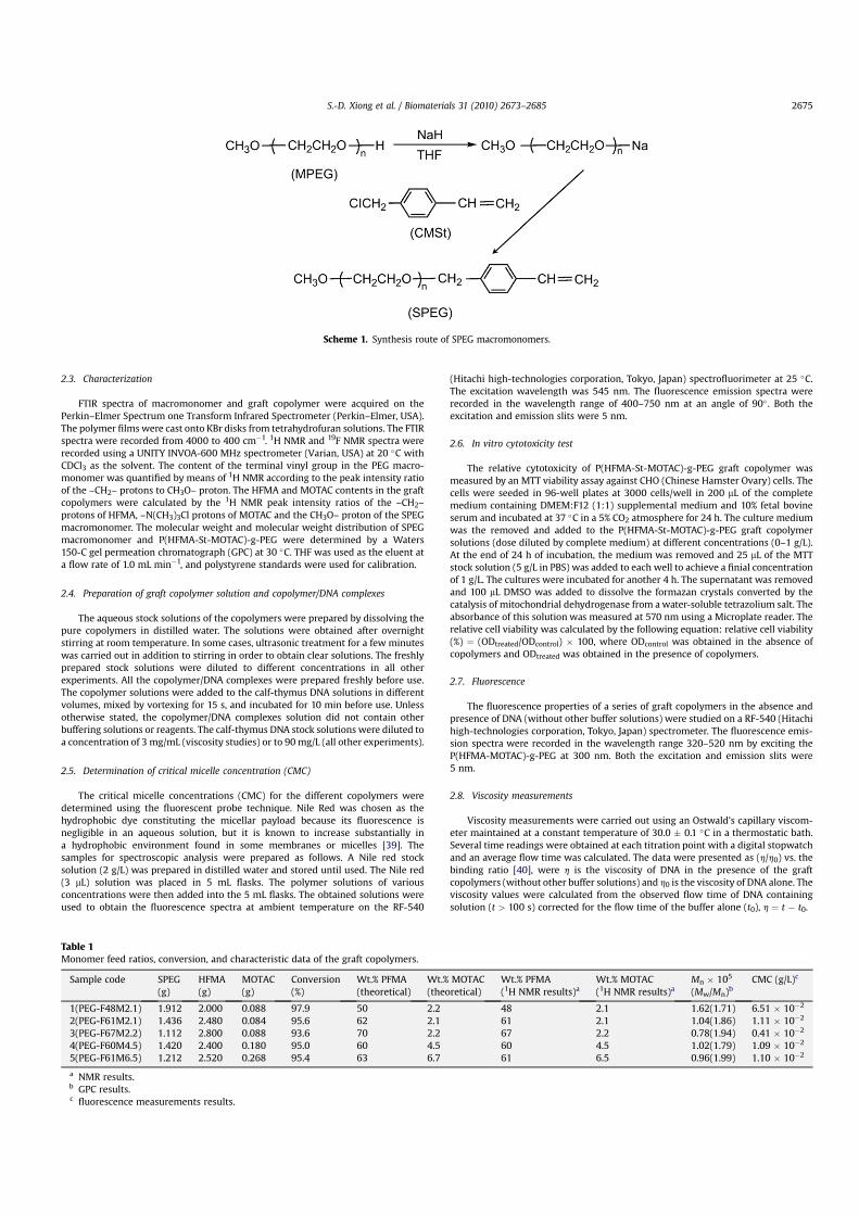
(0.072 g, 3 mmol) was added by stirring at 45 �C for 2 h. After CMSt (1.220 g,8 mmol) was added into the flask, the reaction was conducted at 35 �C for 24 h.The products were precipitated using a large amount of diethyl ether and filtered.They were purified by dissolving in CH2Cl2 and filtered to remove any insolubleimpurities. Finally, the products were precipitated using cold diethyl ether,filtered, and dried under high vacuum [37,38]. The synthesis route of the SPEGmacromonomers is illustrated in Scheme 1. The yield was calculated from theweight of the dry SPEG macromonomers obtained. The composition and contentof the terminal vinyl group in SPEG macromonomers was quantified according tothe 1H NMR spectrum.
2.2.2. Synthesis of P(HFMA-St-MOTAC)-g-PEG graft copolymerThe graft copolymers were synthesized via conventional free radical polymer-
ization in THF. In a typical reaction, SPEG macromonomer (1.420 g, 0.277 mmol),HFMA (2.400 g, 9.600 mmol), MOTAC (0.180 g, 0.867 mmol), AIBN (0.024 g0.146 mmol), and THF (20 mL) were added into a 50 mL round bottom flask con-taining a magnetic stirrer. The flask was then deoxygenated under reduced pressureand backfilled with nitrogen several times. Polymerization was carried out at 75 �Cfor 20 h. The product was isolated by evaporating the solvent in a rotary evaporatorand then the mixture was precipitated in n-hexane. After filtration, the precipitatewas purified by re-precipitation repeatedly in n-hexane. The obtained product wasdried under vacuum at 35 �C for 72 h. Polymerization conversion was determined bygravimetric analysis. The product was analyzed by FTIR, 1H NMR, and 19F NMRspectroscopy. The detailed polymerization conditions are listed in Table 1.
S.-D. Xiong et al. / Biomaterials 31 (2010) 2673–26852674
2.3. Characterization
FTIR spectra of macromonomer and graft copolymer were acquired on thePerkin–Elmer Spectrum one Transform Infrared Spectrometer (Perkin–Elmer, USA).The polymer films were cast onto KBr disks from tetrahydrofuran solutions. The FTIRspectra were recorded from 4000 to 400 cm�1. 1H NMR and 19F NMR spectra wererecorded using a UNITY INVOA-600 MHz spectrometer (Varian, USA) at 20 �C withCDCl3 as the solvent. The content of the terminal vinyl group in the PEG macro-monomer was quantified by means of 1H NMR according to the peak intensity ratioof the –CH2– protons to CH3O– proton. The HFMA and MOTAC contents in the graftcopolymers were calculated by the 1H NMR peak intensity ratios of the –CH2–protons of HFMA, –N(CH3)3Cl protons of MOTAC and the CH3O– proton of the SPEGmacromonomer. The molecular weight and molecular weight distribution of SPEGmacromonomer and P(HFMA-St-MOTAC)-g-PEG were determined by a Waters150-C gel permeation chromatograph (GPC) at 30 �C. THF was used as the eluent ata flow rate of 1.0 mL min�1, and polystyrene standards were used for calibration.
2.4. Preparation of graft copolymer solution and copolymer/DNA complexes
The aqueous stock solutions of the copolymers were prepared by dissolving thepure copolymers in distilled water. The solutions were obtained after overnightstirring at room temperature. In some cases, ultrasonic treatment for a few minuteswas carried out in addition to stirring in order to obtain clear solutions. The freshlyprepared stock solutions were diluted to different concentrations in all otherexperiments. All the copolymer/DNA complexes were prepared freshly before use.The copolymer solutions were added to the calf-thymus DNA solutions in differentvolumes, mixed by vortexing for 15 s, and incubated for 10 min before use. Unlessotherwise stated, the copolymer/DNA complexes solution did not contain otherbuffering solutions or reagents. The calf-thymus DNA stock solutions were diluted toa concentration of 3 mg/mL (viscosity studies) or to 90 mg/L (all other experiments).
2.5. Determination of critical micelle concentration (CMC)
The critical micelle concentrations (CMC) for the different copolymers weredetermined using the fluorescent probe technique. Nile Red was chosen as thehydrophobic dye constituting the micellar payload because its fluorescence isnegligible in an aqueous solution, but it is known to increase substantially ina hydrophobic environment found in some membranes or micelles [39]. Thesamples for spectroscopic analysis were prepared as follows. A Nile red stocksolution (2 g/L) was prepared in distilled water and stored until used. The Nile red(3 mL) solution was placed in 5 mL flasks. The polymer solutions of variousconcentrations were then added into the 5 mL flasks. The obtained solutions wereused to obtain the fluorescence spectra at ambient temperature on the RF-540
(Hitachi high-technologies corporation, Tokyo, Japan) spectrofluorimeter at 25 �C.The excitation wavelength was 545 nm. The fluorescence emission spectra wererecorded in the wavelength range of 400–750 nm at an angle of 90� . Both theexcitation and emission slits were 5 nm.
2.6. In vitro cytotoxicity test
The relative cytotoxicity of P(HFMA-St-MOTAC)-g-PEG graft copolymer wasmeasured by an MTT viability assay against CHO (Chinese Hamster Ovary) cells. Thecells were seeded in 96-well plates at 3000 cells/well in 200 mL of the completemedium containing DMEM:F12 (1:1) supplemental medium and 10% fetal bovineserum and incubated at 37 �C in a 5% CO2 atmosphere for 24 h. The culture mediumwas the removed and added to the P(HFMA-St-MOTAC)-g-PEG graft copolymersolutions (dose diluted by complete medium) at different concentrations (0–1 g/L).At the end of 24 h of incubation, the medium was removed and 25 mL of the MTTstock solution (5 g/L in PBS) was added to each well to achieve a finial concentrationof 1 g/L. The cultures were incubated for another 4 h. The supernatant was removedand 100 mL DMSO was added to dissolve the formazan crystals converted by thecatalysis of mitochondrial dehydrogenase from a water-soluble tetrazolium salt. Theabsorbance of this solution was measured at 570 nm using a Microplate reader. Therelative cell viability was calculated by the following equation: relative cell viability(%) ¼ (ODtreated/ODcontrol) � 100, where ODcontrol was obtained in the absence ofcopolymers and ODtreated was obtained in the presence of copolymers.
2.7. Fluorescence
The fluorescence properties of a series of graft copolymers in the absence andpresence of DNA (without other buffer solutions) were studied on a RF-540 (Hitachihigh-technologies corporation, Tokyo, Japan) spectrometer. The fluorescence emis-sion spectra were recorded in the wavelength range 320–520 nm by exciting theP(HFMA-MOTAC)-g-PEG at 300 nm. Both the excitation and emission slits were5 nm.
2.8. Viscosity measurements
Viscosity measurements were carried out using an Ostwald’s capillary viscom-eter maintained at a constant temperature of 30.0 � 0.1 �C in a thermostatic bath.Several time readings were obtained at each titration point with a digital stopwatchand an average flow time was calculated. The data were presented as (h/h0) vs. thebinding ratio [40], were h is the viscosity of DNA in the presence of the graftcopolymers (without other buffer solutions) and h0 is the viscosity of DNA alone. Theviscosity values were calculated from the observed flow time of DNA containingsolution (t > 100 s) corrected for the flow time of the buffer alone (t0), h ¼ t � t0.
Scheme 1. Synthesis route of SPEG macromonomers.
Table 1Monomer feed ratios, conversion, and characteristic data of the graft copolymers.
Sample code SPEG(g)
HFMA(g)
MOTAC(g)
Conversion(%)
Wt.% PFMA(theoretical)
Wt.% MOTAC(theoretical)
Wt.% PFMA(1H NMR results)a
Wt.% MOTAC(1H NMR results)a
Mn � 105
(Mw/Mn)bCMC (g/L)c
1(PEG-F48M2.1) 1.912 2.000 0.088 97.9 50 2.2 48 2.1 1.62(1.71) 6.51 � 10�2
2(PEG-F61M2.1) 1.436 2.480 0.084 95.6 62 2.1 61 2.1 1.04(1.86) 1.11 � 10�2
3(PEG-F67M2.2) 1.112 2.800 0.088 93.6 70 2.2 67 2.2 0.78(1.94) 0.41 � 10�2
4(PEG-F60M4.5) 1.420 2.400 0.180 95.0 60 4.5 60 4.5 1.02(1.79) 1.09 � 10�2
5(PEG-F61M6.5) 1.212 2.520 0.268 95.4 63 6.7 61 6.5 0.96(1.99) 1.10 � 10�2
a NMR results.b GPC results.c fluorescence measurements results.
S.-D. Xiong et al. / Biomaterials 31 (2010) 2673–2685 2675
2.9. Gel retardation assay
The DNA condensing ability of P(HFMA-St-MOTAC)-g-PEG was examined byagarose gel electrophoresis. The complexes at different weight ratios were preparedby adding an appropriate volume of P(HFMA-St-MOTAC)-g-PEG solution to 100 ng ofpEGFP-C1 (150 ng/mL in 40 mM Tris–HCl buffer solution). The complexes werediluted with 150 mM NaCl solution to 6 mL and then the complexes were incubated at37 �C for 30 min. Afterwards, the complexes were electrophoresed in the 0.7% (w/v)agarose gel containing GelRed (an ethidium-bromide (EtBr) containing 1%) and withTris-acetate (TAE) running buffer at 80 V for 100 min. The DNA was visualized witha UV lamp on a Vilber Lourmat imaging system (France).
2.10. UV–visible spectroscopy
The DNA-binding ability of P(HFMA-St-MOTAC)-g-PEG was determined bymeasuring the intrinsic absorption peaks at 260 nm using a Perkin–Elmer lambda 17UV–vis spectrophotometer at room temperature. In a typical example, the appro-priate volume of DNA solution (90 mg/L) was added to a fixed amount (1 mL) ofpolymer solution (0.12 g/L). After 24 h of incubation at room temperature, thecomplexes were centrifuged at 12 000 rpm for 10 min to precipitate the micelles.The amount of unadsorbed DNA was determined using UV–vis spectrophotometerby incubating 50 mL of the supernatant. All the measurements were made in a quartzcuvette (1 cm in width) in the wavelength range of 200–400 nm and the absorptionpeaks at 260 nm for DNA was used. Deionized water/DNA were used in thecomparative experiments.
2.11. Transmission electron microscopy
The morphology of the graft copolymer micelles and the morphology of graftcopolymer/DNA complexes were characterized with TEM (Tecnai G20, FEI Corp.USA). The sample was stained with phospho-tungstic acid (pH ¼ 6.5), and a drop ofthe sample (concentration of graft copolymer solution was above CMC) was placedon a Formvar-coated copper grid which was dried in air. The TEM images wereobtained at 25 �C at an electron acceleration voltage of 80 kV.
2.12. Particle size measurement
The average hydrodynamic radius and size distribution of the copolymermicelles and graft copolymer/DNA complexes were measured by photon correlationspectroscopy (PCS) (Autosize Loc-Fc-963, Malvern Instrument). The experimentswere performed using the graft copolymer solutions having concentration aboveCMC. The samples prepared by filtering the solutions through cellulose acetatefilters with 0.2 mm pore size were directly poured into a cuvette. The cuvette wasthen set inside a sample holder. Measurements were made at an angle of 90� with679 nm wavelength laser light. All the data were averaged from 3 to 5 parallelmeasurements.
2.13. Stability of copolymer micelles and copolymer/DNA complex particles
The stability of the copolymer micelles in aqueous solutions was evaluated byturbidity measurement and monitoring the time-dependent changes in the micellesize with photon correlation spectroscopy (PCS). The copolymer solutions with theconcentrations above CMC were used in this evaluation. The copolymer micellesolutions were incubated at 37 �C for certain durations (1–15 days), followed byturbidity measurement of the suspension at 550 nm. The stability of copolymer/DNAcomplex particles was examined by monitoring the time-dependent changes in themicelle size (with photon correlation spectroscopy (PCS)) and zeta potentials ofthese particles.
2.14. Zeta potential measurement
The zeta potentials of the graft copolymer/DNA complexes were measured usingNano-ZS ZEN3600 (Malvern Instruments, Southborough, MA) based on the principleof phase analysis light scattering. The zeta potentials quoted were averages obtainedfrom at least six measurements over duplicate samples.
3. Results and discussion
3.1. Synthesis and characterization of SPEG macromonomer andP(HFMA-St-MOTAC)-g-PEG graft copolymers
Recent development of macromonomer techniques has enabledthe preparation of a variety of graft copolymers with a well-definedstructure. In this work, we prepare methoxy poly(ethylene glycol)macromonomer (SPEG) with the p-vinylbenzyl end group. Thesynthesis route of the SPEG macromonomer is illustrated in
Scheme 1. There is no polymerization reaction during the macro-monomer preparation at 35 �C as revealed by the nature of theproduct as well as 1H NMR and FTIR spectra. The quantitativeresults are given in Table 2. It is obvious that the P(HFMA-St-MOTAC)-g-PEG graft copolymer can be easily obtained via copo-lymerization of the SPEG macromonomer, MOTAC, and HFMA usingTHF as the solvent and AIBN as the initiator. The chemical structureand molecular weight of the graft copolymers and SPEG macro-monomer will be discussed in detail later in this paper.
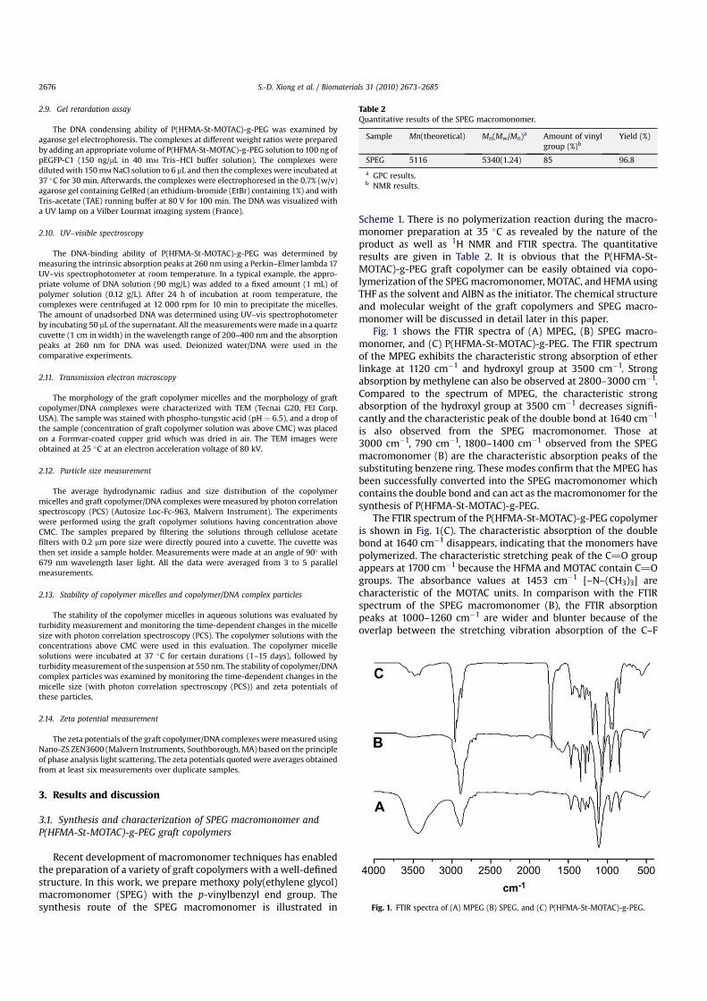
Fig. 1 shows the FTIR spectra of (A) MPEG, (B) SPEG macro-monomer, and (C) P(HFMA-St-MOTAC)-g-PEG. The FTIR spectrumof the MPEG exhibits the characteristic strong absorption of etherlinkage at 1120 cm�1 and hydroxyl group at 3500 cm�1. Strongabsorption by methylene can also be observed at 2800–3000 cm�1.Compared to the spectrum of MPEG, the characteristic strongabsorption of the hydroxyl group at 3500 cm�1 decreases signifi-cantly and the characteristic peak of the double bond at 1640 cm�1
is also observed from the SPEG macromonomer. Those at3000 cm�1, 790 cm�1, 1800–1400 cm�1 observed from the SPEGmacromonomer (B) are the characteristic absorption peaks of thesubstituting benzene ring. These modes confirm that the MPEG hasbeen successfully converted into the SPEG macromonomer whichcontains the double bond and can act as the macromonomer for thesynthesis of P(HFMA-St-MOTAC)-g-PEG.
The FTIR spectrum of the P(HFMA-St-MOTAC)-g-PEG copolymeris shown in Fig. 1(C). The characteristic absorption of the doublebond at 1640 cm�1 disappears, indicating that the monomers havepolymerized. The characteristic stretching peak of the C]O groupappears at 1700 cm�1 because the HFMA and MOTAC contain C]Ogroups. The absorbance values at 1453 cm�1 [–N–(CH3)3] arecharacteristic of the MOTAC units. In comparison with the FTIRspectrum of the SPEG macromonomer (B), the FTIR absorptionpeaks at 1000–1260 cm�1 are wider and blunter because of theoverlap between the stretching vibration absorption of the C–F
Table 2Quantitative results of the SPEG macromonomer.
Sample Mn(theoretical) Mn(Mw/Mn)a Amount of vinylgroup (%)b
Yield (%)
SPEG 5116 5340(1.24) 85 96.8
a GPC results.b NMR results.
Fig. 1. FTIR spectra of (A) MPEG (B) SPEG, and (C) P(HFMA-St-MOTAC)-g-PEG.
S.-D. Xiong et al. / Biomaterials 31 (2010) 2673–26852676
bond at 1100–1260 cm�1 and the stretching vibration absorption ofthe C–O–C bond at 1250 cm�1. This result proves that HFMA,MOTAC and SPEG participate in the polymerization.
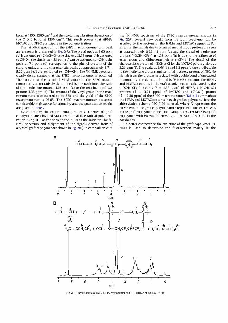
The 1H NMR spectrum of the SPEG macromonomer and peakassignments is presented in Fig. 2(A). The broad peak at 3.65 ppm(b) is assigned to –CH2CH2O–, the singlet at 3.38 ppm (a) is assignedto CH3O–, the singlet at 4.58 ppm (c) can be assigned to –CH2–, thepeak at 7.4 ppm (d) corresponds to the phenyl protons of thestyrene units, and the characteristic peaks at approximately 6.71–5.22 ppm (e,f) are attributed to –CH]CH2. The 1H NMR spectrumclearly demonstrates that the SPEG macromonomer is obtained.The content of the terminal vinyl group in the SPEG macro-monomer is quantitatively determined by the peak intensity ratioof the methylene protons 4.58 ppm (c) to the terminal methoxyprotons 3.38 ppm (a). The amount of the vinyl group in the mac-romonomers is calculated to be 85% and the yield of the SPEGmacromonomer is 96.8%. The SPEG macromonomer possessesconsiderably high active functionality and the quantitative resultsare given in Table 2.
By controlling the experimental protocols, a series of graftcopolymers are obtained via conventional free radical polymeri-zation using THF as the solvent and AIBN as the initiator. The 1HNMR spectrum and assignment of the signals derived from ofa typical graft copolymer are shown in Fig. 2(B). In comparison with
the 1H NMR spectrum of the SPEG macromonomer shown inFig. 2(A), several new peaks from the graft copolymer can beascribed to the protons of the HFMA and MOTAC segments. Forinstance, the signals due to terminal methyl group protons are seenat approximately 0.75–1.3 ppm (g) and the signal of methyleneprotons (–OCH2–CF2–) at 4.39 ppm (h) is due to the influence ofester group and difluoromethylene (–CF2–). The signal of thecharacteristic proton of –N(CH3)3Cl for the MOTAC part is visible at3.21 ppm (l). The peaks at 3.66 (b) and 3.3 ppm (a) are attributableto the methylene protons and terminal methoxy protons of PEG. Nosignals from the protons associated with double bond of unreactedmonomer can be detected from this 1H NMR spectrum. The HFMAand MOTAC contents in the graft copolymers are calculated by the(–OCH2–CF2–) protons (d ¼ 4.39 ppm) of HFMA, (–N(CH3)3Cl)protons (d ¼ 3.21 ppm) of MOTAC and (CH3O–) proton(d ¼ 3.38 ppm) of the SPEG macromonomer. Table 1 summarizesthe HFMA and MOTAC contents in each graft copolymers. Here, theabbreviation scheme PEG-FXMZ is used, where X represents theHFMA wt% in the graft copolymer and Z represents the MOTAC wt%in the graft copolymer. Hence, for example, PEG-F60M4.5 is a graftcopolymer with 60 wt% of HFMA and 4.5 wt% of MOTAC in thebackbones.
To better characterize the structure of the graft copolymer, 19FNMR is used to determine the fluorocarbon moiety in the
Fig. 2. 1H NMR spectra of (A) SPEG macromonomer and (B) P(HFMA-St-MOTAC)-g-PEG.
S.-D. Xiong et al. / Biomaterials 31 (2010) 2673–2685 2677
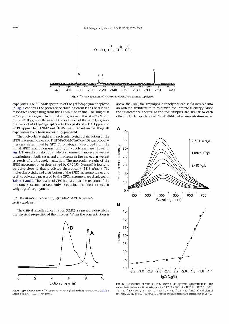
copolymer. The 19F NMR spectrum of the graft copolymer depictedin Fig. 3 confirms the presence of three different kinds of fluorineresonances originating from the HFMA side chains. The singlet at�75.2 ppm is assigned to the end –CF3 group and that at�212.9 ppmto the –CHF2 group. Because of the influence of the –OCH2– group,the peak of –OCH2–CF2– splits into two peaks at �114.3 ppm and�119.6 ppm. The 1H NMR and 19F NMR results confirm that the graftcopolymers have been successfully prepared.
The molecular weight and molecular weight distribution of theSPEG macromonomer and P(HFMA-St-MOTAC)-g-PEG graft copoly-mers are determined by GPC. Chromatograms recorded from theinitial SPEG macromonomer and graft copolymers are shown inFig. 4. These chromatograms indicate a unimodal molecular weightdistribution in both cases and an increase in the molecular weightas result of graft copolymerization. The molecular weight of theSPEG macromonomer determined by GPC (5340 g/mol) is found tobe quite close to that predicted theoretically (5116 g/mol). Themolecular weight and distribution of the SPEG macromonomer andgraft copolymers measured by the GPC instrument are displayed inTables 1 and 2. The results of GPC indicate that the reaction of themonomers occurs subsequently producing the high molecularweight graft copolymers.
3.2. Micellization behavior of P(HFMA-St-MOTAC)-g-PEGgraft copolymer
The critical micelle concentration (CMC) is a measure describingthe physical properties of the micelles. When the concentration is
above the CMC, the amphiphilic copolymer can self-assemble intoan ordered architecture to minimize the interfacial energy. Sincethe fluorescence spectra of the five samples are similar to eachother, only the spectrum of PEG-F60M4.5 at a concentration range
Fig. 3. 19F NMR spectrum of P(HFMA-St-MOTAC)-g-PEG graft copolymer.
Fig. 4. Typical GPC curves of (A) SPEG, Mn ¼ 5340 g/mol and (B) PEG-F60M4.5 (Table 1,Sample 4), Mn ¼ 1.02 � 105 g/mol.
Fig. 5. Fluorescence spectra of PEG-F60M4.5 at different concentrations (Theconcentrations from bottom to top are 8 � 10�4, 2 � 10�3, 4 � 10�3, 8 � 10�3, 1 �10�2,1.3 � 10�2, 1.5 � 10�2, 1.8 � 10�2, 2 � 10�2, 2.4 � 10�2, 2.8 � 10�2 g/L) (A) and plots ofintensity vs. lgC of PEG-F60M4.5 (B). All the measurements are carried out at 25 �C.
S.-D. Xiong et al. / Biomaterials 31 (2010) 2673–26852678
from 8 � 10�4 to 2.8 � 10�2 g/L is shown in Fig. 5(A). It can be seenthat the emission intensity increases with increasing polymerconcentrations. Specifically, at low concentrations, there are smallchanges in the emission intensity. With increasing concentrations,marked changes in the intensity are observed suggesting a transferof Nile Red probe to a more hydrophobic microenvironment andindicating formation of micelles. As shown in Fig. 5(B), plots ofemission intensity vs. lgC of PEG-F60M4.5 are flat at low concen-trations and sigmoidal in the crossover region. The CMC is taken asthe intersection of the tangent to the curve at the inflexion with thehorizontal tangent through the points at the low polymerconcentration. The critical micelle concentrations of the fivesamples are listed in Table 1. As reported in the literature, the CMCis influenced by many variables, such as the PEG content, temper-ature, molecular weight, and polydispersity index. The results hereindicate that the CMC decreases with increasing HFMA contents inthe graft copolymers (PEG-F48M2.1, PEG-F61M2.1, PEG-F67M2.2 inTable 1). This may be due to the unique properties of fluoropol-ymers such as low surface energy, reduced coefficient of friction,oleo- and hydrophobicity. Therefore, the content of HFMA maybecome the main factor affecting the CMC value. The experimentalresults are in good agreement with those previously reported [27].
3.3. In vitro cytotoxicity
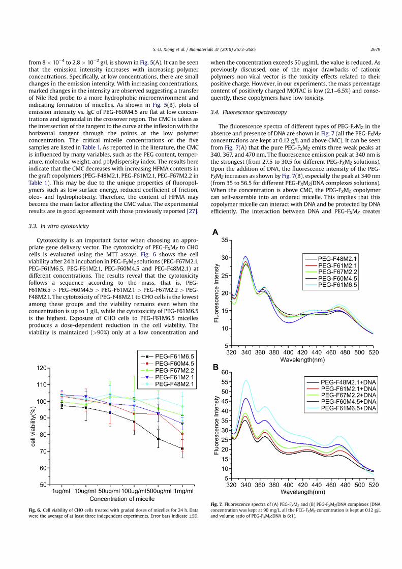
Cytotoxicity is an important factor when choosing an appro-priate gene delivery vector. The cytotoxicity of PEG-FXMZ to CHOcells is evaluated using the MTT assays. Fig. 6 shows the cellviability after 24 h incubation in PEG-FXMZ solutions (PEG-F67M2.1,PEG-F61M6.5, PEG-F61M2.1, PEG-F60M4.5 and PEG-F48M2.1) atdifferent concentrations. The results reveal that the cytotoxicityfollows a sequence according to the mass, that is, PEG-F61M6.5 > PEG-F60M4.5 > PEG-F61M2.1 > PEG-F67M2.2 > PEG-F48M2.1. The cytotoxicity of PEG-F48M2.1 to CHO cells is the lowestamong these groups and the viability remains even when theconcentration is up to 1 g/L, while the cytotoxicity of PEG-F61M6.5is the highest. Exposure of CHO cells to PEG-F61M6.5 micellesproduces a dose-dependent reduction in the cell viability. Theviability is maintained (>90%) only at a low concentration and
when the concentration exceeds 50 mg/mL, the value is reduced. Aspreviously discussed, one of the major drawbacks of cationicpolymers non-viral vector is the toxicity effects related to theirpositive charge. However, in our experiments, the mass percentagecontent of positively charged MOTAC is low (2.1–6.5%) and conse-quently, these copolymers have low toxicity.
3.4. Fluorescence spectroscopy
The fluorescence spectra of different types of PEG-FXMZ in theabsence and presence of DNA are shown in Fig. 7 (all the PEG-FXMZ
concentrations are kept at 0.12 g/L and above CMC). It can be seenfrom Fig. 7(A) that the pure PEG-FXMZ emits three weak peaks at340, 367, and 470 nm. The fluorescence emission peak at 340 nm isthe strongest (from 27.5 to 30.5 for different PEG-FXMZ solutions).Upon the addition of DNA, the fluorescence intensity of the PEG-FXMZ increases as shown by Fig. 7(B), especially the peak at 340 nm(from 35 to 56.5 for different PEG-FXMZ/DNA complexes solutions).When the concentration is above CMC, the PEG-FXMZ copolymercan self-assemble into an ordered micelle. This implies that thiscopolymer micelle can interact with DNA and be protected by DNAefficiently. The interaction between DNA and PEG-FXMZ creates
Fig. 6. Cell viability of CHO cells treated with graded doses of micelles for 24 h. Datawere the average of at least three independent experiments. Error bars indicate �SD.
Fig. 7. Fluorescence spectra of (A) PEG-FXMZ and (B) PEG-FXMZ/DNA complexes (DNAconcentration was kept at 90 mg/L, all the PEG-FXMZ concentration is kept at 0.12 g/Land volume ratio of PEG-FXMZ:DNA is 6:1).
S.-D. Xiong et al. / Biomaterials 31 (2010) 2673–2685 2679
chain entanglement causing changes in the microenvironmentaround PEG-FXMZ. The hydrophobic environment inside the DNAhelix reduces the accessibility of solvent water molecules to thePEG-FXMZ micelle and the PEG-FXMZ mobility is restricted at thebinding site, there is decrease in the vibration modes of relaxationfollowed by increasing fluorescent intensity.
The PEG-FXMZ can bind the double stranded DNA by differentinteractions on the basis of their structure. In the PEG-FXMZ
aqueous system, there are electrostatic interactions, hydrophobicinteractions, and H-bonding, which will influence PEG-FXMZ
binding to the double stranded DNA. According to Fig. 7(B), whenthe content of MOTAC in the same and the hydrophobic charac-teristic contents of HFMA in PEG-FXMZ is increased, the fluores-cence intensity increases slightly compared to pure PEG-FXMZ
(PEG-F48M2.1/DNA, PEG-F61M2.1/DNA and PEG-F67M2.2/DNA inFig. 7(B)). In addition, when the content of HFMA in the PEG-FXMZ
does not change, increasing the MOTAC contents in the PEG-FXMZ
leads to marked changes in the intensity as shown by the emissionspectra compared to pure PEG-FXMZ (PEG-F61M2.1/DNA, PEG-F60M4.5/DNA and PEG-F61M6.5/DNA in Fig. 7(B)). Therefore, theelectrostatic interaction [41] between positively charged MOTACand negatively charged phosphate backbone at the periphery of thedouble helix DNA are the main factor for PEG-FXMZ binding to thedouble stranded DNA. The hydrophobic interaction due to thehydrophobic characteristic of the fluorinated segments is anotherfactor that PEG-FXMZ can bind to the DNA. In addition, as the DNAdouble helix possesses many hydrogen bonding sites, it is likelythat the fluorinated groups of PEG-FXMZ forms hydrogen bondswith the DNA consequently increasing the fluorescent intensity.Hence, H-bonding is another reason for the PEG-FXMZ binding tothe DNA and electrostatic interaction, hydrophobic interaction, andhydrogen bonding exist in this system.
3.5. Viscosity studies
To further explore the interaction characteristics between PEG-FXMZ and DNA, the relative specific viscosity of DNA is examined byvarying the concentrations of the added PEG-FXMZ. Measuring theviscosity of DNA is a classical technique used to analyze the DNA-binding mode in solutions [42]. Hydrodynamic measurements suchas viscosity, sedimentation, rotational diffusion, and so on sensitiveto molecular lengths provide strong evidence of the DNA-bindingmodes, especially in the absence of crystallographic structural data[43]. A classical intercalation model demands that the DNA helixlengthens as base-pairs are separated to accommodate the bindingmolecule, leading to the increase of DNA viscosity. In contrast,a partial, non-classical intercalation of molecule can bend or kinkthe DNA helix and reduce its effective length and in turn itsviscosity [44,45]. When electrostatic binding, groove binding orother outside binding occurs, the viscosity of DNA does not changebasically [43,46].
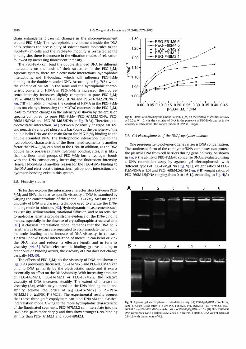
The effects of PEG-FXMZ on the viscosity of DNA are shown inFig. 8. As previously discussed, PEG-F61M6.5 and PEG-F60M4.5 canbind to DNA primarily by the electrostatic mode and it exertsessentially no effect on the DNA viscosity. With increasing amountsof PEG-F48M2.1, PEG-F61M2.1 or PEG-F67M2.2, the relativeviscosity of DNA increases steadily. The extent of increase inviscosity (Dh), which may depend on the DNA-binding mode andaffinity, follows the order of Dh(PEG-F67M2.2) > Dh(PEG-F61M2.1) > Dh(PEG-F48M2.1). The experimental results suggestthat these three graft copolymers can bind DNA via the classicalintercalation mode. Owing to the more hydrophobic characteristicof the fluorinated segments, PEG-F67M2.2 can intercalate into theDNA base-pairs more deeply and thus show stronger DNA-bindingaffinity than PEG-F61M2.1 and PEG-F48M2.1.
3.6. Gel electrophoresis of the DNA/copolymer mixture
One prerequisite to polymeric gene carrier is DNA condensation.The condensed form of the copolymer/DNA complexes can protectthe plasmid DNA from cell barriers during gene delivery. As shownin Fig. 9, the ability of PEG-FXMZ to condense DNA is evaluated usinga DNA retardation assay by agarose gel electrophoresis withdifferent types of PEG-FXMZ/DNA (Fig. 9(A), weight ratios of PEG-FXMZ/DNA is 1.5) and PEG-F60M4.5/DNA (Fig. 9(B) weight ratios ofPEG-F60M4.5/DNA ranging from 0 to 1.6:1.). According to Fig. 4(A)
Fig. 8. Effects of increasing the amount of PEG-FXMZ on the relative viscosities of DNAat 30.0 � 0.1 �C. h is the viscosity of DNA in the presence of PEG-FXMZ and h0 is theviscosity of DNA alone. The concentration of DNA is 3 mg/mL.
Fig. 9. Agarose gel electrophoresis retardation assay: (A) PEG-FXMZ/DNA complexes,Lane 1, naked DNA; lanes 2–6 are PEG-F48M2.1, PEG-F61M2.1, PEG-F67M2.2, PEG-F60M4.5 and PEG-F61M6.5 (weight ratios of PEG-FXMZ/DNA is 1.5); (B) PEG-F60M4.5/DNA complexes, Lane 1, naked DNA; lanes 2–7 are PEG-F60M4.5/DNA weight ratios of0.6–1.6 with increments of 0.2.
S.-D. Xiong et al. / Biomaterials 31 (2010) 2673–26852680
one can see that migration of the plasmid DNA slows when PEG-FXMZ is added. However, the PEG-FXMZ comprising differentMOTAC or HFMA contents shows a different DNA-binding capacity.At a polymer/DNA weight ratio of 1.5, PEG-F60M4.5 and PEG-F61M6.5 can completely retard DNA migration showing a higherDNA-binding capacity than PEG-F67M2.2, PEG-F61M2.1, and PEG-F48M2.1. The results suggest that a high charge density or HFMAcontent in PEG-FXMZ may benefit DNA-binding. As shown inFig. 9(B), an increase in the PEG-F60M4.5 amount causes a gradualdisappearance of the DNA and complete retardation of DNA isachieved at a PEG-F60M4.5/DNA weight ratio of 1:2. These resultsdemonstrate that the copolymers may be used to bind DNA as anefficient gene vector.
3.7. UV–visible spectrophotometry
In addition to gel retardation assay, the DNA-binding ability ofthe graft copolymers is evaluated by UV–visible spectrophotom-etry. The PEG-FXMZ/DNA complexes are centrifuged and thesupernatant is measured at 260 nm which is the absorption peak ofDNA. The results are shown in Table 3. Compared to the H2O/DNAcolumn, after adding PEG-FXMZ, the absorbance of the supernatantis significantly reduced. It shows that most of the DNA adsorbs tothe PEG-FXMZ micelles, and these PEG-FXMZ/DNA aggregates areprecipitated at the bottom of the complex solutions. It also can beseen from Table 3 that in the SPEG-F60D4.5/DNA and SPEG-F61D6.5/DNA system, the decline in the absorbance value is morethan the other three, indicating that SPEG-F60D4.5 and SPEG-F61D6.5 micelles can more easily adsorb DNA compared to theother three. This result is consistent with the gel retardation assay,indicating that high charge density or HFMA contents in PEG-FXMZ
micelles may benefit DNA-binding. It also shows that the PEG-FXMZ
micelles which adsorb DNA can be used as an efficient gene vector.
3.8. Characterization of PEG-FXMZ/DNA particles
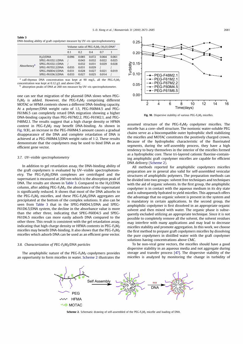
The amphiphilic nature of the PEG-FXMZ copolymers providesan opportunity to form micelles in water. Scheme 2 illustrates the
assumed structure of the PEG-FXMZ copolymer micelles. Themicelle has a core–shell structure. The nonionic water-soluble PEGchains serve as a biocompatible outer hydrophilic shell stabilizingthe micelles and MOTAC constitutes the positively charged crown.Because of the hydrophobic characteristic of the fluorinatedsegments, during the self-assembly process, they have a hightendency to bury themselves in the interior of the micelles formedas a hydrophobic core. These tri-layered cationic fluorine-contain-ing amphiphilic graft copolymer micelles are capable for efficientDNA delivery (Scheme 2).
All methods reported for amphiphilic copolymers micellespreparation are in general also valid for self-assembled vesicularstructures of amphiphilic polymers. The preparation methods canbe divided into two groups: solvent free techniques and techniqueswith the aid of organic solvents. In the first group, the amphiphiliccopolymer is in contact with the aqueous medium in its dry stateand is subsequently hydrated to yield micelles. This approach offersthe advantage that no organic solvent is present in the system andis mandatory in certain applications. In the second group, theamphiphilic copolymer is first dissolved in an appropriate organicsolvent and then mixed with water. The organic phase is subse-quently excluded utilizing an appropriate technique. Since it is notpossible to completely remove all the solvent, the solvent residuesmay interfere with many applications and may lead to decreasemicelles stability and promote aggregation. In this work, we choosethe first method to prepare graft copolymers micelles by dissolvingthe pure copolymers in distilled water with the graft copolymersolutions having concentrations above CMC.
To be non-viral gene vectors, the micelles should have a gooddispersive stability in an aqueous media and not aggregate duringstorage and transfer process [47]. The dispersive stability of themicelles is analyzed by monitoring the change in turbidity of
Scheme 2. Schematic drawing of self-assembled of the PEG-FXMZ micelle and loading of DNA.
Fig. 10. Dispersive stability of various PEG-FXMZ micelles.
Table 3DNA-binding ability of graft copolymer measure by UV–vis spectrophotometry.
Volume ratio of PEG-FXMZ (H2O):DNAa
0.1 0.2 0.4 0.7 1
Absorbencyb
H2O/DNA 0.096 0.085 0.072 0.064 0.061SPEG-F61D2.1/DNA / 0.043 0.032 0.022 0.025SPEG-F61D2.1/DNA / 0.032 0.031 0.029 0.028SPEG-F67D2.2/DNA 0.035 0.031 0.029 / /SPEG-F60D4.5/DNA 0.031 0.028 0.027 0.021 0.019SPEG-F61D6.5/DNA 0.033 0.027 0.025 0.014 /
a calf-thymus DNA concentration was kept at 90 mg/L, all the PEG-FXMZ
concentration was kept at 0.12 g/L and above CMC.b absorption peaks of DNA at 260 nm measure by UV–vis spectrophotometer.
S.-D. Xiong et al. / Biomaterials 31 (2010) 2673–2685 2681
various PEG-FXMZ copolymer micelle solutions (all the PEG-FXMZ
concentrations are kept at 0.12 g/L and above CMC) as shown inFig. 10. Owing to the hydrophobic characteristic of the fluorinatedsegments, they have a high tendency to bury themselves in theinterior of the micelles formed as a hydrophobic core. The hydro-phobic HFMA core compresses the complexes into smaller andmore compact particles, which are very useful for particle stabili-zation, and the outer hydrophilic PEG chains shell also can stabilizethe micelles. As shown in Fig. 10, these cationic fluorine-containingmicelles prepared with PEG-F48M2.1, PEG-F61M2.1, PEG-F60M4.5and PEG-F61M6.5 exhibit no significant change in turbidity evenafter 15 days of incubation at 37 �C. The hydrodynamic diameter ofthose micelles also does not change significantly after 15 days (datanot shown), indicating excellent dispersive stability. The micellesprepared with PEG-F67M2.2 exhibit a gradual decrease in turbidity10 days later. On the one hand, the more hydrophobic the segmentof HFMA, the higher the hydrophobic force and the copolymer ismore prone to secondary or higher level of aggregation. On theother hand, on account of the decrease in the PEG chains, its role in
stabilizing the micelles is weakened, resulting in the formation oflarge micelles during incubation at 37 �C. Hence, the dispersivestability of PEG-F67M2.2 micelles is not good as that of othersamples.
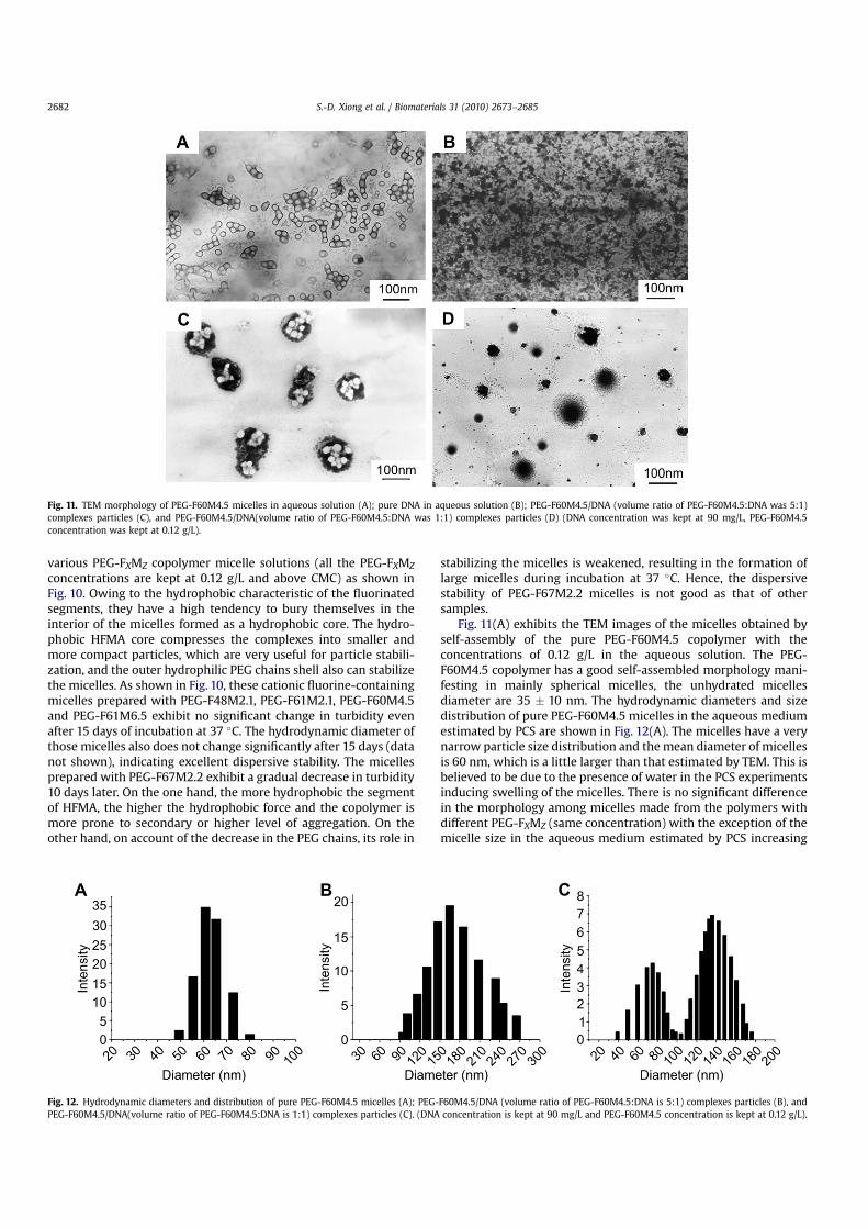
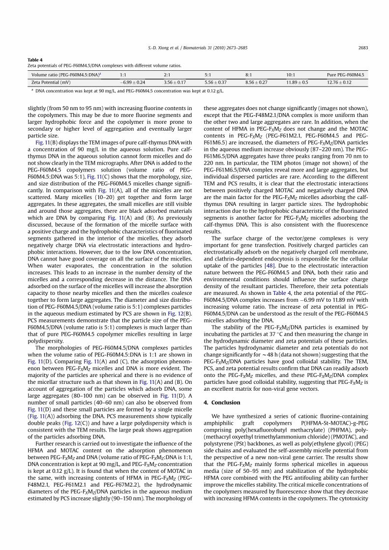
Fig. 11(A) exhibits the TEM images of the micelles obtained byself-assembly of the pure PEG-F60M4.5 copolymer with theconcentrations of 0.12 g/L in the aqueous solution. The PEG-F60M4.5 copolymer has a good self-assembled morphology mani-festing in mainly spherical micelles, the unhydrated micellesdiameter are 35 � 10 nm. The hydrodynamic diameters and sizedistribution of pure PEG-F60M4.5 micelles in the aqueous mediumestimated by PCS are shown in Fig. 12(A). The micelles have a verynarrow particle size distribution and the mean diameter of micellesis 60 nm, which is a little larger than that estimated by TEM. This isbelieved to be due to the presence of water in the PCS experimentsinducing swelling of the micelles. There is no significant differencein the morphology among micelles made from the polymers withdifferent PEG-FXMZ (same concentration) with the exception of themicelle size in the aqueous medium estimated by PCS increasing
Fig. 11. TEM morphology of PEG-F60M4.5 micelles in aqueous solution (A); pure DNA in aqueous solution (B); PEG-F60M4.5/DNA (volume ratio of PEG-F60M4.5:DNA was 5:1)complexes particles (C), and PEG-F60M4.5/DNA(volume ratio of PEG-F60M4.5:DNA was 1:1) complexes particles (D) (DNA concentration was kept at 90 mg/L, PEG-F60M4.5concentration was kept at 0.12 g/L).
Fig. 12. Hydrodynamic diameters and distribution of pure PEG-F60M4.5 micelles (A); PEG-F60M4.5/DNA (volume ratio of PEG-F60M4.5:DNA is 5:1) complexes particles (B), andPEG-F60M4.5/DNA(volume ratio of PEG-F60M4.5:DNA is 1:1) complexes particles (C). (DNA concentration is kept at 90 mg/L and PEG-F60M4.5 concentration is kept at 0.12 g/L).
S.-D. Xiong et al. / Biomaterials 31 (2010) 2673–26852682
slightly (from 50 nm to 95 nm) with increasing fluorine contents inthe copolymers. This may be due to more fluorine segments andlarger hydrophobic force and the copolymer is more prone tosecondary or higher level of aggregation and eventually largerparticle size.
Fig. 11(B) displays the TEM images of pure calf-thymus DNA witha concentration of 90 mg/L in the aqueous solution. Pure calf-thymus DNA in the aqueous solution cannot form micelles and donot show clearly in the TEM micrographs. After DNA is added to thePEG-F60M4.5 copolymers solution (volume ratio of PEG-F60M4.5:DNA was 5:1), Fig. 11(C) shows that the morphology, size,and size distribution of the PEG-F60M4.5 micelles change signifi-cantly. In comparison with Fig. 11(A), all of the micelles are notscattered. Many micelles (10–20) get together and form largeaggregates. In these aggregates, the small micelles are still visibleand around those aggregates, there are black adsorbed materialswhich are DNA by comparing Fig. 11(A) and (B). As previouslydiscussed, because of the formation of the micelle surface witha positive charge and the hydrophobic characteristics of fluorinatedsegments gathered in the interior of the micelles, they adsorbnegatively charge DNA via electrostatic interactions and hydro-phobic interactions. However, due to the low DNA concentration,DNA cannot have good coverage on all the surface of the micelles.When water evaporates, the concentration in the solutionincreases. This leads to an increase in the number density of themicelles and a corresponding decrease in the distance. The DNAadsorbed on the surface of the micelles will increase the absorptioncapacity to those nearby micelles and then the micelles coalescetogether to form large aggregates. The diameter and size distribu-tion of PEG-F60M4.5/DNA (volume ratio is 5:1) complexes particlesin the aqueous medium estimated by PCS are shown in Fig. 12(B).PCS measurements demonstrate that the particle size of the PEG-F60M4.5/DNA (volume ratio is 5:1) complexes is much larger thanthat of pure PEG-F60M4.5 copolymer micelles resulting in largepolydispersity.
The morphologies of PEG-F60M4.5/DNA complexes particleswhen the volume ratio of PEG-F60M4.5:DNA is 1:1 are shown inFig. 11(D). Comparing Fig. 11(A) and (C), the adsorption phenom-enon between PEG-FXMZ micelles and DNA is more evident. Themajority of the particles are spherical and there is no evidence ofthe micellar structure such as that shown in Fig. 11(A) and (B). Onaccount of aggregation of the particles which adsorb DNA, somelarge aggregates (80–100 nm) can be observed in Fig. 11(D). Anumber of small particles (40–60 nm) can also be observed fromFig. 11(D) and these small particles are formed by a single micelle(Fig. 11(A)) adsorbing the DNA. PCS measurements show typicallydouble peaks (Fig. 12(C)) and have a large polydispersity which isconsistent with the TEM results. The large peak shows aggregationof the particles adsorbing DNA.
Further research is carried out to investigate the influence of theHFMA and MOTAC content on the adsorption phenomenonbetween PEG-FXMZ and DNA (volume ratio of PEG-FXMZ:DNA is 1:1,DNA concentration is kept at 90 mg/L, and PEG-FXMZ concentrationis kept at 0.12 g/L). It is found that when the content of MOTAC inthe same, with increasing contents of HFMA in PEG-FXMZ (PEG-F48M2.1, PEG-F61M2.1 and PEG-F67M2.2), the hydrodynamicdiameters of the PEG-FXMZ/DNA particles in the aqueous mediumestimated by PCS increase slightly (90–150 nm). The morphology of
these aggregates does not change significantly (images not shown),except that the PEG-F48M2.1/DNA complex is more uniform thanthe other two and large aggregates are rare. In addition, when thecontent of HFMA in PEG-FXMZ does not change and the MOTACcontents in PEG-FXMZ (PEG-F61M2.1, PEG-F60M4.5 and PEG-F61M6.5) are increased, the diameters of PEG-FXMZ/DNA particlesin the aqueous medium increase obviously (87–220 nm). The PEG-F61M6.5/DNA aggregates have three peaks ranging from 70 nm to220 nm. In particular, the TEM photos (image not shown) of thePEG-F61M6.5/DNA complex reveal more and large aggregates, butindividual dispersed particles are rare. According to the differentTEM and PCS results, it is clear that the electrostatic interactionsbetween positively charged MOTAC and negatively charged DNAare the main factor for the PEG-FXMZ micelles adsorbing the calf-thymus DNA resulting in larger particle sizes. The hydrophobicinteraction due to the hydrophobic characteristic of the fluorinatedsegments is another factor for PEG-FXMZ micelles adsorbing thecalf-thymus DNA. This is also consistent with the fluorescenceresults.
The surface charge of the vector/gene complexes is veryimportant for gene transfection. Positively charged particles canelectrostatically adsorb on the negatively charged cell membrane,and clathrin-dependent endocytosis is responsible for the cellularuptake of the particles [48]. Due to the electrostatic interactionnature between the PEG-F60M4.5 and DNA, both their ratio andenvironmental conditions should influence the surface chargedensity of the resultant particles. Therefore, their zeta potentialsare measured. As shown in Table 4, the zeta potential of the PEG-F60M4.5/DNA complex increases from �6.99 mV to 11.89 mV withincreasing volume ratio. The increase of zeta potential in PEG-F60M4.5/DNA can be understood as the result of the PEG-F60M4.5micelles adsorbing the DNA.
The stability of the PEG-FXMZ/DNA particles is examined byincubating the particles at 37 �C and then measuring the change inthe hydrodynamic diameter and zeta potentials of these particles.The particles hydrodynamic diameter and zeta potentials do notchange significantly for w48 h (data not shown) suggesting that thePEG-FXMZ/DNA particles have good colloidal stability. The TEM,PCS, and zeta potential results confirm that DNA can readily adsorbonto the PEG-FXMZ micelles, and these PEG-FXMZ/DNA complexparticles have good colloidal stability, suggesting that PEG-FXMZ isan excellent matrix for non-viral gene vectors.
4. Conclusion
We have synthesized a series of cationic fluorine-containingamphiphilic graft copolymers P(HFMA-St-MOTAC)-g-PEGcomprising poly(hexafluorobutyl methacrylate) (PHFMA), poly-(methacryl oxyethyl trimethylammonium chloride) (PMOTAC), andpolystyrene (PSt) backbones, as well as poly(ethylene glycol) (PEG)side chains and evaluated the self-assembly micelle potential fromthe perspective of a new non-viral gene carrier. The results showthat the PEG-FXMZ mainly forms spherical micelles in aqueousmedia (size of 50–95 nm) and stabilization of the hydrophobicHFMA core combined with the PEG antifouling ability can furtherimprove the micelles stability. The critical micelle concentrations ofthe copolymers measured by fluorescence show that they decreasewith increasing HFMA contents in the copolymers. The cytotoxicity
Table 4Zeta potentials of PEG-F60M4.5/DNA complexes with different volume ratios.
Volume ratio (PEG-F60M4.5:DNA)a 1:1 2:1 5:1 8:1 10:1 Pure PEG-F60M4.5
Zeta Potential (mV) �6.99� 0.24 3.56� 0.17 5.56� 0.37 8.56� 0.27 11.89� 0.5 12.76� 0.12
a DNA concentration was kept at 90 mg/L, and PEG-F60M4.5 concentration was kept at 0.12 g/L.
S.-D. Xiong et al. / Biomaterials 31 (2010) 2673–2685 2683
study indicates low cytotoxicity. Fluorescence spectroscopy revealselectrostatic interaction, hydrophobic interaction, and hydrogenbonding in the PEG-FXMZ/DNA complexes. Viscosity studies showthat the PEG-F61M6.5 and PEG-F60M4.5 can bind DNA primarilyvia the electrostatic mode whereas PEG-F48M2.1, PEG-F61M2.1 andPEG-F67M2.2 can bind DNA via the classical intercalation mode.The DNA-binding capacity measured by gel retardation assay andUV–vis spectrophotometry discloses that the copolymers havegood binding capacity to DNA and suggests that a high chargedensity or HFMA contents in PEG-FXMZ may benefit DNA-binding.TEM, PCS, and zeta potential results show that adsorption occursreadily between PEG-FXMZ micelles and DNA and these PEG-FXMZ/DNA complex particles have good colloidal stability. These resultsare important to the application of this non-viral vector in genedelivery system.
Acknowledgments
The work was jointly supported by the Key Projects in theNational Science & Technology Pillar Program during the EleventhFive-year Plan Period (No. 2008BAC32B03) and Hong KongResearch Grants Council (RGG) General Research Funds (GRF) No.CityU 112306.
Appendix
Figures with essential colour discrimination. Figs. 6 and 7 andScheme 2 in this article may be difficult to interpret in black andwhite. The full colour images can be found in the on-line version, atdoi:10.1016/j.biomaterials.2009.12.014.
References
[1] Olefsky JM. Diabetes-gene therapy for rats and mice. Nature 2000;408:420–1.[2] Cavazzana-Calvo M, Hacein-Bey S, De Saint Basile G, Gross F, Yvon E,
Nusbaum P, et al. Gene therapy of human severe combined immunodeficiency(SCID)-X1 disease. Science 2000;288:669–72.
[3] El-Aneed A. An overview of current delivery systems in cancer gene therapy.J Control Release 2004;94:1–14.
[4] Zhang XJ, Godbey WT. Viral vectors for gene delivery in tissue engineering.Adv Drug Deliv Rev 2006;58:515–34.
[5] Laporte LD, Rea JC, Shea LD. Design of modular non-viral gene therapy vectors.Biomaterials 2006;27:947–54.
[6] Lehrman S. Virus treatment questioned after gene therapy death. Nature1999;401:517–8.
[7] Bessis N, GarciaCozar FJ, Boissier MC. Immune responses to gene therapyvectors: influence on vector function and effector mechanisms. Gene Ther2004;11:10–7.
[8] Tian HY, Xiong W, Wei JZ, Wang Y, Chen XS, Jing XB, et al. Gene transfection ofhyperbranched PEI grafted by hydrophobic amino acid segment PBLG.Biomaterials 2007;28:2899–907.
[9] Wang CF, Lin YX, Jiang T, He F, Zhuo RX. Polyethylenimine-grafted poly-carbonates as biodegradable polycations for gene delivery. Biomaterials2009;30:4824–32.
[10] Kunath K, Harpe VA, Fischer D, Peterson H, Bickel U, Voigt K, et al. Low-molecular-weight polyethylenimine as a non-viral vector for DNA delivery,comparison of physicochemical properties, transfection efficiency and in vivodistribution with high-molecular-weight polyethylenimine. J Control Release2003;89:113–25.
[11] Itaka K, Yamauchi K, Harada A, Nakamura K, Kawaguchi H, Kataoka K. Polyioncomplex micelles from plasmid DNA and poly(ethylene glycol)-poly(L-lysine)block copolymer as serum-tolerable polyplex system: physicochemical prop-erties of micelles relevant to gene transfection efficiency. Biomaterials2003;24:4495–506.
[12] Cheng H, Zhu JL, Zeng X, Jing Y, Zhang XZ, Zhuo RX. Targeted gene deliverymediated by folate-polyethylenimine-block-poly(ethylene glycol) withreceptor selectivity. Bioconjug Chem 2009;20:481–7.
[13] Rekha MR, Sharma CP. Blood compatibility and in vitro transfection studies oncationically modified pullulan for liver cell targeted gene delivery. Biomate-rials 2009;30:6655–64.
[14] Mao ZW, Ma L, Yan JA, Yan M, Gao CY, Shen JC. The gene transfection efficiencyof thermoresponsive N,N,N-trimethyl chitosan chloride-g-poly(N-iso-propylacrylamide) copolymer. Biomaterials 2007;28:4488–500.
[15] Andersson T, Aseyev V, Tenhu H. Complexation of DNA with poly(methacryloxyethyl trimethylammonium chloride) and its poly(oxyethylene) graftedanalogue. Biomacromolecules 2004;5:1853–61.
[16] Sun TM, Du JZ, Yan LF, Mao HQ, Wang J. Self-assembled biodegradable micellarnanoparticles of amphiphilic and cationic block copolymer for siRNA delivery.Biomaterials 2008;29:4348–55.
[17] Zhu JM, Tang A, Law LP, Feng M, Ho KM, Lee DKL, et al. Amphiphilic core–shellnanoparticles with poly(ethylenimine) shells as potential gene deliverycarriers. Bioconjug Chem 2005;16:139–46.
[18] Chaw CS, Chooi KW, Liu XM, Tan CW, Wang L, Yang YY. Thermally responsivecore-shell nanoparticles self-assembled from cholesteryl end-capped andgrafted polyacrylamides: drug incorporation and in vitro release. Biomaterials2004;25:4297–308.
[19] Wang CH, Wang CH, Hsiue GH. Polymeric micelles with a pH-responsivestructure as intracellular drug carriers. J Control Release 2005;108:140–9.
[20] Choi SW, Yamayoshi A, Hirai M, Yamano T, Takagi M, Sato A, et al. Preparationof cationic comb-type copolymers having high density of PEG graft chains forgene carriers. Macromol Symp 2007;249–250:312–6.
[21] Brus C, Petersen H, Aigner A, Czubayko F, Kissel T. Physicochemical and bio-logical characterization of polyethylenimine-graft-poly(ethylene glycol) blockcopolymers as a delivery system for oligonucleotides and ribozymes. Bio-conjug Chem 2004;15:677–84.
[22] Li LB, Tan YB. Preparation and properties of mixed micelles made of pluronicpolymer and PEG-PE. J Colloid Interface Sci 2008;317:326–31.
[23] Pun SH, Bellocq NC, Liu AJ, Jensen G, Machemer T, Quijano E. Cyclodextrin-modified polyethylenimine polymers for gene delivery. Bioconjug Chem2004;15(4):831–40.
[24] Yang TF, Chen CN, Chen MC, Lai CH, Liang HF, Sung HW. Shell-crosslinkedpluronic L121 micelles as a drug delivery vehicle. Biomaterials 2007;28:725–34.
[25] Gao ZG, Fain HD, Rapoport N. Controlled and targeted tumor chemotherapy bymicellar-encapsulated drug and ultrasound. J Control Release 2005;102:203–22.
[26] Mao J, Ni PH, Mai YY, Yan DY. Multicompartment micelles from hyperbranchedstar-block copolymers containing polycations and fluoropolymer segment.Langmuir 2007;23:5127–34.
[27] Hussain H, Busse K, Kressler J. Poly(ethylene oxide)-and poly(per-fluorohexylethyl methacrylate)-containing amphiphilic block copolymers:association properties in aqueous solution. Macromol Chem Phys2003;204:936–46.
[28] Baradie B, Shoichet MS. Novel fluoro-terpolymers for coatings applications.Macromolecules 2005;38:5560–8.
[29] Krafft MP. Fluorocarbons and fluorinated amphiphiles in drug delivery andbiomedical research. Adv Drug Deliv Rev 2001;47:209–28.
[30] Riess JG. Blood substitutes and other potential biomedical applications offluorinated colloids. J Fluor Chem 2002;114:119–26.
[31] Massa TM, McClung WG, Yang ML, Ho JYC, Brash JL, Santerre JP.Fibrinogen adsorption and platelet lysis characterization of fluorinatedsurface-modified polyetherurethanes. J Biomed Mater Res A 2007;81:178–85.
[32] Ameduri B, Bernard B. Well-architectured fluoropolymers: synthesis, proper-ties and applications. Amsterdam: Elsevier; 2004. pp. 349–54.
[33] Ruiz-Cabello J, Walczak P, Kedziorek DA, Chacko VP, Schmieder AH,Wickline SA, et al. In vivo ‘‘hot spot’’ MR imaging of neural stem cells usingfluorinated nanoparticles. Magn Reson Med 2008;60:1506–11.
[34] Lodge TP, Rasdal A, Li ZB, Hillmyer MA. Simultaneous, segregated storage oftwo agents in a multicompartment micelle. J Am Chem Soc 2005;127:17608–9.
[35] Yang RM, Wang YM, Zhou D. Novel hydroxyethylcellulose-graft-poly acryl-amide copolymer for separation of double-stranded DNA fragments by CE.Electrophoresis 2007;28:3223–31.
[36] Liu M, Fu ZS, Wang Q, Xu JT, Fan ZQ. Study of amphiphilic poly(1-dodecene-co-para-methylstyrene)-graft-poly(ethylene glycol): part I. Preparation of poly-(1-dodecene-co-paramethylstyrene) copolymer and its molecular weightregulation. Eur Polym J 2008;44:3239–45.
[37] Chen MQ, Serizawa T, Kishida A, Akashi M. Graft copolymers havinghydrophobic backbone and hydrophilic branches. XXIII. Particle sizecontrol of poly(ethylene glycol)-coated polystyrene nanoparticlesprepared by macromonomer method. J Polym Sci Part A: Polym Chem1999;37:2155–66.
[38] Manat R, Morten M. POEPOP and POEPS: inert polyethylene glycol crosslinkedpolymeric supports for solid synthesis. Tetrahedron Lett 1996;37:6185–8.
[39] Gillies ER, Jonsson TB, Frechet JMJ. Stimuli-responsive supramolecularassemblies of linear-dendritic copolymers. J Am Chem Soc 2004;126:11936–43.
[40] Eriksson M, Leijon M, Hiort C, Norden B, Graeslund A. Binding of DELTA- andLAMBDA-[Ru(phen)3]2þ to [d(CGCGATCGCG)]2 Studied by NMR. Biochemistry1994;33:5031–40.
[41] Gonzalez-Diaz H, Sanchez-Gonzalez A, Gonzalez-Diaz Y. 3D-QSAR study forDNA cleavage proteins with a potential anti-tumor ATCUN-like motif. J InorgBiochem 2006;100:1290–7.
[42] Satyanarayana S, Dabrowiak JC, Chaires JB. Neither DELTA- nor LAMBDA-tri-s(phenanthroline) ruthenium(II) binds to DNA by classical intercalation.Biochemistry 1992;31:9319–24.
S.-D. Xiong et al. / Biomaterials 31 (2010) 2673–26852684

[43] Satyanarayana S, Dabrowiak JC, Chaires JB. Tris(phenanthroline)ruthenium(II)enantiomer interactions with DNA: mode and specificity of binding.Biochemistry 1993;32:2573–84.
[44] Brodie CR, Grant Collins J, Aldrich-Wright JR. DNA binding and biologicalactivity of some platinum (II) intercalating compounds containing methyl-substituted 1,10-phenanthrolines. Dalton Trans 2004;8:1145–52.
[45] Allardyce CS, Dyson PJ, Ellis DJ, Heath SL. [Ru(h6-p-cymene)Cl2(pta)](pta ¼ 1,3,5-triaza-7-phosphatricyclo-[3.3.1.1]decane): a water solublecompound that exhibits pH dependent DNA binding providing selectivity fordiseased cells. Chem Commun 2001;15:1396–7.
[46] Pasternack RF, Gibbs EJ, Villafranca JJ. Interactions of porphyrins with nucleicacids. Biochemistry 1983;22:2406–14.
[47] Maruyama A, Ishihara T, Kim JS, Kim SW, Akaike T. Nanoparticle DNA carrierwith poly(L-lysine) grafted polysaccharide copolymer and poly(D, L-lactic acid).Bioconjug Chem 1997;8:735–42.
[48] Petersen H, Fechner PM, Martin AL, Kunath K, Stolnik S, Roberts CJ,et al. Polyethylenimine-graft-poly(ethylene glycol) copolymers: influ-ence of copolymer block structure on DNA complexation and biolog-ical activities as gene delivery system. Bioconjug Chem 2002;13:845–54.
S.-D. Xiong et al. / Biomaterials 31 (2010) 2673–2685 2685