Embed Size (px)
Citation preview
LETTERS
The structural basis of protein acetylation by thep300/CBP transcriptional coactivatorXin Liu1,2*, Ling Wang3*, Kehao Zhao1{, Paul R. Thompson3{, Yousang Hwang3, Ronen Marmorstein1,2
& Philip A. Cole3
The transcriptional coactivator p300/CBP (CREBBP) is a histoneacetyltransferase (HAT) that regulates gene expression by acety-lating histones and other transcription factors. Dysregulation ofp300/CBP HAT activity contributes to various diseases includingcancer1–4. Sequence alignments, enzymology experiments and inhi-bitor studies on p300/CBP have led to contradictory results aboutits catalytic mechanism and its structural relation to the Gcn5/PCAF and MYST HATs5–9. Here we describe a high-resolutionX-ray crystal structure of a semi-synthetic heterodimeric p300HAT domain in complex with a bi-substrate inhibitor, Lys-CoA.This structure shows that p300/CBP is a distant cousin of otherstructurally characterized HATs, but reveals several novel featuresthat explain the broad substrate specificity and preference fornearby basic residues. Based on this structure and accompanyingbiochemical data, we propose that p300/CBP uses an unusual ‘hit-and-run’ (Theorell–Chance) catalytic mechanism that is distinctfrom other characterized HATs. Several disease-associated muta-tions can also be readily accounted for by the p300 HAT structure.These studies pave the way for new epigenetic therapies involvingmodulation of p300/CBP HAT activity.
The p300/CBP protein contains several well-defined protein-interaction domains as well as a centrally located 380-residue HATdomain (Supplementary Fig. 1). To obtain direct information onp300/CBP acetyltransferase structure, enzymatic mechanism andinhibition, we prepared homogeneous p300 HAT domain for high-resolution X-ray structure determination. Because the production ofrecombinant p300/CBP HAT domain protein in quantities necessaryfor crystallization was difficult using conventional methods owing tothe promiscuous autoacetylation properties of the p300 HAT, pre-dominantly within a modulatory p300 ‘autoacetylation loop’10,11, wegenerated semi-synthetic human p300 HAT domain by expressedprotein ligation10 (Supplementary Fig. 1b). For the p300 HAT pre-paration, an inactive amino (N)-terminal recombinant fragment wasligated to a synthetic peptide to yield active, minimally acetylatedp300 HAT domain. To further aid in crystallization, we geneticallydeleted 32 residues (1523–1554) of the proteolytically sensitive auto-acetylation loop10, and treated the Lys-CoA inhibitor liganded-p300HAT domain with trypsin to yield a heterodimeric 28 kilodalton(kDa) N-terminal subdomain (N subdomain) and an 11 kDa carboxy(C)-terminal subdomain (C subdomain) that produced crystals ofa p300 HAT–Lys-CoA complex (Supplementary Fig. 1c, d). Thestructure of the protein–inhibitor complex was determined by acombination of multiple and single anomalous dispersion usingselenomethionine- and bromine-derivatized protein and refined to1.7 A resolution (Supplementary Table 1).
The overall fold of the p300 HAT domain consists of a centralb-sheet comprising seven b-strands surrounded by nine a-helicesand several loops, with the last three a-helices and the last b-strandcoming from the C subdomain (Fig. 1a and Supplementary Fig. 1d, e).
*These authors contributed equally to this work.
1Program in Gene Expression and Regulation, The Wistar Institute, 3601 Spruce Street, Philadelphia, Pennsylvania 19104, USA. 2Department of Chemistry, University of Pennsylvania,Philadelphia, Pennsylvania 19104, USA. 3Department of Pharmacology and Molecular Sciences, Johns Hopkins University School of Medicine, 725 North Wolfe Street, Baltimore,Maryland 21205, USA. {Present addresses: Novartis Institute for Biomedical Research, Cambridge, Massachusetts 02139, USA (K.Z.); Department of Chemistry and Biochemistry,University of South Carolina, 631 Sumter Street, Columbia, South Carolina 29208, USA (P.R.T.)
a b
c
p300 Gcn5
Esa1
Figure 1 | Structure of p300 HAT domain and comparison of the p300 HATdomain with other HATs. a, Overall structure of the p300 HAT domain withN and C subdomains coloured in green and pink, respectively. Lys-CoA isshown in yellow stick figure representation. b, p300 HAT domain structurewith GNAT structural homology (A, B and D motifs reported by Neuwald andLandsman13) highlighted in dark blue and additional homology highlightedin purple. The p300 L1 loop is highlighted in red. c, Electrostatic surfacerepresentations of the HAT domains of p300, Gcn5 and Esa1. Lys-CoA in thep300 HAT, and CoA in Gcn5 HAT and Esa1 HAT, are represented in yellowstick representation; the histone substrate of Gcn5 HAT is shown in greenribbon. Electrostatic potentials were calculated with GRASP, drawn to thesame scale, and imported into PyMOL (http://www.pymol.org) for rendering.
Vol 451 | 14 February 2008 | doi:10.1038/nature06546
846Nature Publishing Group©2008

The smaller C subdomain spans the entire structure by cappingopposite ends of the larger N subdomain with secondary structuralelements that are connected by a long loop (L2) that tracks along the‘bottom’ side of the N subdomain. Another unusually long loop (L1)of the N subdomain is intimately associated with Lys-CoA inhibitorbinding. The intimate association of the N and C subdomains isconsistent with their protease resistance and tight heterodimericassociation during purification and crystallization. A comparison ofthe p300 HAT domain with other HAT structures12 shows severaldifferences and some similarities, despite the absence of detectablesequence conservation. Specifically, an overlay of the p300 HATdomain with the HAT domains from yeast Gcn5, a member ofthe Gcn5/PCAF family of HATs, and from yeast Esa1, a member ofthe MYST family of HATs, shows structural conservation withinthe central core region associated with acetyl-CoA cofactor binding13
(Fig. 1b and Supplementary Fig. 2).Other aspects of the p300 HAT domain are very different from
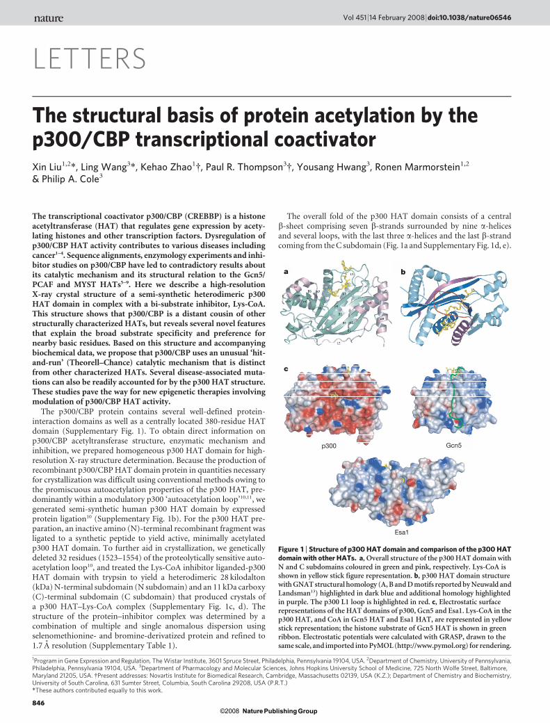
other HATs. In particular, regions flanking the central core regionand the unusually long L1 loop that appears to encapsulate the Lys-CoA inhibitor are unique features of the p300 HAT domain (Fig. 1b).Although the pantetheine arms occupy similar positions with respectto the core domain of all three HATs, the adenosine rings adoptdiverse conformations. The adenosine rings in the Gcn5 HAT andEsa1 HAT structures are less ordered compared with their well-ordered counterpart bound to p300 HAT, potentially one of theconsequences of the L1 loop interactions with the CoA portion ofthe inhibitor (Fig. 2a and Supplementary Fig. 2). Indeed, the L1 loopcontributes about 30% of the total buried solvent accessible surface of1225 A2 of the CoA portion of Lys-CoA. The L1 loop also appears tobe in position to influence protein substrate binding, burying 266 A2
of the lysine portion of the Lys-CoA inhibitor. In addition, com-parison of the electrostatic surface potential of the substrate-bindingsurfaces of the HAT domains of Gcn5, Esa1 and p300 shows signifi-cant divergence (Fig. 1c). Although Gcn5 and Esa1 show deeper andmore apolar substrate-binding pockets, the p300 HAT domainreveals a shallow and highly acidic site, consistent with the differentsubstrate-binding properties of p300 HAT relative to other HATs.
The CoA portion of the Lys-CoA makes extensive interactionswith the p300 HAT domain (Fig. 2a, b). In particular, the pantetheinearm of the CoA portion of the inhibitor makes extensive interactionswith the p300 L1 loop, which in turn appears to help configure theL1 loop to make extensive intramolecular interactions with otherregions of the p300 HAT domain (Fig. 2c). In addition, Arg1410appears to make several key hydrogen bonds with phosphates ofthe Lys-CoA molecule, and we have confirmed its importance in39-phosphate interaction by mutagenesis and inhibition studies with39-dephospho-Lys-CoA14 (Fig. 2d, Supplementary Figs 3 and 4 andSupplementary Table 2). The lysine component of the Lys-CoAinhibitor also makes extensive interactions with the p300 HATdomain (Fig. 2e). Specifically, the e-nitrogen atom makes a hydrogenbond with the main chain oxygen of Trp1436; and three hydrophobicresidues Tyr1397, Trp1436 and Tyr1446, together with Cys1438,form a hydrophobic tunnel that interacts with the aliphatic portionof the lysine side chain.
In addition to the pocket that accommodates the lysine moiety ofLys-CoA, presumably mimicking the lysine side chain from nativeprotein substrates, a second pronounced and highly electronegativepocket is present about 10 A away from the substrate lysine (Fig. 3a).Because p300 substrates show a strong preference for the presence of aLys or Arg side chain three to four residues downstream or upstreamof the primary acetylation site (ref. 9) (Fig. 3b), we considered thepossibility that this region might be important for the binding ofprotein substrates. A narrow, shallow and electronegative grooveconnects these two pockets, with the wall of one side of the groovecomposed of Ser1396 and Tyr1397 (Fig. 3a). The negative potentialof the second pocket is largely formed by the side-chain atoms ofresidues Thr1357, Glu1505, Asp1625 and Asp1628. We performed
single, double, triple and quadruple amino-acid mutagenesis on thementioned residues. As summarized in Fig. 3c and SupplementaryTable 2, these mutants show significantly decreased p300 HAT acti-vity, supporting the importance of this region for peptide substratebinding.
We selected the D1625R/D1628R p300 HAT mutant for furtheranalysis of substrate residue preference. To test the influence of elec-trostatic interactions on substrate sequence selection, we determinedthe steady-state kinetic parameters for several synthetic H4 tail pep-tide analogues (listed in Supplementary Table 3). In H4-15, there aretwo other basic residues (Lys5 and Lys12) near the target Lys8. Here,we compared substrates containing substitutions of Lys5 and Lys12as well as the simple substrate Ac-Lys-NH2 with wild-type andmutant p300 HAT. As shown in Supplementary Table 3, the 22-foldV/K difference between wild-type p300 HAT protein and theD1625R/D1628R mutant for H4-15 substrate drops to about sixfold
Substrate-binding loop L1
a
b c
d e
HH2O
Figure 2 | Lys-CoA recognition by p300. a, Schematic views of p300HAT domain interactions with Lys-CoA. Hydrophobic interactions andhydrogen bonds are indicated with solid and dotted arrows, respectively.b, Representative Fo 2 Fc electron-density omit map contoured at 1.5s forLys-CoA; the L1 loop is highlighted in red. Blow-up regions are indicated forpanels C–E. c, Blow-up view of the tip of the L1 loop. Residues involved inhydrogen-bonding interactions are highlighted in cyan; residues involved inhydrophobic interactions are coloured in red. Green and pink regions of theprotein indicate the N and C subdomains, respectively. All hydrogen bondsinvolving main chain atoms are omitted for clarity. d, Blow-up view ofinteractions stabilizing the ADP–ribose portion of Lys-CoA. e, Blow-up viewof the lysine acetylation reaction centre with all nearby nucleophilic residuesshown in green stick figure.
NATURE | Vol 451 | 14 February 2008 LETTERS
847Nature Publishing Group©2008
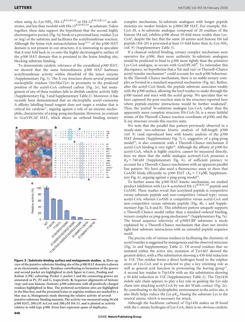
when using Ac-Lys-NH2, H4-12K5A/K12A or H4-12K5Cit/K12A as sub-strates, and less than twofold with H4-15K5D/K12D as substrate. Takentogether, these data support the hypothesis that the second highlyelectronegative pocket (Fig. 3a) binds to a proximal basic residue (Lysor Arg) of the substrate and facilitates the acetyltransferase reaction.Although the lysine-rich autoacetylation loop10,11 of the p300 HATdomain is not present in our structure, it is interesting to speculatethat it may fold back in cis onto the highly electronegative surface ofthe p300 HAT domain that is proximal to the lysine binding site,blocking substrate binding.
To demonstrate catalytic relevance of the crystallized p300 HAT,we showed that the same heterodimeric p300 HAT harboursacetyltransferase activity within threefold of the intact enzyme(Supplementary Fig. 5). The X-ray structure shows several potentialnucleophilic residues (Ser/His/Tyr) in proximity to the predictedposition of the acetyl-CoA carbonyl carbon (Fig. 2e), but muta-genesis of any of these residues fails to abolish catalytic activity fully(Supplementary Fig. 3 and Supplementary Table 2). Moreover, it hasrecently been demonstrated that an electrophilic acetyl-coenzymeA affinity labelling-based reagent does not target a residue that iscritical for catalysis15, arguing against a role of an enzyme nucleo-phile, characteristic of a ping-pong mechanism. However, in contrastto Gcn5/PCAF HAT, which shows an ordered binding ternary
complex mechanism, bi-substrate analogues with longer peptidemoieties are weaker binders to p300/CBP HAT. For example, H4-CoA-20, a bi-substrate analogue composed of 20 residues of thehistone H4 tail, inhibits p300 about 20-fold more weakly than Lys-CoA8, despite the fact that the same 20-amino-acid histone H4 tailpeptide (H4-20) is processed at least 15-fold faster than Ac-Lys-NH2
(ref. 9) (Supplementary Table 3).If a classical ordered binding, ternary complex mechanism were
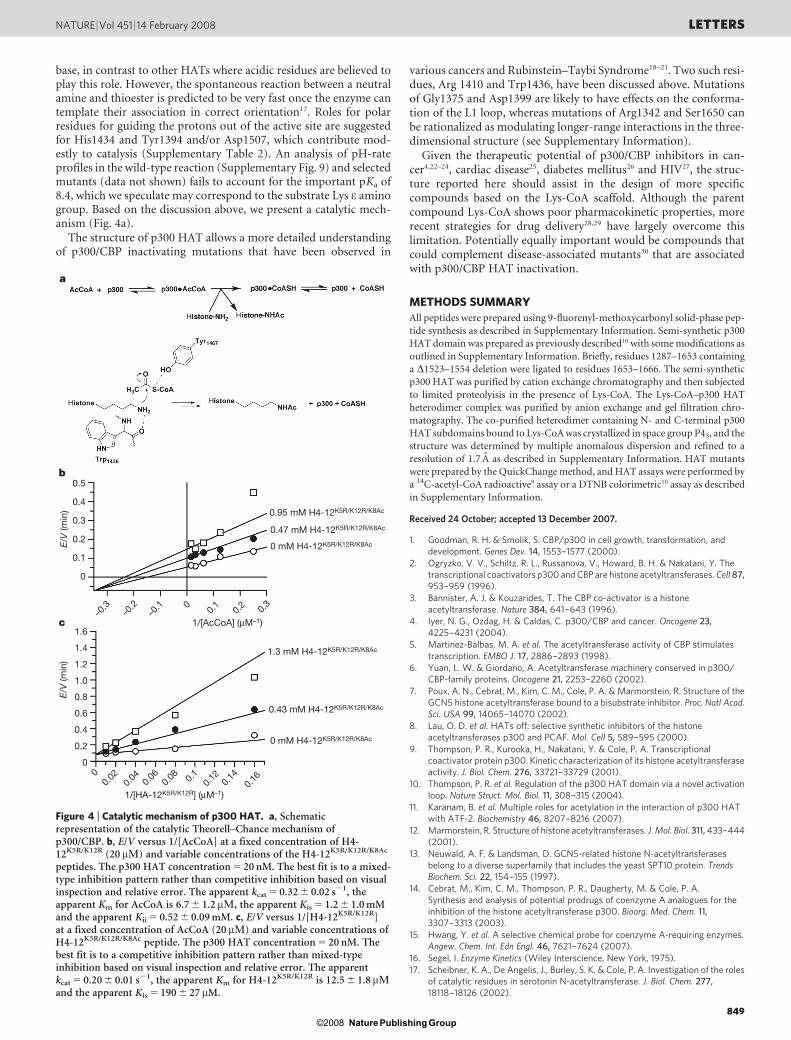
operative for p300, then more authentic bi-substrate analogueswould be predicted to bind to p300 more tightly than the primitiveLys-CoA analogue, as occurs with Gcn5/PCAF8. To rationalize thisdiscrepancy, we hypothesize that a ‘hit-and-run’ or Theorell–Chanceacetyl transfer mechanism16 could account for such p300 behaviour.In the Theorell–Chance mechanism, there is no stable ternary com-plex as formed in a standard sequential mechanism (Fig. 4a). Rather,after the acetyl-CoA binds, the peptide substrate associates weaklywith the p300 surface, allowing the lysyl residue to snake through thep300 tunnel and react with the acetyl group. We speculate that wehave captured the post-reaction state in the structure reported herewhere peptide-enzyme interactions would be further weakened7.Thus, the ‘partial’ bi-substrate analogue Lys-CoA, rather than theapparently more complete structure H4-CoA-20, is a more faithfulmimic of the Theorell–Chance reaction coordinate of p300, and theX-ray structure reveals this reactive state.
We note that the parallel line pattern previously observed in asteady-state two-substrate kinetic analysis of full-length p300(ref. 9) (and reproduced here with kinetic analysis of the p300HAT domain (Supplementary Fig. 7c)), suggestive of a ping-pongmodel16, is also consistent with a Theorell–Chance mechanism ifacetyl-CoA binding is very tight16. Although the affinity of p300 foracetyl-CoA, which is highly reactive, cannot be measured directly,here we show that the stable analogue acetonyl-CoA possesses aKd 5 740 nM (Supplementary Fig. 6), of sufficient potency toaccount for a Theorell–Chance mechanism with an apparent parallelline pattern. We have also used a fluorescence assay to show thatCoASH binds efficiently to p300 HAT (Kd 5 7.3 mM, Supplemen-tary Fig. 6), arguing against a ping-pong model16.
To further assess the p300 HAT kinetic mechanism, we studiedproduct inhibition with Lys-8-acetylated H4-12K5R/K12R peptide andCoASH. These studies reveal that acetylated peptide is competitiveversus substrate peptide and non-competitive (mixed-type) versusacetyl-CoA; whereas CoASH is competitive versus acetyl-CoA andnon-competitive versus substrate peptide (Fig. 4b, c and Supple-mentary Figs 7a, b and 8). This inhibitory pattern uniquely supportsa Theorell–Chance model rather than a standard ordered binding,ternary complex or ping-pong mechanism16 (Supplementary Fig. 7a).The broad sequence selectivity of p300/CBP substrates is nicelyexplained by a Theorell–Chance mechanism that does not involvetight-knit substrate interactions with an extended peptide sequencemotif.
The precise role of various residues in facilitating the chemistry ofacetyl transfer is suggested by mutagenesis and the observed structure(Fig. 2e and Supplementary Table 2). Of several residues that wemutated within the active site, mutation of Tyr1467 showed thegreatest defect, with a Phe substitution showing a 430-fold reductionin V/K. This residue forms a direct hydrogen bond to the sulphuratom of Lys-CoA and is predicted to play a key orienting role aswell as general acid function in protonating the leaving group17.A second key residue is Trp1436 with an Ala substitution showinga 50-fold reduction in V/K (Supplementary Table 2). The Trp1436indole side chain appears to play a key role in guiding the Lys sidechain into attacking acetyl-CoA by van der Waals contact (Fig. 2e).By contributing to the hydrophobic environment in the active site, italso likely helps reduce the Lys pKa, biasing the substrate Lys to theneutral amine, which is necessary for attack.
Although the backbone carbonyl of Trp1436 makes an H-bondwith the e-amino hydrogen of Lys-CoA, there is no obvious catalytic
––––
–––––––––––––––
Rel
ativ
e ac
tivity
(%) 100
80
60
40
20
0
WT
T135
7R
T135
7L
D1625
R/D16
28R
E1505
R/D16
25R/D
1628
R
T135
7R/E
1505
R/D16
25R/D
1628
R
S1396
R/Y13
97R
S1396
W/Y
1397
W
b
c
a
Figure 3 | Substrate-binding surface and mutagenesis studies. a, Blow-upview of the putative substrate-binding site of the p300 HAT domain is shownas an electrostatic surface. Residues contributing to formation of the grooveand second pocket are highlighted in stick figure in Corey, Pauling andKoltun (CPK) colouring. Pocket 1, pocket 2 and the connecting groove areannotated as P1, P2 and G, respectively. b, Sequence alignments of histone(top) and non-histone (bottom) p300 substrates with all positively chargedresidues highlighted in blue. The preferred acetylation sites are highlightedin the blue box, and the proximal lysine or arginine residues are marked by ablue star. c, Mutagenesis study showing the relative activity of severalputative substrate-binding mutants. The activity was measured using 50 nMp300 HAT, 200mM AcCoA and 200mM H4-15, and is plotted as activityrelative to wild-type p300. Error bars represent spans of duplicates.
LETTERS NATURE | Vol 451 | 14 February 2008
848Nature Publishing Group©2008
base, in contrast to other HATs where acidic residues are believed toplay this role. However, the spontaneous reaction between a neutralamine and thioester is predicted to be very fast once the enzyme cantemplate their association in correct orientation17. Roles for polarresidues for guiding the protons out of the active site are suggestedfor His1434 and Tyr1394 and/or Asp1507, which contribute mod-estly to catalysis (Supplementary Table 2). An analysis of pH-rateprofiles in the wild-type reaction (Supplementary Fig. 9) and selectedmutants (data not shown) fails to account for the important pKa of8.4, which we speculate may correspond to the substrate Lys e aminogroup. Based on the discussion above, we present a catalytic mech-anism (Fig. 4a).
The structure of p300 HAT allows a more detailed understandingof p300/CBP inactivating mutations that have been observed in
various cancers and Rubinstein–Taybi Syndrome18–21. Two such resi-dues, Arg 1410 and Trp1436, have been discussed above. Mutationsof Gly1375 and Asp1399 are likely to have effects on the conforma-tion of the L1 loop, whereas mutations of Arg1342 and Ser1650 canbe rationalized as modulating longer-range interactions in the three-dimensional structure (see Supplementary Information).
Given the therapeutic potential of p300/CBP inhibitors in can-cer4,22–24, cardiac disease25, diabetes mellitus26 and HIV27, the struc-ture reported here should assist in the design of more specificcompounds based on the Lys-CoA scaffold. Although the parentcompound Lys-CoA shows poor pharmacokinetic properties, morerecent strategies for drug delivery28,29 have largely overcome thislimitation. Potentially equally important would be compounds thatcould complement disease-associated mutants30 that are associatedwith p300/CBP HAT inactivation.
METHODS SUMMARY
All peptides were prepared using 9-fluorenyl-methoxycarbonyl solid-phase pep-
tide synthesis as described in Supplementary Information. Semi-synthetic p300
HAT domain was prepared as previously described10 with some modifications as
outlined in Supplementary Information. Briefly, residues 1287–1653 containing
a D1523–1554 deletion were ligated to residues 1653–1666. The semi-synthetic
p300 HAT was purified by cation exchange chromatography and then subjected
to limited proteolyisis in the presence of Lys-CoA. The Lys-CoA–p300 HAT
heterodimer complex was purified by anion exchange and gel filtration chro-
matography. The co-purified heterodimer containing N- and C-terminal p300
HAT subdomains bound to Lys-CoA was crystallized in space group P43, and the
structure was determined by multiple anomalous dispersion and refined to a
resolution of 1.7 A as described in Supplementary Information. HAT mutants
were prepared by the QuickChange method, and HAT assays were performed by
a 14C-acetyl-CoA radioactive9 assay or a DTNB colorimetric10 assay as described
in Supplementary Information.
Received 24 October; accepted 13 December 2007.
1. Goodman, R. H. & Smolik, S. CBP/p300 in cell growth, transformation, anddevelopment. Genes Dev. 14, 1553–1577 (2000).
2. Ogryzko, V. V., Schiltz, R. L., Russanova, V., Howard, B. H. & Nakatani, Y. Thetranscriptional coactivators p300 and CBP are histone acetyltransferases. Cell 87,953–959 (1996).
3. Bannister, A. J. & Kouzarides, T. The CBP co-activator is a histoneacetyltransferase. Nature 384, 641–643 (1996).
4. Iyer, N. G., Ozdag, H. & Caldas, C. p300/CBP and cancer. Oncogene 23,4225–4231 (2004).
5. Martinez-Balbas, M. A. et al. The acetyltransferase activity of CBP stimulatestranscription. EMBO J. 17, 2886–2893 (1998).
6. Yuan, L. W. & Giordano, A. Acetyltransferase machinery conserved in p300/CBP-family proteins. Oncogene 21, 2253–2260 (2002).
7. Poux, A. N., Cebrat, M., Kim, C. M., Cole, P. A. & Marmorstein, R. Structure of theGCN5 histone acetyltransferase bound to a bisubstrate inhibitor. Proc. Natl Acad.Sci. USA 99, 14065–14070 (2002).
8. Lau, O. D. et al. HATs off: selective synthetic inhibitors of the histoneacetyltransferases p300 and PCAF. Mol. Cell 5, 589–595 (2000).
9. Thompson, P. R., Kurooka, H., Nakatani, Y. & Cole, P. A. Transcriptionalcoactivator protein p300. Kinetic characterization of its histone acetyltransferaseactivity. J. Biol. Chem. 276, 33721–33729 (2001).
10. Thompson, P. R. et al. Regulation of the p300 HAT domain via a novel activationloop. Nature Struct. Mol. Biol. 11, 308–315 (2004).
11. Karanam, B. et al. Multiple roles for acetylation in the interaction of p300 HATwith ATF-2. Biochemistry 46, 8207–8216 (2007).
12. Marmorstein, R. Structure of histone acetyltransferases. J. Mol. Biol. 311, 433–444(2001).
13. Neuwald, A. F. & Landsman, D. GCN5-related histone N-acetyltransferasesbelong to a diverse superfamily that includes the yeast SPT10 protein. TrendsBiochem. Sci. 22, 154–155 (1997).
14. Cebrat, M., Kim, C. M., Thompson, P. R., Daugherty, M. & Cole, P. A.Synthesis and analysis of potential prodrugs of coenzyme A analogues for theinhibition of the histone acetyltransferase p300. Bioorg. Med. Chem. 11,3307–3313 (2003).
15. Hwang, Y. et al. A selective chemical probe for coenzyme A-requiring enzymes.Angew. Chem. Int. Edn Engl. 46, 7621–7624 (2007).
16. Segel, I. Enzyme Kinetics (Wiley Interscience, New York, 1975).17. Scheibner, K. A., De Angelis, J., Burley, S. K. & Cole, P. A. Investigation of the roles
of catalytic residues in serotonin N-acetyltransferase. J. Biol. Chem. 277,18118–18126 (2002).
0.5
0.4
0.3
0.2
0.1
0
–0.3
–0.2
–0.1 0 0.
30.
20.1
1/[AcCoA] (µM–1)
0.95 mM H4-12K5R/K12R/K8Ac
0.47 mM H4-12K5R/K12R/K8Ac
0 mM H4-12K5R/K12R/K8AcE/V
(min
)
1.6
1.4
1.2
1.0
0.8
0.6
0.4
0.2
0
E/V
(min
)
1/[HA-12K5R/K12R] (µM–1)
00.
020.
04 0.06
0.08 0.
10.
12 0.14
0.16
1.3 mM H4-12K5R/K12R/K8Ac
0.43 mM H4-12K5R/K12R/K8Ac
0 mM H4-12K5R/K12R/K8Ac
H H
HH
a
b
c
Figure 4 | Catalytic mechanism of p300 HAT. a, Schematicrepresentation of the catalytic Theorell–Chance mechanism ofp300/CBP. b, E/V versus 1/[AcCoA] at a fixed concentration of H4-12K5R/K12R (20 mM) and variable concentrations of the H4-12K5R/K12R/K8Ac
peptides. The p300 HAT concentration 5 20 nM. The best fit is to a mixed-type inhibition pattern rather than competitive inhibition based on visualinspection and relative error. The apparent kcat 5 0.32 6 0.02 s21, theapparent Km for AcCoA is 6.7 6 1.2 mM, the apparent Kis 5 1.2 6 1.0 mMand the apparent Kii 5 0.52 6 0.09 mM. c, E/V versus 1/[H4-12K5R/K12R]at a fixed concentration of AcCoA (20 mM) and variable concentrations ofH4-12K5R/K12R/K8Ac peptide. The p300 HAT concentration 5 20 nM. Thebest fit is to a competitive inhibition pattern rather than mixed-typeinhibition based on visual inspection and relative error. The apparentkcat 5 0.20 6 0.01 s21, the apparent Km for H4-12K5R/K12R is 12.5 6 1.8 mMand the apparent Kis 5 190 6 27 mM.
NATURE | Vol 451 | 14 February 2008 LETTERS
849Nature Publishing Group©2008

18. Murata, T. et al. Defect of histone acetyltransferase activity of the nucleartranscriptional coactivator CBP in Rubinstein-Taybi syndrome. Hum. Mol. Genet.10, 1071–1076 (2001).
19. Muraoka, M. et al. p300 gene alterations in colorectal and gastric carcinomas.Oncogene 12, 1565–1569 (1996).
20. Gayther, S. A. et al. Mutations truncating the EP300 acetylase in human cancers.Nature Genet. 24, 300–303 (2000).
21. Kishimoto, M. et al. Mutations and deletions of the CBP gene in human lungcancer. Clin. Cancer Res. 11, 512–519 (2005).
22. Zheng, Y. et al. Selective HAT inhibitors as mechanistic tools for proteinacetylation. Methods Enzymol. 376, 188–199 (2004).
23. Stimson, L. et al. Isothiazolones as inhibitors of PCAF and p300 histoneacetyltransferase activity. Mol. Cancer Ther. 4, 1521–1532 (2005).
24. Iyer, N. G. et al. p300 regulates p53-dependent apoptosis after DNA damage incolorectal cancer cells by modulation of PUMA/p21 levels. Proc. Natl Acad. Sci.USA 101, 7386–7391 (2004).
25. Davidson, S. M. et al. The transcriptional coactivator p300 plays a critical role inthe hypertrophic and protective pathways induced by phenylephrine in cardiaccells but is specific to the hypertrophic effect of urocortin. ChemBioChem 6,162–170 (2005).
26. Zhou, X. Y. et al. Insulin regulation of hepatic gluconeogenesis throughphosphorylation of CREB-binding protein. Nature Med. 10, 633–637 (2004).
27. Varier, R. A. & Kundu, T. K. Chromatin modifications (acetylation/ deacetylation/methylation) as new targets for HIV therapy. Curr. Pharm. Des. 12, 1975–1993(2006).
28. Guidez, F. et al. Histone acetyltransferase activity of p300 is required fortranscriptional repression by the promyelocytic leukemia zinc finger protein. Mol.Cell. Biol. 25, 5552–5566 (2005).
29. Zheng, Y. et al. Synthesis and evaluation of a potent and selective cell-permeablep300 histone acetyltransferase inhibitor. J. Am. Chem. Soc. 127, 17182–17183 (2005).
30. Qiao, Y., Molina, H., Pandey, A., Zhang, J. & Cole, P. A. Chemical rescue of amutant enzyme in living cells. Science 311, 1293–1297 (2006).
Supplementary Information is linked to the online version of the paper atwww.nature.com/nature.
Acknowledgements We appreciate advice on the manuscript from W. Cleland,J. Stivers, A. Mildvan and D. Leahy. We thank the National Institutes of Health andthe FAMRI, Kaufman and Keck foundations for financial support.
Author Contributions X.L. and L.W. designed and performed reported experimentsand prepared manuscript figures and text; K.Z. and P.R.T. developed key reagentsand performed preliminary studies that led to the reported experiments; Y.H.developed a key reagent and designed and interpreted some of the experiments.R.M. and P.A.C. designed and supervised experiments and prepared manuscripttext. All authors read and approved the submitted manuscript.
Author Information The structure of the p300 HAT–Lys-CoA complex has beensubmitted to the Protein Data Bank under accession number 3BIY. Reprints andpermissions information is available at www.nature.com/reprints.Correspondence and requests for materials should be addressed to R.M.([email protected]) or P.A.C. ([email protected]).
LETTERS NATURE | Vol 451 | 14 February 2008
850Nature Publishing Group©2008