Embed Size (px)
Citation preview
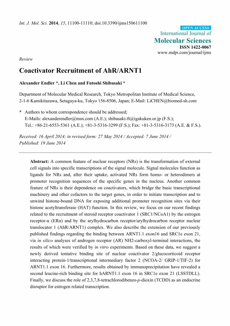
Int. J. Mol. Sci. 2014, 15, 11100-11110; doi:10.3390/ijms150611100
International Journal of
Molecular Sciences ISSN 1422-0067
www.mdpi.com/journal/ijms
Review
Coactivator Recruitment of AhR/ARNT1
Alexander Endler *, Li Chen and Futoshi Shibasaki *
Department of Molecular Medical Research, Tokyo Metropolitan Institute of Medical Science,
2-1-6 Kamikitazawa, Setagaya-ku, Tokyo 156-8506, Japan; E-Mail: [email protected]
* Authors to whom correspondence should be addressed;
E-Mails: [email protected] (A.E.); [email protected] (F.S.);
Tel.: +86-21-6553-5361 (A.E.); +81-3-5316-3299 (F.S.); Fax: +81-3-5316-3173 (A.E. & F.S.).
Received: 16 April 2014; in revised form: 27 May 2014 / Accepted: 7 June 2014 /
Published: 19 June 2014
Abstract: A common feature of nuclear receptors (NRs) is the transformation of external
cell signals into specific transcriptions of the signal molecule. Signal molecules function as
ligands for NRs and, after their uptake, activated NRs form homo- or heterodimers at
promoter recognition sequences of the specific genes in the nucleus. Another common
feature of NRs is their dependence on coactivators, which bridge the basic transcriptional
machinery and other cofactors to the target genes, in order to initiate transcription and to
unwind histone-bound DNA for exposing additional promoter recognition sites via their
histone acetyltransferase (HAT) function. In this review, we focus on our recent findings
related to the recruitment of steroid receptor coactivator 1 (SRC1/NCoA1) by the estrogen
receptor-α (ERα) and by the arylhydrocarbon receptor/arylhydrocarbon receptor nuclear
translocator 1 (AhR/ARNT1) complex. We also describe the extension of our previously
published findings regarding the binding between ARNT1.1 exon16 and SRC1e exon 21,
via in silico analyses of androgen receptor (AR) NH2-carboxyl-terminal interactions, the
results of which were verified by in vitro experiments. Based on these data, we suggest a
newly derived tentative binding site of nuclear coactivator 2/glucocorticoid receptor
interacting protein-1/transcriptional intermediary factor 2 (NCOA-2/ GRIP-1/TIF-2) for
ARNT1.1 exon 16. Furthermore, results obtained by immunoprecipitation have revealed a
second leucine-rich binding site for hARNT1.1 exon 16 in SRC1e exon 21 (LSSTDLL).
Finally, we discuss the role of 2,3,7,8-tetrachlorodibenzo-p-dioxin (TCDD) as an endocrine
disruptor for estrogen related transcription.
OPEN ACCESS
Int. J. Mol. Sci. 2014, 15 11101
Keywords: SRC1; NCoA2; AhR; ARNT; TCDD; ER; AR
1. Basic Signal Transmission Mechanisms of Nuclear Receptors
Nuclear receptors can be categorized not only based on the nature of their DNA binding sites, but
also according to whether they form homodimers, such as the estrogen receptor (ER), progesterone
receptor (PR), androgen receptor (AR), glucocorticoid receptor (GR), and mineralocorticoid receptor
(MR), or heterodimers, such as the retinoic acid receptor (RAR), thyroid hormone receptor (TR), and
vitamin D3 receptor (VDR). The latter one interact with the receptor for 9-cis retinoic acid (RXR) [1].
A common characteristic of all NRs is that, after penetration of hydrophobic chemicals through the cell
membrane, they incorporate their specific ligands into the ligand binding domains (LBD), which are
highly variable within the NRs, due to their ligand specificities [2]. In the absence of ligands, cytosolic
NRs are coupled to heat shock protein hsp90, hsp70, Hop, hsp40, and p23 multi-protein complexes [3],
which enables them to bind ligands. In contrast RAR, TR, and VDR reside in the nucleus, with binding
only to corepressors in their inactive state.
After agonist uptake, NRs undergo dimer formation, leading to accessibility of coactivators and
release of corepressor proteins. The canonical pathway for coactivator recruitment of NRs, after
agonist binding in their LBDs, is a helix 12 realignment with helices 3, 5/6, and 11, which, thereby,
form a lid on the LBD for the engulfed ligands. The LBD contains a conserved activation function 2
(AF2) domain, which is in the ligand activated conformation the connective link to LXXLL domains
in the central nuclear receptor interaction domain (NID) of steroid receptor coactivators (SRCs),
also called the p160 SRC family [4]. SRCs interact with NRs as coactivators and facilitate their
transcription via their histone acetylase (HAT) activity [5,6]. In addition, the activation domain 1 (AD1)
of SRCs binds to the c-terminal SRC interaction domain (SID) of p300, as well as its homolog, the cAMP
response element-binding (CREB) protein (CBP) [7]. The p300/CBP complex plays an important role in
the transcription process through connecting other transcriptional activators, basal transcription factors,
and HATs to the transcriptional machinery [8].
2. AhR/ARNT-Related Transcription
The aryl hydrocarbon receptor (AhR), which is, like ARNT, a member of the basic helix-loop-helix
(bHLH) Per-ARNT-SIM (PAS) family, recognizes the halogenated aromatic hydrocarbon 2, 3, 7,
8-Tetrachlorodibenzodioxin (TCDD) or the polycyclic aromatic hydrocarbon (PAH) 3-methylcholanthrene
(3MC) as ligands. Unliganded AhR resides in the cytosol and forms a complex with hsp90, hsp23 [9],
AhR-activated 9 (ARA9) [10], and hepatitis B virus X-associated protein 2 (XAP2), which is similar to
immunophilin [11]. After ligand uptake, AhR forms an AhR/ARNT1 complex with its obligate partner
ARNT1 and transcribes cytochrome P450s (Phase I), as well as UDP-glucuronosyltransferase and
glutathione-S-transferase (GST) (Phase II) via xenobiotic responsive elements (XREs) within their
promoter regions [12]. Unlike other NRs, a unique major endogenous ligand for the AhR has not been
established, but the following ligands have been reported as agonists: tryptophan (Trp) catabolite
kynurenine (Kyn) [13], indigo and indirubin [14], bilirubin [15], and prostaglandins [16]. According
Int. J. Mol. Sci. 2014, 15 11102
to a hypothesis developed in the early 1990s, enzymes of the cytochrome P450 family originally
evolved as animal-plant “warfare” enzymes to protect themselves from toxic chemicals produced by
plants [17]. Later, Denison and Whitlock proposed that pathways of the cytochrome P450 enzymes
help to maintain homeostasis of endogenous lipophilic substances [18,19].
3. AhR-Coactivator Interactions
The AhR is a ligand-induced transcription factor but lacks an AF2 domain. Since the AF2 domain is
the common link between transcription factors and the SRCs, the question arose how the AhR is
recruiting coactivators. Kim et al. described a coiled-coil coactivator (CoCoA), which is a secondary
coactivator for NRs interacting with the bHLH-PAS domain of p160 proteins, but also functions as
a potent primary coactivator for AhR/ARNT transcription also via bHLH-PAS domain interaction,
underlining, that AhR/ARNT can recruit several coactivators [20]. The authors noted that AhR bound
CoCoA in a ligand-independent manner, and suggested that CoCoA may exist in a cytosolic complex
with AhR before ligand activation, and travels with AhR to its target promoter after ligand binding.
Others demonstrated a TCDD and LXXLL motif-dependent SRC1 interaction with a Q-rich
subdomain of the AhR [21]. Notably, the transcriptional activity of the AhR also depends on the type
of ligand. In the case of AhR agonist application, ordinary transcription with cyclic recruitment of
NCOA1, NCOA2, and NCOA3 (each present on the promoter) simultaneously occurs with
concomitant enhanced CYP1A1 transcription. In contrast, the AhR antagonist 3,3'-diindolylmethane
(DIM) promotes AhR nuclear translocation and p160 coactivator recruitment, but fails to recruit Pol II
or cause histone acetylation, suggesting that ligand-dependent changes in AhR conformation can affect
its transcriptional activity [22].
4. Coactivator Recruitment to the AhR/ARNT Complex
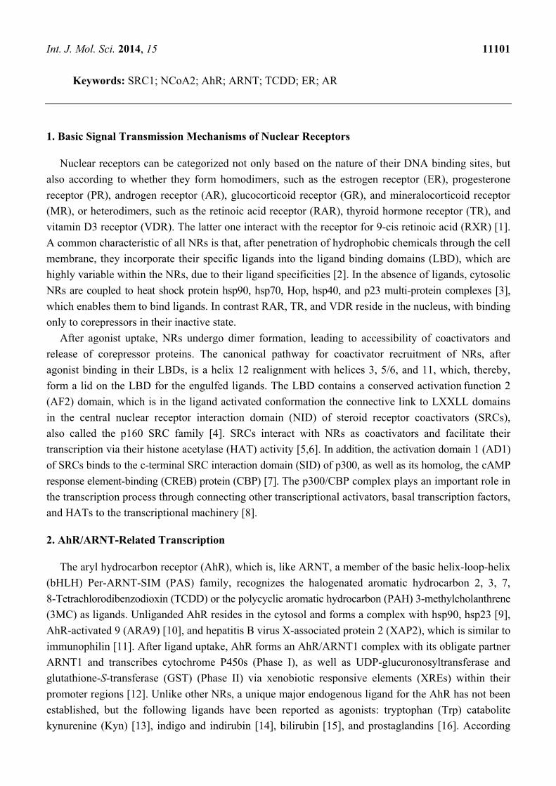
In a recent publication, we described an AF2 domain in ARNT1 (Figure 1) and suggested that the
AhR/ARNT1 complex is a transcription factor complex in which the LBD and the AF2 domain are
distributed on two factors [23]. For complete transcriptional activity of the AhR/ARNT1 complex, two
cyclin degradation boxes adjacent to the ARNT1 AF2 domain are necessary, which lead to accelerated
CYP1A1 transcription (for transcription factor activation via degradation, refer to the review by
Lipford and Deshaies [24]). We found, that the AF2 domains of both ARNT1 and ERα- bind to the
exon 21 of SRC1e, and these interactions are crucial for TCDD- and 17β-estradiol (E2)-related
transcriptions. ARNT1 exists as a splice variant (ARNT1.4) without exon 16, which contains the AF2
domain, and SRC1e also exists as a splice variant without the binding site for ARNT1-AF2 and
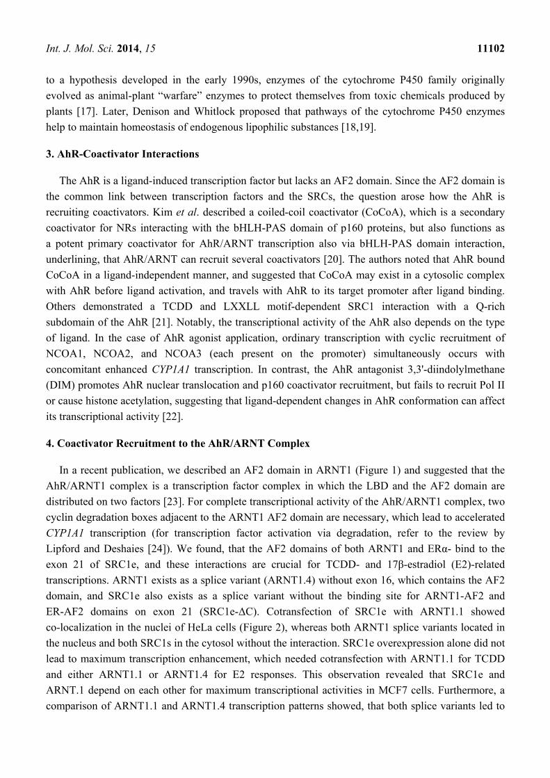
ER-AF2 domains on exon 21 (SRC1e-ΔC). Cotransfection of SRC1e with ARNT1.1 showed
co-localization in the nuclei of HeLa cells (Figure 2), whereas both ARNT1 splice variants located in
the nucleus and both SRC1s in the cytosol without the interaction. SRC1e overexpression alone did not
lead to maximum transcription enhancement, which needed cotransfection with ARNT1.1 for TCDD
and either ARNT1.1 or ARNT1.4 for E2 responses. This observation revealed that SRC1e and
ARNT.1 depend on each other for maximum transcriptional activities in MCF7 cells. Furthermore, a
comparison of ARNT1.1 and ARNT1.4 transcription patterns showed, that both splice variants led to
Int. J. Mol. Sci. 2014, 15 11103
similar activities regarding hypoxia and ER-related responses, whereas TCDD transcription was
dependent on the intact exon 16 of ARNT1.1 [23].
Figure 1. Scheme of AF2 domains. The boxes indicate hydrophobic amino acids beside the
conserved glutamic acid. Underlined amino acids in hAR indicate hydrophobic residues,
which serve as coactivator interface [25].
Figure 2. HeLa cells after cotransfection with the indicated hARNT1.1, hARNT1.4,
SRC1e, and SRC1e-ΔC constructs. Green indicates GFP, red indicates HA-CY3 staining,
and blue indicates nuclear DAPI staining (adapted from [23]). Twenty-four hours after
transfection, fixed cells were incubated with HA antibodies (12CA5 hybridomas, MBL) for
1 h, washed, and then incubated with Cyanine 3 (Cy3)-conjugated secondary antibodies for
30 min. The cells were then washed again and mounted in Vectashield (Vector Laboratories,
Burlingame, CA, USA) mounting medium containing 4,6-diamidino-2-phenylindole (DAPI).
Fluorescence images were visualized using an Olympus 1670 inverted system microscope
(Olympus Optical Co., Ltd, Tokyo, Japan) equipped with a charge-coupled device
(Magnification ×1000).
Int. J. Mol. Sci. 2014, 15 11104
Figure 3. Evaluation of the hARNT1.1 AF-2 binding site in SRC1e exon 21. (A) Scheme
of SRC1e and amino acid composition of exon 21 (upper image) and alignment with
NCOA2 exon 26. Underlined aa are the binding sites for hARNT1.1 AF-2; (B) Comparison
of hydropathy pattern between C-terminal binding peptides for AF2 bindings in hAR [26]
and the common NDPAL sequence in exon 21 of SRC1e and exon 26 of NCOA2;
(C) Analysis of hARNT1.1 and SRC1e exon 16 interactions with immunoprecipitation of
HA- tagged GFP-SRC1e-exon 21 aa stretches and MYC-tagged GFP-hARNT1.1-exon 16
with MYC-beads. Upper panel: WB of MYC-tagged GFP-hARNT1.1-exon 16 after
MYC-beads immunoprecipitation with poly-MYC antibodies; Middle panel: WB of
indicated HA-tagged GFP-SRC1e-exon 21 aa stretches with poly-HA antibodies (cell
lysates); Lower panel: WB after MYC-beads immunoprecipitation with poly HA
antibodies. Abbreviations: PAS, Per/ARNT/SIM domain; bHLH, basic helix-loop-helix
domain; NID, nuclear receptor interaction domain; AD1 and AD2, activation domain 1 and
activation domain 2. (Transfected HeLa cells were lysed and the supernatant was incubated
with anti-myc-conjugated agarose (Sigma-Aldrich, St. Louis, MO, USA) overnight at 4 °C.
The resulting precipitate was washed with RIPA buffer and then boiled in 2xSDS sample buffer
for 10 min. Co-immunoprecipitated HA tagged NDPAL, NDPAK, and LSSTDLL were
analyzed by Western blotting with anti-HA-polyclonal antibodies (Sigma-Aldrich, St. Louis,
MO, USA) followed by horseradish-peroxidase-conjugated anti-rabbit IgG (Thermo Fisher
Scientific Inc., Rockford, IL, USA) incubation for 1 h. Immunoprecipitate analysis was done
using a SuperSignal West Dura chemiluminescent detection system (Thermo Fisher Scientific
Inc., Rockford, IL, USA).
For this review, we further analyzed the binding of SRC1e-exon 21 and the AF2 domain of
ARNT1.1. As shown in Figure 3A, the alignment of SRC1e-exon 21 and NCOA2-exon 26 showed
a common peptide with the amino acid sequence NDPALR. Because the binding between ARNT1
and SRC1e is LXXLL motif independent, we investigated other LXXLL independent interfaces for
Int. J. Mol. Sci. 2014, 15 11105
NR-AF2 domains. After ligand binding, the AR exhibits an amino-carboxy terminal interaction, and a
similar mechanism has been postulated for the PR [27]. For the AR, the three amphipathic 23FQNLF27, 179LKDIL183, and 432WHTLF436 a-helices within the AR interact with its AF2 hydrophobic groove.
Particularly, FQNLF has a five-fold higher affinity for the AR-AF2 domain than for LXXLL motifs in
the NID of SRCs in response to ligand binding. The AR N-terminus also interacts with cyclin D1,
which inhibits the FQNLF-AF2 interaction and leads to reduced transcriptional activity [26].
After ligand-induced folding, the AR amino-terminal domain (NTD) interacts with coactivators in a
NID-independent fashion, as shown by LXXLL mutations in SRC1e and NCOA2 [28]. Regarding
the hydropathy pattern of FQNLF, LKDIL, and WHTLF, the second and third amino acids are
hydrophilic, whereas the fourth and fifth are hydrophobic, which also occurs in NDPAL (Figure 3B).
Further analysis via immunoprecipitation (IP) revealed a binding between NDPAL and exon 16 of
ARNT 1, whereas changing NDPAL to NDPAK, in which K is a highly hydrophilic amino acid,
abrogated the interaction (Figure 3C). During the immunoprecipitation (IP) measurements, control IPs
of the remaining SRC1 exon 21 revealed a second binding interface for hARNT1.1-exon 16, which we
could identify as LSSTDLL (Figure 3C). Since NDPAL is common in SRC1 and NCOA2, we propose
that the same mechanism of AF2–NDPAL binding might also occur with NCOA2, because NCOA2 is
also a coactivator of AHR/ARNT1-related [29] and E2-related [30] transcriptions. However, the other
binding site (LSSTDLL) for the hARNT1.1 AF2 domain is specific for SRC1. None of the binding
peptides are present on SRC3. In humans, 48 nuclear hormone receptors are described [31], and since
the AF-2 domains of ARNT1.1 and ERα both interact with SRC1e exon 21, the question is whether
other NRs also use the binding interface on SRC1e (and probably on NCOA2) for interactions, and
what role the SRC1e-ΔC plays.
5. The Role of TCDD as Endocrine Disrupter
In contrast to 3MC, which is fast degraded into carcinogenic intermediates by the activity of the
AhR/ARNT complex [32], TCDD is a stable chemical, with a half-life of 15.4 months in humans [33].
Therefore, dioxin poses an extremely long-lasting challenge for the cells. Targeted degradation via the
ubiquitin–proteasome pathway is thought to be a cause of TCDD-induced endocrine disruption,
because TCDD initially induces the formation of a nuclear AhR complex, which coordinately recruits
ERα and the proteasome complex, resulting in the degradation of both receptors [34]. Recruitment of
liganded and unliganded ER to XREs by activated AhR has been proposed to modulate AhR signaling, but
there are contrary opinions as to whether the interaction is AhR-activating [35] or downregulating [36].
Furthermore, the reverse direction also has been described, in which the unliganded ER recruits
3MC-activated AhR to EREs, leading to estrogen-related transcription [37]. However, 3MC has
subsequently been recognized as a mixed AhR/ER agonist [38].
In addition to transrepression by transcription factor recruitment to factor-unrelated promoter areas,
coactivator recruitment has been proposed to be a transcription rate-limiting step for the activity of
transcription factors [39–41]. Together with the hypoxia-inducible factor-1α (HIF-1α), ARNT1, also
named hypoxia-inducible factor-1β (HIF-1β), forms the HIF-1 complex and recognizes target genes
via hypoxia-responsive elements (HREs) in their promoter regions after oxygen deprivation [42].
In addition, ARNT1 has been shown to be a potent coactivator of ERs, because their LBDs interact
Int. J. Mol. Sci. 2014, 15 11106
after E2 activation with the c-terminal transactivation domain (TAD) of ARNT1 [43], but this seems to
be cell line specific, since Labrecque and colleagues showed that, in contrast to MCF7 cells, ARNT
exhibited a dioxin-independent co-repressor function for estrogen signaling in human ECC-1
endometrial carcinoma cells [44]. Competition for ARNT was reported to be a transcription
rate-limiting step for ER under hypoxia and during TCDD induction [45], because ARNT was
sequestered to other promoter areas than the EREs. TCDD-related responses, however, were
essentially reduced under hypoxia, but the cause was not ARNT availability limitation [46].
We suggest that ARNT1.4 might represent a minor ARNT1 pool, which maintains complete response
to hypoxia but is reduced transcription activating upon dioxin challenge.
The subcellular localization of SRC1 has been described as nuclear immediately after translation
and then cytosolic within 48 h [47]. In our previous experiments, we derived a similar pattern with
HeLa cells without ARNT1.1 SRC1e binding. With ARNT1.1-SRC1e interaction, however, both
proteins aggregated in the nucleus (Figure 2). Small fractions of the ER were shown to be subject of
constant nucleocytoplasmic shuttling with agonist and antagonist, as well as SRC1 binding reducing
the motility. For SRC1 and unliganded cytosolic ER interactions, the complex is assumed to be
degraded in the cytosol [48]. With the assumption that cytosolic SRC1e is degraded [47,48], SRC1e
appears to be rescued by ARNT1.1 but not by ARNT1.4 and SRC1e-ΔC, which had no effect on
E2-induced transcription, did not accumulate in the nucleus when co-transfected with both
ARNT1s [23]. Since both ARNTs and SRC1es exist as splice variants, we propose that there is a
selective and ligand-independent ARNT1.1-SRC1e accumulation in the nucleus before assembly of
complete transcriptional machineries, which primarily facilitates detoxification but also promotes
E2-related transcription. Depending on the AhR ligands, ERs are directed to XREs via AhR
interaction, and estrogen response is suppressed by ER occupation and degradation [34]. Since we
showed that ERα binds E2 and LXXLL independent to SRC1e, even unliganded ERα might shuttle
SRC1e to the activated AhR complex, which supports the hypothesis that AhR-ER interactions
enhance CYP1A1 transcription [49]. Depending on the cytochrome susceptibility of the AhR ligands,
however, estrogen response might be severely compromised.
6. Conclusions
Coactivator recruitment of NRs includes non-canonical protein interactions, which are independent
of the LXXLL motifs in the NCOAs, as shown for the AR. The ARNT1.1-AF2 domain on exon 16
binds ligand and LXXLL motif independent to two peptide stretches on exon 21 of SRC1e. One of
them is also present on NCOA2 and derived from a comparison with peptides involved in the
AR-specific amino-carboxy terminal interactions, whereas the other one is SRC1 specific. The binding
leads to nuclear localization of ARNT1.1 and SRC1e, which is essential for TCDD-induced
transcription. The ERα AF2 domain also binds to SRC1e exon 21, which is essential for E2-related
transcriptional responses. We suggest that there is a selective nuclear SRC1e-ARNT1.1 assembly
before ligand inductions, which primarily facilitates detoxification but also promotes E2-related
transcription. Since SRC1e is a major co-activating factor for estrogen response, its constant involvement
in TCDD detoxification, in combination with AhR-directed degradation of transcriptionally inactive ER,
leads to E2-related transcription breakdown.
Int. J. Mol. Sci. 2014, 15 11107
Acknowledgments
This work was supported by a Grant-in-Aid from the Ministry of Education, Culture, Sports,
Science and Technology of Japan.
Author Contributions
Alexander Endler and Futoshi Shibasaki prepared the experimental design and wrote the
manuscript, and Li Chen performed the experiments.
Conflicts of Interest
The authors declare no conflict of interest.
References
1. Mangelsdorf, D.J.; Thummel, C.; Beato, M.; Herrlich, P.; Schutz, G.; Umesono, K.; Blumberg, B.;
Kastner, P.; Mark, M.; Chambon, P.; et al. The nuclear receptor superfamily: The second decade.
Cell 1995, 83, 835–839.
2. Germain, P.; Kammerer, S.; Perez, E.; Peluso-Iltis, C.; Tortolani, D.; Zusi, F.C.; Starrett, J.;
Lapointe, P.; Daris, J.P.; Marinier, A.; et al. Rational design of RAR-selective ligands revealed by
RARbeta crystal stucture. EMBO Rep. 2004, 5, 877–882.
3. Pratt, W.B.; Toft, D.O. Regulation of signaling protein function and trafficking by the
hsp90/hsp70-based chaperone machinery. Exp. Biol. Med. 2003, 228, 111–133.
4. McInerney, E.M.; Rose, D.W.; Flynn, S.E.; Westin, S.; Mullen, T.M.; Krones, A.; Inostroza, J.;
Torchia, J.; Nolte, R.T.; Assa-Munt, N.; et al. Determinants of coactivator LXXLL motif
specificity in nuclear receptor transcriptional activation. Genes Dev. 1998, 12, 3357–3368.
5. Spencer, T.E.; Jenster, G.; Burcin, M.M.; Allis, C.D.; Zhou, J.; Mizzen, C.A.; McKenna, N.J.;
Onate, S.A.; Tsai, S.Y.; Tsai, M.J.; et al. Steroid receptor coactivator-1 is a histone
acetyltransferase. Nature 1997, 389, 194–198.
6. Sterner, D.E.; Berger, S.L. Acetylation of histones and transcription-related factors. Microbiol. Mol.
Biol. Rev. 2000, 64, 435–459.
7. Sheppard, H.M.; Harries, J.C.; Hussain, S.; Bevan, C.; Heery, D.M. Analysis of the steroid
receptor coactivator 1 (SRC1)-CREB binding protein interaction interface and its importance for
the function of SRC1. Mol. Cell. Biol. 2001, 21, 39–50.
8. Vo, N.; Goodman, R.H. CREB-binding protein and p300 in transcriptional regulation. J. Biol. Chem.
2001, 276, 13505–13508.
9. Kazlauskas, A.; Sundstrom, S.; Poellinger, L.; Pongratz, I. The hsp90 chaperone complex
regulates intracellular localization of the dioxin receptor. Mol. Cell. Biol. 2001, 21, 2594–2607.
10. Carver, L.A.; LaPres, J.J.; Jain, S.; Dunham, E.E.; Bradfield, C.A. Characterization of the Ah
receptor-associated protein, ARA9. J. Biol. Chem. 1998, 273, 33580–33587.
11. Meyer, B.K.; Pray-Grant, M.G.; Vanden Heuvel, J.P.; Perdew, G.H. Hepatitis B virus X-associated
protein 2 is a subunit of the unliganded aryl hydrocarbon receptor core complex and exhibits
transcriptional enhancer activity. Mol. Cell. Biol. 1998, 18, 978–988.
Int. J. Mol. Sci. 2014, 15 11108
12. Whitlock, J.P., Jr. Induction of cytochrome P4501A1. Annu. Rev. Pharmacol. Toxicol. 1999, 39,
103–125.
13. Opitz, C.A.; Litzenburger, U.M.; Sahm, F.; Ott, M.; Tritschler, I.; Trump, S.; Schumacher, T.;
Jestaedt, L.; Schrenk, D.; Weller, M.; et al. An endogenous tumour-promoting ligand of the
human aryl hydrocarbon receptor. Nature 2011, 478, 197–203.
14. Adachi, J.; Mori, Y.; Matsui, S.; Takigami, H.; Fujino, J.; Kitagawa, H.; Miller, C.A., 3rd;
Kato, T.; Saeki, K.; Matsuda, T. Indirubin and indigo are potent aryl hydrocarbon receptor ligands
present in human urine. J. Biol. Chem. 2001, 276, 31475–31478.
15. Sinal, C.J.; Bend, J.R. Aryl hydrocarbon receptor-dependent induction of cyp1a1 by bilirubin in
mouse hepatoma hepa 1c1c7 cells. Mol. Pharmacol. 1997, 52, 590–599.
16. Seidel, S.D.; Winters, G.M.; Rogers, W.J.; Ziccardi, M.H.; Li, V.; Keser, B.; Denison, M.S.
Activation of the Ah receptor signaling pathway by prostaglandins. J. Biochem. Mol. Toxicol.
2001, 15, 187–196.
17. Gonzalez, F.J.; Nebert, D.W. Evolution of the P450 gene superfamily: Animal-plant “warfare”,
molecular drive and human genetic differences in drug oxidation. Trends Genet. 1990, 6,
182–186.
18. Denison, M.S.; Whitlock, J.P., Jr. Xenobiotic-inducible transcription of cytochrome P450 genes.
J. Biol. Chem. 1995, 270, 18175–18178.
19. Nebert, D.W. Proposed role of drug-metabolizing enzymes: Regulation of steady state levels of
the ligands that effect growth, homeostasis, differentiation, and neuroendocrine functions.
Mol. Endocrinol. 1991, 5, 1203–1214.
20. Kim, J.H.; Stallcup, M.R. Role of the coiled-coil coactivator (CoCoA) in aryl hydrocarbon
receptor-mediated transcription. J. Biol. Chem. 2004, 279, 49842–49848.
21. Kumar, M.B.; Perdew, G.H. Nuclear receptor coactivator SRC-1 interacts with the Q-rich
subdomain of the AhR and modulates its transactivation potential. Gene Exp. 1999, 8, 273–286.
22. Hestermann, E.V.; Brown, M. Agonist and chemopreventative ligands induce differential
transcriptional cofactor recruitment by aryl hydrocarbon receptor. Mol. Cell. Biol. 2003, 23,
7920–7925.
23. Endler, A.; Chen, L.; Zhang, J.; Xu, G.T.; Shibasaki, F. Binding of the ERalpha and ARNT1 AF2
domains to exon 21 of the SRC1 isoform SRC1e is essential for estrogen- and dioxin-related
transcription. J. Cell Sci. 2012, 125, 2004–2016.
24. Lipford, J.R.; Deshaies, R.J. Diverse roles for ubiquitin-dependent proteolysis in transcriptional
activation. Nat. Cell Biol. 2003, 5, 845–850.
25. Hur, E.; Pfaff, S.J.; Payne, E.S.; Gron, H.; Buehrer, B.M.; Fletterick, R.J. Recognition and
accommodation at the androgen receptor coactivator binding interface. PLoS Biol. 2004, 2, E274.
26. Burd, C.J.; Petre, C.E.; Moghadam, H.; Wilson, E.M.; Knudsen, K.E. Cyclin D1 binding to the
androgen receptor (AR) NH2-terminal domain inhibits activation function 2 association and
reveals dual roles for AR corepression. Mol. Endocrinol. 2005, 19, 607–620.
27. Tetel, M.J.; Giangrande, P.H.; Leonhardt, S.A.; McDonnell, D.P.; Edwards, D.P. Hormone-dependent
interaction between the amino- and carboxyl-terminal domains of progesterone receptor in vitro
and in vivo. Mol. Endocrinol. 1999, 13, 910–924.

Int. J. Mol. Sci. 2014, 15 11109
28. Alen, P.; Claessens, F.; Verhoeven, G.; Rombauts, W.; Peeters, B. The androgen receptor
amino-terminal domain plays a key role in p160 coactivator-stimulated gene transcription.
Mol. Cell. Biol. 1999, 19, 6085–6097.
29. Beischlag, T.V.; Wang, S.; Rose, D.W.; Torchia, J.; Reisz-Porszasz, S.; Muhammad, K.;
Nelson, W.E.; Probst, M.R.; Rosenfeld, M.G.; Hankinson, O. Recruitment of the NCoA/SRC-1/p160
family of transcriptional coactivators by the aryl hydrocarbon receptor/aryl hydrocarbon receptor
nuclear translocator complex. Mol. Cell. Biol. 2002, 22, 4319–4333.
30. Hong, H.; Kohli, K.; Trivedi, A.; Johnson, D.L.; Stallcup, M.R. GRIP1, a novel mouse protein
that serves as a transcriptional coactivator in yeast for the hormone binding domains of steroid
receptors. Proc. Natl. Acad. Sci. USA 1996, 93, 4948–4952.
31. Robinson-Rechavi, M.; Carpentier, A.S.; Duffraisse, M.; Laudet, V. How many nuclear hormone
receptors are there in the human genome? Trends Genet. 2001, 17, 554–556.
32. Shimizu, Y.; Nakatsuru, Y.; Ichinose, M.; Takahashi, Y.; Kume, H.; Mimura, J.; Fujii-Kuriyama, Y.;
Ishikawa, T. Benzo[a]pyrene carcinogenicity is lost in mice lacking the aryl hydrocarbon receptor.
Proc. Natl. Acad. Sci. USA 2000, 97, 779–782.
33. Sorg, O.; Zennegg, M.; Schmid, P.; Fedosyuk, R.; Valikhnovskyi, R.; Gaide, O.; Kniazevych, V.;
Saurat, J.H. 2,3,7,8-tetrachlorodibenzo-p-dioxin (TCDD) poisoning in Victor Yushchenko:
Identification and measurement of TCDD metabolites. Lancet 2009, 374, 1179–1185.
34. Wormke, M.; Stoner, M.; Saville, B.; Walker, K.; Abdelrahim, M.; Burghardt, R.; Safe, S.
The aryl hydrocarbon receptor mediates degradation of estrogen receptor alpha through activation
of proteasomes. Mol. Cell. Biol. 2003, 23, 1843–1855.
35. Matthews, J.; Wihlen, B.; Thomsen, J.; Gustafsson, J.A. Aryl hydrocarbon receptor-mediated
transcription: Ligand-dependent recruitment of estrogen receptor alpha to 2,3,7,8-
tetrachlorodibenzo-p-dioxin-responsive promoters. Mol. Cell. Biol. 2005, 25, 5317–5328.
36. Beischlag, T.V.; Perdew, G.H. ER alpha-AHR-ARNT protein-protein interactions mediate
estradiol-dependent transrepression of dioxin-inducible gene transcription. J. Biol. Chem. 2005,
280, 21607–21611.
37. Ohtake, F.; Takeyama, K.; Matsumoto, T.; Kitagawa, H.; Yamamoto, Y.; Nohara, K.; Tohyama, C.;
Krust, A.; Mimura, J.; Chambon, P.; et al. Modulation of oestrogen receptor signalling by
association with the activated dioxin receptor. Nature 2003, 423, 545–550.
38. Abdelrahim, M.; Ariazi, E.; Kim, K.; Khan, S.; Barhoumi, R.; Burghardt, R.; Liu, S.; Hill, D.;
Finnell, R.; Wlodarczyk, B.; et al. 3-Methylcholanthrene and other aryl hydrocarbon receptor
agonists directly activate estrogen receptor alpha. Cancer Res. 2006, 66, 2459–2467.
39. Min, G.; Kim, H.; Bae, Y.; Petz, L.; Kemper, J.K. Inhibitory cross-talk between estrogen receptor
(ER) and constitutively activated androstane receptor (CAR). CAR inhibits ER-mediated
signaling pathway by squelching p160 coactivators. J. Biol. Chem. 2002, 277, 34626–34633.
40. Reen, R.K.; Cadwallader, A.; Perdew, G.H. The subdomains of the transactivation domain of the
aryl hydrocarbon receptor (AhR) inhibit AhR and estrogen receptor transcriptional activity.
Arch. Biochem. Biophys. 2002, 408, 93–102.
41. Spiegelman, B.M.; Heinrich, R. Biological control through regulated transcriptional coactivators.
Cell 2004, 119, 157–167.

Int. J. Mol. Sci. 2014, 15 11110
42. Wenger, R.H. Cellular adaptation to hypoxia: O2-sensing protein hydroxylases, hypoxia-inducible
transcription factors, and O2-regulated gene expression. FASEB J. 2002, 16, 1151–1162.
43. Brunnberg, S.; Pettersson, K.; Rydin, E.; Matthews, J.; Hanberg, A.; Pongratz, I. The basic
helix-loop-helix-PAS protein ARNT functions as a potent coactivator of estrogen receptor-dependent
transcription. Proc. Natl. Acad. Sci. USA 2003, 100, 6517–6522.
44. Labrecque, M.P.; Takhar, M.K.; Hollingshead, B.D.; Prefontaine, G.G.; Perdew, G.H.;
Beischlag, T.V. Distinct roles for aryl hydrocarbon receptor nuclear translocator and ah receptor
in estrogen-mediated signaling in human cancer cell lines. PLoS One 2012, 7, e29545.
45. Ruegg, J.; Swedenborg, E.; Wahlstrom, D.; Escande, A.; Balaguer, P.; Pettersson, K.; Pongratz, I.
The transcription factor aryl hydrocarbon receptor nuclear translocator functions as an estrogen
receptor beta-selective coactivator, and its recruitment to alternative pathways mediates
antiestrogenic effects of dioxin. Mol. Endocrinol. 2008, 22, 304–316.
46. Pollenz, R.S.; Davarinos, N.A.; Shearer, T.P. Analysis of aryl hydrocarbon receptor-mediated
signaling during physiological hypoxia reveals lack of competition for the aryl hydrocarbon
nuclear translocator transcription factor. Mol. Pharmacol. 1999, 56, 1127–1137.
47. Amazit, L.; Alj, Y.; Tyagi, R.K.; Chauchereau, A.; Loosfelt, H.; Pichon, C.; Pantel, J.;
Foulon-Guinchard, E.; Leclerc, P.; Milgrom, E.; et al. Subcellular localization and mechanisms
of nucleocytoplasmic trafficking of steroid receptor coactivator-1. J. Biol. Chem. 2003, 278,
32195–32203.
48. Maruvada, P.; Baumann, C.T.; Hager, G.L.; Yen, P.M. Dynamic shuttling and intranuclear
mobility of nuclear hormone receptors. J. Biol. Chem. 2003, 278, 12425–12432.
49. Matthews, J.; Gustafsson, J.A. Estrogen receptor and aryl hydrocarbon receptor signaling
pathways. Nucl. Recept. Signal 2006, 4, e016.
© 2014 by the authors; licensee MDPI, Basel, Switzerland. This article is an open access article
distributed under the terms and conditions of the Creative Commons Attribution license
(http://creativecommons.org/licenses/by/3.0/).