Embed Size (px)
Citation preview
THE SPONGES COLLECTED IN PORTO RICO IN 1899 BY THEU. S. FISH COMMISSION STEAMER FISH HAWK.
BY
H. V. "W"ILSON,Professor ofBioloirY ill the University ofNorth Caroiiua,
375
THE SPONGES COLLECTED IN PORtO RICO IN 1899 BY THE U. S. FISHCOMMISSION STEAMER FISH HAWK.
By H. V. WILSON,
Professor ofBiology in the U,tiversity ofNorth Carolina.
The following report has been put, at the request of the Commission, in such shapethat it may be used for the identification of forms by those unprovided with the specialliterature bearing' OIl the different species. Some description of each species, whetherknown or new, is therefore given. The descriptions in all cases apply particularlyto the collection specimens. It has seemed unnecessary to give complete lists of thesynonymy. Under each species reference is made to the memoir containing theoriginal description. The additional references are to works in which importantredescriptions have been given, and which for the most part are readily accessible.
It is needless to dilate on the limitations imposed on one who undertakes to describea collection of sponges. The histological condition of the material is, of necessity, verypoor. Many specimens are broken; and frequently a species is represented by but asingle specimen. Species founded on such data are, of course, provisional. Subsequent study of the animals in their habitat, particularly observations on the indi~idualdifferences due to mere locality, age, or regularly recurring physiological condition(for example, alteration in the surface associated with the closing and opening ofpores and oscula), will naturally lead to a more precise conception of the systematicposition of those forms, of which the collection specimens are examples.
The question whether a specimen or two, differing in certain respects from adescribed species (itself frequently based on a study of 11 very small number ofpreserved specimens), is to be recorded as a new species or as a variety is one asfamiliar as vexing. Where in the sponges the difference is one of shape only (as inthe cases of Piloohrotafibrosa val'. globula'r1;!m'm,is, p. 385, or Aplysina flageUy'o1'1nlsval'. amomala; p. 407), even though this difference be a very considerable one, there canbe no doubt that it is right to group the differing forms as varieties, roun.d a typeform having the same skeletal elements and arrangement, canal system, and histological structure; or indeed to merge them in the type. Where, as for instance inthe case of Hircinia fmtlda val'. cuspidata (p. 406), the differences are slight butdefinite, and concern various parts of the anatomy, the question becomes more complex and is obviously one that must be decided in each particular case from thestandpoint of expediency. In cases of this sort, where we do not know whether theindividual peculiarities are inheritable, and thus racially distinctive, or whetherthe offspring of the" varying" forms start afresh on the same footing with those ofthe type, the term variety clearly can have no precise meaning. This fact does not,
377
378 BULLETIN OF THE UNITED STATES FISH OOMMISSION.
however, do away with its practical value as an index of slight differences-differences so slight as to make it probable that the forms exhibiting them intergrade andinterbreed, or would interbreed, with the type.
Microscopic sections sufficiently good to show the plan of the canal system (inparticular the size, shape, and connection with exhalent cavities; of the flagellated
. chambers) are necessary for the diagnosis of most horny and calcareous sponges; lessnecessary for the silicious forms. .
For the study of the skeleton: (1) It is sometimes necessary to bisect the wholesponge in order to learn the relative arrangement of internal and peripheral portions.(2) The arrangement of spicules, spicule-fiber, or horny fiber, may best be learnedfrom thick sections. These may conveniently be cut free-hand after a very shortimbedding in paraffin. If''arm turpentine dissolves the paraffin very quickly, andthe sections may be mounted in balsam. (3) For the demonstration of horny matterwhere scanty (as in certain chalinine sponges) and for the study of IIircinla filaments,glycerin preparations are far superior to balsam ones. (4) In the case of the hornysponges it is advisable to prepare macerated skeletons a~ well as sections. Warmcarefully a piece of sponge or a thick slice in caustic potash, not allowing fluid to boil.Leave in the fluid until the skeleton can be cleaned by gently squirting upon itwith pipette. (5) In the case of silicious sponges the arrang-ement of the skeletonmay often be learned in a somewhat similar way. Warm a thick slice in potash, notallowing the slice to disintegrate; compress between slide and covel', admitting freshwater into the space until the skeleton has been cleared of the macerated debris.(6) Surface preparations of the ectosome are usually necessary; surface layer maybe stripped or sliced off. (7) For a study of the individual megascleres it is onlynecessary to boil a bit of sponge in potash, wash, and examine the sediment. Microscleres are often overlooked in this way. For the microscleres a small piece may beboiled on the slide, though I prefer to make a section, or dehydrate and soak in cloveoil a fragment, teasing it then on the slide and mounting in balsam. .
Unless special mention is made, measurements of spicules gi ven are the averagemaximum measurements, the diameter given being the greatest diameter of spicule.Colors mentioned are those of alcoholic specimens.
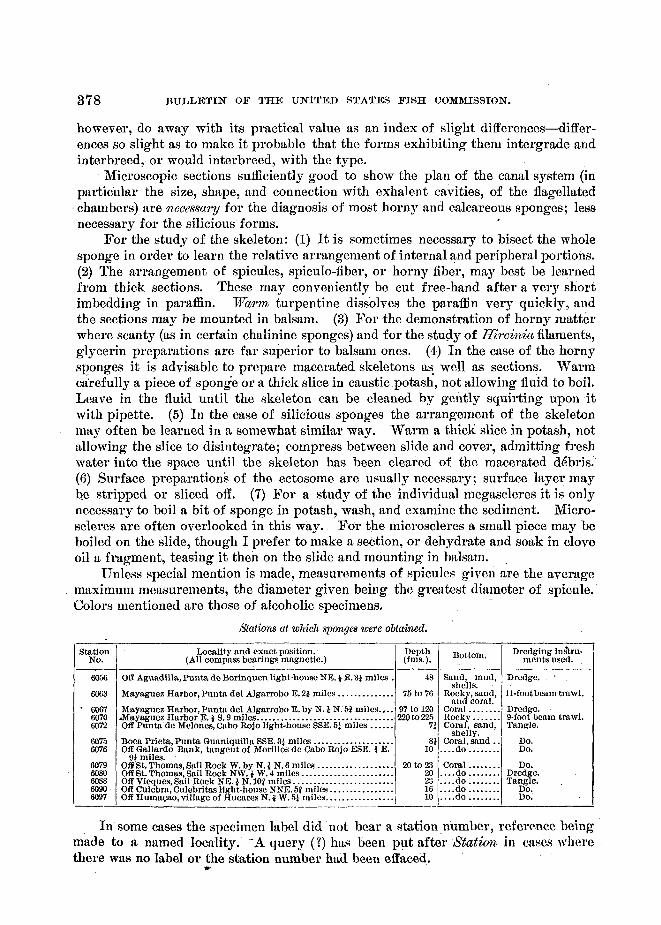
Stations at which spongl!8 were obtained.
Station Locality and exact position. Depth Dredging Instru- I
No. (All compass bearings mngnetic.) (fms.), Bottom, menta used.~ -~
6056 Off Agnadilla, Punta de Borinqnen Iight-house NE.l E. Sf miles . 48 Sand, mud, Dredge.shells.
6063 Mayagnez Harbor, Punta del Algurrobo E.2! miles ............•. 76 to 76 R~~~c~~~;~'ll-footbeam trawl.
- 6067 Mayagnez Harbor, Pnnta del Algarrobo E. by N.t N:Vf miles.... 97 to 120 Coral ........ Dredge.6070 ..Mayaguez Harbor E. i S.9 miles.................................. 220to 22, Roeky . , ..... s-toot beam trawl.0072 Off Punta de Melones, Cabo Rojo light-house SSE. 5! miles ..•.•• n Coral, sand, Tangle.
shelly.0075 Boca Prieta, Punta Guanlqutlla SSE. 3! miles .•.•....•.... _....•. 81 Coral, sand .. Do.0076 Off Gallardo Bank, tangent of Morillos de Cabo Rojo ESE. i E. 10 .... do ........ Do.
9t miles.6079 OtiSt. Thomas,Sall Roek W. by N.t N.6 miles ................... 20 to 23 Coral ........ Do.6080 Off St. Thomas, Sail Roek NW. 1 W. 4 miles....... _............... 20 ....do ........ Dredie.6088 Off Vieques, Sail Rock NE. t N.1Oi miles ......................... 2~ ....do ........ Tang e.6090 Off Culebra, Culebrltas Hght-house NNE. 5i miles ................ 16 ....do ........ Do.6097 Off Humacao, village of Hucares N.t W.5t miles................. 1 10 ....do ........ Do.
----_.-.
In some cases the specimen label did not bear a station number, reference beingmade to a named locality.-A query (?) has been put after Station in CUHes wherethere was no label or the station number had been effaced.. '
SPONGES COLLECTED IN PORTO RICO. 379
DEFINITIONS OF SYSTEMATIC TERMS USED IN THIS REPORT.
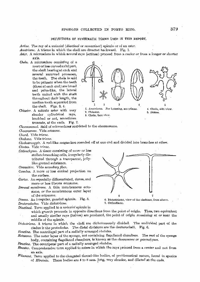
5
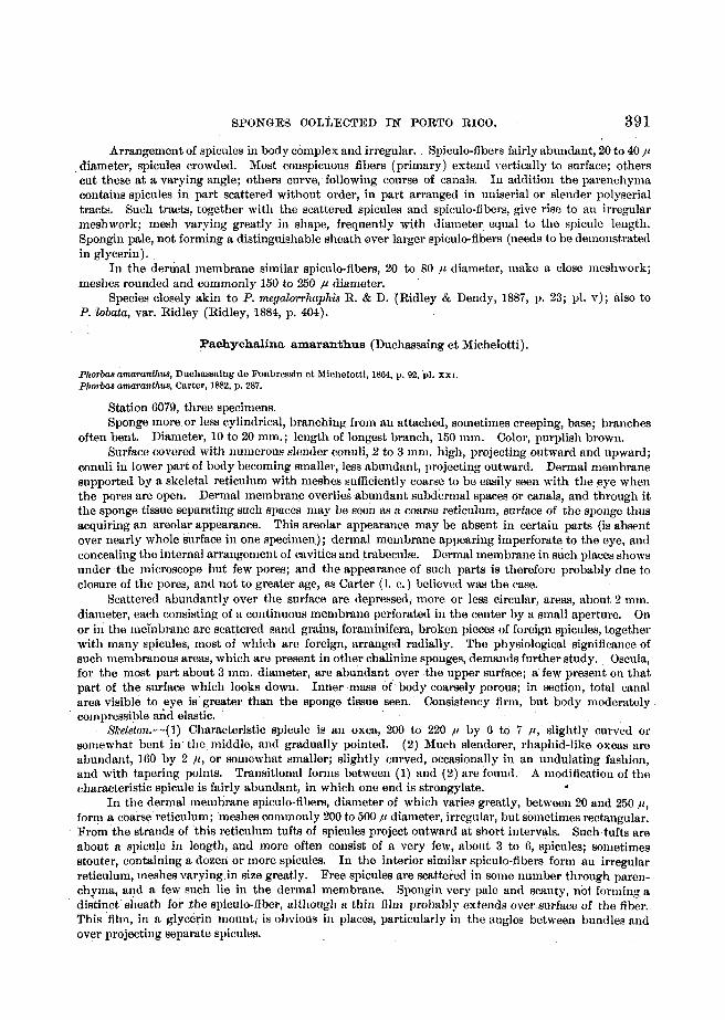
7
4. Chela, side view.5. Desma.
43
6
6. Dlehotrirene, view of the cladome, from above.7. Orthodleenc.
21
s
1. Anntritene, For lettering, see trleene.2. Chluster,3. Chela, facc vlcw.
Actine. The ray of a uniaxial (diacl'ina.l or monactinal) spicule or of an aster;Anatrimne. A tritene in which the cladi are directed backward. Fig. l.Aster. A microsclere in which several rays (actines) proceed from a center or from a longer or shorter
axis.Chela. A microsclere consisting of a
more or less curved axial part,the shaft bearing at each endseveral recurved processes,the teeth. The chela is said Cto be palmate when the teeth(threeat each end) are broadand palm-like, the lateralteeth united with the shaftthroughout their length, themedian tooth separated fromthe shaft. Figs. 3, 4.
Chiaster. A minute aster with veryslender cylindrical rays,knobbed or not, sometimestruncate, at the ends. Fig. 2.
Choanosomul. Said of microscleres restricted to the choanosome.Cluxmosome. Vide ectosome.Chord. Vide t1'iltne.C1Cldome.Vide triwne.Cladostronyyle. A rod-like megasclere rounded off at one end and divided into branches at other.Cladus. Vide triame.ColienchYlI/et. A tissue consisting of more or less
stellate branching cells, irregularly distributed through a transparent, jellylike ground substance.
Connective. Vide eecondarsj fiber.Conulus. A more or less conical projection on
the surface.Cortex. An especially differentiated, dense, and
more or less fibrous ectosome.Dermal membrane. A thin membranous ecto
some, or the membranous outer layerof the ectosome.
Desma. An irregular, gnarled spicule. Fig. 5.Deuierocladus. Vide dicliotrucne.Diactinal. Term applied to a uniaxial spicule in
which growth proceeds in opposite directions from the point of origin. Thus, two equivalentand usually similar rays (halves) are produced, the point of origin remaining at or near themiddle of the spicule.
Dicliotriume. A triame in which the cladi are dichotomously divided. The undivided part of thecladus is the protocladus. The distal divisions are the deuterocladi. Fig. 6.
Ecaaine. The centrifugal part of a radially arranged rhubdu«.Ectosome. The outer layer of the sponge,not containing flagellated chambers. The rest of the sponge
body, containing flagellated chambers, is known as the choanosome or parenchyma.Esactine. The centripetal part of a radially arranged rhabdus.Euaster. Comprehensive term applied to asters in which the rays 'proceed from a center and not from
an axis.Filament. Term applied to the elongated thread-like bodies, of problematical nature, found in species
of Hircinia. These bodies are 4 to 8 111m. long, very slender, and dilated at the ends.
380 BULLETIN OF THE UNITED STATES FISH OOMMISSION.
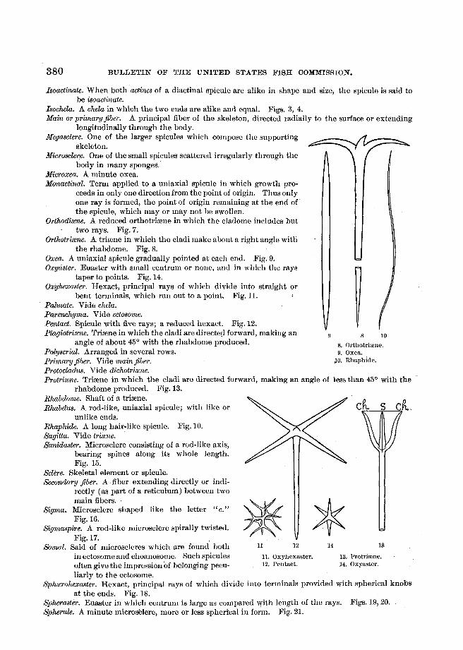
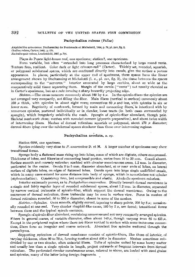
10
13
8
13. Protrlrene.14. Oxyaster,
8. Orthotrlrene.9. Oxea.
,10. Rhnphide,
9
1412
11. Oxyhexaster.12. Pentaet.
11
Isoactinate. When both actines of a diactinal spicule are alike in shape and size, the spicule is said tobe isoactinaie.
Ieochela. A chela in which the two ends are alike and equal. Figs. 3, 4.Main or primary fiber. A principal fiber of the skeleton, directed radially to the surface or extending
longitudinally through the body.Megasclere. One of the larger spicules which compose the supporting
skeleton.Microsclere, One of the small spicules scattered irregularly through the
body in many sponges.Microxea. A minute oxea.Monactinal. Term applied to a uniaxial spicule in which growth pro
ceeds in only one direction from the point of origin. Thus onlyone ray is formed, the point of origin remaining at the end of'the spicule, which mayor may not he swollen.
Orthoduene. A reduced orthotrirene in which the cladorne includes buttwo rays. Fig. 7.
Orthotritene. A truene in which the cladi make abont a right angle withthe rhabdome. Fig. 8.
Oxea. A uniaxial spicule gradually pointed at each end. Fig. 9.Oxyaster. Euaster with small centrum or none, and in which the rays
taper to points. Fig. 14.Oxyhexaster. Hexact, principal rays of which divide into straight or
bent terminals, which run out to a point. Fig. 11.Palmate. Vide chela.Parenchyma. Vide ectosome.Pentact. Spicule with five rays; a reduced hexact. Fig. 12.Plagiotriame. Trirene in which the eladi are directed forward, making an
angle of about 45° with the rhabdome produced.Polyserial. Arranged in several rows.Primary fiber. Vide main fiber.Protocladus. Vide dichotriamc.Proirucne. Trirene in which the cladi are directed forward, making an angle of less than 45° with the
rhabdome produced. Fig. 13.Rhabdome. Shaft of a trirene,Rhabdus. A rod-like, uniaxial spicule; with like or
unlike ends.Rhaphide. A long hair-like spicule. Fig. 10.Sagitta. Vide triwne.Sanidaster. Microselere consisting of a rod-like axis,
bearing spines along its whole length.Fig. 15.
Sclere. Skeletal element or spicule.Secondary fiber. A fiber extending directly or indi-
rectly (as part of a reticnlum) between twomain fibers.
Sigma. Microsclere shaped like the letter "c."Fig. 16.
Sigmaspire. A rod-like microsclere spirally twisted.Fig. 17.
Somal. Said of microscleres which are found bothin ectosome and choanosome. Such spiculesoften give the impression of belonging peculiarly to the ectosome.
Splucrohexaster, Hexaet, principal rays of which divide into terminals provided with spherical knobsat the ends. Fig. 18.
Spheraster. Euaster in which centrum is large as compared with length of the rays. Figs. 19,20.Spherule. A minute microselere, more or less spherical in form. Fig. 21.
SPONGEtl COLLEOTED IN PORTO RICO. 381
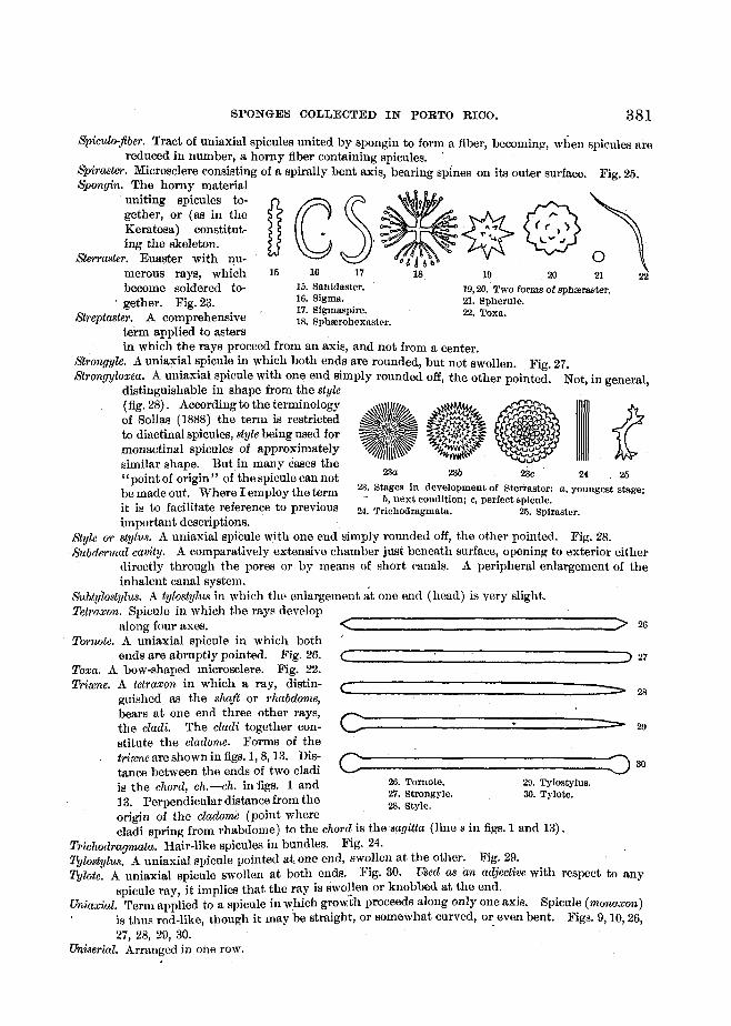
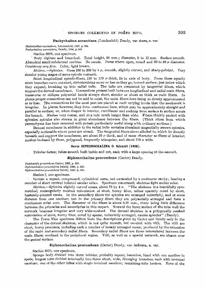
2524
29. Tylostylus.30. Tylote.
•23a 23b 23c
23. Stages in development of Bterraster: a, youngest stage;b, next condition; e, perfectspicule.
24. 'I'rtchodragmata. 25. Splrastcr.
c-----------..:...------::=>...;= 29
C----------::J 30
Spiculo-fiber, Tra~t of uniaxial spicules united by spongin to form a fiber, becoming, wlien spicules arereduced 1Il number, a horny fiber containing spicules. .
Spiraster. Microsclere consisting of a spirally bent axis, bearing spines on its outer surface. Fig. 25.
bP~¢~~~~Ef4~r~ fi rry 5.f~":*.~: 0~·mg the skeleton. U~ .'" :,;; ~ .
Sterroster. Euaster with nu- d d AAbOmerous rays, which 15 16 17 18 19 20 21 22
become soldered to- 15. Sanldaster. 19,20. Twoforms ofsphreraster.gether. Fig. 23. 16. Sigma. 21. Spherule.
17. Blgmasplro. 22 TStreptaster. A comprehensive 18. Sphrerohexaster. . oxa,
term applied toastersin which the rays proceed from an axis, and not from a center.
Strongyle. A uniaxi.al ~picuI~ in w~ich both en~s are rounded, but not swollen. Fig. 27.Strongyloxea. A uniaxial spicule with one end simply rounded off, the other pointed. Not in general
distinguishable in shape from the style ' ,(fig. 28). According to the terminologyof Sollas (1888) the term is restrictedto diactinal spicules, style being used formonaetinal spicules of approximatelysimilar shape. But in many cases the"point of origin" of the spicule can notbe made out. Where I employ the termit is to facilitate reference to previousimportant descriptions.
Style or stylu.~. A uniaxial spicule with one end simply rounded off, the other pointed. Fig. 28.Subdermal cavity. A comparatively extensive chamber just beneath surface, opening to exterior either
directly through the pores or by means of short canals. A peripheral enlargement of theinhalent canal system. .
Subtylostylus. A tylostylus in which the enlargement at one end (head) is very slight.Tetraxon. Spicule in which the rays develop
along four axes. < > 26
Tornote. A uniaxial spicule in which bothends are abruptly pointed. Fig. 26. C... -'J 27
Toxa. A bow-shaped microsclere. Fig. 22.Triame. A tetraxon in which a ray, distin- c-------------------_L... ~ ":::>~ 28
guished as the shaft. or rhabdome,bears at one end three other rays,the cladi. The cladi together constitute the cladome. Forms of thetriame are shown in figs. 1,8,13. Distance between the ends of two cladiis the chord, ch.-ch. in figs. 1 and 26. Tornote.
d f I 27. Strongyle.13. Perpendicular istance rom t ie 28. Style.origin of the cladome (point wherecladi spring from rhabdome) to the chord is the sagitta (line 8 in figs. 1 and 13).
'J1richodragmata. Hair-like spicules in bundles. Fig. 24.Tyl08tylu8. A uniaxial spicule pointed at. one end, swollen at the other. Fig. 29.Tulote. A uniaxial spicule swollen at both ends. Fig. 30. Used as an adjective with respect to any
spicule ray, it implies that the ray is swollen or knobbed at the end.Uniaxial. Term applied to a spicule in which growth proceeds along only one axis. Spicule (mona,xon). is thus rod-like, though it may be straight, or somewhat curved, or. even bent. Figs. 9, 10,26,
27, 28, 29, 30.Uniserial. Arranged in aile row.
382 BULLETIN OF THE UNITED STATES l!'ISH OOMMISSION.
Class 1. CALCAREA Bowerbank.
Order HETEROCCELA PolejaeffWith flagellated chambers, remaining parts of inner surface covered with pavement epithelium.
Family LEUCONIDJE (LEUCt>NES) Hackel,
The usually round flagellated chambers communicate with the central cavity by means of exhalentcanals. Spicules irregularly.scattered.
Genus PERICHARAX Po16jaeft' (1883).
With distinct subdermal cavities.
Pericharax carteri val'. homoraphis, Pol6jaeff.Pericharax carteri val'. homoruphls, PolCjaeff, 1883, p.s, pI. II, fig. 5; pl. VII, fig. 8.
Station 6090, seven specimens; station ?, two specimens.Sizes range from a greatest length of 25 mm, to one of 10 mm, Variety of shape exhibited is
interesting. A tubular or vase-like shape, expanded above, tapering below into peduncle-like portion,arid with a single terminal osculum, is represented. The vase may be very low and wide; may beespecially expanded above, and furnished with two oscula at the upper end; or the body may be of amassive, somewhat. flattened character, with an osculum on upper surface and another at the margin.In a specimen of the massive shape one surface is convex, the other concave, with the appearance ofhaving been attached; one osculum at the margin.
Color.-Specimens from station 6090, a fairly dark brown; those from station 'I, very light brown.Skeleton.-(1) Regular gastric quadriradiate spicules; facial rays straight, smooth, sharp-pointed,
about 150 p long; apical ray, sharp-pointed, straight or curved, often irregularly bent, sometimesrudimentary, usually 200 to 250 p long. (2) Triradiate spicules of parenchyma differing from quadriradiates only in absence of fourth ray. Among them are scattered very large (similar) triradiateswith rays sometimes exceeding 1 mm. in length; connected with smaller triradiates by intermediatestages. (3) Dermal and subdermal triradiate spicules, similar to the smaller triradiates of parenchyma(in val'. heieroraphi» becoming sagittal and irregular).
Class II. NON-CALCAREA Vosmaer.
Subclass 1. TRIAXONIA F. E;. Schulze.
Order HEXACTINELLIDA O. Schmidt.With silicious spicules belonging to the triaxial type, or readily derivable from it.
Family MJEANDROSPONGIDJE Zittel.
Dictyonine hexactinellida (i. e., having the large parenchymal hexacts united in a firmly connectedframework). Body consists of a connected system of labyrinthine anastomosing tubes, between whichthere is a connected interstitial system of intercanals.
Genus MARGARITELLA O. 80hmidt (1880).
With the single species.
Margaritella cceloptychioides O. Schmidt.Margarilella cwloplycltioidc8. Schmidt, 1880,p. 54, pI. VII, fig. 7.Margari/.eUa cwloptychioides, Schulze, 1887,p. 351,pI. cr, figs. 3-8.
Station 6056, nine fragments, all flattened pieces about 10 mm. thick, the largest 66 mm. wide.Only fragments have been seen. From these 0; Schmidt inferred that the body has a shallow
cup-like form. The intercanals are commonly of a cylindrical shape, but externally are more qr lessunited so as to give rise to the mreandriform furrows seen on outer surface of the sponge. The furrowsor separate intercanals are covered in by a delicate dermal membrane perforated by pores and
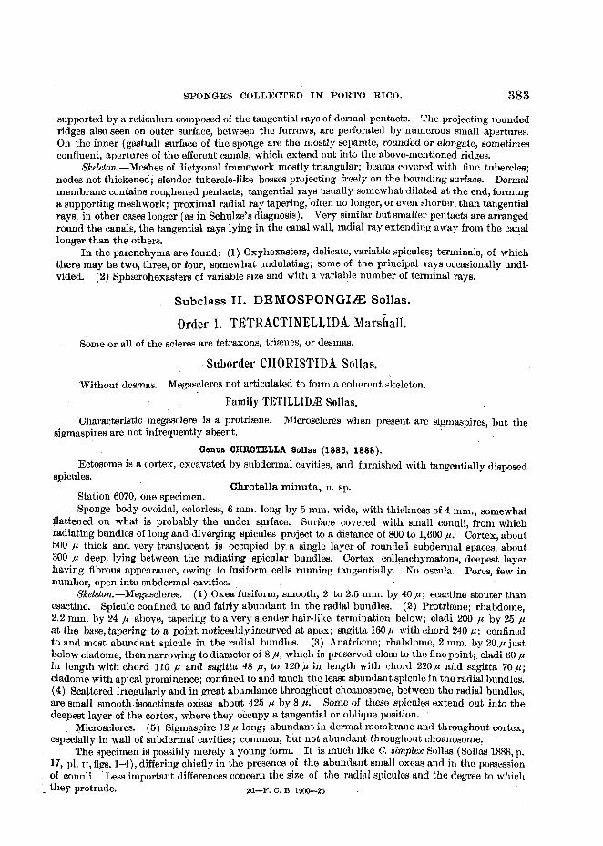
SPONGES COI.I.IWTED IN PORTO RICO. 383
supported by a reticulum composed of the tangential rays of dermal pentaets. The projecting roundedridges also seen on outer surface, between the furrows, are perforated by numerous small apertures.On the inner (gastral) surface of the sponge are the mostly separate, rounded or elongate, sometimesconfluent, apertures of the efferent canals, which extend out into the above-mentioned ridges.
Skeleton.-Meshes of dictyonal framework mostly triangular; beams covered with fine tubercles;nodes not thickened; slender tubercle-like bosses projecting freely on the bounding surface. Dermalmembrane contains roughened pentaets; tangential rays usually somewhat dilated at the end, forminga supporting meshwork; proximal radial ray tapering, 'often no longer, or even shorter, than tangentialrays, in other cases longer (as in Schulze's diagnosis). Very similar but smaller pentacts are arrangedround the canals, the tangential rays lying in the canal wall, radial ray extending away from the canallonger than the others.
In the parenchyma are found: (1) Oxyhexasters, delicate, variable spicules; terminals, of whichthere may be two, three, or four, somewhat undulating; some of the principal rays occasionally undivided. (2) Sphrerohexasters of variable size and with a variable number of terminal rays.
Subclass II. DEMOSPONGllE Sollas.
Order 1. TETRACTINELLIDA Marshall.Some or all of the scleres are tetraxons, trirenes, or desmas.
Suborder CHORISTIDA Sollas.Without desmas, Megascleres not articulated to form a coherent skeleton.
Family TETILLlD£ Sollas.
'Characteristic megasclere is a protrisene, Microscleres when present are sigmaspires, but thesigmaspires are not infrequently absent.
Genus OHROTELLA SoUas (1886, 1888).
Ectosome is a cortex, excavated by subdermal cavities, and furnished with tangentially disposedspicules.
. Chrotella minuta, n. sp.Station 6070, one specimen.Sponge body ovoidal, colorless, 6 nun. long by 5 mm, wide, with thickness of 4 mm., somewhat
flattened on what is probably the under surface, Surface covered with small conuli, from whichradiating bundles of long and diverging spicules project to a distance of 800 to 1,600 u, Cortex, about500 J.l thick and very translucent, is occupied by a single layer of rounded subdermal spaces, about300 J.l deep, lying between the radiating spicular bundles. Cortex collenchymatous, deepest layerhaving fibrous appearance, owing to fusiform cells running tangentially. No oscula. Pores, few innumber, open into subdermal cavities.
Skeleton.-Megascleres. (1) Oxea fusiform, smooth, 2 to 2.5 mm, by 40 J.l; ecactine stouter thanesactine. Spicule confined to and fairly abundant in the radial bundles. (2) Protrirene; rhabdome,2.2 mm, by 24 J.l above, tapering to a very slender hair-like termination below; cladi 200 p by 25 pat the base, tapering to a point, noticeably incurved at apex; sagitta 160 jt with chord 240 J.l; confinedto and most abundant spicule in the radial bundles. (3) Anatrirene; rhabdome, 2 mm, by 20p justbelow cladome, then narrowing to diameter of 8 u, which is preserved close to the fine point; cladi 60 pin length with chord 110 J.l and sagitta 48 J.l, to 120 J.l in length with chord 220 J.l and sagitta 70 J.l;cladome with apical prominence; confined to and much the least abundant spicule in the radial bundles.(4) Scattered irregularly and in great abundance throughout choanosome, between the radial bundles,are small smooth isoactinate oxeas about 425 J.l by 8 u, Some of these spicules extend out into thedeepest layer of the cortex, where they occupy a tangential or oblique position.
Microscleres. (5) Sigmaspire 12 p long; abundant in dermal membrane and throughout cortex,especially in wall of subdermal cavities; common, but not abundant throughout choanosome,
The specimen is possibly merely a young form. It is much like C. simplex-Sollas (Sellas 1888, p.17, pI. II, figs. 1-4), differing chiefly in the presence of the abundant small oxeas and in the possessionof conuli. .Less important differences concern the size of the radial spicules and the degree to which
• they protrude, 2d-F. C. B. 1900-25
3'84 BULLETIN O.!" THE UNITED STATES FISH COMMISSION.
Family STELLETID£ SolIas.
Megascleres are oxeas and orthotriicnes, or plagiotrisenes, or dichotrircnes, frequently with anatrirenes in addition. Microscleres include euasters, but never spirasters nor sterrasters.
Genus PILOCHROTA Sollas (1886, 1888).
"Oscules usually distinct. Pores in sieves leading into radial incurrent canals, which are notconstricted on passing through the fibrous layer of the cortex. Ectosome differentiated to form acortex, which usually consists of a middle collenchymatous layer, an outer thin fibrous layer, and aninner thicker fibrous layer." There is but one form of aster, and this is a chiaster,
Pilochrota variabilis, n. sp.
Station 6079, two specimens; station 6090, three specimens; station ?, ten specimens.The sponges here described resemble several of Sollas's species (P. lueckeli, P. pachydermata. P.
purpurea, P. lendenfeldi), but the limitations laid down in Sollas's descriptions (1888) make itimpossible to refer my specimens to any of these species.
Body spheroidal, often flattened. One specimen attached by its under .surface obliquely to coral;the others free, many with foreign particles adhering to surface. Younger forms occasionally foundwith short (2 mm. or less in length) slender rhizoids on under surface; such rhizoids being prolongationsof soft tissue almost without megascleres. Color in alcohol whitish or purplish-brown. The largerspecimens measure mostly 20 to 25 mm. in diameter; the smallest,apparently young forms, 5 to 7 mm.
. in diameter.In several of the larger specimens a single large osculum, 2 to 3 mm. diameter, is present, leading
into a deep cloaca, on the inner wall of which the apertures of excurrent canals may' be seen. Such anosculum may be situated in center of upper surface or nearer one side, then opening obliquely. Inthe attached specimen one such osculum is present on the side, low down, below the margin, whiletwo other very small oscula, appearing as apertures in smooth membranous areas, are similarly situated.In one of the large specimens there are no oscula, but several smooth membranous areas on the surface.In another of the larger specimens a single small osculum is present in the center of a similar smoothmembranous area. Most of the young (small) forms are without oscula, but in two cases a very smallosculum is situated in center of upper surface. All oscula are surrounded by a smooth membranousborder, narrow in the case of the larger ones. Appearances indicate that oscula may be opened anddosed.
Pores of variable size, but easily distinguishable with lens, lying in meshes of the network formedby cladi of the cortical orthotrisenes. Surface as seen with lens may be practically smooth andobviously porous, or punctate with minute elevations. Such elevations may be enlarged, appearing asareas of rounded polygonal shape, with pores in channels between-this appearance more obvious toeye than when sponge is examined with lens or objective, since the elevated areas themselves areporous.
Cortex in the larger specimens about 700 fl., in the younger forms only 500 fl. thick; fibrous withabundant densely granular cells, often in groups, in the outer half. Numerous subdermal cavities inthe deeper portion of cortex form a conspicuous layer. These are connected with short, wide (sometimes rounded) canals, which pass outward, branching near surface (often bifurcating), each branchterminating in a pore.
Skeleton.-Megascleres. (1) Orthotrisene; rhabdome, tapering to a fine often whiplash-like end,1,350 to 1,700 fl. by 12 to 24 u ; cladi 100 to 250 fl. (increasing with length of rhabdome), slightly curvedoutward, then straightening or recurved near tip; center of cladome depressed. (2) Very slenderorthotrizenes of variable size, probably young stages of the preceding, the cladi directed slightlyforward so as to give to the spicule the plagiotrireno character; rhabdome tapering to fine long point.A common size has a rhabdome about 500 by 4 u, cladi 20 to 24 u, (3) Anatrirene with depressedapex, rhabdome tapering to point, 1,530 to 2,210 fl. by 20 to 24 fl.; cladi stout, tapering to sharp point,with length 44 to 84 u, and sagitta 20 to 80 fl., the sagitta increasing with the length of cladus, asdoes length of cladus with that of rhabdome. Cladome is thus comparatively' shallow or quite deep.(4) Oxea smooth, tapering to sharp points, 1,020 to 1,600 fl. by 12 to 16 u, with much smaller formsof same spicule. (5) Ectosomal oxeas slightly curved, 168 to 200 j.t by 6 to 8 u, Such spicules occurscattered very sparsely through outer part' of cortex, arranged vertically to surface and slightly project-
SJ'ONGES COJ..LEC'l'ED IN PORTO RICO. 385
ing. The variations (many no doubt due to difference in the stage of development) in the size andshape of the megascleres, above indicated, may all be encountered in the same individual.
In the center of the sponge, oxeas (4) are the only megascleres. Spicules (1), (2), (3), (4), allradiate outward from central portion as far as the cortex. Just within cortex the cladi of the orthotrirenes may be arranged in conspicuous concentric layers, the larger spicules on the outside. In otherspecimens this concentric arrangement of the cladi is not conspicuous, the orthotrisenes of this regionnot being abundant, and nearly all comparatively small. From the interior separate spicules orbundles, containing each a few divergingspicules, radiate out through cortex to surface. Such spiculesare large orthotrioones (1), accompanied byanatrioones (3), with occasional oxeas (4). Cladomes ofthe orthotrisenes lie just beneath the surface. Anatrioones are frequently found projecting from surface.
Microscleres. (6) Chiasters with a very small centrum or none, the rays very slender, minutelyroughened, and somewhat tylote. Ohoanosomal chiasters have diameter 12 to 16 # and about 8 rays;somal, diameter 10 to 12 # and about 12 rays. In some specimens the chiasters are rare throughoutthe sponge; in other specimens they may be abundant in the cortex and comparatively rare in theinterior. .
Pilochrota fibrosa (0. Schmidt) Sollas var, globulariformis, 11. var,
Ancorina ftbrosa, Sehmidt,1870, p.67.Ptlocltl'ola flbrosa, Sollas,1888, p. 180.
Station 6079, one specimen.Schmidt (1. c.) gave the name of Ancorina fibrosa to a sponge of irregular, incrusting habitus, with
a clearly differentiated cortex. Schmidt says the megascleres are similar to those of A. simplicissl:maSchm. In this latter sponge (Schmidt, 1868, p. 18; Taf. III, fig, 9; Taf. IV, fig. 9) are found oxeas,anatrirenes (Anker "mit abwarts gekehrten Spitzen"}, and plagiotrioones. Sollas (1. e.) examinedone of Schmidt's preparations of this sponge and discovered chiasters, also determining size of oxea.He does not, however, in his diagnosis, mention the anatrioones.
ThePorto Rico form, although differing in habitus from Schmidt's specimen, agrees with it inthe character of its spicules, and must therefore be identified as belonging to the same species. Thespecimen is spheroidal, 35 mm, ill diameter, with aile osculum, 4 mm. in diameter, .leading into acloaca-like cavity, into which efferent canals open. Surface uneven and. almost completely covered withbroken pieces of shell. Color, reddish-brown.
Skeleton.-Megascleres. (1) Oxea 1.43 mm, by 0.027 mm., smooth, tapering to points. (2) Plagiotrimnes very abundant; rhabdome about 1 mm, long, 24 # thick above, becoming very slenderbelow, tapering to fine point; c1adi 80 # long, nearly straight, tapering gradually to a point. ,Anatrioone not abundant; rhabdome about 1.45 mm, long, 16# thick above, becoming very slender below,tapering to fine point; cladi 52 # long, sagitta 44 u,
Microscleres, (4) Chiasters are abundant and alike in ectosome and choanosome; total diameter12 p; 110 differentiated centrum; rays tylote, 6 to 12.
Genus TRIBRACHIUM Weltner (1882).
Sponge produced into a special cloacal tube, the megaseleros of which are orthodisenes, Thecharacteristic microsclere is a sanidaster.
Tribrachium schmidtii WeItner.Tribrachiuan. sclmlJidlU, Weltner, 1882.TribrachiurJI schlllidHi, Sol las, 1888, p. 154, pl. XVII; pl. XLI, fig. 5.
Station 6067, four specimens.Sponge body is spheroidal, 5 to 8 mm, diameter; cloacal tube 3 to 5 mm, long. The Porto Rico
specimens differ from the type as described hy Sollas in the character of the somal trirenes. These inSollas's description are orthotriwnes, but he mentions that" sometimes one or more cladi are bifurcate" (1. c., p. 154); and in the explanation of fig. 17, pI. XVII, he mentions that dichotrioones Occurnearthe-base of the cloacal tube, but not elsewhere. In the Porto Rico specimens the somal trisenes areall dichotrioones, but in view of the agreement in other respects with Sollas's description, it does notseem ad visable to make a new species on this evidently variable characteristic.
386 BULLETIN OF THE UNITED STATES FISH OOMMISSION.
Skeleton.-Megasc1eres: (1) Oxeas about 2.8 mm. by 34 u, (2) Somal dichotrisenes: Rhabdomeabout 2.5 mm. by 68 j.t above. Cladi stout; protocladus about 150 j.t by 68j.t; deuterocladus about 170 'pby 50 j.t at base, tapering to point; deuterocladi of same spicule not always of same length. Here andthere are found dichotrirenes with rhabdome much slenderer than in the type; and with cladi longerand also much slenderer than in the type. (3) Cloacal orthoditenes, with rhabdome 2 mm, by 25 j.t
above; cladi 475 j.t long. No anatrieenes observed, although such spicules were present in Sellas'sspecimens.
Microscleres: (4) Banidasters 12 to 14 j.t long, are abundant in parenchyma and very abundant indermal membrane. (5) Sollas says "oxyasters would appear to be characteristically absent." He,however, observed a single spicule. In the Porto .Ricospecimens, a few minute and somewhat irregularasters having a total diameter 6 to «», with a small and variable number of slender rays (chiastertype),are present. In a small percentage of the sanidasters, the actines become fewer in number and longer,while the axis decreases in length. The asters just mentioned are, I think, to be regarded asexcessively shortened sanidasters.
Family GEODIID£ Gray.
Megascleres include trisenes. Characteristic microsclere is a sterraster.
Genus CAMINUS O. Schmidt (1862)
Megascleres are orthotrieenes and rhabdi; anatritenes and protrirenes absent. .Sterraster isspherical, and the somal microsclere a spherule. Osculum leads into a cloaca.
Caminus sphreroconia Sollas.
Caminu8 eptueroconia, Sollas, 1888,p. 214, pI. XXVII.
Station?, one specimen.Specimen is massive, obconical, 80 mm. high; two oscula on upper surface.Skeleton.-Spicules in general somewhat smaller than in Sellas's specimen (from Bahia). Mcgas
cleres: (1) Strongyle about 450 to 500}l by 12 u, (2) Orthotrieene: rhabdome 240 p by 8 p; cladusabout 148u, Microscleres: (3) Sterraster about 44 j.t diameter. (4) Spherule, 4 j.t diameter.
Order z. CARNOSA Topsent.Sponges without megascleres, Microscleres present or absent; when present, they are asters 'or
4-rayed spicules.Suborder OLIGOSILICINA Vosmaer.
Sponges either entirely withou~ skeleton, or with only stellate microscleres.
Family CHONDROSID£ F. E. Schulze.
With a fibrous cortex. Flagellated chambers opening by special canaliculi into exhalentcavities. Surface slippery.
Genus CHONDRILLA O. Schmidt (1862),
Bulbous or incrusting sponges with stellate spicules (spherasters associated in some species withoxyaaters) .
Chondrilla nucula O. Schmidt.
Clwndrilla nucula O. Schmidt, 1862, p. 39, pI. III, figs. 22, 22a.CltolldriUa nucula F. E. Schulze, 1877, p. 24, Taf. IX.Chondrilla nucula Lendenfeld, 1896, p. 34, Taf. I, VI, VIII, IX.
Station 6079, two specimens incrusting Pachychalina amaramihus,Skeleton.-Spicules are spherasters, especially abundant in the cortex and accompanying the
canals. In the Porto Rico forms (in one and the same individual) there is a considerable variation inthe details of shape, as well as in the size, of the spicules. Schmidt, for Mediterranean forms, puts thetotal diameter at 27 u, with smaller sizes. Schulze and Lendenfeld, for Mediterranean forms; putthe diameter respectively at 10 to 20 j.t and 13 to 28 u. In the Porto Rico specimens the cortical
SPONGES COLLECTED IN PORTO RICO. 387
spicules vary but little. Total diameter from ray apex to ray apex is 28 p or close to it, with a raylength of 6 u, The spicule resembles that figured by Schmidt, rather than the other extreme presentin Schulze's specimens; differing from the latter in having a longer, sharper ray, outline of which isnot convex, but is either straight or slightly concave. About nine rays are seen round the edge whenthe equator of the spicule is brought into focus.
In the interior of the sponge the spicules vary more. Spherasters similar to those of the cortexare abundant, with a ray length 6 to 8/1. Smaller sizes of same spicule, down to 10 p diameter, arepresent. Forms with relatively larger body and shorter rays are also fairly common. In these, totaldiameter of which is 12 to 20 p, the rays are mere projections, 2 p and less in length, on the surface;outline of ray straight or frequently convex. Such spicules. resemble those described by Schulze.Intermediate forms between the two extremes are fairly common; total diameter, 28 to 32 p; raylength, 3 to 4 p; rays with straight or slightly concave outline.
Order 3. MONAXONIDA Ridley & Dendy.
Silicious sponges in which the megascleres are uniaxial.
Suborder 1. HADROMERINA Topsent.
Compact sponges, with skeleton radiately arranged, or without order, rarely forming spieulofibers, not reticulated, and usually without spongin. Megascleres, monactinal or diactinal, as a rule ofa single kind. Microscleres, when present, some form of aster or microxea, ~ever chelie or sigmata.
Family COPPATIID£ Topscnt.
Megascleres diactinal. Body massive, rarely cyathiform. Mieroscleres absent, or when presenteuasters with which streptasters may be associated.
Genus COPPATIAS SoUas (1888).
Megascleres arranged partly in radiating fibers, partly scattered loosely in the choanosome; in theectosome they lie tangentially. Microscleres are euasters.
Ooppatias solidissima, n, sp.
Station 6079, two specimens, both fragments.Both fragments elongated, flattened lobes, about 20 mm. thick and twice as wide, each prolonged
above into a slender digitate solid process, rounded at the end. Total length of longest fragment 120IIIIll. Oscula, few in number, about 3 mm, diameter, all on one side: Consistency firm and hard;surface nearly covered with incrustations. Color: Exterior, dark Slate-brown, with tinge of purple;colorless within.
Subdermal cavities fairly welldeveloped, Granular pigment cells (brown), 16 to 20 p diameter,are scattered through whole body, but are densely crowded in dermal membrane, and 'internal to itfor a varying distance, in places throughout the thickness occupied by the eortlcal brushes of spicules.Pores, about 40 Po diameter, are grouped, four or fi ve together, in small transparent pore areas, separatedby heavily pigmented tissue. The arrangement is such that the pigmented tissue forms a network oftrabeculre. (In this species, as elsewhere, the appearance of the dermal membrane must vary fromtime to time.)
Skelelon.-Megascleres: (1) Oxea, smooth, somewhat curved, 1 mm, by 28 u, Smaller sizes,doubtless younger stages of this, the chief, spicule, are common. These large oxeas are arranged: a, inradial cortical brushes; b, from some of the latter, bundles are prolonged into the interior; c, innumerous longitudinal main bundles, which are not very distinctly marked off from one another, thebundle arrangement most distinct in transverse section; d, scattered irregularly through the spongebody. The oxen is occasionally found with one rounded end (st.rongyloxea). It is noteworthythat occasionally a trirene spicule is found properly placed amoug the c6r'iical oxeas, In looking
388 BULLETIN OF THE UNI'fED S1'ATES FISH COMMISSION.
through a number of preparations, I have seen three such spicules. Their position possibly .indicatesthat they are not foreign. (2) Small smooth oxeas (microxeas Sollas), 60 by 3}1-, are abundant inthe dermal membrane, where they are arranged tangentially; also scattered sparsely through interior.
Microscleres: (3) Minute chiasters, 6 }1- diameter, incrusting the dermal membrane in greatabundance. Similar but somewhat larger chiasters, 8 to 10}1- diameter, are sparsely scattered throughinterior.
In spiculation this species closely approaches Coppaiia« (Stelletinopsi.~) purpureus Carter (Carter1886, p. 459), from Port 'Vestern, South Australia. Carter's description does not include details as todistribution of spicules.
Family TETHYIDj£ Topsent.
Body spheroidal or massive, with radiately arranged skeleton, and a more or less differentiatedeetosome. 'Vhen microscleres are present, the chief microsclere is some form of euaster.
Genus TETHYA Lamarck (1815).
Ectosome differentiated into a well-developed fibrous cortex. Megascleres are fusiform strongyloxeas, Microscleres are euasters of two kinds (spherasters arid chiasters).
Tethya seychellensis (E. P. Wright) Sollas.
Alemo seye1lellensig, E. P. Wright, 1881, 1'.13, 1'1.1.Teth1la geye1lellengig, Sol las, 1888, 1'.427, 1'1.XI.IV, figs. 1-6.
Ensenada Honda, Culebra, one specimen.Sponge more or less spherical, attached or free, surface conulose, conules sometimes appearing as
wide polygonal plates with denticulated margins. Oscules one or more in number; pores in sieves,situated in the depressions between the conules, leading into extensive intercortical cavities (Sollas).
Skeleton.-Megascleres. (1) Large strongyloxeas, 1.2 to 1.5 mm. long, with diameter 20 to 24 }1-.Similar smaller spicules of varying size down to 300 by 4/1 are abundant.
Microscleres. (2) Cortical spheraster, 40 fl diameter. (3) Somal chiaster, 8 to 10 fl diameter.(4) Choanosomal aster, with a ray length 20 to 28 u.
Tethya 1yncurium (Lin.) Lamarck.
Tethya limcurtu.m, Sol las 1888, 1'.435,1'1. XI.III, Jigs. 1.5-18; 1'1. xr.rv, figs. 17-19.
Playa de Ponce light-house reef, three specimens; Ponce reefs, two specimens,Body spheroidal, attached. Diameter of spooimons ]5 to 22 mrn.: small oscula present on some,
absent on others. Buds on some of the specimens, Outer non-fibrous part of cortex 800 pinnerfibrous part 650 }1- thick. . '
8keleton.-Megascleres. (1) Large strongyloxeas, 1.2 mm, by 20 JI to 1.4 mm. by 2811; oxeateend rounded, though often quite small. Essentially similar spicules of varying size, down to 340 by4 fl, are abundant; in the smaller forms the oxeate end is sharp-pointed.
Microsoleres, (2) Cortical spheraster, 72 It diameter. (3) Somal and choanosomal chiaster, 12 to16 }1- diameter.
Genus TUBERELLA Keller (1880).
Without a fibrous cortex, and without microse/eres. Chief megascleres are fusiform strongyloxeas, with small strongyloxeas at the periphery.
Tuberella aaptos (0. Bchmidt) Topsent.
Ancoriiu: aapio«; O. Schmidt, 1864, p.33, Taf. IY, lig.ll.Suberiies <taptug, Leudenfeld, 1896, p.140, Tnf. V1I, XII.
Tuberella aapios, Topscnt, 1900, p. ~8.5, 1'1.VIII.
Mayaguez Harbor, one specimen.Body is massive, higher than broad, 60 by 35 mm., attached below to coral; subdivided above
into two short lobes, one with small (nearly closed '1) terminal osculum opening into a short axialcavity. Surface smooth or covered with short papillre. Consistency firm and fleshy. Color: Surfaceof specimen very dirty; clean parts light brown. In a gross section peripheral layer is slate colored;interior light.
SPONGES COLLECTED IN' PORTO RICO.· 389
Skeleton.-Strong spiculo-flbers about 0.5 mm, thick course, at oblique angles to vertical axis ofbody, through interior; breaking up 5 mm. or thereabouts from surface into smaller bundles, whichradiate out to the surface. These fibers cross frequently, fusing at the points of intersection, and thusgive rise to scattered, sometimes star-like, centers, from which the fibers seem to radiate. The outersmaller radiating bundles frequently (always?) arise from peripherally situated nodal points of thissort. Abundant scattered spicules lie between the fibers and peripheral bundles. At the surface areclosely set diverging bundles of small strongyloxeas, .
Megascleres, (1) Chief spicule is a 'smooth fusiform strongyloxea, 1.5 mm, by 40 j.l or thereabouts;strongylate end greatly narrowed. Smaller stages in development of the same spicule are found.Occasionally oxeate end is rounded, spicule becoming nearly isoactinate. (2) Strongyloxeas of aboutsame size as (1), not fusiform; with basal end rounded but not narrowed, and with tapering end alsorounded at apex; not common. Both (1) and (2) make up the fibers and peripheral bundles, andare also found scattered. (3) Small strongyloxeas, frequently about 300 by 71', with basal end roundednot narrowed, make up the surface brushes; also scattered in interior, together with smaller slendererforms, probably stages in development of same spicule.
Suborder II. HALICHONDRINA Ridley & Dendy.
Typically noncorticate; skeleton usually reticulate; megaseleres usually either oxeas or styles.
Family HOMORRHAPHIDlE Ridley & Dendy.
Megascleres all diactinal, either oxeas or strongyles; no microscleres.
Subfamily RENIERINJE Ridley & Dendy.
The spicules may be united together by a small proportion of spongin, but are never completelyenveloped in it.
Genus PETROSIA Vosmaer (1886).
Sponge usually hard; skeleton more or less confused; spicules oxeate to strongylato, packedtogether in tracts.
Petrosia ha,lichondrioides, n. sp,
Station 6079, one specimen.Sponge a cake-shaped fragment, about 50 mm. diameter, with a thickness of 15 mm.: outer surface
convex and bearing one small excentrically placed osculum 3 by 1.5 mm. Surface even and slightlypilose. Consistency very dense and firm, though not hard; sponge becoming hard and brittle ondrying. Color: Exterior, chocolate-brown; interior, somewhat. lighter. .
Canal system of such a character that the sponge body is divided into trabeculre of a more or Iessuniform width (commonly about 60 j.t), the canals between being as wide or wider than the trabeeuhe. C
In the superficial region, the trabeculae and intervening canals in some places, but ..not universally,run more or less vertically to the surface; ill the interior, they pursue a meandering course. Spiculesin the trabeculas form tracts (scarcely bundlesj.vwhich are vaguely defined, because the spicules areso loosely packed, without perceptible spongin. The tracts vary in thiokness and distinctness, thelarger ones sometimes running more or less vertically to the surface, again pursuing a tangentialcourse. Following the anastomoses of the meandering trabeoulro, the tracts of spicules form a quiteirregular and vaguely defined reticulum. The tracts of spicules in the trabeculre of the -superflcialregion pass, often very obliquely, into vaguely ueflned brushes of spicules which project radially fromthe surface.
Dermal membrane indistinctly 'differentiated from subjacent tissue, perforated by numerous,diffusely scattered, rounded pores about 40 It in diameter. The spicules lying between the pores giverise to a loose reticulum. These spicules are merely the outermost layer of the main skeleton.
/Spicules are slender 'for a Petrosino Oxea 160 I' by 4 to 5/1, smooth and slightly curved; verycommonly somewhat irregularly bent in the middle, or sometimes with It slight prominence Itt thatpoint.
390 BULLETIN OF THE UNITED STATES FISH OOMMISSION.
On comparing this species with a more typical Petrosia, such as P. (Schmidtia) aulopora Schmidt,we find that the regularity of arrangement in the canal system and skeleton of the latter, produced bythe crossing of radial and tangential canals and spicule bundles, is absent in the Porto Rico species.Again, in the latter the spicules are muchless closely bound together to form bundles, and the canalsseparating the trabeculse are not so wide. Hence P. halichondrioides is the denser of the two species.The loosely arranged spicules of the Porto Rico species suggest a close affinity with Halichorulria.The spicules of the Porto Rico form are slenderer than in P. aulopora. In a specimen of the ,latterspecies, in the Museum of Comparative Zoology, the oxea measures 152 by 7 to 8 u, is smooth, sharppointed (with exceptions), and somewhat curved or bent at the middle.
Genus FOLIOLINA O. Schmidt (1870).
Sponge a hollow stem with horizontal, lamellate processes embracing the stem. Upper end closed:Oscula absent. Texture loose, spongy. Spicules rather stout oxeas, which form polyserial tracts aswell as a network. Tracts developed especially in the stem, though also radiating out into theprocesses.
Foliolina peltata O. Schmidt.
Foliolina ]Jeltata, O. Schmidt, 1870, p. 42, pl. IV, fig. 4.
Station 6067, seven specimens.One specimen nearly perfect, 120 mm. long, with stem diameter of () mm.; radial length of lateral
processes, 7 to 10 mm, Processes, of which there are six in this specimen, are at upper end of stem;lower end broken across, open. There are six other fragmentary specimens, in three of which thestem divides near upper end:
The lamellate appendages are flattened hollow lobes into which the axial cavity of stem isprolonged. Appendage convex above, concave below, and more or less incised round the margin.The only pores visible are on lower surface of appendages.
Skeleton. -Oxea, 320 by 10 u, somewhat curved or bent in the middle, rounding off at each endto a sharp point. Wall of the stem, which is firm and 0.5 IllIP.. thick, is supported by a dense renierinereticulum strengthened internally by polyserial bundles, which cross so as to prorluce a coarse network;superficially"armed with projecting spicules. Skeleton of lower wall of appendage consists of a reticulum strengthened internally by radiating bundles of spicules. Upper wall supported by a reticulum,without the bundles; outer surface armed with projecting spicules. Cavity of lobe contains a gooddeal of sand. Soft parts of this interesting sponge have been almost completely macerated out.
Subfamily CHALININ.l:E (CHALINE.l:E) O. Schmidt.
Spongin unites, usually envelops, the spicules so as to give rise to a distinctly fibrous skeleton.
Genus PACHYCHALINA O. Schmidt (1868).
Not tubular; skeleton composed of stout fibers, containing numerous spicules, arranged in severalrows.
Pachychalina mollis, n. sp,
Station 6072, ten specimens; station 6075, one specimen; station 6079, two specimens.Sponge body branching from the base. Branches, which themselves may divide, subcyliudrical,
long, 8,to 15 mm, diameter. Surface nearly smooth. Consistency compressible and elastic; rigidityinsufficient (in the wet specimen) for the body to stand erect; body, on drying, becoming stiff andcapable of .standing, retaining its elasticity. Oscula 2 to 3 mm. diameter,arranged on one side of thebranch in a not strictly uniserial row, commonly 10 to)5 mm. apart. Height of a specimen, from baseto tip of longest branch, 400 mm. Color, grayish-brown, but with abundant traces of purplish-red,probably the natural color.
Skeleton.-Characteristic spicule is an oxea, about 140 hy 7 u, slightly curved and terminatingsuddenly in points. Oxea occasionally straight: rarely with one end strongylate; still more rarely,with one end strongylate and the other tylote. A slender moJification cstage in the growth?) ofthe characteristic oxea, present in some abundance; 140 by 2 to a J1., with tapering ends.
SPONGES OOLLEOTED IN PORTO RIOO. 391
Arrangement of spicules in body complex and irregular. Spiculo-fibers fairly abundant, 20 to 40 jl
,diameter, spicules crowded. Most conspicuous fibers (primary) extend vertically to surface; otherscut these at a varying angle; others curve,' following course of canals. In addition the parenchymacontains spicules in part scattered without order, in part arranged in uniserialor slender polyserialtracts. Such tracts, together with the scattered spicules and spiculo-flbers, give rise to an irregularmeshwork; mesh varying greatly in shape, frequently with diameter equal to the spicule length.Spongin pale, not forming a distinguishable sheath ewer larger spiculo-flbera (needs to be demonstratedin glycerin).
In the dermal membrane similar spiculo-flbers, 20 to 80 II diameter, make a close meshwork;meshes rounded and commonly 150 to 250 jl diameter.
Species closely akin to P. megalorrhaphiB R. & D. (Ridley & Dendy, 1887, p. 23; pl. v); also toP. lobata, var. Ridley (Ridley, 1884, p. 404).
Pachychalina amaranthuB (Duchassaing et Michelotti).
Phorba8 amaranthUB, Duchassalng dc Fonbressln et Michelotti, 1864, p. 92, pl. XXI.Phorbo« amaranthu8, Carter, 1882, p. 287.
Station 6079, three specimens.Sponge more or less cylindrical, branching from an attached, sometimes creeping, base; branches
often bent. Diameter, 10 to 20 mm.: length of longest branch, 150 mm. Color, purplish brown.Surface covered with numerous slender conuli, ,2 to 3 mm, high, projecting outward and upward;
conuli in lower part of body becoming smaller, less abundant, projecting outward: Dermal membranesupported by a skeletal reticulum with meshes sufficiently coarse to be easily seen with the eye whenthe pores are open. Dermal membrane overlies abundant subdermal spaces or canals, and through itthe sponge tissue separating such spaces may be seen as a coarse reticulum, surface of the sponge thusacquiring an areolar appearance. This areolar appearance may be absent in certain parts (is absentover nearly whole surface in one specimen); dermal membrane appearing imperforate to the eye, andconcealing the internal arrangement of cavities and trabeculre. Dermal membrane in such places showsunder the microscope but few pores; and the appearance of such parts is therefore probably due toclosure of the pores, and not to greater age, as Carter (I. c.) believed was the case.
Scattered abundantly over the surface are depressed, more or less circular, areas, about 2 mm.diameter, each consisting of a continuous membrane perforated in the center by a small aperture. Onor in the meinbrane are scattered sand grains, foraminifera, broken pieces of foreign spicules, togetherwith many spicules, most of which are foreign, arranged radially. The physiological significancc ofsuch membranous areas, which are present in other chalinine sponges, demands further study. Oscula,for thc most part about 3 mm. diameter, are abundant over the upper surface; a few present on thatpart of the surface which looks down. Inner mass of body coarsely porous; in section, total canalarea visible to eye is' greater than the sponge tissue seen. Consistency firm, but body moderatelycompressible and elastic. .
Skeleton.-(1) Characteristic spicule is an oxea, 200 to 220 )I by 6 to 7 II, slightly curved orsomewhat bent in' the middle, and gradually pointed. (2) Much slenderer, rhaphid-like oxeas areabundant, 160 by 2 II, or somewhat smaller; slightly curved, occasionally in lUI undulating fashion,and with tapering points. Transitional forms between (1) and (2) are found. A modification of thecharacteristic spicule is fairly abundant, in which one end is strongylate.
In the dermal membrane spieulo-flbers, diameter of which varies greatly, between 20 and 250 IIIorma coarse reticulum; meshes commonly 200 to 500 II 'diameter, irregular, but sometimes rectangular:From the strands of this reticulum tufts of spicules project outward at short intervals. Such tufts areabout a spicule in length, and more often consist of a very few, about 3 to 6, spicules; sometimesstouter, containing a dozen or more spicules. In the interior similar spicule-fibers form an irregularreticulum, meshes varying in size greatly. Free spicules are scattered in some number through parenchyma, and a few such lie in the dermal membrane. Spongin very pale and scanty, not forming adistinct sheath for the spiculo-fiber, although a thin film probably extends over surface of the fiber.This 'film, in a glycerin mount, is obvious in places, particularly ill the angles between bundles andover projecting separate spicules.
392 BUI..LETIN 0.1<' THB UNITED STATES I!'ISH OOMMISSION
Pachychalina rubena (Pallas)
Amphimcdon. arbore8een8. Duchassaing dc Fonbrcssin et Michelotti, 1864,p. 79, pI. XIV,fig. 2.Chalina ruben8, Carter, 1882; p.276.Euchalinopsis rubcu8, Lendenfeld, 1887,p. 744.
Playa de Ponce light-house reef, one specimen; station?, one specimen.Form variable, but often "extended into long processes characterized by large round vents.
Texture firm, resilient. Color dark or ligh; crimson-red" (Carter). Thickly set, rounded, squarish,or polygonal subdermal spa ces, which are continued directly into canals, give the surface a porousappearance. In places, particularly at the upper end of specimens, these spaces have the lineararrangement shown by Duchassaing et Michelotti (1. c., pl. XIV, fig. 2), the tissue between the spacescorresponding to the "nervures." Interior excavated by large cavities, about as wide as thecomparatively solid tissue separating them. Margin of the oscula (" vents") not usually elevated asin Carter's specimens, but as a rule forming a sharp inwardly projecting edge..
Skeleton.-The oxeas measure commonly about 160 by 4 u. In the spiculo-fibers the spicules arenot arranged very compactly, not filling the fiber. Main fibers (vertical to surface) commonly about100 jl thick, with spicules in about eight rows; connectives 60 p and less, with spicules in six orfewer rows. Regularity of meshwork, formed by main and connecting fibers; is interfered with bynumerous spicules, scattered individually or in slender, loose tracts (in both cases surrounded byspongin), which irregularly subdivide the mesh. Spongin of spiculo-fiber abundant, though pale.Skeletal meshwork close; meshes with rounded corners (glycerin preparation), and about twice widthof intervening fibers. Meshes of dermal reticulum squarish or polygonal, about 170 p diameter;dermal fibers lying over the subdermal spaces slenderer than those over intervening regions.
Pachychalina areolata, n. sp,
Station 6088, one specimen.Species evidently very close to P. amaranthue D. et M. A larger number of specimens may show
transitional forms.Sponge body a flattened mass rising up into lobes, some of which are digitate, others compressed.
Thickness of lobes, and likewise of connecting basal portion, varies from 10 to 20 mm, Conuli absent.Surface smooth and coarsely reticular; studded with circular membranous areas, 1.2 mm. in diameter,perforated in the center. Oscula 3 to 5 mm, diameter abundant, at 01' neal' ends of lobes, on lateralsurface of digitate lobes, on edges of flattened lobes. Oscula open into large single undivided canals,which in many cases extend for some distance into body of sponge, which is nevertheless not tubular(siphonochaline}. Consistency firm, but compressible and elastic. Alcoholie specimen colorless.
Interior extremely porous, as in Paclojchalirui omartuuhus. Directly beneath dermal membrane isa single and fairly regular layer of rounded subdermal spaces, about 1.2 mm. in diameter, separatedby narrow vertical trabeculee of spicule-tiber, which support the dermal membrane. Owing to thecoarseness of dermal reticulum, these trabeeuhe may be seen in surface view. Pores in meshes ofdermal reticulum rounded, 50 to 350 J.L diameter; absent in some of the meshes.
Skcleton.-Spicules. Oxea smooth, slightly curved, tapering to sharp points, 200 by 8 jl; occasionally rounded at one end. Very slender rhaphid-like oxeas, 160 by 2 J.L, are found; transitional formsbetween these and the typical spicule common.
Spongin of spiculo-fiber abundant, containing numerous and not very eompactly arranged spicules.Fibers in general coarse, of variable diameter, often about 140 It, though varymg from 80 to 425 u,Except in the peripheral region where there are fibers vertical to surface with some transverse connectives, fibers form an irregular and coarse network. Abundant free spicules scattered through theparenchyma.
Supporting reticulum of dermal membrane consists of spiculo-flbers, like those of interior, ofvarying thickness, ~ften 60 to 70 J.L, forming meshes about 600 J.L in diameter. Meshes sometimes subdivided by one Or two slender, often uniserial fibers. Tufts of spicules united by some horny matterand usually less than a single spicule in length, project outwards at frequent intervals fromdermalreticulum. The perforated circular membranous areas, referred to above, are loaded with sand g'rainsand spicules, many of the latter being foreign fragments.
SPONGES OOLLEOTED IN PORTO RICO. 393
Pachychalina aurantiaca (Lendenfeld) Dendy, val'. dura, n. val'.
Oladochalina mwanUaca, Lendenfeld, 1887, p.768.Pachychalina a"mnliaca, Dendy, 1894, p.241.
Station 6079, one specimen.. Body digitate and branched. Total height, 90 mm.: diameter, 8 to 15 mm, Surface smooth.
Abundant small subdermal cavities. No oscula. Pores where open, round and 60 to 80 p. diameter.Consistency very.firm. Color, light brown. .
Skeleton.-Spicules. Oxea 160 to 200 by 4 u, smooth, slightly curved, and sharp-pointed. Veryslender young stages of same spicule common.
Stout longitudinal spiculo-fibers, 130 to 170 }t thick, lie in axis of body. From these equallystout branches curve outward, dichotomizing more or less as they go, toward surface; just below whichthey expand, breaking up into radial tufts. The tufts are. connected by tangential fibers, whichsupport the dermal membrane. Connectives present both between longitudinal and radial main fibers,transverse or oblique polyserial bands always short, slender or about as thick as main fibers. Inplaces proper connectives can not be said to exist, the main fibers here being so closely approximatedas to fuse. The connectives for the most part are placed at such varying levels that the meshwork isirregular. In places, however, they form continuous lines, which may be approximately straight andparallel to surface; or, when deeper in interior, curvilinear and arching from surface to surface acrossthe branch. Meshes very coarse, and as a rule much longer than wide. Fibers thickly packed withspicules; spicules also strewn in great abundance between the fibers. (Thick slices from whichparenchyma has been removed with potash particularly useful along with ordinary sections.)
Dermal membrane in addition to the radial tufts contains abundant tangentially strewn spicules,especially noticeable where pores {lore dosed. The tangential fibers above alluded to, which lie directlybeneath and support the membrane, are about 50 It thick, and of same character as fibers of interior;meshes inclosed by them, polygonal, frequently triangular, and about 170 tl wide.
Genus SIPHONOCHALINA O. Schmidt (1868).
Tubular forms; tubes smooth both inside and out, each with a large opening at the summit.
Siphonochalina procumbens (Carter) Dandy.
Paiuloscula procumocns Carter, 1882, p. 635.Sipllonocllalina l)1'oCl!mbcns Dendy, 1890, p. 355.Sipho1lochali"a 11l'owmbcnB Dendy, 1894, p. 245.
Station?, one specimen.Sponge a repent, compressed, cylindrical mass, not excavated by a continuous cavity; hearing a
number of short vertical inflated oscular tubes. Specimen macerated; skeleton light amber color.8keleton.-Spicules slightly eurvedoxeas, about 75 by 4 tt."The skeleton is a beautifully sym
metrical, rectangularly meshed reticulation of stout, horny tiber, rather sparsely cored by short,hastately-pointed oxeas. In the secondary fibers the spicules are arranged uniserially, and at somedistance from one another; but in the primary fibers they are polyserially arranged and form acontinuous axial core. The diameter of the fibers is about 0.07 mm., there being little differencebetween the primaries and secondaries in this respecLToward the inner surface of the tube wall thenetwork becomes irregular and very wide-meshed. The dermal skeleton is a polygonally meshedreticulation of stont, horny fiber, cored by sparse, uniserially arranged, oxeate spicules" (Dendy).. The Porto Rico specimen differs from the descriptions given by Carter and Dendy only ill thecharacter of the dermal skeleton, which is not quite smooth, but covered with villi. The villi areshort, horny processes, including each a number of loosely arranged oxeas; produced by the extensionof the main lind secondary radial fibers. Secondary radial fibers are those intercalated between themain fibers; conflned t.o the peripheral region. Villi, as well as a special network, are absent overthe gastral surface.
Siphonochalina procumbens (Cartel') Dendy, val'. infirma, n. var.
Station 6079, one specimen.Sponge body divided into three tubular, probably repent, branches, fused with one another in
spots: longest tube divided terminally into three short, wide, diverging brnnchos, each with terminalosculum; one of the other tubes with single terminal osculum; remaining tube broken. None of the
394 BULL}i~'l'IN OF THE UNITED STATES FISH COMMISSION.
short upright oscular tubes characteristic' of tYP,e are present. Paragastric cavity continuou.~ throughoutsponge, and about 15 nun, diameter. Tube wall thinner than in type, only 2 to 3 mm, thick. Length oflongest tube, 150 mm, Color dark amber, with purplish tint.
Skeleton.-Fibers of the general skeleton slenderer than in type, about 40 It diameter. Dermalskeleton, a network of slender fibers, commonly about 8 It thick, with meshes 100 to 120 Jl diameter.From dermal surface small horny villi project, containing a few, sometimes only one, spicule; villi notexclusively situated at nodes of network, viz, not all peripheral extensions of radial fibers. Gastralsurface also provided with a special skeletal network very similar to that of dermal surface; villi longerand thicker than on dermal surface, spicules of a villus often forming a pretty compact bundle. Theorigin of the dermal and gastral networks can readily be made out in this variety. In places the mostsuperficial meshes of the general skeleton are simply subdivided by finer fibers, which extend betweenand only exceptionally overlie the coarser skeletal fibers. Elsewhere the system of fine fibers hasreached a further stage of development and forms a continuous reticulum overlying the skeletal fibers.
Siphonochalina spiculosa Dendy.
SipltOnocha1"ina spiculosa Dendy, 1890, p.354, pl. I,VIII, figs. 2, 2a; pI. I,XII, fig. 3.
Station 6079, three specimens, fragmentary.The habitus of these fragments is interesting when compared with the typical Siplumochalina
"form of body: One specimen is 160 mm. long, of a cylindrical or in places flattened cylindrical shape;diameter 10 to 20 mm., except in the middle where body is twice as thick; broken off at both ends.On this specimen are five of the large pseudogastral orifices, which in Siplumochaiina are typicallyfound at the ends of tubular branches. Here all five are lateral, and distributed round the surface;
. two are flush with the surface; two are at'the ends of very short, wide elevations; and' one is near,but not at, the end of a somewhat more marked elevation. Another specimen consists of a cylindricalpiece 60 by 20 mm., broken off at both ends, and bearing a somewhat slenderer and longer branch, alsobroken at the end. On this specimen are two of the large apertures, both strictly lateral (not at thesummit of any elevation whatever).
Pseudogaater is narrow, somewhat winding in its course, diameter for the most part about 5 mm.;in places pseudogaster so narrowed as to be nearly interrupted. The transition in habitus and withrespect to the pseudogastral system offered by these specimens to the Pnclojcluilina type is obvious.Should the pseudogaster become quite discontinuous and the orifices strictly lateral, the sponge wouldhave assumed the character of a Paclutchalina. In both S. iniermedia R. & D. and S. annulata R. & D.(Ridley & Dendy, 1887, pI. VII), it may be noticed that some of the orifices are not at the ends oftubular branches, but at the summit of small protuberances on the side of a branch or main axis.
The outer surface in two of the specimens, where intact, is marked with abundant depressedmembranous areas, more or less circular, perforated in the center, about 1 mm. diameter. Such areas,frequently connected together so as to form grooves or channels of varying length, often meandering.In the third specimen, surface shows faintly marked, irregular indentations, but the membranous areasare absent. The membranous areas overlie subdermal spaces of corresponding shape, Color of intactsurface reddish purple. Consistency firm, hard; body incompressible.
8keleton.-Spicules. Characteristic oxea, 140 to 160 Jl by 4 to 6 fl. Very slender Iorms of the samespicule, often about 120 by 2 itor somewhat smaller, are fairly eommon; transitional forms to eharacteristie spicule present. A modification of characteristic oxea, with one end strongylate.Ioccasiouallyfound. Spiculo-fibers stout, containing very numerous spicules, with but little spongin. Primary fibersabout 60 Jl diameter, and somewhat slenderer secondary fibers, may be distinguished, meshwork more orless rectangular,' but rather irregular; meshes wide. On inner surface of tube-wall longitudinal fibers'distinguishable. Spicules also scattered freely in parenchyma. "The dermal skeleton on the outersurface of the tubes consists of a fairly close, polygonally meshed reticulation of stout spicula-fiber,containing a very large proportion of spicules and but Iittlo spongin; the average diameter of the fibersis about 0.03 mm. The ends of the primary fibers of the main skeleton form projecting nodes in thedermal network" (Dendy).
Genus SPINOSELLA Vosmaer (1887).
Tubular forms; inner surface of tubes smooth, outer covered with "spines, warts, or prominentridges" (Dendy).
SPONGES OOI.LEC'l'ED IN PORTO lUOO.
Spinosella sororia (Duchassaing et Michelotti) Dendy.
395
Tuba sororia, Duchassatng de Fonbrossln et Michelotti, 1864, p. 46, pl. VIlI, fig. 1.Siphonochalina 1,al'yracca, O. Schmidt, 1870, p. 33.Sl'inosclla sororia, Dendy, 1890, p. 360.
Station 6063, one specimen (dried); station 6079, one specimen,The dried specimen consists of eight tubes, somewhat fused, radiating from the base. Other
specimen consists of three tubes, somewhat fused, radiating in one plane, ill a fan-like fashion, frombase. All tubes open terminally by large apertures. Typical tube 150 by 25 mm.
Skeleton.-Spicules are slightly curved oxeas 75 to 90 fl. by 3 fl.. Main skeleton consists of apolygonally meshed reticulation of stout horny fiber, sparsely cored with sr)icules, which are muchmore abundant in the primary than in the secondary fibers. Dermal skeleton, a polygonal reticulation of slender fibers, 8 to 24 fl. thick; fibers cored by uniserially arranged spicules; meshwork close,side of mesh about equalling length of spicule.
Inner surface of tubes marked with longitudinal veins, in which the skeletal fibers are stouterand make, a closer reticulation than elsewhere; veins project freely round margin of terminal orifice.Inner surface also provided with projecting plumose bundles of spicules. Outer surface covered with'strong spines, which project upward and outward from above-mentioned veins.
Family HETERORRHAPHIDlE Ridley & Dendy.
Skeleton reticulate, never plumose. Megascleres of various forms. Microscleres usually present,but never chelre,
Genus OCEANAl'IA Norman (1869).
"Sponge consisting of a central body with closed or open tubular processes (fistulre) projectingfrom it. Megascleres oxea or strongyla, Microscleres in the form of sigmata, or altogether absent.Skeleton usually coarsely spiculo-fibrous; with a bast-like reticulation beneath the dermal membrane." . (Dendy.)
Oceanapia oleracea(O. Schmidt).
Rldzochalitu: olcmcea, O. Schmidt, 1870, p. 35, pl. IV, fig. 1.
Station 6079, one specimen.Body tuber-like, with tubular processes arising from both upper and lower surfaces. In Schmidt's
specimens" the flattened upper end is surrounded by tubes, dividing two or three times, and endinginnumerous vesicular inflations clustered in rows." In the Porto Rico specimen the upper processesare stout and stiff; and do not divide; vesicular inflations elongate, narrowing toward apex. Thelower processes, "roots," are also undivided, long, slender, and flaccid. '
Skeleton.-The only spicules are oxeas, about 140 by 5 fl., with variable points; points oftensuddenlysharpened, end of spicule becoming concave; or end may taper gradually, without terminalconcavity; or end may rarely be rounded. Spicule-fiber forms a reticulum. Spicules very abundantin the fiber, also scattered in meshes of reticulum. .
Genus TEDANIA Gray (1867).
"Megascleres of two kinds: (1) Monaotinal: smooth styli forming the main skeleton; (2) Diactinal;tylota, strongyle, or tornota, typically dermal. Microscleres always present in the form of hair-like'rhaphides." (Dendy). '
Tedania digitata O. Schmidt.
Renicre d'ig'itata, O. Schmidt, 1862, p. 75, pl, VII, fig. 11.7Cdania, O. Schmidt, 1870, p. 43.7Cdan'ianigrcsccns, Vosmaer, 1887, p. 338. .7Cdania digitata, Ridley & Dendy, 1887, p. 51, plate XI, fig. 3.Tcdania digitata, Dendy, 1887, p. 168.Tcdania lJrucci, H. V. Wilson, 1894, p. 320, pis. XIX, XX.
Mayaguez Harbor, several fragments.The fragments, which seem to have formed a large massive Sponge, are coarsely porous, tear very
easily, and are of a brown-yellow color. Preservation very imperfect.
396 J3UI~LE'l'IN 01<' THJ~ UNrrJW STATES FISH COMMISSION.
Skeleton.-Spicules: (1) Style slightly curved and smooth, 240 by 8 /l, the chief spicule. (2) Tylote220 by 4 /l,with slightly enlarged heads, which are 'minutely spined, sometimes only very slightlyso spined or not at all. (3) The rhaphides are oxeas from 200 by 2 /l down through successive sizesto spicules only 40p long. The large forms, about 200/l long, are the more common. In these oneend is often less slender and tapering than the other, and a most minute roughening of the surfacecan be made out. The spicules are scattered irregularly through body, though there are ill-definedpolyserial tracts (largely made up of styles), some of which extend vertically to the surface; alsobrushes of tylotes supporting the dermal membrane. 'Where several of the styles intersect they arecemented together by a small amount of spongin, and thus a vague and quite irregular reticulum isformed.
Schmidt (1870, p.43) has shown how extremely variable is the habitus of the widely-spreadsponges possessing the spicules mentioned above, and declines to erect new species for the WestIndian forms.
Family DESMACIDONID£ Ridley & Dendy.
Subfamily ECTYONINlE R. & D.
"Megasclera of various forms, usually monactinal.ing chelee."
Microsclera al ways present and al ways includ- '
"Skeleton fiber echinated by laterally projecting spicules."
Genus MICROCIONA Bowerbank (Topsent emend. 1894).
Incrusting sponges. Skeleton a basal plate bearing short, upright plumose columns. MegascIeresmonactinal, smooth and spined. Microscleres: isoehehe, often accompanied by toxas,. sometimes bysigmas.
Microciona spinosa, n. sp.
Station 6079, two specimens.Sponge is a thin, firm incrustation covering a conglomerate mass of branched millepore coral and
small lamellibranch shells. Total size of mass in one specimen 110 by 60 mm., in other 80 by 50 mm,Incrustation 0.5 mm. or less in thickness, and closely beset with spine-like radiating processes, frequently divided terminally, 1 to 2 mm, high and about 600 /l thick. (Where incrustation is apparentlyyoung th'e body is particularly thin and the radiating processes are just beginning to develop.) Fromends and sides of the processes, and from the general surface, stout styli project 200 /l or more beyonddermal membrane. The styli, which are the echinating spicules of the horny skeleton, may be in smalltufts or distributed singly. Oolor a dull pink.
Skeleton.-Horny skeleton consists of an extremely thin basal membrane, bearing stout radiatingcolumns, latter forming the support of the spine-like processes. In the thicker parts of the incrustation the basal membrane may develop on its outer surface rather vaguely marked ridge-like thickenings,which by their union give rise to a strengthening network of tangential bands, thus suggesting on amost minute scale the arrangement of the trabeeulse in the honey-combed species of Echinoclathria.(Ridley & Dendy, 1887, pl. 31.)
Spieules.-Megascleres: (1) Stylus smooth, with the merest trace of a constriction just belowrounded end, tapering to sharp point, 340 by 20u, with smaller sizes present; echinating the radiatingcolumns, issuing for the most part in tufts-c-espeoially from ends, though also from sides-of columns;projecting, also, ,singly or in small tufts, from the basal membrane; also included as an axial string(though in places absent)in radiating columns; included muchless abundantly in basal membrane.(2) Slender subtylostylus, smooth and tapering to point, 280 fJ. by 3/l a common size; abundant inparenchyma; also included, but not abundantly, in the several parts of the horny skeleton, especiallyin basal plate. Microscleres: (3) Small palmate isochelre 12 to 14 /l long, very abundant throughoutparenchyma. (4) Toxa 64 /llong, smooth, and so slender a~ to be inconapicuous in balsam preparations; fairly numerous in parenchyma, abundant in places. (5) Rhaphid oxea 300 /liong, straight orsomewhat curved; sparsely present here and there in parenchyma. These spicules are probablyelongated toxas,as in some species of Clathrio: I have not, howcvcr.seen transitional stages, owingpossibly to the scant numbers of this spicule,
A-comparison of the Porto Rico form with Microcionu. prolifer« Verrill (Verrill & Smith, 1874,p. 741) is interesting. The latter species is an incrusting fonn on shells and stones, common from
SPONGES COLLE01.'ED IN PORTO RICO. 397
Cape Cod to South Carolina. In it the surface exhibits no spinose projections, but bears, at comparatively great intervals, large projecting processes. Beneath the comparatively uniform surface, buriedin the sponge body, are, however, small, closely set, horny columns, projecting upward from, thebasal plate. These columns may anastomose, the radial growth which leads to the production ofthe columns continuing from the new level 'thus formed. 'Where the sponge body is thin and thecolumns are free from one another, the horny skeleton is essentially like that of M. spinosa, exceptthat the columns are closer and the surface is not molded over them, as in the latter form. Themegascleres are very similar in the two species. In],f. prolifer« the large stylus measures about 250by 16)li and the head, which mayor may not .be (in same specimen) most minutely tuberculate, issomewhat more marked (tylostylote condition) than in ],f. spinosa. Isochelre 16)l long, and toxas 40)llong, are present, but in very small number. The data given relate to Beaufort (North Carolina)specimens of M. prolifera. The indication is that the two sets of forms may intergrade.
Genus CLATHRIA O. Schmidt (1862).
"Skeleton a reticulation of fibel', usually with much spongin, cored by smooth styli and echinatedby spined styl~; Typical microscleres, small palmate isochelse " (Dendy). In addition to isocheloo,toxas may be present.
Clathria clathrata (0. Schmidt).
Tenacia clathrata, O. Schmidt, 1870, p. 56.
Station 6079, one specimen.From Schmidt's description, the following is drawn: Sponge body. consists of cylindrical branches
which may anastomose freely or may be widely divergent. Horny skeleton exceedingly welldeveloped. Fibers yellow, echinated abundantly with spined styli; including long styli, morecommonly tylostyli, and many spined styli. In the spined stylus the shaft, immediately belowspinose head, is smooth; rest of shaft spinose; the sharp-pointed end again smooth.Cheloo something.over 10 )llong. Very slender toxas and sigmas? (Spangen) present. Ends of the horny fibers extendout into the easily separable dermal layer. From each such end radiate spicules, not united byspongin, in a thick bundle. In the dermal layer are numerous brushes, spicules in abrush radiatingin very divergent fashion.' Between the brushes lie the same spicules, partly in tracts, partly scattered.
In the Porto Rico specimen the body is divided dichotomously into a few slender subeylindrical'branches, 5 to 8 mm. diameter. Branches widely divergent on one side of specimen, on other confluent, in typical Claihrin. fashion. Surface smooth, except for scattered, mostly small, protuberances.No oscula visible. Consistency firm, hard. Color, light gray. Total height of specimen, which isnot entire, 150 mm,
Skeleton.-Reticulum, formed by the stout, horny fiber, is dense and massive, nearly filling thesponge body, but stopping short of the surface. From it extend out, vertically to surface, closely set,short, strong, horny processes, which are very thickly echinated with the spined styli.
Spicules: (1) From the ends of the processes, comparatively stout, smooth styli, 300 by 10)l orsomewhat smaller, radiate; mingled with these may be equally long but slenderer subtylostyli orstyli. (2) Megascleres, imbedded in horny fibers and scattered in parenchyma, are chiefly slender,smooth, subtylostyli, 200 by 4 )l to 350 by 6 )l; with some strictly stylote forms. (3) Spined stylus,,echinating and included in horny fiber, 50 to 60)l by b u, (4) Dermal brushes and tracts made up ofsmooth, slender subtylostyli, 100 by 2 )l to 200 by 4 II. Mlcroscleres. (5) Isochelre commonly 12 )llong; smaller ones present. (6) Toxas about 60 fl long, together with elongated toxas, passing bytransitions into greatly elongated rhaphides, all together forming a loose bundle. No sigmas found.
Cl~thria jugosa, n. sp.
San Antonio Bridge, San Juan, one specimen.I follow Dendy (1895, p. 31) Inmerging Rhaphidophlus Ehlers (1870) in Clathria. IfRhaphi
dophlus were to he retained, the species here described would fall in that genus. But such a form asClathria clathrata Schmidt shows how impracticable it is to divide forms with, from those without, adermal crust.
Sponge body laminate, 130 by 50 mm. with thickness of 10 mrn. Lamina bifurcates at one end,plane of division being that in which lamina is compressed; both surfaces covered with conuli about
398 BULLETIN OF THE UNITED STATES FISH COMMISSION.
2 mm. high. Over most of the surface the conuli are arranged very distinctly in rows, which extendin parallel lines across the lamina; conuli close together, so that the rows form ridges, interveningdepressions appearing as furrows 2 to 3 mm. wide. Over a part of the surface the conuli are irregularlydistributed. Dermal membrane distinct and tough. No pores visible. A few small apertures,presumably oscula, 1 mm, and less in diameter, perforate the dermal membrane at the bottom of thefurrows. Sponge very firm, flexible, and elastic. Color, light gray.
Skeleton.-Main skeleton consists of an irregular and dense reticulum of stout spicule-fiber, processes from which extend up into conuli.' Spiculo-fiber very variable in thickness, largely composedof horny matter, cored with (I) smooth styli 250 to 300 # by 4 u, tapering to sharp point, sometimesslightly bent. (2) Spined styli, 60 by 6 #, echinating the fiber; also scattered in parenchyma. Theshaft directly beneath spined head is smooth; rest of shaft spinose, terminating in smooth, tapering,sharp-pointed end (like corresponding spicule in Clathria clathrata Schmidt). (3) Styli similar to (1),but somewhat thicker and longer, lie freely scattered in parenchyma. (4) Stouter styli, 200 to 250 #by 8 #;also occur, both in parenchyma and in spiculo-fiber as well. (5) From projecting points ofthe skeletal reticulum multispicular bands of slender styli, similar to (1), radiate out toward thesurface, expanding and becoming continuous with the d~rmal brushes. (6) There is a dermal crust,consisting of closely-set brushes of diverging styli similar to (1); pointed ends of styli, projecting aconsiderable distance beyond the surface. '
Mioroscleres.. (7) Isoehelee 12 to 14 # long, abundant. (8) Toxas smooth, abundant. Whensmall, 50 to 60 # long, the shape is typical, although the curving is frequently such that the spiculedoes not all lie in one plane. All transitions are found between the toxas and long, hair-like oxeatespicules, up to 300 # in length, which may be nearly straight or variously curved; Toxas and hairlike spicules frequently form loose bundles.
Genus AGELAB Duchassaing et Michelotti (1864).~,
"Of various form; with well-developed, horny fiber echinated by verticillately spined stylotespicules. No microsclera and no other megasclera " (Ridley & Dendy).
Agelas schmidtii, n. sp,Siplwnochalinopsi8 sp., O. Schmidt, 1880,p. ,80.
Station 6079, one specimen.Sponge body elongated, tubular, with a few short branches. Diameter of tube,for the most part,
about 12 mm.; thickness of tube wall about 2 mm. Total length of specimen 130 mm. Surface smoothand finely pilose. Consistencyfirm and, in the thinner places, parchment-like. Color yellowish-brown,with a tinge of washed-out red here and there.
Oscula, 1.5 to 2.5 mm., are found at the ends of branches; also scattered over surface of the spongebody in general. In some cases osculum appears lIB a perforation of a smooth, depressed, membranousarea, which may be rounded or irregularly elongate. In other cases osculum is not surrounded by sucha membranous border. Membranous areas of this sort, which are not perforated, are scattered overthe general surface. It seems probable that oscula may appear in such areas.
Dermal membrane is pierced by the thickly scattered, radially projecting spicules, which echinatethe superficial skeletal fibers. Membrane also thickly incrusted with the spores and hyphse of afungus. No pores visible on outer surface of sponge. Membrane lining the tubular cavity exhibitsscattered pores or pore areas: pores 20 to 40 # diameter. Flagellated chambers, 16# diameter.
Skeleton.-8keleton consists of a reticulum of horny fiber, echinated sparsely in the interior,abundantly at the surface, with verticillately spined stylotes, Primary fibers, 50 to 60 # thick, about500 ft apart; extending more or less radially, often very obliquely, from inner to outer surface; bothechinated and cored by the stylotes, coring spicules abundant in some places, scanty in others.Secondary fibers, 30 to 40 #, echinated, but not cored, by stylotes, though a spicule, im bedded longitudinally, may here and there be, found; forming with one another, and with the main fibers, irregularlyarranged rounded polygonal meshes of variable diameter, commonly 140 to 250 II.
The stylote spicule, in addition to echinating and coring the horny fiber, is scattered freely inparenchyma. Spicule varies a good deal in size, from 200 by 8 # to 90 by 4 #; base usually truncated,occasionally pointed; distal end tapering to sharp point; lIB a rule four spines in each whorl. Spinesare relatively longer, and the whorls more conspicuous, in the smaller and medium-sized spicules thanin the largest.
SPONGES COLLECTED IN PORTO RICO. 399
Schmidt (loc. cit. ) divides his Chalinopsis species into two groups, the one including solid forms(Pachychalinopsi!3), the other tubular forms (~iphonochalinopsis). Of the latter group he had but asingle specimen, which he mentions was 9 em. high, with a wall 3 mm. thick, and with spicules likethose of Chalinopsis (Agelas). Schmidt thought it unnecessary to found a species name on such slightmaterial. Schmidt's specimen in the Museum of Comparative Zoology is essentially identical withmine, although the wall is thicker, color is dark brown, and the whorls of spines on the spicules aresomewhat more distinct, the numberin a whorl usually exceeding four (6 to 8, about).
Carter (1883, p.312) makes mention of a British Museum specimen of Agelas (Eclyon) whichconsists of several thick (3l in. maximum), hollowcylinders, It seems to me unnecessary at presentto separate the tubular forms of Agelas -c Chalinopsis) from the solid forms.
Family AXINELLID£ Ridley & Dendy.
,. Skeleton typicallynon-reticulate, consisting of ascending axes of fibers frorri which arise subsidiaryfibers radiating to the surface. Fibers typically plumose. Megasclera chiefly styli, to which oxea and(or) strongyla may be added. Microsclera rarely present, never chelae." (R. & D.)
Genus PHAKELLIA Bowerbank (1864).
,. Sponge more or less flabellate or cup-shaped. Skeleton often more or less reticulate. Megasclerastyli and often oxea. No microsclera" C,R. & D.). Styli may be represented by tylostyli.
Phakellia Iobata, n, sp,
Station?, four specimens.Sponge body usually a flattened vertical lamina 5 to 7 mm. thick; wide in the larger specimens,
narrow and even club-shaped in the smallest; narrowing below to an irregular and, in somecases atany rate, incrusting base. Lamina in its upper portion is split into lobes; other lobes arise through theexcessive development of ridges on either of the flattened surfaces. The underlying flabellate character is thus partially disguised by a frutescent appearance. The irregularity in the general shape maybe further increased through the partial coalescence of lobes, and through the curving of the laminaby which local concavities may be produced. ,
Surface smooth and in a drying sponge velvety in appearance, owing to the fine projecting spicules.Very few oscula to be seen; these small, 1 to 2 mm. diameter, and disposed irregularly over one of theflattened surfaces. Pores not visible. Dermal membrane, containing the cortical brushes of spicules, isthick and well marked off from the internal body. Sponge firm, the bundles of spiculo-flber havinga cartilage-like consistency. Color, grayish brown, the inner spiculo-flber much darker. Height oflargest specimen 150 mm. ..
Skeletpn.-Internal skeleton made up of coarse bundles which radiate upward through spongebody, occupying a large part of the interior. These are composed of correspondingly arranged,' vaguelydefined spiculo-fiber (tractsthe spicules united by a small amount of horny matter) and scatteredspicules, the whole forming a loose reticulum. From this internal skeleton bundles pass out to thesurface, where they meet and mingle with closely set cortical brushes of small, diverging spicules.Latter project a short distance beyond the surface. •
Spicules. (1) Tylostyli, composing the cortical brushes, 200 to 300Jl by 3 to 4 Jl, With conspicuousrounded heads which frequently are pointed or slightly knobbed at tb...~apex. (2) Similar but muchlarger tylostyli, commonly 650 to 850 u by 10 to 15 u, although forms of all sizes are found, gradingdown to the small ones at the surface; composing the bundles, and also scattered freely. In the largetylostyli the head, which frequently exhibits a low, knob-like elevation at the apex, is not so conspicuously developed as in the smaller forms. The tylostylote character of the spicules is a markedfeature of the species.
In its external appearance this species resembles the frutescent (staudenartig) forms mentionedbut not specifically described by O. Schmidt (1880, p. 81). It is evidently closely related to P.fiobdlat« R. & D. (1887, p. 171); name of this species now changed to P. jacksoniana Dendy (1896,p. 236). The brushes of surface spicules are not developed round an axial larger spicule, as in thelatter species. 2d-F C. B.1900-26
400 BULLETIN OF THE UNITED STATES FISH OOMMISSION.
Genus AXINELLA O. Schmidt (1862).
"Sponge typically ramose, but may be massive. Skeleton fiber plumose. Megasclera st)l,l.oteand sometimes oxeate. No microsclera." (R. & D.)
Axinella. reticulata Ridley & Dendy.
AxineUa reucuuua Ridley & Dendy, 1887.p.l84, pI. XXXVII, figs, 4, 4u.
Station 3079, one specimen.Specimen consists of a cylindrical, upright lobe, 80 mm. by 20 mm., rising-from an enlarged and
broken basal part. Dermal membrane minutely reticulate, containing a few scattered spicules, and thebrush-like ends of the radial fibers. Reticulation of dermal membrane (best seen with lens, surfaceunder fluid) not due to skeleton, .but to thickenings of dermal membrane itself. Rather numerousoscula, 2 to 3 mm. diameter, distributed over surface, not as in type at summits of oscular tubes.Surface beset with numerous small conuli. Consistency firm, sponge almost incompressible. Color,salmon-pink (type, pale yellow).
Skeleton.-" There is an extremely irregular reticulation of scattered spicules, among which one candistinguish rather loose plumose fibers running more.or less vertically towards the surface" (R. & D.).In the Porto Rico specimen there are longitudinal fibers, from which radial fibers curve outwardtoward surface. Fibers are loose, but coarse and distinct, except in regions where the scattered spiculesare crowded. Spicules in the fibers very abundant.
Spicules. (1) Chief spicule is a stout, smooth, slightly curved style, 340 by 16 p (type, 450 by20 p), usually with a bend toward the base. (2) Smooth, slightly curved oxeas, of about same size asthe styles, are infrequently met with.
Genus THRINACOPHORA Ridley (1886).
"Sponge ramose, with a dense central axis of spiculo-fiber: megasclera styli and (or) oxea, and(in some species) cladostrongyla, Microsclera present in the form 'of trichodragmata." (R. & D.)
Thrinacophora spinosa, n. sp.
Station 6072, three specimens.Sponge body cylindrical, erect, and branching from the base. Branches lateral, but all more or
less parallel; tapering gradually toward the end; after forming, may again fuse with one another.Surface covered with closely set, stiff, conuli, 2 to 3 mm, long, and about 2 to 3 mm. apart; each tapering distally to a point; and all pointing outward and more or less upward. A few divergent spicules(spicule 1), about equal in size, in number up to 5 or 6, protrude from apex of each conulus, Oscula,2 mm. and less in diameter, are scattered, not abundantly, over surface of. branches. Commondiameter, excluding connli, about 7 mm; height of largest specimen 220 mm. ,Color, brown.Between axial core and dermal membrane are fairly numerous subdermal spaces and canals.
Skeleton.-Skeleton of axial core consists of compact mass of spiculo-fiber;from which bundlesradiate obliquely upward and outward into thesuperficial conuli, as may best be seen in a maceratedsponge. Spiculo-fiber consists of abundant spongin, in which some of the spicules are completelyimbedded, while most are only partially imbedded. Spicule-fiber of axial core forms a network;meshes more or less rounded in transverse section, elongated in longitudinal section.
Characteristic spicules of the spiculo-flber are (1) style, alJout 1,100 by 10 to 12,tt, smooth andevenly rounded at the base, tapering to a point, slightly curved. (2) Oxea, about 250 by 8,tt,smooth, tapering to not very sharp points, slightly curved or bent at the middle. Smaller forms ofthese two spicules are fairly common, style measuring often only 800 by 8,tt, oxea 200 by 6 u, A lesscharacteristic spicule is (3) style, very slender and of very variable length; often 800 to 1,100 p by5 p; much smaller forms, down to 400 p by 3 to 5,tt, also common; frequently somewhat curved in anundulating fashion.
Microsclera. In the dermal membrane (4) trichodragmata are abundant; bundles measuringabout 120,tt by 8 to 12 u, I have not been able to find them in the interior of the sponge, but thispart of body is' badly macerated.

SPONGES OOLLEOTED IN PORTO RIOO.
Order 4. KERATOSA Grant.
Sponges in which the skeleton is composed of horny fibers without proper spicules.
Family SPONGID£ Potejaetf.
"'401
Skeleton consists of reticulating fibers with very slender axial core. Flagellated ehambers small,opening by special canaliculi into exhalent cavities.
Genus CHALINOPSILLA Lendenfeld (1889).
Branching, generally digitate Spongidre, with smooth surface and reticulate dermal skeleton.Connecting fibers generally unbranched, forming with the simple main fibers a network with squaremeshes 0.2 to 1 mm, wide. Imitating Chalinids, "
Chalinopsilla pilosa, n. sp.
Station 6080, one specimen.Sponge body solid, consisting of two diverging somewhat flattened cylindrical processes united at
the. base. One process has a length of 100 mm., the other of about 30 mm. ; diameter varying from 8to 17 mm. Both processes have rounded ends. Color a dull purple, pinkish inside.
Surface with minute conuli, which may be rather vaguely arranged to form ridges. Main skeletalfibers protrude slightly, giving surface a pilose character. Pores thickly scattered, leading into smallrounded subdermal cavities. Small oscula, 1 to 2 mm. diameter, are found on the sides and at the endsof the digitate processes. Several of the more oonspicuous lead into longitudinal efferent canals, whichfor some distance course along the sides of the digitate processes, separated from the exterior only bythe dermal membrane; latter sunken so that the position of the canal is indicated by a superficialgroove.
Skeleton.-Longitudinal fibers lie in the axis, sending off radial branches, which pass upward andoutward toward the surface. The axial main fibers are about 400 u apart, somewhat .closer togetherthan the radial main fibers, the interval between which is about 550 fl. Axial and radial fibers arealike, about 40 fl thick, with a granular core about one-third the thickness of fiber, containing spiculefragments very sparsely imbedded. Connecting fibers about 24 Jl thick, without inclusions, meetingmain fibers with an expanded base, which is frequently perforated. When the perforation is large thefiber appears to arise by two roots. In most connecting fibers in a glycerin preparation a very thinaxial granular core may be indistinctly made out (doubtless universally present). Spongin in both mainand connecting fibers faintly stratified. Connecting fibers may be quite simple, stretching from mainfiber to main fiber, thus giving rise to large rectangular meshes. Such meshes may be subdivided by
. the intercalation between two of the main radial fibers of one or two comparatively short radial fibers,the resulting meshes being still rectangular. Or the connecting fibers are frequently somewhat bentand branched, so as to give rise to irregularly polygonal meshes, 'commonly with a diameter o;le-halfor one-third the interval between the main fibers.
The radial fibers, as already mentioned, protrude some little distance beyond the surfaee. Themost superficial connectives lie in the dermal membrane, and thus form a dermal retic~. Thecloseness of the meshwork varies greatly. In places the meshes are 200 to 250 fl in diamMr, whileelsewhere the diameter may be three times as great. Fibers of the dermal reticulum are alike andsomewhat slenderer than the average skeletal fiber, about 20 It in diameter.
The species resembles C. dichotoma (Lendenfeld, 1889,p.142, pI. 2, fig. 4; pI. 3, figs. 3, 11) perhapsmore closely than it does the other species of tlte genus. It differs from C. dichotoma mainly in thecharacter of the surface, iii' the frequent irregularity of the skeletal meshwork, in the variable characterof the dermal reticulum, in the greater slenderness of the fibers in general, and in the exceeding scarcityof foreign inclusions.
Genus EUSPONGIA Bronn (1859).
Skeletal network pretty evenly developed throughout the, in general, massive body; fibers slenderand meshes very small. Simple main fibers usually with inclusions, and finer connecting fibers withoutinclusions easily distinguishable, the latter branching and continually anastomosing,
409' BULLETIN O~' THJ<~ UNITED STA'fES FISH OOMMISSION.
Euspongia officinalis (Linmeus) val'. rotunda Lendenfeld.
E't8pongia officinaiie val'. ,·ot",ula. Lendenfeld, 1889,p. 269.
Ensenada Honda, Culebra, two small, flattened, massive specimens; station (?), 'one somewhatlarger, elongated, massive specimen 80 mm. high, waterworn. Upper surface covered with minutesharp-pointed conuli, which disappear on the sides as the edge of' attached under surface is reached.Color of upper surface blackish, fading away on the sides into livid.
Skeleton.-Main fibers have a diameter commonly between 40 and 60 fl, occasionally widening inspots; about 800 fl apart; abundantly cored with spicule fragments, rarely with sand grains. Secondaryfibers measure, for the most part about 20 fl diameter. Mesh is polygonal, frequently five-sided, withboth rounded and angular corners, diameter commonly in neighborhood of 200 j.l, but varyingconsiderably.
The distinctive features of the numerous varieties of the "bath sponge" are of such a vagueintangible character, that I refer the Porto Rico specimens to a particular variety with considerablehesitation.
Genus HIFFOSFONGIA F. E. Sohulze (1879).
Sponges with fine skeletal fibers, forming a network with comparatively small meshes, 0.1 to0.5 mm.: in the network thicker main fibers mayor may not be distinguishable. Body permeated bya system of large canals (vestibular spaces), the intervening sponge iissue appearing as septa betweenthe canals.
Hippospongia intestinalis (Lamarck) Ridley.
Spongia intesUnali8; Lamarck, 1813, p.434.Spongelia velata, H~'att,1877, p.534, pl. XVII, fig. 8.Hippospongia inte8ttnali8, val'. RidleY,1884, p.590, pl. LUI, fig. D.
Station 6079, five specimens.Ridley (1.c.) says: "The tortuous perforated tubes are sometimes single, but sometimes form
confused reticulate masses; * * l(. their diameter varies from about 5 to 20 mm." The Porto Ricoforms differ from Ridley's and Lamarck's in the great scantiness .of main sand-cored fibers. As tothe relationship between my specimens and the skeletons described by Carter (1881,p. 366), under thename of Hircinia claihraia, 1 am nnable to reach an opinion. Ridley regards Carter's species as avariety of Hippospongia intestinolis.
Sponge body in four specimens divided into two or three elongate lobes, extending out from thecentral body, which is insignificant, appearing merely as a fusion of, the lobes. Lobes which maybranch are in general free from the substratum, mostly subrepent, though in some cases ascending;irregularly cylindrical, rounded at the end, with length of about 30 to 50 mm., and diameter of about20 to 30 mm. Lobes may be incrusting, in which case they become flattened, attached surface takingshape of substratum. The fifth specimen has a slender, somewhat tortuous, subcylindrical body,30 mm. long by 8 mm. wide, attached by entire under surface to a Hircinia variabilis.
Surface, which is much incrusted, especially with polyzoa, is for the most part smooth, thoughareas of very small sharp conuli are found here and there. Color of surface varies from nearly blackto livid purple; light-brown inside.L. vestibular spaces having the shape of irregularly cylindrical canals, 5 to 10 mm.diametcr,extend longitudinally through the lobes and excavate the central body. Diameter of the spaces as arule considerably exceeds the thickness of the sponge tissue lying between them. Spaces separatedfrom the exterior by the dermal membrane alone, or by only a thin sheet of sponge tissue; connectingwith exterior by numerous rounded or elongate ape~tures, 2 to 5 mm, diameter, which are apt to formgroups. Such apertures (pseudoscula) are present both at the ends and over the surface of the lobes,being mere perforations of the dermal membrane, which in immediate neighborhood of aperture, orgroup of apertures, forms an especially smooth area, usually, but not always, depressed below thegeneral surface. Such areas are sometimes seen unperforated.
The lining of the vestibular spaces i8 smooth only where the wall consists of thin membrane. Itis for the most part roughened by minute and irregularly intersecting ridges produced into excessivelyminute conuli. Ridges are supported by the most superficial, tangentially lying, skeletal fibers (ridges,conuli, and fibers must be examined with a lens). There are degrees in the roughening: in places, theridges are comparatively far apart, with intervening smooth areas, and here the general surface becomes
SPONGES COLLECTED IN PORTO RICO. 403
nearly smooth. In the lining membrane of the vestibular spaces are many groups of pores, but theyare not so numerous as in Schulze's figure of the, in many respects, similar form Oacosponqia cavernosa(1879a, Ta£. XXXVII, fig. 14); nor are the individual groups (pore-areas) so well defined. Frequently,in place of a group of small apertures, there is one comparatively large opening. Flagellated chambers,25 to 30 jl diameter.
Skeleton.-The macerated and dried skeleton is hard, though compressible and elastic; reddishbrown externally, lighter inside, and especially light on attached surfaces. Skeletal network consistsof fibers mostly about 40 jl diameter, without inclusions, forming polygonal meshes; diameter of typicalmesh, 350 jl; abundant smaller meshes, and larger ones up to 500 II diameter common. In places,both in the interior and on the surface, the meshwork becomes much closer, meshes here havingdiameter of about 100 jl or even less; fibers a thickness of 10 to 20 u, On surfaces of attachment thisfine network may form a continuous coating (diteliform veil). Sponge spicules are abundant in thedermal membrane and in the membrane lining the vestibular spaces.
Main fibers are variable in abundance. In particular parts of the sponge they may be abundant,about 1 mm. apart, but in general they are sparsely scattered, interval between them being about 5 to6 mm, They extend, branching acutely as they go, from the attached surface, or simply from theinterior out to the dermal surface, meeting the latter often very obliquely. Main fibers are frequentlyso curved that through a part of their course they lie tangentially in the walls of the vestibular spaces(course of the fibers is best seen in macerated and dried skeletons). They are cored with sand grainsand sponge spicules; may be simple and about 60 jl in thickness, or double that thickness, and withirregular perforations so as to be fascicular.
Genus CACOSPONGIA O. Schmidt (1862).
Meshes of skeletal network large, many easily distinguishable with unassisted eye. Main andconnecting fibers clearly differentiated. Dried skeleton less elastic and more brittle than in Euspongia;some of tlie fibers, at any rate, thick as compared with Eusponqia:
Cacospongia spongeliformis, n. sp,
Station 6072, one specimen; station 6079, one specimen.Sponge body cylindrical, somewhat branching. Diameter 5 to '7mm.; larger specimen 250 mm.
long. Sponge solid; with evident subdermal cavities between outer ends of radiating main fibers.Surface covered with small conical conuli about 0.5 nun. high, and 1 to 2 mm. apart; in places arrangedso as to produce vaguely marked longitudinal ridges. Small oscula, 1 to 2 mm. diameter, distributedsparsely over surface. Flagellated l!hambers about 36 by 32 u, with distinct. canaliculi leading into theefferent canals, quite like those of Cacosponpia scalari.~ as figured by Schulze (1879a, Ta£. XXXVII, fig. 12).Consistency rather yielding; rigidity scarcely great enough for the slender sponge body' to stand erect.Color: one specimen dull lilac, color faded out in spots; other specimen colorless.
Skeleton.-There are main longitudinal fibers 80 to 120 jl thick, and from 500 to 1,000 jl apart,packed thickly with sand grains and some spicule fragments; acutely forking, branches terminating inthe conuli. Secondary fibers extending between main fibers, and behveen their branches, have adiameter commonly between 20 and 40 II; mostly without, or with very few foreign inclusions, althoughthe larger ones are pretty abundantly cored with inclusions (sand grains, spicule fragments). Sec-
, ondary fibers [oinrnain fibers in some cases by an expanded base; in others by such a base with oneor more perforations; in others again, the perforations are large enough to divide the 'base into two orthree distinct roots. Many of the secondary fibers simple" passing undivided from main fiber tomain fiber; others branch, forming coarse, irregular networks, with a mesh frequently about 400 jl
diameter, variation in general being 250 to 850 II. " ,In the dermal membrane are many broken and entire foreign spicules, also sand grains and'fora
minifer shells. Conuli round termination of main fibers particularly full of broken spicules. A commensal alga, apparently identical with the form Oseillaria s]Jongeli<1~,discovered by Schulze in Sponqeli«pallescens (Schulze, 1879b, p. 147; Ta£. vrrr.flgs. 9,10), is abundant throughout the body, although mostabundant in the peripheral region.
, The species in habitus resembles Spongelia eleqan» Nardo (Schmidt, 1862, p. 28; Taf. III, fig. 5).Its skeleton is very similar to that of Cacosponqia vesiculifem Polejaeff (Polejaeff, 1884, p, 59; pl. IV,
fig. 2; pl. \'I, fig. 9); and this, as Polejaeff has pointed out, is essentially Spongelia-like. The peculiarcortical cells present in C. nesiculifera are absent in the Porto Rico form.
404 BULLETIN os THE UNITED STATES FISH OOMMISSION.
Genus STELOSPONGOS O. Schmidt (1870).
Fibers of the skeletal network comparatively stout; distinct radiating bundles (fascicular fibers)always developed, in which, radial main fibers and short transverse connectives may be distinguished.
Ste1ospongos sp.Btationz, one specimen.I am unable more exactly to identify a macerated skeleton, having the shape of a flattened
irregular mass 210 by 70 mm. with a thickness of 20 to 40 mm. I append the following descriptionfor the use of those who may collect in the Porto Rico waters:
Body consists of trabeculas and plates anastomosed together with irregular spaces between.Surface in places covered with conuli 1 to 2 mm. high, and about 2 mm. apart, which may be arrangedin rows; and which in such region's may fuse more or less completely to form imperfect ridges. Betweenthe conuli lie the closely set round openings, 1 to 2 mm. in diameter, of canals running vertically tosurface. Elsewhere conuli are absent; and here are found closely set oscula about 2 mm. diameter, thebounding wall of each osculum being a short tubular projection about 1 to 2 mm. high. Transitionalstages between these two conditions are found, from a consideration of which it becomes plain thatthe tubular oscular projections are formed by a fusion of conuli.
In still other regions, in place of the conuli or oscular projections, the surface may exhibit grooves1 to 2 mm. wide and deep, sometimes mreandriform. Such grooves are here and there imperfectlycovered over by a few fibers. On the bottom of the groove the round openings of canals may be seen,or the groove appears as the oblique superficial continuation of an oscular aperture.
Conuli are the, extensions of the fascicular fibers. Wall of oscular tube made, up of closely setfascicular fibers with looser network between. A very loose open network of fibers, with meshesplainly visible to the eye (up to 1 mm. diameter) extends superficially between the conuli or oscularprojections. The fascicles include radial, more or less parallel, fibers about 50 J.l thick, some of which(sometimes one, sometimes more) are cored with spicule fragments, sand grains, and foraminifer shells.Between these run short, simple connecting fibers, often about 20 J.l thick, commonly giving rise to ascalariform arrangement. Skeletal network of interior with irregularly polygonal meshes; diameter
_of fiber commonly 40 to 50 u, Foreign inclusions found, only in the radial fibers of the fascicles.Consistency, for a horny sponge, hard 'and rigid. Color of skeleton, light brown.
Genus RIRCINIA Nardo (1834).
Skeletal reticulum in general coarse, meshes 0.5 to 3 mm. wide; tracts of finer reticulation maybe developed. More or less fascicular radial (main) fibers always present; and parts of internalnetwork may also become fascicular. Fascicular fibers vary from a nearly simple condition, in whichthe' horny mass 'of the fiber exhibits scattered mesh-like perforations, to a state in which theperforations are so numerous and large as to give the fiber the character of a bundle. Filamentspresent in the parenchyma.
Hircinia acuta (Duchassaing et Michelotti) Hyatt,
PolytlWl'8e8 aCtlta, Duehnssatng et Michelotti, 1864, p. 72, pl. Xlll, fig. 3.Hirciuia aC1tta, Hyatt, 1877, p, 518, pI. xv, figs. 20, 21; pl. XVIf fig. 26.
Station?, two specimens.Smaller specimen massive, irregular, about 50 in111. high; with one osculum, 2 by 3 111111., on the
upper surface at apex of rounded protuberance; four smaller oscula flush with the surface on one side.Larger specimen also massive, 100 mm. high with transverse diameter of 50 mm.; two oscula about3 mm. diameter at apex of truncated conical protuberances on upper surface; one smaller osculum atapex Qf fistular protuberance on upper surface. From the bases of the prominent conuli radiate lines,the most conspicuous of which are ridges which pass from conulus to conulus, thus dividing thesurface up into a system of polygonal depressed areas, diameter of which may be as great as 20 mm.(Hyatt). In the Porto Rico specimens conuli are about 2 mm. high, 4 to G-rnm. apart; frequentlydivided, as Duchassaing et Michelotti state, at the summit into two or three very small projections,each of which marks the termination of one of the ridges separating the surface areas. Dermal membrane very tough. Subdermal cavities, often about 2 mm. diameter, fairly abundant; in places so
SPONGES COLLEOTED IN PORTO RIOO. 405
extensively developed as widely to separate the dermal membrane from underlying tissue except inimmediate neighborhood of skeletal pillars supporting the conuli. In the interior, canals 1 to 2 mm,diameter are abundant. On the surface are several small funnel-shaped depressions, leading intotubular holes occupied by messmates. Color: surface light graYi interior about the same; sandy fiberbrown; horny matter itself amber. Living sponge, according to Duchassainget Michelotti, is blackish..
On one and the same specimen surface in places is porous to the eye, in places non-porous. In thenon-porous areas sand grains and bits of spicules are uniformly distributed through the dermal membrane, there being no reticular arrangement and 'no visible pores. In the porous regions the sandgrains and bits of spicules present in the dermal membrane are arranged so as to form a reticulum,with more or less rounded meshes having a diameter of about 100 u, the intervening sand cords themselves having a diameter of about 50 u, The dermal membrane in each meshis, as a rule, perforatedby pores, of which there may only be one, more often several, up to nine; diameter of pores 40 to 80 u,Porous and non-porous regions fade gradually into each other. Moreover, in the non-porous regionsscattered here and there, in places in some abundance, are small more or less well defined circularareas approaching the size of the reticular pore areas. The dermal membrane occupying such areas'is without pores and without sand grains, the arrangement of the latter round margin of area suggesting that such areas are spots from which the sand grains are withdrawing to become concentrated inbands as in the reticular region. The facts in general suggest that the pores and reticular arrangement of the sand grains may appear and disappear.
Skeleton.-In the inner portion of the sponge the skeleton consists of a coarse irregular network,main threads of which are irregularly disposed fascicular fibers with diameter 400 to 700 Ii. Between'the fascicles extends a very loose reticulum with meshes from about 500 to 1,200 )t diameter, formedby fibers frequently about 80)l thick in the middle, which are usually simple, but which invicinity ofthe nodes may widen out and become fascicular. Extensive areas, sometimes 3 mm. in, diameter, arehere and there left unoccupied by the skeleton. The distinction between main fascicular fibers andthe.Jntervening comparatively simple network can not always be made out: in places network canonly be described as irregular and consisting of fibers which are simple or more or less fascicular.
From this inner skeleton strong fascicular fibers, 0.5 to 1 mm. diameter, radiate outward andupward, terminating in the conuli. Near its peripheral end the fascicular fiber narrows, becomingdenser, and runs out to a point. The tracts of tissue between the radiating bundles, which are in theneighborhood of 10 mm, long and 3 to 4 mm, apart, are unoccupied by fibrous skeleton, except in thecases (which do not seem to be common) where a connecting fiber extends between the radiatingbundles. Such connecting fibers as I have seen vary in diameter from 85 to 170 u, and are simple exceptat the ends, where they become fascicular. .
Meshes of fascicular fibers (both internal and radiating fibers), and the individual fibers of thebundle, vary greatly in size. Meshes, which frequently are elongated in the direction of the fiber, maydn places appear as mere rounded perforations, frequently 100 to 200 Ii in diameter, in a continuousmass of horny matter. Elsewhere the structure is much more open, but with large and small meshesin close neighborhood, meshes measuring in typical cases 850 by 170)l, 800 by 300u, 180 by 180u, Theindividual fibers frequently have adiameter of about 50 Ii, but vary between 20 and 100u,
Sand grains, together with broken pieces of spicules and some foraminifer shell'S' are presentand usually abundant in all the fibers of the skeleton, both the individual fibers of the fascicles andthe separate simple fibers. Similar foreign particles unassociated with horny matter are scatteredfreely through the parenchyma, and are abundant in the dermal membrane and tissue directlybeneath it, forming a layer from about 40 to 120 Ii thick. Throughout the sponge body the charncteristie "filaments" are exceedingly abundant, in many places exhibiting an arrangement in bundles.Filaments are without spots; diameter in the middle region about 6 Ii; terminal enlargpment about8)t wide.
Hircinia variabilis F. E. Schulze.
Hll'clnia variabill8, F. E. Schulze, 1879c,p. 13, Tnf. I, flgs, 1-5; Tnf.lII. Ilg.l; Tnf.lv, Ilgs.1-15.Hireimia variabill8, Lcndenfeld, 1889,p.557, pl. 36, figs, il-H.
Station 6079, two specimens.Shape very variable in the species. One of the Porto Rico specimens, a hemispherical mass
attached by whole under surface to a Hipposponqia iutestillalis, horizontal diameter about 40 mm.,
406 BULLETIN OF THE UNITED STATES FISH OOM.MISSIO~.
with no evident oscula. Other specimen very irregular in shape, partly incrusting on shells, alsoassociated with Hippospongia intestinalis; about 90 mm, long, with greatest width of 50 mrn.: withseveral very small oscula, and one osculum 3 mm, diameter. According to Lendenfeld, the oscula are"always large and conspicuous." Pores uniformly distributed.Conuli 1 to 2 min. high, 1 to 3 mm,apart, and rather blunt. Subdermal cavities extensive, having the character of tangentially disposedcanals. Filaments abundant, and about 6 JI thick in the middle. Color: exterior, a rather lightreddish purple; grayish inside.
Skeleton.-Main fibers about 200 JI thick, and 1 to 2 mm, apart, radiate from interior. Thesefibers for the most part simple, here and there becoming fascicular; cored with sand grains andspicule fragments. Similar foreign particles are found sparsely distributed in the connecting fibers.Lendenfeld says: "The connecting fibers are generally slightly branched, and are attached to the mainfibers by two or more roots. The connecting fibers on an average are 50 Jl thick, The larger meshesare about 1 mm. wide and irregularly polygonal." This description of the connecting fibers appliesto parts of the Porto Rico specimens; but a commoner condition is one in which the connectingfibers form a reticulum with meshes 300 to 500 Jl in diameter.
Hircinia fretida (0. Schmidt) F. E. Schulze var. cuspidata, n. var~
Sarcotrogu8jceUdus, O. Schmidt, 1862, p.36.Hirciniajcetida, F. E. Schulze, 1879 c, p.29, Tat II, fig. 3; Taf. III, figs. 2, 3.Hirciniajceti§lit, Lendenfeld, 1889, p, 577.
Station 6079, one specimen.Variety differs from Mediterranean type in having very small, sharp conuli; in absence of a
differentiated axial fiber in the main bundles; in abundance of foreign bodies with, which the fibersin general are cored; in greater diameter of the filaments.
Sponge massive, amorphous, about 80 mm. high. Conuli about 1 mm. high, conical, and 2 to 3111m. apart. Several rounded oscula over upper end and over dark surface (see below), one 4,mm.diameter, others 1.5 to 2 mm. diameter. Filaments very abundant, 6 to 8 tc thick in middle. Color,blackish-brown above and on' one surface; basal portion, which is somewhat peduncular, and lowerpart of other surface, much lighter.
8keletoll.-Main fibers radiating and projecting into conuli, densely (i. e., meshes small) fascicular;about 0.5 mm. thick and 1.5 to 2 mm, apart;' individual fibers often about 50 u, cored abundantly withsand grains, spicule fragments, and foraminifer shells. Connecting fibers freely cored with sandgrains and some spicules, commonly about 40 fl thick; forming band-like ret-icula in the plane in whichthe main fibers lie (Schulze's figu~es, 2, 3, TaL III, are characteristic). These band-like reticula inthe Porto Rico specimen vary in radial length from 0.5 mm, to about 3 mm.; meshes fine. Betweensuccessive band-like reticula, large rounded meshes 1.5.to 2 mm. diameter, occupy the space separatingthe main fibers. Such meshes frequently much longer in a radial direction than wide; radial diameternp to about 4 mm, Connecting fibers just below dermal membrane, simple or only slightly reticular.
Family APLYSINID£ Vosmaer.
Skeletal fibers without inclusions llIHI with thick and conspicuous axial core; Flagellated chambers small.
Genus APLYSINA Nardo (1884).
"Spongiclre with small ciliated chambers 0.025-0.035 mm, wide, and a skeleton composed of aloose network of pithed fibers, which are not clearly distinguished into main and connecting fibers.The surface.is eonulatedand not protected 'by a stout sand cortex." (Lendenfeld).
Aplysina flagelliformis (Carter) Lendenfeld.
Hircinia jlagellijormis, Carter, 1886, p. 373.AplysinajlagellijormiB, Lcndenfeld, 188~. p. 412,
Station 6079, two specimens.Body cylindrical, branching; diameter 5 to 7 mm.; length of longest specimen 100 mm, Con
sistency firm, but not hard, Color, a dull dark red. "The surface is uneven or slightly undulating,and covered with very small conuli 0.3 mm. high, which are about 1.2 IllJl1. apart. In the specimenswith more slender branches the conuli are smaller and closer together than in the stouter -specimens.
SPONGES COLI,ECTED IN POR'ro RICO. 407
The oscula are I to 2 mm. wide, scattered 01' arranged more 01' less regularly in longitudinal rows."(Lendenfeld.) Lendenfeld's specimens measured 8 to 15 mm, diameter, and reached a length of700 mm.
Skeleton.-Skeleton is a uniform reticulum. Mesh polygonal, sometimes oval, axes about equal orunequal; diameter 600 JI to 1.2 nun, Fiber about 100 to 120 JI thick; core commonly 0.3 of entirethickness, though frequently greater or less. Fiber bright amber. Macerated skeleton moderatelycompact and firm, though compressible and elastic.
Carter (1.c., p. 373) says, skeleton includes sand-cored main libel'S ending in eonuli on surface,and transparent lateral libel'S "interuniting the sand-cored filaments." No such distinction exists inmy specimens, which I might, therefore, conclude are not to be identified with Carter's. Von Len
.denfeld, however, who has examined Carter's type specimen, does not mention this distinction in hisdiagnosis.
Aplysina :fl.agelliformis (Carter) Lendenfeld val'. anomala, n. val'.
, LlIjfaria nuciformis, Duohassaing et Michelotti. 1864,p. 60, pI. x, fig. 2.
Station 6079, one specimen.Specimen consists of two vase-like. tubes united basally, 60 by 50 mm.iand 70 by 40 mm., respec
tively, the narrower tube tapering below. Cavity in each tube about 10 mm. diameter, extendingnearly the length of the tube; ,opening above by large terminal aperture, which is surrounded in onecase by a :rim of smooth membrane. A few small oscula about 1 mm. diameter are scattered oversurface. Surface extremely uneven, produced into somewhat meandering rounded ridges or roundedoutgrowths, 5 to 7 mm. wide and about as high, with depressions between. The depressions are commonly a little wider than the separating ridges. At several points on the sponge surface, the ridgesproject as free subcylindrical processes, up to 20 mill. long. Such processes are in no wise differentfrom short portions of the body in the elongated (typical) forms of this species. Consistency firm,rather fleshy above, hard below. Color, dull red.
The surface in its finer character (alike 011 and between the above-mentioned ridges) is intermediate between the type and my val'. rugosa. In places, especially in the more fleshy portions, theconuli are no higher than in Lendenfeld's type specimens; and the ridges caused by. the most superficin], tangentially lying, skeletal fibers are so slightly developed that the latter part of Lendenfeld'sdeseription, "surface is uneven or slightly undulating," applies very well. Over most of the surfacethe conuli, which are sharp, are 0.5 mm. or something over in height, and about 1.5 111m. apart, theconnecting ridges being feebly developed. In places the surface ridges connecting the conuli arehigher than elsewhere, and sharp-edged. Such ridges may appear as short, independent, meandering structures; or they may intersect; the furrows between the ridges thus becoming converted intoseparate, depressed, polygonal areas. In the latter case the surface acquires a honeycombed appearance; the cell-like areas having a diameter of 1.5 to 2 mm., with a depth of about 1 mm,
Skeleton.-Skeleton in color and texture is like that of the type; average diameter of the meshbeing somewhat higher, meshes as large as 700 by 1,500 p occurring. Fiber diameter 120 to 140 JI,with core 0.1 to 0.15 total thickness.
This tubular variety is closer to val'. rugosa than to the specimens which I have assigned to thetype. So close is the resemblance to var.' ruqosa, that if one of the projecting subcylindrical processeshad been found as a fragment, I should undoubtedly have assigned it to this variety.
Aplysina :fl.agelliformis (Carter) Lendenfeld val'. rugosa, n. val'.
Station 6076, one specimen; Station 6079, seven specimens.Body cylindrical, branching; diameter 10 to 15 mm.; greatest length 400 mm, Color, reddish
brown to purple. Sponge hard. Skeleton a uniform reticulum. Skeletal fiber bright amber; diameter eOll1monly 120 to 180u, with a pith from 0.2 to 0.3 whole thickness; pith occasionally occupyingscarcely more than 0.1 whole thickness. ' Diameter of the polygonal, often rounded or oval mesh,commonly 750 to 1,500 II; one axis of mesh usually longer than the other.
Over the surface small oscula 1 to 2 mrn. diameter are scattered, in some cases forming longitudinal rows. Surface here and there approaches condition found in the type. In general the surface is characterized by the development of sharp ridges connecting the conuli. The ridges may, asin val'. onomala, appear as independent meandering structures. More commonly they intersect,giving rise t.o the honeycombed surface already described as occasionally found in val'. anomalct.,
408 BULLETIN OF THE UNITED STATES FISH OOMMISSION.
Ap1ysina fenestrata Carter.
BponpiaJelle8trata, Duchassaing ct Michelotti, 1864,p. 36, pl. III, fig. 7.AplysinaJeneslrala, Carter, 1882,p. 272;
Station 6079, one specimen.Carter describes the species as " massive, sessile, lobate, hollow," with "vents large, on the prom
inent parts of the body." In the figure given by Duchassaing et Michelotti, the large apertures arerepresented on the apices of mammillary lobes. The Porto Rico specimen is an irregular mass, 150mm. long, with an average thickness of 50 mm., and apparently without a snrface of attachment.It may have been held in place by a branching coral, or something of that sort. The mass is excavated by large concavities, some of which extend entirely throngh the body. It is thus imperfectlyand very irregularly divided into lobes. At one end is a rounded aperture, 18 mm. diameter, leadinginto a cloaca-like depression 35 mm. deep, having a smooth inner wall. At the opposite end is asimilar but smaller aperture, leading into a shallower depression. These apertures probably correspond to the "vents" of Carter's specimens. Sponge body itself is solid, and with no discoverabletrue oscula. Color: surface black, purplish-brown where water-worn; interior, olive-brown.
Surface is divided iuto polygonal concave areas, having all average diameter of about 5 mm. Suchareas sometimes shallow, mostly deep; sometimes regularly 5 or 6 sided; again rounded or irregular,often owing to confluence. (Figure given by Duchassaing et Michelotti is schematic.) Dermal membrane lining concavities, shining. Where the concave areas are deep, the separating ridges becomethin walls (as in the description of D. et 1\1.). From the sharp edges of the bounding ridges, skeletalfibers may protrude, occasionally to such an extent that the lamellar structure of the skeleton isshown.
Dermal membrane over almost entire surface, quite smooth and without pores. 'Here and therea tract is found with a few pores, and in which the reticulate condition exhibited by the dermal membrane of many horny sponges (Aplysina fragilis for example) is imperfectly developed.
8keleton.':"'Skeletal fibers form strong reticulate Iamelhe, extending directly inwardfrom bounding edges of the surface areas. These lamellas meet one another at about a right angle. Skeleton isthus given a honeycomb-like character; the outer open end of each "cell" of the honeycombembracing one of the surface areas. In each lamella the meshes are squarish, or polygonal, and 1 to2 mm, diameter. Fiber about 200 Jl thick, of deep amber color; core 0.3 to 0.5 whole thickncss,anddistinct (indistinct, as given by Carter). Sand grains sparsely scattered in parenchyma.
There seems to be no doubt that the Porto Rico specimen belongs to. the same species as thesponges described by Carter under A. [enestraia. Von Lendenfeld, however (1889, p. 413), lists thisspecies as a synonym of A. archeri, although the two are very different.
Ap1ysina fragilis, n. sp,
Station 6097, one specimen.Sponge incrusting on coral, becoming massive at one end. Greatest length, 80 mm.; thickness,
5 to 15 mm. Color: surface dark violet, almost black; reddish violet inside. Low conuli in neighborhood of 1 mm. high, scattered over surface, usually 3 to 4' mm. apart, in places nearly disappearing.Surface reticulate except at extreme edge of specimen, where it is smooth. Reticulate character iscaused by a network of low solid ridges,85 to 170 Jl thick, including depressed areas about 1 mm.diameter, which are, however, more or less subdivided by lower and narrower ridges. In eachdepressed area there is a considerable number of pores, 12 to 30 Jl diameter, opening into subdermalcavities. No large oscula to be seen. A few small round 'apertures (oscula?), 0.5 mm, or less indiameter, are scattered over surface.
Subdermal cavities numerous and fairly extensive. Canals of interior numerous and large. Inouter layer of ectosome arc crowded granular cells giving color to surface. Remaining ectosomelighter in color, containing numerous fine spindle or branched cells in a clear ground substance. Asimilar light-colored tissue is abundant round some of the larger canals.
8kelelon.-Skeletal fibers form reticulate lamellre, lying vertical to surface and meeting one anotherabout at right angles, so as to produce a honeycomb-like structure. From outer edge of lamellar singlefibers extend radially into conuli. Diameter of the" cell" formed by.Iamelho, about 4 mm. The
SPONGES OOLLECTED IN PORTO RIOO. 409
lamellre themselves rather vaguely developed (as compared with A.jenC8trata), owing to fact that themeshes are not uniform in size, often irregular and large; diameter of mesh frequently 2 mm., or evenlarger. Skeletal fiber light brown to dark reddish-brown, fragile; 100/to 250 p, thick; core, 0.8 to 0.9whole thickness. Sand grains and foreign spicules scattered in parenchyma, especially in basal part.
Aplysina hirsuta (Hyatt) Lendenfeld.
Verongia hirsllta, Hyatt, 1875, p. 403.veronpia hireuta; PoJejaeff, ]884, p. 70, pl. ]0, figs. 1, 2, 3.AplyBina Idreuia, Londenfeld, ]889, p. 415.
Station 6079, one specimen,Sponge consists of two cylindrical tubes, 40 mm. and 60 mm., respectively, in diameter, fused
throughout their course so as to produce a roughly cylindrical, somewhat flattened mass, 320 by100 mm, Basal surface of. mass flat, slightly smaller than the likewise flattened upper end. A verylarge osculum, narrowed in: one diameter, at upper end of each tube. Small oscula, 2 to 3 mm,diameter, abundant on lateral walls. Thickness of tube wall fairly uniform, about 12 mm,
Sponge rather fleshy and compressible. Surface very uneven, elevated here, depressed there;marked by sharp, more or less meandering ridges, or by separate conuli, both about 1 mm, high.Intervening7 smooth, depressed areas, 2 to 3 mID. wide, may be polygonal, or may not be so definitelycircumscribed, in which case they appear as vaguely marked furrows.
Skeleton.-Skeleton a uniform reticulum. Fiber about 100}l diameter; pith one-third whole thick.ness. Mesh polygonal or irregular, 1.5 to 3 mm. wide. Skeletal fibers, protruding from the conuliand surface ridges, unite to form an extra-superficial network, having an average thickness of 5 mm.,in places twice that thickness. Beneath this network the dermal membrane is intact. A similar butmuch thinner extra-superficial growth is found, in patches, on inner walls of the tubes. Maceratedskeleton very compressible; loose.
Von Lendenfeld says "sponge rose color. The skeleton is dark brown." The Porto Rico specimen is purplish, showing a lighter olive tint in the hollows. Color of fiber in the interior is a brightamber; extra-superficial fiber is light brown.
Genus DENDROBPONGIA Hyatt (1876).
Skeleton composed of dendritic fibers, which may anastomose to a slight extent, hut do not forma .reticulum, as in Aplysinn.
Dendrospongia crassa Hyatt.
Dendroepotutia ct'a8sa, Hyatt, 1875, p. 401, pI. ]3, fjgs. 1, 2, 7.AplY8ina crcssc, Lendenfeld, 1889, p. 423, pl. 35, fig. 3, pl. 38, fig. 7.
Station 6079, three specimens; station ?, one specimen.Sponge irregularly lobate, lobes massive; in some cases attached to coral, then passing at the base
into incrusting condition. Porto Rico specimens have greatest length of 100 to 120mm, Surface covered with low, sharp conuli, often about 5 mm. apart; skeletal fibers frequently projecting from conuli.Color: Surface dark violet, almost black; interior dark violet, appearing reddish in sections.
Dermal membrane over nearly entire surface, smooth, shining, and without pores. Here andthere are tracts in which surface is reticulate. Reticulate appearance is caused by a network of low, flat,solid ridges about 140 fl wide, which inclose depressed pore areas about 260 by I~Ofl. Pores of eacharea considerable in number (15 in a typical case), 12 to 20 fl in diameter, opening into subdermalcavities. In spots the reticulate condition of surface is distinct, hut the pores are absent. In otherplaces the reticulate condition is imperfectly developed. The varying character of the surface suggeststhat not/only do the pores appear and disappear, but that with them possibly comes and goes thereticulate arrangement. Minute apertures (oscnla?), about 0.5 mm. diameter, scattered over surfacein some abundance. An occasional osculum, 2 to 3 mm, diameter, is also found.
Ectosome densely crowded with spindle-shaped cells; light in color. Similar tissue extends intointerior, especially abundant round 'larger canals. Subdermal cavities are found here and there, but ingeneral their place is taken by small canals in the ectosome. Flagellated chambers in one of myspecimens measures 32 by 24 u,
Skell'loll.-The dendritic fibers composing the skeleton may anastomose to some extent, without,however, producing a reticulum; although an occasional squarish mesh, about 2 nun. diameter, may
410 BULLE'.rIN OJ!' 'l'Hl<~ UNITED STATES FISH COMMISSION.
be formed. Diameter of fiber, for the most part, about 500 Ji; core 0.6 to 0.8 whole thickness.grains scattered sparsely, more abundantly in places, through parenchyma.
The peculiar character of the skeleton marks off Dendrosponqia from the species of Aplysina.interesting similarity between Dendrosporujia and the Aplysillidre has been. pointed out byLendenfeld (1889, p. 424).
Family SPONGELIDJE F. E. Schulze.
Sand
TheVon
Flagellated chambers open directly, by means of a wide mouth, into exhalent cavities. Skeletalfiber with thin axial core. Fibers in general cored with foreign inclusions.
Genus SPONGELIA Nardo (1834).
Flagellated chambers large and sac-shaped. Skeletal fibers form an irregular .retieulum. Mainfibers abundantly cored with inclusions; connectives also so cored, or more or less free from inclusions.
Spongelia pallescens (0. Schmidt) subsp. fragilis var. ramosa, F. E. Schulze.. . .
SpongeUa paUe8cen8, O. Schmidt, 1862, p.30, 'tal. III, fig. 8.Spongelia pallescc'ns subsp. jragilis var, ?'Omosa,1'. E. Schulze, 187~ b, pp.l50, 154, Tar. V-VIII.SpongcUa paUcscen8, Polejaeff, 1884, p. 42, pI. III, fig. 1.SpangeUcijragiU8 vur.irreqularls, Lendenfeld, 1889, p. 662, pl. 37, fig.IO.
One specimen, "off Punta de Melones."Sponge consists of several upright, digitate (some cylindrical, some flattened) lobes, united at the
base and here and there fused laterally. Total height of mass, 60 mm.: greatest width, 50 mm.Diameter of lobes,5 to 15 nun. Lobes, with exception of one, solid and without terminal osculum.Exceptional lobe tubular, with a terminal osculum 3 mill.. diameter leading into an axial cavity.Remaining oscula (three) about 3 mm. diameter, and on sides of Iobes. Flagellated chambers measure60 to 70 Ji by 40 to 50 II. Conuli, 1 mm. high, 2 to 3 mm, apart. Consistency fleshy, yielding, andsomewhat elastic, with sufficient firmness for the sponge lobes perfectly to retain their natural shapeand position when sponge is removed from the fluid. Color, blackish gray.
From the conuli radiate band-like ridges, which branch and anastomose so as to form a network,in meshes. of which lie the pore areas. Only a few of the ridges are supported by skeletal fibers.Most contain merely scattered sand grains and spicule fragments, such as are found throughout thedermal membrane; also many fibrous cells; also, as a rule, closely packed cords of the commensal alga,Oscillaria eponqclue (Schulze, 1879 b, p.147, Taf. VIII, figs. 9, 10). The latter is extremely abundant inand just below the dermal membrane, but is nearly absent from the interior. The band-like ridgesare everywhere distinct to the eye, and in regions where the pores are opened to their widest extent.they become very obvious. .
Skeleton.-Main longitudinal fibers, 350)1 thick, narrowing down in places to diameter of 170 )1,
lie about 2 mm. apart; somewhat fascicular, and densely crowded with sand grains, spicule fragments,and foraminifer shells. Main fibers branch at acute angles, branches extending outward and upwardto terminate in the conuli. Connectives vary in thickness from 100 to 60 u, arid also are for the mostpart thickly crowded with inclusions. In some of the smaller fibers, the inclusions are only abundantenough to form an axial string.
The system. of connectives is fundamentally ladder-like, giving rise to large squarish meshes,each occupying the whole space between a pair of main fibers and having a longitudinal or radiallength of about 1.5 mm. Connectives separating successive meshes of this sort are occasionally simple,but usually branch and form secondary reticula with meshes commonly 800 to 700 )1 diameter. Thelarge squarish meshes, just alluded to, are in places. broken up into smaller ones by the branching ofthe connectives. Connectives extending between outer ends of main fibers are in many places, butnot everywhere, sufficiently branched to give rise to a superficial reticulum with meshes commonly400 to 700 II diameter.
SPONGES COLLEOTED IN PORTO RICO.
LITERATURE REFERENCES.
411
CARTER H. J. 1881. Supplementary report on specimens dredged up from the Gulf of Manaar, etc., Ann. Mag. Nat. Hist., ser. 5, vol. VII. .
---. 1882. Some sponges from the West Indies and Acapulco, etc. Ann. Mag. Nat. Hist., ser. 5,vol. [x. . .
1883. Contributions to our knowledge of the Spongida. Ann. Mag. ])Tat. Hist., ·ser. 5, vol. XII.---. 1886. Supplement to the descriptions of Mr. J. Bracebridge Wilson's Australian sponges.
Ann. Mag. Nat. Hist., ser, 5, vol. XVIII. .DENDY, A. 1887. The sponge fauna of Madras. Ann. Mag. Nat. HISt., ser. 5, vol. xx,'---. 1890. Observations on the West Indian Chalinine sponges, etc. Trans. Zool. Soc. London,
vol. XII, part 10.---. 1894. Catalogue of non-calcareous spon~es, collected by J. Bracebridge Wilson, etc. Part 1.
. Proc. Roy. Soc. Victoria. Melbourne.--. 1895. Idem. Part 2. Ibid.--. 1896. Idem. Part 3. Ibid.DllCHASSAING DE FONBRESSIN ET MICHEWTTI. 1864. Spongiaires de la Mer Caraibe, Haarlem.HYATT, A. 1875. Revision of North American Poriferre. Part 1. Mem. Boston Soc. Nat. Hist., vol. 2.--.1877. Idem. Part. 2. Ibid.LENDENFELD, R. von. 1887. Die Chalineen d. Australischen Gebietes. Zool. Jahrb, Bd. 2.---. 1889. A Monograph of the horny sponges. London.---. 1896. Die Clavulina der Adria. Abh. der Kaiserl, Leop.-CaroJ. Deutsch. Akad. der Natur-
forscher, Bd. LXIX.LAMARCK, J. B. 1813.* Sur les Polypiers empates: Eponges. Ann. Mus. Hist. Nat., xx.POLl!:JAEFF, N. 1883. Report on the scientific results of the voyage of H. M. S. Challenger. Zoology,
vol. VIII. Report on the Calcarea.---. 1884. Idem. Zoology, vol. XI. Report on the Keratosa.RIDLEY, S. O. 1884. Report on the zoological collections made in the Indo-Pacific Ocean during the
voyage of H. M. S. Alert, 1881-82. Spongiida. London.RIDLEY, S. 0., and DENDY, A. 1887. Report on the scientiflc results of the voyage of H. M. S. Challenger.
Zoology, vol. xx. Report on the Monaxonida.SC'IlMIDT, O. 1862. Die Spongien des Adriatischen Meeres. Leipzig..---. 1864. Supplement der Spong. d. Adr, Meeres. Leipzig.---. 1868. Die Spongien der Ki.iste von Algier. Leipzig.---. 1870. Grundzuge einer Spongien-Fauna des Atlantischen Gebietes. Leipzig.---. 1880. Die Spongien des Meerbusens von Mexico (und des Caraibischen Meeres). Jena,SCHULZE, F. E. 1877. Untersuchungen fiber den Bau u. die Entwicklung der Spongien. Die FamiIie
der Ohondrosidre. Zeitschr. f. wiss, Zool. XXIX Bd.1879a. Idem. Die Familie derSpongidre. Ibid. XXXII Bd.
--., 1879 b. Idem. Die Gattung Spon~elia. Ibid. XXXII Bd,1879c. Idem. Die Gattun~ Hircmia Nardo und Oligoceras n. g. Ibid. XXXIII Bd.
--. 1887. Report on. the scientiflc results of the voyage of H. M. S. Challenger. Zoology, vol.XXI. Report on the Hexactinellida.
SOLLAS, W. J., 1888. Report on the scientific results of the voyage of H. M. S.Challenger. Zoology,vol. xxv. Report on the Tetractinelhda..
TOPSEN1', E., 1894. Une rMorme dans la classification des Halichondrina. Mem, Soc. Zool. de France, VII.---, 1898. Introduction a l'etude monographique des Monaxonides de France. Classification des
. Hadromerina, Arch. de Zool. expo et gen. (3) VI.---, 1900. Etude monographique des Spongiaires de France, III. Monaxonida (Hadromerina).
Arch. de Zool. expo et gen. (3) VIII.VERRILl" A. E., and SMITH, S. I., 1874. Report 011 the invertebrate animals of Vineyard Sound and,
adjacent waters. Report U. S. Comrn. Fish and Fisheries. Washin~ton.
VOSMAER, G. C. J., 1887. Die Klassen U. Ordnungen d. Thierreichs. Bd. 2. Spongien. Leipzig U.Heidelberg.
WELTNER, W., 1882.* Beitriige zur Kenntniss d. Spongien. Inaug.-Dissert. Freiburg.WILSON, 'H. V., 1894. Observations on gemmule and egg development of marine sponges. Journ.
Morphology, vol. IX.WRIGHT, E. P., 1881.* On a new genus and species of sponge (Alemo seychellensis) , etc. Trans. Roy.
Irish Acad., vol. XXVIII (Sci.).
*I have been unable to consult directly the memoirs marked with an asterisk.