Embed Size (px)
Citation preview

fmicb-09-01127 May 28, 2018 Time: 10:3 # 1
ORIGINAL RESEARCHpublished: 29 May 2018
doi: 10.3389/fmicb.2018.01127
Edited by:Dominique Sanglard,
Université de Lausanne, Switzerland
Reviewed by:Attila Gacser,
University of Szeged, HungaryElizabeth R. Ballou,
University of Birmingham,United Kingdom
*Correspondence:Paula Sampaio
†These authors have contributedequally to this work.
Specialty section:This article was submitted toFungi and Their Interactions,
a section of the journalFrontiers in Microbiology
Received: 25 February 2018Accepted: 14 May 2018Published: 29 May 2018
Citation:Oliveira-Pacheco J, Alves R,
Costa-Barbosa A,Cerqueira-Rodrigues B,
Pereira-Silva P, Paiva S, Silva S,Henriques M, Pais C and Sampaio P
(2018) The Role of Candida albicansTranscription Factor RLM1
in Response to Carbon Adaptation.Front. Microbiol. 9:1127.
doi: 10.3389/fmicb.2018.01127
The Role of Candida albicansTranscription Factor RLM1 inResponse to Carbon AdaptationJoão Oliveira-Pacheco1,2†, Rosana Alves1†, Augusto Costa-Barbosa1,Bruno Cerqueira-Rodrigues3, Patricia Pereira-Silva1, Sandra Paiva1, Sónia Silva4,Mariana Henriques4, Célia Pais1 and Paula Sampaio1*
1 Department of Biology, CBMA, School of Sciences, University of Minho, Braga, Portugal, 2 UCD School of Biomolecularand Biomedical Science, University College Dublin, Dublin, Ireland, 3 ICVS, School of Health Sciences, University of Minho,Braga, Portugal, 4 CEB, School of Engineering, University of Minho, Braga, Portugal
Candida albicans is the main causative agent of candidiasis and one of the mostfrequent causes of nosocomial infections worldwide. In order to establish an infection,this pathogen supports effective stress responses to counter host defenses and adaptsto changes in the availability of important nutrients, such as alternative carbon sources.These stress responses have clear implications on the composition and structure ofCandida cell wall. Therefore, we studied the impact of lactate, a physiologically relevantcarbon source, on the activity of C. albicans RLM1 transcriptional factor. RLM1 isinvolved in the cell wall integrity pathway and plays an important role in regulating theflow of carbohydrates into cell wall biosynthesis pathways. The role of C. albicans RLM1in response to lactate adaptation was assessed in respect to several virulence factors,such as the ability to grow under cell wall damaging agents, filament, adhere or formbiofilm, as well as to immune recognition. The data showed that growth of C. albicanscells in the presence of lactate induces the secretion of tartaric acid, which has thepotential to modulate the TCA cycle on both the yeast and the host cells. In addition,we found that adaptation of C. albicans cells to lactate reduces their internalizationby immune cells and consequent % of killing, which could be correlated with a lowerexposure of the cell wall β-glucans. In addition, absence of RLM1 has a minor impact oninternalization, compared with the wild-type and complemented strains, but it reducesthe higher efficiency of lactate grown cells at damaging phagocytic cells and inducesa high amount of IL-10, rendering these cells more tolerable to the immune system.The data suggests that RLM1 mediates cell wall remodeling during carbon adaptation,impacting their interaction with immune cells.
Keywords: C. albicans, candidiasis, carbon adaptation, lactate, alternative carbon sources, RLM1, cell wallremodeling
INTRODUCTION
Candida albicans is an opportunistic pathogenic fungus responsible for a wide spectrum ofinfections in immunocompromised individuals, ranging from superficial mycosis to systemic anddisseminated candidiasis (Pfaller and Diekema, 2007; Brown et al., 2012). These infections areestimated to cause 400,000 deaths each year, remaining by far the most common of all invasivefungal infections (Brown et al., 2012; Campion et al., 2015).
Frontiers in Microbiology | www.frontiersin.org 1 May 2018 | Volume 9 | Article 1127

fmicb-09-01127 May 28, 2018 Time: 10:3 # 2
Oliveira-Pacheco et al. Candida albicans RLM1 in Carbon Adaptation
This pathogen thrives within distinct niches in the humanhost, including the skin, the oral cavity, the gut, and thegenitourinary tracts (Southern et al., 2008). These niches differconsiderably in terms of nutrients, pH, and local microbiotaand, in order to survive and proliferate, C. albicans must adaptto the changing host environment. This extraordinary flexibilityto adapt to the different environmental conditions activates theexpression of several virulence factors and affects the resistance ofthis pathogen to multiple stresses (Brown et al., 2014; Hall, 2017;Miramón and Lorenz, 2017).
Like most microorganisms, C. albicans possesses a dynamiccell wall that responds efficiently to host-imposed stresses,including changes in carbon sources (Ene et al., 2012a, 2013;Ballou et al., 2016) or exposure to antifungal drugs (Wheeleret al., 2008). This protection is conferred by a carbohydrate-basedmatrix containing chitin, β-glucans, and mannoproteins, eachof which has an important role in innate immune recognition(Netea et al., 2008). For instance, the recognition of β-glucansby the receptor dectin-1, which is present at the cell surface ofimmune cells, promotes phagocytosis and killing by macrophagesand neutrophils (Brown and Gordon, 2001; Hardison and Brown,2012). Consequently, any change in the structure of cell wall willtherefore impact innate immune recognition and virulence (Hall,2017).
Much of what is known about the fungal cell wall integrity(CWI) results from studying the yeast model Saccharomycescerevisiae, where the CWI mitogen-activated protein kinasepathway (also known as the PKC pathway) is the main systemresponsible to repair the cell wall and maintain the cell integrity(Lipke and Ovalle, 1998; Levin, 2011). The targets of the CWIpathway activation are the Swi4-Swi6 cell cycle box-bindingfactor (SBF) (Baetz et al., 2001) and the major effector theMADS-box transcription factor RLM1 (Dodou and Treisman,1997; Watanabe et al., 1997; Jung et al., 2002). Although thispathway is conserved in C. albicans, the role of RLM1 as themain transcriptional regulator of the cell wall stress responsesis not conserved in this pathogenic species and other additionaltranscription factors, such as Cas5, have been proposed as keyregulators in this pathway (Bruno et al., 2006; Rauceo et al., 2008;Blankenship et al., 2010; Xie et al., 2017). Even so, C. albicansRLM1 gene has been shown to be required for cell wall integrity,at least under Caspofungin, Calcofluor White, and Congo Redstresses (Bruno et al., 2006; Delgado-Silva et al., 2014). This genehas also an increased genetic variability that has been associatedwith strain susceptibility to different stress conditions, with somegenetic variations enhancing resistance (Sampaio et al., 2009).Additionally, the absence of RLM1 alters the cell wall content,specifically the chitin and the mannan layers, increasing celladhesion in vitro and reducing virulence in vivo (Delgado-Silva et al., 2014). Some findings also suggested that this geneparticipates in the cell wall biogenesis, particularly in regulatingthe flow of carbohydrates into cell wall biosynthesis pathways(Delgado-Silva et al., 2014).
Here, we explore the involvement of C. albicans RLM1 on cellwall biogenesis and virulence during carbon source adaptation.To approach this, C. albicans cells were grown in the presence oflactate, a particularly abundant metabolite in several host niches
(Owen and Katz, 1999; Barelle et al., 2006). Exposure to thealternative carbon source lactate is particularly relevant as it hasbeen shown to affect the cell wall architecture of C. albicans (Eneet al., 2012a, 2013; Ballou et al., 2016). In order to understandwhether RLM1 is involved in this process, two RLM1 mutantstrains adapted to lactate were characterized with respect toseveral virulence factors, such as the ability to grow under cellwall damaging agents, filament, adhere, or form biofilm. Theinvolvement of RLM1 in host-pathogen interaction was alsoassessed, providing new insights into the role of C. albicans RLM1in cell wall regulatory responses and pathogenicity.
MATERIALS AND METHODS
Strains and Growth ConditionsFive C. albicans strains were used during this study: thewild-type SC5314 strain (Gillum et al., 1984), two RLM1mutant strains (SCRLM1M4A and SCRLM1M4B), and twoRLM1 complemented strains (SCRLM1K2A and SCRLM1K2B)(Delgado-Silva et al., 2014). All strains were stored as frozenstocks with 30% (v/v) glycerol and routinely cultured on YPDagar plates (1% yeast extract, 2% peptone, 2% dextrose, and2% agar) stored at room temperature. For all experiments, apre-inoculum was prepared by collecting one colony from theYPD plate and allowing the cells to adapt to minimal mediumcontaining either 2% glucose or 2% lactate, and 0.67% yeastnitrogen base (YNB) without amino acids (pH 5.2–5.6) at 30◦C,overnight (for glucose) or during 24 h (for lactate). The inoculumwas then prepared with adapted cells into new medium, dilutingthe cells to an optical density (OD600nm) of 0.1, and allowing thecells to grow.
High-Performance LiquidChromatography (HPLC)Candida albicans cells were grown in YNB 2% glucose or YNB2% lactate (30◦C, 200 rpm) as previously described and, atdifferent time points, 1 mL of the cell cultures were harvested andcentrifuged (5 min, 5000 g). The supernatants were then preparedand analyzed for the detection of different organic acids, glucose,glycerol, and ethanol by HPLC. Culture supernatant sampleswere treated with 10% trichloroacetic acid to remove proteincontaminants, centrifuged for 15 min at 14,000 rpm, and thenfiltered through a 0.22-µm filter before analysis. HPLC analysiswas performed in a Rezex 8 µm ROA-organic acid H+ (8%) high-performance liquid chromatography column (Phenomenex) withan Elite LaChrom (VWR Hitachi) chromatography system,according to Collins et al. (2014). A total of 2.5 mM H2SO4 wasused for the mobile phase, the column was maintained at 60◦C,and detection was by refractive index measurement with an EliteLaChrom L-2490 RI detector (VWR Hitachi) at 40◦C. Samplesfrom at least five independent replicates were analyzed.
Susceptibility AssaysFungal cells were incubated overnight in YNB 2% glucose or YNB2% lactate (30◦C, 200 rpm), diluted to OD600nm = 0.05–0.1 and
Frontiers in Microbiology | www.frontiersin.org 2 May 2018 | Volume 9 | Article 1127
fmicb-09-01127 May 28, 2018 Time: 10:3 # 3
Oliveira-Pacheco et al. Candida albicans RLM1 in Carbon Adaptation
left to grow until OD ∼1 with fresh medium. Drop tests wereperformed by spotting 5 µL of the serially diluted cell suspensiononto YNB 2% glucose or 2% lactate agar plates supplementedwith the following compounds: 200 µg.mL−1 Calcofluor White,100 µg.mL−1 Congo Red, 90 ng.mL−1 Caspofungin, 10 mMCaffeine and 0.035% (w/v) SDS. Plates were incubated for 48 hat 30◦C before observation. A minimum of three independentreplicates was performed.
Filamentation TestsAll strains were grown overnight in YNB 2% glucose or YNB 2%lactate (30◦C, 200 rpm), diluted to OD600 nm = 0.05–0.1 and left togrow until OD∼1 with fresh medium. Cell were then spin down,rinsed two times with PBS, diluted to the same volume withDulbecco’s modified Eagle’s medium (DMEM) and incubatedat 37◦C and 5% CO2. In order to observe the filamentation,yeast cells were stained with Calcofluor White and monitoredby fluorescence microscopy (Leica, DM5000B). Hyphal lengthwas then measured with ImageJ 1.51s (NIH, United States).A minimum of 50 cells from each condition was measured inimages taken from three independent replicates.
Adhesion and Biofilm Formation AssaysTo determine the impact of different carbon sources on adhesionand biofilm formation ability, 24-well microplates (OrangeScientific, Braine-l’Alleud, Belgium) were filled with C. albicanscell suspensions (1 mL containing 1 × 10⁶ cells) grown oneach carbon source until reaching stationary growth phase (20 hfor glucose-grown cells and 40 h for lactate-grown cells) asdescribed above, and incubated at 37◦C, 120 rpm. Adhesion andbiofilm formation were assessed through quantification of totalbiomass by crystal violet (CV) staining (Stepanovic et al., 2000).The measurements were performed after 2 h of incubation foradhesion ability and the biofilm formation was assessed after 24and 48 h. At 24 h, 500 µL of cultured medium was removed andreplaced by fresh medium. After the defined times of incubation,the medium was aspirated and non-adherent cells removed bywashing the wells with sterile ultra-pure water. For total biomassquantification, cells were fixed with 1 mL of methanol during15 min. After that, the methanol was removed, the plates wereallowed to dry at room temperature and 1 mL of CV (1% v/v)was added to each well. After 5 min, the wells were gently washedwith sterile, ultra-pure water and 1 ml of acetic acid (33% v/v)was added to release and dissolve the stain. The absorbance ofthe obtained solution was read in triplicate in a microtiter platereader (SpectraMax Plus) at 570 nm. Results were presented asabsorbance/area of the wells (abs.cm−2) from three independentreplicates.
Scanning Electron Microscopy (SEM)The biofilm structure was observed by SEM. For that, biofilmswere formed in 24-well polystyrene microtiter plates (OrangeScientific, Braine-l’Alleud, Belgium) with 1 mL of 1 × 10⁵ cellssuspensions, as described previously. After 48 h of incubation theformed biofilms were washed with PBS, dehydrated with alcohol(using 70% ethanol for 10 min, 90% ethanol for 10 min, and100% ethanol for 20 min) and air-dried. Prior to observation,
the base of the wells was mounted onto aluminum stubs, sputtercoated with gold, and observed with an Ultra-high resolutionField Emission Gun Scanning Electron Microscopy (FEG-SEM;Nova NanoSem 200, FEI Company, United States).
Quantification of β-Glucan ExposureQuantification of β-glucans was performed as describedpreviously (Ballou et al., 2016), with some modifications. Briefly,C. albicans cells were grown in YNB 2% glucose or YNB 2%lactate and collected at stationary phase, as described above.A total of 2.5× 10⁶ cells were counted with a hemocytometer, andwashed with cold FACS buffer (1× PBS, 1% FBS, 0.5 mM EDTA).Cells were then resuspended in 100 µl FACS buffer + 5 ng.µL−1
anti-β-1,3-glucans (BioSupplies) and incubated in the dark onice for 1 h. Cells were washed (5000 rpm, 3 min) twice in FACSbuffer, resuspended in 100 µL FACS buffer plus 1:200 anti-mouseIgG conjugated to Alexafluor 647 (Invitrogen) and incubatedin the dark on ice for 1 h. Cells were washed as above, fixed in4% formaldehyde, diluted in FACS buffer, washed again, andanalyzed by flow cytometry (BD LSR II, Becton Dickinson)using FACSDiva software. For each experiment, at least 20,000events were acquired for each sample. As a control, aliquotsfrom all cells to be analyzed were pooled, diluted to 2.5 × 10⁶
cells and treated as above except that no anti-β-1,3-glucans wasadded. Median fluorescence intensities (MFIs) were determinedusing FlowJo software (Tree Star, v 10.2) and reported foreach sample. Plots are representative of two independentassays.
Phagocytosis AssaysAll experiments were conducted with the approval of the EthicalCommittee Board of the Portuguese Veterinary Directorate andthey all adhered to local and institutional policy requirements.Single-cell suspensions of bone marrow cells were preparedby aseptically removing femurs from C57BL/6J wild-type mice.Bones were cut on both ends and marrow was flushed withice-cold supplemented RPMI 1640 (10% heat-inactivated FBS,10 mM HEPES, 1 mM NaPyruvate, 2 mM L-glutamine,50 mg.ml−1 streptomycin, and 50 U/mL penicillin, all fromMerck). Bone marrow cells were then resuspended in RPMI1640 supplemented with M-CSF (20 ng.mL−1 Peprotech) andseeded in a 24-well plate at 5 × 10⁵ cells/well. Cells wereincubated for 7 days at 37◦C with 5% CO2. After 4 days ofincubation, 1 mL of fresh RPMI 1640 supplemented with M-CSFwas added. For fluorescence microscopy assays, macrophageswere incubated in the presence of sterile glass coverslips(diameter 13 mm). On the day of co-culture, macrophageswere washed with sterile PBS and fresh medium was added.C. albicans grown in YNB 2% glucose or YNB 2% lactateuntil stationary phase, as described before, were fixed withformol/ethanol (1:9) for 10 min, washed five times with sterilePBS, and incubated for 10 min with Sytox Green at roomtemperature in the dark. Yeast cells were then washed withsterile PBS to remove unbound dye and brought to thedesired cell density in RPMI 1640. Macrophages were incubatedwith labeled yeast suspensions at a multiplicity of infection(MOI) of 5Y:1M ratio, for 30 min at 37 ◦C and 5% CO2.
Frontiers in Microbiology | www.frontiersin.org 3 May 2018 | Volume 9 | Article 1127

fmicb-09-01127 May 28, 2018 Time: 10:3 # 4
Oliveira-Pacheco et al. Candida albicans RLM1 in Carbon Adaptation
After incubation plates were kept on ice to stop phagocytosis,and wells rinsed twice with PBS to remove unbound yeasts.Macrophages and associated yeasts were then incubated withPropidium Iodide (PI) at a final concentration of 6 µg.mL−1,for 5 min at room temperature (Carneiro et al., 2014).Cells were analyzed by flow cytometry (BD LSR II, BectonDickinson) and by fluorescence microscopy (Leica DM5000B).Cytometry data was analyzed using FlowJo software (Tree Star,v 10.2) and fluorescence microscopy images using ImageJ cellcounter software. All experiments were done in duplicate andresults were obtained from three independent experimentalassays.
Host Viability AssaysThe yeast killing assay was performed as previously describedby (McKenzie et al., 2010). Macrophages and yeast cellsadapted to each carbon source, were cultured in 96-welltissue culture plate (SpectraMaxPlus), and incubated for 1 hat MOI 5Y:1M ratio. After incubation, the 96-well tissueculture plate was centrifuged for 2 min at 750 g and80 µL of supernatant was transferred to a new 96-wellmicroplate and stored at −80◦C for further analysis. Thefinal volume was restored by adding 80 µL of 10% saponinfollowed by gently up and down pipetting in order to lysemacrophages and release the adhered cells. Wells with Candidaalone and incubated in the same conditions represented100% viability. Serial 10-fold dilutions were then plated onYPD agar plates and incubated at 30◦C for 24 h. Lactatedehydrogenase (LDH) activity was measured in the supernatantof the yeast killing assay. Each reaction contained 40 µL ofextracellular LDH, 250 µl of NADH (0.28 mM), and 10 µLof pyruvate (0.32 mM). Both NADH and pyruvate solutionswere prepared in 0.05 M phosphate buffer pH 7.4. NADHconversion to NAD+ was spectrophotometrically evaluatedin a microplate reader (Molecular Devices, SPECTRAmaxPlus 384) at 340 nm, every 10 s for 3 min, at 30◦C(Korzeniewski and Callewaert, 1983). MTT assay was used toevaluate metabolic activity of 105 cells/mL macrophages (cellline RAW 264.7) with tartaric acid (0.075 g/L). Briefly, aftertartaric acid incubation, for 24 and 72 h, the supernatantswere removed and cells were incubated with MTT solution(final concentration 0.45 mg/mL) for 2 h at 37◦C and 5%CO2. Then, the supernatant was discarded, the formazancrystals resuspended in DMSO/Ethanol 1:1 (v/v) and finalabsorbance measured at 570 nm in the SPECTRAmax Plus384 microplate reader. TNF-α and IL-10 levels were quantifiedusing commercially available sandwich ELISA kits (Quantikine,R&D Systems, Abingdon, United Kingdom and KMC0102,Biosource, Camarillo, CA, United States, respectively) accordingmanufacturer’s instructions. All experiments were carried out intriplicate.
Statistical AnalysesStatistical analyses were performed using Graph Pad Prism (v. 7)and significance was determined using two-way ANOVA withTukey’s multiple comparison test. All tests were performed witha confidence level of 95%.
RESULTS
Characterization of C. albicans Growthand Metabolism on LactateWe have proposed that the major role of C. albicans RLM1 is inthe biogenesis of the cell wall, particularly in regulating the flowof carbohydrates into cell wall biosynthesis pathways (Delgado-Silva et al., 2014). However, these conclusions were based inexperiments using C. albicans cells grown on media containing2% glucose, leaving the effects of other host carbon sources, suchas lactate, largely unexplored. Here, we have used a set of RLM1null mutants previously constructed from the prototrophic wild-type model strain SC5314 (Delgado-Silva et al., 2014) to evaluatephenotype and impact on immune recognition when cells areexposed to different carbon sources: glucose or lactate.
Slow growth of the mutant strains may lead to differencesin phenotypes, thus wild-type, mutant, and complementedC. albicans cells were grown on minimal medium in which thesole carbon sources were glucose or lactate (Figure 1). As wereported previously (Delgado-Silva et al., 2014), the growth rateof the different strains was unaffected in minimal medium withglucose (Figure 1). The doubling time of the mutant in glucosewas 4.9 h, with no significant difference to the wild-type strain(4.5 h) or complemented strain (4.5 h). In contrast, growth wassignificantly slower for all strains, when lactate was the solecarbon source (Figure 1). The doubling time of the mutant inlactic acid was 17.7 h, with no significant difference to the wild-type strain (17.3 h) or complemented strain (16.5 h). Therefore,we can conclude that loss of RLM1 did not affect growth rate onany carbon source.
We have also evaluated the production of several metabolitesby the different strains during growth either in glucose orin lactate (Figure 2). After 20 h of growth in the presenceof glucose, this carbon source was totally consumed and, asexpected, all strains produced mainly glycerol, ethanol, andacetic acid (Figure 2). In contrast, during growth in lactate, theconsumption of this carbon source was slower, stabilized after
FIGURE 1 | Growth of C. albicans RLM1 wild-type, mutant, andcomplemented strains. C. albicans wild-type (WT), homozygous mutants(1rlm1/1rlm1), and complemented (1rlm1+RLM1) strains were grown inminimal YNB medium containing 2% of glucose (black lines) or lactate (graylines), as the sole carbon source. Growth was monitored by optical density.Results presented are mean values and standard deviation (n ≥ 5).
Frontiers in Microbiology | www.frontiersin.org 4 May 2018 | Volume 9 | Article 1127

fmicb-09-01127 May 28, 2018 Time: 10:3 # 5
Oliveira-Pacheco et al. Candida albicans RLM1 in Carbon Adaptation
FIGURE 2 | Identification of C. albicans metabolites during growth on different carbon sources by HPLC. C. albicans wild-type (WT), homozygous mutants(1rlm1/1rlm1), and complemented (1rlm1+RLM1) strains were grown in YNB 2% glucose (A) or lactate (B). At different time-points glucose and lactic acidconsumption and the variations in glycerol, ethanol, acetic acid and tartaric acid levels were monitored by HPLC. Results represent mean + SD (n ≥ 5).
Frontiers in Microbiology | www.frontiersin.org 5 May 2018 | Volume 9 | Article 1127

fmicb-09-01127 May 28, 2018 Time: 10:3 # 6
Oliveira-Pacheco et al. Candida albicans RLM1 in Carbon Adaptation
45 h of growth and less than 20% of the initial amount wasconsumed (Figure 2). This consumption was significant after 45 h(P < 0.05) for the WT and complemented strains but not forthe mutant (P = 0.2056). During this time, no ethanol, aceticacid, nor glycerol was detected (Figure 2), since this is a non-fermentative carbon source. Curiously, the production of a smallamount of tartaric acid was observed only with lactate-growncells (Figure 2). However, we can conclude that loss of RLM1 didnot significantly affect the metabolic usage of each carbon source.
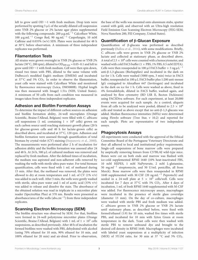
C. albicans RLM1 Hypersensitivity toCongo Red Is Rescued by Growth onLactateIn order to evaluate the impact of an alternative carbon source onthe role of C. albicans RLM1, we determined the sensitivity of thewild-type, 1rlm1/1rlm1 mutant and complemented strains to arange of cell wall-perturbing agents in the presence of glucose orlactate. In glucose medium, strains were able to grow well in thepresence of all the compounds except in caspofungin, with thehomozygous mutants being more sensitive. The mutant strainspresented hypersensitivity to Congo Red when compared withcomplemented and parental strains (Figure 3A), as previouslyreported (Bruno et al., 2006; Delgado-Silva et al., 2014). Incontrast, in the presence of lactate, all strains were hypersensitiveto SDS, Caffeine, and Caspofungin (Figure 3B). In the absenceof a functional RLM1, C. albicans cells grown in the presence oflactate showed more sensitivity to Caspofungin, when comparedto their glucose counterparts. However, the hypersensitivity toCongo Red observed with these cells grown in glucose diminishedgreatly. This result indicates that when grown in lactate cells aremore sensitive to cell wall stressing agents but, curiously, theabsence of a functional RLM1 rescues the hypersensitivity to CRobserved in glucose grown cells (Figure 3B).
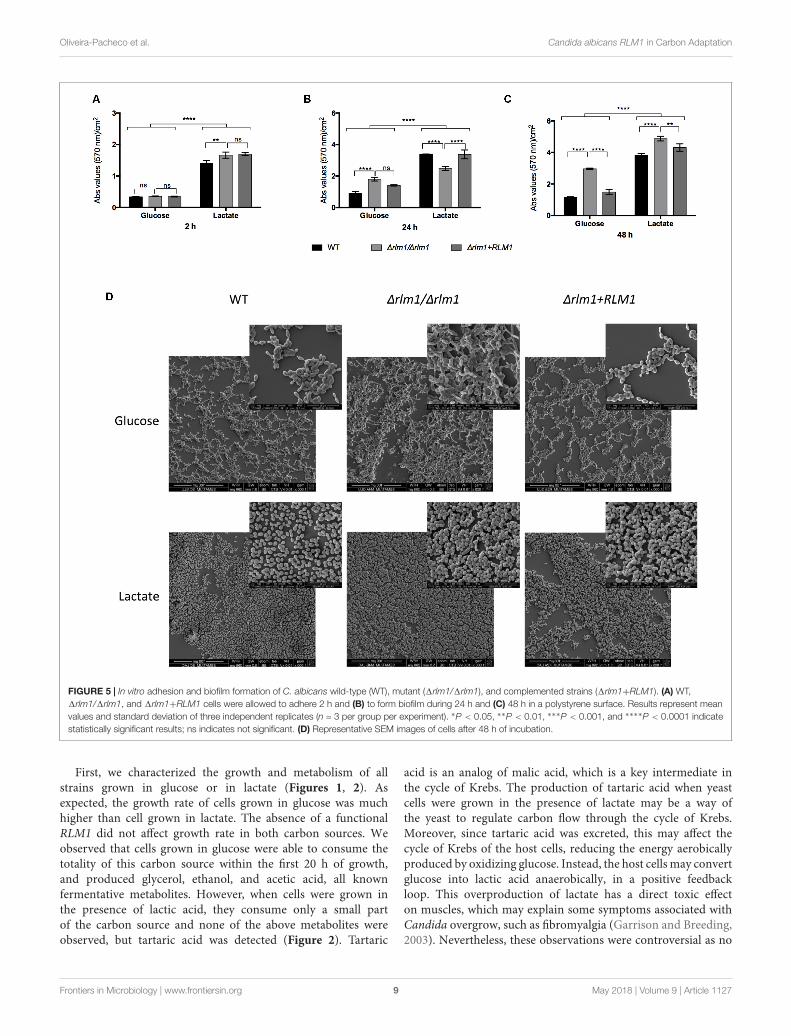
The Transcription Factor RLM1 IsImportant for C. albicans Filamentationand Biofilm FormationFilamentation and biofilm formation represent two of themajor virulence factors contributing to Candida pathogenesis.A previous work using 1rlm1/1rlm1 mutant strains grown onglucose-containing media has shown a higher upregulation ofproteins involved in adhesion and biofilm formation (Delgado-Silva et al., 2014). Additionally, some studies have demonstratedthat lactate-grown cells display higher ability to adhere and formbiofilm when compared to glucose-grown cells (Ene et al., 2012b;Alves et al., 2017). Based on these studies, 1rlm1/1rlm1 mutantstrains were tested regarding their ability to filament (Figure 4),to adhere to a polystyrene surface (Figure 5A), and to formbiofilm after 24 h (Figure 5B) and 48 h (Figure 5C) of incubation,in the presence of lactate.
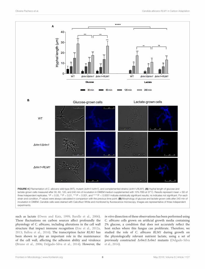
For filamentation analysis, glucose- and lactate-grown cellswere incubated in induced media for 30, 60, 120, and 240 min,stained with Calcofluor White and monitored by fluorescencemicroscopy. Independently of the condition, all strains wereable to filament and hyphae length increased with time. Inglucose-grown cells, filaments were visible right after 30 min
of incubation, while in lactate-grown cells only after 60 min(Figure 4). Moreover, lactate-grown cells presented shorterhyphae than glucose-grown cells. The lack of a functional RLM1affected filamentation of cells adapted to both carbon sources,and although the differences in hyphae length were visible in earlytime points, only after 240 min of incubation the differences weresignificant (P < 0.001). This difference was more pronounced incells grown on glucose (Figure 4).
Then, all strains were evaluated regarding their ability toadhere to a polystyrene surface. After 2 h of incubation, lactate-grown cells showed a higher ability to adhere when comparedwith the glucose-grown cells (P< 0.0001), as previously reported(Ene et al., 2012b), and no differences were seen regarding the1rlm1/1rlm1 mutant strain (Figure 5A). Considering biofilmformation, cells grown in lactate were able to form more biofilmthan cells grown in glucose. As expected, 1rlm1/1rlm1 mutantformed more biofilm than the wild-type in presence of glucoseat 24 and 48 h of incubation (Figures 5B,C). However, in thepresence of lactate, at 24 h of incubation the mutant presentedlower biofilm formation but at 48 h the amount of biofilmformed was higher than the WT and complemented strains(Figures 5B,C). SEM analyses confirmed the differences inbiofilm formation after 48 h of incubation (Figure 5D). Theseresults showed that lactate-grown cells presented higher biomassthan their glucose counterparts and the mutant presented higherbiofilm formation in both conditions, after biofilm maturation.
Overall, these results indicated that RLM1 is important forfilamentation, adhesion, and biofilm formation and that thesephenotypes were similar for cells adapted to glucose or to lactate,suggesting that they are independent of the carbon source.
C. albicans RLM1 Does Not AffectImmune Recognition but Is Important forImmune ActivationIn order to determine the importance of C. albicans RLM1in cell wall remodeling, we tested whether the growth ofthe different strains in the presence of the alternative carbonsource lactate would influence the interaction with phagocyticcells (Figure 6). We showed previously that the absence ofC. albicans RLM1 on glucose grown cells significantly altersthe proportions of the major cell wall components, enhancingthe chitin content and reducing the glucans and mannanscontent (Delgado-Silva et al., 2014). However, nothing aboutthe exposure of these components was previously observed.Thus, as the host immune defenses rely on the recognition ofconserved molecular patterns in the fungal cell wall, particularlythe glucans, we analyzed the exposure of β-glucans at the cellsurface of C. albicans cells grown either in the presence ofglucose or in the lactate. All cells were stained with anti-β-1,3-glucans and analyzed by flow cytometry. Glucose-grown cellsexhibited significantly (P < 0.0001) higher levels of β-glucanexposure than lactate-grown cells (Figure 6A), consistent withpreviously published data (Ballou et al., 2016). Although themutant seemed to present a different pattern of β-glucansexposure in both carbon sources, the differences were notsignificant.
Frontiers in Microbiology | www.frontiersin.org 6 May 2018 | Volume 9 | Article 1127

fmicb-09-01127 May 28, 2018 Time: 10:3 # 7
Oliveira-Pacheco et al. Candida albicans RLM1 in Carbon Adaptation
FIGURE 3 | Sensitivity of C. albicans wild-type (WT), mutant (1rlm1/1rlm1), and complemented (rlm11 +RLM1) strains to several agents that affect cell integrity.(A) Serial 10-fold dilutions of overnight cultures were spotted on YNB 2% glucose or (B) YNB 2% lactate plates with 10 mM Caffeine, 90 ng.ml−1 Caspofungin,200 µg.ml−1 Calcofluor White, 100 µg.ml−1 Congo Red, and 0.035% SDS for 2 days at 30◦C. Images are representative of three independent experiments.
To evaluate phagocytosis of C. albicans cells, we useda previously described assay (Carneiro et al., 2014), whichallowed the identification of different macrophage populationsby differential staining. In this way, macrophages with onlyinternalized C. albicans cells (sytox green-stained) and with bothinternalized and surface adhered cells (PI and sytox green doublestained) were clearly distinguished. As previously described,results showed that glucose-grown cells were internalizedmore efficiently by murine bone marrow-derived macrophages(BMDMs) compared to lactate-grown cells (Figure 6B) (Eneet al., 2013). In contrast, lactate-grown cells displayed higherlevels of adhesion than glucose-grown cells (Figure 6B).However, no significant changes were observed between the wild-type and 1rlm1/1rlm1 mutant strains under these conditions(Figure 6B). Representative fluorescence microscopy analyses areshown in Figure 6C.
Additionally, the effect of RLM1 on phagocyte interaction andactivation was assessed. For that, macrophages were infected withC. albicans cells, previously grown in glucose or lactate, in a MOIof 5 yeasts to 1 macrophage for 1 h. The uptake of live fungalcells by macrophages was measured by colony-forming units(CFUs) and presented in percentage of yeast killing (Figure 7A).Results indicated that wild-type and complemented lactate-grown cells were less efficiently killed by macrophages than theirglucose counterparts (Figure 7A), as previously reported (Eneet al., 2013). However, no significant differences were observedbetween the 1rlm1/1rlm1 mutant strain adapted to glucose incomparison to lactate, suggesting that the mutant lost part of itsresistance to killing.
The cell damage caused by the different strains was alsoquantified by measuring the amount of lactate dehydrogenase(LDH) released by murine macrophages after 1 h of incubationwith yeast cells (Figure 7B). Results showed that the wild-typeand complemented strains adapted to glucose led to a lowerproduction of LDH than their lactate counterparts, resulting inhigher percentage of viable macrophages (P < 0.001; Figure 7B).
In contrast, the1rlm1/1rlm1 mutant strain was able to cause lessdamage in the murine macrophages when cells where grown onlactate, in comparison with glucose, indicating that it also lost itsability to kill macrophages when grown in lactate. These resultssuggest that RLM1 is involved in cell damage but only when cellsare grown in lactate.
Cell activation was also evaluated by quantifying IL-10 andTNF-α after 1 h of co-incubation (Figures 7C,D). Results showedthat glucose-grown cells stimulated less production of TNF-α (Figure 7D) and IL-10 (Figure 7C), compared to lactate-grown cells, consistent with previously published data (Ene et al.,2013). Furthermore, the 1rlm1/1rlm1 mutant strain grown inlactate showed higher levels of IL-10 when compared with theirwild-type (P < 0.001) and complemented (P < 0.001) strains(Figure 7C), while when grown in glucose the results were theopposite. The same results were observed considering TNF-αsecretion (Figure 7D), confirming that RLM1 is relevant forimmune activation. Overall, these results indicate thatRLM1 doesnot mediate immune recognition but is important in immune cellresistance and activation, particularly in cells grown in lactate.
Finally, in order to evaluate whether tartaric acid couldcontribute for cellular host cytotoxicity, we incubatedmacrophages with this organic acid and evaluate metabolicactivity by MTT assay. Results showed that, after 72 h ofincubation, tartaric acid reduces cellular viability by around 35%(Figure 8).
DISCUSSION
Candida albicans has the ability to survive and proliferate withindistinct niches in the human host. This flexibility requiresa rapid adaptation to local conditions, forcing the pathogento utilize the alternative nutrients that are available. Someof these niches contain glucose, the preferred carbon sourceof C. albicans, while others contain different carboxylic acids
Frontiers in Microbiology | www.frontiersin.org 7 May 2018 | Volume 9 | Article 1127
fmicb-09-01127 May 28, 2018 Time: 10:3 # 8
Oliveira-Pacheco et al. Candida albicans RLM1 in Carbon Adaptation
FIGURE 4 | Filamentation of C. albicans wild-type (WT), mutant (1rlm1/1rlm1), and complemented strains (1rlm1+RLM1). (A) Hyphal length of glucose andlactate-grown cells measured after 30, 60, 120, and 240 min of incubation in DMEM medium supplemented with 10% FBS at 37◦C. Results represent mean + SD ofthree independent replicates. ∗P < 0.05, ∗∗P < 0.01, ∗∗∗P < 0.001, and ∗∗∗∗P < 0.0001 indicate statistically significant results; ns indicates not significant. For eachstrain and condition, P-values were always calculated in comparison with the previous time point. (B) Morphology of glucose and lactate-grown cells after 240 min ofincubation in DMEM. Candida cells were stained with Calcofluor White and monitored by fluorescence microscopy. Images are representative of three independentexperiments.
such as lactate (Owen and Katz, 1999; Barelle et al., 2006).These fluctuations on carbon sources affect profoundly thephysiology of C. albicans, including alterations in the cell wallstructure that impact immune recognition (Ene et al., 2012a,2013; Ballou et al., 2016). The transcription factor RLM1 hasbeen shown to play an important role in the maintenanceof the cell wall, affecting the adhesion ability and virulence(Bruno et al., 2006; Delgado-Silva et al., 2014). However, the
in vitro dissection of these observations has been performed usingC. albicans cells grown on artificial growth media containing2% glucose, a condition that does not accurately reflect thehost niches where this fungus can proliferate. Therefore, westudied the role of C. albicans RLM1 during growth onthe physiologically relevant nutrient lactate, using a set ofpreviously constructed 1rlm1/1rlm1 mutants (Delgado-Silvaet al., 2014).
Frontiers in Microbiology | www.frontiersin.org 8 May 2018 | Volume 9 | Article 1127
fmicb-09-01127 May 28, 2018 Time: 10:3 # 9
Oliveira-Pacheco et al. Candida albicans RLM1 in Carbon Adaptation
FIGURE 5 | In vitro adhesion and biofilm formation of C. albicans wild-type (WT), mutant (1rlm1/1rlm1), and complemented strains (1rlm1+RLM1). (A) WT,1rlm1/1rlm1, and 1rlm1+RLM1 cells were allowed to adhere 2 h and (B) to form biofilm during 24 h and (C) 48 h in a polystyrene surface. Results represent meanvalues and standard deviation of three independent replicates (n = 3 per group per experiment). ∗P < 0.05, ∗∗P < 0.01, ∗∗∗P < 0.001, and ∗∗∗∗P < 0.0001 indicatestatistically significant results; ns indicates not significant. (D) Representative SEM images of cells after 48 h of incubation.
First, we characterized the growth and metabolism of allstrains grown in glucose or in lactate (Figures 1, 2). Asexpected, the growth rate of cells grown in glucose was muchhigher than cell grown in lactate. The absence of a functionalRLM1 did not affect growth rate in both carbon sources. Weobserved that cells grown in glucose were able to consume thetotality of this carbon source within the first 20 h of growth,and produced glycerol, ethanol, and acetic acid, all knownfermentative metabolites. However, when cells were grown inthe presence of lactic acid, they consume only a small partof the carbon source and none of the above metabolites wereobserved, but tartaric acid was detected (Figure 2). Tartaric
acid is an analog of malic acid, which is a key intermediate inthe cycle of Krebs. The production of tartaric acid when yeastcells were grown in the presence of lactate may be a way ofthe yeast to regulate carbon flow through the cycle of Krebs.Moreover, since tartaric acid was excreted, this may affect thecycle of Krebs of the host cells, reducing the energy aerobicallyproduced by oxidizing glucose. Instead, the host cells may convertglucose into lactic acid anaerobically, in a positive feedbackloop. This overproduction of lactate has a direct toxic effecton muscles, which may explain some symptoms associated withCandida overgrow, such as fibromyalgia (Garrison and Breeding,2003). Nevertheless, these observations were controversial as no
Frontiers in Microbiology | www.frontiersin.org 9 May 2018 | Volume 9 | Article 1127

fmicb-09-01127 May 28, 2018 Time: 10:3 # 10
Oliveira-Pacheco et al. Candida albicans RLM1 in Carbon Adaptation
FIGURE 6 | Immune recognition of C. albicans wild-type (WT), mutant (∆rlm1/∆rlm1), and complemented strains (rlm11+RLM1). (A) Flow cytometry analysis ofβ-glucan exposure for cells grown either in YNB 2% Glucose (red) or in YNB 2% Lactate (blue). MFIs are indicated at the top of each panel and plots arerepresentative of two independent replicate experiments (n = 3 per group per experiment). (B) Flow cytometry analysis of phagocytosis after 30 min of incubationwith macrophages. Graphs represent the % of macrophages with internalized yeast cells (left), and the % of macrophages with internalized and adhered yeasts(right). ∗P < 0.05, ∗∗P < 0.01, ∗∗∗P < 0.001, and ∗∗∗∗P < 0.0001 indicate statistically significant results; ns indicates not significant. Plots are representative of threeindependent replicate experiments (n = 3 per group per experiment). (C) Representative micrographs showing macrophages with internalized (green labeled) oradhered (red labeled) yeast cells grown either in YNB 2% glucose or in YNB 2% Lactate.
evidence has been found for the production of tartaric acidas a metabolic end product by Candida (Lord et al., 2005).Here, we show that C. albicans produces tartaric acid whengrowing in the presence of lactate and this metabolite, at aconcentration identified in our studies, reduces cell viability.This is particularly interesting as lactate is present in severalniches within the host, including within the gastrointestinaltract and vagina (Owen and Katz, 1999; Barelle et al., 2006),suggesting that when cells grow in these niches, the productionof tartaric acid may explain some symptoms associated withCandida infections.
We then compared the same typical cell wall phenotypesdescribed for glucose-grown C. albicans 1rlm1/1rlm1 mutantcells by performing the tests in parallel with the controlstrains grown in the presence of lactate (Figure 3). Wefound that lactate-grown cells, independently of the mutation,presented hypersensitivity to different stresses that affected the
cell wall, such as Caffeine, Caspofungin, and SDS (Figure 3B).The cell wall of C. albicans lactate-grown cells is describedas being thinner, presenting less β-glucans and mannansand is more porous than their glucose counterparts (Eneet al., 2012a,b). The differences observed between the twoconditions reflect the alterations in the cell wall compositiondue to the carbon source and the porosity may explain thecomplete absence of growth of lactate-adapted cells on SDS.Since these phenotypes were similar for all strains, it isprobable that similar cell wall alterations observed previously(Ene et al., 2012a,b) may also occur in the mutant strain.Moreover, in lactate grown cells, the absence of a functionalRLM1 reverted the hypersensitivity to Congo Red (Figure 3B)observed when cells were grown in glucose (Figure 3A).It has been described that Congo Red interacts with cellwall polysaccharides, exhibiting a particularly high affinity forchitin and β-glucans (Klis et al., 2002). Thus, the fact that
Frontiers in Microbiology | www.frontiersin.org 10 May 2018 | Volume 9 | Article 1127

fmicb-09-01127 May 28, 2018 Time: 10:3 # 11
Oliveira-Pacheco et al. Candida albicans RLM1 in Carbon Adaptation
FIGURE 7 | Host viability and immune response to C. albicans wild-type (WT), mutant (1rlm1/1rlm1), and complemented (1rlm1+RLM1) cells grown on glucose orlactate after 1 h of infection. (A) Killing of yeast cells adapted to glucose or lactate and incubated with macrophages at 5:1 ratio. Results are expressed as thepercentage of yeast killing. (B) Assessment of host viability by measuring LDH released by murine macrophages. Concentrations of (C) IL-10 and (D) TNF-αdetected in culture supernatants of murine macrophages after incubation with the yeast cells. ∗P < 0.05, ∗∗P < 0.01, ∗∗∗P < 0.001, and ∗∗∗∗P < 0.0001 indicatestatistically significant results; ns indicates not significant. Plots are representative of three independent replicate experiments (n = 3 per group per experiment). Mφ
indicates macrophages alone (near glucose) or incubated with LPS (near lactate).
FIGURE 8 | Host viability after incubation with tartaric acid. Macrophageswere incubated with tartaric acid at a final concentration of 0.075 g/L for 24and 72 h. Results express the percentage of macrophage viability. ∗P < 0.05,∗∗P < 0.01, ∗∗∗P < 0.001, and ∗∗∗∗P < 0.0001 indicate statisticallysignificant results; ns indicates not significant. Plots are representative of threeindependent replicate experiments (n = 3 per group per experiment).C– indicates macrophages incubated with DMEM alone.
in lactate-grown cells, β-glucans are masked may reduce theavailability of Congo Red to β-glucans, rendering the mutantmore resistant.
Candida albicans has evolved multiple strategies, including theexpression of several virulence factors, to overcome the differentenvironmental conditions imposed by the host (Calderone andFonzi, 2001; Mayer et al., 2013). As many of these factorsrely on morphology changes, we also tested whether RLM1could be involved in some virulence mechanisms such ashyphal growth, adhesion and biofilm formation during carbonadaptation. Under our growth conditions, we showed that the1rlm1/1rlm1 strain presented shorter hypha than the WTor complemented strains when grown in glucose as well asin lactose, indicating that RLM1 is involved in C. albicansfilamentation independently of the carbon source (Figure 4).As we reported previously, the 1rlm1/1rlm1 strain showed areduction in the cell wall mannans (Delgado-Silva et al., 2014)and loss of cell wall O-mannans was associated with impairedhyphal growth (McKenzie et al., 2010). Thus, the defect infilamentation observed could be correlated with the cell wallcomposition.
Additionally, C. albicans RLM1 has been described asa negative regulator of in vitro biofilm formation, as the1rlm1/1rlm1 mutant strain forms more biofilm than thewild-type in presence of glucose (Delgado-Silva et al., 2014).This gene also seems to regulate negatively some of thesame targets of BCR1, a well-known transcription factor that
Frontiers in Microbiology | www.frontiersin.org 11 May 2018 | Volume 9 | Article 1127

fmicb-09-01127 May 28, 2018 Time: 10:3 # 12
Oliveira-Pacheco et al. Candida albicans RLM1 in Carbon Adaptation
governs biofilm formation (Nobile et al., 2006). We observedthat in the presence of lactate, all strains produced morebiofilm, but as previously reported, the 1rlm1/1rlm1 strainformed even more biofilm, than in the presence of glucose(Figure 5). This enhanced biofilm formation in lactate maybe directly correlated with the higher ability (around threetimes more) of the lactate grown cells to adhere. Theseresults indicate that the role of RLM1 as a negative regulatorof in vitro biofilm formation is independent of the carbonsource.
Finally, we studied the involvement of RLM1 in host-pathogen recognition. The cell wall of microbial pathogensis the first point of contact with the host defenses. Then,any modification on the cell surface, especially on thepathogen-associated molecular patterns (PAMPs), such asβ-glucans, α-, and β-mannans, phosphomannans and chitin,impacts the immune detection. The host metabolite lactatehas been shown to modulate the exposure of some PAMPsin C. albicans, affecting the recognition of the fungus by thehost phagocytes (Ene et al., 2012a, 2013; Brown et al., 2014;Ballou et al., 2016). During growth in lactate, the β-glucansare actively masked by the outer mannans layer (Ballou et al.,2016).
In our experiments, RLM1 does not seem to beinvolved in β-glucans masking (Figure 6A), which isconsistent with the phagocytosis results, since no significantdifferences were observed between the wild-type and the1rlm1/1rlm1 mutant in both carbon sources (Figures 6B,C).Moreover, our results confirmed that cells grown inlactate were less internalized when compared with theirglucose counterparts (Figure 6) (Ballou et al., 2016),but remained attached to the macrophages (Figure 6B).Once more RLM1 does not seem to be involved in thismechanism.
Although no significant differences were observed in thepercentage of yeast killing between 1rlm1/1rlm1 and theircounterpart strains, within each condition (carbon source),our results confirm previous data that lactate grown cellsare more resistance to phagocyte killing (Ene et al., 2013).However, it is curious to observe that when the mutant wasgrown in lactate, the resistance to yeast killing was lower andnot statistically different when grown in glucose, indicatingthat without RLM1 C. albicans loses its resistance acquiredwhen cells grow in lactate (Figure 7A). Since previous resultsindicate that RLM1 is involved in cell wall architecture,when cells were grown in glucose, this loss of resistance tokilling could also be due to changes in cell wall structureand composition and account for its lower virulence inthe disseminated mouse model of infection (Delgado-Silvaet al., 2014). The levels of LDH released by macrophages,an indicator of cell damage, were increased for the cellsgrown in the presence of lactate, indicating that these cellsalthough being taking up by the macrophages less efficiently,are more prone to kill these phagocytic cells (Figure 7B).Interestingly, this is not verified for the 1rlm1/1rlm1mutant. As seen previously (Figure 7A), the mutant ismore susceptible to macrophage killing, which explains the
lower ability to escape from macrophages by damaging thephagocyte. Previous data demonstrated that hyphal extensionis a key factor promoting fungal escape from phagocytes,therefore the fact that 1rlm1/1rlm1 mutant produces shorterhyphae might also contribute to the lower capacity to damagemacrophages.
The amount of IL-10 and TNF-α was also quantified inorder to evaluate the ability of the fungus to induce anti-and pro-inflammatory responses, respectively (Figures 7C,D).As expected, the cytokines profile of the C. albicans in lactategrown cells points to an anti-inflammatory response, giventhe increased levels of IL-10 in this conditions. However, thelevels of IL-10 in the 1rlm1/1rlm1 mutant were even higher.The masking of β-glucans on the surface of lactate growncells reduces not only the phagocytosis, which is stimulatedvia dectin-1, but also the secretion of cytokines, which isunder the control of the transduction pathways upon activationof dectin-1 (Steele et al., 2003). In our study, we observedthat the levels of TNF-α were lower with the mutant, whencompared with the wild-type and complemented strains, inglucose-grown cells, but slightly higher in lactate-grown cells.This would suggest that β-glucans in the mutant strain grownin glucose would be less exposed than their counterparts whilein lactate-grown cells it would be the opposite. RegardingIL-10, the higher concentration observed when macrophageswere incubated with the mutant cells grown in lactate, couldalso be explained by the fact that the β-glucans’ maskingof the mutant was much lower than in the wild-type andcomplemented strains. However, and once more, the differenceswere not significant. The fact that interpreting relative MFIfor individual runs across strains is sensitive to growthrate, growth phase, staining uptake, and cell size distributioncould contribute to not reach statistical significance. Theseslight differences in β-glucans exposure would not influencephagocytosis but could account for the differences in host cellactivation.
Taken together, these results confirmed that C. albicanscells grown in the presence of lactate were less internalizedand killed by macrophages and suggest that RLM1, althoughnot being involved in yeast cells internalization, seems tobe involved in the killing by macrophages and inflictingdamage to host cells, what could be related to the lowercapacity of the mutant to filament. This interaction mediatescytokine levels, rendering the lactate-grown cells less visibleto the phagocytic cells, as previously reported (Ene et al.,2013). However, although the 1rlm1/1rlm1 induced a higheranti-inflammatory response, the modifications in the cell wallrendered the mutant less resistant to action of the immunesystem.
CONCLUSION
This study reveals that regardless of the carbon source, C. albicansRLM1 is involved in filamentation and biofilm formation,which could be directly correlated with the architecture andcomposition of the cell wall. It also showed that some phenotypes
Frontiers in Microbiology | www.frontiersin.org 12 May 2018 | Volume 9 | Article 1127

fmicb-09-01127 May 28, 2018 Time: 10:3 # 13
Oliveira-Pacheco et al. Candida albicans RLM1 in Carbon Adaptation
were dependent on the carbon source, such as resistance tomacrophage killing and ability to damage macrophages, whichcan also be correlated to changes in the cell wall. Thus, thecarbon source that presents in different host niches, such aslactate, affects the remodeling of the C. albicans cell wall, withthe potential to also modulate host metabolism, and suggests thatRLM1 plays an important role in this pathway.
AUTHOR CONTRIBUTIONS
PS and CP designed the experiments. JO-P, RA, BC-R, AC-B,and PP-S carried out the experiments. JO-P, RA, BC-R, CP, andPS analyzed and interpreted the data. JO-P, RA, and PS wrotethe manuscript with additional input from BC-R, SP, SS, MH,and CP.
FUNDING
This study was supported by the Portuguese National FundingAgency for Science, Research and Technology FCT. RAreceived FCT Ph.D. fellowship (PD/BD/113813/ 2015) andAC-B received FCT Ph.D. fellowship SFRH/BD/133513/2017.The work on CBMA was supported by FEDER throughPOFC-COMPETE and by FCT through strategic fundingUID/BIA/04050/2013. The work on CEB was supported by Pest-OE/EQB/LA0023/2013, from FCT, “BioHealth – Biotechnologyand Bioengineering approaches to improve health quality,” Ref.NORTE-07-0124-FEDER-000027, co-funded by the ProgramaOperacional Regional do Norte (ON.2 – O Novo Norte), QREN,FEDER, and the project “Consolidating Research Expertise andResources on Cellular and Molecular Biotechnology at CEB/IBB,”Ref. FCOMP-01-0124-FEDER-027462.
REFERENCESAlves, R., Mota, S., Silva, S., Rodrigues, C. F., Alistair, J. P., Henriques, M.,
et al. (2017). The carboxylic acid transporters Jen1 and Jen2 affect thearchitecture and fluconazole susceptibility of Candida albicans biofilm inthe presence of lactate. Biofouling 33, 943–954. doi: 10.1080/08927014.2017.1392514
Baetz, K., Moffat, J., Haynes, J., Chang, M., and Andrews, B. (2001). Transcriptionalcoregulation by the cell integrity mitogen- activated protein kinase Slt2 and thecell cycle regulator Swi4. Mol. Cell. Biol. 21, 6515–6528. doi: 10.1128/MCB.21.19.6515-6528.2001
Ballou, E. R., Avelar, G. M., Childers, D. S., Mackie, J., Bain, J. M.,Wagener, J., et al. (2016). Lactate signalling regulates fungal β-glucan maskingand immune evasion. Nat. Microbiol. 2:16238. doi: 10.1038/nmicrobiol.2016.238
Barelle, C. J., Priest, C. L., MacCallum, D. M., Gow, N. A., Odds, F. C., and Brown,A. J. (2006). Niche-specific regulation of central metabolic pathways in a fungalpathogen. Cell. Microbiol. 8, 961–971. doi: 10.1111/j.1462-5822.2005.00676.x
Blankenship, J. R., Fanning, S., Hamaker, J. J., and Mitchell, A. P. (2010). Anextensive circuitry for cell wall regulation in Candida albicans. PLoS Pathog.6:e1000752. doi: 10.1371/journal.ppat.1000752
Brown, A. J. P., Brown, G. D., Netea, M. G., and Gow, N. A. R. (2014). Metabolismimpacts upon Candida immunogenicity and pathogenicity at multiple levels.Trends Microbiol. 22, 614–622. doi: 10.1016/j.tim.2014.07.001
Brown, G. D., Denning, D. W., Gow, N. A. R., Levitz, S. M., Netea, M. G., andWhite, T. C. (2012). Hidden killers: human fungal infections. Sci. Transl. Med.4:165rv13. doi: 10.1126/scitranslmed.3004404
Brown, G. D., and Gordon, S. (2001). Immune recognition. A new receptor forbeta-glucans. Nature 413, 36–37. doi: 10.1038/35092620
Bruno, V. M., Kalachikov, S., Subaran, R., Nobile, C. J., Kyratsous, C., andMitchell, A. P. (2006). Control of the C. albicans cell wall damage response bytranscriptional regulator Cas5. PLoS Pathog. 2:0204–0210. doi: 10.1371/journal.ppat.0020021
Calderone, R., and Fonzi, W. (2001). Virulence factors of Candida albicans. TrendsMicrobiol. 9, 327–335. doi: 10.1016/S0966-842X(01)02094-7
Campion, E. W., Kullberg, B. J., and Arendrup, M. C. (2015). Invasive Candidiasis.N. Engl. J. Med. 373, 1445–1456. doi: 10.1056/NEJMra1315399
Carneiro, C., Vaz, C., Carvalho-Pereira, J., Pais, C., and Sampaio, P. (2014). A newmethod for yeast phagocytosis analysis by flow cytometry. J. Microbiol. Methods101, 56–62. doi: 10.1016/j.mimet.2014.03.013
Collins, T., Barroca, M., Branca, F., Padrão, J., Machado, R., and Casal, M.(2014). High level biosynthesis of a silk-elastin-like protein in E. coli.Biomacromolecules 15, 2701–2708. doi: 10.1021/bm5005564
Delgado-Silva, Y., Vaz, C., Carvalho-Pereira, J., Carneiro, C., Nogueira, E.,Correia, A., et al. (2014). Participation of Candida albicans transcription factorRLM1 in cell wall biogenesis and virulence. PLoS One 9:e86270. doi: 10.1371/journal.pone.0086270
Dodou, E., and Treisman, R. (1997). The Saccharomyces cerevisiae MADS-boxtranscription factor Rlm1 is a target for the Mpk1 mitogen-activated proteinkinase pathway. Mol. Cell. Biol. 17, 1848–1859. doi: 10.1128/MCB.17.4.1848
Ene, I. V., Adya, A. K., Wehmeier, S., Brand, A. C., Maccallum, D. M., Gow,N. A. R., et al. (2012a). Host carbon sources modulate cell wall architecture, drugresistance and virulence in a fungal pathogen. Cell. Microbiol. 14, 1319–1335.doi: 10.1111/j.1462-5822.2012.01813.x
Ene, I. V., Cheng, S. C., Netea, M. G., and Brown, A. J. P. (2013). Growth ofCandidaalbicans cells on the physiologically relevant carbon source lactate affects theirrecognition and phagocytosis by immune cells. Infect. Immun. 81, 238–248.doi: 10.1128/IAI.01092-12
Ene, I. V., Heilmann, C. J., Sorgo, A. G., Walker, L. A., De Koster, C. G., Munro,C. A., et al. (2012b). Carbon source-induced reprogramming of the cell wallproteome and secretome modulates the adherence and drug resistance of thefungal pathogen Candida albicans. Proteomics 12, 3164–3179. doi: 10.1002/pmic.201200228
Garrison, R. L., and Breeding, P. C. (2003). A metabolic basis for fibromyalgia andits related disorders: the possible role of resistance to thyroid hormone. Med.Hypotheses 61, 182–189. doi: 10.1016/S0306-9877(02)00294-3
Gillum, A. M., Tsay, E. Y., and Kirsch, D. R. (1984). Isolation of the Candidaalbicans gene for orotidine-5′-phosphate decarboxylase by complementation ofS. cerevisiae ura3 and E. coli pyrF mutations. Mol. Gen. Genet. 198, 179–182.doi: 10.1007/BF00328721
Hall, R. A. (2017). Adapting to change: interactions of Candida albicans with itsenvironment. Future Microbiol. 12, 931–934. doi: 10.2217/fmb-2017-0130
Hardison, S. E., and Brown, G. D. (2012). C-type lectin receptors orchestrateantifungal immunity. Nat. Immunol. 13, 817–822. doi: 10.1038/ni.2369
Jung, U. S., Sobering, A. K., Romeo, M. J., and Levin, D. E. (2002). Regulationof the yeast Rlm1 transcription factor by the Mpk1 cell wall integrityMAP kinase. Mol. Microbiol. 46, 781–789. doi: 10.1046/j.1365-2958.2002.03198.x
Klis, F. M., Mol, P., Hellingwerf, K., and Brul, S. (2002). Dynamics of cellwall structure in Saccharomyces cerevisiae. FEMS Microbiol. Rev. 26, 239–256.doi: 10.1111/j.1574-6976.2002.tb00613.x
Korzeniewski, C., and Callewaert, D. M. (1983). An enzyme-release assay fornatural cytotoxicity. J. Immunol. Methods 64, 313–320. doi: 10.1016/0022-1759(83)90438-6
Levin, D. E. (2011). Regulation of cell wall biogenesis in Saccharomyces cerevisiae:the cell wall integrity signaling pathway. Genetics 189, 1145–1175. doi: 10.1534/genetics.111.128264
Lipke, P. N., and Ovalle, R. (1998). Cell wall architecture in yeast: new structureand new challenges. J. Bacteriol. 180, 3735–3740.
Lord, R. S., Burdette, C. K., and Bralley, J. A. (2005). Significance ofurinary tartaric acid. Clin. Chem. 51, 672–673. doi: 10.1373/clinchem.2004.036368
Mayer, F. L., Wilson, D., and Hube, B. (2013). Candida albicans pathogenicitymechanisms. Virulence 4, 119–128. doi: 10.4161/viru.22913
Frontiers in Microbiology | www.frontiersin.org 13 May 2018 | Volume 9 | Article 1127

fmicb-09-01127 May 28, 2018 Time: 10:3 # 14
Oliveira-Pacheco et al. Candida albicans RLM1 in Carbon Adaptation
McKenzie, C. G. J., Koser, U., Lewis, L. E., Bain, J. M., Mora-Montes, H. M., Barker,R. N., et al. (2010). Contribution of Candida albicans cell wall componentsto recognition by and escape from murine macrophages. Infect. Immun. 78,1650–1658. doi: 10.1128/IAI.00001-10
Miramón, P., and Lorenz, M. C. (2017). A feast for Candida: metabolic plasticityconfers an edge for virulence. PLoS Pathog. 13:e1006144. doi: 10.1371/journal.ppat.1006144
Netea, M. G., Brown, G. D., Kullberg, B. J., and Gow, N. A. R. (2008). An integratedmodel of the recognition of Candida albicans by the innate immune system.Nat. Rev. Microbiol. 6, 67–78. doi: 10.1038/nrmicro1815
Nobile, C. J., Andes, D. R., Nett, J. E., Smith, F. J., Yue, F., Phan, Q. T., et al. (2006).Critical role of Bcr1-dependent adhesins in C. albicans biofilm formationin vitro and in vivo. PLoS Pathog. 2:0636–0649. doi: 10.1371/journal.ppat.0020063
Owen, D. H., and Katz, D. F. (1999). A vaginal fluid simulant. Contraception 59,91–95. doi: 10.1016/S0010-7824(99)00010-4
Pfaller, M. A., and Diekema, D. J. (2007). Epidemiology of invasive candidiasis:a persistent public health problem. Clin. Microbiol. Rev. 20, 133–163.doi: 10.1128/CMR.00029-06
Rauceo, J. M., Blankenship, J. R., Fanning, S., Hamaker, J. J., Deneault, J.-S., Smith,F. J., et al. (2008). Regulation of the Candida albicans cell wall damage responseby transcription factor Sko1 and PAS kinase Psk1. Mol. Biol. Cell 19, 2741–2751.doi: 10.1091/mbc.E08-02-0191
Sampaio, P., Nogueira, E., Loureiro, A. S., Delgado-Silva, Y., Correia, A., andPais, C. (2009). Increased number of glutamine repeats in the C-terminal ofCandida albicans Rlm1p enhances the resistance to stress agents. Antonie VanLeeuwenhoek 96, 395–404. doi: 10.1007/s10482-009-9352-5
Southern, P., Horbul, J., Maher, D., and Davis, D. A. (2008). C. albicanscolonization of human mucosal surfaces. PLoS One 3:e2067. doi: 10.1371/journal.pone.0002067
Steele, C., Marrero, L., Swain, S., Harmsen, A. G., Zheng, M., Brown, G. D., et al.(2003). Alveolar macrophage-mediated killing of Pneumocystis carinii f. sp.
muris involves molecular recognition by the Dectin-1 beta-glucan receptor.J. Exp. Med. 198, 1677–1688. doi: 10.1084/jem.20030932
Stepanovic, S., Vukovic, D., Dakic, I., Savic, B., and Svabic-Vlahovic, M. (2000).A modified microtiter-plate test for quantification of staphylococcal biofilmformation. J. Microbiol. Methods 40, 175–179. doi: 10.1016/S0167-7012(00)00122-6
Watanabe, Y., Takaesu, G., Hagiwara, M., Irie, K., and Matsumoto, K. (1997).Characterization of a serum response factor-like protein in Saccharomycescerevisiae, Rlm1, which has transcriptional activity regulated by the Mpk1(Slt2) mitogen-activated protein kinase pathway. Mol. Cell. Biol. 17, 2615–2623.doi: 10.1128/MCB.17.5.2615
Wheeler, R. T., Kombe, D., Agarwala, S. D., and Fink, G. R. (2008). Dynamic,morphotype-specific Candida albicansβ-glucan exposure during infectionand drug treatment. PLoS Pathog. 4:e1000227. doi: 10.1371/journal.ppat.1000227
Xie, J. L., Qin, L., Miao, Z., Grys, B. T., Diaz, J. C., Ting, K., et al. (2017).The Candida albicans transcription factor Cas5 couples stress responses, drugresistance and cell cycle regulation. Nat. Commun. 8:499. doi: 10.1038/s41467-017-00547-y
Conflict of Interest Statement: The authors declare that the research wasconducted in the absence of any commercial or financial relationships that couldbe construed as a potential conflict of interest.
Copyright © 2018 Oliveira-Pacheco, Alves, Costa-Barbosa, Cerqueira-Rodrigues,Pereira-Silva, Paiva, Silva, Henriques, Pais and Sampaio. This is an open-accessarticle distributed under the terms of the Creative Commons Attribution License(CC BY). The use, distribution or reproduction in other forums is permitted, providedthe original author(s) and the copyright owner are credited and that the originalpublication in this journal is cited, in accordance with accepted academic practice.No use, distribution or reproduction is permitted which does not comply with theseterms.
Frontiers in Microbiology | www.frontiersin.org 14 May 2018 | Volume 9 | Article 1127