Embed Size (px)
Citation preview
The Rim15-Endosulfine-PP2ACdc55 Signalling ModuleRegulates Entry into Gametogenesis and Quiescence viaDistinct Mechanisms in Budding YeastSourav Sarkar, Jacob Z. Dalgaard, Jonathan B. A. Millar, Prakash Arumugam*
Division of Biomedical Cell Biology, Warwick Medical School, University of Warwick, Coventry, United Kingdom
Abstract
Quiescence and gametogenesis represent two distinct survival strategies in response to nutrient starvation in buddingyeast. Precisely how environmental signals are sensed by yeast cells to trigger quiescence and gametogenesis is not fullyunderstood. A conserved signalling module consisting of Greatwall kinase, Endosulfine and Protein Phosphatase PP2ACdc55
proteins regulates entry into mitosis in Xenopus egg extracts and meiotic maturation in flies. We report here that ananalogous signalling module consisting of the serine-threonine kinase Rim15, the Endosulfines Igo1 and Igo2 and theProtein Phosphatase PP2ACdc55, regulates entry into both quiescence and gametogenesis in budding yeast. PP2ACdc55
inhibits entry into gametogenesis and quiescence. Rim15 promotes entry into gametogenesis and quiescence byconverting Igo1 into an inhibitor of PP2ACdc55 by phosphorylating at a conserved serine residue. Moreover, we show thatthe Rim15-Endosulfine-PP2ACdc55 pathway regulates entry into quiescence and gametogenesis by distinct mechanisms. Inaddition, we show that Igo1 and Igo2 are required for pre-meiotic autophagy but the lack of pre-meiotic autophagy isinsufficient to explain the sporulation defect of igo1D igo2D cells. We propose that the Rim15-Endosulfine-PP2ACdc55
signalling module triggers entry into quiescence and gametogenesis by regulating dephosphorylation of distinct substrates.
Citation: Sarkar S, Dalgaard JZ, Millar JBA, Arumugam P (2014) The Rim15-Endosulfine-PP2ACdc55 Signalling Module Regulates Entry into Gametogenesis andQuiescence via Distinct Mechanisms in Budding Yeast. PLoS Genet 10(6): e1004456. doi:10.1371/journal.pgen.1004456
Editor: Gregory P. Copenhaver, The University of North Carolina at Chapel Hill, United States of America
Received July 8, 2013; Accepted May 9, 2014; Published June 26, 2014
Copyright: � 2014 Sarkar et al. This is an open-access article distributed under the terms of the Creative Commons Attribution License, which permitsunrestricted use, distribution, and reproduction in any medium, provided the original author and source are credited.
Funding: PA’s laboratory and SS were funded by a research grant from BBSRC (BB/G00353X/1). The funders had no role in study design, data collection andanalysis, decision to publish, or preparation of the manuscript.
Competing Interests: The authors have declared that no competing interests exist.
* Email: [email protected]
Introduction
The ability of cells to sense deleterious changes in environment
and mount an appropriate physiological and metabolic response is
essential for cellular survival. Response to nutrition starvation in
budding yeast has been an extremely powerful model to study this
biological trait [1]. Upon complete nutrient starvation, yeast cells
enter either gametogenesis or quiescence. Diploid yeast cells
undergo gametogenesis when subjected to nitrogen starvation in
the absence of glucose and in the presence of a non-fermentable
carbon source. They undergo one round of DNA replication
followed by two rounds of nuclear divisions to form 4 haploid
spores which can stay dormant for long periods of time. Haploid
and diploid cells enter quiescence when subjected to nutrient
starvation or when treated with a drug called rapamycin, an
inhibitor of the TOR (Target of Rapamycin) signalling pathway.
Quiescence (-also referred to as G0) is a reversible non-proliferative
state characterized by low rates of transcription and translation,
increased stress-tolerance, elevated rate of macroautophagy and
synthesis of storage carbohydrates (trehalose and glycogen). Many
of the G0-features like increased macroautophagy, low rates of
transcription and translation are also characteristic of quiescent
mammalian cells suggesting that the core features of quiescence
program are conserved [2,3]. Ablation of G0-entry/exit control
mechanisms is frequently linked to either reduced life span
(especially in unicellular organisms) or cellular transformation (in
multi-cellular organisms) [4,5].
In budding yeast, entry into quiescence is controlled by the
master regulator Rim15, a member of the AGC (named after
protein kinase A, G and C families) group of serine-threonine
kinases [6]. Activity of Rim15 is controlled by two nutrient
signalling pathways namely the Ras/Protein Kinase A (Ras/PKA)
and the Target of Rapamycin Complex 1 (TORC1) pathways. The
TORC1 pathway responds to the availability of nitrogen source in
the growth medium [2,5]. In contrast, the Ras/PKA pathway
responds to levels of glucose in the growth medium [2,5]. Both
pathways positively regulate cell proliferation in response to
nutrient availability and thereby inhibit entry into G0. PKA
phosphorylates Rim15 at five consensus PKA phosphorylation
sites to inhibit its kinase activity and promote its retention in the
cytosol [6]. Apart from PKA and TORC1 pathways, Rim15 also
integrates signalling from Sch9 kinase (ortholog of mammalian
Akt/S6 kinase) and Pho85-Pho80 kinase (phosphate-sensing)
pathways [7]. Signalling through TORC1, Sch9 and Pho85/
Pho80 pathways phosphorylate Rim15 at Thr-1075 and inhibit its
nuclear localization. Nutrient deprivation inhibits signalling
through these four pathways which results in dephosphorylation
of Rim15 at its five PKA sites and Thr-1075 leading to its
activation and translocation to the nucleus. Activated Rim15
stimulates stress-responsive transcription factors Msn2/4 and post-
PLOS Genetics | www.plosgenetics.org 1 June 2014 | Volume 10 | Issue 6 | e1004456
diauxic shift transcription factor Gis1 which in turn activate
transcription of several genes required for survival in G0. Rim15
phosphorylates endosulfines, a highly conserved family of cAMP
regulated phosphoproteins to promote entry into quiescence [8].
Endosulfines, following phosphorylation by Rim15, protect
mRNA, which are transcriptionally controlled by Msn2/4 and
Gis1, from degradation via the 59-39 mRNA decay pathway by
inhibiting Dhh1 (decapping activator) and Ccr4 (deadenylation
factor) [8].
Entry into gametogenesis in yeast is mainly regulated at the level
of transcription of IME1 which encodes the master transcription
factor for early-meiosis genes (EMGs) [9]. Like G0, entry into
gametogenesis is negatively regulated by TORC1 and Ras/PKA
pathways. Ime1 is recruited to promoters of EMGs and activates
their transcription [10]. During vegetative growth, a DNA-binding
protein Ume6 binds to promoters of EMGs and represses their
expression by associating with the Sin3/Rpd3 histone deacetylase
and Isw2 chromatin remodelling complexes [11,12]. Absence of
glucose and nitrogen in the medium results in the replacement of
Sin3/Rpd3 and Isw2 by Ime1 at the EMG promoter regions [13].
It was proposed that Ime1 activates transcription of EMGs by
converting Ume6 from a repressor into an activator [10]. This
model was consistent with the observations that Ime1 physically
interacts with Ume6 and that cells lacking Ume6 fail to sporulate
efficiently [10,14,15]. However, this model was disputed by
subsequent studies which showed that interaction of Ime1 with
Ume6 facilitates Ume6 destruction and meiotic gene induction
[16]. Rim15 has been implicated in the removal of histone
deacetylase complex from the promoters of EMGs [13] but
precisely how this is achieved is not known.
In this paper, we demonstrate that endosulfines are required for
entry into gametogenesis and quiescence in budding yeast.
Phosphorylation of endosulfine by Rim15 results in its association
with the protein phosphatase PP2ACdc55 and inhibition of its
phosphatase activity. We show that the Rim15-endosulfine-
PP2ACdc55 signalling module regulates entry into quiescence and
gametogenesis by distinct mechanisms. We also demonstrate that
this signalling module is required for pre-meiotic autophagy which
is necessary for gametogenesis in budding yeast. Remarkably a
similar signalling module regulates M-phase progression during
mitosis and meiosis in higher eukaryotes. In Xenopus egg extracts,
the Greatwall kinase phosphorylates a-endosulfine (ENSA) and
Arpp19 at a conserved serine residue, which then inhibits PP2A-
B55d to promote entry into mitosis [17,18]. Depletion of
Greatwall kinase and endosulfine in Drosophila leads to mitotic
defects suggests that the module regulates entry into mitosis in flies
[19,20]. Inactivation of endosulfine in flies causes a failure in
oocyte progression from prophase I to metaphase I indicating that
this module regulates entry into M-phase during meiosis [21]. Our
results therefore expand the repertoire of functions for this highly
conserved signalling module that regulates distinct biological
processes in different systems.
Results
Endosulfines are required for entry into gametogenesisSince Rim15 is required for expression of early meiotic genes
[22] we examined the function of endosulfine in gametogenesis.
Budding yeast has two endosulfines Igo1 and Igo2. We first
assessed the ability of wild type, igo1D, igo2D and igo1D igo2Dstrains to sporulate. While wild type, igo1D and igo2D strains
sporulated with an efficiency of $65%, only 3% of igo1D igo2Dcells formed spores (Figure 1A). To determine the precise function
of endosulfines in spore formation, we induced wild type and igo1Digo2D cells to enter meiosis by transferring them to Sporulation
medium (SPM). We examined expression of early meiotic proteins
Ime1 and Rec8 (by Western blotting), pre-meiotic DNA replica-
tion (flow cytometry) and nuclear division (DAPI staining). Wild
type cells replicated their DNA after 5 hours into SPM (Figure 1B),
expressed Ime1 and Rec8 (Figure 1D), and underwent two rounds
of nuclear division to form tetranucleate spores (Figure 1C).
However igo1D igo2D cells failed to express both Rec8 and Ime1,
did not undergo pre-meiotic DNA replication and remained
mononucleate even after 12 hours into SPM (Figure 1B–1D).
These results indicate that endosulfines are required for entry into
gametogenesis in budding yeast. Induction of sporulation [23]
involves arresting cells in stationary phase by growth in nutrient
medium contacting acetate as a carbon source for 16 hours. To
rule out the possibility that the failure of endosulfine mutant cells
to sporulate was due to their inability to exit from stationary phase,
we induced logarithmically growing wild type and igo1D igo2D cells
to enter gametogenesis. Wild type but not igo1D igo2D cells
underwent pre-meiotic DNA replication and spore formation
(Figure S1), indicating that endosulfines are required for entry into
gametogenesis. The spores formed in igo1D igo2D cells at a low
frequency had viabilities similar to wild type spores (data not
shown) suggesting that endosulfines are required for efficient entry
into gametogenesis but not for rest of the sporulation program.
Phosphorylation of Igo1 at S64 is required for efficiententry into gametogenesis
Phosphorylation of endosulfine at a conserved serine residue
(Figure 1E) by Greatwall kinase is required for entry into mitosis in
Xenopus egg extracts [17,18]. Phosphorylation at the correspond-
ing Serine residue (Serine-64) in budding yeast Igo1 by Rim15
kinase is required for entry into G0 [8]. To test whether
phosphorylation at S-64 also regulates entry into gametogenesis,
we tested the ability of phospho-inhibitory igo1-S64A mutant to
sporulate. About 50% of igo1D igo2D cells expressing wild type
Igo1 sporulated in comparison to just 2% of control igo1D igo2Dcells. In contrast, only 10% of igo1D igo2D cells expressing
Author Summary
The fundamental property of a cell is to sense changes inthe environment and then respond in a way thatmaximizes its chances of survival. When diploid buddingyeast cells are subjected to complete nutrient starvationthey have two possible fates, namely quiescence andgametogenesis. Quiescent cells have reduced rates oftranscription and translation and increased stress toler-ance. Gametogenesis results in production of haploidspores that can survive for long periods of time. In thispaper, we report a signalling module that regulates entryinto both quiescence and gametogenesis in buddingyeast. The module consists of three molecular componentsnamely a serine-threonine kinase Rim15, a phosphatasePP2ACdc55 and a conserved protein called as endosulfine.PP2ACdc55 negatively regulates entry into gametogenesisand quiescence. Upon nutrient starvation, Rim15 becomesactive and phosphorylates endosulfine. This convertsendosulfine to an inhibitor of PP2ACdc55 and therebyleading to entry into quiescence and gametogenesis.Remarkably, an analogous module consisting of Greatwallkinase, PP2A-B55d and endosulfine regulates entry intomitosis in frog egg extracts and meiotic maturation in fliessuggesting that this signalling module is highly conservedand co-opted during evolution to control distinct biolog-ical processes in different organisms.
Module for Entry into Gametogenesis & G0 in Yeast
PLOS Genetics | www.plosgenetics.org 2 June 2014 | Volume 10 | Issue 6 | e1004456

Module for Entry into Gametogenesis & G0 in Yeast
PLOS Genetics | www.plosgenetics.org 3 June 2014 | Volume 10 | Issue 6 | e1004456
Igo1-S64A sporulated (Figure 1F). The sporulation efficiency of
igo1D igo2D cells expressing the phospho-mimetic mutant Igo1-
S64D was 1.7 fold more than that of Igo1-S64A expressing igo1Digo2D cells (Figure 1F). This effect of S64D mutation on
sporulation efficiency was independent of Rim15 function (Figure
S2). These results suggest that phosphorylation of Igo1 at Serine-
64 by Rim15 is required for efficient entry into gametogenesis.
Phosphorylation of Igo1 at Serine-64 occurs at a constant level
during the mitotic cell cycle [24]. To examine the phosphorylation
of Igo1 at Serine-64 during entry into gametogenesis, we induced
IGO1-myc8 and igo1-S64A-myc8 cells to enter gametogenesis by
transferring them to SPM. Analysis of DNA content by flow
cytometry indicated that pre-meiotic DNA replication was
initiated after 3 hours into SPM and completed by 5 hours in
both strains (Figure S3B). We prepared whole cell extracts and
analysed electrophoretic mobility of Igo1 by Phos-tag affinity gel
electrophoresis and SDS-PAGE. Phos-tag specifically retards the
mobility of phosphoproteins [25]. We observed a phos-tag
dependent mobility shift of wild type Igo1 but not Igo1-S64A.
This upshifted band in wild type cells was present before transfer
to SPM and was detectable up to 2 hours after transfer (Figure
S3A). As expression of early meiotic genes like Ime1 and Rec8 is
detectable even after 1 h in SPM (Figure 1C), we conclude that
Igo1 is phosphorylated at S-64 during entry into gametogenesis
but dephosphorylated subsequently.
Endosulfine contains a conserved protein kinase A site RK/
RXS/T at its C-terminus (Figure S4A). Since PKA inhibits entry
into gametogenesis, we reasoned that phosphorylation at this site
might have an opposite effect to that mediated by Rim15
phosphorylation of S-64. However replacement of the Serine-
105 in Igo1 with alanine or aspartate did not affect sporulation
(Figure S4B).
Depletion/absence of the PP2A regulatory subunit Cdc55suppress the gametogenesis- and G0- entry defects ofendosulfine mutant cells
Phosphorylated endosulfine promotes entry into mitosis in
Xenopus egg extracts by inhibiting the Cdk-antagonizing protein
phosphatase PP2A-B55d [17,18]. We have demonstrated that
PCLB2CDC55 cells which express CDC55 from the mitosis-specific
promoter PCLB2, fail to undergo meiotic nuclear divisions and form
monads [23]. The meiotic nuclear division defect of PCLB2CDC55
cells can be suppressed by net1-6Cdk, a mutant allele encoding the
nucleolar protein Net1 lacking 6 Cdk recognition sites [23]. We
also noted that PCLB2CDC55 cells underwent pre-meiotic DNA
replication earlier than wild type cells [23] suggesting that
PP2ACdc55 might negatively regulate entry into gametogenesis.
We therefore investigated whether budding yeast proteins Rim15,
endosulfine and PP2ACdc55 regulate entry into gametogenesis and
G0. If PP2ACdc55 and Rim15/endosulfines play opposing roles in
entry into gametogenesis and endosulfines promote entry into
gametogenesis only by antagonising PP2ACdc55 , we reasoned that
inactivation of PP2ACdc55 might suppress the sporulation defect of
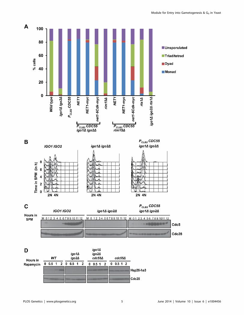
igo1D igo2D and rim15D cells. While 80% of wild type cells formed
spores, only about 10% and 18% of igo1D igo2D and rim15D cells
respectively, did. Remarkably igo1D igo2D and rim15D cells
carrying a meiotic-null allele of CDC55 (PCLB2CDC55) formed
monads (75%) like PCLB2CDC55 cells (Figure 2A). Crucially,
combining net1-6Cdk with PCLB2CDC55 igo1D igo2D and
PCLB2CDC55 rim15D cells resulted in efficient formation of tetrads
(Figure 2A). The ability of PCLB2CDC55 to suppress igo1D igo2Dwas specific as deletion of a gene encoding an alternative PP2A
regulatory subunit Rts1 had no effect on sporulation efficiency of
igo1D igo2D cells (Figure 2A).
To confirm suppression of igo1D igo2D by PCLB2CDC55 we
induced wild type, igo1D igo2D and igo1D igo2D PCLB2CDC55 cells
to enter meiosis by transferring them to SPM. Wild type cells
completed pre-meiotic DNA replication after 4 hours (Figure 2B),
and expressed Cdc5 (a marker for mid-meiosis) after 7 hours in
SPM (Figure 2C). In contrast, igo1D igo2D cells did not initiate
DNA replication (Figure 2B) and failed to express Cdc5 even after
12 hours in SPM (Figure 2C). Crucially igo1D igo2D PCLB2CDC55
cells completed pre-meiotic DNA replication (3–4 hours) and
expressed Cdc5 (5–6 hours) (Figure 2B–C). These results indicate
that PP2ACdc55 and Rim15/endosulfine play opposing roles in
regulating entry into gametogenesis.
We then determined whether PP2ACdc55 also negatively
regulates entry into quiescence. Wild type, igo1D igo2D, cdc55Dand igo1D igo2D cdc55D cells were treated with rapamycin and
entry into G0 was monitored by assaying expression of Hsp26, a
gene that is specifically induced during entry into G0 [8]. While
wild type cells induced expression of Hsp26 after 2 hours following
rapamycin treatment, the igo1D igo2D cells failed to express Hsp26
(Figure 2D). Crucially, both cdc55D cells and igo1D igo2D cdc55Dcells expressed Hsp26 even in the absence of rapamycin treatment.
These results indicate that the Rim15-endosulfine-PP2ACdc55
pathway regulates entry into gametogenesis and quiescence in
budding yeast.
Phosphorylation of Igo1 at S64 by Rim15 converts it intoan inhibitor of PP2ACdc55
To test whether phosphorylation of Igo1 at S64 results in
increased association with PP2ACdc55, we performed an in vitro
binding assay. We purified wild type Igo1, Igo1-S64A and Igo1-
S64D from bacterial cells by attaching a Maltose Binding Peptide
(MBP) to their N-termini. We then incubated endosulfine (and its
variants) bound to amylose resin via the MBP with yeast extracts
containing Cdc55-TAP (Tandem Affinity Purification). Specifical-
ly Igo1-S64D but not WT Igo1/Igo1-S64A physically interacted
with Cdc55 in vitro (Figure 3A). We then tested whether
phosphorylation of wild type endosulfine by Rim15 results in
Figure 1. Endosulfines are required for entry into gametogenesis. A) Wild-type, igo1D, igo2D and igo1D igo2D cells were incubated for24 hours on sporulation plates and number of sporulated (includes monad, dyad, Tri-/tetrads) and unsporulated cells were counted using a lightmicroscope. The experiment was repeated 3 times and 200 cells were counted every time for each strain. B) Wild-type and igo1D igo2D cells weretransferred to sporulation medium (SPM) and DNA content was measured by flow cytometry. C) Kinetics of nuclear division of cells in B was measuredafter staining cells with DAPI (n = 200). D) Analysis of expression of early meiotic proteins in cells described in B. Whole-cell extracts of hourly culturein SPM were subjected to western analysis using anti-PK (to detect Ime1), anti-HA (to detect Rec8) and Cdc28 antibody (loading control). The laneindicated as M contains mitotic extracts and * indicate a non-specific band. E) Conserved Rim15/Greatwall kinase site present in endosulfines fromSaccharomyces cerevisiae (S.c), Kluyveromyces lactis (K.l.), Candida glabrata (C.g.), Caenorhabditis elegans (C.e.), Drosophila melanogaster (D.m.),Arabidopsis thaliana (A.t.) and humans (H.s.) is indicated. F) igo1D igo2D cells and igo1D igo2D cells containing either pRS303-IGO1-myc8 or pRS303-IGO1-S64A-myc8 or pRS303-IGO1-S64D-myc8 were incubated for 24 hours on sporulation plates and number of sporulated (includes monad, dyad,triads/tetrads) and unsporulated cells were counted using a light microscope. Values are expressed as mean 6 s.e.m of 3 independentmeasurements. *P,0.01 (Student’s t-test).doi:10.1371/journal.pgen.1004456.g001
Module for Entry into Gametogenesis & G0 in Yeast
PLOS Genetics | www.plosgenetics.org 4 June 2014 | Volume 10 | Issue 6 | e1004456
Module for Entry into Gametogenesis & G0 in Yeast
PLOS Genetics | www.plosgenetics.org 5 June 2014 | Volume 10 | Issue 6 | e1004456
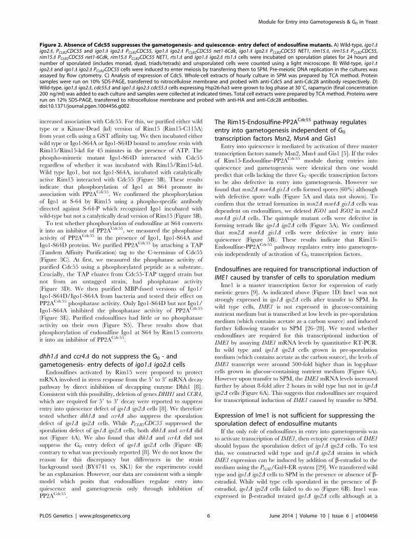
increased association with Cdc55. For this, we purified either wild
type or a Kinase-Dead (kd) version of Rim15 (Rim15-C115A)
from yeast cells using a GST affinity tag. We then incubated either
wild type or Igo1-S64A or Igo1-S64D bound to amylose resin with
Rim15/Rim15-kd for 45 minutes in the presence of ATP. The
phospho-mimetic mutant Igo1-S64D interacted with Cdc55
regardless of whether it was incubated with Rim15/Rim15-kd.
Wild type Igo1, but not Igo1-S64A, incubated with catalytically
active Rim15 interacted with Cdc55 (Figure 3B). These results
indicate that phosphorylation of Igo1 at S64 promote its
association with PP2ACdc55. We confirmed the phosphorylation
of Igo1 at S-64 by Rim15 using a phospho-specific antibody
directed against S-64-P which recognized Igo1 incubated with
wild-type but not a catalytically dead version of Rim15 (Figure 3B).
To test whether phosphorylation of endosulfine at S64 converts
it into an inhibitor of PP2ACdc55, we measured the phosphatase
activity of PP2ACdc55 in the presence of Igo1, Igo1-S64A and
Igo1-S64D proteins. We purified PP2ACdc55 by attaching a TAP
(Tandem Affinity Purification) tag to the C-terminus of Cdc55
(Figure 3C). At first, we measured the phosphatase activity of
purified Cdc55 using a phosphorylated peptide as a substrate.
Crucially, the TAP eluates from Cdc55-TAP tagged strain but
not from an untagged strain, had phosphatase activity
(Figure 3D). We then purified MBP-fused versions of Igo1/
Igo1-S64D/Igo1-S64A from bacteria and tested their effect on
PP2ACdc55 phosphatase activity. Only Igo1-S64D but not Igo1/
Igo1-S64A inhibited the phosphatase activity of PP2ACdc55
(Figure 3E). Purified endosulfines had little or no phosphatase
activity on their own (Figure S5). These results show that
phosphorylation of endosulfine Igo1 at S64 by Rim15 converts
it into an inhibitor of PP2ACdc55.
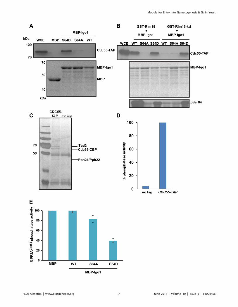
dhh1D and ccr4D do not suppress the G0 - andgametogenesis- entry defects of igo1D igo2D cells
Endosulfines activated by Rim15 were proposed to protect
mRNA involved in stress response from the 59 to 39 mRNA decay
pathway by direct inhibition of decapping enzyme Dhh1 [8].
Consistent with this possibility, deletion of genes DHH1 and CCR4,
which are required for 59 to 39 decay were reported to suppress
entry into quiescence defect of igo1D igo2D cells [8]. We therefore
tested whether dhh1D and ccr4D also suppress the sporulation
defect of igo1D igo2D cells. While PCLB2CDC55 suppressed the
sporulation defect of igo1D igo2D cells, both dhh1D and ccr4D did
not (Figure 4A). We also found that dhh1D and ccr4D did not
suppress the G0 entry defect of igo1D igo2D cells (Figure 4B)
contrary to what was previously reported [8]. We do not know the
reason for this discrepancy but differences in the strain
background used (BY4741 vs. SK1) for the experiments could
be an explanation. However, our data are consistent with a simple
model which posits that endosulfines regulate entry into
quiescence and gametogenesis only through inhibition of
PP2ACdc55.
The Rim15-Endosulfine-PP2ACdc55 pathway regulatesentry into gametogenesis independent of G0
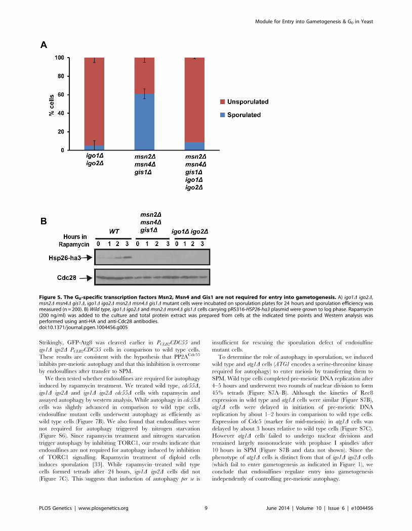
transcription factors Msn2, Msn4 and Gis1Entry into quiescence is mediated by activation of three master
transcription factors namely Msn2, Msn4 and Gis1 [5]. If the roles
of Rim15-Endosulfine-PP2ACdc55 module during entries into
quiescence and gametogenesis were identical then one would
predict that cells lacking the three G0–specific transcription factors
to be also defective in entry into gametogenesis. However we
found that msn2D msn4D gis1D cells formed spores (60%) although
with defective spore walls (Figure 5A and data not shown). To
confirm that the tetrad formation in msn2D msn4D gis1D cells was
dependent on endosulfines, we deleted IGO1 and IGO2 in msn2Dmsn4D gis1D cells. The quintuple mutant cells were defective in
forming tetrads like igo1D igo2D cells (Figure 5A). We confirmed
that msn2D msn4D gis1D cells were defective in entry into
quiescence (Figure 5B). These results indicate that Rim15-
Endosulfine-PP2ACdc55 pathway regulates entry into gametogen-
esis independently of activation of G0 transcription factors.
Endosulfines are required for transcriptional induction ofIME1 caused by transfer of cells to sporulation medium
Ime1 is a master transcription factor for expression of early
meiotic genes [9]. As indicated above (Figure 1D) Ime1 was not
strongly expressed in igo1D igo2D cells after transfer to SPM. In
wild type cells, IME1 is not expressed in glucose-containing
nutrient medium but is transcribed at low levels in pre-sporulation
medium (which contains acetate as a carbon source) and induced
further following transfer to SPM [26–28]. We tested whether
endosulfines are required for this transcriptional induction of
IME1 by assaying IME1 mRNA levels by quantitative RT-PCR.
In wild type and igo1D igo2D cells grown in pre-sporulation
medium (which contains acetate as the carbon source), the levels of
IME1 transcript were around 500-fold higher than in log-phase
cells grown in glucose-containing nutrient medium (Figure 6A).
However upon transfer to SPM, the IME1 mRNA levels increased
further by about 8-fold after 2 hours in wild type but not in igo1Digo2D cells (Figure 6A). This suggests that endosulfines are required
for transcriptional induction of IME1 caused by transfer to SPM.
Expression of Ime1 is not sufficient for suppressing thesporulation defect of endosulfine mutants
If the only role of endosulfines in entry into gametogenesis was
to activate transcription of IME1, then ectopic expression of IME1
should bypass the sporulation defect of igo1D igo2D cells. To test
this, we constructed wild type and igo1D igo2D strains in which
IME1 expression can be induced by addition of b-estradiol to the
medium using the PGAL/Gal4-ER system [29]. We transferred wild
type and igo1D igo2D cells to SPM in the presence or absence of b-
estradiol. While wild type cells sporulated in the presence of b-
estradiol, igo1D igo2D cells failed to do so (Figure 6B). Ime1 was
expressed in b-estradiol treated igo1D igo2D cells although at a
Figure 2. Absence of Cdc55 suppresses the gametogenesis- and quiescence- entry defect of endosulfine mutants. A) Wild-type, igo1Digo2D, PCLB2CDC55 and igo1D igo2D PCLB2CDC55, igo1D igo2D PCLB2CDC55 net1-6Cdk, igo1D igo2D PCLB2CDC55 NET1, rim15D, rim15D PCLB2CDC55,rim15D PCLB2CDC55 net1-6Cdk, rim15D PCLB2CDC55 NET1, rts1D and igo1D igo2D rts1D cells were incubated on sporulation plates for 24 hours andnumber of sporulated (includes monad, dyad, triads/tetrads) and unsporulated cells were counted using a light microscope. B) Wild-type, igo1Digo2D and igo1D igo2D PCLB2CDC55 cells were induced to enter meiosis by transferring them to SPM. Pre-meiotic DNA replication in the cultures wasassayed by flow cytometry. C) Analysis of expression of Cdc5. Whole-cell extracts of hourly culture in SPM was prepared by TCA method. Proteinsamples were run on 10% SDS-PAGE, transferred to nitrocellulose membrane and probed with anti-Cdc5 and anti-Cdc28 antibody respectively. D)Wild-type, igo1D igo2D, cdc55D and igo1D igo2D cdc55D cells expressing Hsp26-ha3 were grown to log phase at 30uC, rapamycin (final concentration200 ng/ml) was added to each culture and samples were collected at indicated times. Total cell extracts were prepared by TCA method. Proteins wererun on 12% SDS-PAGE, transferred to nitrocellulose membrane and probed with anti-HA and anti-Cdc28 antibodies.doi:10.1371/journal.pgen.1004456.g002
Module for Entry into Gametogenesis & G0 in Yeast
PLOS Genetics | www.plosgenetics.org 6 June 2014 | Volume 10 | Issue 6 | e1004456
Module for Entry into Gametogenesis & G0 in Yeast
PLOS Genetics | www.plosgenetics.org 7 June 2014 | Volume 10 | Issue 6 | e1004456
lower level compared to wild type cells (Figure 6C). Since
endosulfines have been implicated in mRNA stability [8], we
tested whether the difference in the Ime1 levels in the two
strains was due to difference in the IME1 transcript levels.
Quantitative RT-PCR analyses revealed that the IME1 tran-
script levels were induced to similar extent in wild type and
igo1D igo2D strains and remained relatively unchanged up to
8 hours following induction (Figure 6D). This suggests that
endosulfines are not required for regulating IME1 mRNA
stability. Decreased Ime1 levels in igo1D igo2D cells could be
caused by either decreased translational efficiency of IME1
mRNA or decreased Ime1 stability. These results indicate that
endosulfines promote entry into gametogenesis independently of
regulating IME1 expression.
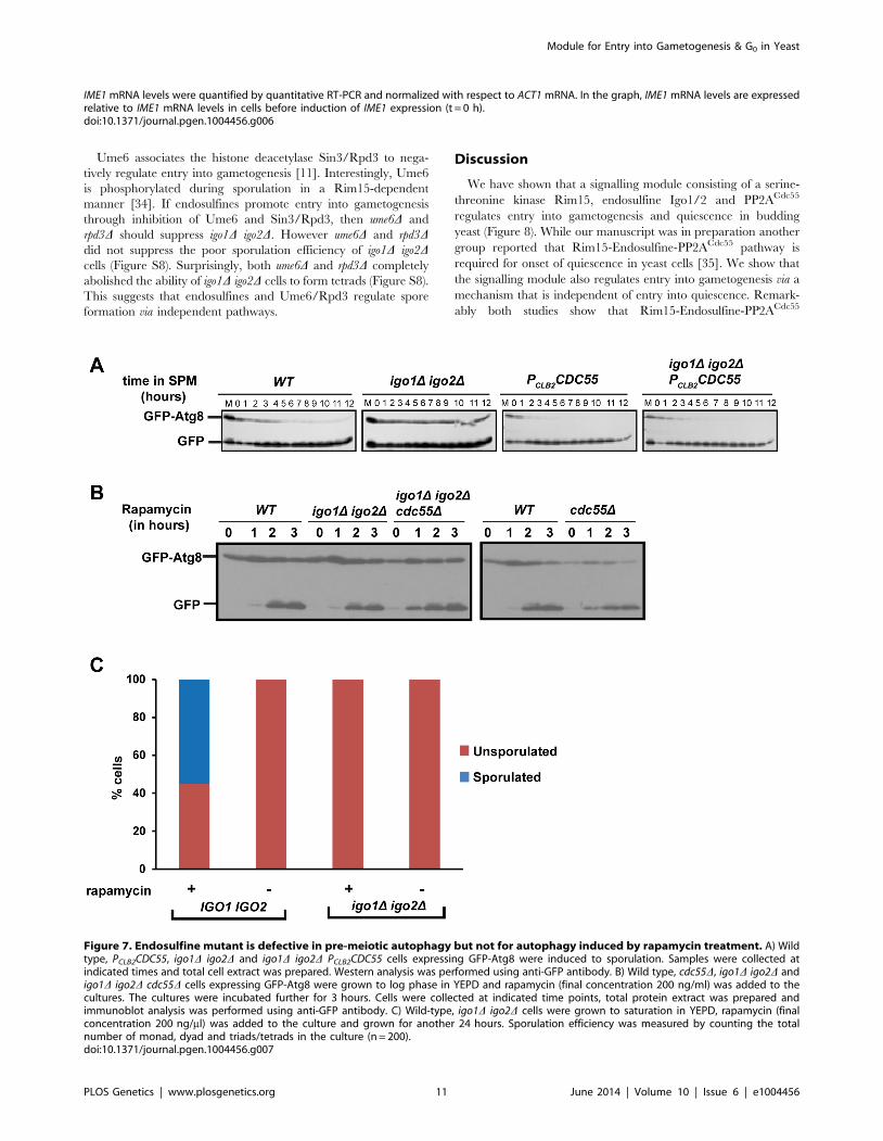
Endosulfines are required for pre-meiotic autophagyRim15 is required for autophagy induced by inhibition of PKA
and Sch9 but not for autophagy induced by rapamycin treatment
[30]. Since autophagy is required for spore formation in yeast [31],
we tested whether endosulfines are required for autophagy during
entry into gametogenesis. Autophagy can be assayed by following
proteolytic cleavage of GFP-Atg8, which is a N-terminal fusion of
GFP to Atg8 (a ubiquitin-like protein required for formation of
autophagosomal membranes) [32]. We induced wild type,
PCLB2CDC55, igo1D igo2D and igo1D igo2D PCLB2CDC55 cells to
enter meiosis by transferring them to SPM and assayed autophagy.
In wild type cells, GFP-Atg8 underwent proteolytic cleavage after
2 hours into SPM (Figure 7A). In contrast, GFP-Atg8 remained
intact in igo1D igo2D cells even after 12 hours in SPM (Figure 7A).
Figure 3. Igo1 associates and inhibits the phosphatase activity of PP2ACdc55 in a phosphorylation dependent manner. A) Either MBPor MBP fused to Igo1, Igo-S64A and Igo1-S64D were purified from bacteria and equal amount of protein was incubated with yeast cell expressingCdc55-TAP. The proteins bound to the beads were run on 10% SDS-PAGE and probed with anti-TAP antibody. The MBP purified proteins werevisualized by coomassie staining of SDS-PAGE gels. WCE denotes whole cell extracts. B) GST-Rim15 and GST-Rim15-kd were purified from yeast cells.Equal amounts of MBP fused Igo1, Igo1-S64A and Igo1-S64D were subjected to in vitro phosphorylation using GST-Rim15 and GST-Rim15-kdrespectively. MBP fused proteins were then pulled down with amylose beads and mixed with soluble protein extracts from a yeast strain expressingCdc55-TAP. The proteins bound to the beads were analysed by western blotting using an anti-TAP antibody. Purified MBP-tagged proteins werevisualized by coomassie staining of SDS-PAGE gels. Phosphorylation of Igo1 by Rim15 at S-64 was assayed using a phospho-specific antibody. WCEdenotes whole cell extracts. C) TAP eluates from CDC55-TAP and untagged strains were analysed by silver staining. D) Phosphatase activity of TAPeluates from CDC55-TAP and untagged strains was measured using a colorimetric assay (Millipore). E) Phospho-mimetic mutant of Igo1 (Igo1S64D)inhibits the phosphatase activity of PP2ACdc55. Purified Cdc55 was incubated with equal amount (25 mg) of MBP-Igo1, MBP-Igo1-S64A and MBP-Igo1-S64D respectively. MBP was used as a control. The mixture was then incubated with 500 mM phosphopeptide (Millipore). The release of freephosphate was measured by colorimetric assay (Millipore).doi:10.1371/journal.pgen.1004456.g003
Figure 4. dhh1D and ccr4D do not suppress the G0 - and gametogenesis- entry defects of endosulfine mutant cells. A) Sporulationefficiency of igo1D igo2D, igo1D igo2D PCLB2CDC55, igo1D igo2D dhh1D and igo1D igo2D ccr4D cells was measured (n = 200). B) Wild type, igo1D igo2D,cdc55D, igo1D igo2D cdc55D, igo1D igo2D dhh1D and igo1D igo2D ccr4D cells expressing HSP26-ha3 were grown to log phase at 30uC. Rapamycin(final concentration 200 ng/ml) was added to the culture and processed as described above. Whole cell extracts were subjected to SDS-PAGEfollowed by western analysis using anti-HA and anti-Cdc28 antibodies.doi:10.1371/journal.pgen.1004456.g004
Module for Entry into Gametogenesis & G0 in Yeast
PLOS Genetics | www.plosgenetics.org 8 June 2014 | Volume 10 | Issue 6 | e1004456
Strikingly, GFP-Atg8 was cleaved earlier in PCLB2CDC55 and
igo1D igo2D PCLB2CDC55 cells in comparison to wild type cells.
These results are consistent with the hypothesis that PP2ACdc55
inhibits pre-meiotic autophagy and that this inhibition is overcome
by endosulfines after transfer to SPM.
We then tested whether endosulfines are required for autophagy
induced by rapamycin treatment. We treated wild type, cdc55D,
igo1D igo2D and igo1D igo2D cdc55D cells with rapamycin and
assayed autophagy by western analysis. While autophagy in cdc55Dcells was slightly advanced in comparison to wild type cells,
endosulfine mutant cells underwent autophagy as efficiently as
wild type cells (Figure 7B). We also found that endosulfines were
not required for autophagy triggered by nitrogen starvation
(Figure S6). Since rapamycin treatment and nitrogen starvation
trigger autophagy by inhibiting TORC1, our results indicate that
endosulfines are not required for autophagy induced by inhibition
of TORC1 signalling. Rapamycin treatment of diploid cells
induces sporulation [33]. While rapamycin–treated wild type
cells formed tetrads after 24 hours, igo1D igo2D cells did not
(Figure 7C). This suggests that induction of autophagy per se is
insufficient for rescuing the sporulation defect of endosulfine
mutant cells.
To determine the role of autophagy in sporulation, we induced
wild type and atg1D cells (ATG1 encodes a serine-threonine kinase
required for autophagy) to enter meiosis by transferring them to
SPM. Wild type cells completed pre-meiotic DNA replication after
4–5 hours and underwent two rounds of nuclear division to form
45% tetrads (Figure S7A–B). Although the kinetics of Rec8
expression in wild type and atg1D cells were similar (Figure S7B),
atg1D cells were delayed in initiation of pre-meiotic DNA
replication by about 1–2 hours in comparison to wild type cells.
Expression of Cdc5 (marker for mid-meiosis) in atg1D cells was
delayed by about 3 hours relative to wild type cells (Figure S7C).
However atg1D cells failed to undergo nuclear divisions and
remained largely mononucleate with prophase I spindles after
10 hours in SPM (Figure S7B and data not shown). Since the
phenotype of atg1D cells is distinct from that of igo1D igo2D cells
(which fail to enter gametogenesis as indicated in Figure 1), we
conclude that endosulfines regulate entry into gametogenesis
independently of controlling pre-meiotic autophagy.
Figure 5. The G0-specific transcription factors Msn2, Msn4 and Gis1 are not required for entry into gametogenesis. A) igo1D igo2D,msn2D msn4D gis1D, igo1D igo2D msn2D msn4D gis1D mutant cells were incubated on sporulation plates for 24 hours and sporulation efficiency wasmeasured (n = 200). B) Wild type, igo1D igo2D and msn2D msn4D gis1D cells carrying pRS316-HSP26-ha3 plasmid were grown to log phase. Rapamycin(200 ng/ml) was added to the culture and total protein extract was prepared from cells at the indicated time points and Western analysis wasperformed using anti-HA and anti-Cdc28 antibodies.doi:10.1371/journal.pgen.1004456.g005
Module for Entry into Gametogenesis & G0 in Yeast
PLOS Genetics | www.plosgenetics.org 9 June 2014 | Volume 10 | Issue 6 | e1004456
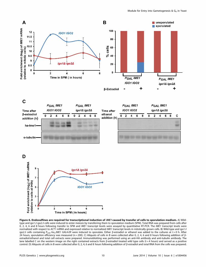
Figure 6. Endosulfines are required for transcriptional induction of IME1 caused by transfer of cells to sporulation medium. A) Wild-type and igo1D igo2D cells were induced to enter meiosis by transferring them to sporulation medium (SPM). Total RNA was prepared from cells after0, 2, 4, 6 and 8 hours following transfer to SPM and IME1 transcript levels were assayed by quantitative RT-PCR. The IME1 transcript levels werenormalized with respect to ACT1 mRNA and expressed relative to normalized IME1 transcript levels in mitotically grown cells. B) Wild-type and igo1Digo2D cells containing PGAL-ha3-IME1 GAL4-ER were induced to sporulate. Either b-estradiol or ethanol was added to the cultures at t = 0 h. After24 hours, sporulation efficiency was measured (n = 200). C) Aliquots of cells in B were collected after 0, 2, 4, 6 and 8 hours following addition of b-estradiol/ethanol and total cell extracts were prepared. Immunoblotting was performed using an anti-HA antibody and anti-tubulin antibody. Thelane labelled C on the western image on the right contained extracts from b-estradiol treated wild type cells (t = 4 hours) and served as a positivecontrol. D) Aliquots of cells in B were collected after 0, 2, 4, 6 and 8 hours following addition of b-estradiol and total RNA from the cells was prepared.
Module for Entry into Gametogenesis & G0 in Yeast
PLOS Genetics | www.plosgenetics.org 10 June 2014 | Volume 10 | Issue 6 | e1004456
Ume6 associates the histone deacetylase Sin3/Rpd3 to nega-
tively regulate entry into gametogenesis [11]. Interestingly, Ume6
is phosphorylated during sporulation in a Rim15-dependent
manner [34]. If endosulfines promote entry into gametogenesis
through inhibition of Ume6 and Sin3/Rpd3, then ume6D and
rpd3D should suppress igo1D igo2D. However ume6D and rpd3Ddid not suppress the poor sporulation efficiency of igo1D igo2Dcells (Figure S8). Surprisingly, both ume6D and rpd3D completely
abolished the ability of igo1D igo2D cells to form tetrads (Figure S8).
This suggests that endosulfines and Ume6/Rpd3 regulate spore
formation via independent pathways.
Discussion
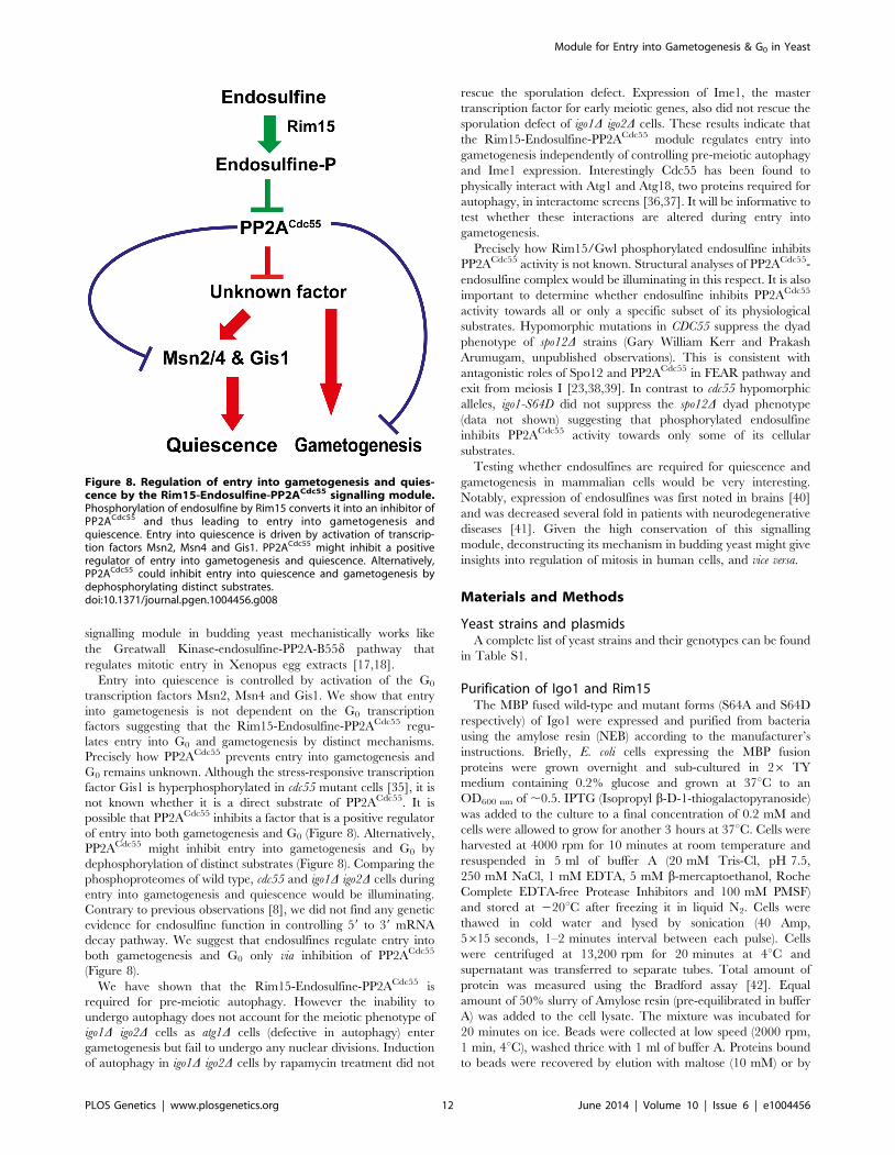
We have shown that a signalling module consisting of a serine-
threonine kinase Rim15, endosulfine Igo1/2 and PP2ACdc55
regulates entry into gametogenesis and quiescence in budding
yeast (Figure 8). While our manuscript was in preparation another
group reported that Rim15-Endosulfine-PP2ACdc55 pathway is
required for onset of quiescence in yeast cells [35]. We show that
the signalling module also regulates entry into gametogenesis via a
mechanism that is independent of entry into quiescence. Remark-
ably both studies show that Rim15-Endosulfine-PP2ACdc55
IME1 mRNA levels were quantified by quantitative RT-PCR and normalized with respect to ACT1 mRNA. In the graph, IME1 mRNA levels are expressedrelative to IME1 mRNA levels in cells before induction of IME1 expression (t = 0 h).doi:10.1371/journal.pgen.1004456.g006
Figure 7. Endosulfine mutant is defective in pre-meiotic autophagy but not for autophagy induced by rapamycin treatment. A) Wildtype, PCLB2CDC55, igo1D igo2D and igo1D igo2D PCLB2CDC55 cells expressing GFP-Atg8 were induced to sporulation. Samples were collected atindicated times and total cell extract was prepared. Western analysis was performed using anti-GFP antibody. B) Wild type, cdc55D, igo1D igo2D andigo1D igo2D cdc55D cells expressing GFP-Atg8 were grown to log phase in YEPD and rapamycin (final concentration 200 ng/ml) was added to thecultures. The cultures were incubated further for 3 hours. Cells were collected at indicated time points, total protein extract was prepared andimmunoblot analysis was performed using anti-GFP antibody. C) Wild-type, igo1D igo2D cells were grown to saturation in YEPD, rapamycin (finalconcentration 200 ng/ml) was added to the culture and grown for another 24 hours. Sporulation efficiency was measured by counting the totalnumber of monad, dyad and triads/tetrads in the culture (n = 200).doi:10.1371/journal.pgen.1004456.g007
Module for Entry into Gametogenesis & G0 in Yeast
PLOS Genetics | www.plosgenetics.org 11 June 2014 | Volume 10 | Issue 6 | e1004456
signalling module in budding yeast mechanistically works like
the Greatwall Kinase-endosulfine-PP2A-B55d pathway that
regulates mitotic entry in Xenopus egg extracts [17,18].
Entry into quiescence is controlled by activation of the G0
transcription factors Msn2, Msn4 and Gis1. We show that entry
into gametogenesis is not dependent on the G0 transcription
factors suggesting that the Rim15-Endosulfine-PP2ACdc55 regu-
lates entry into G0 and gametogenesis by distinct mechanisms.
Precisely how PP2ACdc55 prevents entry into gametogenesis and
G0 remains unknown. Although the stress-responsive transcription
factor Gis1 is hyperphosphorylated in cdc55 mutant cells [35], it is
not known whether it is a direct substrate of PP2ACdc55. It is
possible that PP2ACdc55 inhibits a factor that is a positive regulator
of entry into both gametogenesis and G0 (Figure 8). Alternatively,
PP2ACdc55 might inhibit entry into gametogenesis and G0 by
dephosphorylation of distinct substrates (Figure 8). Comparing the
phosphoproteomes of wild type, cdc55 and igo1D igo2D cells during
entry into gametogenesis and quiescence would be illuminating.
Contrary to previous observations [8], we did not find any genetic
evidence for endosulfine function in controlling 59 to 39 mRNA
decay pathway. We suggest that endosulfines regulate entry into
both gametogenesis and G0 only via inhibition of PP2ACdc55
(Figure 8).
We have shown that the Rim15-Endosulfine-PP2ACdc55 is
required for pre-meiotic autophagy. However the inability to
undergo autophagy does not account for the meiotic phenotype of
igo1D igo2D cells as atg1D cells (defective in autophagy) enter
gametogenesis but fail to undergo any nuclear divisions. Induction
of autophagy in igo1D igo2D cells by rapamycin treatment did not
rescue the sporulation defect. Expression of Ime1, the master
transcription factor for early meiotic genes, also did not rescue the
sporulation defect of igo1D igo2D cells. These results indicate that
the Rim15-Endosulfine-PP2ACdc55 module regulates entry into
gametogenesis independently of controlling pre-meiotic autophagy
and Ime1 expression. Interestingly Cdc55 has been found to
physically interact with Atg1 and Atg18, two proteins required for
autophagy, in interactome screens [36,37]. It will be informative to
test whether these interactions are altered during entry into
gametogenesis.
Precisely how Rim15/Gwl phosphorylated endosulfine inhibits
PP2ACdc55 activity is not known. Structural analyses of PP2ACdc55-
endosulfine complex would be illuminating in this respect. It is also
important to determine whether endosulfine inhibits PP2ACdc55
activity towards all or only a specific subset of its physiological
substrates. Hypomorphic mutations in CDC55 suppress the dyad
phenotype of spo12D strains (Gary William Kerr and Prakash
Arumugam, unpublished observations). This is consistent with
antagonistic roles of Spo12 and PP2ACdc55 in FEAR pathway and
exit from meiosis I [23,38,39]. In contrast to cdc55 hypomorphic
alleles, igo1-S64D did not suppress the spo12D dyad phenotype
(data not shown) suggesting that phosphorylated endosulfine
inhibits PP2ACdc55 activity towards only some of its cellular
substrates.
Testing whether endosulfines are required for quiescence and
gametogenesis in mammalian cells would be very interesting.
Notably, expression of endosulfines was first noted in brains [40]
and was decreased several fold in patients with neurodegenerative
diseases [41]. Given the high conservation of this signalling
module, deconstructing its mechanism in budding yeast might give
insights into regulation of mitosis in human cells, and vice versa.
Materials and Methods
Yeast strains and plasmidsA complete list of yeast strains and their genotypes can be found
in Table S1.
Purification of Igo1 and Rim15The MBP fused wild-type and mutant forms (S64A and S64D
respectively) of Igo1 were expressed and purified from bacteria
using the amylose resin (NEB) according to the manufacturer’s
instructions. Briefly, E. coli cells expressing the MBP fusion
proteins were grown overnight and sub-cultured in 26 TY
medium containing 0.2% glucose and grown at 37uC to an
OD600 nm of ,0.5. IPTG (Isopropyl b-D-1-thiogalactopyranoside)
was added to the culture to a final concentration of 0.2 mM and
cells were allowed to grow for another 3 hours at 37uC. Cells were
harvested at 4000 rpm for 10 minutes at room temperature and
resuspended in 5 ml of buffer A (20 mM Tris-Cl, pH 7.5,
250 mM NaCl, 1 mM EDTA, 5 mM b-mercaptoethanol, Roche
Complete EDTA-free Protease Inhibitors and 100 mM PMSF)
and stored at 220uC after freezing it in liquid N2. Cells were
thawed in cold water and lysed by sonication (40 Amp,
5615 seconds, 1–2 minutes interval between each pulse). Cells
were centrifuged at 13,200 rpm for 20 minutes at 4uC and
supernatant was transferred to separate tubes. Total amount of
protein was measured using the Bradford assay [42]. Equal
amount of 50% slurry of Amylose resin (pre-equilibrated in buffer
A) was added to the cell lysate. The mixture was incubated for
20 minutes on ice. Beads were collected at low speed (2000 rpm,
1 min, 4uC), washed thrice with 1 ml of buffer A. Proteins bound
to beads were recovered by elution with maltose (10 mM) or by
Figure 8. Regulation of entry into gametogenesis and quies-cence by the Rim15-Endosulfine-PP2ACdc55 signalling module.Phosphorylation of endosulfine by Rim15 converts it into an inhibitor ofPP2ACdc55 and thus leading to entry into gametogenesis andquiescence. Entry into quiescence is driven by activation of transcrip-tion factors Msn2, Msn4 and Gis1. PP2ACdc55 might inhibit a positiveregulator of entry into gametogenesis and quiescence. Alternatively,PP2ACdc55 could inhibit entry into quiescence and gametogenesis bydephosphorylating distinct substrates.doi:10.1371/journal.pgen.1004456.g008
Module for Entry into Gametogenesis & G0 in Yeast
PLOS Genetics | www.plosgenetics.org 12 June 2014 | Volume 10 | Issue 6 | e1004456
adding 26SDS sample buffer to the beads followed by incubation
at 95uC for 5 minutes.
GST-tagged wild-type or mutant forms of Rim15 was purified
from yeast cells. Briefly, cells carrying the plasmids (encoding
either wild-type or mutant Rim15) were grown to log phase in SD
–URA medium at 30uC containing 2% raffinose. After allowing
the cultures to reach an OD600 nm ,1.0, YPG (1% Yeast extract,
2% bactopeptone and 2% galactose) was added to the culture and
grown for another 4 hours at 30uC. Cells were collected, washed
in cold water and frozen in liquid N2 and stored at 280uC. Cells
were thawed , resuspended in lysis buffer (50 mM Tris-Cl, pH 7.5,
100 mM NaCl, 1 mM EDTA, 1% NP-40, 1 mM PMSF and
Roche Complete EDTA-free Protease Inhibitors) and lysed by
using glass beads. Total amount of protein was measured; equal
amount of protein was mixed with 150 ml of 50% slurry of GST
beads (pre-equilibrated in lysis buffer) and rotated at 4uC for
1 hour. Beads were collected and washed once with lysis buffer,
twice with lysis buffer +250 mM NaCl and twice with lysis buffer +500 mM NaCl. The GST-fused proteins were eluted using
10 mM reduced glutathione.
In vitro interaction between Igo1 and Cdc55Yeast cells expressing Cdc55-TAP were grown to log phase at
30uC in YEPD medium, harvested at 4000 rpm for 5 minutes at
4uC. The cell pellets were stored at 280uC after freezing it in
liquid N2. The pellet was thawed, resuspended in yeast lysis buffer
(50 mM Tris-Cl, pH 7.5, 100 mM NaCl, 1 mM EDTA, 1% NP-
40, 1 mM PMSF and Roche Complete EDTA-free Protease
Inhibitors) and lysed by using glass beads. Protein concentration
was measured by Bradford method. MBP fused wild-type and
mutant Igo1 proteins were purified as described above. Equal
amounts of bead bound proteins were added to equal amounts of
total yeast cell extract. The mixture was incubated on ice for
20 minutes. The beads were collected by centrifugation, washed
three times with lysis buffer, resuspended in SDS sample buffer,
boiled and run on 10% SDS-PAGE.
GST fused Rim15 and Rim15-kd proteins were purified as
described above and the purified protein was used to phosphor-
ylate purified MBP-fused Igo1. The reaction was carried out in
kinase buffer (50 mM Tris-Cl, pH7.5, 20 mM MgCl2, 1 mM
DTT) containing 1 mM ATP at room temperature for 45 min-
utes. Beads were collected, mixed with equal amount of yeast cell
extract containing Cdc55-TAP and incubated on ice for 20 min-
utes. The beads were washed three times with lysis buffer,
resuspended in 26 SDS sample buffer, boiled and analyzed by
Western blotting following SDS-PAGE.
Phosphatase assayPhosphatase assay was carried out using the Ser/Thr phospha-
tase assay kit containing a phospho-peptide as a substrate (from
Millipore). Briefly, strain expressing TAP-tagged Cdc55 was
grown in 1 litre of YEPD medium to log-phase. Cells were
harvested, resuspended in 5 ml of yeast lysis buffer and soluble
extracts were prepared by bead beating. The extract was mixed
with 0.2 ml of IgG sepharose beads (pre-equilibrated in lysis
buffer) and the mixture was incubated for 2 hours on a rotary
wheel at 4uC. The beads were precipitated, washed 4 times with
lysis buffer, once with TEV cleavage buffer (10 mM Tris-Cl,
pH 7.5, 150 mM NaCl, 0.5 mM EDTA, 0.1% NP-40 and 1 mM
DTT) and resuspended in 350 ml of TEV cleavage buffer. The
bound protein was cleaved and eluted from the beads after
incubating overnight at 4uC with 15 U of TEV protease
(Invitrogen). The eluted protein was then used for phosphatase
assay. Purified Cdc55 was mixed with equal amount (25 mg) of
MBP-fused Igo1 or Igo1-S64A or Igo1-S64D or MBP alone and
incubated for 20 minutes on ice. The mixture was then incubated
with 500 mM of phospho-peptide for 1 hour at 30uC. The reaction
was terminated by addition of malachite green solution provided
with the kit and absorbance was measured at 620 nm.
In situ immunofluorescenceFor in situs, cells from 1 ml of yeast culture were fixed for
15 minutes with 3.7% formaldehyde, pelleted and resuspended in
100 mM K-phosphate buffer (pH 6.4) containing 3.7% formal-
dehyde and kept overnight on ice. Immunostaining was performed
as previously described [23]. The following primary antibodies
were used: monoclonal rat anti-a-tubulin 1:500 (Serotec), mono-
clonal mouse anti-HA 1:500 (Covance). Secondary antibodies,
pre-absorbed against sera from other species used in labeling, were
conjugated with Cy3 or Cy5 (Chemicon) and diluted 1:500 (Cy3)
or 1:50 (Cy5). DNA was visualized by staining with DAPI.
MicroscopyImages were acquired using a Nikon TE-2000 inverted
microscope with a 10061.49 N.A. objective lens equipped with
a Photometrics Coolsnap-HQ2 liquid cooled CCD camera
(Photometrics, Tucson, AZ). 16 Z-stacks (spacing = 0.2 mm)
Exposure times of 1 second were used for both Cy3 and Cy5,
and 0.25 seconds for DAPI. Images were analysed using
Metamorph (version 7.5.2.0 MAG Biosystems Software).
ImmunoblottingWhole cell extracts were prepared by cell breakage with glass
beads in 20% Trichloroacetic acid. Cell pellets were resuspended
in 26 SDS sample buffer, neutralized with 2M Tris base and
proteins were denatured by heating the samples at 95uC for 59.
After centrifugation, protein samples were electrophoresed on
10% SDS-PAGE gels. The HA epitope was detected by mouse
monoclonal antibody 16B12 at 1:1000. Goat anti-Cdc5 (Santa
Cruz SC-6733) antibody, Goat anti-Cdc28 (Santa Cruz-6708)
antibody, mouse anti-Pk (Serotec) antibody, mouse anti-GFP
(Roche) antibody and rabbit anti-TAP antibody (Pierce) were all
used at 1:1000 dilution. Myc epitope was detected using the 9E10
antibody (Cambridge Biosciences) at 1:1000 dilution. Phospho-
specific antibody was raised against the phosphorylated synthetic
peptide KRKYFDpSGDYALC (pS indicates phosphoserine) by
Eurogentec.
For phos-tag gels, TCA extracts were prepared as above and
analysed as previously described [25] with a few modifications.
Briefly 12.5% polyacrylamide gels were prepared and Phos-Tag
(Wako) was added at its final concentration of 50 mM to the
separating gel mixture before polymerization. Electrophoresis was
performed at a constant current of 30 mA at room temp. After
electrophoresis, gels were first soaked in Transfer Buffer (25 mM
Tris, 192 mM Glycine, 10% methanol) containing 1 mM EDTA
for 20 minutes (2610 minutes) and then in Transfer Buffer for
30 minutes (3610 minutes). Electrotransfer onto PVDF mem-
brane was done at a constant voltage of 36 V for 16 hours at 4uC.
Silver stainingAfter running the protein sample on 10% SDS-PAGE, the gel
was washed once with water and then fixed with 100 ml of fixative
(50% methanol and 5% acetic acid) for 2 hours. The gel was
washed once with 100 ml of 20% ethanol and twice with water.
The gel was sensitized with 100 ml of 0.02% sodium thiosulfate for
1 minute and washed immediately with water. The gel was
incubated with 100 ml of silver nitrate (0.1% in water) solution
Module for Entry into Gametogenesis & G0 in Yeast
PLOS Genetics | www.plosgenetics.org 13 June 2014 | Volume 10 | Issue 6 | e1004456
containing 20 ml of 37% formaldehyde and kept for 20 minutes at
4uC in dark. The gel was then washed again with water and
100 ml of developing solution (2.5% sodium carbonate 0.0185%
formaldehyde) was added. After the bands were visible, 5% acetic
acid was added to terminate the reaction.
Analysis of mRNA by quantitative RT-PCRTotal RNA was extracted from yeast cell pellets using the
MasterPure Yeast RNA purification kit (Epicentre). RNA integrity
was confirmed by agarose gel electrophoretic analysis after
denaturation with formamide. Reverse transcription reactions
were performed on 0.5 mg of DNAase I-treated RNA with Oligo-
dT, using the GoScript Reverse Transcription System (Promega).
Quantitative real-time PCR primers for analyzing IME1 and
ACT1 were designed as previously described [43] and their
specificity was confirmed by melt curve analyses. cDNA reactions
were diluted 100-fold, and triplicate quantitative real-time
PCRs were performed in a Rotor-Gene Q (Qiagen) using the
26 Rotor-Gene SYBR Green PCR kit (Qiagen). Reactions were
analyzed using RotorGene Q software by the comparative CT
method, normalizing IME1 mRNA levels against the ACT1
reference gene.
Other techniquesInduction of sporulation was carried out as previously described
[44]. To measure sporulation efficiency of yeast strains on solid
media, cells were streak purified on YEPD plates. Three single
colonies were patched onto YEPD plates. After 24 h of growth at
30uC, cells were patched onto Sporulation plates (0.82% Sodium
acetate, 0.19% Potassium chloride, 0.035% Magnesium sulphate,
0.12% Sodium chloride and 1.5% Agar) and incubated at 30uCfor 24 h. Sporulation efficiency was assayed using a light
microscope. To induce GAL1-IME1 expression, b-estradiol was
added to the cultures at the final concentration of 1 mM. The
DNA content of sporulating cells was measured by flow cytometry
as previously described [45].
Supporting Information
Figure S1 The sporulation defect of endosulfine mutant cells is
not due to their failure to exit from stationary phase. Wild-type
and igo1D igo2D cells were grown to mid-log phase in YEPA
medium. Cells were then transferred to sporulation medium
(SPM). A) DNA content was measured by flow cytometry over a
period of 24 hours. B) Spore formation in the two strains after
24 hours was assayed by light microscopy.
(PDF)
Figure S2 The effect of phospho-mimetic mutation igo1-S64D
on sporulation efficiency is independent of Rim15 function.
rim15D cells and igo1D igo2D cells containing either pRS303-IGO1-
myc8 or pRS303-IGO1-S64A-myc8 or pRS303-IGO1-S64D-myc8 or
rim15D pRS303-IGO1-S64D-myc8 were incubated for 24 hours on
sporulation plates and percentage of sporulated cells were counted
using a light microscope. Values are expressed as mean 6 s.e.m of
3 independent measurements.
(PDF)
Figure S3 The endosulfine Igo1 is phosphorylated at S-64
during entry into gametogenesis. A) Strains expressing either Igo1-
myc8 or Igo1S64A-myc8 cells induced to sporulate. Cells were
collected at indicated time points and TCA extracts were
prepared. Protein samples were loaded on phos-tag or normal
SDS-PAGE gels and analysed by western blotting using anti-Myc
antibody. B) DNA content in the two cultures was measured by
flow cytometry over a period of 8 hours.
(PDF)
Figure S4 The conserved PKA site in Igo1 is dispensable for
entry into gametogenesis. A) Conserved PKA site at the C-termini
of budding yeast endosulfines Igo1, Igo2 and human endosulfines
ENSA and ARPP-19. B) Wild type, igo1D igo2D, igo1-S105D igo2Dand igo1-S105A igo2D cells were incubated on sporulation plates for
24 hours and the number of sporulated (includes monad, dyad,
Tri-/terads) and unsporulated cells were counted using a light
microscope.
(PDF)
Figure S5 Purified endosulfine has no phosphatase activity.
25 mg of purified MBP, MBP-Igo1, MBP-Igo1S64A and MBP-
Igo1S64D was incubated with 500 mM phosphopeptide (Milli-
pore). The release of free phosphate was measured using a
colorimetric assay (Millipore). TAP eluates from CDC55-TAP and
untagged strains were used as positive and negative controls
respectively for the phosphatase assay.
(PDF)
Figure S6 Endosulfines are not required for autophagy induced
by nitrogen starvation. Wild type, cdc55D, igo1D igo2D and igo1Digo2D cdc55D cells expressing GFP-Atg8 were grown to log phase
in YEPD and then transferred to nitrogen deprivation medium.
The cultures were incubated further for 3 hours. Cells were
collected at indicated time points, total protein extract was
prepared and immunoblot analysis was performed using anti-GFP
antibody.
(PDF)
Figure S7 The atg1D strains enter gametogenesis but fail to
undergo any meiotic nuclear divisions. Wild-type and atg1D cells
were induced to enter meiosis by transferring them to SPM. A)
Pre-meiotic DNA replication in the cultures was assayed by flow
cytometry. B) Kinetics of nuclear division of cells was measured
after staining cells with DAPI (n = 100). Rec8 expression was
monitored by in situ immunofluorescence using an anti-HA
antibody. C) Whole-cell extracts from meiotic cultures taken every
hour from 0–10 hours was prepared by TCA method. Protein
samples were run on 10% SDS-PAGE, transferred to nitrocellu-
lose membrane and probed with anti-Cdc5 and Cdc28 antibody
respectively.
(PDF)
Figure S8 ume6D and rpd3D do not suppress the sporulation
defect of igo1D igo2D cells. Wild-type, ume6D, rpd3D, igo1D igo2D,
igo1D igo2D ume6D and igo1D igo2D rpd3D cells were incubated for
24 hours on sporulation plates and number of sporulated (includes
monad, dyad, Tri-/tetrads) and unsporulated cells were counted
using a light microscope. The experiment was repeated 3 times
and 200 cells were counted every time for each strain.
(PDF)
Table S1 List of yeast strains used. All yeast strains are
derivatives of SK1 and have the following markers, unless
otherwise stated. ho::LYS2/ho::LYS2, ura3/ura3, leu2::hisG/leu2::hisG,
trp1::hisG/trp1::hisG, his3::hisG/his3::hisG, lys2/lys2.
(PDF)
Acknowledgments
We would like to thank Kim Nasmyth, Nancy Kleckner, Aaron Mitchell,
Yona Kassir, Daniel Klionsky, Claudio Virgilio, Fulvio Reggiori, Yoshinori
Ohsumi and Stephen Royle for strains, antibodies, plasmids and reagents.
Module for Entry into Gametogenesis & G0 in Yeast
PLOS Genetics | www.plosgenetics.org 14 June 2014 | Volume 10 | Issue 6 | e1004456
Author Contributions
Conceived and designed the experiments: SS PA. Performed the
experiments: SS PA. Analyzed the data: SS JZD JBAM PA. Contributed
reagents/materials/analysis tools: SS JZD JBAM PA. Wrote the paper: SS
PA.
References
1. Broach JR (2012) Nutritional control of growth and development in yeast.
Genetics 192: 73–105.2. Gray JV, Petsko GA, Johnston GC, Ringe D, Singer RA, et al. (2004) ‘‘Sleeping
beauty’’: quiescence in Saccharomyces cerevisiae. Microbiol Mol Biol Rev 68:187–206.
3. Kaeberlein M (2010) Lessons on longevity from budding yeast. Nature 464:
513–519.4. Malumbres M, Barbacid M (2001) To cycle or not to cycle: a critical decision in
cancer. Nat Rev Cancer 1: 222–231.5. De Virgilio C (2012) The essence of yeast quiescence. FEMS Microbiol Rev 36:
306–339.
6. Pedruzzi I, Dubouloz F, Cameroni E, Wanke V, Roosen J, et al. (2003) TORand PKA signaling pathways converge on the protein kinase Rim15 to control
entry into G0. Mol Cell 12: 1607–1613.7. Swinnen E, Wanke V, Roosen J, Smets B, Dubouloz F, et al. (2006) Rim15 and
the crossroads of nutrient signalling pathways in Saccharomyces cerevisiae. CellDiv 1: 3.
8. Talarek N, Cameroni E, Jaquenoud M, Luo X, Bontron S, et al. (2010)
Initiation of the TORC1-regulated G0 program requires Igo1/2, which licensespecific mRNAs to evade degradation via the 59-39 mRNA decay pathway. Mol
Cell 38: 345–355.9. Kassir Y, Adir N, Boger-Nadjar E, Raviv NG, Rubin-Bejerano I, et al. (2003)
Transcriptional regulation of meiosis in budding yeast. Int Rev Cytol 224: 111–
171.10. Rubin-Bejerano I, Mandel S, Robzyk K, Kassir Y (1996) Induction of meiosis in
Saccharomyces cerevisiae depends on conversion of the transcriptionalrepresssor Ume6 to a positive regulator by its regulated association with the
transcriptional activator Ime1. Mol Cell Biol 16: 2518–2526.
11. Kadosh D, Struhl K (1997) Repression by Ume6 involves recruitment of acomplex containing Sin3 corepressor and Rpd3 histone deacetylase to target
promoters. Cell 89: 365–371.12. Goldmark JP, Fazzio TG, Estep PW, Church GM, Tsukiyama T (2000) The
Isw2 chromatin remodeling complex represses early meiotic genes uponrecruitment by Ume6p. Cell 103: 423–433.
13. Pnueli L, Edry I, Cohen M, Kassir Y (2004) Glucose and nitrogen regulate the
switch from histone deacetylation to acetylation for expression of early meiosis-specific genes in budding yeast. Mol Cell Biol 24: 5197–5208.
14. Bowdish KS, Yuan HE, Mitchell AP (1995) Positive control of yeast meioticgenes by the negative regulator UME6. Mol Cell Biol 15: 2955–2961.
15. Steber CM, Esposito RE (1995) UME6 is a central component of a
developmental regulatory switch controlling meiosis-specific gene expression.Proc Natl Acad Sci U S A 92: 12490–12494.
16. Mallory MJ, Cooper KF, Strich R (2007) Meiosis-specific destruction of theUme6p repressor by the Cdc20-directed APC/C. Mol Cell 27: 951–961.
17. Gharbi-Ayachi A, Labbe JC, Burgess A, Vigneron S, Strub JM, et al. (2010) Thesubstrate of Greatwall kinase, Arpp19, controls mitosis by inhibiting protein
phosphatase 2A. Science 330: 1673–1677.
18. Mochida S, Maslen SL, Skehel M, Hunt T (2010) Greatwall phosphorylates aninhibitor of protein phosphatase 2A that is essential for mitosis. Science 330:
1670–1673.19. Yu J, Fleming SL, Williams B, Williams EV, Li Z, et al. (2004) Greatwall kinase:
a nuclear protein required for proper chromosome condensation and mitotic
progression in Drosophila. J Cell Biol 164: 487–492.20. Rangone H, Wegel E, Gatt MK, Yeung E, Flowers A, et al. (2011) Suppression
of scant identifies Endos as a substrate of greatwall kinase and a negativeregulator of protein phosphatase 2A in mitosis. PLoS Genet 7: e1002225.
21. Von Stetina JR, Tranguch S, Dey SK, Lee LA, Cha B, et al. (2008) alpha-Endosulfine is a conserved protein required for oocyte meiotic maturation in
Drosophila. Development 135: 3697–3706.
22. Vidan S, Mitchell AP (1997) Stimulation of yeast meiotic gene expression by theglucose-repressible protein kinase Rim15p. Mol Cell Biol 17: 2688–2697.
23. Kerr GW, Sarkar S, Tibbles KL, Petronczki M, Millar JB, et al. (2011) Meiotic
nuclear divisions in budding yeast require PP2A(Cdc55)-mediated antagonism of
Net1 phosphorylation by Cdk. J Cell Biol 193: 1157–1166.
24. Juanes MA, Khoueiry R, Kupka T, Castro A, Mudrak I, et al. (2013) Budding
yeast greatwall and endosulfines control activity and spatial regulation of
PP2A(Cdc55) for timely mitotic progression. PLoS Genet 9: e1003575.
25. Kinoshita-Kikuta E, Aoki Y, Kinoshita E, Koike T (2007) Label-free kinase
profiling using phosphate affinity polyacrylamide gel electrophoresis. Mol Cell
Proteomics 6: 356–366.
26. Shah JC, Clancy MJ (1992) IME4, a gene that mediates MAT and nutritional
control of meiosis in Saccharomyces cerevisiae. Mol Cell Biol 12: 1078–1086.
27. Chu S, DeRisi J, Eisen M, Mulholland J, Botstein D, et al. (1998) The
transcriptional program of sporulation in budding yeast. Science 282: 699–705.
28. Kassir Y, Granot D, Simchen G (1988) IME1, a positive regulator gene of
meiosis in S. cerevisiae. Cell 52: 853–862.
29. Benjamin KR, Zhang C, Shokat KM, Herskowitz I (2003) Control of landmark
events in meiosis by the CDK Cdc28 and the meiosis-specific kinase Ime2.
Genes Dev 17: 1524–1539.
30. Yorimitsu T, Zaman S, Broach JR, Klionsky DJ (2007) Protein kinase A and
Sch9 cooperatively regulate induction of autophagy in Saccharomyces
cerevisiae. Mol Biol Cell 18: 4180–4189.
31. Tsukada M, Ohsumi Y (1993) Isolation and characterization of autophagy-
defective mutants of Saccharomyces cerevisiae. FEBS Lett 333: 169–174.
32. Klionsky DJ, Cuervo AM, Seglen PO (2007) Methods for monitoring autophagy
from yeast to human. Autophagy 3: 181–206.
33. Zheng XF, Schreiber SL (1997) Target of rapamycin proteins and their kinase
activities are required for meiosis. Proc Natl Acad Sci U S A 94: 3070–3075.
34. Xiao Y, Mitchell AP (2000) Shared roles of yeast glycogen synthase kinase 3
family members in nitrogen-responsive phosphorylation of meiotic regulator
Ume6p. Mol Cell Biol 20: 5447–5453.
35. Bontron S, Jaquenoud M, Vaga S, Talarek N, Bodenmiller B, et al. (2013) Yeast
endosulfines control entry into quiescence and chronological life span by
inhibiting protein phosphatase 2A. Cell Rep 3: 16–22.
36. Ptacek J, Devgan G, Michaud G, Zhu H, Zhu X, et al. (2005) Global analysis of
protein phosphorylation in yeast. Nature 438: 679–684.
37. Ho Y, Gruhler A, Heilbut A, Bader GD, Moore L, et al. (2002) Systematic
identification of protein complexes in Saccharomyces cerevisiae by mass
spectrometry. Nature 415: 180–183.
38. Bizzari F, Marston AL (2011) Cdc55 coordinates spindle assembly and
chromosome disjunction during meiosis. J Cell Biol 193: 1213–1228.
39. Rock JM, Amon A (2009) The FEAR network. Curr Biol 19: R1063–1068.
40. Virsolvy-Vergine A, Leray H, Kuroki S, Lupo B, Dufour M, et al. (1992)
Endosulfine, an endogenous peptidic ligand for the sulfonylurea receptor:
purification and partial characterization from ovine brain. Proc Natl Acad
Sci U S A 89: 6629–6633.
41. Kim SH, Lubec G (2001) Brain alpha-endosulfine is manifold decreased in
brains from patients with Alzheimer’s disease: a tentative marker and drug
target? Neurosci Lett 310: 77–80.
42. Bradford MM (1976) A rapid and sensitive method for the quantitation of
microgram quantities of protein utilizing the principle of protein-dye binding.
Anal Biochem 72: 248–254.
43. Kahana S, Pnueli L, Kainth P, Sassi HE, Andrews B, et al. (2010) Functional
dissection of IME1 transcription using quantitative promoter-reporter screening.
Genetics 186: 829–841.
44. Kiburz BM, Amon A, Marston AL (2008) Shugoshin promotes sister
kinetochore biorientation in Saccharomyces cerevisiae. Mol Biol Cell 19:
1199–1209.
45. Epstein CB, Cross FR (1992) CLB5: a novel B cyclin from budding yeast with a
role in S phase. Genes Dev 6: 1695–1706.
Module for Entry into Gametogenesis & G0 in Yeast
PLOS Genetics | www.plosgenetics.org 15 June 2014 | Volume 10 | Issue 6 | e1004456