Embed Size (px)
Citation preview
RESEARCH ARTICLE
The Rho GTPase Rif signals through IRTKS, Eps8 and WAVE2 togenerate dorsal membrane ruffles and filopodiaThankiah Sudhaharan1, Kai Ping Sem1, Hwi Fen Liew1, Yuan Hong Yu1, Wah Ing Goh1,2, Ai Mei Chou1 andSohail Ahmed1,*
ABSTRACTRif induces dorsal filopodia but the signaling pathway responsible forthis has not been identified. We show here that Rif interacts with theI-BAR family protein IRTKS (also known as BAIAP2L1) through itsI-BAR domain. Rif also interacts with Pinkbar (also known asBAIAP2L2) in N1E-115 mouse neuroblastoma cells. IRTKS and Rifinduce dorsal membrane ruffles and filopodia. Dominant-negative Rifinhibits the formation of IRTKS-induced morphological structures,and Rif activity is blocked in IRTKS-knockout (KO) cells. To furtherdefine the Rif–IRTKS signaling pathway, we identify Eps8 andWAVE2 (also known as WASF2) as IRTKS interactors. We find thatEps8 regulates the size and number of dorsal filopodia andmembrane ruffles downstream of Rif–IRTKS signaling, whereasWAVE2 modulates dorsal membrane ruffling. Furthermore, ourdata suggests that Tir, a protein essential for enterohemorrhagicEscherichia coli infection, might compete for Rif for interaction withthe I-BAR domain of IRTKS. Based on this evidence, we propose amodel in which Rho family GTPases use the I-BAR proteins, IRSp53(also known as BAIAP2), IRTKS and Pinkbar, as a centralmechanism to modulate cell morphology.
KEY WORDS: IRTKS, Eps8, WAVE2, Dorsal filopodia, Dorsal ruffles
INTRODUCTIONThe Rho GTPases Rif (for ‘Rho in filopodia’) and Cdc42 are knownto signal to the actin cytoskeleton (Hall, 2012). Both GTPases cangenerate filopodia, dynamic structures that are thought to beinvolved in cell sensing mechanisms. Cdc42 partners with theInverse bin-amphiphysin-Rvs (I-BAR) protein IRSp53 (insulinreceptor substrate protein 53 kDa; also known as BAIAP2) ininducing filopodia formation (for a review, see Scita et al., 2008).IRSp53 has a Cdc42-binding site and an SH3 domain, both ofwhich are crucial for filopodia formation (Krugmann et al., 2001;Govind et al., 2001). The IRSp53 SH3 domain interacts with Mena(also known as ENAH) (Krugmann et al., 2001), Eps8 (Disanzaet al., 2006), WAVE2 (also known as WASF2) (Suetsugu et al.,2006; Goh et al., 2012), mDia1 (Goh et al., 2011) and dynamin 1(Chou et al., 2014) to induce filopodia. These data led to a model forfilopodia formation mediated by IRSp53 where membraneprotrusion modulated by its I-BAR domain is coupled to actinpolymerization modulated by its SH3-domain-binding partners
(Ahmed et al., 2010). Cdc42 is thought to activate and localizeIRSp53 (Disanza et al., 2006). As for Rif, it has been found topartner the actin nucleators mDia1 and mDia2 in forming peripheralfilopodia (Goh et al., 2011; Pellegrin and Mellor, 2005).
The I-BAR family of proteins contain five members: IRSp53,IRTKS (insulin receptor tyrosine kinase substrate; also known asBAIAP2L1 or BAI1-associated protein 2-like 1), MIM (missing inmetastasis; also known as MTSS1), ABBA (actin-bundling proteinwith BAIAP2 homology; also known as MTSS1L), and Pinkbar(Planar intestinal-and kidney-specific BAR domain protein; alsoknown as BAI1-associated protein 2-like protein 2, BAIAP2L2).The I-BAR domain induces membrane tubulation with variation foreach family member (Saarikangas et al., 2009; Mattila et al., 2007).I-BAR proteins are thought to dimerize allowing membranecurvature to be induced (Zhao et al., 2011). The domain structureof I-BAR family proteins suggest coupling of I-BAR domain-induced membrane deformation with signaling pathways associatedwith the actin cytoskeleton (Scita et al., 2008) that could give rise toactin-based cell membrane protrusions. Such protrusions have beenshown to play roles in pathological conditions such as infection andmetastasis. For example, enteropathogenic and enterohemorrhagicEscherichia coli trigger the formation of an actin-based membraneprotrusion on the apical surface of host cells known as the actinpedestal. IRSp53 and IRTKS have been implicated in this process.In particular, IRTKS is subverted by the E. coli proteins translocatedintimin receptor (Tir) and Tir cytoskeleton coupling protein (TccP)(Vingadassalom et al., 2009), and Tir is known to bind the I-BARdomains of IRTKS and IRSp53 (Vingadassalom et al., 2009; Weisset al., 2009). Invadosomes are another type of actin-based cellmembrane protrusion that facilitate the migration of cancer cells (seeLinder et al., 2011 for a recent review). Thus, it is important tounderstand the signaling pathways that lead to the formation ofactin-based membrane protrusions at the dorsal surface of cells.
Rif has been shown to induce dorsal protrusions in wide-field(Goh et al., 2011) and confocal imaging (Pellegrin and Mellor,2005) studies. However, to follow dorsal morphology quantitativelyit is important to use a technique such as scanning electronmicroscopy (SEM). The signaling pathway by which Rif generatesdorsal structures, such as dorsal membrane ruffles and filopodia, isunknown. Using affinity purification and mass spectrometryanalysis, we identify IRTKS as a Rif interactor. Both IRTKS andRif induce dorsal filopodia and membrane ruffles. We also identifyEps8 and WAVE2 as modulators of this dorsal morphologydownstream of Rif and IRTKS. Thus, we establish a newpathway, Rif to IRTKS to Eps8 and WAVE2, responsible for thegeneration of dorsal filopodia and membrane ruffles.
RESULTSTo identify Rif targets we used a tandem affinity purification tagapproach (Rigaut et al., 1999). A number of proteins that boundReceived 25 August 2015; Accepted 27 May 2016
1Neural Stem Cell Laboratory, Institute of Medical Biology, 8A Biomedical Grove,Immunos, 138648 Singapore. 2Mechanobiology Institute, National University ofSingapore, 5A Engineering Drive 1, 117411 Singapore.
*Author for correspondence ([email protected])
S.A., 0000-0002-5208-8193
2829
© 2016. Published by The Company of Biologists Ltd | Journal of Cell Science (2016) 129, 2829-2840 doi:10.1242/jcs.179655
Journal
ofCe
llScience
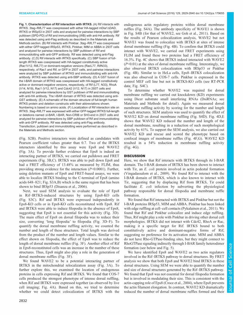
specifically to Rif were identified. In this paper, we analyze one ofthese Rif-binding proteins – IRTKS. To confirm that Rif doesindeed interact with IRTKS we carried out pulldown experimentsusing HA-tagged proteins. As controls, we used IRSp53 and aconstitutively active version of mDia1 (mDia1ΔDAD). Rifinteracted with IRTKS and the mDia1ΔDAD, but not withIRSp53 (Fig. 1A). Next, we asked whether other members of theI-BAR family of proteins interacted with Rif. Our results show Rifinteracts with Pinkbar in addition to IRTKS but not with MIM,ABBA or IRSp53 (Fig. 1B). To determine the Rho family GTPasespecificity of IRTKS we used RhoA, Cdc42 and Rac in pulldownexperiments. IRTKS showed high specificity for Rif independentlyof its GTP- or GDP-bound status (Fig. 1C,D). The location of theRif-binding site on IRTKS was identified using a series of deletionconstructs. Rif bound to the I-BAR domain but not to other regionsof IRTKS (Fig. 1E,F).To analyze the morphological signaling pathway that the Rif–
IRTKS complex is involved in, we used the mouse neuroblastomacell line N1E-115. Expression of IRTKS itself induced filopodiaformation, both peripheral and dorsal, as well as dorsal membraneruffles (Fig. 2A), and this phenotype of IRTKS is similar to thatinduced by Rif (Ellis and Mellor, 2000; Fan and Mellor, 2012; Gohet al., 2011). When IRTKS was expressed with constitutively activeRif (mutation Q77L), denoted RifL77, there was a synergy, givingan enhanced phenotype (Table 1). In contrast, when IRTKS wasexpressed with dominant-negative Rif (mutation T33N), denotedRifN33, there was a reduction in the phenotype, as seen by a 50%reduction in filopodia formation.To confirm that Rif and IRTKS interacted in a cellular context,
we carried out fluorescence resonance energy transfer (FRET)experiments using fluorescence lifetime imaging microscopy(FLIM). These FLIM experiments also revealed the location ofthe Rif–IRTKS interaction within the cell. There was a decreaseof GFP–Rif lifetime in the presence of IRTKS (P<0.003) yieldinga FRET efficiency of 6.3%, indicating that Rif and IRTKS dointeract (Fig. 2B). The Rif–IRTKS complex appeared to belocated on peripheral and dorsal filopodia and membrane ruffles.To locate the region in Rif responsible for the interaction withIRTKS, we generated RifS51A, RifE54N and RifΔCAAXdeletion mutations and examined localization and interactionusing confocal imaging and FLIM-FRET measurement,respectively. Initially, we carried out confocal imaging by co-expressing GFP–IRTKS with mCherry–Rif or its mutants. Theconfocal data (Fig. S1A) suggests that there is distinct localizationof Rif mutants in N1E 115 cells. RifE51A and RifE54Ncolocalized with IRTKS along the edges of cells whereas theRifΔCAAX mutant localized in the nucleus of cells. The FLIMdata (Fig. S1B) revealed that the RifS51A had a minimalinteraction with IRTKS compared with RifQ77L, followed byRifE54N and RifΔCAAX. These data suggest that the amino acidE54 and the CAAX motif are essential for Rif interaction withIRTKS and generation of dorsal structures.To further investigate the idea that Rif signals through IRTKS to
generate dorsal actin-based structures, we generated IRTKS-knockout (KO) mice (Fig. S1C) and then derived KO mouseembryonic fibroblast (MEF) cell lines from them. IRTKS-KO micehad no obvious phenotype. Rif failed to generate dorsal filopodiaand membrane ruffles in IRTKS-KO cells (Fig. 2C), whereas Rifgenerated intense peripheral edge ruffling (arrowhead, Fig. 2C) andlong peripheral protrusions (arrowhead, Fig. 2C) in IRTKS KOcells. On rescue of these KO cells with IRTKS, the Rif–IRTKSdorsal phenotype was restored (Fig. 2C). Given that Rif interacts
with Pinkbar (based on pulldown and immunoblot experiments)there was a possibility that the edge ruffling and long protrusionsinduced by Rif in IRTKS KO cells are due to Pinkbar. Interestingly,we found that Pinkbar located with Rif in edge ruffling sites but notwith the long protrusions (Fig. 2C). As controls, we showed that theIRTKS I-BAR domain alone and the IRTKS SH3 domain mutantare not able to rescue the lack of dorsal phenotype in IRTKS KOcells (Fig. 2D).
Next we turned to SEM to analyze and quantify dorsal filopodiaand membrane ruffles. In control cells expressing GFP or mRubywe did not detect surface protrusions (Fig. 2E). Upon expression ofIRTKS or Rif alone, structures resembling small filopodia wereseen, designated here as filopodial tips (Fig. 2E; Fig. S1D). Withcoexpression of IRTKS and Rif, dorsal filopodia and membraneruffles were clearly seen (Fig. 2E, bottom right panel). In IRTKSKO cells, overexpression of Rif induced only dorsal filopodial tips.Interestingly, Rif also induced edge ruffling and long peripheralprotrusions in IRTKS KO cells (Fig. 2E, right panel). In contrast,Rif expression in IRTKS KO cells rescued with IRTKS revealedboth dorsal filopodia and membrane ruffles (Fig. 2E, bottom panel).Fig. 2F shows a quantification of the results presented in Fig. 2E. Inorder to test the role of Rif in a more physiological manner, wecarried out 3D imaging of cells in 3D culture in a collagen type Imatrix (Fig. S2A; Movies 1 and 2). All cells generated surfaceprotrusions that are not seen in the control experiments. Thus, inboth 2D and 3D cultures, Rif induces filopodia and membraneruffles.
Having established that Rif signals through IRTKS to generatedorsal filopodia and membrane ruffles, we next investigatedpossible partners of IRTKS that could be involved in this process.Based on the fact that the SH3 domain of IRSp53 plays a crucial rolein peripheral filopodia formation through proteins such as Mena,Eps8 and mDia1, we looked for SH3-domain-binding partners ofIRTKS. A mitochondrial localization signal was placed on IRTKS–mRuby with the idea that proteins interacting with IRTKS wouldrelocalize to mitochondrial sites (Kanaji et al., 2000). HeLa cellscoexpressing mito-IRTKS–mRuby and potential GFP-taggedinteracting partners (WAVE1, WAVE2, Mena, Eps8, VASP,Espin, mDia1, mDia2, N-WASP, ezrin, dynamin 1 and dynamin2) were generated, and colocalization analysis carried out
Table 1. IRTKS induces filopodia in N1E-115 cells
Protein(s)expressed
Filopodiaper cell
Filopodiallength (µm)
Filopodiallifetime (s)
GFP–actin 1.2±1.02 7.16±0.82 212±64RifQL+GFP–actin*
8.5±2.36 4.37±0.28 162±14
mCherry–ABP**
1.3±1.03 4.68±1.01 166±17
GFP–IRTKS+mCherry–ABP
10.0±0.22 3.60±0.44 113±8
GFP–IRTKS+mRFP–RifL77
14.3±4.19 ND ND
GFP–IRTKS+mRFP–RifN33
5.1±2.57 ND ND
Cells were transfected with GFP–IRTKS and mCherry–Lifeact and imagedusing dual-channel (GFP and mCherry) time-lapse microscopy at 24 h posttransfection. Filopodia number, length and lifetime were then scored asdescribed previously (Lim et al., 2008). Data is expressed as mean±s.d.(n=3–7). Data from *Goh et al., 2011 and **Goh et al., 2012 is shown forcomparison, but excluded from statistical analysis. ND, not detected.
2830
RESEARCH ARTICLE Journal of Cell Science (2016) 129, 2829-2840 doi:10.1242/jcs.179655
Journal
ofCe
llScience
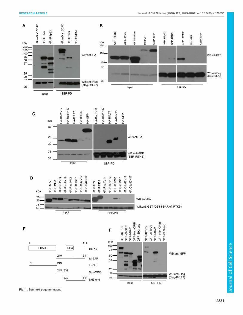
Fig. 1. See next page for legend.
2831
RESEARCH ARTICLE Journal of Cell Science (2016) 129, 2829-2840 doi:10.1242/jcs.179655
Journal
ofCe
llScience
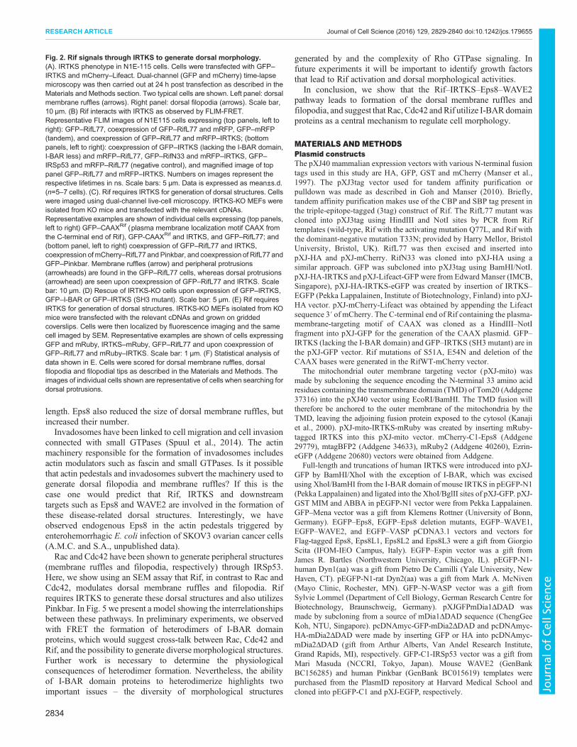
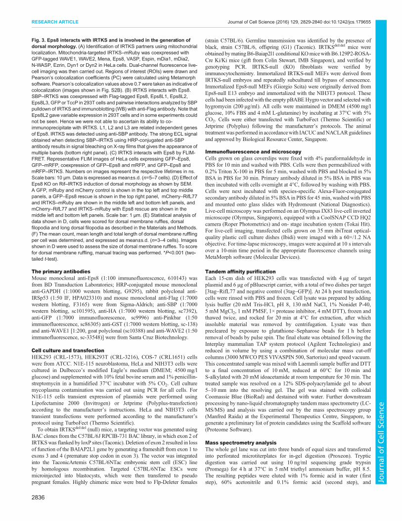
(Fig. S2B). Positive interactors were defined as candidates withPearson coefficient values greater than 0.7. Two of the IRTKSinteractors identified by this assay were Eps8 and WAVE2(Fig. 3A). To provide further evidence that Eps8 is indeed aninteracting partner of IRTKS, we carried out pulldown and FRETexperiments (Fig. 3B,C). IRTKS was able to pull down Eps8 andhad a FRET efficiency of 13.48% as measured by FLIM, thusproviding strong evidence for the interaction (P<0.0001). Further,using deletion mutants of Eps8 and FRET-based assays, we wereable to localize IRTKS binding to the C-terminal of Eps8 (aminoacids 648–821; Fig. S3A,B) which is the same region that has beenshown to bind IRSp53 (Disanza et al., 2006).Next, we used SEM analysis to evaluate the role of Eps8
in Rif–IRTKS-induced structures by using Eps8-KO cells(Fig. S3C). Rif and IRTKS were expressed independently inEps8-KO cells or in Eps8-KO cells reconstituted with Eps8. Rifand IRTKS were able to induce filopodia in the absence of Eps8,suggesting that Eps8 is not essential for this activity (Fig. 3D).The main effect of Eps8 on dorsal filopodia was to reduce theirsize, converting ‘long filopodia’ to filopodia (Fig. 3D–F). Toquantify the dorsal membrane ruffling activity, we counted thenumber and length of these structures. Total length was derivedfrom the product of the number and length values. Similar to theeffect shown on filopodia, the effect of Eps8 was to reduce thelength of dorsal membrane ruffles (Fig. 3F). Another effect of Rifin Eps8-reconstituted cells was an increase in the number of thesestructures. Thus, Eps8 might also play a role in the generation ofdorsal membrane ruffles (Fig. 3F).We found WAVE2 to be a potential interacting partner of
IRTKS in the mitochondrial relocalization assay (Fig. 3A). Tofurther explore this, we examined the location of endogenousproteins in cells expressing Rif and IRTKS. We found that COS-7cells produced the strongest phenotype of intense dorsal ruffling,when Rif and IRTKS were expressed together (as observed by livecell imaging; Fig. 4A). Based on this, we tried to determinewhether we could detect colocalization of IRTKS with the
endogenous actin regulatory proteins within dorsal membraneruffles (Fig. S4A). The antibody specificity of WAVE1 is shownin Fig. S4B (for that of WAVE2, see Goh et al., 2011). Based onthe results of Pearson colocalization analysis, WAVE2 but notWAVE1 was found to colocalize with IRTKS at sites of intensedorsal membrane ruffling (Fig. 4B). To confirm that IRTKS couldinteract with WAVE2, we carried out FRET experiments usingFLIM and found these two proteins had a FRET efficiency of16.3%. Fig. 4C shows that IRTKS indeed interacted with WAVE2(P<0.01) at the sites of dorsal membrane ruffling. Interestingly, wealso observed IRTKS colocalization with IRSp53 or Pinkbar(Fig. 4B). Similar to in HeLa cells, Eps8–IRTKS colocalizationwas also observed in COS-7 cells. Pinkbar is expressed in thecontrol MEF cell line but not in IRTKS-KO cells (see RT-PCRdata; Fig. S4C).
To determine whether WAVE2 was required for dorsalmembrane ruffling we carried out knockdown (KD) experimentsusing Dicer substrate small interfering RNAs (DsiRNAs; seeMaterials and Methods for detail). Again we measured dorsalmembrane ruffling activity by scoring for the number and lengthof such structures. SEM analysis was used to quantify the effect ofWAVE2 KD on dorsal membrane ruffling (Fig. S4D). Fig. 4D,Eshows that WAVE2 KD reduced the number and length of thedorsal membrane, resulting in a reduction of such morphologicalactivity by 61%. To support the SEM analysis, we also carried outWAVE2 KD and rescue and scored the phenotype based onconfocal images of membrane ruffles (Fig. 4F,G). WAVE2 KDresulted in a 54% reduction in membrane ruffling activity(Fig. 4G).
DISCUSSIONHere, we show that Rif interacts with IRTKS through its I-BARdomain. The I-BAR domain of IRTKS has been shown to interactwith Tir, an E. coli protein involved in actin pedestal formation(Vingadassalom et al., 2009). We found Rif to interact with theI-BAR domain of IRTKS, which is also known to interact withTir, suggesting that by displacing Rif from IRTKS, Tir couldfacilitate E. coli infection by subverting the physiologicalpathway responsible for dorsal filopodia and membrane ruffleformation.
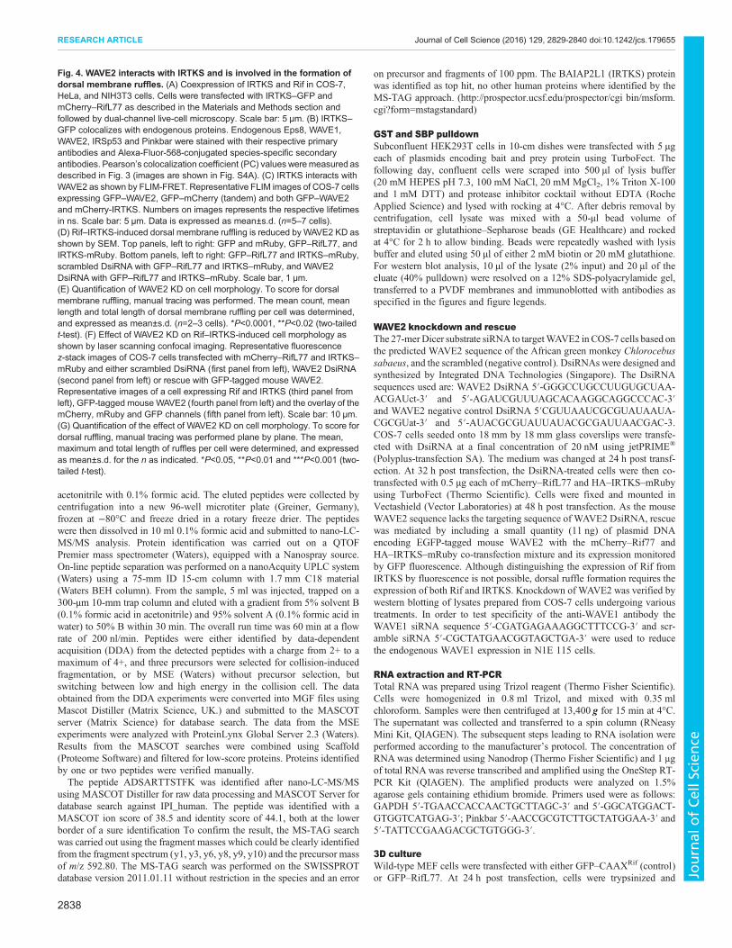
We found that Rif interacted with IRTKS and Pinkbar but not theI-BAR proteins IRSp53, MIM and ABBA. Pinkbar has been linkedwith edge ruffling at cell–cell contacts (Pykalainen et al., 2011). Wefound that Rif and Pinkbar colocalize and induce edge ruffling.Thus, Rif might play a role with Pinkbar in driving other dorsal cellmorphologies. IRTKS did not interact with Cdc42, RhoA or Rac,making it a specific target for Rif. IRTKS bound to bothconstitutively active and dominant-negative forms of Rif,suggesting no preference for its activation state. MIM and ABBAdo not have Rho-GTPase-binding sites, but they might connect toRhoGTPase signaling indirectly through I-BAR family heterodimerformation (see below and Fig. 5).
We have identified Eps8 and WAVE2 as two actin regulatorsinvolved in the Rif–IRTKS pathway to dorsal structures. By FRETanalysis we show that both Eps8 and WAVE2 bind IRTKS in thesedorsal structures. Using SEM we were able to quantify the numberand size of dorsal structures generated by the Rif–IRTKS pathway.We found that Eps8 was not essential for dorsal filopodia formationbut is involved in modulating their size. This is consistent with theactin-capping role of Eps8 (Croce et al., 2004), where Eps8 preventsthe actin filament elongation. In contrast, WAVE2 KD dramaticallyreduced the number of dorsal membrane ruffles formed and their
Fig. 1. Characterization of Rif interaction with IRTKS. (A) Rif interacts withIRTKS. 3tag–RifL77 was coexpressed with either HA-tagged mDia1ΔDAD,IRTKS or IRSp53 in 293T cells and analyzed for pairwise interactions by SBPpulldown (SPD-PD) of Rif and immunoblotting (WB) with anti-HA antibody. Rifwas detected using anti-Flag antibody as described in the Materials andMethods section. (B) Rif interacts with Pinkbar. 3tag–RifL77 was coexpressedwith either GFP-tagged IRSp53, IRTKS, Pinkbar, MIM or ABBA in 293T cellsand analyzed for pairwise interactions by SBP pulldown of Rif andimmunoblotting with anti-GFP antibody. Rif was detected using anti-Flagantibody. (C,D) Rif interacts with IRTKS specifically. (C) SBP fusion of full-length IRTKS was coexpressed with HA-tagged constitutively active(Rac1V12, RifL77) or dominant-negative versions (RacL77, RifN33),respectively, of Rac1 and Rif, or GFP in 293T cells, and pairwise interactionswere analyzed by SBP pulldown of IRTKS and immunoblotting with anti-HAantibody. IRTKS was detected using anti-SBP antibody. (D) A GST fusion ofthe I-BAR domain of IRTKS was coexpressed with HA-tagged constitutivelyactive or dominant-negative versions, respectively, of Rif (L77, N33), RhoA(V14, N19), Rac1 (V12, N17) and Cdc42 (V12, N17) in 293T cells andanalyzed for pairwise interactions by GST pulldown of Rif and immunoblottingwith anti-HA antibody. The I-BAR domain of IRTKS was detected using anti-GST antibody. (E) A schematic of the domain structure of full-length humanIRTKS protein and deletion constructs with their abbreviations shown.Numbering is based on amino acids. (F) Localization of Rif interaction site onIRTKS. 3tag–RifL77 was expressed with either GFP-tagged IRTKS full-lengthor deletions named ΔI-BAR, I-BAR, Non-CRIB or SH3-end in 293T cells andanalyzed for pairwise interactions by SBP pulldown of Rif and immunoblottingwith anti-GFP antibody. Rif was detected using anti-Flag antibody.Transfection, pulldown and immunoblotting were performed as described inthe Materials and Methods section.
2832
RESEARCH ARTICLE Journal of Cell Science (2016) 129, 2829-2840 doi:10.1242/jcs.179655
Journal
ofCe
llScience
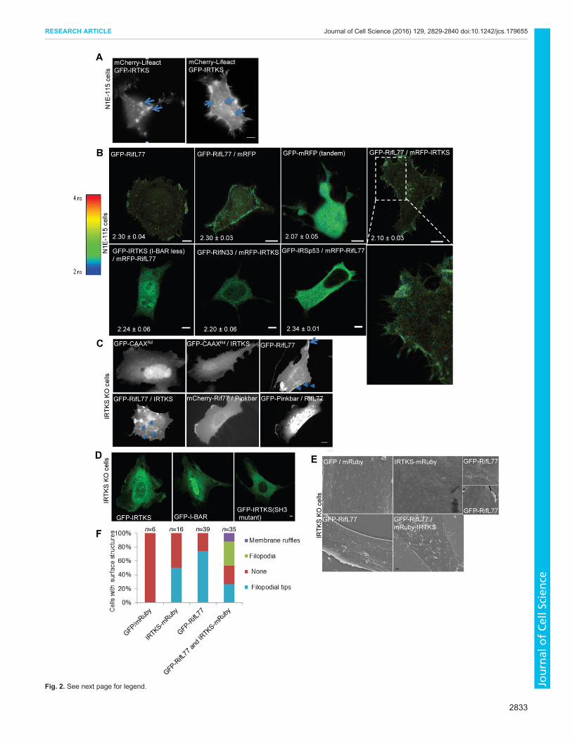
Fig. 2. See next page for legend.
2833
RESEARCH ARTICLE Journal of Cell Science (2016) 129, 2829-2840 doi:10.1242/jcs.179655
Journal
ofCe
llScience
length. Eps8 also reduced the size of dorsal membrane ruffles, butincreased their number.Invadosomes have been linked to cell migration and cell invasion
connected with small GTPases (Spuul et al., 2014). The actinmachinery responsible for the formation of invadosomes includesactin modulators such as fascin and small GTPases. Is it possiblethat actin pedestals and invadosomes subvert the machinery used togenerate dorsal filopodia and membrane ruffles? If this is thecase one would predict that Rif, IRTKS and downstreamtargets such as Eps8 and WAVE2 are involved in the formation ofthese disease-related dorsal structures. Interestingly, we haveobserved endogenous Eps8 in the actin pedestals triggered byenterohemorrhagic E. coli infection of SKOV3 ovarian cancer cells(A.M.C. and S.A., unpublished data).Rac and Cdc42 have been shown to generate peripheral structures
(membrane ruffles and filopodia, respectively) through IRSp53.Here, we show using an SEM assay that Rif, in contrast to Rac andCdc42, modulates dorsal membrane ruffles and filopodia. Rifrequires IRTKS to generate these dorsal structures and also utilizesPinkbar. In Fig. 5 we present a model showing the interrelationshipsbetween these pathways. In preliminary experiments, we observedwith FRET the formation of heterodimers of I-BAR domainproteins, which would suggest cross-talk between Rac, Cdc42 andRif, and the possibility to generate diverse morphological structures.Further work is necessary to determine the physiologicalconsequences of heterodimer formation. Nevertheless, the abilityof I-BAR domain proteins to heterodimerize highlights twoimportant issues – the diversity of morphological structures
generated by and the complexity of Rho GTPase signaling. Infuture experiments it will be important to identify growth factorsthat lead to Rif activation and dorsal morphological activities.
In conclusion, we show that the Rif–IRTKS–Eps8–WAVE2pathway leads to formation of the dorsal membrane ruffles andfilopodia, and suggest that Rac, Cdc42 andRif utilize I-BARdomainproteins as a central mechanism to regulate cell morphology.
MATERIALS AND METHODSPlasmid constructsThe pXJ40 mammalian expression vectors with various N-terminal fusiontags used in this study are HA, GFP, GST and mCherry (Manser et al.,1997). The pXJ3tag vector used for tandem affinity purification orpulldown was made as described in Goh and Manser (2010). Briefly,tandem affinity purification makes use of the CBP and SBP tag present inthe triple-epitope-tagged (3tag) construct of Rif. The RifL77 mutant wascloned into pXJ3tag using HindIII and NotI sites by PCR from Riftemplates (wild-type, Rif with the activating mutation Q77L, and Rif withthe dominant-negative mutation T33N; provided by Harry Mellor, BristolUniversity, Bristol, UK). RifL77 was then excised and inserted intopXJ-HA and pXJ-mCherry. RifN33 was cloned into pXJ-HA using asimilar approach. GFP was subcloned into pXJ3tag using BamHI/NotI.pXJ-HA-IRTKS and pXJ-Lifeact-GFP were from Edward Manser (IMCB,Singapore), pXJ-HA-IRTKS-eGFP was created by insertion of IRTKS–EGFP (Pekka Lappalainen, Institute of Biotechnology, Finland) into pXJ-HA vector. pXJ-mCherry-Lifeact was obtained by appending the Lifeactsequence 3′ of mCherry. The C-terminal end of Rif containing the plasma-membrane-targeting motif of CAAX was cloned as a HindIII–NotIfragment into pXJ-GFP for the generation of the CAAX plasmid. GFP–IRTKS (lacking the I-BAR domain) and GFP–IRTKS (SH3 mutant) are inthe pXJ-GFP vector. Rif mutations of S51A, E54N and deletion of theCAAX bases were generated in the RifWT-mCherry vector.
The mitochondrial outer membrane targeting vector (pXJ-mito) wasmade by subcloning the sequence encoding the N-terminal 33 amino acidresidues containing the transmembrane domain (TMD) of Tom20 (Addgene37316) into the pXJ40 vector using EcoRI/BamHI. The TMD fusion willtherefore be anchored to the outer membrane of the mitochondria by theTMD, leaving the adjoining fusion protein exposed to the cytosol (Kanajiet al., 2000). pXJ-mito-IRTKS-mRuby was created by inserting mRuby-tagged IRTKS into this pXJ-mito vector. mCherry-C1-Eps8 (Addgene29779), mtagBFP2 (Addgene 34633), mRuby2 (Addgene 40260), Ezrin-eGFP (Addgene 20680) vectors were obtained from Addgene.
Full-length and truncations of human IRTKS were introduced into pXJ-GFP by BamHI/XhoI with the exception of I-BAR, which was excisedusing XhoI/BamHI from the I-BAR domain of mouse IRTKS in pEGFP-N1(Pekka Lappalainen) and ligated into the XhoI/BglII sites of pXJ-GFP. pXJ-GST MIM and ABBA in pEGFP-N1 vector were from Pekka Lappalainen.GFP–Mena vector was a gift from Klemens Rottner (University of Bonn,Germany). EGFP–Eps8, EGFP–Eps8 deletion mutants, EGFP–WAVE1,EGFP–WAVE2, and EGFP–VASP pCDNA3.1 vectors and vectors forFlag-tagged Eps8, Eps8L1, Eps8L2 and Eps8L3 were a gift from GiorgioScita (IFOM-IEO Campus, Italy). EGFP–Espin vector was a gift fromJames R. Bartles (Northwestern University, Chicago, IL). pEGFP-N1-human Dyn1(aa) was a gift from Pietro De Camilli (Yale University, NewHaven, CT). pEGFP-N1-rat Dyn2(aa) was a gift from Mark A. McNiven(Mayo Clinic, Rochester, MN). GFP–N-WASP vector was a gift fromSylvie Lommel (Department of Cell Biology, German Research Centre forBiotechnology, Braunschweig, Germany). pXJGFPmDia1ΔDAD wasmade by subcloning from a source of mDia1ΔDAD sequence (ChengGeeKoh, NTU, Singapore). pcDNAmyc-GFP-mDia2ΔDAD and pcDNAmyc-HA-mDia2ΔDAD were made by inserting GFP or HA into pcDNAmyc-mDia2ΔDAD (gift from Arthur Alberts, Van Andel Research Institute,Grand Rapids, MI), respectively. GFP-C1-IRSp53 vector was a gift fromMari Masuda (NCCRI, Tokyo, Japan). Mouse WAVE2 (GenBankBC156285) and human Pinkbar (GenBank BC015619) templates werepurchased from the PlasmID repository at Harvard Medical School andcloned into pEGFP-C1 and pXJ-EGFP, respectively.
Fig. 2. Rif signals through IRTKS to generate dorsal morphology.(A). IRTKS phenotype in N1E-115 cells. Cells were transfected with GFP–IRTKS and mCherry–Lifeact. Dual-channel (GFP and mCherry) time-lapsemicroscopy was then carried out at 24 h post transfection as described in theMaterials and Methods section. Two typical cells are shown. Left panel: dorsalmembrane ruffles (arrows). Right panel: dorsal filopodia (arrows). Scale bar,10 µm. (B) Rif interacts with IRTKS as observed by FLIM-FRET.Representative FLIM images of N1E115 cells expressing (top panels, left toright): GFP–RifL77, coexpression of GFP–RifL77 and mRFP, GFP–mRFP(tandem), and coexpression of GFP–RifL77 and mRFP–IRTKS; (bottompanels, left to right): coexpression of GFP–IRTKS (lacking the I-BAR domain,I-BAR less) and mRFP–RifL77, GFP–RifN33 and mRFP–IRTKS, GFP–IRSp53 and mRFP–RifL77 (negative control), and magnified image of toppanel GFP–RifL77 and mRFP–IRTKS. Numbers on images represent therespective lifetimes in ns. Scale bars: 5 µm. Data is expressed as mean±s.d.(n=5–7 cells). (C). Rif requires IRTKS for generation of dorsal structures. Cellswere imaged using dual-channel live-cell microscopy. IRTKS-KO MEFs wereisolated from KO mice and transfected with the relevant cDNAs.Representative examples are shown of individual cells expressing (top panels,left to right) GFP–CAAXRif (plasma membrane localization motif CAAX fromthe C-terminal end of Rif ), GFP-CAAXRif and IRTKS, and GFP–RifL77; and(bottom panel, left to right) coexpression of GFP–RifL77 and IRTKS,coexpression of mCherry–RifL77 and Pinkbar, and coexpression of RifL77 andGFP–Pinkbar. Membrane ruffles (arrow) and peripheral protrusions(arrowheads) are found in the GFP–RifL77 cells, whereas dorsal protrusions(arrowhead) are seen upon coexpression of GFP–RifL77 and IRTKS. Scalebar: 10 µm. (D) Rescue of IRTKS-KO cells upon expression of GFP–IRTKS,GFP–I-BAR or GFP–IRTKS (SH3 mutant). Scale bar: 5 µm. (E) Rif requiresIRTKS for generation of dorsal structures. IRTKS-KO MEFs isolated from KOmice were transfected with the relevant cDNAs and grown on griddedcoverslips. Cells were then localized by fluorescence imaging and the samecell imaged by SEM. Representative examples are shown of cells expressingGFP and mRuby, IRTKS–mRuby, GFP–RifL77 and upon coexpression ofGFP–RifL77 and mRuby–IRTKS. Scale bar: 1 µm. (F) Statistical analysis ofdata shown in E. Cells were scored for dorsal membrane ruffles, dorsalfilopodia and filopodial tips as described in the Materials and Methods. Theimages of individual cells shown are representative of cells when searching fordorsal protrusions.
2834
RESEARCH ARTICLE Journal of Cell Science (2016) 129, 2829-2840 doi:10.1242/jcs.179655
Journal
ofCe
llScience
Fig. 3. See next page for legend.
2835
RESEARCH ARTICLE Journal of Cell Science (2016) 129, 2829-2840 doi:10.1242/jcs.179655
Journal
ofCe
llScience
The primary antibodiesMouse monoclonal anti-Eps8 (1:100 immunofluorescence, 610143) wasfrom BD Transduction Laboratories; HRP-conjugated mouse monoclonalanti-GAPDH (1:1000 western blotting, G9295), rabbit polyclonal anti-IRSp53 (1:50 IF, HPA023310) and mouse monoclonal anti-Flag (1:7000western blotting, F3165) were from Sigma-Aldrich; anti-SBP (1:7000western blotting, sc101595), anti-HA (1:7000 western blotting, sc7392),anti-GFP (1:7000 immunofluorescence, sc9996) anti-Pinkbar (1:50immunofluorescence, sc86305) anti-GST (1:7000 western blotting, sc-138)and anti-WAVE1 [1:200, goat polyclonal (sc10388) and anti-WAVE2 (1:50immunofluorescence, sc-33548)] were from Santa Cruz Biotechnology.
Cell culture and transfectionHEK293 (CRL-1573), HEK293T (CRL-3216), COS-7 (CRL1651) cellswere from ATCC. N1E-115 neuroblastoma, HeLa and NIH3T3 cells werecultured in Dulbecco’s modified Eagle’s medium (DMEM; 4500 mg/lglucose) and supplemented with 10% fetal bovine serum and 1% penicillin-streptomycin in a humidified 37°C incubator with 5% CO2. Cell culturemycoplasma contamination was carried out using PCR for all cells. ForN1E-115 cells transient expression of plasmids were performed usingLipofectamine 2000 (Invitrogen) or Jetprime (Polyplus-transfection)according to the manufacturer’s instructions. HeLa and NIH3T3 cellstransient transfections were performed according to the manufacturer’sprotocol using TurboFect (Thermo Scientific).
To obtain IRTKSdel/del (null) mice, a targeting vector was generated usingBAC clones from the C57BL/6J RPCIB-731 BAC library, in which exon 2 ofIRTKSwas flanked by loxP sites (Taconic). Deletion of exon 2 resulted in lossof function of the BAIAP2L1 gene by generating a frameshift from exon 1 toexons 3 and 4 (premature stop codon in exon 3). The vector was integratedinto the TaconicArtemis C57BL/6NTac embryonic stem cell (ESC) lineby homologous recombination. Targeted C57BL/6NTac ESCs weremicroinjected into blastocysts, which were then transferred to pseudopregnant females. Highly chimeric mice were bred to Flp-Deleter females
(strain C57BL/6). Germline transmission was identified by the presence ofblack, strain C57BL/6, offspring (G1) (Taconic). IRTKSdel/del mice wereobtained bymatingB6-Baiap2l1 conditional KOmicewith B6.129P2-ROSA-Cre Ki/Ki mice (gift from Colin Stewart, IMB Singapore), and verified bygenotyping PCR. IRTKS-null (KO) fibroblasts were verified byimmunocytochemistry. Immortalized IRTKS-null MEFs were derived fromIRTKS-null embryos and repeatedly subcultured till bypass of senescence.Immortalized Eps8-null MEFs (Giorgio Scita) were originally derived fromEps8-null E13 embryo and immortalized with the NIH3T3 protocol. Thesecells had been infectedwith the empty pBABEHygro vector and selectedwithhygromycin (200 µg/ml). All cells were maintained in DMEM (4500 mg/lglucose, 10% FBS and 4 mM L-glutamine) by incubating at 37°C with 5%CO2. Cells were either transfected with TurboFect (Thermo Scientific) orJetprime (Polyplus) following the manufacturer’s protocols. The animaltreatmentwas performed in accordancewith IACUCandNACLARguidelinesand approved by Biological Resource Center, Singapore.
Immunofluorescence and microscopyCells grown on glass coverslips were fixed with 4% paraformaldehyde inPBS for 10 min and washed with PBS. Cells were then permeabilized with0.2% Triton X-100 in PBS for 5 min, washed with PBS and blocked in 5%BSA in PBS for 30 min. Primary antibody diluted in 5% BSA in PBS wasthen incubated with cells overnight at 4°C, followed by washing with PBS.Cells were next incubated with species-specific Alexa-Fluor-conjugatedsecondary antibody diluted in 5%BSA in PBS for 45 min, washed with PBSand mounted onto glass slides with Hydromount (National Diagnostics).Live-cell microscopy was performed on an Olympus IX83 live-cell invertedmicroscope (Olympus, Singapore), equipped with a CoolSNAP CCD HQ2camera (Roper Photometrics) and on- stage incubation system (Tokai Hit).For live-cell imaging, transfected cells grown on 35 mm ibiTreat optical-quality plastic cell culture dishes (Ibidi) were imaged with a 60×/1.2 NAobjective. For time-lapse microscopy, images were acquired at 10 s intervalsover a 10-min time period in the appropriate fluorescence channels usingMetaMorph software (Molecular Devices).
Tandem affinity purificationEach 15-cm dish of HEK293 cells was transfected with 4 µg of targetplasmid and 6 µg of pBluescript carrier, with a total of two dishes per target[3tag–RifL77 and negative control (3tag–GFP)]. At 24 h post transfection,cells were rinsed with PBS and frozen. Cell lysate was prepared by addinglysis buffer (20 mM Tris-HCl, pH 8, 130 mM NaCl, 1% Nonidet P-40,5 mMMgCl2, 1 mM PMSF, 1× protease inhibitor, 4 mM DTT), frozen andthawed twice, and rocked for 20 min at 4°C for extraction, after whichinsoluble material was removed by centrifugation. Lysate was thenprecleared by exposure to glutathione–Sepharose beads for 1 h beforeremoval of beads by pulse spin. The final eluate was obtained following theInterplay mammalian TAP system protocol (Agilent Technologies) andreduced in volume by using a combination of molecular mass cut-offcolumns (3000MWCO PES VIVASPIN 500, Sartorius) and speed vacuum.This concentrated sample was mixed with Laemmli sample buffer and DTTto a final concentration of 10 mM, reduced at 60°C for 10 min andS-alkylated with 20 mM idoacetamide at room temperature for 30 min. Thetreated sample was resolved on a 12% SDS-polyacrylamide gel to about5–10 mm into the resolving gel. The gel was stained with colloidalCoomassie Blue (BioRad) and destained with water. Further downstreamprocessing by nano-liquid chromatography tandemmass spectrometry (LC-MS/MS) and analysis was carried out by the mass spectroscopy group(Manfred Raida) at the Experimental Therapeutics Centre, Singapore, togenerate a preliminary list of protein candidates using the Scaffold software(Proteome Software).
Mass spectrometry analysisThe whole gel lane was cut into three bands of equal sizes and transferredinto perforated microtiterplates for in-gel digestion (Proxeon). Trypticdigestion was carried out using 10 ng/ml sequencing grade trypsin(Promega) for 4 h at 37°C in 5 mM triethyl ammonium buffer, pH 8.5.The resulting peptides were eluted with 1% formic acid in water (firststep), 60% acetonitrile and 0.1% formic acid (second step), and
Fig. 3. Eps8 interacts with IRTKS and is involved in the generation ofdorsal morphology. (A) Identification of IRTKS partners using mitochondriallocalization. Mitochondria-targeted IRTKS–mRuby was coexpressed withGFP-tagged WAVE1, WAVE2, Mena, Eps8, VASP, Espin, mDia1, mDia2,N-WASP, Ezrin, Dyn1 or Dyn2 in HeLa cells. Dual-channel fluorescence live-cell imaging was then carried out. Regions of interest (ROIs) were drawn andPearson’s colocalization coefficients (PC) were calculated using Metamorphsoftware. Pearson’s colocalization values above 0.7 were taken as indicative ofcolocalization (images shown in Fig. S2B). (B) IRTKS interacts with Eps8.SBP–IRTKS was coexpressed with Flag-tagged Eps8, Eps8L1, Eps8L2,Eps8L3, GFP or TccP in 293T cells and pairwise interactions analyzed by SBPpulldown of IRTKS and immunoblotting (WB) with anti-Flag antibody. Note thatEps8L2 gave variable expression in 293T cells and in some experiments couldnot be seen. Hence we were not able to ascertain its ability to co-immunoprecipitate with IRTKS. L1, L2 and L3 are related independent genesof Eps8. IRTKS was detected using anti-SBP antibody. The strong ECL signalobtained when detecting SBP–IRTKS using HRP-conjugated anti-SBPantibody results in signal bleaching on X-ray films that gives the appearance ofmultiple bands (bottom right panel). (C) IRTKS interacts with Eps8 by FLIM-FRET. Representative FLIM images of HeLa cells expressing GFP–Eps8,GFP–mRFP, coexpression of GFP–Eps8 and mRFP, and GFP–Eps8 andmRFP–IRTKS. Numbers on images represent the respective lifetimes in ns.Scale bars: 10 µm. Data is expressed as mean±s.d. (n=5–7 cells). (D) Effect ofEps8 KO on Rif–IRTKS induction of dorsal morphology as shown by SEM.A GFP, mRuby and mCherry control is shown in the top left and top middlepanels, a GFP–Eps8 rescue is shown in the top right panel, mCherry–RifL77and IRTKS–mRuby are shown in the middle left and bottom left panels, andmCherry–RifL77 and IRTKS–mRuby with Eps8 rescue are shown in themiddle left and bottom left panels. Scale bar: 1 µm. (E) Statistical analysis ofdata shown in D, cells were scored for dorsal membrane ruffles, dorsalfilopodia and long dorsal filopodia as described in the Materials and Methods.(F) The mean count, mean length and total length of dorsal membrane rufflingper cell was determined, and expressed as mean±s.d. (n=3–4 cells). Imagesshown in D were used to assess the size of dorsal membrane ruffles. To scorefor dorsal membrane ruffling, manual tracing was performed. *P<0.001 (two-tailed t-test).
2836
RESEARCH ARTICLE Journal of Cell Science (2016) 129, 2829-2840 doi:10.1242/jcs.179655
Journal
ofCe
llScience
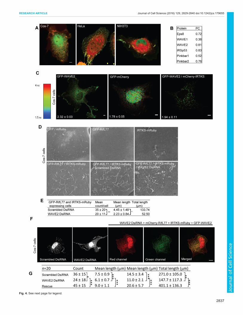
Fig. 4. See next page for legend.
2837
RESEARCH ARTICLE Journal of Cell Science (2016) 129, 2829-2840 doi:10.1242/jcs.179655
Journal
ofCe
llScience
acetonitrile with 0.1% formic acid. The eluted peptides were collected bycentrifugation into a new 96-well microtiter plate (Greiner, Germany),frozen at −80°C and freeze dried in a rotary freeze drier. The peptideswere then dissolved in 10 ml 0.1% formic acid and submitted to nano-LC-MS/MS analysis. Protein identification was carried out on a QTOFPremier mass spectrometer (Waters), equipped with a Nanospray source.On-line peptide separation was performed on a nanoAcquity UPLC system(Waters) using a 75-mm ID 15-cm column with 1.7 mm C18 material(Waters BEH column). From the sample, 5 ml was injected, trapped on a300-µm 10-mm trap column and eluted with a gradient from 5% solvent B(0.1% formic acid in acetonitrile) and 95% solvent A (0.1% formic acid inwater) to 50% B within 30 min. The overall run time was 60 min at a flowrate of 200 nl/min. Peptides were either identified by data-dependentacquisition (DDA) from the detected peptides with a charge from 2+ to amaximum of 4+, and three precursors were selected for collision-inducedfragmentation, or by MSE (Waters) without precursor selection, butswitching between low and high energy in the collision cell. The dataobtained from the DDA experiments were converted into MGF files usingMascot Distiller (Matrix Science, UK.) and submitted to the MASCOTserver (Matrix Science) for database search. The data from the MSEexperiments were analyzed with ProteinLynx Global Server 2.3 (Waters).Results from the MASCOT searches were combined using Scaffold(Proteome Software) and filtered for low-score proteins. Proteins identifiedby one or two peptides were verified manually.
The peptide ADSARTTSTFK was identified after nano-LC-MS/MSusing MASCOT Distiller for raw data processing and MASCOT Server fordatabase search against IPI_human. The peptide was identified with aMASCOT ion score of 38.5 and identity score of 44.1, both at the lowerborder of a sure identification To confirm the result, the MS-TAG searchwas carried out using the fragment masses which could be clearly identifiedfrom the fragment spectrum (y1, y3, y6, y8, y9, y10) and the precursor massof m/z 592.80. The MS-TAG search was performed on the SWISSPROTdatabase version 2011.01.11 without restriction in the species and an error
on precursor and fragments of 100 ppm. The BAIAP2L1 (IRTKS) proteinwas identified as top hit, no other human proteins where identified by theMS-TAG approach. (http://prospector.ucsf.edu/prospector/cgi bin/msform.cgi?form=mstagstandard)
GST and SBP pulldownSubconfluent HEK293T cells in 10-cm dishes were transfected with 5 µgeach of plasmids encoding bait and prey protein using TurboFect. Thefollowing day, confluent cells were scraped into 500 µl of lysis buffer(20 mM HEPES pH 7.3, 100 mM NaCl, 20 mM MgCl2, 1% Triton X-100and 1 mM DTT) and protease inhibitor cocktail without EDTA (RocheApplied Science) and lysed with rocking at 4°C. After debris removal bycentrifugation, cell lysate was mixed with a 50-µl bead volume ofstreptavidin or glutathione–Sepharose beads (GE Healthcare) and rockedat 4°C for 2 h to allow binding. Beads were repeatedly washed with lysisbuffer and eluted using 50 µl of either 2 mM biotin or 20 mM glutathione.For western blot analysis, 10 µl of the lysate (2% input) and 20 µl of theeluate (40% pulldown) were resolved on a 12% SDS-polyacrylamide gel,transferred to a PVDF membranes and immunoblotted with antibodies asspecified in the figures and figure legends.
WAVE2 knockdown and rescueThe 27-mer Dicer substrate siRNA to targetWAVE2 inCOS-7 cells based onthe predicted WAVE2 sequence of the African green monkey Chlorocebussabaeus, and the scrambled (negative control). DsiRNAs were designed andsynthesized by Integrated DNA Technologies (Singapore). The DsiRNAsequences used are: WAVE2 DsiRNA 5′-GGGCCUGCCUUGUGCUAA-ACGAUct-3′ and 5′-AGAUCGUUUAGCACAAGGCAGGCCCAC-3′and WAVE2 negative control DsiRNA 5′CGUUAAUCGCGUAUAAUA-CGCGUat-3′ and 5′-AUACGCGUAUUAUACGCGAUUAACGAC-3.COS-7 cells seeded onto 18 mm by 18 mm glass coverslips were transfe-cted with DsiRNA at a final concentration of 20 nM using jetPRIME®
(Polyplus-transfection SA). The medium was changed at 24 h post transf-ection. At 32 h post transfection, the DsiRNA-treated cells were then co-transfected with 0.5 µg each of mCherry–RifL77 and HA–IRTKS–mRubyusing TurboFect (Thermo Scientific). Cells were fixed and mounted inVectashield (Vector Laboratories) at 48 h post transfection. As the mouseWAVE2 sequence lacks the targeting sequence of WAVE2 DsiRNA, rescuewas mediated by including a small quantity (11 ng) of plasmid DNAencoding EGFP-tagged mouse WAVE2 with the mCherry–Rif77 andHA–IRTKS–mRuby co-transfection mixture and its expression monitoredby GFP fluorescence. Although distinguishing the expression of Rif fromIRTKS by fluorescence is not possible, dorsal ruffle formation requires theexpression of both Rif and IRTKS. Knockdown of WAVE2 was verified bywestern blotting of lysates prepared from COS-7 cells undergoing varioustreatments. In order to test specificity of the anti-WAVE1 antibody theWAVE1 siRNA sequence 5′-CGATGAGAAAGGCTTTCCG-3′ and scr-amble siRNA 5′-CGCTATGAACGGTAGCTGA-3′ were used to reducethe endogenous WAVE1 expression in N1E 115 cells.
RNA extraction and RT-PCRTotal RNA was prepared using Trizol reagent (Thermo Fisher Scientific).Cells were homogenized in 0.8 ml Trizol, and mixed with 0.35 mlchloroform. Samples were then centrifuged at 13,400 g for 15 min at 4°C.The supernatant was collected and transferred to a spin column (RNeasyMini Kit, QIAGEN). The subsequent steps leading to RNA isolation wereperformed according to the manufacturer’s protocol. The concentration ofRNAwas determined using Nanodrop (Thermo Fisher Scientific) and 1 μgof total RNAwas reverse transcribed and amplified using the OneStep RT-PCR Kit (QIAGEN). The amplified products were analyzed on 1.5%agarose gels containing ethidium bromide. Primers used were as follows:GAPDH 5′-TGAACCACCAACTGCTTAGC-3′ and 5′-GGCATGGACT-GTGGTCATGAG-3′; Pinkbar 5′-AACCGCGTCTTGCTATGGAA-3′ and5′-TATTCCGAAGACGCTGTGGG-3′.
3D cultureWild-type MEF cells were transfected with either GFP–CAAXRif (control)or GFP–RifL77. At 24 h post transfection, cells were trypsinized and
Fig. 4. WAVE2 interacts with IRTKS and is involved in the formation ofdorsal membrane ruffles. (A) Coexpression of IRTKS and Rif in COS-7,HeLa, and NIH3T3 cells. Cells were transfected with IRTKS–GFP andmCherry–RifL77 as described in the Materials and Methods section andfollowed by dual-channel live-cell microscopy. Scale bar: 5 µm. (B) IRTKS–GFP colocalizes with endogenous proteins. Endogenous Eps8, WAVE1,WAVE2, IRSp53 and Pinkbar were stained with their respective primaryantibodies and Alexa-Fluor-568-conjugated species-specific secondaryantibodies. Pearson’s colocalization coefficient (PC) values weremeasured asdescribed in Fig. 3 (images are shown in Fig. S4A). (C) IRTKS interacts withWAVE2 as shown by FLIM-FRET. Representative FLIM images of COS-7 cellsexpressing GFP–WAVE2, GFP–mCherry (tandem) and both GFP–WAVE2and mCherry-IRTKS. Numbers on images represents the respective lifetimesin ns. Scale bar: 5 µm. Data is expressed as mean±s.d. (n=5–7 cells).(D) Rif–IRTKS-induced dorsal membrane ruffling is reduced by WAVE2 KD asshown by SEM. Top panels, left to right: GFP and mRuby, GFP–RifL77, andIRTKS-mRuby. Bottom panels, left to right: GFP–RifL77 and IRTKS–mRuby,scrambled DsiRNA with GFP–RifL77 and IRTKS–mRuby, and WAVE2DsiRNA with GFP–RifL77 and IRTKS–mRuby. Scale bar, 1 µm.(E) Quantification of WAVE2 KD on cell morphology. To score for dorsalmembrane ruffling, manual tracing was performed. The mean count, meanlength and total length of dorsal membrane ruffling per cell was determined,and expressed as mean±s.d. (n=2–3 cells). *P<0.0001, **P<0.02 (two-tailedt-test). (F) Effect of WAVE2 KD on Rif–IRTKS-induced cell morphology asshown by laser scanning confocal imaging. Representative fluorescencez-stack images of COS-7 cells transfected with mCherry–RifL77 and IRTKS–mRuby and either scrambled DsiRNA (first panel from left), WAVE2 DsiRNA(second panel from left) or rescue with GFP-tagged mouse WAVE2.Representative images of a cell expressing Rif and IRTKS (third panel fromleft), GFP-tagged mouse WAVE2 (fourth panel from left) and the overlay of themCherry, mRuby and GFP channels (fifth panel from left). Scale bar: 10 μm.(G) Quantification of the effect of WAVE2 KD on cell morphology. To score fordorsal ruffling, manual tracing was performed plane by plane. The mean,maximum and total length of ruffles per cell were determined, and expressedas mean±s.d. for the n as indicated. *P<0.05, **P<0.01 and ***P<0.001 (two-tailed t-test).
2838
RESEARCH ARTICLE Journal of Cell Science (2016) 129, 2829-2840 doi:10.1242/jcs.179655
Journal
ofCe
llScience
resuspended in fetal bovine serum at a concentration of 1×10⁵ cells/ml. Thecollagen matrix was prepared and neutralized by mixing 200 µl of type Icollagen (Corning) with 250 µl of DMEM medium (Thermo FisherScientific) in a pre-cooled 15 ml falcon tube; 23 µl of NaOH (Sigma) wasadded to collagen to reach neutral pH. Then, 250 µl of cell suspension wasthen added to the neutralized collagen mixture and plated immediately in achambered coverglass (Nunc). The collagen mixture was then allowedto solidify completely at 37°C for 3 h. Upon solidification, the gel wasoverlaid with 200 µl of DMEM and incubated overnight at 37°C. On thefollowing day, 3D imaging was carried out using an inverted confocalmicroscope.
Fluorescence lifetime microscopyFLIM experiments were performed on a Time-Correlated Single PhotonCounting (TCSPC) system (PicoQuant, Germany) attached to an OlympusFV-1000 confocal microscope (Olympus, USA) with a 60×1.2 W objective(Lam et al., 2012). The excitation light source was a 485-nm pulsed diodelaser controlled by a Sepia II driver (PicoQuant) with a 488- or 559-nmdichroic mirror and a 520/30 emission filter. Individual photon arrivals weredetected using a SPAD detector and events were recorded by a PicoHarp 300TCSPC module. Lifetime analysis was carried out using Symphotimesoftware (PicoQuant). Mono- and bi-exponential fittings were obtained forGFP alone and in the presence of mRFP respectively. The percentage FLIM-FRET efficiency was calculated as FRET=100×[1−(lifetime of donor withFRET/lifetime of donor without FRET)].
Scanning electron microscopySEM samples were prepared by growing Eps8-KO, IRTKS-KO or COS-7cells on IBIDgridded 35-mmdishes (Zwaenepoel et al., 2012). The cells weretransfected with either free GFP or mRuby as controls and fusion plasmids ofRifL77, Eps8 and IRTKS alone or in pairs using Turbofect. TheWAVE2KDexperiment was performed along with a control experiment in COS-7 cellsexpressing GFP–RifL77 and IRTKS–mRuby. At 16 h post transfection, cellswere fixed using 2.5% glutaraldehyde in 0.1 M sodium cacodylate buffer(pH 7.4) and incubated for at least 20 min at room temperature. Transfectedcells were identified under a fluorescencemicroscope for their locations on the
grid and recorded. After fixation, the samples were washed with bufferfollowed by another 1 h post fixation with 1% osmium tetroxide in 0.1 Msodiumcacodylate at 4°C.The sampleswere extensivelywashedwith distilledwater and subsequently dehydrated through a gradient concentration of 25%,50%, 75%, 95% and 100% ethanol in distilled water. Following two changesof 100% ethanol, the samples were transferred to a Leica Critical Point Dryer(EMCPD 030) for critical point drying to preserve fine cell surface structures.Samples were sputter coatedwith 2-nmgold and observed using a JEOLFieldEmission Scanning Electron Microscope JSM6701F with an acceleratingvoltage of 5 kV. Cells imaged in fluorescence microscopy were identified inSEM by referring to the marking of the grid.
Colocalization analysisThe degree of colocalization between two proteins at various cellregions was determined by Pearson’s coefficient, as previously described(Bu et al., 2009). A value of 0.7 and above was taken as indicatingcolocalization.
Definition of structures analyzed by SEMFilopodia tips are defined as small protrusions seen on the dorsal surface.Short filopodia are defined as structures that are smaller than 1 µm, whereasfilopodia are defined as structures that are between 1 and 5 µm in length.Dorsal membrane ruffles are defined as sheet-like structures on the dorsalsurface of cells (Fig. S1D).
Scoring for dorsal filopodia and membrane ruffling using SEMThe scoring for dorsal filopodia and ruffling was performed usingImageJ software by manual tracing on SEM images for IRTKS-KO,Eps8-KO and COS-7 cells. An average of 2–5 cells of each cell type werescored. The measured lengths were transferred onto an Excel spreadsheetand the mean and total length calculation for filopodia and ruffling wasperformed.
Statistical analysisAll quantitative results were obtained from at least three independentexperiments, and expressed as the mean±s.d. Experimental data were
Fig. 5. Rho family GTPases signal through I-BAR proteins to control cell morphology. The data presented in this paper suggests that Rif is responsible fordorsal membrane ruffles and filopodia, whereas Cdc42 and Rac promote peripheral structures. Heterodimerization of I-BAR family proteins allow crosstalkbetween Rho-family-GTPase-mediated signaling pathways and facilitate the formation of a greater diversity of morphological structures. AlthoughMIM and ABBAdo not bind Rac, Cdc42 or Rif, theymight be linked to signaling pathways regulated by these Rho family GTPases by forming heterodimers with other I-BAR familyproteins. Most importantly, our model suggests that Rif, Rac and Cdc42 utilize I-BAR proteins as a central mechanism for regulating cell morphology.
2839
RESEARCH ARTICLE Journal of Cell Science (2016) 129, 2829-2840 doi:10.1242/jcs.179655
Journal
ofCe
llScience
analyzed by two tailed t-test with unequal variance. Differences amongsamples were considered statistically significant when P<0.05.
AcknowledgementsThe authors would like to thank John Lim for his help in image analysis and ShupingLin help in for colocalization analysis.
Competing interestsThe authors declare no competing or financial interests.
Author contributionsT.S. and S.A. conceived and designed the experiments and wrote the manuscript.T.S., K.P.S., Y.H.Y., W.I.G. and A.M.C. performed the experiments.
FundingThis work was funded by the Agency for Science, Technology and Research(A-STAR), Singapore.
Supplementary informationSupplementary information available online athttp://jcs.biologists.org/lookup/doi/10.1242/jcs.179655.supplemental
ReferencesAhmed, S., Goh, W. I. and Bu, W. (2010). I-BAR domains, IRSp53 and filopodiumformation. Semin. Cell Dev. Biol. 21, 350-356.
Bu,W., Chou, A. M., Lim, K. B., Sudhaharan, T. and Ahmed, S. (2009). The Toca-1-N-WASP complex links filopodial formation to endocytosis. J. Biol. Chem. 284,11622-11636.
Chou, A. M., Sem, K. P., Wright, G. D., Sudhaharan, T. and Ahmed, S. (2014).Dynamin1 is a novel target for IRSp53 protein and works withmammalian enabled(Mena) protein and Eps8 to regulate filopodial dynamics. J. Biol. Chem. 289,24383-24396.
Croce, A., Cassata, G., Disanza, A., Gagliani, M. C., Tacchetti, C., Malabarba,M. G., Carlier, M.-F., Scita, G., Baumeister, R. andDi Fiore, P. P. (2004). A novelactin barbed-end-capping activity in EPS-8 regulates apical morphogenesis inintestinal cells of Caenorhabditis elegans. Nat. Cell Biol. 6, 1173-1179.
Disanza, A., Mantoani, S., Hertzog, M., Gerboth, S., Frittoli, E., Steffen, A.,Berhoerster, K., Kreienkamp, H.-J., Milanesi, F., Di Fiore, P. P. et al. (2006).Regulation of cell shape by Cdc42 is mediated by the synergic actin-bundlingactivity of the Eps8-IRSp53 complex. Nat. Cell Biol. 8, 1337-1347.
Ellis, S. and Mellor, H. (2000). The novel Rho-family GTPase rif regulatescoordinated actin-based membrane rearrangements. Curr. Biol. 10, 1387-1390.
Fan, L. and Mellor, H. (2012). The small Rho GTPase Rif and actin cytoskeletalremodelling. Biochem. Soc. Trans. 40, 268-272.
Goh, L. L. and Manser, E. (2010). The RhoA GEF Syx is a target of Rnd3 andregulated via a Raf1-like ubiquitin-related domain. PLoS ONE 5, e12409.
Goh, W. I., Sudhaharan, T., Lim, K. B., Sem, K. P., Lau, C. L. and Ahmed, S.(2011). Rif-mDia1 interaction is involved in filopodium formation independent ofCdc42 and Rac effectors. J. Biol. Chem. 286, 13681-13694.
Goh, W. I., Lim, K. B., Sudhaharan, T., Sem, K. P., Bu, W., Chou, A. M. andAhmed, S. (2012). mDia1 and WAVE2 proteins interact directly with IRSp53 infilopodia and are involved in filopodium formation. J. Biol. Chem. 287, 4702-4714.
Govind, S., Kozma, R., Monfries, C., Lim, L. and Ahmed, S. (2001). Cdc42Hsfacilitates cytoskeletal reorganization and neurite outgrowth by localizing the 58-kD insulin receptor substrate to filamentous actin. J. Cell Biol. 152, 579-594.
Hall, A. (2012). Rho family GTPases. Biochem. Soc. Trans. 40, 1378-1382.
Kanaji, S., Iwahashi, J., Kida, Y., Sakaguchi, M. and Mihara, K. (2000).Characterization of the signal that directs Tom20 to the mitochondrial outermembrane. J. Cell Biol. 151, 277-288.
Krugmann, S., Jordens, I., Gevaert, K., Driessens, M., Vandekerckhove, J. andHall, A. (2001). Cdc42 induces filopodia by promoting the formation of an IRSp53:Mena complex. Curr. Biol. 11, 1645-1655.
Lam, C. S., Mistri, T. K., Foo, Y. H., Sudhaharan, T., Gan, H. T., Rodda, D., Lim,L. H., Chou, C., Robson, P., Wohland, T. et al. (2012). DNA-dependent Oct4-Sox2 interaction and diffusion properties characteristic of the pluripotent cell staterevealed by fluorescence spectroscopy. Biochem. J. 448, 21-33.
Lim, K. B., Bu, W., Goh, W. I., Koh, E., Ong, S. H., Pawson, T., Sudhaharan, T.and Ahmed, S. (2008). The Cdc42 effector IRSp53 generates filopodia bycoupling membrane protrusion with actin dynamics. J. Biol. Chem. 283,20454-20472.
Linder, S., Wiesner, C. and Himmel, M. (2011). Degrading devices: invadosomesin proteolytic cell invasion. Annu. Rev. Cell Dev. Biol. 27, 185-211.
Manser, E., Huang, H. Y., Loo, T. H., Chen, X. Q., Dong, J. M., Leung, T. and Lim,L. (1997). Expression of constitutively active alpha-PAK reveals effects of thekinase on actin and focal complexes. Mol. Cell. Biol. 17, 1129-1143.
Mattila, P. K., Pykalainen, A., Saarikangas, J., Paavilainen, V. O., Vihinen, H.,Jokitalo, E. and Lappalainen, P. (2007). Missing-in-metastasis and IRSp53deform PI(4,5)P2-rich membranes by an inverse BAR domain-like mechanism.J. Cell Biol. 176, 953-964.
Pellegrin, S. and Mellor, H. (2005). The Rho family GTPase Rif induces filopodiathrough mDia2. Curr. Biol. 15, 129-133.
Pykalainen, A., Boczkowska, M., Zhao, H., Saarikangas, J., Rebowski, G.,Jansen, M., Hakanen, J., Koskela, E. V., Peranen, J., Vihinen, H. et al. (2011).Pinkbar is an epithelial-specific BAR domain protein that generates planarmembrane structures. Nat. Struct. Mol. Biol. 18, 902-907.
Rigaut, G., Shevchenko, A., Rutz, B., Wilm, M., Mann, M. and Seraphin, B.(1999). A generic protein purification method for protein complex characterizationand proteome exploration. Nat. Biotechnol. 17, 1030-1032.
Saarikangas, J., Zhao, H., Pykalainen, A., Laurinmaki, P., Mattila, P. K.,Kinnunen, P. K. J., Butcher, S. J. and Lappalainen, P. (2009). Molecularmechanisms of membrane deformation by I-BAR domain proteins. Curr. Biol. 19,95-107.
Scita, G., Confalonieri, S., Lappalainen, P. and Suetsugu, S. (2008). IRSp53:crossing the road of membrane and actin dynamics in the formation of membraneprotrusions. Trends Cell Biol. 18, 52-60.
Spuul, P., Ciufici, P., Veillat, V., Leclercq, A., Daubon, T., Kramer, I. J. andGenot, E. (2014). Importance of RhoGTPases in formation, characteristics, andfunctions of invadosomes. Small GTPases 5, e28195.
Suetsugu, S., Kurisu, S., Oikawa, T., Yamazaki, D., Oda, A. and Takenawa, T.(2006). Optimization of WAVE2 complex-induced actin polymerization bymembrane-bound IRSp53, PIP(3), and Rac. J. Cell Biol. 173, 571-585.
Vingadassalom, D., Kazlauskas, A., Skehan, B., Cheng, H.-C., Magoun, L.,Robbins, D., Rosen,M. K., Saksela, K. and Leong, J. M. (2009). Insulin receptortyrosine kinase substrate links the E. coli O157:H7 actin assembly effectors Tirand EspF(U) during pedestal formation. Proc. Natl. Acad. Sci. USA 106,6754-6759.
Weiss, S. M., Ladwein, M., Schmidt, D., Ehinger, J., Lommel, S., Stading, K.,Beutling, U., Disanza, A., Frank, R., Jansch, L. et al. (2009). IRSp53 links theenterohemorrhagic E. coli effectors Tir and EspFU for actin pedestal formation.Cell Host Microbe 5, 244-258.
Zhao, H., Pykalainen, A. and Lappalainen, P. (2011). I-BAR domain proteins:linking actin and plasma membrane dynamics. Curr. Opin Cell Biol. 23, 14-21.
Zwaenepoel, I., Naba, A., Da Cunha, M. M. L., Del Maestro, L., Formstecher, E.,Louvard, D. and Arpin, M. (2012). Ezrin regulates microvillus morphogenesis bypromoting distinct activities of Eps8 proteins. Mol. Biol. Cell 23, 1080-1095.
2840
RESEARCH ARTICLE Journal of Cell Science (2016) 129, 2829-2840 doi:10.1242/jcs.179655
Journal
ofCe
llScience