Embed Size (px)
Citation preview

FUNCTIONAL ANALYSIS OF AN INTEGRATED GTPASE REGULATING THE
CELLULAR POOL AND DISTRIBUTION PROFILE OF INTRAFLAGELLAR
TRANSPORT PARTICLES IN CHLAMYDOMONAS REINHARDTII
A Thesis
by
DAVID ALEJANDRO SILVA
Submitted to the Office of Graduate Studies of Texas A&M University
in partial fulfillment of the requirements for the degree of
MASTER OF SCIENCE
Approved by:
Chair of Committee, Hongmin Qin Committee Members, Michael Manson Hays Rye Head of Department, U.J. McMahan
December 2012
Major Subject: Microbiology
Copyright 2012 David Alejandro Silva

ii
ABSTRACT
Cilia and flagella are sensory organelles, found in the majority of eukaryotic
organisms that play a vital role in the general physiology, health and early development
of humans. Intraflagellar transport (IFT) is tasked with building and maintaining the
entire ciliary structure by facilitating the transport of axonemal precursors, trafficking of
ciliary membrane proteins and turnover products. Currently, there are no complete
models detailing how ciliated organisms regulate the entry and exit of IFT particles, a
multi-meric adaptor complex that ferries flagellar proteins. In this thesis, I focus on
small Rab-like protein IFT22, an IFT-particle integrated protein with predicted GTPase
activity, as a potential regulatory component of IFT particle trafficking in
Chlamydomonas.
Using an artificial microRNAs strategy, I show that IFT22 regulates the available
cellular pool of IFT particles and the distribution profile of the IFT particles between the
cytoplasm and the flagellar compartment. Additionally, I demonstrate how the putative
constitutive active mutant of IFT22 is able properly localize to the peri-basal body and
enter the flagellar compartment using immunofluorescence and immunoblot analysis of
flagella extracts. Finally, preliminary RNAi data suggests IFT25 the IFT
particle/motor/BBSome assembly downstream of IFT22 regulation, evident from the
depletion of kinesin-2 subunit FLA10, IFT-dynein-2 subunit D1bLIC and BBsome
component BBS3from whole cell extracts of IFT25 knockdown transformants.

iii
ACKNOWLEDGEMENTS
I would like to thank my committee chair, Dr. Qin, and my committee members,
Dr. Rye, and Dr. Manson, for their guidance and support throughout the course of this
research. I remembered emailing Dr. Qin during the summer of 2009 asking for chance
to work in her lab. Thankfully, because of her, I had to opportunity to attend graduate
school here at A&M. Also a special thanks to Dr. Manson for the rounds of beer shared.
Thanks also go to my friends, colleagues and the department faculty for making
my time at Texas A&M University a great experience. I also want to extend my
gratitude to the Louis Stokes Alliances for Minority Participation Organization, which
provided me with enough fellowship money to dedicate myself entirely to research. You
cannot imagine how grateful I am to you for your help. To all my Fellows, thank you
for all the laughs and good times. BTD VI!
Thanks to my mom and dad for their encouragement and support over the past 25
years. I would not be here if it were not for your love and dedication. To my brother
Andrew, I could not have asked for a better friend than you. You kept me sane over the
past two years and I am thankful we got to spend more time together before we go start
our own families. To my Sigma Lambda Beta brothers, thank you for having my back
over the past 6 years. You were my home away from home, and could not ask for a
better family to look after Andrew when I leave College Station. And finally, to my
best friend and fiancée Crystal, thank you for your love, support and patience during
these two years. You are my rock. I am coming home, baby!

iv
TABLE OF CONTENTS
ABSTRACT .............................................................................................................. ii
ACKNOWLEDGEMENTS ...................................................................................... iii
TABLE OF CONTENTS .......................................................................................... iv
LIST OF FIGURES ................................................................................................... v
CHAPTER
I INTRODUCTION ............................................................................ 1
Background .................................................................................... 1
II THE RABL5 HOMOLOG IFT22 REGULATES THE CELLULAR POOL SIZE AND THE DISTRIBUTION OF IFT PARTICLES INTO THE FLAGELLAR COMPARTMENT OF CHLAYMDOMONAS REINHARDTII…….. 23
Introduction .................................................................................... 24 Results ............................................................................................ 26
Discussion ...................................................................................... 41 Materials and Methods ................................................................... 47
III SUMMARY AND CONCLUSION……… ....................................... 55
Future Directions………………………………………………… 56
REFERENCES .......................................................................................................... 59
Page

v
LIST OF FIGURES
Figure Page
1 Intraflagellar transport at a glance…………………………………...….. 3
2 IFT22 is an IFT protein………………………………………………….. 18
3 IFT22 co-fractionates only with complex B on sucrose gradients………. 20
4 IFT22 is stably expressed in IFT-A and IFT-B mutant backgrounds........ 22
5 Knockdown of IFT22 depletes the cellular pool of IFT particles and
increases the distribution of IFT proteins into the flagellar compartment.
28
6 Over-expression of IFT22 increases cellular and flagellar levels of IFT
particle proteins………………………………………………………...
30
7 Cells with several fold increase of IFT22 have severely shortened
flagella fill with IFT particle proteins……………………………………
33
8 Transcriptional levels of ift22 and ift27 amiRNA cell lines…………….. 35
9 IFT22 enters the flagellar compartment…………………………………. 37
10 Transgenic IFT22 and IFT22-GTP co-localize with IFT46 at the peri-
basal body and flagellar compartment…………………………………...
38
11 Depletion of IFT25 leads to the depletion of IFT motors and BBS3……. 40

1
CHAPTER I
INTRODUCTION*
BACKGROUND
Cilia and flagella are long, slender structures protruding from the body of ciliated
cells and are composed of a microtubule-based core known as the axoneme. The main
structural element is an array of microtubule pairs, each pair consisting of a fully
enclosed a-subfiber fused to the incomplete b-subfiber with fewer tubulin subunits. In
the “9+2” arrangement, nine doublet pairs of subfibers are linked together by nexin
proteins that form an enclosed cylinder around a central pair of singlet subfibers. The
outer doublet ring and the central pair of microtubules are connected by structures
known as radial spokes.1 The axoneme originates from the basal body, a modified form
of the centriole consisting of nine triplet microtubules which anchors the cilia to the
plasma membrane. The area between the triplet microtubules of the basal body and
doublet pairs of the axoneme is referred to as the transition zone. Proteinaceous
extensions from this area called transition fibers serve to mark the enclosure of the
flagellar compartment and create a semi-selective barrier from the cytoplasm.
Cilia are categorized into two groups: primary (nonmotile) and motile cilia. Although
they have a similar basic structure, the biological functions of primary and motile cilia
can be very different. Primary cilia typically lack the central pair of microtubules known
_____________________
* Portions of the Biology of Cilia and Ciliopathies Chapter reprinted with permission from Current Frontiers and Perspectives in Cell Biology by Silva, D.A., Richey, E., and Qin, H, 2012, InTech, New York. Copyright 2012 David Alejandro Silva

2
as the “9+0” arrangement, and are also missing accessory proteins important for
generating the ciliary waveform stroke. This form of cilia is predominately considered
as a sensory antenna for the cell due to a highly specialized profile of membrane proteins
and an ability to extend into the luminal space of various tissues. Motile cilia/flagella
were once considered important only for the locomotion of single-celled organisms.
Recent discoveries in ciliary research have demonstrated how essential motile cilia are
for various physiological processes, ranging from mucous clearing in the trachea to
aiding in establishing proper left-right symmetry in developing organisms.2 The terms
cilia and flagella will be used interchangeably for the remainder of the thesis report
because of their structural similarities.
Intraflagellar Transport
Intraflagellar transport (IFT) is the bi-directional movement of non-membrane
protein particles which travel along the axoneme, between the space of the ciliary
membrane and the microtubule core of the flagella. IFT was originally discovered by
Kozminksi and colleagues in 1993, using digital interference contrast (DIC) microscopy
to visualize the continuous movement of “bulges” beneath the flagella membrane of the
bi-flagellated green algae Chlamydomonas reinhardtii. 3. Anterograde movement,
towards the ciliary tip or plus end of microtubules, is powered by heterotrimeric kinesin-
2 and retrograde movement is driven by cytoplasmic dyein1b. 4-14 A multi-meric protein
complex known as the IFT particle is comprised of two large protein sub-complexes,
IFT-A and IFT-B, and attaches to IFT motor complex.5 The axoneme is undergoing

3
Figure 1 Intraflagellar transport at a glance. A) A simplified version of intraflagellar transport depicts the important components involved in ciliogenesis.1 Canonical heterotrimeric kinesin-2 powers the anterograde movement of IFT particles and their cargo to the ciliary distal tip. IFT dynein-2 is ferried in the inactivated form until the IFT train reaches the tip; it is at this point that retrograde movement is activated and shuttles turnover products to back into the cell body. B) A TEM cross section capturing IFT trains traveling between the flagellar membrane and the axoneme.15
A
B

4
constant protein turnover at its tip, meaning tubulin and other accessory proteins must be
constantly replenished to maintain the length of the flagella.16 The well-conserved IFT
motors and particle are tasked with assembling and maintaining the entire cilia structure
by serving as an adaptor for the transport of axonemal precursors. Additionally, the IFT
assembly ferries in ciliary membrane proteins using a secondary adapter complex
known, the BBSome, as an intermediate.17,18 The following sections will discuss the
various members of the IFT machinery and the transition zone as a semi-selective
barrier.
Anterograde Movement: Kinesin-2
The anterograde IFT motor heterotrimeric kinesin-2 was first isolated in sea-
urchin.19 Homologs are found in a variety of ciliated organisms, including Tetrahymena,
Caenorhabditis elegans, and humans. In Chlamydomonas, FLA10 and FLA8 comprise
the motor portion of the complex and together they interact with kinesin-associated
protein FLA3.20 First evidence for the role of the kinesin-II in anterograde movement
arose from the characterization of a FLA10 temperature sensitive mutant. While
incubated at the permissive temperature (22°C), fla10ts possess two, full-length flagella.
Following a shift to the non-permissive temperature (32°C), FLA10 subunit denatures
and IFT proteins are significantly depleted within the first hour.5 Cessation of IFT
results in the dismantling of the axoneme and the entire ciliary structure is eventually
retracted into the cell body due to the constant protein turnover. In addition, an isolated

5
null mutant for the FLA10 subunit produces no flagella21, further demonstrating the
importance of kinesin-2 to ciliogenesis.
These observations, however, are not entirely consistent among all ciliated
organisms. Mutations in the kinesin-2 motor subunits of other model organisms do not
result in a cilia-less (bald) cell phenotype because of a secondary, homodimeric kinesin
known as OSM-3 in C. elegans and KIF17 in Homo sapiens.13 Studies investigating the
function of OSM-3 conclude that the canonical kinesin-2 motor and OSM-3 work in a
concerted effort to build sensory cilia in C. elegans.13 Single null mutants in KLP-11
(FLA8) and KAP-1 (FLA3) in C. elegans appear to form intact sensory cilia due to the
redundancy of OSM-3 function in ciliogenesis.22 However, perturbations of OSM-3
results in the loss of the ciliary distal segment comprised of singlet microtubule
extensions beyond the doublet axoneme core of C. elegans cilia. In these mutants, the
heterotrimeric anterograde motor still allows formation of the middle segment.
The transferring of the IFT particle from the canonical kinesin-II to OSM-3 may
simply insure the proper and sequential construction of the cilia. However, a previous
study observed the speed of OSM-3 actually increases when kinesin-II is
compromised23, suggesting that kinesin-2 may in fact be negatively regulating OSM-3.
If so, kinesin-2 would ultimately be involved in determining the re-supply rate of
axonemal precursors to the flagella compartment. Defects in retrograde IFT clearly
demonstrate the negative impact excess precursors and turnover products have on
normal ciliary formation and function. Therefore, the accumulation of axonemal
6
components caused by an influx of proteins ferried by OSM-3 could also unbalance the
natural turnover vs. assembly in favor of creating longer cilia. This exact phenotype has
been observed in kinesin-2 mutants. Recently, a null mutant for kinesin-3 KLP-6, a cell-
specific kinesin found in C. elegans males, demonstrated a slower procession of OSM-
3/KAP-1-associated IFT particle within the ciliary compartment.24 Although it was
observed moving independently of the canonical IFT particle/motor complex, KLP-6
function may have a positive influence on ciliary length.
Recently, research has shifted its focus to understanding the process involved in
regulating the recruitment of kinesin-2 to the transition zone. Defects in FLA3, a kinesin
associated protein, lead to the mislocalization of kinesin-2 and the subsequent
production of a bald phenotype in Chlamydomonas.25 Isolation of DYF-11 null mutant,
a homolog of human microtubule-interacting protein (MIP)-T3 and IFT54 in C. elegans,
reveals that this protein may function as an anchoring protein for the priming/loading of
the entire IFT motor/particle complex onto the transition zone of cilia.26 KIF17, an
OSM-3 homolog of C. elegans, was discovered to be under the regulation of a RAN
gradient across the transition zone in human primary cilia. This mechanism was shown
to operate much like the RAN gradient active in regulating the trafficking of proteins
across the nuclear pore complex.27 A ciliary localization signal (CLS) at the tail end of
KIF17 was shown to contribute to a Ran-GTP inhibited interaction with the nuclear
import protein importin-β2. Due to the conserved nature of IFT, it is tempting to

7
speculate similar CLS signals may exist to regulate the trafficking of other proteins
across the transition zone.
Retrograde Movement: IFT-dynein 2
IFT-dynein-2, previously known as dynein 1b, powers the retrograde movement of
IFT particles.12 To date, four proteins are confirmed members of the dynein 2 complex:
heavy chain DHC1b, light chain LC8, light intermediate chain D1bLIC, and an
intermediate chain FAP133. 7-9,14,28-30 C. elegans null mutants defective in dynein
components undergo normal anterograde movement but accumulate large amounts of
IFT proteins and turnover products within the ciliary compartment.22 Retrograde-
defective cilia are severely truncated and contain protein aggregates that appear as
noticeable large, electron-dense clots. These results have been observed in
Chlamyomonas31, suggesting IFT dynein is responsible for the retrograde movement of
the IFT. Anterograde movement remains active in these mutants; however, the
characteristic bulbous cilia are present as a result of axonemal turnover outpacing the
dysfunctional retrograde IFT. It has become fairly evident that IFT particles do not
passively diffuse out of the flagella compartment and turnover products must be actively
removed by dynein 2 in order to allow unhindered trafficking of the IFT trains.
The current model for retrograde activation is fragmented at best. IFT-dynein is
carried into the compartment in an inactivated form as part of the IFT cargo. Once it
reaches the tip, a poorly understood remodeling occurs, ultimately initiating the dynein-
powered return of the IFT train back into the cell body.32,33 During retrograde
8
movement, the kinesin must be inactivated, though it is unknown whether kinesin-2 is
directly removed by the IFT particle as part of the turnover cargo or if it is possibly
diffused out. IFT-dynein is historically shown to associate with complex A in
Chlamydomonas, primarily due to IFT-A temperature sensitive mutants exhibiting
similar phenotypes as retrograde mutants. During remodeling at the distal tip, IFT-A
likely facilitates the activation of dynein-2 in order to initiate retrograde movement 34,
although a detailed mechanistic model is lacking.
The newest addition to the retrograde movement model suggests OSM-3 and
kinesin-II may directly transport IFT dynein, independently of the IFT particle in C.
elegan. 11 The conclusion is derived from IFT-dynein undergoing normal IFT
transportation speeds despite the uncoupling of IFT complex A/kinesin-2, and complex
B/OSM-3. Another novel concept suggests IFT172 may mediate the interaction between
inactivated dynein and the IFT particle during anterograde movement.34 Additionally, a
recent study in C. elegans revealed the presence of a novel retrograde dynein motor,
specific to outer labial quadrant neurons, which are able to form full functional cilia in
canonical IFT dynein mutants.11
The IFT Particle
By comparing the flagellar proteome of a fla10ts mutant after incubation at the
non-permissive temperature to the wild-type flagellar proteome, Cole and colleagues
biochemically observed the depletion of some proteins from the flagellar compartment.5
After further analysis, members of the IFT particle were discovered. Results from this

9
study also revealed the IFT particle was actually comprised of two sub-complexes, IFT-
A and IFT-B, which to date consist of 6 and 12 polypeptides, respectively.20 A recent
study revealed the structural organization of the IFT-B subcomplex, and demonstrated
that it can be further separated into two tetrameric subdomains: IFT25/27/74/81/72 and
IFT52/46/88/70.35-37 IFT52 was shown to function as the interface between the
IFT74/81 and IFT52/46/88/70, while IFT74/81 serves as the intermediate bridge
between IFT25/27 and IFT52/46/88/70. The IFT-A complex is understood to be
composed of IFT144/121/140/121/139/4333; a recent study uncovered the structural
arrangement of the IFT-A complex.38
A majority of the IFT members are enriched in WD40 and tetracopeptide repeats
(TPR), multi-protein binding domains that possibly form a circularized beta-propeller
structure and alpha helical solenoid, respectively. These binding motifs are thought to
function as scaffolding elements for IFT sub-complex assembly.20 WAA is another
binding motif present in IFT proteins, though the nature of the motif it is poorly
understood. IFT core proteins contribute to the overall structural integrity of their
respective sub-complexes, evident by the subsequent instability and cytoplasmic
depletion of complex-mates following disruption of the core proteins. Flagella
morphology is typically affected in one of two ways depending on which complex is
compromised: structurally sound but severely truncated cilia (IFT-B mutants) or short
bulbous flagella filled with electron dense clots (IFT-A).1,13,31 The resulting phenotypes
reveal the how the two complexes contribute differently to ciliogenesis. The short

10
flagella in IFT-B mutants suggest IFT-B operates in anterograde movement and the
protein buildup in the flagella compartment in IFT-A mutants is reminiscent of
retrograde IFT mutants (Fig. 3). Nonetheless, the many parts of IFT machinery must
work in a concerted effort to strike an efficient balance between retrograde and
anterograde transportation dynamics.
IFT Complex A
The function of individual IFT-A sub-complex proteins are mostly unknown.
Much like the IFT-B complex, disruption or depletion of a single IFT-A protein leads to
the instability of the complex and subsequent depletion from the cell body.34 Mouse
IFT122 was shown to regulate members of the sonic hedgehog pathway in a number of
ways by affecting the localization of certain proteins differently than IFT-A and IFT
dynein mutants.39 In Drosophila, ciliary TRPV ion channels were undetectable in
IFT140 mutant on a Western blot; although the mRNA levels remained static, IFT140
may function in the post-translational stabilization of the ion channels.40
The more predominant understanding of the IFT-A function comes from its
importance to retrograde movement. At the permissive temperature, electron-dense
bulges are present within the cilia of temperature-sensitive Chlamydomonas mutants in
IFT139 (fla17) and IFT144 (fla15). A shift to the non-permissive temperature leads to
the complete breakdown of retrograde IFT, resulting in lolli-pop shaped bulbs filled with
IFT-B proteins.32,33 This phenotype was also observed in IFT dynein mutants, thought
to arise from the possible hindrance of retrograde IFT activation, and ultimately leading
11
to the buildup of turnover products and IFT particles within the flagellar compartment.41
In C. elegans, IFT-A directly interacts with kinesin-II while IFT-B is transported by
OSM-3. IFT-A and IFT-B are linked together by the BBSome, a secondary adaptor
complex important for ciliary membrane biogenesis.23,42
IFT Complex B
Defects in the IFT-B complex typically lead to a bald phenotype, making any
biochemical analysis a challenge to determine the individual function of each IFT-B
protein. Most IFT-B core proteins are currently only known to serve as structural
components, though some have been experimentally shown to function as direct
adaptors for specific flagella cargo. In the ift46 mutant, IFT-B complex partially
assembles the complex B core proteins although stability is severely affected, evident by
the presence of structural sound yet short flagella in Chlamydomonas.43 Further study
showed IFT46 serves as an adaptor protein for the specific transport of ODA16, a
component of the outer dynein arms.44 A subset of IFT-B proteins could potentially
function as mechanical components of an anchoring mechanism designed to facilitate the
loading of IFT-B onto the transition zone. Mutant strain ift88, the first IFT protein to be
implicated in disease 45, produces a structurally sound IFT-B complex that is unable to
load onto the transition zone.46 The absence of IFT54/MIP-T3 causes the entire IFT
motor/particle complex, with the exception of OSM-3, to mislocalize in C. elegans.26
Additionally, there are also peripheral proteins associated with complex B: IFT57,
IFT20, IFT172, and IFT80.35 IFT20 is a particularly interesting protein, because it is the

12
only IFT protein that can localize to the Golgi apparatus. The current model suggests
IFT20, using IFT57 as an intermediate to the IFT-B in zebrafish 47, is involved in
directing vesicles transporting ciliary-specific proteins to the basal body, and
participating in the trafficking of membrane proteins into the flagellar compartment.48
IFT172 readily dissociates from the IFT particle and was shown to be important for
retrograde movement. Using the temperature sensitive mutant fla11ts (IFT172),
Pederson et al. 2006 concluded that IFT172 directly interacts with CrEB1, a protein
exclusively located at the flagella tip, and accumulates IFT-B but not IFT-A nor IFT
dynein proteins in the flagella.41 Recently, evidence of IFT172 involvement in the
flagellar entry of IFT-dynein was detected in Chlamydomonas. Upon incubation at the
non-permissive temperature, IFT-dynein is depleted from the flagella compartment of
the temperature sensitive IFT172 mutant while the rest of IFT particle remains at wild-
type levels.34
The Transition Zone
As mentioned before, the transition fibers mark the entrance of the flagella
compartment by tethering the plasma membrane to the base of the flagella. The ciliary
proteome contains various proteins concentrated at levels not found in the cytoplasm,
implying an inherent selectivity to the transition zone barrier (TZ).49 Although the
complete regulatory pathway remains poorly understood, various studies have begun to
demonstrate the complexity of the flagella gating mechanism. Recent biochemical
characterization of cep290 mutant in Chlamydomonas revealed that the protein CEP290

13
functions as an intricate member of the TZ proteins.50 CEP290 is part of the
MKS/MKSR/NPHP proteins (Meckel-Gruber syndrome/related and nephronophthisis),
shown to localize at the base of the flagella. Together these proteins form the transition
zone and function as the ciliary selective barrier, evident by the accumulation of non-
ciliary proteins in the cilia of various TZ mutants.51,52 Although IFT anterograde
movement was normal and retrograde slightly slower, the cep290 flagella accumulated
IFT-B proteins and BBS4, yet had a reduction of IFT-A, some membrane proteins and
axonemal precursors. This phenotype suggests CEP290 plays a role in the mechanical
selectivity of the TZ; it could be possible that IFT-B binding/priming at the transition
zone requires CEP290, which could explain the mostly unhindered movement of IFT
and the buildup of IFT-B and not IFT-A.
Additional selectivity mechanisms have become more apparent, such as the
requirement of ciliary transport signal (CTS) for access to the flagella compartment.53
The VxPx motif has been shown to be important for the targeting and entry of ciliary
membrane proteins polycistin-1 and rhodopsin. In contrast, a recent study discovered a
mechanism for molecular retention, whereby passive diffusion into the ciliary membrane
is inhibited by a transferable retention signal.54 Podoclayxin was shown to contain a
four- amino acid PDZ binding motif that facilitated its interaction with NA+/H+
exchanger 3 regulatory factor NHERF1, a protein attached to the apical actin
cytoskeleton.55 The conserved four- amino acid sequence in the PDZ binding motif was
shown to be sufficient to prevent passive diffusion into the ciliary membrane domain.54

14
Although the ciliary entry is passive, the ciliary membrane protein retention appears to
require the protein to be firmly attached to the axoneme. Thus, a ciliary retention signal
is likely to be necessary for membrane protein accumulation in the ciliary compartment.
Additionally, much like the gating system for the nuclear pore complex, a RAN-GTP has
been found to exist between the cilia and cytosol that is important for import of KIF17,
via its Ran-GTP dependent association with importin-β2.27
GTPases in Intraflagellar Transport
Research in small GTPases has demonstrated various instances in which GTPases
play a direct or supplementary role in the trafficking of the IFT particle. ADP-
ribosylating factor–like 13 (ARL-13), BBS3 and the BBSome are involved in the
targeting and entry of flagellar membrane proteins into the compartment.53 ARL-13 and
ARL-3 are small G-proteins antagonistically operating to maintain the stability of IFT
particles during middle segment transport in C. elegans (IFT A and B).56,57 ARL-13
may also have roles involved in maintaining axonemal integrity since null mutant
animals have a variety of gross ciliary abnormalities.56-58 It has been suggested that
ARL13 may in fact regulate the coupling of IFT-A and IFT-B, while ARL3 regulates
IFT-B interaction with OSM-3; together they regulate the integrity of the IFT machinery
in C. elegans.57 Rab8 is recruited to the transition zone by Rabin8 (Rab8GEF),
following stimulation from BBS1, a core member of the BBSome; this ultimately results
in the fusion of post-Golgi vesicles shuttling ciliary membrane proteins near the basal
body.59,60 Dominant negative and constitutive active constructs demonstrate the impact

15
that the nucleotide state of Rab8 has on its entry into the ciliary compartment and its role
in ciliogenesis.17 Arf4 and Rab11 form a complex with Arf GTPase activating protein
ASAP1 and FIP3 to package and transport rhodopsin from the trans-golgi-network to
photoreceptor cilia.61 This interaction between rhodopsin and Arf4 is dependent on a
VxPx motif, a ciliary localization signal also found in other ciliary membrane proteins.53
Recently, the VxPx motif has been shown to be essential for the trafficking of polycistin-
1 protein, and to be involved in the recruitment of Rab8, thereby promoting fusion of
ciliary membrane protein-containing vesicles.62 Another small GTPase, Rab23, was
found to be responsible for the turnover of sonic hedgehog signaling protein,
Smoothened, from the ciliary compartment.63
IFT-particle Integrated GTPases
Two mysterious members of the IFT-B complex, IFT27 and IFT22, are the only
small GTPases known to directly interact with the IFT particle. IFT25 is a
phosphoprotein of unknown function that serves as a binding and stabilizing partner to
IFT27.64 Partial depletion of the complex B subunit IFT27, a small GTPase with high
sequence homology to Rab-like 4 (RABL4) 65,66, reduces the levels of both complex A
and B proteins in C. reinhardtii.65\ Due to its categorization as a small GTPase, it is
unlikely that IFT27 contributes as a structural component in the stability of the A and B
complexes. Recent work on IFT27 confirmed its GTP binding and GTPase activity along
with solving the crystal structure of the sub-complex IFT25/27. However, both the

16
mechanism and the pathway through which IFT27 regulates the cellular amount of IFT
particles remain to be identified.
IFT22 has been the more controversial of the two, since in studies with C.
elegans and Trypanosome IFT22 homologs produced conflicting results. C. elegans
IFTA-2, the RABL5 homolog in C. elegans, was predicted to be a complex B-associated
protein because it was observed to move together specifically with IFT complex B, when
complexes A and B traffic separately.67 A putative constitutive active form (GTP-
locked) of the IFTA-2 (IFT22 homolog) can enter the ciliary compartment while
dominant negative (GDP-locked) diffusely localizes throughout the neuronal cell body
and is notably excluded from the ciliary compartment. The result suggests that only the
GTP-bound form of IFTA-2 participates in active IFT traffic. 68 More importantly, the
null IFTA-2 (IFT22) mutant had intact sensory cilia, effectively suggesting IFTA-2 is
not essential to ciliogenesis. However, the IFTA-2 null mutants exhibited extended
lifespans, reminiscent of insulin IGF-1-like signaling pathway defects, and a failure to
enter dauer formation, conveying IFTA-2 regulates specific signaling activities in the
cilia.68 Conversely, the RNAi knockdown experiments in Trypanosome RABL5 lead to
the buildup of IFT particles in the flagella compartment and subsequent shortening of the
flagella.69 This phenotype is similar to mutants with defective retrograde IFT 12,70,71,
suggesting RABL5 is essential to ciliogenesis.

17
Recent Developments in IFT22/RABL5
A documented characteristic of IFT particle proteins is their dependence on the
kinesin-II motor for transport into the flagellar compartment. Preliminary experiments
used C. reinhardtii mutant fla10-1 72,73, a temperature sensitive (ts) strain harboring a
point mutation in the kinesin-II motor subunit FLA10 74, to determine whether the entry
of IFT22 into the flagella is FLA10-dependent.5,6 Ciliogenesis is functionally normal at
the permissive temperature (18°C) though anterograde movement is abolished when
shifted to the non-permissive temperature (32°C). The immunoblot in Figure 2A
demonstrates the gradual reduction of IFT22 from the flagella compartment over the
course of 2 hours; the zero point marks the temperature shift from the 18°C to 32°C.
The pattern of IFT22 depletion mimics those of tested IFT-A complex protein IFT139
and the IFT-B complex proteins IFT81 and IFT46, confirming IFT22 requires the
kinesin-II motor to gain entry into the flagella compartment. Additionally, a co-
immunoprecipation using α-IFT22 antibody was used to determine if IFT22 physically
interacts with other IFT particle proteins. Figure 2B clearly demonstrates the enrichment
of both IFT-A complex IFT139 and IFT-B complex IFT81 (Fig 2B); kinesin-II subunit
FLA10 and the dynein retrograde subunit DHC1b. The results suggest IFT22 is tightly
associated with the IFT particle and IFT motors.
Following sucrose density centrifugation, complex A and complex B proteins
from whole cell lysates settle into two distinct peaks near the 16S region.64 This well-

18
Figure 2 IFT22 is an IFT protein.75 Western blot depicts flagella time-course sample from fla10ts after incubation at 32°C. (0 to 2hrs) Coomassie Blue gels serves as loading control. (B) Immunoprecipitation was performed using IFT22 antibody against soluble flagellar proteins isolated from 32liters cc125 culture. Control lane contains protein A-Sepharose beads only. Coomassie blue stain included.

19
established attribute allowed the initial classification of IFT particle proteins as distinct
members of either IFT-A and IFT-B complexes.5 In light of this, a sucrose density
gradient focusing on the sedimentation pattern of IFT22 would definitively confirm
whether IFT22 is an IFT-A or IFT-B protein. Preliminary work used wild-type protein
extracts to perform the sucrose density gradient experiment depicted in Figure 3A,
complex A subunit IFT139 and complex B subunit IFT81 sediment into two peaks,
approximately two fractions apart. IFT22 peaks in the same fractions as IFT81,
suggesting it is indeed an IFT complex B protein. Figure 3 Panel 2B contains the sucrose
density gradient results from ift88, a mutant strain containing an insertional mutation
within IFT-B complex protein IFT8845; IFT-B complex remains relatively intact. In the
absence of IFT88, complex B proteins dissociate more readily and will sediment nearly
four fractions away from IFT-A protein IFT139. IFT22 repeatedly co-sedimented with
IFT46, further supporting the conclusion that IFT22 is a member of the IFT-B complex
core proteins.
Typically, core IFT proteins contribute to the overall integrity of their respective
sub-complexes. When IFT proteins are individually disrupted, either by mutation or
artificially, complexes-mates are also negatively affected. We prepared protein extracts
of a various IFT-A and IFT-B core protein mutants and performed a Western blot (Fig.
3) to monitor the expression levels of IFT22 when the IFT particle complex is
compromised. The strains ift46, bld1/IFT52, and ift88 were selected for their IFT-B
mutant backgrounds. The cc125 strain represents the wild-type control; bld2 (defect in

20
Figure 3 IFT22 co-fractionates only with complex B on sucrose gradients.75 Whole cell lysates from wild-type cc125 (panel A) and ift88 mutant (panel B) were fractionated through a 12-mL 10-25% sucrose density gradient. The resulting fractions were separated by SDS-PAGE and were analyzed by immunoblotting using antibodies of IFT139 (A), IFT81 (B), IFT46 (B), and IFT22.

21
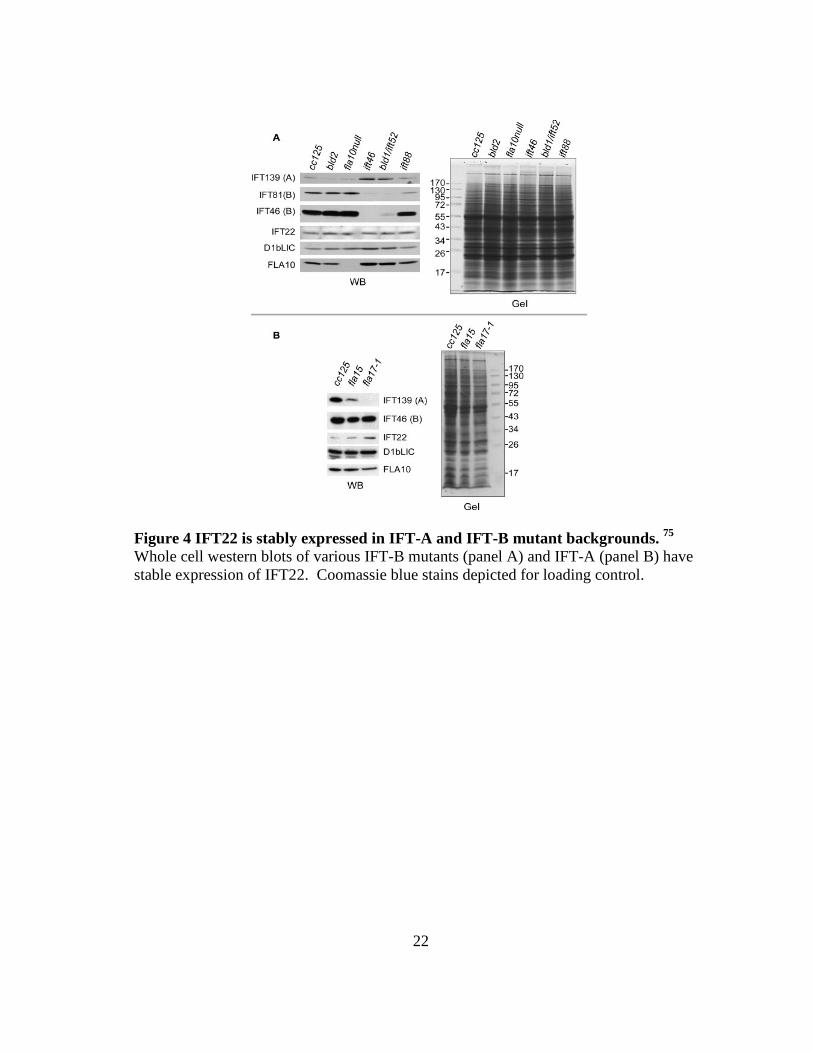
basal body; flagella-less) and fla10null demonstrate whether IFT22 levels are affected by
the absence of an intact flagella or compromised motor, respectively. IFT-B proteins
were significantly depleted in the IFT-B mutant backgrounds (Fig 4A) while IFT-A
protein IFT139 expression levels were increased. IFT139 was also significantly
depleted in fla17-1 while IFT46 remained relatively unchanged in fla15 and fla17-1
strains, mutants for IFT144 and IFT139, respectively (Fig. 4B).33 The results are
consistent with previous reports 34,43,45,65,76 and demonstrates the overall contribution of
IFT core proteins to their respective complexes and the need for all components to
properly integrate in order to yield a structurally sound IFT particle. Remarkably, IFT22
remains unchanged in the IFT-B mutant backgrounds, suggesting it does not require an
intact IFT-B complex to be stably expressed. Taken together with the IFT-A mutant
data, the results show that depletion of either IFT-A or IFT-B affects the normal
expression levels of IFT22.
22
Figure 4 IFT22 is stably expressed in IFT-A and IFT-B mutant backgrounds. 75 Whole cell western blots of various IFT-B mutants (panel A) and IFT-A (panel B) have stable expression of IFT22. Coomassie blue stains depicted for loading control.

23
CHAPTER II
THE RABL5 HOMOLOG IFT22 REGULATES THE CELLULAR POOL SIZE AND
THE DISTRIBUTION OF IFT PARTICLES INTO THE FLAGELLAR
COMPARTMENT OF CHLAYMDOMONAS REINHARDTII*
Cilia and flagella, sensory and motile structures protruding from the cell body, rely
on the continuous bidirectional traffic of intraflagellar transport (IFT) particles to ferry
precursors into flagella for assembly. Cells synthesize a large pool of IFT particle
proteins in the cell body, but only a small portion engages in active transport within the
flagella at any given time. The atypical small G protein Rab-like 5 (RABL5) has been
shown to move in an IFT-like manner in the flagella, but its function in ciliogenesis is
controversial. We previously demonstrated IFT22, the Chlamydomonas reinhardtii
homolog of RABL5, is a bona fide IFT particle complex B subunit and remains stably
expressed in IFT-B compromised mutants. In this thesis report, we report the depletion
of IFT22 leads to a smaller pool of both IFT complex A and B proteins. We observed
that a smaller cellular pool of IFT particles does not result in a reduced distribution of
IFT particles to flagella or significant defects to flagella formation. Instead, the flagellar
compartment undergoes an influx of IFT particle proteins, including IFT22 itself.
___________________
*Reprinted with permission from RABL5 homologue IFT22 regulates the cellular pool size and the amount of IFT particles distributed to the flagellar compartment in Chlamydomonas reinhardtii by Silva DA, Huang, X, Behal RH, Cole DG, Qin H. Cytoskeleton, 69 (1) 33-48 Copyright 2012 John Wiley and Sons

24
Moreover, cells over-expressing IFT22 also accumulate IFT particles in their flagella.
Our data indicates that IFT22 controls the cellular levels of both complex A and B in C.
reinhardtii, and thus plays a critical role in determining the cellular availability of IFT
particles. Although it may not directly carry any precursors for flagellar assembly,
IFT22 regulates how many IFT particles actively participate in ferrying precursors into
flagella.
This chapter will also describe the construction of two artificial microRNAs
optimized to target the non-coding 3’ unstranslated region of IFT27 and the generation
of a super-active variant of IFT22 produced through site-directed mutagenesis for in vivo
and biochemically studies. Finally, we will also discuss preliminary data suggesting
IFT25/IFT27 may function downstream of IFT22 to regulate the assembled IFT
machinery complex including the IFT motors and BBS3.
INTRODUCTION
The IFT particle research has demonstrated how IFT-A and IFT-B complexes are
distinct and structurally-independent entities within the IFT particle.43 The stability of
each complex has repeatedly been shown to depend on the proper integration of
complex-respective subunits.32,43,45,77 In addition to the self-contained regulation of each
complex, the cellular amount of complex A and B has been shown to also be regulated
concomitantly. Examples of IFT particle regulation are discussed in the IFT particle-
integrated GTPases section of Chapter 1 ( 65 and this report).

25
Flagella are long and narrow structures where protein “traffic jams” can occur
when an excessive accumulation of IFT particles or defective IFT-dynein can impede
normal IFT trafficking along the axoneme. This particular scenario is observed in
multiple retrograde-defective mutants 7,8,11,14,22,28,30,41,68,71 and RABL5 depleted T.
brucei.69 Normally, IFT particles concentrate at the peri-basal body region at the base of
the flagella78, and only a small portion of total IFT particles enter the flagella to undergo
IFT.64,79 This small, active portion of IFT particles is responsible for ferrying flagellar
precursors from the cytosol to the flagellar compartment. Since ciliary proteins are
synthesized in the cell body, and because the transition zone functions as physical barrier
between the cell body and the flagellar compartment 50-52,80, the entry of precursors is
understood to be a critical step in ciliogenesis 1,44 that must require an intuitive
regulatory mechanism to control. Currently, little is known about how the size of the
cellular pool of IFT particles is controlled or about how the number of IFT particles are
allocated to active IFT is regulated.
To gain insights into the regulatory role of RABL5 in flagellar assembly, we
have focused on the RABL5 homolog of C. reinhardtii, in which routine biochemical
analysis of flagellar proteins is relatively easy. We previously identified an IFT particle
protein IFT22 through biochemical purification and reported IFT22 as the
Chlamydomonas homolog of RABL5.64 Additionally we also determined IFT22 is an
integral component of IFT particle complex B. In this thesis, we report the partial
depletion of IFT22 specifically reduces the levels of both complex A and B proteins,

26
while the IFT motors remained relatively unaffected. In light of this, we conclude IFT22
regulates the cellular pool size of IFT polypeptides. Moreover, based on biochemical
analysis of flagella isolated from cells expressing abnormal amounts of IFT22, we
demonstrate IFT22 regulates the amount of IFT particles distributed to the flagella.
These results provide new insights into how the cell regulates the partitioning of IFT
particles between the cell body and the flagella.
RESULTS
The Cellular Pool Size of Complex A and B Are Simultaneously Reduced in IFT22
Knockdown Cells
An RNAi interference strategy was used to investigate whether depletion of
IFT22 affects cellular levels of either IFT-A or IFT-B proteins in Chlamydomonas.
Three constructs were designed and introduced into cc125 wild-type cells: two
constructs expressed long, double-stranded RNA targeting the either the coding region
or the noncoding 3’ untranslated region (UTR) of IFT22 (Xiaomeng Huang,
unpublished). The third construct was designed as an artificial microRNA (amiRNA),
optimized to target a sequence near the translation initiation site of IFT22 mRNA.
Whole-cell extracts of transformants were prepared and analyzed for depletion of IFT22
by immunoblotting; each construct was transformed and screened four times, yielding
over 600 individual transformants. Resulting immunoblots revealed the construct
targeting the coding region of IFT22 was non-efficient, though the amiRNA and dsRNA
with the 3’UTR target were highly effective in reducing the expression of IFT22.
27
Approximately 10% of the transformants achieved a 50% or greater reduction of IFT22
cellular levels. The knockdown effect of the amiRNA was more reproducible and stably
maintained compared to transformants harboring the IFT22 3’UTR dsRNAi, and thus
was selected as the primary RNAi construct for the remainder of the study.
Transformants with an approximate 80% were selected for further analysis to determine
the cellular condition of IFT particle proteins.
The cellular levels of IFT particle proteins and IFT motor subunits were
measured by immunoblotting whole-cell extracts of IFT22 knockdown transformants.
IFT particle proteins from both IFT-A and IFT-B sub-complexes exhibited a reduction in
amiRNA knockdown cells, though they were observed to decrease to different extents
(Figs. 5 A1 and 6A, results on the strain b8-46 in Fig. 8A). Retrograde IFT motor
subunit D1bLIC was slightly reduced while the protein levels of anterograde IFT motor
subunit FLA10 remained unaffected at the cellular level. The approximate extent of
IFT protein depletion was determined by measuring immunoblot band intensity relative
to wild-type protein extracts (Fig. 5A3). The reduction of both complex A and complex
B proteins in the IFT22 knockdown cells indicated that IFT22 is essential in maintaining
the normal cellular levels of IFT particle polypeptides.
Knockdown of IFT22 Leads to an Influx of IFT Particles into the Flagella To examine the effect of IFT22 depletion on flagellar assembly, we
measured the length of flagella in IFT22-knockdown strains (Fig. 5B-B1). No gross
differences in flagellar morphology were observed. Strains b8-8 and b8-48 had normal

28
Figure 5 Knockdown of IFT22 depletes the cellular pool of IFT particles and increases the distribution of IFT proteins into the flagellar compartment. Depletion of IFT22 leads to a smaller cellular pool of complex A and B (panel A), but an influx of IFT particles to flagella (panel B). (A1 and A2) Immunoblot analysis of whole-cell extracts showed IFT22-knockdown cells had reduced levels of IFT particle subunits, slightly reduced D1bLIC, and unchanged FLA10. (A3) Band intensities of the IFT particle proteins and IFT motors in the IFT22 knockdown strains from panel A1 were plotted as a percent of wild-type cells. (B1) Average flagella lengths of cells from wild-type cc125 and IFT22-knockdown strains were compared. The mean length and the number of flagella measured are noted. (B2 and B3) The IFT22- knockdown cells accumulate IFT particle proteins, but not the motor proteins, in their flagella. Antibodies used against IFT proteins are as noted (A1 and B2). Equal loading of the samples is shown by the Coomassie Blue-stained gel in panel A2 and B3.

29
differences in flagellar morphology were observed. Strains b8-8 and b8-48 had normal
flagella length though the flagella from the strain b8-17, were slightly shorter than wild-
type (Fig. B-B1, b8-17 mean length = 9.302 μm; wt = 10.94 μm, p < 0.001).
To address whether the knockdown of IFT22 led to a reduced distribution of IFT
particles to flagella, we checked the levels of IFT particles and motors in flagella
isolated from the three IFT22-knockdown strains. Strikingly, the flagellar amount of
IFT particles was not reduced. Levels of complex A proteins, complex B proteins, and
IFT22 were increased in the flagella of IFT22 knockdown cells, (Figs. 5B-B2, 5B-B3,
and 8A). The flagella of b8-17 were shorter than normal and experienced the greatest
increase of IFT particles. The anterograde motor subunit FLA10 remained at the wild-
type level and the retrograde motor subunit D1bLIC was slightly reduced (Fig. 5B-B2),
or were unchanged (Fig. A). Collectively, these data showed that when the normal
cellular expression of IFT22 is inhibited, more IFT particles, but not IFT motors, were
distributed to the flagella compartment. The accumulation of flagellar IFT particles in
IFT22-depleted cells is consistent with previous observations in RABL5-depleted T.
brucei 69, suggesting IFT22 plays a conserved role in flagellar assembly.
IFT22 Over-Expression Cells Contain Increased Amounts of IFT Particle Proteins
in Both Cellular and Flagellar Compartments
Unexpectedly, we observed an approximate 50% over-expression of IFT22 in
130 transformants generated via amiRNA (Fig. 6). To understand how the cells respond
to a surplus of IFT22, we selected a few cell lines to analyze in detail (Fig. 6A).

30
Figure 6 Over-expression of IFT22 increases cellular and flagellar levels of IFT particle proteins. The whole-cell extracts (panel A) and flagella extracts (panel B) from the IFT22 over-expression strains were analyzed by immunoblot. (A1) Whole-cell extracts from cells of wild-type (cc125), over-expression strains (b8-7, b8-34, b8-41, b8-47, b8-57, and b8-60), and the knockdown strain (b8-46). (B1) The IFT22 over-expression cells accumulated IFT particle proteins in their flagella. Whole-cell and flagellar extracts from the over-expression strains were analyzed by immunoblotting proteins using noted antibodies. Equal loading of the samples is shown by the Coomassie Blue-stained gel in panel A2 and B2.

31
Immunoblot analysis of whole-cell extracts showed that the IFT22 over-expression
strains had increased cellular levels of complex A protein IFT139. The amount of the
IFT-dynein subunit D1bLIC was either unchanged or slightly reduced. Increased levels
of complex B and the anterograde motor subunit FLA10 were also observed but were
only prominent in strain b8-34, which experienced the most significant increase in IFT22
expression. These results indicated that over-expression of IFT22 upregulates the
expression of IFT particle proteins. Moreover, this regulation affects IFT particle
complexes A and B, and the IFT motor kinesin FLA10, but does not influence the
expression of IFT-dynein.
The levels of IFT proteins were also measured in flagella isolated from several
IFT22 over-expression strains (Fig. 6B). No obvious changes of flagellar IFT proteins
were found in with strain b8-57, which only had a mild over-expression of IFT particles
(Fig. 6A). However, a significant increase of IFT particle proteins were observed in the
flagella isolated from both b8-34 and b8-47 strains. This was especially true for the b8-
34 strain which also displayed the highest levels of whole cell IFT particle proteins (Fig.
6A). Despite the dramatic changes to IFT particle proteins amounts, no change was
detected for the IFT-dynein subunit D1bLIC, and only a mild increase was seen for
FLA10. Thus, IFT22 over-expression increased the level of flagellar IFT particles
within the cell body, but not IFT-dynein.
We also examined the flagella morphology of the over-expression strains under
the microscope (Fig. 7). The flagella lengths of the strains b8-57 and b8-47, which had

32
moderate over-expression of IFT22, were normal. Severe flagellar defects were observed
in strain b8-34, which experienced the highest increase in IFT particles at both the
cellular and the flagellar levels. The defects included short flagella and unequal lengths
of a cell’s two flagella (Fig. 7A). A close examination revealed that the short flagella
were often swollen and formed membrane bulges along the lateral membrane and at the
tip of the flagella (Fig. 7A). Similar membrane bulges, previously observed in the
flagella of retrograde IFT mutants, were found to be due to aggregation of IFT
particles.7,14,28,32 Though not experimentally established, these aggregates are thought to
jam the IFT traffic, block the precursor assembly site, ultimately inhibiting flagellar
assembly.7 The flagellar assembly defects in the strain b8-34 are likely caused by a
similar process. However, since b8-34 was the only strain overexpressing IFT22 several
folds, it remains unclear whether such a dramatic overexpression was solely due to the
effect of IFT22 amiRNA. It should also be noted that the b8-34 strain was the only one
among the overexpression strains that displayed a flagellar assembly defect, thus the
significance of IFT22 overexpression on flagellar assembly needs to be verified.
IFT22 Artificial miRNA Acts at the Translational Level
The sequence of the amiRNA of IFT22 targets the site of translation initiation,
likely blocking translation initiation without triggering mRNA cleavage and decay.81-84
To check if the artificial miRNA acts at the level of translation or of mRNA transcription
or stability, semi-quantitative RT-PCR was performed on both the IFT22

33
Figure 7 Cells with several fold increase of IFT22 have severely shortened flagella fill with IFT particle proteins. (A) DIC images of wild-type cc125 cells and b8-34 cells. The flagella of b8-34 cells were shorter than normal. Unequal lengths of two flagella were common in the population of b8-34 liquid cultures. Membrane bulges (indicated as arrows) were also common at the flagellar tip or along the flagella. Scale bar, 5 μm. (B) Histograms showing the length distribution of flagella from wild-type cc125, and the over-expression strain b8-34 and b8-47 cells. The b8-34 strain, which had several-fold increase of IFT22, had shorter flagella (mean length = 5.018 μm, n = 54) compared to the cc125 (10.72 μm, n = 51) and the b8-47 (10.39 μm, n = 51) cells.

34
knockdown strains and the over-expression strains. The IFT22 knockdown strains b8-17,
b8-46, which contained reduced cellular and elevated flagellar levels of IFT particle
proteins (Figs. 5, and 6), had increased or unchanged levels of IFT22 and IFT27 mRNA
compared to that of wild-type cells (Figs. 8). The over-expression strain b8-34 had
increased levels of IFT22 and IFT27 mRNA (Fig. 8). These data indicated that, in the
case of IFT22, the repression via amiRNA was exerted at the step of translation instead
of transcription, without major degradation of mRNA. The inhibition of translation
likely induced a negative feedback that upregulated the production of IFT22 mRNA. The
total level of IFT22 protein was therefore determined by the combined effects of miRNA
inhibition and upregulation of transcription. Indeed, both silencing and over-expression
effects were observed among the IFT22 miRNA transgenic lines.
Constitutive Active IFT22 Enters the Flagella Compartment
In order to establish the impact the predicted GTPase activity has on the
trafficking of IFT22 into and out of the flagella compartment, several mutations were
introduced into IFT22 to generate putative constitutive active and dominant negative
(Xiaomeng Huang, unpublished data). The constitutive active variant was generated by
mutating the P-loop glutamine at the 14th amino acid to a valine (Q14V), and the
threonine at the 19th residue was replaced by asparagine (T19N) to yield the dominant
negative form of IFT22. Additionally, each variant along with a wild-type IFT22 were
supplemented with a HA and GFP tag in order to effectively detect the localization of

35
Figure 8 Transcriptional levels of ift22 and ift27 amiRNA cell lines. Semi-quantitative RT-PCR was performed with RNA isolated from wild-type cc125 and IFT22 amiRNA strains. DNA fragments of genes encoding IFT22, IFT27, and the β subunit of GBLP were amplified.

36
the IFT22 variants within the cell extracts. All three constructs were individually
transformed into a cw92 background, a mutant with a cell-wall defect in C. reinhardtii,
permitting the use of a simpler transformation procedure. Over 50 transformants per
variant were isolated following selection with varying degrees of transgenic IFT22
expression. Of those, a small group of representative transformants for each construct
were selected for further testing.
In order to determine if the IFT22-Q14V and IFT22-T19N could successfully
enter the flagella, cell body and flagella extracts were prepared and analyzed using
immunoblotting. The IFT22:HA:GFP construct was detected in the flagella extract,
suggesting the large GFP tag does not inhibit the trafficking of the transgenic IFT22
(Fig. 9). Much like the C. elegans homologue IFTA-2 68, IFT22-Q14V was detected in
the flagella extract, thus confirming its entry the flagella (Fig. 9A). Compared to the
wild-type IFT2, the constitutive variant had a smaller percentage of total expressed
transgenic protein entering the flagella (Fig. 9A). To further verify the entry of the
constitutive active IFT22, selected transformants were subjected to immunofluorescence.
IFT22-Q14V properly localized to the basal body of the cw92, confirming its normal
recruitment to the base of the flagella is not impaired (Fig. 9). Sporadic localization was
observed throughout the axoneme, suggesting it may remain associated with the IFT
particle when trafficking within the flagella compartment. Despite multiple
transformations, the dominant negative variant would not sufficiently and consistently

37
Figure 9 IFT22 enters the flagellar compartment. IFT-GTP enters the flagellar compartment. Panel A contains immunoblots of whole cell and flagella extracts from selected transformants expressing IFT22-WT:HA:GFP and IFT22-Q14V:HA:GFP, denoted as WT and Q14V, respectively. A lower percentage of the GTP-locked IFT22 were shown to enter the flagella compared to the wild-type IFT22. Panel B shows the equal loading control of whole cell and flagella extracts using a Coomassie-Blue stain.

38
Figure 10 Transgenic IFT22 and IFT22-GTP co-localize with IFT46 at the peri-basal body and flagellar compartment. The wild-type cw92 cells were stained with anti-HA and anti-IFT46 antibodies. Both IFT22:HA:GFP and IFT22 (Q14V) HA:GFP localize to peri-basal body region concomitantly with IFT46. It should be noted that the reason for the high concentration of transgenic IFT22 and constitutive active IFT22 in the cytoplasm is unknown. Bar, 10 μm

39
express in cw92 to permit the same biochemical verification procedures; as a result,
further testing was not possible.
Both IFT27 and IFT22 have a serine residue on the 79th and 67th amino acid,
respectively, in place of the canonical glutamine residue. Although GTPases have
evolved multiple methods to achieving GTP hydrolysis, the glutamine to serine mutation
in Rab-like proteins IFT27 and IFT22 was predicted to yield a lower GTP turnover rate.
A recent study restored the canonical glutamine residue of IFT27 and observed a
significant increase in GTP hydrolysis.66 A super-active variant of IFT22 was
constructed to be introduced into C. reinhardtii to observe its localization patterns and
behavior in vivo. A mutation was introduced into IFT22 at S67Q and supplemented
with an HA tag for quick identification of successful transformants. Additionally, two
constructs containing the IFT22 (S67Q):HA insertion were designed to add either a GFP
for immunofluorescence and total internal reflection fluorescence (TIRF) microscopy
experiments or His6 for future large scale purification. Additionally, paromomycin and
bleomycin resistance markers were added to the GFP and His-tagged IFT22-S67Q
construct, respectively. Generated plasmids were named as follows:
pBluescriptIIKS+ift22 (S67Q): HA:GFP-PMM and
pBluescriptIIKS+ift22(S67Q):HA:His6-Ble.
Knockdown of IFT25 Results in Cytoplasmic Depletion of BBS3 and IFT motors
A previous study and this thesis work observed the depletion of IFT-A and IFT-B
proteins following the knockdown of IFT27 65 and IFT22 in addition to their stable

40
Figure 11 Depletion of IFT25 leads to the depletion of IFT motors and BBS3. Panel A is a whole cell extract immunoblot of IFT25 knockdown transformants with a range of IFT25 expression. Four transformants (panel A; 1-44, 1-63, 1-78, and 1-98) with lowest residual expression of IFT25 showed depleted levels of FLA10, D1bLIC and BBS3 (Panel C). Equal loading of the samples is shown by the Coomassie Blue-stained gel in Panel B.
A B
C

41
expression in IFT-B mutant backgrounds. In an effort to determine whether IFT27
functions downstream of IFT22 or vice versa, an RNAi construct targeting the 3’UTR of
IFT25 (Zhenchuan Fan, unpublished data), the binding partner of IFT27, was generated
and transformed into cc125 cells. Unfortunately, the results proved to be inconclusive as
IFT22 protein levels would not follow a consistent trend in 25RNAi transformants (data
not shown). However, selected transformants with an approximate 80% reduction of
IFT25 (Fig 11A) replicating the results observed following knockdown of IFT27.65
Surprisingly, BBSome subunit BBS3, kinesin-II subunit FLA10, and retrograde dynein
subunit D1bLIC protein levels were also significantly depleted from the cell body (Fig.
11C). No significant defects were observed in the flagella of selected transformants with
the highest level of IFT25 knockdown (data not shown). To verify the results, two
artificial microRNAs were generated and optimized to target different regions of the
IFT27 3’UTR using the same procedure employed in the building of the IFT22 artificial
microRNA. Two constructs were generated and named as follows:
pChlamiRNA3intIFT27-1 and pChlamiRNA3intIFT27-2.
DISCUSSION
Cellular Pool Regulation of IFT Particle Proteins
The C. reinharditii cell maintains a large pool of IFT particle proteins in the cell
body79, and only a small portion of the pool participates in active IFT within the flagella
at any given time.5 The pool size of complex B is approximately ten times larger than
that of complex A 44, despite the fact that the assembled IFT particle from flagella has a

42
1:1 stoichiometry between complex A and complex B proteins. The difference in the
pool size of complex A and B is thought to reflect the non-overlapping functions of these
two complexes.64 In this study, we show that IFT22 regulates the cellular pool of IFT
particles, as reduced cellular levels of both complex A and B proteins are observed in
IFT22 RNAi knockdown cells in C. reinhardtii. Thus, IFT22 is required to maintain the
pool size of the IFT particle proteins, which in turn determines the availability of IFT
particles.
This study describes an IFT22 over-expression strain which expresses IFT22
protein at levels several folds greater than normal (or wild-type) that also results in
increases in complexes A and B and in FLA10. FLA10 is present at wild-type levels in
various IFT particle mutants, including IFT22-knockdown strains, which all have
reduced levels of IFT particles. Therefore, the cell has an apparent baseline level of
anterograde motor FLA10, which is regulated independently of IFT particle production.
The observation that FLA10 accumulates along with IFT particles upon over-expression
of IFT22 suggests FLA10 can be positively regulated by responding to the surplus in the
IFT-particle pool. In contrast, the expression of the retrograde motor IFT-dynein is
insensitive to the changes of the cellular amount of IFT particles. The mechanism
controlling the expression of IFT-dynein appears to be at least partially independent
from control of IFT particle and FLA10 expression.

43
IFT22 Regulates Amount of IFT Particles Distributed to the Flagellar
Compartment
Normally, the majority of IFT particles concentrate around the basal body and on
the cytoplasmic side of the transition zone. As previous studies have shown, the cell
only allocates approximately 20% of complex A and 2% of complex B to participate in
active IFT transport within the flagella.64,79 We observed partial depletion of IFT22
causes a decrease in the cytoplasmic pool of IFT particles. However, the absolute levels
IFT particle proteins including IFT22 itself, actually increase within the flagella
compartment. This build-up did not extend to IFT motors as both the anterograde motor
subunit FLA10 and the retrograde IFT motor subunit D1bLIC displayed a normal
distribution ratio between the cell body and the flagella. These results indicate that
IFT22 is involved in regulating the specific distribution of IFT particles to the flagella.
The exact role of IFT22 in regulating the distribution of IFT particles remains
unclear. Since the changes of IFT22 expression level also alter the cellular level of IFT
particles, it is possible that the up-regulation of IFT A and B complex in the flagella in
the IFT22 knockdown cells is simply due to the disruption of IFT particles. It is also
possible that IFT22 is specifically involved in regulating the distribution of IFT particles.
Nonetheless, the absolute amount of IFT22 cannot be the determining factor in the
distribution of IFT particles, as both over-expression and depletion generate increased
flagellar localization. We hypothesize that because IFT22 is a small GTPase, the

44
distribution or availability of IFT particles for IFT transport could potentially be
determined by the ratio between the GTP-bound and GDP-bound forms of IFT22.
The C. elegans homolog of IFT22, when in the GDP-bound form, is excluded
from the ciliary compartment, indicating that only the GTP-bound form is capable of
entering cilia.68 We also duplicated this result by biochemically confirming the presence
of the constitutive active IFT22-Q14V within prepared flagella extracts. Since we
observe a smaller percentage of the constitutive active variant detected in the flagella
compartment, it is possible the endogenous IFT22 out-competes the transgenic IFT22-
GTP for binding sites on IFT complex B. It is also possible that disrupting the normal
GTP-GDP cycling of IFT22 impairs ability of the IFT particle/IFT-GTP locked complex
to efficiently traffic into the flagella. Additionally, we also observed puncta localization
throughout the axoneme, suggesting IFT22-GTP remains bound to the IFT particle while
undergoing IFT within the flagellar compartment. Performing a sucrose density gradient
of cell lines expressing the transgenic the constitutive active, dominant negative and
super-active variants of IFT22 would definitively confirm whether the nucleotide status
of IFT2 dictates its association with the IFT particle both in the cell body and the
flagellar compartment.
Based on the data of previous studies and our own experiments, we hypothesize
that the ratio between the GTP-bound versus GDP-bound IFT22 in the cell body is
tightly controlled. The cell maintains this ratio by adjusting the number of IFT particles
containing GTP-bound IFT22, which are the only version capable of entering the

45
flagellar compartment. Normally, only a small fraction of IFT particles would enter the
flagella. However, as the ratio between the GTP-bound and GDP-bound IFT22
increases, the cell responds by sending additional IFT particles to the flagellar
compartment in order to maintain. Increased amounts of flagellar IFT particles have
been observed in IFT22-knockdown cells in C. reinhardtii (this thesis), T. brucei 69 and
in the IFT22-over-expression cells in C. reinhardtii (this report). Based on our
hypothesis, the ratio between the GTP-bound and GDP-bound IFT22 should increase in
cells that express abnormally low or high amounts of IFT22.
This hypothesis also provides a reasonable explanation for the observation that
the C. elegans IFTA-2 null mutant does not display an obvious defect in ciliogenesis or
IFT 68 but rather defects in specific signaling activities within the cilia. The absence of
IFTA-2 may alter the distribution of IFT particles to the cilia, which in turn could
change the levels of some ciliary signaling molecules. However, such changes in the
numbers of IFT particles would not have to disrupt the balance of anterograde and
retrograde IFT to such a degree as to cause an obvious defect in ciliary assembly. Future
research will be needed to establish the effect of GTP-bound and GDP-bound IFT22 on
localization of IFT particles to the cilia and flagella.
IFT25 May Regulate the Assembled IFT Particle/Motor/BBSome Complex
Downstream of IFT22
This work and a previous study 65 have shown that the depletion of both IFT22
and IFT27 results in a reduction of both IFT-A and IFT-B complex proteins. Our over-
expression and knockdown of IFT22 significantly depleted the expression levels of IFT

46
particle proteins while the protein levels of kinesin-2 and IFT dynein-2 subunits were
comparatively unaffected. Conversely, our knockdown of IFT25, the stabilizing partner
of IFT27, resulted in the depletion of not only IFT particle proteins, but also both the
retrograde and anterograde motors along with BBSome subunit BBS3. Previous studies
have suggested IFT25/27 could be involved in the regulating the entry of the IFT
machinery in the flagella compartment64, though its impact on ciliogenesis may be
species-dependent. Null IFT25 mutant mice were non-viable but tested positive for cilia
formation; defects in Hedge-hog signaling were determined to be the cause of death.85
Interestingly enough, the source of the signaling defects was the abolished trafficking of
notable Hedgehog proteins including Patched-1, Smoothened, and Gli2 85, suggesting
IFT25 is also involved in the trafficking of ciliary membrane proteins. BBS3 itself does
not contribute to the structural integrity of the assembled BBSome, however it is
important for the recruitment of the BBSome to primary cilia.86 Currently the only
known direct interaction between the IFT particle and the BBSome is between C.
elegans DYF-2, IFT-A protein IFT144 in C. reinhardtii, and BBsome core protein
BBS1.87
Taken together, the data suggest IFT25 may be directly or indirectly involved in
regulating the assembled IFT particle/motor/BBsome complex or the interaction between
the BBSome and the IFT particle. Biochemical pull-down assays and direct interaction
assays between IFT25 and the BBSome would definitively reveal if IFT25 serves as
bridge between the BBSome and the IFT particle. It is tempting to speculate the recently

47
confirmed GTPase activity of IFT27 could serve as the regulatory trigger to control the
assembly or loading of the entire IFT particle/motor/BBSome complex prior to the entry
of assembled complex into the flagella.
MATERIALS AND METHODS
Strains and Culture Conditions
Chlamydomonas reinhardtii wild-type strain cc125, mutant strains, ift88 (CC-
3943) 45, ift46 43, bld1/ift52 (CC-477) 76, fla10 null (CC-4180) 21, bld2 (CC478) 88,
temperature-sensitive (ts) flagella assembly mutants fla10ts (fla10-1 allele, CC-1919),
fla15 (CC-3861), and fla17-1 (CC-3862) 32,33,70 were obtained from the Chlamydomonas
center (http://www.chlamy.org). The ts mutants were cultured on Tris-acetate-phosphate
(TAP) solid plates or in M1 liquid media with constant aeration in a Conviron
programmed at 18°C with a light–dark cycle of 14:10 h. Other strains, if not otherwise
specified, were grown on TAP solid plates or in TAP liquid media at 22 °C in
continuous light with constant aeration.
Antibodies
Bacterial-expressed N-terminal His6-tagged full length IFT22 recombinant
protein (pTrcHis vector, Invitrogen) was used as the antigen for polyclonal antibody
production in rabbits (The Woodlands, TX). Aliquots of immune serum were tested
against blots of diluted recombinant IFT22 and sucrose gradient-purified IFT proteins to
verify the avidity and specificity of the serum. Anti-IFT22 antisera were affinity purified
from the GST-IFT22 proteins bound to the nitrocellulose membrane (Protran BA83, 0.2

48
mm, Whatman, Dassel, Germany). GST-IFT22 full-length fusion protein was made from
the pGEX-2T vector (GE healthcare), and purified before being used for purification.
Other antibodies used in this study include antibodies against -tubulin (clone B-5-1-2,
ascites fluid; Sigma), IC69 (clone 1869A; Sigma), Antibodies against C. reinhardtii
proteins IFT25 64, IFT27 65, IFT139, IFT172, IFT81 5, IFT72 35, IFT46 43, FLA10 5,
DHC1b 8 and D1bLIC 28were also previously reported. Polyclonal antisera were raised
against Chlamydomonas IFT122 in rabbit and will be described at a later time.
Plasmid Construction and Chlamydomonas Transformation
The miRNA construct pChlamiRNA3int-ift22 was created as detailed
previously.89 The target sequence of ift22 was scanned via http://wmd2.weigelworld/cgi-
bin/mirnatools.pl for potential artificial miRNA target sites, and a 21-bp sequence
(TCTATCTTCAACCCGTGCTGT) targeting the ift22 exon 1, the sequence right after
the translation initiation codon, was chosen for further study. Two 90-mer
oligonucleotides (ift22-ami-forward: 5′ ctagtACAGCACGGGTTGAAGTTA
GAtctcgctgatcggcaccatgggggtggtggtgatcagcgctaTCTATCTTCAACCCGTGCTGTg 3′
and ift22-ami-reverse: 5′
ctagcACAGCACGGGTTGAAGATAGAtagcgctgatcaccaccacccccatggtgccgatcagcgagaT
CTAACTTCAACCCGTGCTGTa 3′, Invitrogen) containing the targeted sequence in
opposite directions separated by a 42-mer spacer sequence (lower case) were annealed in
vitro and treated with T4 polynucleotide kinase (PNK, Fermentas) for phosphorylation.
The miRNA vector pchlamiRNA3int was digested with SpeI (Fermentas) followed with

49
CIAP (Fermentas) treatment for dephosphorylation. The 90-bp DNA oligo was digested
with SpeI and inserted into the pchlamiRNA3int vector. The correct miRNA colony was
screened by nucleotide sequencing.
The construction of the IFT27 miRNA followed the same procedure denoted
above; two 21-bp sequences located in the 3’UTR of IFT27, 5’
TATCATTACATAAGCAGACAC 3’ and 5’ TCTTAGACCGGGATCTCTCAT3,’were
designated as optimal targeting sites for the miRNA construct. The following pairs of
90-mer oligo nucleotides were used to generate constructs pChlamiRNA3int-ami27-1
and pChlamiRNA3int-ami27-2: ( ami27-For_1 5'
ctagtGTGTCTGCTTATGTAAAGATAtctcgctgatcggcaccatgggggtggtggtgatcagcgctaTA
TCATTACATAAGCAGACACg 3' and ami27Rev_1 5'
ctagcGTGTCTGCTTATGTAATGATAtagcgctgatcaccaccacccccatggtgccgatcagcgagaT
ATCTTTACATAAGCAGACACa 3); the second pair consists of (ami27For_2
5'ctagtATGAGAGATCCCGGTCAAAGAtctcgctgatcggcaccatgggggtggtggtgatcagcgcta
TCTTAGACCGGGATCTCTCATg 3' and ami27Rev_2
5'ctagcATGAGAGATCCCGGTCTAAGAtagcgctgatcaccaccacccccatggtgccgatcagcgaga
TCTTTGACCGGGATCTCTCATa 3', Invitrogen).
Constructs, pBluescriptIIKS+-ift22 (QV) HA- GFP-PMM and pBluescriptIIKS+-
ift22 (TN) HA- GFP-PMM were generated prior to the initiation of this thesis work
(Xiaomeng Huang, unpublished data). The construction of the pBluescriptIIKS+ift22
(S67Q)HA:GFP-PMM and pBluescriptIIKS+ift22 (S67Q)HA:His6 consisted of excising

50
full gene of IFT22, including endogenous promoter, coding region plus a HA tag, from
the pBluescriptIIKS+-ift22 HA- GFP-PMM using a double digest reaction of Not1 and
EcoR1. The IFT22:HA fragment was then inserted into a pGEM vector also digested by
prepared by the Not1 and EcoR1, thereby forming the construct pGEM ift22:HA. The
S67Q mutation was introduced using the QuikChange Site-Directed Mutagenesis Kit
(Stratagene) according to the manufacturer’s instructions with the following parameters:
16 cycles; 95ºC (30secs); 55ºC (1min); 68ºC (11min); 4ºC (storage). Mutagenesis was
carried out using primers ift22-S67Q-F (5’ - GGA CGT GTC GGG GCA GGT GCA
GTA CCA G - 3’) and ift22-S67Q –R (5’ - CTG GTA CTG CAC CTG CCC CGA CAC
GTC C – 3’); following mutagenesis, the resulting construct was sequenced and
confirmed for production of pGEM-ift22(S67Q):HA. The ift22 containing the super-
active mutation plus HA tag were excised from pGEM-ift22(S67Q):HA using a
Not1/EcorR1 double digest and subjected to a secondary digest of Sca1 to break the
pGEM vector. The ift22(S67Q):HA fragment was isolated by gel purification and
inserted into Not1/EcoR1 prepared and isolated pBluescriptIIKS+ -GFP-PMM and
pBluescriptIIKS+ His6-Ble vectors. The resulting constructs containing a GFP and
His6-tagged IFT22-S67Q variant were named as follows:
pBluescripIIKS+ift22(S67Q):HA:GFP-PMM and
pBluescripIIKS+ift22(S67Q):HA:His6-Ble, respectively.

51
Whole-Cell Extracts Sample Preparation for Direct Electrophoresis Analysis
When only small samples were needed, extracts were prepared from 1 ml of cells
grown to log phase in TAP medium. After removing supernatant, the cell pellets were
resuspended with 54 μl of Buffer A (0.1M Na2CO3), 6 μl of DTT and 40 μl of Buffer B
(5% SDS, 30% sucrose), followed by vortexing for 45 min at 20 °C. Samples were spun
down at 10,000 rpm for 5 min, and the supernatant was collected. 50 μl of each protein
sample was mixed with 40 μl of water and 10 μl of 10X Laemmli buffer. The samples
were mixed well by vortexing for 10 sec. Samples were then boiled for 3 min, followed
by a brief spin, before loading for SDS-PAGE. When larger amounts of protein were
needed, the sample size and the extraction solution were increased proportionally. The
protein concentration of the samples was first measured by the Amino Black method, as
previously described 65, and then adjusted based on the total intensity of the lanes stained
by Coomassie Blue measured using the Image Lab™ Software (Bio-Rad).
SDS-PAGE and Immunoblotting Assay
Protein samples in 1 × Laemmli buffer were separated by 10% SDS-PAGE and
then electrotransferred onto a nitrocellulose membrane (Protran BA83, 0.2 mm,
Whatman, Dassel, Germany). Before immunoblotting, the membrane was stained with
Ponceau S to ensure that proteins were properly transferred. Thereafter, the membrane
was blocked with 5% nonfat dry milk in TBS (10 mM Tris, pH 7.5, 166 mM NaCl) plus
0.05%Tween-20. Primary antibodies were diluted in the blocking solution and then
incubated with the membrane overnight at 4°C. After washing three times with TBS plus

52
0.05% Tween-20, horseradish peroxidase-conjugated secondary antibodies (Pierce
Biotechnology, Rockford, IL) were incubated with the membrane for 1 h followed by
washing three times with TBS plus 0.05% Tween-20. Chemiluminescence was used to
detect the primary antibodies. The intensities of the immunoblot bands were quantified
by the Image Lab™ Software (Bio-Rad).
RNA Isolation and Reverse Transcriptase (RT)-PCR Analysis
Total cell RNA from wild-type cc125 and amiRNA cells was purified with
TRIzol (Invitrogen) according to the manufacturer's instructions followed by DNase I
(RNase-free) (Fermentas) treatment to remove contaminating DNA. RNA samples were
purified by RNeasy Mini Kit (QIAGEN), and the concentration was determined by
NanoDrop. Reverse transcription reactions were carried out with equal amounts of RNA
from each sample using SuperScript III Reverse Transcriptase (Invitrogen), according to
the manufacturer's instructions, with oligo (dT)20. By using the resulting cDNA as the
template, fragments containing sequences from the genes encoding IFT22, IFT27, and
the beta subunit of G protein-like protein (GBLP), which was used as a control for equal
amounts of input RNA, were amplified by PCR. A fragment of 118 bp containing exon 4
and 5 of the ift22 sequence was amplified with the primer IFT22qPF1 (5′-CAGT
CTTACGTGGCCATCAA-3′) and primer IFT22qPR1 (5′-
GGACGTCCAAATGCGAGTAT-3′). A 110 bp fragment containing sequence from
ift27 was amplified with the primer 27QPF2 (5′-TCTCGGTGGAGCTC TTTCTG-3′)
and primer27QPR3 (5′-GCTCACA TCGAACACGAGAA-3′). A 141 bp fragment of

53
containing sequence from gblp was amplified with the primer GBLP3 (5′-
GTCATCCACTGCCTGTGCTTCT-3′) and primer GBLP4 (5′-GGC
CTTCTTGCTGGTGATGTT-3′). Five microliter of PCR product from each sample was
used for 0.8% agarose gel electrophoresis.
Microscopy and Phenotype Assay
Wild-type cc125 and IFT22 amiRNA cells were cultured in TAP liquid media
and grown to log phase. A hundred microliters of cells were taken at the same time of
the same day and then fixed by 1 × Lugol's iodine solution. Following the fixation,
preparations were quickly photographed by the Olympus IX70 Inverted Microscope and
Simple PCI Imaging System at 100× magnification. The lengths of flagella from 134–
164 randomly selected cells from each strain were measured using ImageJ
(http://rsbweb.nih.gov/ij/). Student's t test analysis of length data was performed using
JMP®, Version 9.0., SAS Institute, Cary, NC, 2010. The data was processed with
GraphPad Prism 5 (GraphPad Software), and flagellar lengths are presented as mean
value (μm).
Immunofluoresent Microscopy
The same method described previously 90 was used. The primary antibody -HA
was purchased from Roche and used at a 1:25 dilution, IFT46 was used at a 1:200
dilution, the secondary antibodies were chosen according to the suggestions of the
manufacturer. An Olympus IX-70 inverted fluorescence microscope at 100×

54
magnification was used to observe the staining and the images were captured by the PCI
software package through an Image Point CCD camera (Photometrics)

55
CHAPTER III
SUMMARY AND CONCLUSION
The purpose of my thesis project was to investigate the mechanisms involved in
regulating the entry/exit of IFT particles from the flagellar compartment, and how these
mechanisms contribute to ciliogenesis.
In Chapter II, I demonstrated how depleting or over-expressing IFT22 in C.
reinhardtii using an artificial microRNA resulted in a reduction or increase of the
available cellular pool of IFT particles, respectively. Surprisingly, both the depletion
and over-expression of IFT22 caused an influx of IFT particles into the flagella
compartment. Using a transgenic IFT22 mutant, I showed that the IFT22 GTP-bound
properly localized to the peri-basal body, enter the flagella and is likely to remain
associated with the IFT particle while undergoing normal IFT trafficking in the flagella.
Additionally, I demonstrated how the knockdown of IFT25 using RNAi resulted in a
reduction of the cellular amount of BBsome subunit BBS3, kinesin-II subunit FLA10
and IFT dynein-2 subunit D1bLIC. Taken together, the data suggests IFT22 and IFT25,
possibly in conjunction with IFT27, function in a sequential regulatory mechanism
where IFT22 specifically regulates the availability of IFT particle for IFT trafficking and
IFT25/IFT27 may directly or indirectly regulate the fully assembled IFT machinery prior
to its entry into the flagellar compartment.

56
This thesis provides valuable insight into a potentially novel mechanism involved
in regulating the sequential assembly or loading of IFT particle/motor/BBSome complex
prior to the entry of IFT machinery into the flagellar compartment.
FUTURE DIRECTIONS
The IFT and cilia field in general has experienced a surge in interest after it was
discovered that human genetic diseases, now collectively known as ciliopathies, are
linked to defects in ciliogenesis. There are still many unexplored questions in the IFT
community and thus many areas that could benefit from further study. In this section, I
will address future directions that could use my work as a foundation for studies
investigating regulatory mechanisms involved in the maintenance and trafficking of the
IFT machinery. A recent study confirmed the GTP binding and GTPase activity of
IFT27, an integrated protein of the IFT particle.66 In the same light, confirming GTPase
enzyme activity of IFT22 would be beneficial to understanding the function of IFT22.
In the same study, researchers restored a canonical glutamine important for enzyme
activity that is mutated to a serine in IFT27 and observed a significant increase in GTP
hydrolysis.66 Since IFT22 has the same serine residue as IFT27 and is likely to have low
intrinsic GTPase activity, it is logical to assume there is potential GAP that facilitates
that GTP hydrolysis activity of IFT22. Exploratory pull-down assays may lead to the
discovery of IFT22-GAP that would be an important component to IFT particle
regulation. It is currently unclear if the nucleotide status of IFT22 regulates its
association with the IFT particle or if it is constitutively bound to IFT-B complex.

57
Performing a sucrose density gradient against cell extracts containing putative
constitutively active, dominant negative and super-active variants of IFT22 would
definitely confirm whether IFT22 is constitutively associated with the IFT particle. This
scenario would support my hypothesis of the nucleotide status of IFT22 serves as a
signaling beacon for the marking or targeting of the IFT particle for assembly with the
remaining components of the IFT machinery. Although this information would support
our hypothesis of IFT22 regulating the IFT particle pool through its GTP-GDP cycling,
it does not convey whether IFT22 remains bound to the IFT particle throughout normal
IFT trafficking within the flagella compartment. We have already demonstrated that the
GFP tag on IFT22:HA:GFP does not hinder its recruitment to the peri-basal body region
or its entrance into the flagella compartment. Using TIRF microscopy, it would be
possible to track transgenic IFT22 variants during in vivo IFT trafficking within the
flagella to determine if IFT-GTP remains associated with the IFT particle during IFT.
My amiRNA analysis showed that IFT22 regulates the cellular pool of IFT
particles and the IFT particle distribution profile between the cytoplasmic and flagella
compartment. An obvious shortcoming of RNAi studies is that the endogenous proteins
levels are not completely eliminated. The discovery of an IFT22 null mutant could be
used for rescue experiments using not only wild-type IFT22 but also the constitutive
active, dominant negative and super-active variants of IFT22. The in vivo behavior of
the IFT22 mutants and their impact on the ciliogenesis would provide a clearer picture of
IFT22 true function in IFT particle regulation.

58
The influence of IFT25 over the cellular protein levels of FLA10, D1bLIC and
BBS3 require more extensive research. My proposed use of IFT27 amiRNA would
reinforce our current RNAi data for IFT25 knockdown strains but it will not reveal
whether IFT25 directly interacts with the BBSome or if IFT25 regulates the entire
IFTparticle/motor/BBSome assembly. Pull-down experiments using IFT25 could
provide evidence for the direct interaction of IFT25 and BBSome components. If a
direct linkage is discovered, it would be interesting to see if the nucleotide status of
IFT27 plays a role in the interaction.

59
REFERENCES
1 Ishikawa, H. & Marshall, W. F. Ciliogenesis: building the cell's antenna. Nature Reviews. Molecular Cell Biology 12, 222-234, (2011).
2 Pazour, G. J. & Witman, G. B. The vertebrate primary cilium is a sensory
organelle. Current Opinion in Cell Biology 15, 105-110 (2003). 3 Kozminski, K. G., Johnson, K. A., Forscher, P. & Rosenbaum, J. L. A motility in
the eukaryotic flagellum unrelated to flagellar beating. Proceedings of the National Academy of Sciences of the United States of America 90, 5519-5523 (1993).
4 Rosenbaum, J. Intraflagellar transport. Current Biology 12, R125, (2002). 5 Cole, D. G. Diener, D.R., Himelblau, A.L., Beech, P.L., Fuster, J.C., &
Rosenbaum, J.L. Chlamydomonas kinesin-II-dependent intraflagellar transport (IFT): IFT particles contain proteins required for ciliary assembly in Caenorhabditis elegans sensory neurons. The Journal of Cell Biology 141, 993-1008 (1998).
6 Kozminski, K. G., Beech, P. L. & Rosenbaum, J. L. The Chlamydomonas
kinesin-like protein FLA10 is involved in motility associated with the flagellar membrane. The Journal of Cell Biology 131, 1517-1527 (1995).
7 Pazour, G. J., Wilkerson, C. G. & Witman, G. B. A dynein light chain is essential
for the retrograde particle movement of intraflagellar transport (IFT). The Journal of Cell Biology 141, 979-992 (1998).
8 Pazour, G. J., Dickert, B. L. & Witman, G. B. The DHC1b (DHC2) isoform of
cytoplasmic dynein is required for flagellar assembly. The Journal of Cell Biology 144, 473-481 (1999).
9 Rompolas, P., Pedersen, L. B., Patel-King, R. S. & King, S. M. Chlamydomonas
FAP133 is a dynein intermediate chain associated with the retrograde intraflagellar transport motor. Journal of Cell Science 120, 3653-3665, (2007).
10 Asai, D. J., Rajagopalan, V. & Wilkes, D. E. Dynein-2 and ciliogenesis in
Tetrahymena. Cell Motility and the Cytoskeleton 66, 673-677, (2009).

60
11 Hao, L., Efimenko, E., Swoboda, P. & Scholey, J. M. The retrograde IFT machinery of C. elegans cilia: two IFT dynein complexes? PloS ONE 6, e20995, (2011).
12 Scholey, J. M. Intraflagellar transport motors in cilia: moving along the cell's
antenna. The Journal of Cell Biology 180, 23-29, (2008). 13 Scholey, J. M. Intraflagellar transport. Annual Review of Cell and Developmental
Biology 19, 423-443, (2003). 14 Porter, M. E., Bower, R., Knott, J. A., Byrd, P. & Dentler, W. Cytoplasmic
dynein heavy chain 1b is required for flagellar assembly in Chlamydomonas. Molecular Biology of the Cell 10, 693-712 (1999).
15 Pigino, G. Geimer, S., Lanzavecchia, S., Paccagnini, E. Canetele, F., Diener,
D.R., Rosenbaum, & J.L. Lupetti, P. Electron-tomographic analysis of intraflagellar transport particle trains in situ. The Journal of Cell Biology 187, 135-148, (2009).
16 Marshall, W. F. & Rosenbaum, J. L. Intraflagellar transport balances continuous
turnover of outer doublet microtubules: implications for flagellar length control. The Journal of Cell Biology 155, 405-414, (2001).
17 Nachury M.V., Loktev A.V., Zhang Q., Westlake C.J., Peränen J., Merdes A.,
Slusarski D.C., Scheller R.H., Bazan J.F., Sheffield V.C., & Jackson P.K... A core complex of BBS proteins cooperates with the GTPase Rab8 to promote ciliary membrane biogenesis. Cell 129, 1201-1213 (2007).
18 Lechtreck, K. F. Johnson E.C., Sakai T., Cochran D., Ballif B.A., Rush J., Pazour
G.J., Ikebe M., & Witman G.B.. The Chlamydomonas reinhardtii BBSome is an IFT cargo required for export of specific signaling proteins from flagella. The Journal of Cell Biology 187, 1117-1132, (2009).
19 Cole, D. G. Chinn S.W., Wedaman K.P., Hall K., Vuong T., & Scholey J.M..
Novel heterotrimeric kinesin-related protein purified from sea urchin eggs. Nature 366, 268-270, (1993).
20 Cole, D. G. The intraflagellar transport machinery of Chlamydomonas
reinhardtii. Traffic 4, 435-442 (2003).

61
21 Matsuura, K., Lefebvre, P. A., Kamiya, R. & Hirono, M. Kinesin-II is not essential for mitosis and cell growth in Chlamydomonas. Cell Motility and the Cytoskeleton 52, 195-201, (2002).
22 Signor, D. Wedaman K.P., Orozco J.T., Dwyer N.D., Bargmann C.I., Rose L.S.,
& Scholey J.M. Role of a class DHC1b dynein in retrograde transport of IFT motors and IFT raft particles along cilia, but not dendrites, in chemosensory neurons of living Caenorhabditis elegans. The Journal of Cell Biology 147, 519-530 (1999).
23 Snow, J. J. Ou G., Gunnarson A.L., Walker M.R., Zhou H.M., Brust-Mascher I.,
& Scholey J.M. Two anterograde intraflagellar transport motors cooperate to build sensory cilia on C. elegans neurons. Nature Cell Biology 6, 1109-1113 (2004).
24 Morsci, N. S. & Barr, M. M. Kinesin-3 KLP-6 regulates intraflagellar transport
in male-specific cilia of Caenorhabditis elegans. Current Biology 21, 1239-1244, (2011).
25 Mueller, J., Perrone, C. A., Bower, R., Cole, D. G. & Porter, M. E. The FLA3
KAP subunit is required for localization of kinesin-2 to the site of flagellar assembly and processive anterograde intraflagellar transport. Molecular Biology of the Cell 16, 1341-1354 (2005).
26 Li, C. Inglis P.N., Leitch C.C., Efimenko E., Zaghloul N.A., Mok C.A., Davis
E.E., Bialas N.J., Healey M.P., Héon E., Zhen M., Swoboda P., Katsanis N., & Leroux M.R. An essential role for DYF-11/MIP-T3 in assembling functional intraflagellar transport complexes. PLoS Genetics 4, e1000044, (2008).
27 Dishinger, J. F. Kee, H.L., Jenkins, P.M., Fan, S., Hurd, T.W., Hammond, J.W.,
Truong, Y.N., Margolis, B., Martens, J.R., & Verhey, K.J.. Ciliary entry of the kinesin-2 motor KIF17 is regulated by importin-beta2 and RanGTP. Nature Cell Biology 12, 703-710, (2010).
28 Hou, Y., Pazour, G. J. & Witman, G. B. A dynein light intermediate chain,
D1bLIC, is required for retrograde intraflagellar transport. Molecular Biology of the Cell 15, 4382-4394, (2004).
29 Perrone, C. A. Tritschler D., Taulman P., Bower R., Yoder B.K., Porter M.E. A
novel dynein light intermediate chain colocalizes with the retrograde motor for intraflagellar transport at sites of axoneme assembly in Chlamydomonas and mammalian cells. Molecular Biology of the Cell 14, 2041-2056 (2003).

62
30 Schafer, J. C., Haycraft, C. J., Thomas, J. H., Yoder, B. K. & Swoboda, P. XBX-1 encodes a dynein light intermediate chain required for retrograde intraflagellar transport and cilia assembly in Caenorhabditis elegans. Molecular Biology of the Cell 14, 2057-2070, (2003).
31 Pedersen, L. B. & Rosenbaum, J. L. Intraflagellar transport (IFT) role in ciliary
assembly, resorption and signalling. Current Topics in Developmental Biology 85, 23-61, (2008).
32 Piperno, G. Siuda E., Henderson S., Segil M., Vaananen H., Sassaroli M. Distinct
mutants of retrograde intraflagellar transport (IFT) share similar morphological and molecular defects. The Journal of Cell Biology 143, 1591-1601 (1998).
33 Iomini, C., Li, L., Esparza, J. M. & Dutcher, S. K. Retrograde intraflagellar
transport mutants identify complex A proteins with multiple genetic interactions in Chlamydomonas reinhardtii. Genetics 183, 885-896, (2009).
34 Williamson, S. M., Silva, D. A., Richey, E. & Qin, H. Probing the role of IFT
particle complex A and B in flagellar entry and exit of IFT-dynein in Chlamydomonas. Protoplasma 249, 851-856, (2012).
35 Lucker, B. F. Behal R.H., Qin H., Siron L.C., Taggart W.D., Rosenbaum J.L.,
Cole D.G. Characterization of the intraflagellar transport complex B core: direct interaction of the IFT81 and IFT74/72 subunits. The Journal of Biological Chemistry 280, 27688-27696, (2005).
36 Lucker, B. F., Miller, M. S., Dziedzic, S. A., Blackmarr, P. T. & Cole, D. G.
Direct interactions of intraflagellar transport complex B proteins IFT88, IFT52, and IFT46. The Journal of Biological Chemistry 285, 21508-21518, (2010).
37 Taschner, M., Bhogaraju, S., Vetter, M., Morawetz, M. & Lorentzen, E.
Biochemical mapping of interactions within the intraflagellar transport (IFT) B core complex: IFT52 binds directly to four other IFT-B subunits. The Journal of Biological Chemistry 286, 26344-26352, (2011).
38 Behal, R. H. Miller M.S., Qin H., Lucker B.F., Jones A., Cole D.G. Subunit
interactions and organization of the Chlamydomonas reinhardtii intraflagellar transport complex A proteins. The Journal of Biological Chemistry 287, 11689-11703, (2012).

63
39 Qin, J., Lin, Y., Norman, R. X., Ko, H. W. & Eggenschwiler, J. T. Intraflagellar transport protein 122 antagonizes Sonic Hedgehog signaling and controls ciliary localization of pathway components. Proceedings of the National Academy of Sciences of the United States of America 108, 1456-1461, (2011).
40 Lee, E., Sivan-Loukianova, E., Eberl, D. F. & Kernan, M. J. An IFT-A protein is
required to delimit functionally distinct zones in mechanosensory cilia. Current Biology 18, 1899-1906, (2008).
41 Pedersen, L. B., Geimer, S. & Rosenbaum, J. L. Dissecting the molecular
mechanisms of intraflagellar transport in Chlamydomonas. Current Biology 16, 450-459, (2006).
42 Ou, G., Blacque, O. E., Snow, J. J., Leroux, M. R. & Scholey, J. M. Functional
coordination of intraflagellar transport motors. Nature 436, 583-587 (2005). 43 Hou, Y. Qin H., Follit J.A., Pazour G.J., Rosenbaum J.L., & Witman G.B.
Functional analysis of an individual IFT protein: IFT46 is required for transport of outer dynein arms into flagella. The Journal of Cell Biology 176, 653-665, (2007).
44 Ahmed, N. T., Gao, C., Lucker, B. F., Cole, D. G. & Mitchell, D. R. ODA16 aids
axonemal outer row dynein assembly through an interaction with the intraflagellar transport machinery. The Journal of Cell Biology 183, 313-322, (2008).
45 Pazour, G. J. Dickert B.L., Vucica Y., Seeley E.S., Rosenbaum J.L., Witman
G.B., & Cole D.G. Chlamydomonas IFT88 and its mouse homologue, polycystic kidney disease gene tg737, are required for assembly of cilia and flagella. The Journal of Cell Biology 151, 709-718 (2000).
46 Richey, E. A. & Qin, H. Dissecting the sequential assembly and localization of
intraflagellar transport particle complex B in Chlamydomonas. PloS ONE 7, e43118, (2012).
47 Krock, B. L. & Perkins, B. D. The intraflagellar transport protein IFT57 is
required for cilia maintenance and regulates IFT-particle-kinesin-II dissociation in vertebrate photoreceptors. Journal of Cell Science 121, 1907-1915 (2008).
48 Follit, J. A., Tuft, R. A., Fogarty, K. E. & Pazour, G. J. The intraflagellar
transport protein IFT20 is associated with the Golgi complex and is required for cilia assembly. Molecular Biology of the Cell 17, 3781-3792 (2006).

64
49 Rosenbaum, J. L. & Witman, G. B. Intraflagellar transport. Nature Reviews.
Molecular Cell Biology 3, 813-825, (2002). 50 Craige, B., Tsao C.C., Diener D.R., Hou Y., Lechtreck K.F., Rosenbaum J.L., &
Witman G.B. CEP290 tethers flagellar transition zone microtubules to the membrane and regulates flagellar protein content. The Journal of Cell Biology 190, 927-940, (2010).
51 Omran, H. NPHP proteins: gatekeepers of the ciliary compartment. The Journal
of Cell Biology 190, 715-717, (2010). 52 Williams, C. L., Li C., Kida K., Inglis P.N., Mohan S., Semenec L., Bialas N.J.,
Stupay R.M., Chen N., Blacque O.E., Yoder B.K., & Leroux M.R. MKS and NPHP modules cooperate to establish basal body/transition zone membrane associations and ciliary gate function during ciliogenesis. The Journal of Cell Biology 192, 1023-1041, (2011).
53 Nachury, M. V., Seeley, E. S. & Jin, H. Trafficking to the ciliary membrane: how
to get across the periciliary diffusion barrier? Annual Review of Cell and Developmental Biology 26, 59-87, (2010).
54 Francis, S. S., Sfakianos, J., Lo, B. & Mellman, I. A hierarchy of signals
regulates entry of membrane proteins into the ciliary membrane domain in epithelial cells. The Journal of Cell Biology 193, 219-233, (2011).
55 Takeda, T., McQuistan, T., Orlando, R. A. & Farquhar, M. G. Loss of glomerular
foot processes is associated with uncoupling of podocalyxin from the actin cytoskeleton. Journal of Clinical Investigation 108, 289-301, (2001).
56 Cevik, S., Hori Y., Kaplan O.I., Kida K., Toivenon T., Foley-Fisher C., Cottell
D., Katada T., Kontani K., Blacque O.E.. Joubert syndrome Arl13b functions at ciliary membranes and stabilizes protein transport in Caenorhabditis elegans. The Journal of Cell Biology 188, 953-969, (2010).
57 Li, Y., Wei, Q., Zhang, Y., Ling, K. & Hu, J. The small GTPases ARL-13 and
ARL-3 coordinate intraflagellar transport and ciliogenesis. The Journal of Cell Biology 189, 1039-1051, (2010).
58 Duldulao, N. A., Lee, S. & Sun, Z. Cilia localization is essential for in vivo
functions of the Joubert syndrome protein Arl13b/Scorpion. Development 136, 4033-4042, (2009).

65
59 Westlake, C. J., Baye L.M., Nachury M.V., Wright K.J., Ervin K.E., Phu L.,
Chalouni C., Beck J.S., Kirkpatrick D.S., Slusarski D.C., Sheffield V.C., Scheller R.H., & Jackson P.K. Primary cilia membrane assembly is initiated by Rab11 and transport protein particle II (TRAPPII) complex-dependent trafficking of Rabin8 to the centrosome. Proceedings of the National Academy of Sciences of the United States of America 108, 2759-2764, (2011).
60 Knodler, A. , Feng S., Zhang J., Zhang X., Das A., Peränen J., & Guo W.
Coordination of Rab8 and Rab11 in primary ciliogenesis. Proceedings of the National Academy of Sciences of the United States of America 107, 6346-6351, (2010).
61 Mazelova, J. Astuto-Gribble L., Inoue H., Tam B.M., Schonteich E., Prekeris R.,
Moritz O.L., Randazzo P.A., & Deretic D. Ciliary targeting motif VxPx directs assembly of a trafficking module through Arf4. The EMBO Journal 28, 183-192, (2009).
62 Ward, H. H., Brown-Glaberman U., Wang J., Morita Y., Alper S.L., Bedrick
E.J., Gattone V.H. 2nd, Deretic D., & Wandinger-Ness A. A conserved signal and GTPase complex are required for the ciliary transport of polycystin-1. Molecular Biology of the Cell 22: 3289–3305 (2011).
63 Boehlke, C., Bashkurov M., Buescher A., Krick T., John A.K., Nitschke R.,
Walz G., & Kuehn E.W. Differential role of Rab proteins in ciliary trafficking: Rab23 regulates smoothened levels. Journal of Cell Science 123, 1460-1467, (2010).
64 Wang, Z., Fan, Z. C., Williamson, S. M. & Qin, H. Intraflagellar transport (IFT)
protein IFT25 is a phosphoprotein component of IFT complex B and physically interacts with IFT27 in Chlamydomonas. PloS ONE 4, e5384, (2009).
65 Qin, H., Wang, Z., Diener, D. & Rosenbaum, J. Intraflagellar transport protein 27
is a small G protein involved in cell-cycle control. Current Biology 17, 193-202, (2007).
66 Bhogaraju, S., Taschner, M., Morawetz, M., Basquin, C. & Lorentzen, E. Crystal
structure of the intraflagellar transport complex 25/27. The EMBO Journal 30, 1907-1918, (2011).

66
67 Ou, G., Koga M., Blacque O.E., Murayama T., Ohshima Y., Schafer J.C., Li C., Yoder B.K., Leroux M.R., & Scholey JM.. Sensory ciliogenesis in Caenorhabditis elegans: assignment of IFT components into distinct modules based on transport and phenotypic profiles. Molecular Biology of the Cell 18, 1554-1569, (2007).
68 Schafer, J. C., Winkelbauer M.E., Williams C.L., Haycraft C.J., Desmond R.A.,
& Yoder B.K.. IFTA-2 is a conserved cilia protein involved in pathways regulating longevity and dauer formation in Caenorhabditis elegans. Journal of Cell Science 119, 4088-4100, (2006).
69 Adhiambo, C., Blisnick, T., Toutirais, G., Delannoy, E. & Bastin, P. A novel
function for the atypical small G protein Rab-like 5 in the assembly of the trypanosome flagellum. Journal of Cell Science 122, 834-841, (2009).
70 Iomini, C., Babaev-Khaimov, V., Sassaroli, M. & Piperno, G. Protein particles in
Chlamydomonas flagella undergo a transport cycle consisting of four phases. The Journal of Cell Biology 153, 13-24 (2001).
71 Tam, L. W., Wilson, N. F. & Lefebvre, P. A. A CDK-related kinase regulates the
length and assembly of flagella in Chlamydomonas. The Journal of Cell Biology 176, 819-829, (2007).
72 Huang, B., Rifkin, M. R. & Luck, D. J. Temperature-sensitive mutations
affecting flagellar assembly and function in Chlamydomonas reinhardtii. The Journal of Cell Biology 72, 67-85 (1977).
73 Adams, G. M., Huang, B. & Luck, D. J. Temperature-Sensitive, Assembly-
Defective Flagella Mutants of Chlamydomonas reinhardtii. Genetics 100, 579-586 (1982).
74 Walther, Z., Vashishtha, M. & Hall, J. L. The Chlamydomonas FLA10 gene
encodes a novel kinesin-homologous protein. The Journal of Cell Biology 126, 175-188 (1994).
75 Silva, D. A., Huang, X., Behal, R. H., Cole, D. G. & Qin, H. The RABL5
homolog IFT22 regulates the cellular pool size and the amount of IFT particles partitioned to the flagellar compartment in Chlamydomonas reinhardtii. Cytoskeleton 69, 33-48, (2012).

67
76 Brazelton, W. J., Amundsen, C. D., Silflow, C. D. & Lefebvre, P. A. The bld1 mutation identifies the Chlamydomonas osm-6 homolog as a gene required for flagellar assembly. Current Biology 11, 1591-1594 (2001).
77 Mukhopadhyay, S., Wen X., Chih B., Nelson C.D., Lane W.S., Scales S.J., &
Jackson P.K. TULP3 bridges the IFT-A complex and membrane phosphoinositides to promote trafficking of G protein-coupled receptors into primary cilia. Genes & Development 24, 2180-2193, (2010).
78 Deane, J. A., Cole, D. G., Seeley, E. S., Diener, D. R. & Rosenbaum, J. L.
Localization of intraflagellar transport protein IFT52 identifies basal body transitional fibers as the docking site for IFT particles. Current Biology 11, 1586-1590 (2001).
79 Cole, D. G. & Snell, W. J. SnapShot: Intraflagellar transport. Cell 137, 784-784
e781, (2009). 80 Garcia-Gonzalo, F. R., Corbit, K.C., Sirerol-Piquer, M.S., Ramaswami, G., Otto,
E.A., Noriega, T.R., Seol, A.D., Robinson, J.F., Bennett, C.L., Josifova, D.J., García-Verdugo, J.M., Katsanis, N., Hildebrandt, F., & Reiter, J.F. A transition zone complex regulates mammalian ciliogenesis and ciliary membrane composition. Nature Genetics 43, 776-784, (2011).
81 Sonenberg, N. & Dever, T. E. Eukaryotic translation initiation factors and
regulators. Current Opinion in Structural Biology 13, 56-63 (2003). 82 Bartel, D. P. MicroRNAs: genomics, biogenesis, mechanism, and function. Cell
116, 281-297 (2004). 83 Bartel, D. P. & Chen, C. Z. Micromanagers of gene expression: the potentially
widespread influence of metazoan microRNAs. Nature Reviews. Genetics 5, 396-400, (2004).
84 Gebauer, F. & Hentze, M. W. Molecular mechanisms of translational control.
Nature Reviews. Molecular Cell Biology 5, 827-835, (2004). 85 Keady, B. T., Samtani, R., Tobita, K., Tsuchya, M., San Agustin, J.T., Follit,
J.A., Jonassen, J.A., Subramanian, R., Lo, C.W., & Pazour, G.J. IFT25 links the signal-dependent movement of Hedgehog components to intraflagellar transport. Development Cell 22, 940-951, (2012).
68
86 Jin, H., White, S.R., Shida, T., Schulz, S., Aguiar, M., Gygi, S.P., Bazan, J.F., & Nachury, M.V. The conserved Bardet-Biedl syndrome proteins assemble a coat that traffics membrane proteins to cilia. Cell 141, 1208-1219, (2010).
87 Wei, Q., Zhang, Y., Li, Y., Zhang, Q., Ling K., & Hu, J. The BBSome controls
IFT assembly and turnaround in cilia. Nature Cell Biology 14, 950-957, (2012). 88 Dutcher, S. K., Morrissette, N. S., Preble, A. M., Rackley, C. & Stanga, J.
Epsilon-tubulin is an essential component of the centriole. Molecular Biology of the Cell 13, 3859-3869, (2002).
89 Molnar, A., Bassett, A., Thuenemann, E., Schwach, F., Karkare, S., Ossowski,
S., Weigel, D., & Baulcombe, D. Highly specific gene silencing by artificial microRNAs in the unicellular alga Chlamydomonas reinhardtii. The Plant Journal for Cell and Molecular Biology 58, 165-174, (2009).
90 Qin, H., Diener, D. R., Geimer, S., Cole, D. G. & Rosenbaum, J. L. Intraflagellar
transport (IFT) cargo: IFT transports flagellar precursors to the tip and turnover products to the cell body. The Journal of Cell Biology 164, 255-266, (2004).