Embed Size (px)
Citation preview
Retrospective Theses and Dissertations Iowa State University Capstones, Theses andDissertations
1981
The response of liver and spleen to Escherichia colibacteremia in turkeysLawrence Harry ArpIowa State University
Follow this and additional works at: https://lib.dr.iastate.edu/rtd
Part of the Animal Sciences Commons, and the Veterinary Medicine Commons
This Dissertation is brought to you for free and open access by the Iowa State University Capstones, Theses and Dissertations at Iowa State UniversityDigital Repository. It has been accepted for inclusion in Retrospective Theses and Dissertations by an authorized administrator of Iowa State UniversityDigital Repository. For more information, please contact [email protected].
Recommended CitationArp, Lawrence Harry, "The response of liver and spleen to Escherichia coli bacteremia in turkeys " (1981). Retrospective Theses andDissertations. 7151.https://lib.dr.iastate.edu/rtd/7151
INFORMATION TO USERS
This was produced from a copy of a document sent to us for microfilming. While the most advanced technological means to photograph and reproduce this document have been used, the quality is heavily dependent upon the quality of the material submitted.
The following explanation of techniques is provided to help you understand markings or notations which may appear on this reproduction.
1. The sign or "target" for pages apparently lacking from the document photographed is "Missing Page(s)". If it was possible to obtain the missing page(s) or section, they are spliced into the film along with adjacent pages. This may have necessitated cutting through an image and duplicating adjacent pages to assure you of complete continuity.
2. When an image on the film is obliterated with a round black mark it is an indication that the film inspector noticed either blurred copy because of movement during exposure, or duplicate copy. Unless we meant to delete copyrighted materials that should not have been filmed, you will find a good image of the page in the adjacent frame. If copyrighted materials were deleted you will find a target note listing the pages in the adjacent frame.
3. When a map, drawing or chart, etc., is part of the material being photographed the photographer has followed a definite method in "sectioning" the material. It is customary to begin filming at the upper left hand corner of a large sheet and to continue from left to right in equal sections with small overlaps. If necessary, sectioning is continued again—beginning below the first row and continuing on until complete.
4. For any illustrations that cannot be reproduced satisfactorily by xerography, photographic prints can be purchased at additional cost and tipped into your xerographic copy. Requests can be made to our Dissertations Customer Services Department.
5. Some pages in any document may have indistinct print. In all cases we have filmed the best available copy.
University Microfilms
International 300 N. ZEEB RD., ANN ARBOR, Ml 48106
8122498
ARP, LAWTŒXCE HARRY
THE RESPONSE OF LIVER AND SPLEEN TO ESCHERICHIA COLI BACTEREMIA IN TURKEYS
Iowa State University PH.D. 1981
University Microfilms
I n tern sti on âl 300 N. zeeb Road, Ann Arbor, MI 48106
PLEASE NOTE:
In all cases this material has been filmed in the best possible way from the available copy. Problems encountered with this document have been identified here with a check mark V .
1. Glossy photographs or pages ^
2. Colored illustrations, paper or print
3. Photographs with dark background
4. Illustrations are poor copy
5. Pages with black marks, not original copy
6. Print shows through as there is text on both sides of page
7. Indistinct, broken or small print on several pages
8. Print exceeds margin requirements
9. Tightly bound copy with print lost in spine
10. Computer printout pages with indistinct print
11. Page{s) lacking when material received, and not available from school or author.
12. Page(s) seem to be missing in numbering only as text follows.
13. Two pages numbered . Text follows.
14. Curling and wrinkled pages
15. Other
University Microfilms
International
The response of liver and spleen
to Escherichia coli bacteremia in turkeys
by
Lawrence Harry Arp
A Dissertation Submitted to the
Graduate Faculty in Partial Fulfillment of the
Requirements for the Degree of
DOCTOR OF PHILOSOPHY
Major: Veterinary Pathology
Approved;
^arge of Major Wor
the Major Departi^rft
For the Gradiytfe College
Iowa State University
Ames, Iowa
1981
Signature was redacted for privacy.
Signature was redacted for privacy.
Signature was redacted for privacy.
ii
TABLE OF CONTENTS
Page
GENERAL INTRODUCTION 1
LITERATURE REVIEW 3
Escherichia coli infections of poultry 3
Role of liver and spleen in bacteremia of animals 4
Anatomy of the avian liver and spleen 6
Bacteremia and septicemia caused by coli 8
Virulence factors of invasive E. coli 10
INTERACTION OF BLOOD-BORNE ESCHERICHIA COLI WITH PHAGOCYTES OF SPLEEN AND LIVER IN TURKEYS
14
ABSTRACT 15
INTRODUCTION 16
MATERIALS AND METHODS 17
Escherichia coli strains 17
Turkeys 17
Experimental design 17
Necropsy procedure 18
Quantitation of bacteria 18
Histopathologic examination 18
Electron microscopy 19
RESULTS 20
Bacterial titers 20
ill
Histopathologic examination
Electron microscopic examination
20
28
DISCUSSION
EFFECT OF PASSIVE IMMUNIZATION ON PHAGOCYTOSIS OF BLOOD-BORNE ESCHERICHIA COLI IN SPLEEN AND LIVER OF TURKEYS
ABSTRACT
INTRODUCTION
MATERIALS AND METHODS
Bacteria
Turkeys
Passive immunization
Necropsy procedure and specimen collection
Estimation of percentage of coll inoculum in selected tissues
Histology
Electron microscopy
RESULTS
Experimental design
Clearance of coli from circulating blood
Localization of coli in tissue
Histologic examination
Electron microscopic examination
DISCUSSION
40
44
45
46
47
47
47
47
47
48
49
49
50
50
51
51
56
59
67
iv
PATHOLOGY OF SPLEEN AND LIVER IN TURKEYS INOCULATED WITH ESCHERICHIA COLI
ABSTRACT 71
INTRODUCTION 72
MATERIALS AND METHODS 73
Escherichia coll strains 73
Turkeys 73
Experimental design 73
Necropsy and collection of specimens 73
Light microscopy 75
Electron microscopy 77
RESULTS 78
Response to i.v. inoculation of jE. coll (experiments 1-4) 78
Response to i.t. inoculation of coll 90
DISCUSSION 100
GENERAL SUMMARY AND DISCUSSION 104
LITERATURE CITED 107
ACKNOWLEDGMENTS 120
ABSTRACT 45
INTRODUCTION 46
MATERIALS AND METHODS 47
GENERAL INTRODUCTION
Infections of young poultry caused by Escherichia coli are commonly
seen as airsacculitis, pericarditis, and septicemia (14, 72). Bacteremia
is recognized as an important feature of natural E^. coli infections and
of experimental infections initiated by the inoculation of E_. coli
parenterally or into the respiratory tract (4, 27, 57). In a recent study,
virulent E. coli was detectable in blood of turkeys within 12 h of aerosol
exposure but avirulent coli did not produce detectable bacteremia (4).
It is unclear whether avirulent coli lacks the capacity to invade the
bloodstream or whether avirulent ]E. coli is so rapidly cleared from blood
that it is undetectable by blood culture.
Clearance of E^. coli from the blood of mammals is accomplished most
efficiently by the mononuclear phagocyte system (23) of the liver and
spleen (7, 62, 78). In mammals, the efficiency with which bacteria are
cleared from blood is dependent upon both bacterial and host factors.
Encapsulated bacteria and bacteria having other antiphagocytic surface
components tend to resist removal from the bloodstream (68, 93). However,
animals with adequate levels of serum opsonins clear virulent JE. coli
from blood as efficiently as they clear avirulent strains (7, 78).
Impaired opsonization is commonly observed in human patients with severe
gram-negative bacteremia (20, 90).
Little is known of the response of avian species to coli
bacteremia. The objectives of the present research were i) to compare
the interaction of blood-borne virulent and avirulent E. coli with
2
phagocytes in spleen and liver of turkeys, ii) to determine the effect
of specific antibody on the clearance of virulent E. coli from blood
and localization in tissues, and iii) to characterize the lesions in
spleen and liver of turkeys associated with coli bacteremia.
This dissertation is presented in alternate format including 3
manuscripts which are submitted or accepted for publication in scientific
journals. The first manuscript has been accepted for publication in the
American Journal of Veterinary Research. The second manuscript has been
submitted to Infection and Immunity, and the third manuscript has been
submitted to Avian Pathology. The format used for this dissertation is
that of the journal, Infection and Immunity. A literature review precedes
the first manuscript and a general summary and discussion follow the final
manuscript. Literature cited throughout the dissertation is listed once,
at the end of the dissertation.
The Ph.D. candidate, Lawrence H. Arp, was the principal or sole
investigator for each of these studies. The coauthor of the first study,
Norman F. Cheville, assisted with interpretation of histologic and electron
microscopic results and in the preparation of the manuscript.
3
LITERATURE REVIEW
Escherichia coli infections of poultry. Escherichia coli has been
recognized as a cause of disease in poultry (43) and other avian species
(41) since the late 1800s. Numerous manifestations of jE. coli infection
in poultry have been described, including disease of chicks.(16, 33),
enteritis (56), salpingitis (21, 32), alrsacculltis (27, 89), coli-
septicemia (27, 86), osteomyelitis and synovitis (25, 52), and coli-
granuloma (34, 85). Diseases of poultry caused by coli have been
reviewed by Sojka (72) and Gross (31). Manifestations of jE. coli
infection and their sequelae are sometimes grouped as acute colisepti-
cemia, subacute fibrlnopurulent serosltls, and chronic granulomatous
pneumonitis/hepatitis/enteritis (14). The natural disease is most
commonly subacute and characterized by pericarditis, perihepatitis,
alrsacculltis, synovitis, panophthalmitis, and salpingitis (14, 73). The
subacute disease is a common sequel in birds surviving acute colisepti-
cemia.
Study and control of jE. coll infections of poultry have been
difficult due to the complex pathogenesis of the disease. Epizootics of
coli infection are commonly associated with viral or mycoplasmal
respiratory disease, vaccination, or environmental stress (28, 64, 65).
Escherichia coli from poultry vary in virulence, susceptibility to antl-
bacterials, and expression of surface antigens (73). Colncidently with
the trend toward intensive confinement housing of poultry, there has
been an Increased incidence of poultiry diseases caused by E. coli in the
last two decades (57, 72).
4
The typical manifestations of the natural disease, septicemia and
airsacculitis-pericarditis, have been reproduced experimentally by
inoculation of poultry with virulent E. coli (28, 57, 73). Disease has
been produced in poultry by virulent coli inoculated into air sacs
(21, 30), intratracheally (21), intravenously (73), intraperitoneally
(84), and by aerosol (4, 29). Oral or intranasal inoculation of coli
does not produce significant disease in poultry (57).
Bacteremia is recognized as an important feature of natural coli
infections and of experimental infections produced by inoculation of
coli parenterally or into the respiratory tract (4, 27, 57). A large
percentage of chicks are bacteremic 6 h after the inoculation of coli
into air sacs (57). In a recent study, bacteremia was produced in young
turkeys 12 h after aerosol exposure to virulent coli but not after
exposure to avirulent coli (4). The mechanism whereby JE. coli enter
the bloodstream from respiratory passages remains a nystery (14),
however, coli bacteremia is a common denominator for most, if not
all, manifestations of JE. coli infections in poultry.
Role of liver and spleen in bacteremia of animals. Transient
bacteremia is thought to occur commonly in most animals. Bacteria may
enter the bloodstream from periodontal tissues during chewing and dental
procedures or from areas of local infection on skin and mucosal surfaces
(8, 9). In spite of the absence of specific opsonins, most bacteria are
readily cleared from circulating blood by the mononuclear phagocyte
system (23) (reticuloendothelial system) of the spleen, liver, lung,
bone marrow and lymphoid tissues (61). Bacteria which resist clearance
5
from blood are usually endowed with a capsule or other antiphagocytic
surface component (68, 93). Generally, such bacteria behave as extra
cellular parasites and cause acute septicemic disease in animals (67, 93).
The rate of clearance from blood and the organ distribution of
bacteria are determined by the bacterial species and strain, the presence
of specific or nonspecific opsonins, and the species and age of the
animal host (13, 61). The major portion of blood-bome bacteria are
cleared by the mononuclear phagocytes of the liver (Kupffer cells) and
spleen (7, 78). A minor portion of blood-bome bacteria are trapped in
lung, kidney and bone marrow (6). Blood leukocytes are sometimes
important in bacteremia, especially when clearance of bacteria by spleen
and liver is slow (61). Profound leukopenia is commonly associated with
bacteremia because of sequestration of leukocytes in capillary beds,
particularly in the lung (62). Intravascular phagocytosis of bacteria
by blood leukocytes is enhanced in the late stages of bacteremia by the
presence of fibrin and platelet thrombi in the microcirculation (61).
Quantitative studies of organ distribution of blood-bome bacteria
in mammals indicate that most bacteria are trapped in the liver when
clearance is rapid (7, 61, 74). Bacterial trapping in the spleen is
equally avid but quantitatively less important because of the small size
of the spleen relative to the liver (7). Therefore, easily phagocytized
and optimally opsonized bacteria are rapidly cleared from the blood in
the liver;, bacteria which resist clearance from blood are poorly phago
cytized in the liver (7). In contrast, most bacteria are avidly
6
phagocytized in the spleen whether the bacteria are opsonized or
not (7). The greater capacity of splenic macrophages, compared with
hepatic macrophages, to phagocytize nonopsonized bacteria may be due to
the sluggish flow of blood through the splenic parenchyma conçared to
the hepatic sinusoids (7). A slower blood flow may enhance the attach
ment of bacteria to fixed macrophages (7).
Anatomy of the avian liver and spleen. The avian liver consists of
anastomotic sheets of hepatocytes which enclose a labyrinth of irregular
sinusoids. In contrast to the mammalian liver, sheets of hepatocytes
are a single cell thick and lobular structure is not discrete. Portal
canals, composed of vessels and ducts within a scant fibrous stroma,
irregularly penetrate the hepatic parenchyma. Blood enters the liver by
way of the hepatic arteries and portal veins. Small terminal branches of
the portal vein interdigitate with those of the hepatic venous system
and both systems communicate through the network of sinusoids. Arterial
blood enters the hepatic sinusoids both directly and by first passing
through the peribiliary capillary plexus to the portal venous system
(35, 59).
Walls of the sinusoids are composed of a discontinuous layer of
fenestrated endothelial cells and scattered mononuclear phagocytes
(Kupffer cells). Kupffer cells bulge into the lumen of the sinusoids
and avidly phagocytize colloids and particulate material in circulating
blood. Ultrastructurally, avian Kupffer cells have an irregular cyto
plasmic border, an irregular shaped nucleus, numerous lysosomes, few
mitochondria, and scant rough endoplasmic reticulum (35, 59).
7
The avian spleen consists of approximately equal amounts of red and
white pulp superimposed on delicate framework consisting of reticular
cells and fibers. True trabeculae are absent, but the reticular frame
work is particularly dense around arteries of the white pulp. Red pulp
is a loose, spongy tissue composed of cellular cords containing lympho
cytes, macrophages and circulating blood cells. White pulp is composed
of lymphoid tissue associated with branches of the splenic arteries. At
the periphery of the white pulp, small penicillar arteries branch 3 to 5
times to form sheathed capillaries (ellipsoids, Schweigger-Seidel
sheaths). Periarteriolar lymphoid tissue is primarily thymic dependent,
but bursal dependent germinal centers are often found adjacent to central
and penicillar arterioles. Periellipsoidal lymphoid tissue is bursal
dependent (22, 36). Blood entering the spleen first traverses the white
pulp via trabecular arteries, central arteries, penicillar arterioles,
and sheathed capillaries to terminal arterial capillaries which enter
the red pulp sinuses. Blood percolates through the red pulp to venous
sinuses which empty into collecting veins and finally trabecular
veins (22, 35).
An important function of splenic reticular sheaths (ellipsoids) is
the trapping of particles 100 Â to 1 |j,m in diameter from circulating
blood. Particles of carbon or latex and staphylococci are found within
reticular sheaths minutes after intravenous injection, whereas Candida
albicans (diameter 3 to 5 p,m) are localized exclusively in the red pulp.
In cross-section, reticular sheaths consist of 2 or 3 layers of macro
phages with numerous pseudopodia intermingled with a network of reticular
8
fibers. At the center is the reticular sheath capillary with tall
endothelial cells surrounded by a thick, discontinuous basal lamina and
reticular fibers. Cytoplasmic protrusions of the endothelial cells
commonly extend through the gaps in the basal lamina. Macrophages,
laden with antigen, migrate from the reticular sheath back along peni-
cillar arterioles to their bifurcation where germinal centers form
in periarteriolar lymphoid tissue (92).
Bacteremia and septicemia caused by E^. Septicemia
caused by JE. coll is a serious, potentially fatal disease of poultry and
caged birds, calves, pigs, foals (70, 72), and humans (42, 45). For
many animal species, bacteremic coll is an important threat for the
very young or the aged, and for animals with defective specific and non
specific host defense mechanisms. Neonatal animals are at risk to coli-
septicemia when transfer of maternal antibody to offspring is defective
(70), Coliseptlcemla of poultry is commonly asssociated with concurrent
viral or mycoplasmal respiratory disease, environmental stress, vac
cination and intensive confinement housing (72). In humans admitted to
hospitals, the incidence of gram-negative bacteremia (^. coll is the most
Important agent) and associated fatality nearly doubled from 1965 to
1974 (42). The Increase of grammegative bacteremias of hospital patients
is a reflection of Increased patient age, extensive use of sophisticated
diagnostic and therapeutic procedures and the use of antineoplastic
agents (42). Impaired opsonization is commonly associated with gramr-
negatlve bacteremia in human patients (20, 90). The frequency of shock
9
or death, as a sequel to gram-negative rod bacteremia In humans, parallels
an early reduction of the serum complement component, C3 (46).
Escherichia coll bacteremia has been studied experimentally in
rabbits (7, 11, 62), mice (7, 74), guinea pigs (7), rats (63, 88) and
nonhuman primates (24). When JE. coll is injected into the ear vein of
rabbits, approximately two-thirds of the circulating bacteria are removed
in transit through the liver and spleen (62). Clearance of coll from
blood is rapid for the first 20 min and moderate from 40 to 90 min at
which time numbers of E^. coll increase steadily. Progressive leukopenia
is associated with early bacteremia due to sequestration of leukocytes
in liver and lung. Pulmonary trapping of coll appears to follow
pulmonary sequestration of leukocytes, particularly when bacteremia is
of 3 to 5 h duration. There is little evidence for survival of coll
within phagocytes (62).
Overwhelming bacteremia with pyrexia and vascular collapse
(septicemia) occurs in rabbits fed coll in drinking water followed by
treatment with nitrogen mustard and rectal instrumentation (11).
Bacteremia does not occur in the absence of granulocytopenia or pelvic
instrumentation with a rectal probe (11).
32 Clearance of P-labeled JE. coll from the blood of mice is an
exponential function of time until less than 10% of the injected bacteria
remain; then the rate of clearance decreases progressively (7). The
32 majority of P-labeled coll, when Injected intravenously into mice,
is phagocytlzed by mononuclear phagocytes of the liver and spleen.
Small numbers of coll are found in lungs and traces in the kidneys (7).
10
Passive immunization of mice with specific antibody against jE. coli
lipopolysaccharide increases the rate of phagocytosis of blood-borne
coli. A direct relationship exists between the antibody titer and
the rate of phagocytosis until an antibody excess is reached (7). It
appears that clearance of coli from blood of mice is inefficient in
the absence of antibodies (7), and the rate of clearance of coli from
germ-free mice is less than in conventional mice (7). Decomplementation
of mice and nonhuman primates results in a markedly reduced rate of
coli clearance from the blood (24, 74). Phagocytosis of E^. coll by
Kupffer cells in isolated, perfused rat liver is enhanced by specific
antibody, conçlement, and another heat-labile factor which is absorbed
by zymosan (88).
Virulence factors of invasive E. coli. The capacity of bacteria to
produce disease is often related to specialized structures and physico-
chemical properties of the bacterial surface. Although properties of
the bacterial surface are associated with invasiveness, resistance to
phagocytosis, and colonization, the virulence of bacteria is rarely
determined by a single factor (54). Mast strains of coli which cause
bacteremia in humans have a more negative surface charge than !E. coli
isolated from urine or feces (75). The negative surface charge of
bacteremic coli is attributed to the capsular polysaccharide K
antigens (75). However, in one study the amount of K antigen produced
by bacteremic coli was not significantly greater than that produced by
fecal and urine isolates (47). The poor correlation between bacteremic
coli and K antigen production is explained in part by the fact that
11
virulence usually depends more on the overall properties of the bacterial
surface than on the quantity of K and 0 antigens (76). Not all coli
produce capsular antigens, but virulent strains usually do (54). Pro
duction of K antigens by coll of human origin is associated with
resistance to phagocytosis, resistance to killing by complement and
inefficient opsonization, particularly by the alternative complement
pathway (10, 20, 38, 77).
Strains of E. coli having smooth hydrophilic 0 antigens tend to resist
phagocytosis whether or not K antigens are present (75), and resistance
to phagocytosis depends on the presence of complete polysaccharide side
chains in the cell wall antigens (48, 67). An coli mutant lacking
colitose in its cell wall was 100 times more susceptible to phagocytosis
by mouse neutrophils than the parent strain (48). Mutations affecting
the synthesis of the 0-specific side chains may result in rough (R)
mutants which are both more easily phagocytized and more sensitive to
serum components than coli with complete 0 antigens (54). The super
ficial polysaccharide side chains of the lipopolysaccharide molecules
may serve to keep antibodies and complement at a distance from the
susceptible outer membrane and cytoplasmic membrane (15). Therefore,
smooth coli strains resist the bactericidal effects of serum, whereas
rough strains are susceptible to serum (54, 82).
A significant association exists in E . coli between the presence
of a colicine V (Col V) plasmid and the ability to cause septicemia,
particularly in cattle and chickens (71). Of 166 invasive strains of
coll isolated from chickens, 126 strains (76%) produced Col V (70).
12
Pathogenicity trials of Col V+ and Col V- forms, of an 078:K80 coll
strain, revealed that the Col V+ form is about 30 times more lethal than
the Col V- form for chickens (70). Creation of Col V- forms of coli
by treatment with sodium lauiryl sulfate is accompanied by a decrease in
pathogenicity (70). The increased virulence of Col V+ coli does not
appear to be due to a lethal effect of colicine V or any other substance
synthesized by the Col V plasmid, but it is conceivable that Col V+
coli are structurally different than Col V- E^. coli (70). It was
speculated that colicine V might form a proteinaceous coat around
organisms that produce it and thus make them more resistant to phago
cytosis and the effects of complement (70). In human volunteers, Col V+
E. coli have a greater ability to survive in, and to colonize, the
alimentary tract (70).
The Vir plasmid, associated with some strains of coli and sal
monella, is responsible for synthesis of a heat-labile, acid-sensitive,
nondialyzable toxin which is lethal for rabbits, mice and chickens (69).
Intravenous inoculation of chickens with membrane-sterilized supernatant
from Vir+ E^. coli cultures causes death within 6 to 24 h (69). At
necropsy, chickens have large amounts of serous fluid in the abdominal
cavity and pericardial sac (69). Of 166 invasive strains of coli
isolated from chickens, none of the strains contained the Vir plasmid
(70). Thus, the Vir plasmid, unlike the Col V plasmid, is not commonly
found among invasive JE. coli strains from poultry (70). A chick-lethal
toxin, distinct from endotoxin and enterotoxins of jE. coli, has been
recovered from ]E. coli strains of bovine and avian origin. The toxin is
heat-labile, antigenic, high in protein and inactivated by pronase.
13
trypsin, amylase and pancreatic lipase (83). The chick-lethal toxin
resembles the toxin produced by E^. coli containing the Vir plasmid (44).
Both toxins are heat-labile, of high molecular weight and associated
with the culture supernatant (70, 83).
Numerous types of pili (fimbriae) have been observed on coli and
other bacteria (55). The natural function of pili is uncertain, but at
least some types act as adhesins between the bacterial cell and host
epithelium (55). Infections of the mammalian intestine and urinary
bladder are often initiated by the specific attachment of coli to the
mucosal surface mediated by pili (50, 80). In a recent study, most
strains of coli which were virulent for turkeys were piliated when
cultured in vitro (5). It was speculated that pili may be important
mediators in the colonization of the respiratory tract by coli (5).
In addition, production of pili has been attributed to possession of the
Vir plasmid by some coli strains (44). On virulent Neiseria gonor-
rheae, pili enable the bacteria to resist phagocytosis (58). In contrast,
piliated coli isolated from human blood and urine are more easily
phagocytized than nonpiliated strains (66). Whether surface pili are
important virulence factors of invasive coli has yet to be established.
14
INTERACTION OF BLOOD-BORNE ESCHERICHIA COLI
WITH PHAGOCYTES OF SPLEEN AND LIVER IN TURKEYS
L. H. Arp and N. F. Cheville
Manuscript accepted for publication in
the American Journal of Veterinary Research
From the National Animal Disease Center, Science and Education
Administration, Agricultural Research, United States Department of
Agriculture, P.O. Box 70, Ames, lA 50010
Presented at the 1980 Annual Meeting of the American Veterinary
Medical Association, Washington, D.C.
No product endorsements are implied herein
15
ABSTRACT
The response of splenic and hepatic macrophages to blood-bome
virulent and avirulent Escherichia coli was studied in 3-week-old
turkeys. Bacterial titers in blood, spleen, and liver were determined
for 20 rain after intravenous injection of jE. coli. Spleen and liver
were examined by light and electron microscopy. Blood titers of avlrulent
coli were reduced to 1/3,000 of their original level in 20 min,
whereas titers of virulent coli were only slightly reduced. The JE.
coli localized in macrophages of hepatic sinusoids and splenic reticular
sheaths (ellipsoids). In liver, phagocytosis was more efficient for
avirulent coli than for virulent coll. In splenic macrophages,
phagosomal membranes were separated from Ingested avirulent JE. coli by a
prominent space, whereas phagosomal membranes surrounding virulent
coll were wavy and closely apposed to the bacterial surface. The
appearance of phagosomes may reflect the capacity of splenic macrophages
to kill intracellular E. coll. Cultural and histopathologic results
indicate that virulent coli resist trapping and killing by macro
phages of spleen and liver.
16
INTRODUCTION
Diseases of turkeys caused by Escherichia coli vary from acute
fatal septicemia of young birds to subacute disease in older birds with
fibrinopurulent serositis or diffuse granulomatous inflammation (14).
Strains of coli from turkeys vary greatly in their virulence for
poults inoculated intravenously (i.v.) (14). The initial interaction of
virulent bacteria with tissues of a nonimmune host is often dependent on
specialized components of the bacterial surface (68, 93).
Virulent coli strains rapidly cause bacteremia in turkeys
exposed by aerosol (4) and in chickens inoculated by the air sac route
(57). Preliminary experiments with several coli strains from turkeys
indicated that avirulent coli is rapidly cleared from blood of
turkeys, whereas virulent JE. coli resists clearance. In mammals,
clearance of bacteria from circulating blood depends largely on the
capacity of macrophages in spleen and liver to eliminate the bacteria
before significant multiplication occurs (7).
The purpose of the present study was to compare the interaction of
blood-bome virulent and avirulent coli with phagocytes in spleen and
liver of turkeys. Clearance rates of coli from the bloodstream,
sites of phagocytosis in spleen and liver, and the interactions between
phagocytes and intracellular coll were investigated.
17
MATERIALS AND METHODS
^s_c^^ich^ coll strains. The virulent jE. coli serotype used was
078:K80;H9 (V078), and the avirulent JE. coli serotype was 02:K-:H6
(AV02) (4). Both strains were Isolated from liver of turkeys that died
of colisepticemia. The V078 strain is piliated in broth culture and
causes mortality in young turkeys when injected i.v. (5). The AV02
strain is weakly piliated or nonpiliated in broth culture and does not
cause mortality in young turkeys (5). Each strain was grown 18 h in
trypticase-soy broth, washed once in phosphate-buffered saline solution
0 (PBSS,pH 7.2), and suspended to a concentration of 3 x 10 colony-
forming units (CPU)/ml for inoculation. The coli suspensions were
quantitated and maintained at 5° C during use. The V078 inoculum was
piliated and the AV02 inoculum was nonpiliated, as determined by D-
mannose-sensitive hemagglutination (5).
Turkeys. One-day-old Broad-Breasted White turkeys were obtained
commercially and raised to 3 weeks of age in isolation rooms as previously
described (4).
Experimental design. At 2 weeks of age, 29 turkeys were randomly
assigned to 2 groups of 12 birds each and a control group of 5 birds.
At 3 weeks of age, the 12 birds of each group were Inoculated i.v. with
1 ml of coli inoculum and control birds were injected with PBSS.
Three birds each from the 2 principal groups were killed at postinocula-
tion min (PIM) 2, 5, 10, and 20. Control birds were killed at PIM 2
(no. =2) and 20 (no. = 3).
18
Necropsy procedure. At the given postinoculation times, a 1 ml
blood sanple was taken from the wing vein and then birds were immobilized
(succinyl choline given i.v.) and killed (decapitation). Within 30 s,
birds were dipped in a disinfectant solution and opened for collection
of spleen and liver specimens. Sera of control birds were tested by
tube agglutination for antibody against V078 and AV02.
Quantitation of bacteria. Blood samples were diluted 10-fold in
PBSS 7 times. Appropriate dilutions (0.5 ml) were inoculated onto blood
agar plates (BAP). The plates were incubated overnight at 37° C and CFU
were counted.
A 1- to 2-g specimen of liver and half of the spleen of each bird
were collected and placed in vials at 5° C. After the specimens were
weighed, they were mixed with 9 ml of cold PBSS in TenBroeck grinders
for trituration. Tissue specimens were triturated for 30 s, then serially
diluted and processed as described for blood samples. Tissues were
maintained at 5° C during processing and all were inoculated onto BAP
within 2 h of collection. Bacterial isolates were confirmed as the
inoculated strain by slide agglutination with rabbit antiserum against
V078 and AV02.
Histopathologic examination. Specimens of spleen and liver were
fixed in 10% neutral buffered formalin for 48 h. Blocks were trimmed,
dehydrated in alcohols, embedded in paraffin, and sectioned at 5 im.
Sections were stained with hematoxylin and eosin (H & E), H & E and
periodic acid-Schiff (PAS), Giemsa, and modified Dieterle (87) stains.
Bacteria stained by the modified Dieterle technique were counted in
19
cross sections of splenic reticular sheaths [synonyms: ellipsoids,
Schweigger-Seidel sheaths] (22) and in liver sinusoids with the aid of a
grid reticle at 1,000 X magnification. Bacteria in 10 sheaths in spleen
and 10 grid fields (0.12 mm x 0.12 mm) in liver were counted.
Electron microscopy. Cubes (0.5 mm) of spleen and liver were fixed
in 2.5% buffered glutaraldehyde for 4 h, washed in sodium cacodylate
buffer, postfixed in 1% buffered osmium tetroxide, washed in buffer, and
dehydrated through graded ethanols and propylene oxide to embedment in
ep6xy resin (Epon 812). Ultrathin sections were cut with a diamond
knife, stained with 2% uranyl acetate and Reynold's lead citrate, and
examined with an electron microscope. Sections from 1 or 2 blocks of
each tissue were examined.
20
RESULTS
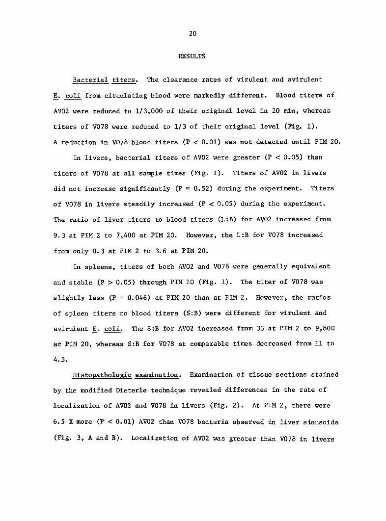
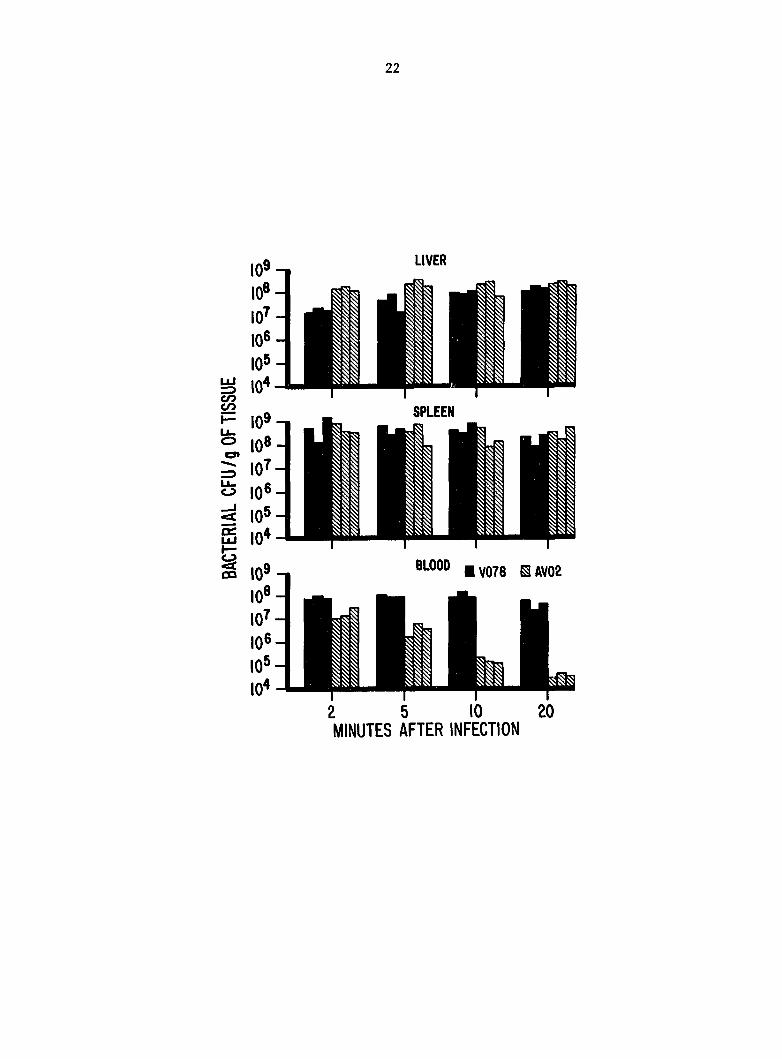
Bacterial titers. The clearance rates of virulent and avirulent
]E. coll from circulating blood were markedly different. Blood titers of
AV02 were reduced to 1/3,000 of their original level in 20 min, whereas
titers of V078 were reduced to 1/3 of their original level (Fig. 1).
A reduction in V078 blood titers (P < 0.01) was not detected until PIM 20.
In livers, bacterial titers of AV02 were greater (P < 0.05) than
titers of V078 at all sample times (Fig. 1). Titers of AV02 in livers
did not increase significantly (P = 0.52) during the experiment. Titers
of V078 in livers steadily Increased (P < 0.05) during the experiment.
The ratio of liver titers to blood titers (L:B) for AV02 Increased from
9.3 at PIM 2 to 7,400 at PIM 20. However, the L:B for V078 increased
from only 0.3 at PIM 2 to 3.6 at PIM 20.
In spleens, titers of both AV02 and V078 were generally equivalent
and stable (P > 0.05) through PIM 10 (Fig. 1). The titer of V078 was
slightly less (P = 0.046) at PIM 20 than at PIM 2. However, the ratios
of spleen titers to blood titers (S:B) were different for virulent and
avirulent E. coll. The S:B for AV02 increased from 33 at PIM 2 to 9,800
at PIM 20, whereas S:B for V078 at comparable times decreased from 11 to
4.5.
Histopathologic examination. Examination of tissue sections stained
by the modified Dieterle technique revealed differences in the rate of
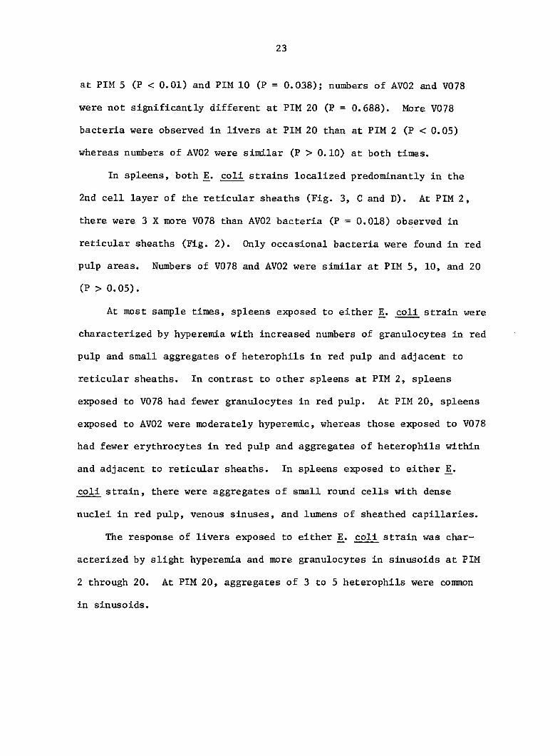
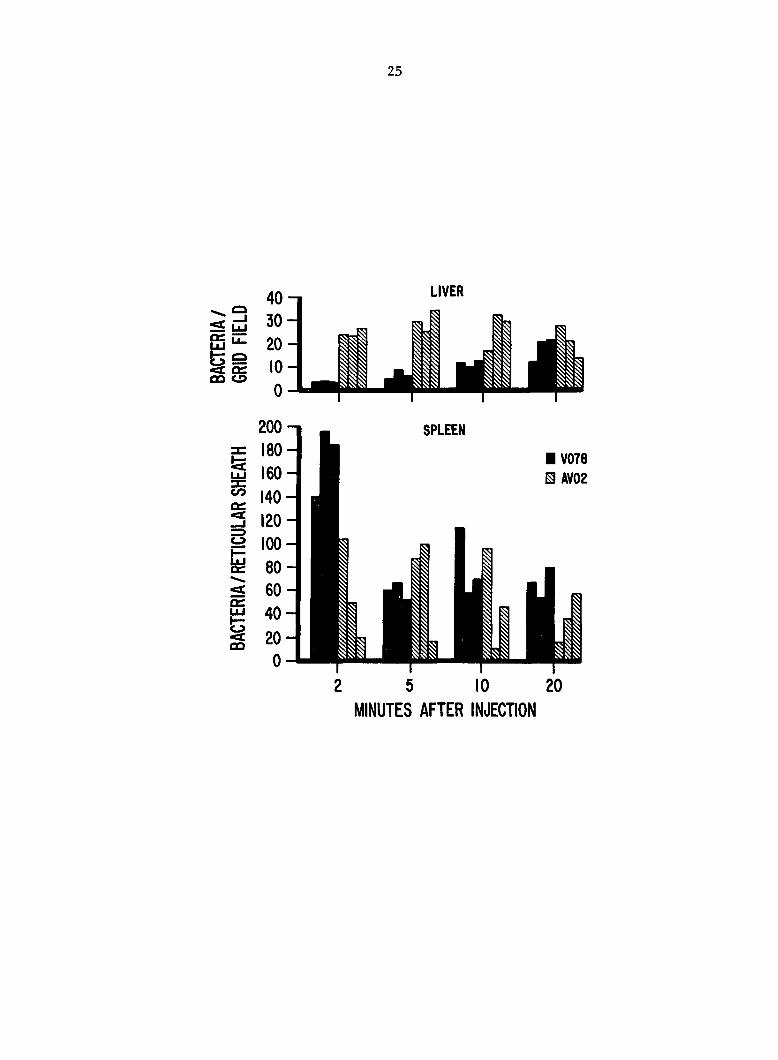
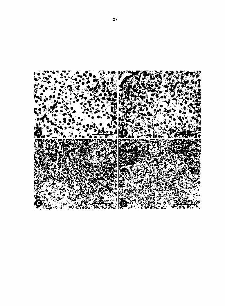
localization of AV02 and V078 in livers (Fig. 2). At PIM 2, there were
6.5 X more (P < 0.01) AV02 than V078 bacteria observed in liver sinusoids
(Fig. 3, A and B). Localization of AV02 was greater than V078 in livers
Fig. 1. Colony-forming units (CFU) of Escherichia coli per g in
blood, spleen, and liver after i.v. inoculation.
22
SPLEEN
V078 BAV02
2 5 10 MINUTES AFTER INFECTION
23
at PIM 5 (P < 0.01) and PIM 10 (P = 0.038); numbers of AV02 and V078
were not significantly different at PIM 20 (P = 0.688). More V078
bacteria were observed in livers at PIM 20 than at PIM 2 (P < 0.05)
whereas numbers of AV02 were similar (P > 0.10) at both times.
In spleens, both E^. coli strains localized predominantly in the
2nd cell layer of the reticular sheaths (Fig. 3, C and D). At PIM 2,
there were 3 X more V078 than AV02 bacteria (P = 0.018) observed in
reticular sheaths (Fig. 2). Only occasional bacteria were found in red
pulp areas. Numbers of V078 and AV02 were similar at PIM 5, 10, and 20
(P > 0.05).
At most sample times, spleens exposed to either E^. coli strain were
characterized by hyperemia with increased numbers of granulocytes in red
pulp and small aggregates of heterophils in red pulp and adjacent to
reticular sheaths. In contrast to other spleens at PIM 2, spleens
exposed to V078 had fewer granulocytes in red pulp. At PIM 20, spleens
exposed to AV02 were moderately hyperemic, whereas those exposed to V078
had fewer erythrocytes in red pulp and aggregates of heterophils within
and adjacent to reticular sheaths. In spleens exposed to either
coli strain, there were aggregates of small round cells with dense
nuclei in red pulp, venous sinuses, and lumens of sheathed capillaries.
The response of livers exposed to either ]E. coli strain was char
acterized by slight hyperemia and more granulocytes in sinusoids at PIM
2 through 20. At PIM 20, aggregates of 3 to 5 heterophils were common
in sinusoids.
Fig. 2. Number of Escherichia coll observed histopathologically in
liver sinusoids and splenic reticular sheaths after i.v. inoculation.
25
LIVER
200-1
z 180 -
g 160
I 140
G 120
p 100
5 80
5 60 oc |±î 40
M 20
0
SPLEEN
• V078 B AV02
2 5 10 20
MINUTES AFTER INJECTION
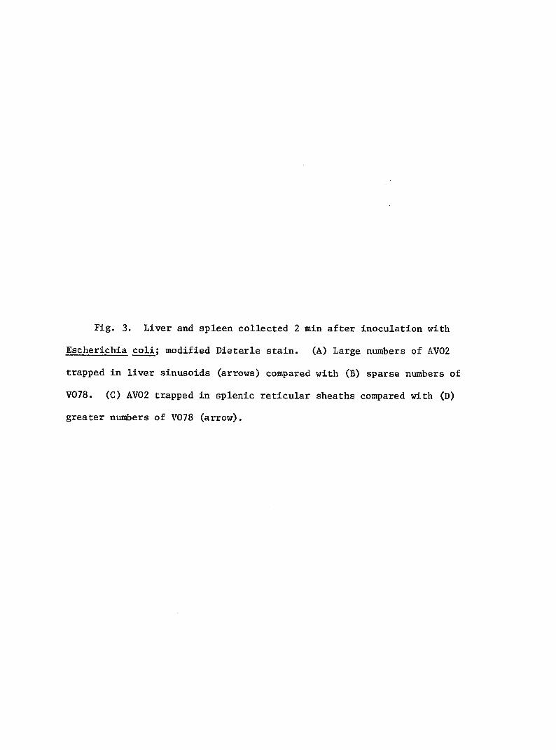
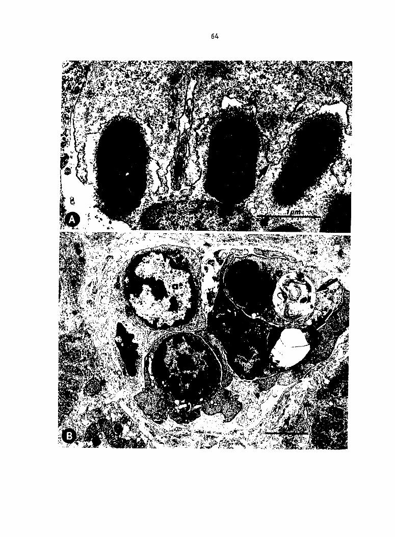
Fig. 3. Liver and spleen collected 2 min after inoculation with
Escherichia coli; modified Dieterle stain. (A) Large numbers of AV02
trapped in liver sinusoids (arrows) compared with (B) sparse numbers of
V078. (C) AV02 trapped in splenic reticular sheaths compared with (D)
greater numbers of V078 (arrow).
27
i
K
I
*
Si M
%F &W)S
28
Electron microscopic examination. Splenic reticular sheath capillar
ies were characterized by tall endothelium incompletely surrounded by a
basal lamina and a thick network of reticular fibers. Reticular sheaths
were composed of 2 or 3 layers of macrophages with numerous pseudopodia
intermingled with reticular fibers.
At PIM 2, bacteria were found within phagosomes of reticular
sheath macrophages in all spleens exposed to coli. Occasionally,
bacteria were in the process of being ingested or were located between
macrophages (Fig. 4). Changes in spleens collected at PIM 5 and 10 were
comparable with those at PIM 2, except that heterophils and eosinophils
were commonly observed near reticular sheaths. Many phagosomes were
surrounded by electron-dense homogeneous granular material (Fig. 4).
Some phagosomes were in the process of fusion with lysosomes (Fig. 5).
In general, phagosomes containing AV02 were larger and more regular than
those containing V078 (Fig 6 and 7). Phagosomal membranes around V078
appeared wavy and closely apposed to the bacterial surface. Few bacteria
were found within macrophages or heterophils of the red pulp.
At PIM 20, fewer bacteria were seen in spleens exposed to AV02 than
to V078. Small round cells with dense, clumped chromatin were common in
reticular sheaths and red pulp of all spleens exposed to coli.
These cells occasionally contained concentric membranous structures
similar to large myelin figures observed in thrombocytes of chickens (81)
(Fig. 8A). Endothelium of sheathed capillaries was occasionally necrotic
in spleens exposed to either coli strain.
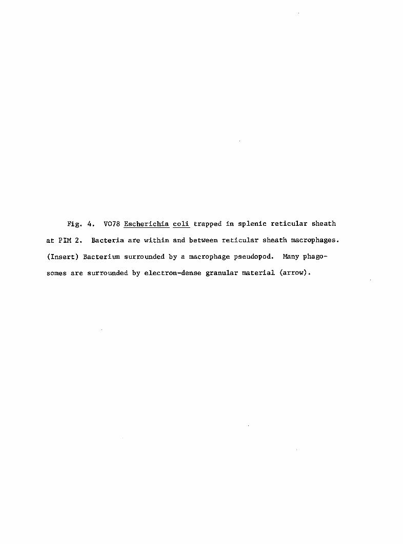
Fig. 4. V078 Escherichia coli trapped in splenic reticular sheath
at PIM 2. Bacteria are within and between reticular sheath macrophages.
(Insert) Bacterium surrounded by a macrophage pseudopod. Many phago
somes are surrounded by electron-dense granular material (arrow).
30
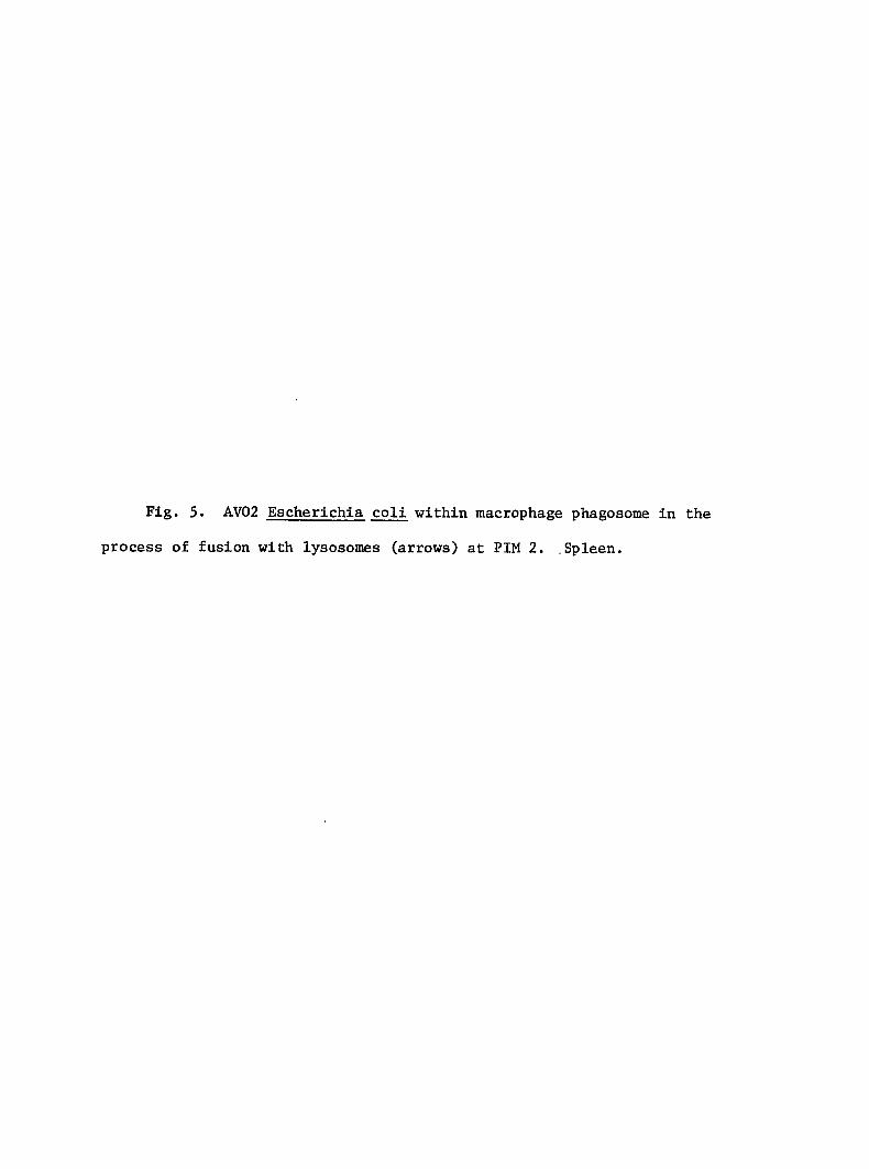
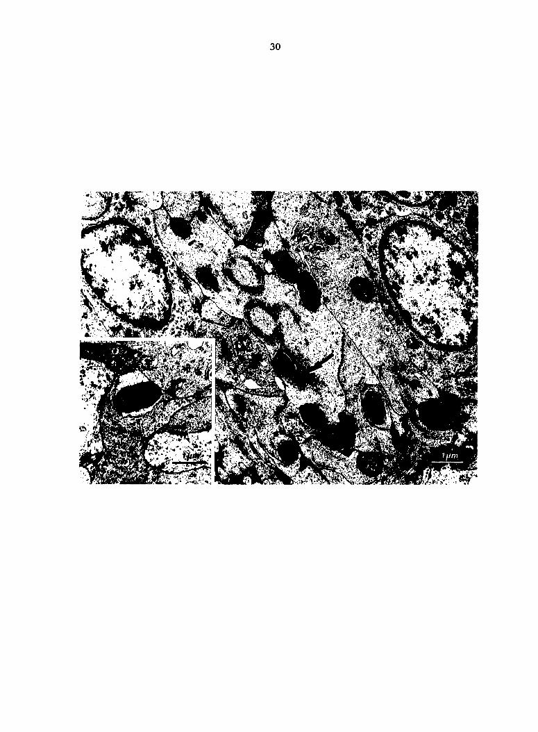
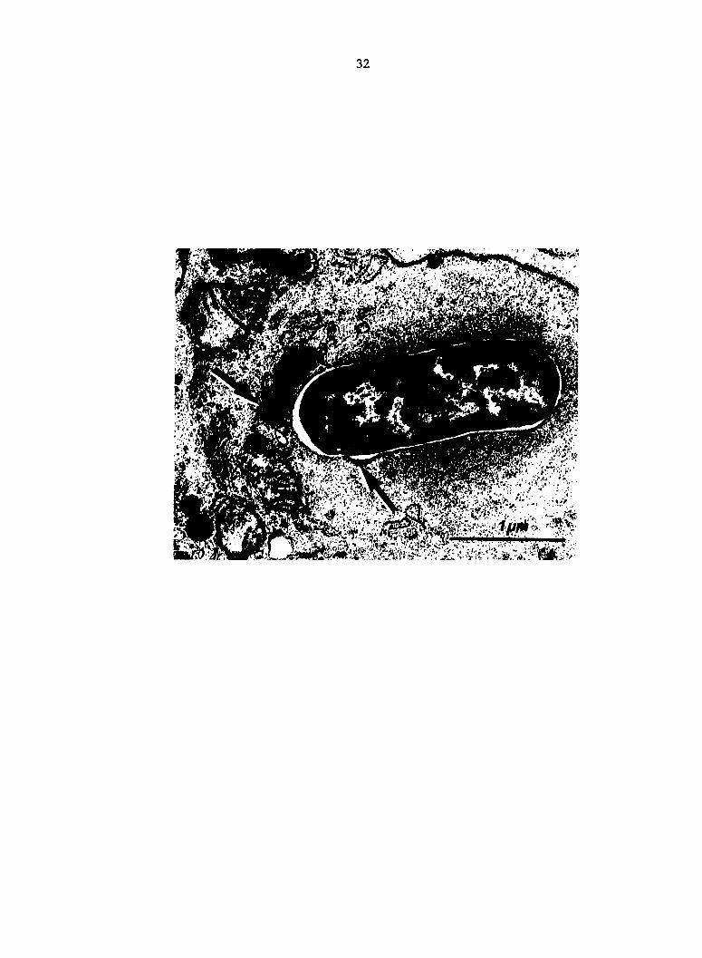
Fig. 5. AV02 Escherichia coli within macrophage phagosome in the
process of fusion with lysosomes (arrows) at PIM 2. Spleen.
32
,3MM
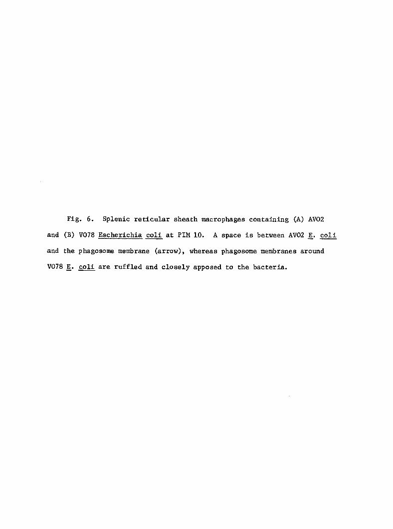
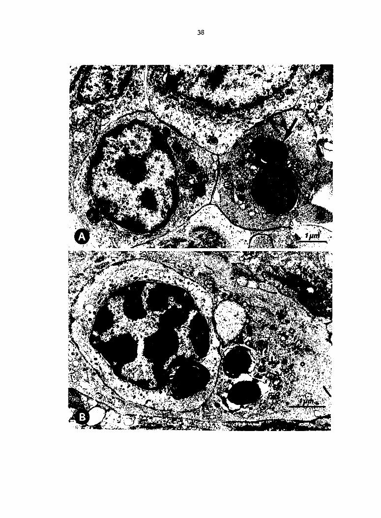
Fig, 6. Splenic reticular sheath macrophages containing (A) AV02
and (B) V078 Escherichia coli at PIM 10. A space is between AV02 coll
and the phagosome membrane (arrow), whereas phagosome membranes around
V078 E^. coli are ruffled and closely apposed to the bacteria.
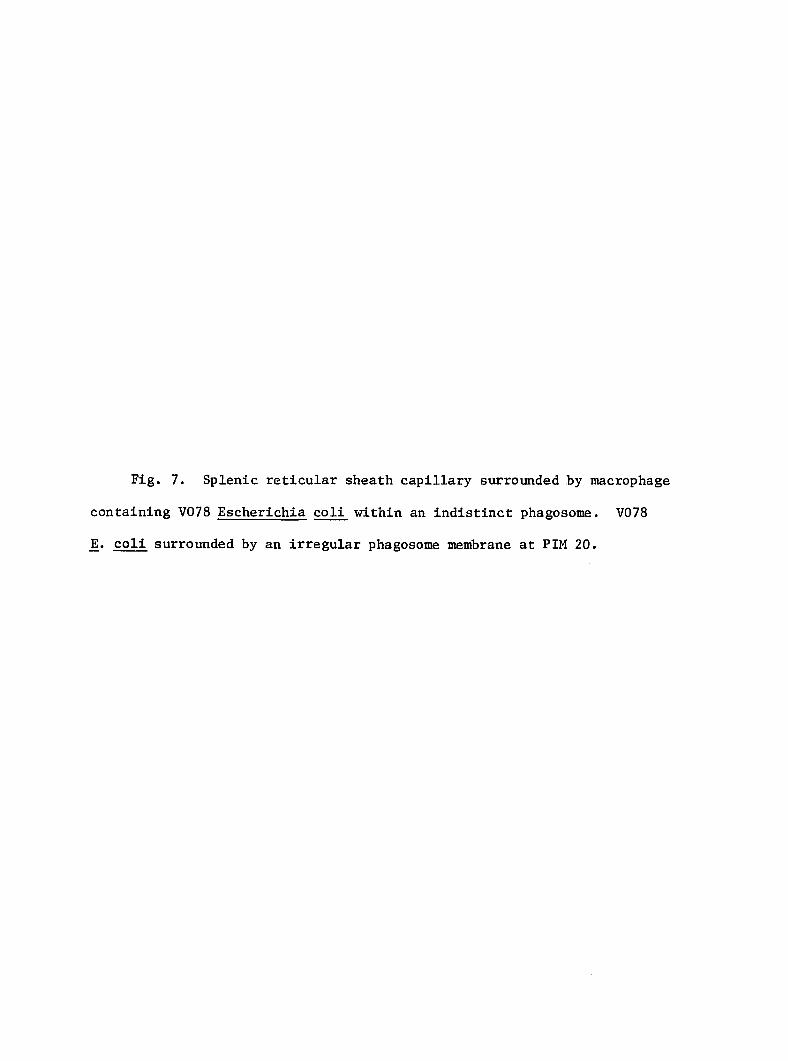
Fig. 7. Splenic reticular sheath capillary surrounded by macrophage
containing V078 Escherichia coli within an indistinct phagosome. V078
JE. coli surrounded by an irregular phagosome membrane at PIM 20.
36
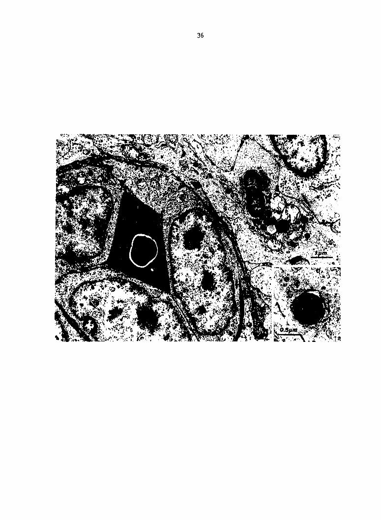
Fig. 8. (A) Macrophage (left) and thrombocyte in splenic red pulp
at PIM 20. Complex myelin figure type cytoplasmic inclusion of thrombo
cyte (81) (arrow). (B) Thrombocyte and V078 Escherichia coli contained
in a liver sinusoid-lining macrophage.
39
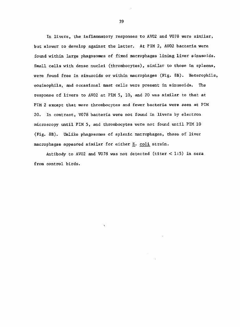
In livers, the inflammatory responses to AV02 and V078 were similar,
but slower to develop against the latter. At PIM 2, AV02 bacteria were
found within large phagosomes of fixed macrophages lining liver sinusoids.
Small cells with dense nuclei (thrombocytes), similar to those in spleens,
were found free in sinusoids or within macrophages (Fig. 8B). Heterophils,
eosinophils, and occasional mast cells were present in sinusoids. The
response of livers to AV02 at PIM 5, 10, and 20 was similar to that at
PIM 2 except that more thrombocytes and fewer bacteria were seen at PIM
20. In contrast, V078 bacteria were not found in livers by electron
microscopy until PIM 5, and thrombocytes were not found until PIM 10
(Fig. 8B). Unlike phagosomes of splenic macrophages, those of liver
macrophages appeared similar for either coli strain.
Antibody to AV02 and V078 was not detected (titer < 1:5) in sera
from control birds.
40
DISCUSSION
The cultural and histopathologic results of this study indicate
that V078 bacteria resist clearance from circulating blood, whereas AV02
bacteria are cleared at an exponential rate. Livers were more effective
in trapping AV02 than V078. Due to the large size of the liver, effective
trapping of AV02 in liver macrophages had a major effect on early clear
ance of these bacteria from blood.
Why there were 3 X more V078 than AV02 bacteria observed in retic
ular sheaths at PIM 2 is unclear, but at least 2 mechanisms are possible.
First, bacteria of the V078 inoculum may not have been homogeneous with
respect to expression of surface components affecting phagocytosis, thus
a small population of bacteria did not resist phagocytosis. Second,
initial rapid phagocytosis of V078 may have adversely affected these
macrophages to inhibit further phagocytosis.
Different rates of killing of V078 and AV02 are indicated by the
ratios of spleen and liver titers to blood titers (S:B, L:B). Organ
titers reflect numbers of bacteria in circulating blood relative to
fixed organ tissues plus trapped viable bacteria. However, organ titers
probably underestimate the real number of viable bacteria since phagocytes
laden with bacteria may not be sufficiently disrupted during trituration
to register each bacterium as a CFU. Changes in organ titer are deter
mined by the blood titer, plus the rate of trapping, minus the rate of
killing. The present study was designed to exclude effects of bacterial
multiplication since these strains have generation times of nearly 20
min after a 1 h lag phase in vitro (5). The fact that S;B and L;B were

41
much higher for AV02 than for V078 indicates greater bacterial trapping
of AV02 in spleen and liver. Relatively stable spleen and liver titers
concomitant with falling blood titers (AV02) Indicate that rates of
trapping and killing in spleen and liver are roughly equivalent and are
at least partially responsible for the decrease of circulating bacteria.
In early bacteremia, it is unlikely that significant numbers of bacteria
are cleared from the blood by organs other than spleen and liver (7, 61).
The low S:B and L:B of V078 indicate that V078 was slowly trapped in
spleen and liver, slowly killed after phagocytosis, and therefore,
slowly cleared from the blood stream.
The inflammatory process in spleens differed slightly depending on
the JE. coll strain used. Initial depletion of granulocytes from splenic
red pulp at PIM 2 is probably a reflection of profound leukopenia that
has been observed immediately after the i.v. injection of bacteria (61).
The lack of hyperemia in spleens 20 min after exposure to V078 indicates
that splenic blood flow was decreased compared with normal spleens and
those exposed to AV02. Decreased blood flow through spleens could
inhibit clearance, since a constant fraction of circulating bacteria are
removed with each organ passage (61) The greater tendency for aggregates
of heterophils to infiltrate reticular sheaths of spleens exposed to
V078 indicates that V078 bacteria were not effectively neutralized by
sheath macrophages.
The ultrastructure of splenic reticular sheaths in the present
study was essentially like that described for chickens by White and
Gordon (92). However, we did not distinguish 2 distinct reticular
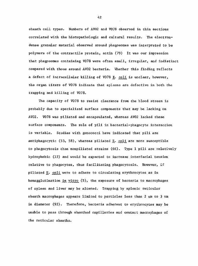
42
sheath cell types. Numbers of AV02 and V078 observed in thin sections
correlated with the histopathologic and cultural results. The electron-
dense granular material observed around phagosomes was interpreted to be
polymers of the contractile protein, actin (79) It was our impression
that phagosomes containing V078 were often small, irregular, and indistinct
compared with those around AV02 bacteria. Whether this finding reflects
a defect of intracellular killing of V078 coli is unclear, however,
the organ titers of V078 indicate that spleens are defective in both the
trapping and killing of V078.
The capacity of V078 to resist clearance from the blood stream is
probably due to specialized surface components that may be lacking on
AV02. V078 was piliated and encapsulated, whereas AV02 lacked these
surface components. The role of pili in bacterial-phagocyte interaction
is variable. Studies with gonococci have indicated that pili are
antiphagocytic (53, 58), whereas piliated JE. coli are more susceptible
to phagocytosis than nonpiliated strains (66). Type 1 pili are relatively
hydrophobic (12) and would be expected to increase interfacial tension
relative to phagocytes, thus facilitating phagocytosis. However, if
piliated coli were to adhere to circulating erythrocytes as in
hemagglutination in vitro (5), the exposure of bacteria to macrophages
of spleen and liver may be altered. Trapping by splenic reticular
sheath macrophages appears limited to particles less than 2 um to 3 um
in diameter (92). Therefore, bacteria adherent to erythrocytes may be
unable to pass through sheathed capillaries and contact macrophages of
the reticular sheaths.
The role of a surface capsule (K antigen) on V078, like the role of
pili, is not clearly understood. Strains of coli with sufficient K
antigen to resist killing by complement are also poorly phagocytized
when injected into mice (38). However, IE. coli strains isolated from
human patients with bacteremia do not have excessive amounts of K
antigen (47). In a recent study, most E^. coli organisms isolated from
livers of turkeys produced K antigens, but production of K antigens did
not correlate with virulence for turkeys (14). Two coli serotypes
(01:K-:H7 and 02:K-:H5) were virulent for turkeys although they lacked K
antigens. Stendahl et al. (76) found that strains of ]E. coli having
smooth hydrophilic 0 antigens tend to resist phagocytosis whether or not
K antigens are present. Further study is required to determine the
nature of surface components of virulent E^. coli that enable these
bacteria to resist destruction by turkey phagocytes.
44
EFFECT OF PASSIVE IMMUNIZATION ON PHAGOCYTOSIS OF BLOOD-BORNE
ESCHERICHIA COLI IN SPLEEN AND LIVER OF TURKEYS
Lawrence H. Arp
Manuscript submitted to
Infection and Immunity
From the National Animal Disease Center, Science and Education
Administration, Agricultural Research, United States Department of
Agriculture, P.O. Box 70, Ames, lA 50010
No product endorsements are implied herein.
45
ABSTRACT
Clearance of Escherichia coll from blood and localization in
tissues were studied in passively immune and nonimmune turkeys after
intravenous inoculation. In passively immune turkeys, coll bacteria
were rapidly cleared from blood and localized predominantly in liver and
spleen. Bacteria were within or between macrophages of reticular
sheaths and red pulp in spleen, and sinusoids of liver. Ultra-
structurally, extracellular bacteria were covered by an extraneous
coat. Moderate interstitial hemorrhage and aggregates of thrombocytes
were found in lungs of passively immune birds. In nonimmune turkeys,
persistence of large numbers of coli in blood and high mortality
were associated with inefficient localization of JE. coli in liver.
These studies indicate that rapid clearance of virulent coli from
blood requires antibody-dependent phagocytosis by hepatic macrophages.
46
INTRODUCTION
Bacteremia is a feature of clinical and subclinical infections of
turkeys caused by Escherichia coli. Virulent strains of coli
readily cause bacteremia in normal poultry exposed by aerosol (4) or
air-sac inoculation (57). Colisepticemia may develop in birds exposed
to large numbers of virulent coli at a time when their specific or
nonspecific defenses are conçromised.
In young turkeys, avirulent coli are rapidly cleared from
circulating blood, whereas virulent E^. coli resist clearance (3).
Hepatic macrophages are the principal agents for the clearance of
avirulent JE. coli from blood, but these cells are less effective against
virulent jE. coli (3). A recent study showed that passive immunization
with hyperimmune serum protected young turkeys from lethal challenge
with a virulent coli strain (1). In contrast, other investigators
have suggested there is no relationship between 0 and K serum agglutinins
and protection against colisepticemia (17, 18). Although some experi
mental vaccines protected against challenge with virulent jE. coli, sera
from only 4 of 28 vaccinated birds had 0 or K agglutinin titers greater
than 1:5 (17, 18).
The purpose of the present study was to determine the effect of
antibody on: (i) the clearance of coll from circulating blood, (ii)
the localization of blood-borne coll in spleen, liver, lung, and bone
marrow, and (iii) the interaction of JE. coli and phagocytes in spleen,
liver, and lung.
47
MATERIALS AND METHODS
Bacteria. The coll serotype 078:K80:H9 was used (1, 3, 4). The
strain was originally isolated from the liver of a turkey that died with
colisepticemia. For each experiment, bacteria were grown for 24 h in
trypticase-soy broth; washed once in phosphate-buffered saline (PBS), pH
7.2; and resuspended to the desired concentration for inoculation.
Turkeys. Three-week-old Broad-Breasted White turkeys were used in
each experiment. Turkeys were obtained commercially and raised to 3
weeks of age in isolation rooms as previously described (4).
Passive immunization. Hyperimmune serum was produced in 8-week-old
turkeys against the coli strain used in this study. Turkeys were
inoculated intravenously (i.v. ) 3 times at 5 day Intervals with E^. coli
inoculum prepared as described above. The inoculum contained 10^, 10^,
g and 10 colony-forming units (CPU) of coli for the first, second, and
third inoculations, respectively. Serum was harvested 5 days after the
third inoculation, diluted with PBS to an 0-agglutinin titer of 1:256,
and stored at -70" C until used. Treatment of a sample of the serum with
2-mercaptoethanol reduced the titer to 1:8. In each experiment, turkeys
were injected i.v. with 1 ml of diluted hyperimmune serum 48 h before
inoculation with jE. coli.
Necropsy procedure and specimen collection. At the appropriate
times after coli inoculation, a 1 ml blood sanple was collected from
the wing vein; birds were immobilized by i.v. injection of succinyl
choline and killed by decapitation. Within 30 s, birds were dipped in a
disinfectant solution and opened aseptically for collection of spleen.
48
liver, lung, and proximal tibia. Blood samples were diluted 10-fold in
ice-cold PBS containing 1% Grobax^ (Roche Diagnostics) as an anticoagu
lant. Blood was further diluted 10-fold in PBS 6 times. One-half ml of
each dilution was inoculated onto blood agar plates. Plates were in
cubated overnight at 37° C, and CPU were counted. A 1- to 2-g specimen
of liver, half of the spleen, and the right lung of each bird were
collected aseptically, weighed, and processed to determine the number of
CFU/g of tissue as previously described (3). Bacterial isolates were
confirmed as the inoculated strain by slide agglutination tests.
Estimation of percentage of coli inoculum in selected tissues.
Total numbers of coli in spleen, liver, lung, and blood were calculated
by multiplying the number of coli CFU/g of tissue by the estimated
weight of the organ. The weight of spleen and liver were estimated from
percentage body-weight means determined in 5, 3-week-old turkeys;
expressed as the mean percentage of body-weight, the spleen was 0.0821%
and the liver was 2.046%. The blood weight was estimated, based on
values for White Leghorn chickens of similar weight (49), to be 8% of
the body-weight. Since a whole lung was removed from each bird, actual
lung-weight means were multiplied by 2 and used to estimate the total
number of coli CPU in lungs. The percentage of coli inoculum in
selected tissues was estimated by the following formula:
% E. coli inoculum in tissue = ^°* CFU in tissue — X 100
No. of E. coli CPU in inoculum
49
Histology. Specimens of spleen, liver, lung, and proximal tibia
were fixed in 10% neutral buffered formalin for 48 h, and bone specimens
were decalcified in Decalcifying Solution® (Scientific Products) for 4 h.
Blocks were trimmed, dehydrated in ethanols, embedded in paraffin, and
sectioned at 5 ii.m. Sections were stained with hematoxylin and eosin
(H & E), Giemsa, and modified Dieterle (87) stain. Additional specimens
of spleen, liver, and lung were fixed in Bouin's solution for 8 h,
washed in 50% ethanol, dehydrated, embedded, sectioned, and stained with
H & E.
Electron microscopy. Cubes (0.5 mm) of spleen, liver, and lung
were fixed in 2.5% buffered glutaraldehyde for 3 h, washed in 0.1 M
sodium cacodylate buffer with 0.05 M sucrose, postfixed in 1% buffered
osmium tetroxide, washed in buffer, and dehydrated througih graded
ethanols and propylene oxide to embedment in epoxy resin (Epon 812).
Ultrathin sections were cut with a diamond knife, stained with 2% uranyl
acetate and Reynold's lead citrate, and examined with an electron
microscope. Sections from 2 or 3 blocks of each tissue were examined.
50
RESULTS
Experimental design. The study consisted of 3 experiments.
Experiment 1 was designed to determine the clearance rate of coli
from blood of passively immune (Ab+) and nonimmune (Ab-) turkeys.
Experiments 2 and 3 were designed to determine sites of E^. coli locali
zation and coli-phagocyte interactions in tissues of Ab+ and Ab-
turkeys. In experiment 1, 5 Ab+ and 5 Ab- turkeys were inoculated i.v.
with 8 X 10 CFU of coli. For determining the number of coli
CFU/ml of blood, a 0.5-ml blood sangle was collected from the wing vein
of each turkey at 1, 10, 30, 60, and 120 min after the injection of
coli. In experiment 2, 3 Ab+ and 3 Ab- turkeys were inoculated i.v.
9 with 6 X 10 CFU of E^. coli. A 0.5 ml blood sample was collected, and
the turkeys were killed 1 min after inoculation with JE. coli. Numbers
of viable bacteria in blood, spleen, liver, and lung were determined;
specimens of spleen, liver, lung, and bone marrow were studied with
light and electron microscopy. Experiment 3 was like experiment 2
9 except that the inoculum contained 10 CFU of jE. coli, and turkeys were
killed 20 min after inoculation.
Blood was collected from 10 uninoculated control turkeys (5 Ab+ and
5 Ab-) for determination of serum agglutinin titers against the live jE.
coli inoculum. The Ab+ birds had agglutinin titers of 1:4 to 1:8,
whereas Ab- birds had titers of less than 1:4. The 5 Ab+ birds were
used as negative controls for bacterial culture and microscopic examina
tion.
51
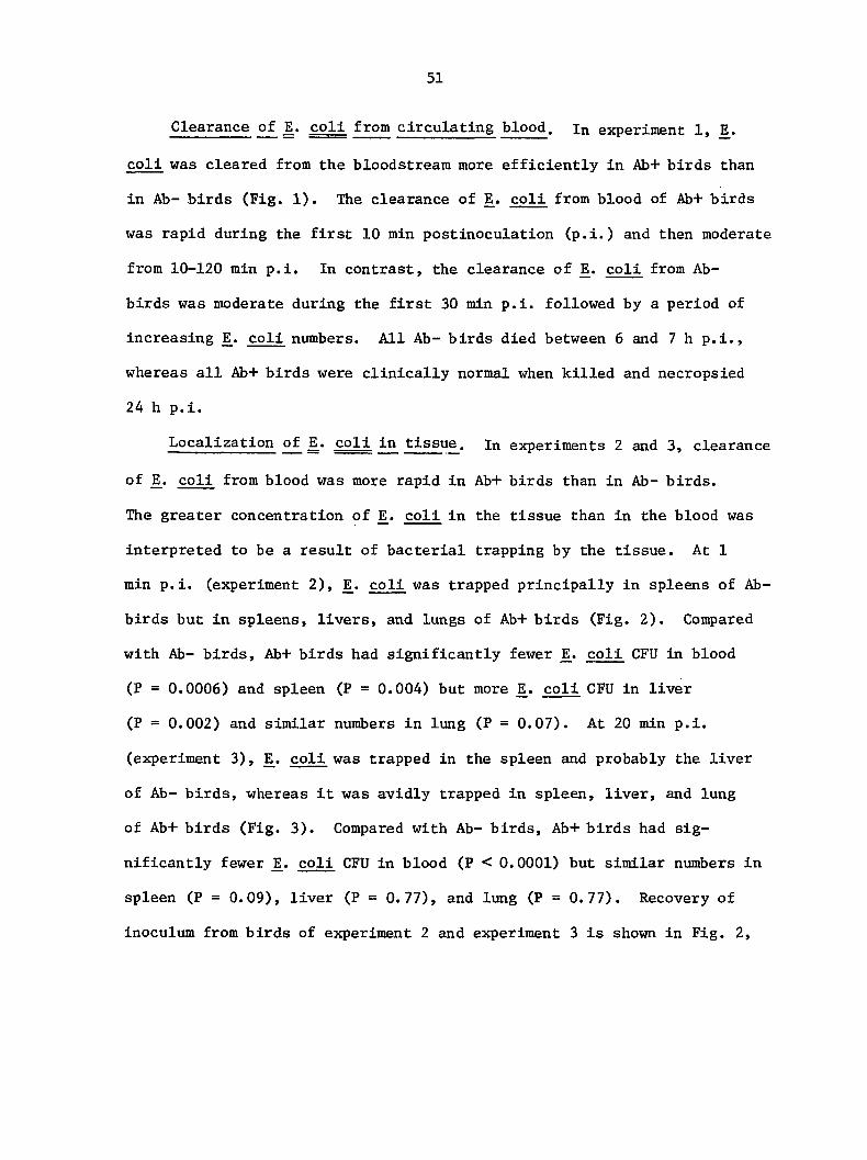
Clearance of JE. coll from circulating blood, in experiment 1,
coll was cleared from the bloodstream more efficiently in Ab+ birds than
in Ab- birds (Fig. 1). The clearance of E. coli from blood of Ab+ birds
was rapid during the first 10 min postinoculation (p.i.) and then moderate
from 10-120 min p.i. In contrast, the clearance of coli from Ab-
birds was moderate during the first 30 min p.i. followed by a period of
increasing jE. coli numbers. All Ab- birds died between 6 and 7 h p.i.,
whereas all Ab+ birds were clinically normal when killed and necropsied
24 h p.i.
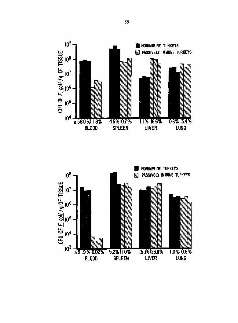
Localization of jE. coj^ in tissue, in experiments 2 and 3, clearance
of coli from blood was more rapid in Ab+ birds than in Ab- birds.
The greater concentration of E. coli in the tissue than in the blood was
interpreted to be a result of bacterial trapping by the tissue. At 1
min p.i. (experiment 2), E^. coli was trapped principally in spleens of Ab-
birds but in spleens, livers, and lungs of Ab+ birds (Fig. 2). Compared
with Ab- birds, Ab+ birds had significantly fewer coli CFU in blood
(P = 0.0006) and spleen (F = 0.004) but more coll CFU in liver
(P = 0.002) and similar numbers in lung (P = 0.07). At 20 min p.i.
(experiment 3), E. coli was trapped in the spleen and probably the liver
of Ab- birds, whereas it was avidly trapped in spleen, liver, and lung
of Ab+ birds (Fig. 3). Compared with Ab- birds, Ab+ birds had sig
nificantly fewer coli CFU in blood (P < 0.0001) but similar numbers in
spleen (P = 0.09), liver (P = 0.77), and lung (P = 0.77). Recovery of
inoculum from birds of experiment 2 and experiment 3 is shown in Fig. 2,
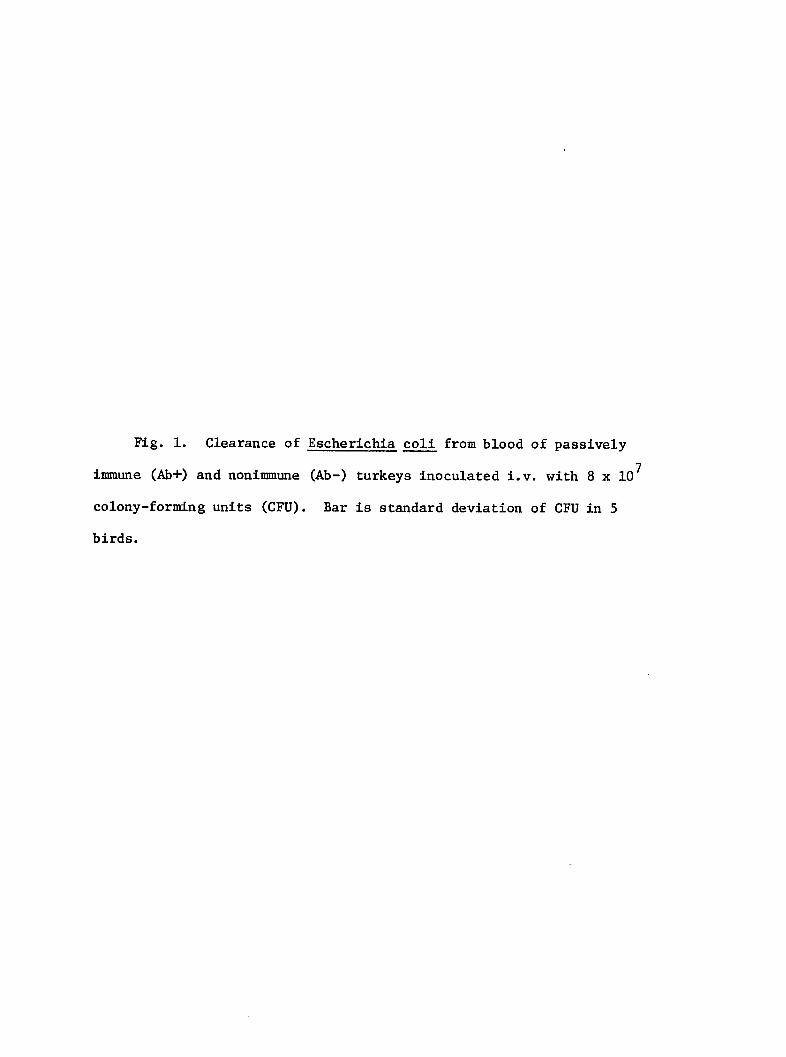
Fig. 1. Clearance of Escherichia coll from blood of passively
immune (Ab+) and nonimmune (Ab-) turkeys inoculated i.v. with 8 x 10^
colony-forming units (CPU). Bar is standard deviation of CFU in 5
birds.
53
S §
s
10^ -
10® -
IO5 -
10* -
10 -
10^ -— MSSIVELV IMMUNE
10' 4—1 1 1
0 10 30 60
MINUTES AFTER INOCULATION
Fig. 2 Localization of Escherichia coli in tissues of 3 passively
immune (Ab+) and 3 nonimmune (Ab-) turkeys 1 min after i.v. inoculation
9 with 6 X 10 colony-forming units (CFU).
^Estimated % of E. coli inoculum in whole tissue.
Fig. 3. Localization of Escherichia coli in tissues of 3 passively
immune (Ab+) and 3 nonimmune (Ab-) turkeys 20 min after i.v. inoculation
Û of 10 colony-forming units (CFU).
^Estimated % of E. coli inoculum in whole tissue.
55
• NONIMMUNE TURKEYS
^ PASSIVELY IMMUNE TURKEYS
BLOOD SPLEEN LIVER LUNG
• NONIMMUNE TURKEYS
PASSIVELY IMMUNE TURKEYS
BLOOD SPLEEN LIVER LUNG
56
and Fig. 3, respectively. In each experiment, the recovery of viable E.
coll from tissues of Ab+ birds was about 1/3 of that from Ab- birds.
Histologie examination. At 1 min p.i. (experiment 2), spleens of Ab+
birds contained clumps of 5-20 E^. coll in reticular sheaths (ellipsoids)
and red pulp (Fig. 4A). Spleens from Ab- birds contained large numbers
of E^. coll evenly distributed in reticular sheaths but few coll
associated with red pulp (Fig. 4B). In livers from Ab+ birds, E. coll
were numerous, and clumps of 5-10 bacteria were associated with macro
phages lining the sinusoids (Fig. 4C). Only scant numbers of coll
were observed in livers of Ab- birds. Lungs from Ab+ birds contained
clumps of 5-20 coll in small blood vessels associated with air
passages (air capillaries) at the periphery of parabronchl (tertiary
bronchi). Occasionally, coll were contained within aggregates of
thrombocytes. Bacteria were rarely observed in lungs of Ab- birds and
then only Individually, within small blood vessels. Small numbers of
coll were in vascular sinusoids of bone marrow from proximal tibias,
however, differences between Ab+ and Ab- birds were not apparent. At 20
min p.i. (experiment 3), numbers of JE. coll were not adequate for micro
scopic interpretation of localization in tissues.
Lungs of Ab+ birds at 1 min p.i. (experiment 2) were characterized by
diffuse interstitial hemorrhage, aggregation of thrombocytes in small
blood vessels, hyperemia, and mild serous exudation into small airways
(Fig. 4D). Lung lesions in Ab+ birds at 20 min p.i. (experiment 3) were
similar but less severe. In all Ab- birds, lungs were normal except for
hyperemia and occasional aggregates of thrombocytes in small blood
vessels. Lesions in spleens were most pronounced at 20 min p.i.
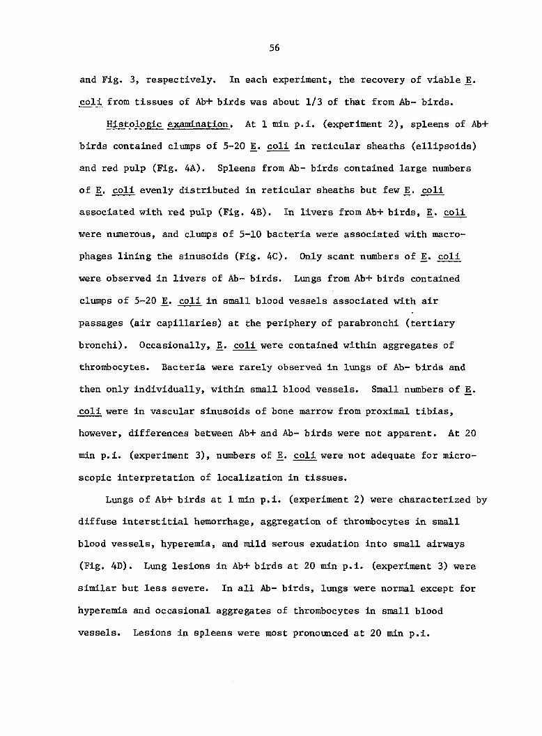
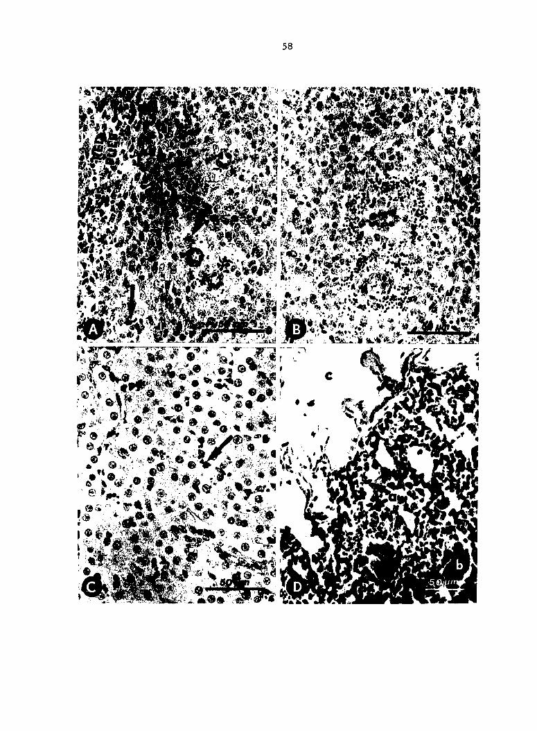
Fig. 4. Histopathology and localization of Escherichia coll in
turkeys killed 1 mln after inoculation (experiment 2). A. Passively
immune (Ab+) birds: clumps of coll (arrows) trapped in splenic
reticular sheaths and red pulp. Modified Dieterle stain. B. Nonimmune
(Ab-) birds : Escherichia coll trapped diffusely throughout splenic
reticular sheaths. Modified Dieterle stain. C. Ab+ birds: numerous
E^. coll (arrow) associated with sinusoid-lining macrophages in livers.
Modified Dieterle stain. D. Ab+ birds; aggregates of thrombocytes (a)
in capillaries and Interstitial hemorrhage (b) in lung. Lumen (c) of
parabronchus. H & E stain.
58
wmmm
!iœM !r ' & •££ . ;
59
(experiment 3). In spleens of Ab+ and Ab- birds, there was ischemia of
red pulp, with increased numbers of heterophils and thrombocytes. Clumps
of heterophils localized in red pulp of all birds, but in Ab- birds there
were also accumulations of heterophils in and around reticular sheaths.
Sinusoids of livers from Ab+ birds contained increased numbers of hetero
phils and thrombocytes. Heterophils or thrombocytes were often in clumps
of 10-30 cells. Livers of Ab- birds at 1 min p.i. (experiment 2) were
normal, but those at 20 min p.i. (experiment 3) contained increased
numbers of heterophils and thrombocytes. Histologically, sections of bone
marrow from Ab+ and Ab- birds did not differ appreciably from those of
controls.
Electron microscopic examination. In tissues from Ab+ birds at 1
min p.i. (experiment 2), extracellular E. coli were enveloped in an
extraneous coat of fibrillar/granular electron-dense material (Fig. 5A).
In the case of JE. coll phagocytized in Ab+ birds, the phagosomal membrane
was closely apposed to the extraneous coat. No such coat was observed
on coll from Ab- birds (Fig. 5B). In Ab+ birds phagosomes with JE.
coll often contained a homogeneous electron-dense substance between the
phagosomal membrane and the bacterial surface. A substance of similar
appearance was sometimes observed in lysosomes fused with or adjacent to
phagosomes containing IE. coli (Fig. 5C).
In spleen, most E^. coli were found within phagosomes of reticular
sheath macrophages. Some bacteria were incompletely surrounded by
pseudopods of macrophages. Although extraneous coats were often con
tinuous between extracellular E^. coli from Ab+ birds, macrophage
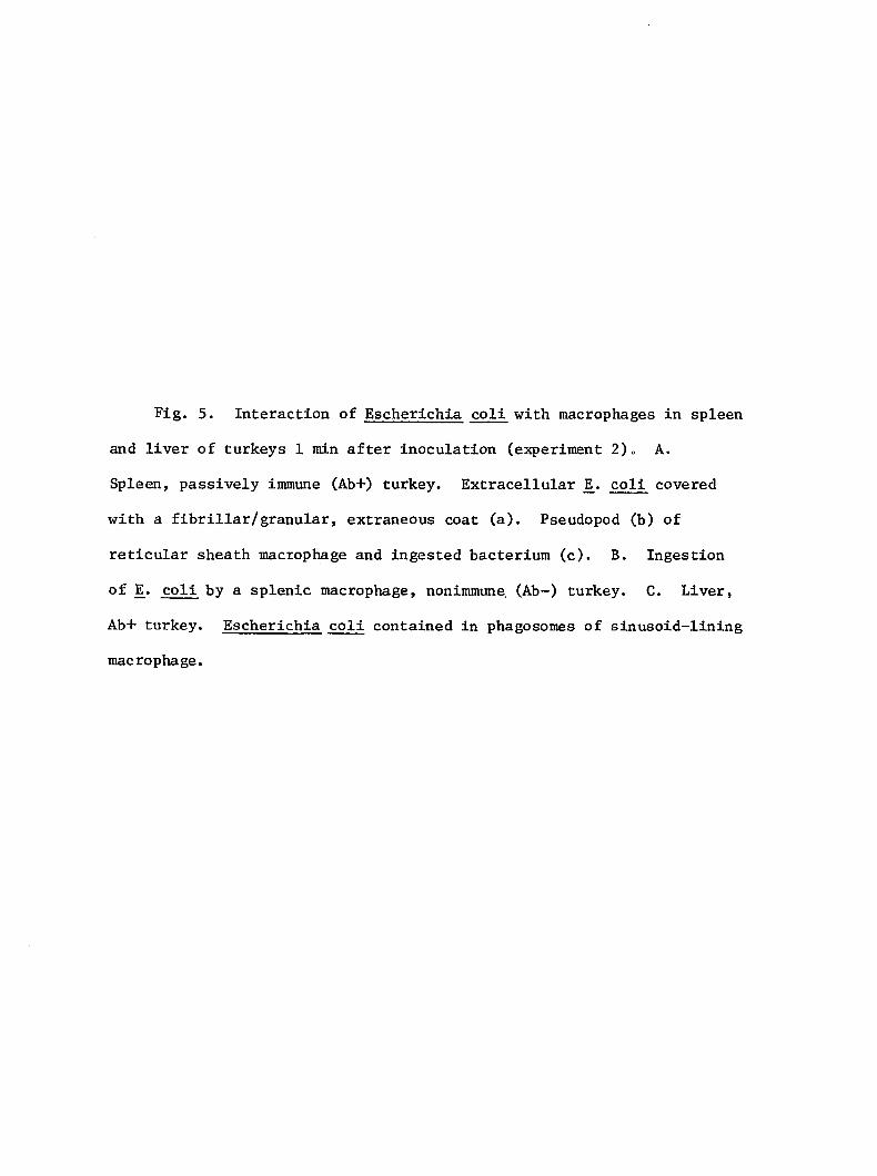
Fig. 5. Interaction of Escherichia coli with macrophages in spleen
and liver of turkeys 1 min after inoculation (experiment 2)o A.
Spleen, passively immune (Ab+) turkey. Extracellular IE. coli covered
with a fibrillar/granular, extraneous coat (a). Pseudopod (b) of
reticular sheath macrophage and ingested bacterium (c). B. Ingestion
of coli by a splenic macrophage, nonimmune (Ab-) turkey. C. Liver,
Ab+ turkey. Escherichia coli contained in phagosomes of sinusoid-lining
macrophage.
m
62
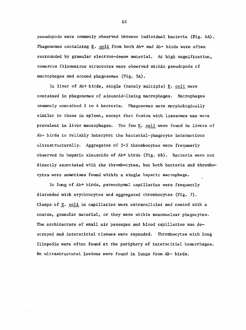
pseudopods were commonly observed between Individual bacteria (Fig. 6A).
Phagosomes containing E^. coli from both Ab+ and Ab- birds were often
surrounded by granular electron-dense material. At high magnification,
numerous filamentous structures were observed within pseudopods of
macrophages and around phagosomes (Fig, 5A).
In liver of Ab+ birds, single (rarely multiple) ]E. coli were
contained in phagosomes of sinusoid-lining macrophages. Macrophages
commonly contained 2 to 4 bacteria. Phagosomes were morphologically
similar to those in spleen, except that fusion with lysosomes was more
prevalent in liver macrophages. Too few coli were found in livers of
Ab- birds to reliably interpret the bacterial-phagocyte interactions
ultrastructurally. Aggregates of 2-5 thrombocytes were frequently
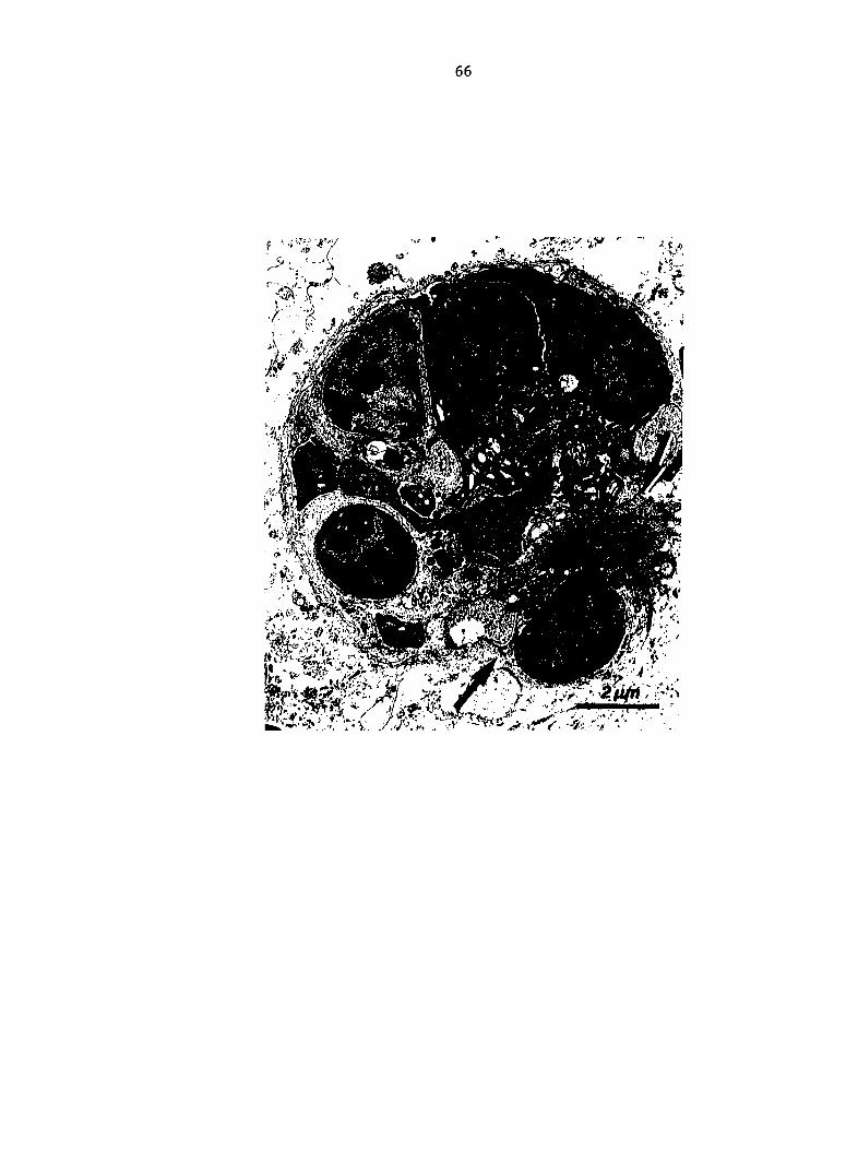
observed in hepatic sinusoids of Ab+ birds (Fig. 6B). Bacteria were not
directly associated with the thrombocytes, but both bacteria and thrombo
cytes were sometimes found within a single hepatic macrophage.
In lung of Ab+ birds, parenchymal capillaries were frequently
distended with erythrocytes and aggregated thrombocytes (Fig. 7).
Clumps of coli in capillaries were extracellular and coated with a
coarse, granular material, or they were within mononuclear phagocytes.
The architecture of small air passages and blood capillaries was de
stroyed and interstitial tissues were expanded. Thrombocytes with long
filopodia were often found at the periphery of interstitial hemorrhages.
No ultrastructural lesions were found in lungs from Ab- birds.
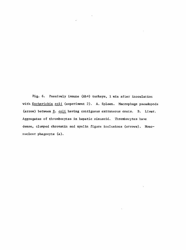
Fig. 6. Passively immune (Ab+) turkeys, 1 min after inoculation
with Escherichia coli (experiment 2). A. Spleen. Macrophage pseudopods
(arrow) between coli having contiguous extraneous coats. B. Liver.
Aggregates of thrombocytes in hepatic sinusoid. Thrombocytes have
dense, clumped chromatin and myelin figure inclusions (arrows). Mono
nuclear phagocyte (a).
"IS'"-
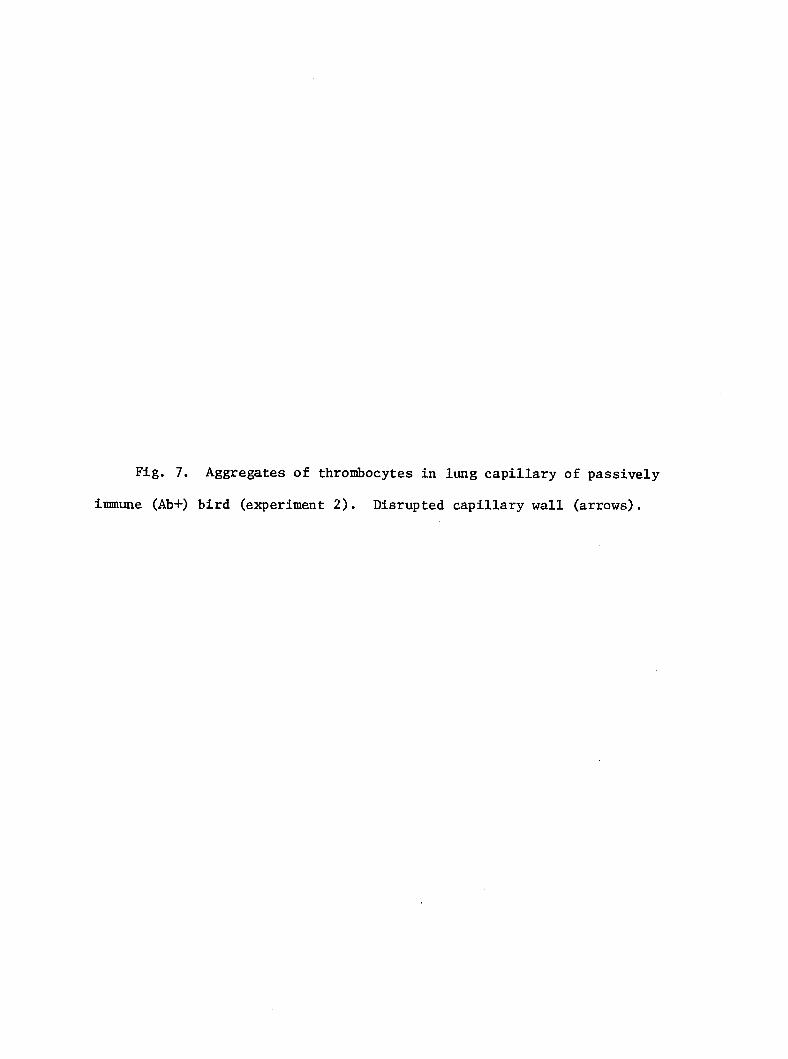
Fig. 7. Aggregates of thrombocytes in lung capillary of passively
immune (Ab+) bird (experiment 2). Disrupted capillary wall (arrows).
67
DISCUSSION
The results of the present study in turkeys concur with those of
previous studies of bacteremia in mammals; that is, rapid clearance of
virulent coli from blood requires antibody-dependent phagocytosis by
hepatic macrophages. The role of antibody in the clearance of bacteria
from circulating blood has been studied in small laboratory mammals (7,
37, 63, 74, 91). In mice and rabbits, coli is cleared from blood
principally by mononuclear phagocytic cells of the spleen and liver (7,
61). The avidity with which bacteria are trapped is determined by the
nature of the bacterial surface (68) and the presence or absence of
serum opsonins (7). The efficiency with which coli is trapped in
livers of mice and rats depends on the level of specific antibody (7,
39). Easily phagocytized bacteria and opsonized bacteria are efficiently
cleared from blood by hepatic macrophages (7).
In a previous study, virulent coli resisted trapping by hepatic
macrophages, whereas avirulent coli was avidly trapped in the liver
(3). Although splenic phagocytes trap coli effectively in the pres
ence or absence of antibody, the small size of the avian spleen, compared
with the liver, limits its relative effect on severe coli bacteremia.
Trapping of bacteria in the lung of mammals is minimal compared with
that in the spleen and liver. In the present study, antibody enhanced
localization of 12. coli in the lung, but this enhancement may have been
partially due to vascular effects of thrombocyte aggregation.
68
The presence of clumps of JE. coll in tissues of Ab+ birds, but not in
tissues of Ab- birds, suggests that antibody mediated the intravascular
agglutination of bacteria. Such aggregation would be expected to increase
the efficiency of phagocytosis. Finding clunçs of E. coli in splenic
reticular sheaths of Ab+ birds is inconsistent with the concept that in
chickens particles greater than 2-3 um in diameter are unlikely to pass
between endothelial cells of reticular sheath capillaries (92). It may
be that coli clumps are loosely arranged and therefore allow passage
of bacteria between cells of the reticular sheath capillaries in single-
file fashion.
Aggregation of platelets is sometimes associated with intravascular
injection of large numbers of bacteria (37). Aggregates of thrombocytes
are found in the splenic red pulp and hepatic sinusoids of turkeys
injected i.v. with coli (3). In this study, the presence of antibody
was associated with extensive aggregation of thrombocytes in lung
9 capillaries of birds inoculated with 6 x 10 CPU of coli. When other
Ab+ birds were inoculated similarly with coli (results not included),
they died with severe respiratory distress in less than 10 min. The
rapid development of thrombosis in lung capillaries was probably
mediated by thrombocyte aggregation. The severity of lung lesions in
Ab+ birds appeared to be directly related to the size of inoculum, since
lesions were mild in experiment 3. It seems unlikely that a similar
pulmonary vascular disorder would occur in the natural disease, since
the onset of bacteremia is more gradual.
69
Although there was histologic and ultrastructural evidence of
intravascular agglutination of bacteria in Ab+ birds, most coli were
phagocytized individually. This is not unreasonable if one considers
the "zipper" mechanism of phagocytosis proposed by Griffin et al. (26).
These authors suggested that movement of the phagocyte membrane around a
particle is dependent on sequential receptor-ligand interactions which
enhance close apposition of the two surfaces. Granular electron-dense
material observed around phagosomes and within pseudopods of macrophages
is probably polymerized actin (79).
The extraneous coat observed on extracellular coli from Ab+
birds is similar to the "beard" described by Horn et al. (37) on staphylo
cocci exposed to serum. Haemophilus influenzae, when complexed with
type-specific antibodies and viewed with the electron microscope, has a
similar appearing coat (60). Such coats were never observed on E . coli
from Ab- birds. Therefore, the extraneous coats were probably composed
of specific antibody against coli, although other plasma proteins may
have contributed.
70
PATHOLOGY OF SPLEEN AND LIVER IN TURKEYS
INOCULATED WITH ESCHERICHIA COLI
Lawrence H. Arp
Manuscript prepared for submission
to Avian Pathology
From the National Animal Disease Center, Science and Education
Administration, Agricultural Research, United States Department
Agriculture, P.O. Box 70, Ames, lA 50010
No product endorsements are Implied herein.
71
ABSTRACT
Lesions of spleen and liver were studied in 3-week-old turkeys
infected with Escherichia coll either intravenously or intratracheally.
The character and severity of lesions depended on the magnitude and
duration of coli bacteremia. Bacteremia resulting from intratracheal
inoculation persisted 3 to 5 days at low titer and produced only mild
lesions in spleen and liver. Spleens of birds inoculated intravenously
had necrosis and fibrinopurulent exudates in reticular sheaths (ellip
soids) • Red pulp and venous sinuses were hyperemic and contained aggre
gates of thrombocytes. Periellipsoidal and periarterial lymphoid tissues
were hyperplastic beginning by the third day of infection.
Livers of birds killed i^ extremis were markedly hyperemic, and
centrolobular hepatocytes were vacuolated or necrotic. Small hepatic
veins were occluded by aggregates of thrombocytes and fibrin. Bacilli
were numerous in hepatic sinusoids and phagocytes. Phagosomes of hepatic
macrophages frequently contained up to 10 normal appearing bacteria,
whereas bacteria within phagosomes of heterophils showed ultrastructural
degenerative changes. These studies indicate that the liver may be an
important site of bacterial colonization in turkeys with progressive
E. coli bacteremia.
72
INTRODUCTION
Escherichia coli infections of young poultry are commonly seen as
airsacculitis, pericarditis, and septicemia (72). Virulent JE. coli
readily produce bacteremia in poultry exposed by aerosol (4) or by air-
sac inoculation (57). Local infection of the avian respiratory tract by
JE. coli may progress to septicemia and death, or survival with systemic
lesions (14).
Clearance of JE. coli from the blood of mammals is primarily accom
plished by fixed macrophages in the liver and spleen (7, 62, 780. In
turkeys, the efficiency with which JE. coli is cleared from blood in
spleen and liver is influenced by the JE. coli strain and the immune
status of the bird (2, 3). Virulent coli resists phagocytosis by
hepatic macrophages, whereas blood-borne avirulent JE. coll is rapidly
sequestered in liver (3). In the presence of specific antibody, virulent
coli bacteria localize in liver, spleen, and lung (2). Avirulent and
opsonized coli are effectively cleared from blood of mice by hepatic
macrophages, but virulent JE. coll is not (7).
The pathologic changes in spleen and liver of chickens with coll-
septlcemla has been briefly described by Truscott et al. (84). Recent
studies of coli bacteremia in turkeys have focused on the initial
response of spleen and liver to the intravenous inoculation of large
numbers of E. coll (3). The purpose of this study was to determine the
lesions in spleen and liver of turkeys with JE. coll bacteremia.
73
MATERIALS AND METHODS
Escherichia coll strains. Two virulent strains (ECl and EC27) and
one avirulent strain (EC3) were used. The serotype of strains ECl,
EC27, and EC3 was 078:K80:H9, 078:K3;H9, and 02:K-:H6, respectively.
ECl and EC3 were used in previous studies (3, 4). All strains were
originally isolated from liver of turkeys that died of colisepticemia.
Strains were grown in trypticase-soy broth 24 h at 37° C, washed once in
phosphate-buffered saline (PBS, pH 7.2), and suspended in PBS to the
concentration desired for inoculation (Table 1). The actual number of
colony-forming units (CPU)/ml of each inoculum was determined by plate
count.
Turkeys. In each experiment, one-day-old Broad-Breasted White
turkeys were obtained commercially and raised to 3 weeks of age in
isolation rooms (4).
Experimental design. Experiments were designed to determine the
sequential changes in spleen and liver and the duration and magnitude of
bacteremia in turkeys inoculated with JE. coll. Turkeys were given 1 ml
of coll inoculum intravenously (i.v.) in the wing vein or intra-
tracheally (i.t.) through a polypropylene catheter passed 5 cm down the
trachea. In each experiment, 2 birds were necropsied at intervals
(Table 1) up to 7 days postinoculation (p.i.). From 4 to 6 turkeys in
each experiment were injected with only PBS and served as controls.
Necropsy and collection of specimens. Immediately before necropsy,
a 0.5 ml sample of blood was collected from the wing vein and placed in
4.5 ml of ice-cold PBS containing 1% Grobax (Roche Diagnostics) as an
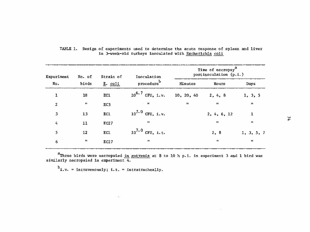
TABLE 1. Design of experiments used to determine the acute response of spleen and liver in 3-week-old turkeys inoculated with Escherichia coli
Experiment
No.
No. of
birds
Strain of
E. coli
Inoculation
procedure
Time of necropsy postinoculation (p.i.)
Minutes Hours Days
1
2
3
4
5
6
18
I?
13
11
12
ECl
EC3
ECl
EC27
ECl
EC27
10^'7 CFU, i.v.
10 '° CFU, i.v.
10^'° CFU, i. t.
10, 20, 40 2, 4, 8
2, 4, 6, 12
1, 3, 5
2 , 8 1, 3, 5, 7
Three birds were necropsied in extremis at 8 to 10 h p.i. in experiment 3 and 1 bird was similarly necropsied in experiment 4.
^i.v. = intravenously; i.t. = intratracheally.
75
anticoagulant. Serial, 10-fold dilutions of blood in PBS were inoculated
onto blood agar plates for determination of colony-forming units (CPU)/ml
of blood (3). The remaining fraction of the 1/10 dilution of blood was
mixed with an equal volume of brain-heart infusion broth and incubated
at 37° C.
At necropsy, turkeys were immobilized by an i.v. injection of succinyl
choline and killed by decapitation. Birds were dipped in a disinfectant
solution and opened, gross lesions were recorded, and spleen and liver
specimens were collected.
The development of bacteremia was studied in 10 turkeys inoculated
i. t. with JE. coli; sequential blood sançles were collected up to 7 days
p.i. (Table 2). Numbers of coli in blood were determined as described
above.
Just prior to each experiment, sera from 10 to 20 percent of the
turkeys were tested for antibody against the E. coli inoculum by tube
agglutination.
Light microscopy. Specimens of spleen and liver were fixed in 10%
neutral buffered formalin, dehydrated in alcohols, embedded in paraffin,
and sectioned at 5-6 ujn. Sections were stained with hematoxylin and
eosin (H & E), Giemsa, and modified Dieterle (87) stains. Additional
specimens of spleen and liver were fixed in Bouin's solution 12 h,
washed in water and 70% ethanol, dehydrated, embedded, sectioned, and
stained with H & E.
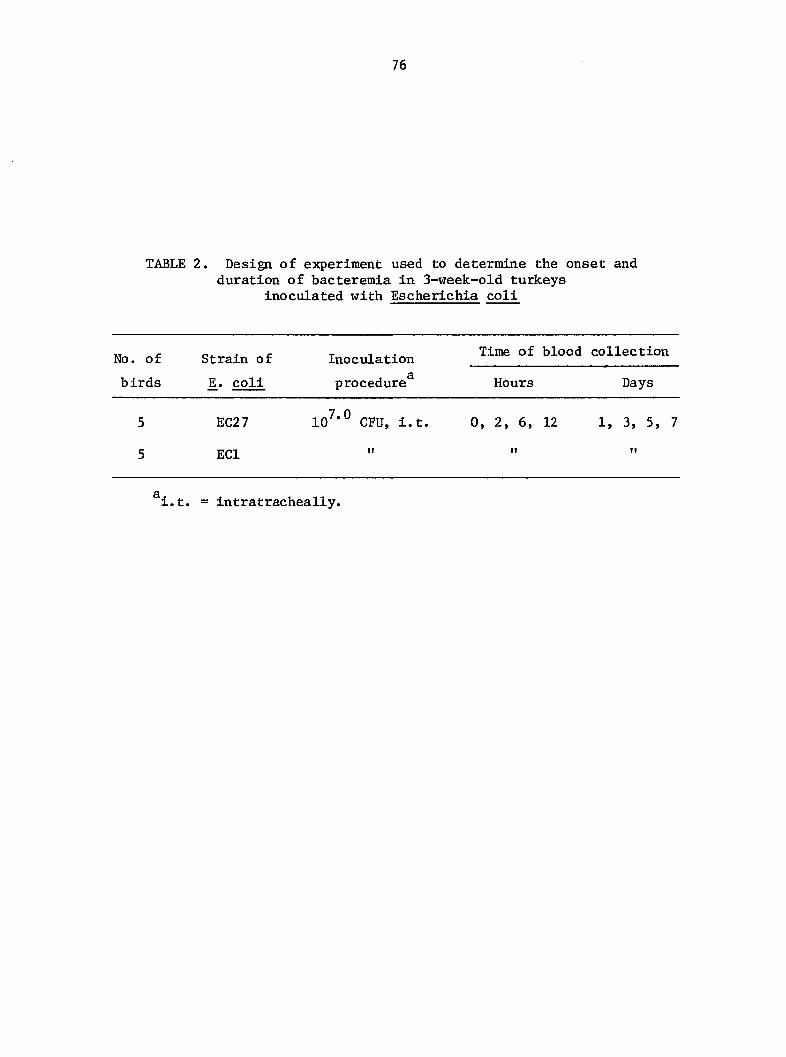
76
TABLE 2. Design of experiment used to determine the onset and duration of bacteremia in 3-week-old turkeys
inoculated with Escherichia coli
NO. of Strain of Inoculation collection
birds coli procedure Hours Days
5 EC27 lo/'O CPU, i.t. 0, 2, 6, 12 1, 3, 5, 7
5 ECl " " "
i.t. = intratracheally.
77
Electron microscopy. Specimens of spleen and liver were collected
from birds killed at 6 h p.i. and in extremis in experiment 3. Cubes
(0.5mm) of tissue were fixed in 2.5% buffered glutaraldehyde for 2 h,
washed in 0.1 M phosphate buffer pH 7.4, postfixed in 2% osmium tetroxide,
washed in buffer, and dehydrated through graded ethanols and propylene
oxide to embedment in epoxy resin (Epon 812). Ultrathin sections were
cut with a diamond knife, stained with 2% uranyl acetate and Reynold's
lead citrate, and examined with an electron microscope.
78
RESULTS
Response to 1.v. inoculation of coll (experiments 1-4). Spleens
were generally dark and slightly enlarged although some spleens exposed
to ECl or EC27 were pale and mottled at less than 1 day p.i. Micro
scopically, spleens were characterized by hyperemia, heterophil infil
tration, fibrinopurulent exudation and necrosis in reticular sheaths,
necrosis of isolated cells, thrombocyte aggregation, and lymphoid hyper
plasia. The onset, duration, and severity of lesions varied with the
virulence of the coli strain (Fig. 1). Red pulp spaces were dilated
and filled with erythrocytes. Heterophil infiltration was diffuse in red
pulp but intense in reticular sheaths (ellipsoids) (Fig. 2). There was
localization of heterophils in and around reticular sheaths accompanied
by necrosis and fluid exudation, particularly in birds exposed to ECl or
EC27. In birds inoculated with EC3, there was only slight cellular and
fluid exudation into reticular sheaths. Aggregates of thrombocytes were
common in the red pulp and venous sinuses of birds at 2 to 6 h p.i.
Beginning at 3 days p.i., lymphocytes surrounding reticular sheaths
(periellipsoidal) had abundant cytoplasm and large basophilic nuclei
with a prominent nucleolus; mitotic figures were common (Fig. 3).
Numbers of lymphoid follicles were increased in some birds as early as 5
days p.i.
Spleens of birds killed extremis were diffusely necrotic and
hyperemic. Small numbers of bacilli were scattered in necrotic tissue
and rarely formed colonies. Ultrastructurally, cells associated with
reticular sheaths of spleens were swollen and intercellular spaces were
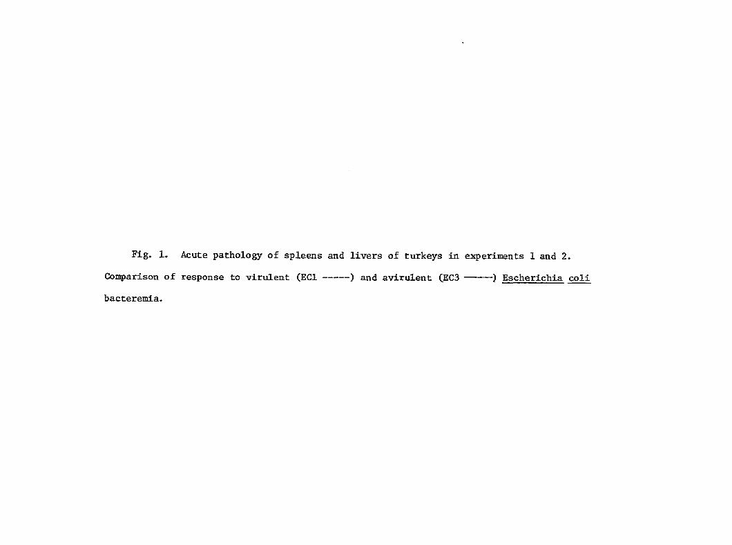
Fig. 1. Acute pathology of spleens and livers of turkeys in experiments 1 and 2.
Comparison of response to virulent (ECl ) and avirulent (ECS ) Escherichia coli
bacteremia.
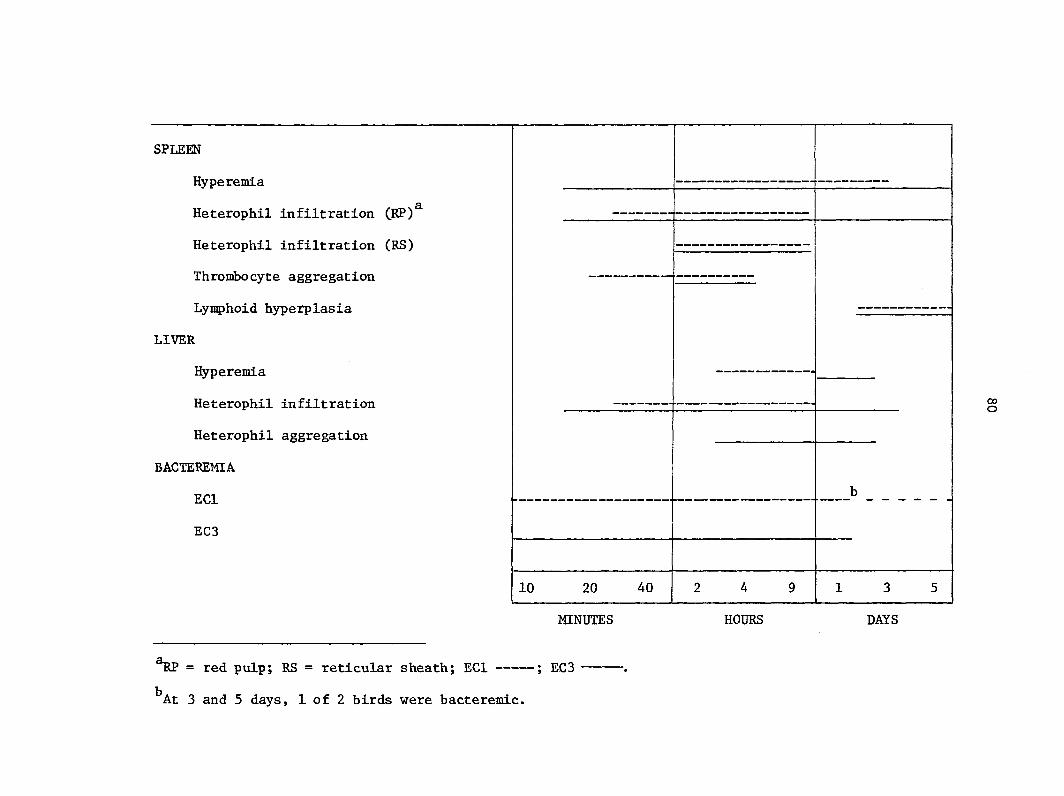
SPLEEN
Hyperemia
Heterophil infiltration CRP)^
Heterophil infiltration (RS)
Thrombocyte aggregation
Lymphoid hyperplasia
LIVER
Hyperemia
Heterophil infiltration
Heterophil aggregation
BACTEREMIA
ECl
EC3
1 1 1 1 1 1 i
1 1
1 1
1 1
i 1
1 1
1 1
1 1 1 1 1 1 1
10 20 40 2 4 9 13 5
MINUTES HOURS DAYS
RP = red pulp; RS = reticular sheath; ECl ; EC3
^At 3 and 5 days, 1 of 2 birds were bacteremic.
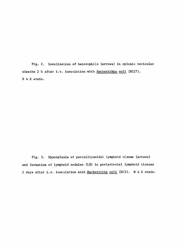
Fig. 2. Localization of heterophils (arrows) in splenic reticular
sheaths 2 h after i.v. inoculation with Escherichia coli (EC27).
H & E stain.
Fig. 3. Hyperplasia of periellipsoidal lymphoid tissue (arrows)
and formation of lymphoid nodules (LN) in periarterial lymphoid tissues
5 days after i.v. inoculation with Escherichia coli (EC3). H & E stain.
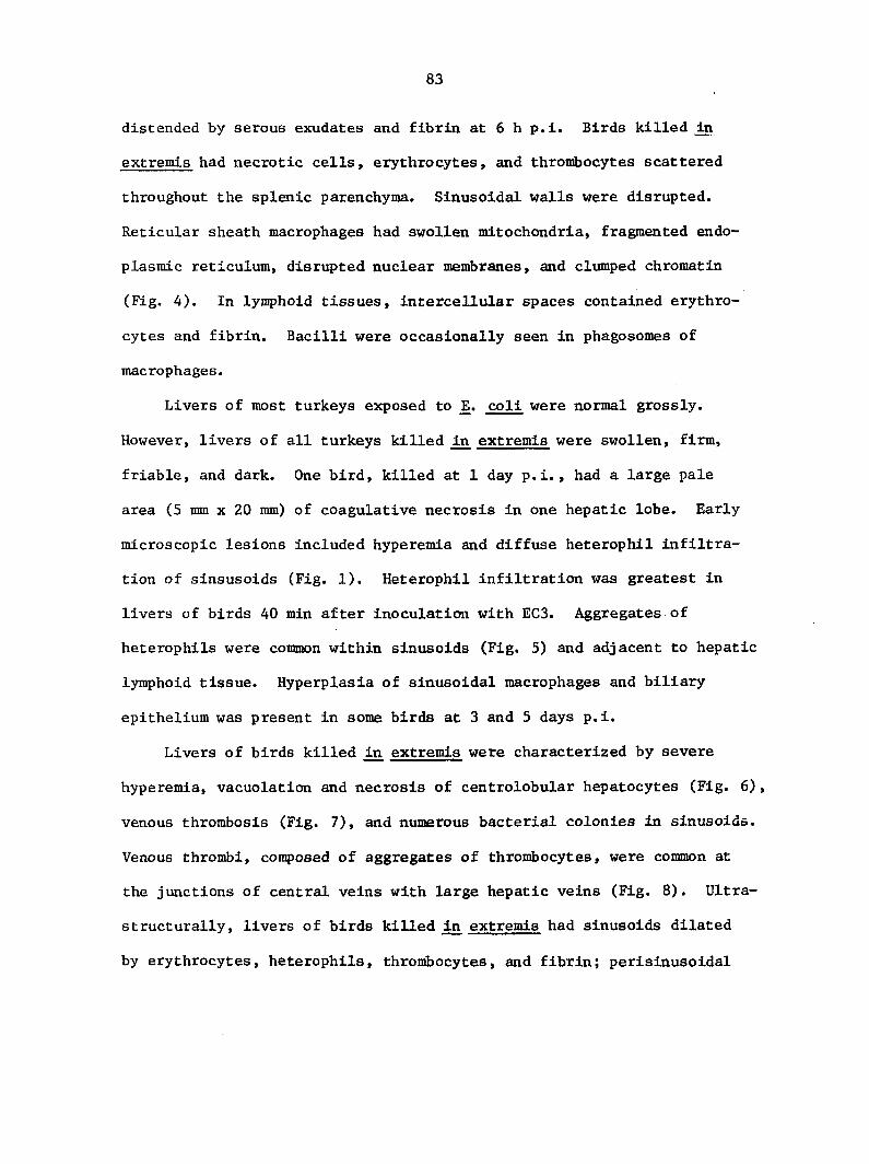
83
distended by serous exudates and fibrin at 6 h p.i. Birds killed in
extremis had necrotic cells, erythrocytes, and thrombocytes scattered
throughout the splenic parenchyma. Sinusoidal walls were disrupted.
Reticular sheath macrophages had swollen mitochondria, fragmented endo
plasmic reticulum, disrupted nuclear membranes, and clumped chromatin
(Pig. 4). In lymphoid tissues, intercellular spaces contained erythro
cytes and fibrin. Bacilli were occasionally seen in phagosomes of
macrophages.
Livers of most turkeys exposed to coll were normal grossly.
However, livers of all turkeys killed in extremis were swollen, firm,
friable, and dark. One bird, killed at 1 day p.i., had a large pale
area (5 mm x 20 mm) of coagulative necrosis in one hepatic lobe. Early
microscopic lesions included hyperemia and diffuse heterophil infiltra
tion of sinsusoids (Fig. 1). Heterophil infiltration was greatest in
livers of birds 40 min after inoculation with EC3. Aggregates of
heterophils were common within sinusoids (Fig. 5) and adjacent to hepatic
lymphoid tissue. Hyperplasia of sinusoidal macrophages and biliary
epithelium was present in some birds at 3 and 5 days p.i.
Livers of birds killed in extremis were characterized by severe
hyperemia, vacuolation and necrosis of centrolobular hepatocytes (Fig. 6),
venous thrombosis (Fig. 7), and numerous bacterial colonies in sinusoids.
Venous thrombi, composed of aggregates of thrombocytes, were common at
the junctions of central veins with large hepatic veins (Fig. 8). Ultra-
structurally, livers of birds killed in extremis had sinusoids dilated
by erythrocytes, heterophils, thrombocytes, and fibrin; perislnusoidal
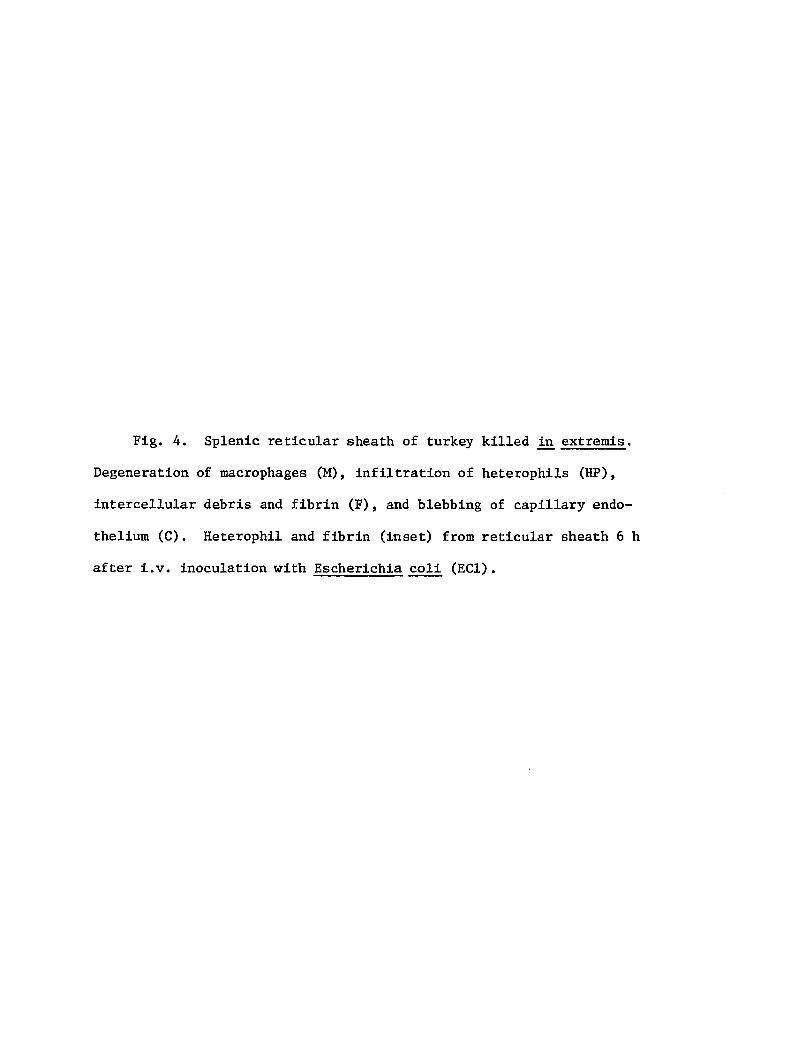
Fig. 4. Splenic reticular sheath of turkey killed extremis.
Degeneration of macrophages (M), infiltration of heterophils (HP),
intercellular debris and fibrin (F), and blebbing of capillary endo
thelium (C). Heterophil and fibrin (inset) from reticular sheath 6 h
after i.v. inoculation with Escherichia coli (ECl).
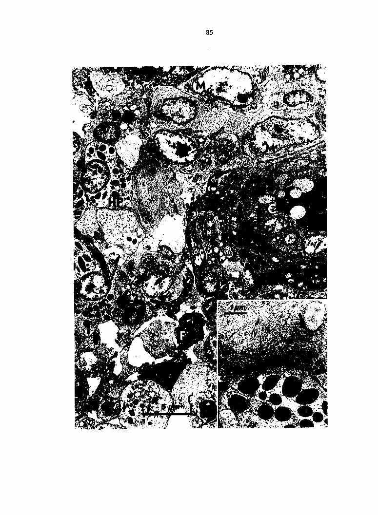
Fig. 5. Aggregates of heterophils (arrow) in hepatic sinusoids
4 h after i.v. inoculation with Escherichia coli (EC3). H & E stain.
Fig. 6. Vacuolation and necrosis of centrolobular hepatocytes in
a turkey killed extremis 10 h after i.v. inoculation with Escherichia
coli (EC27). Islands of hepatocytes around portal canals (PC) appear
normal. H & E stain.
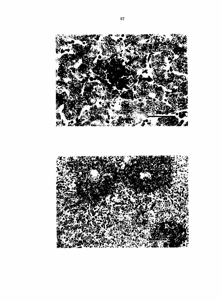
87
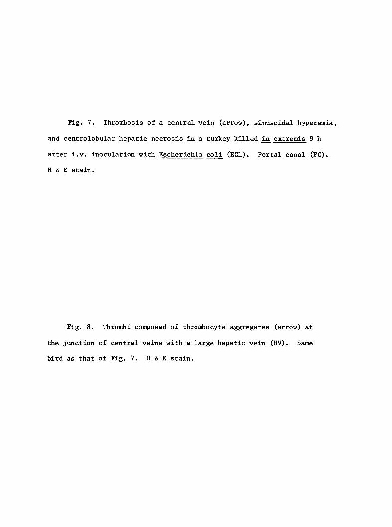
Fig. 7. Thrombosis of a central vein (arrow), sinusoidal hyperemia,
and centrolobular hepatic necrosis in a turkey killed extremis 9 h
after i.v. inoculation with Escherichia coli (ECl). Portal canal (PC).
H & E stain.
Fig. 8. Thrombi composed of thrombocyte aggregates (arrow) at
the junction of central veins with a large hepatic vein (HV). Same
bird as that of Fig. 7. H & E stain.
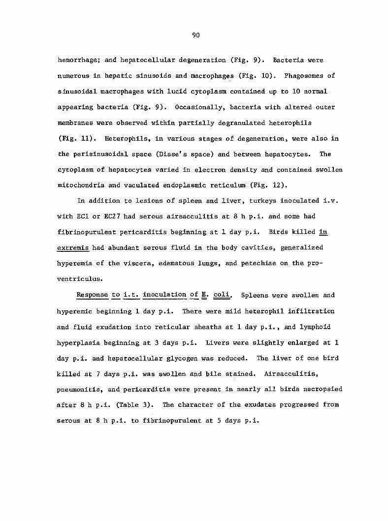
90
hemorrhage; and hepatocellular degeneration (Fig. 9). Bacteria were
numerous in hepatic sinusoids and macrophages (Fig. 10). Phagosomes of
sinusoidal macrophages with lucid cytoplasm contained up to 10 normal
appearing bacteria (Fig. 9). Occasionally, bacteria with altered outer
membranes were observed within partially degranulated heterophils
(Fig. 11). Heterophils, in various stages of degeneration, were also in
the perisinusoidal space (Disse's space) and between hepatocytes. The
cytoplasm of hepatocytes varied in electron density and contained swollen
mitochondria and vaculated endoplasmic reticulum (Fig. 12).
In addition to lesions of spleen and liver, turkeys inoculated i.v.
with ECl or EC27 had serous airsacculitis at 8 h p.i. and some had
fibrinopurulent pericarditis beginning at 1 day p.i. Birds killed in
extremis had abundant serous fluid in the body cavities, generalized
hyperemia of the viscera, edematous lungs, and petechiae on the pro-
ventriculus.
Response to i.t. inoculation of E^. coli. Spleens were swollen and
hyperemic beginning 1 day p.i. There were mild heterophil infiltration
and fluid exudation into reticular sheaths at 1 day p.i., and lymphoid
hyperplasia beginning at 3 days p.i. Livers were slightly enlarged at 1
day p.i. and hepatocellular glycogen was reduced. The liver of one bird
killed at 7 days p.i. was swollen and bile stained. Airsacculitis,
pneumonitis, and pericarditis were present in nearly all birds necropsied
after 8 h p.i. (Table 3). The character of the exudates progressed from
serous at 8 h p.i. to fibrinopurulent at 5 days p.i.

Fig. 9. Liver: Perisinusoidal space is expanded and contains
erythrocytes (E) and a heterophil (HP). Sinusoidal macrophage (SM) has
many normal appearing bacteria in a phagosome (inset). Acute cell
swelling, hepatocytes (H). From a turkey killed in extremis 8 h after
i.v. inoculation with Escherichia coll (ECl).
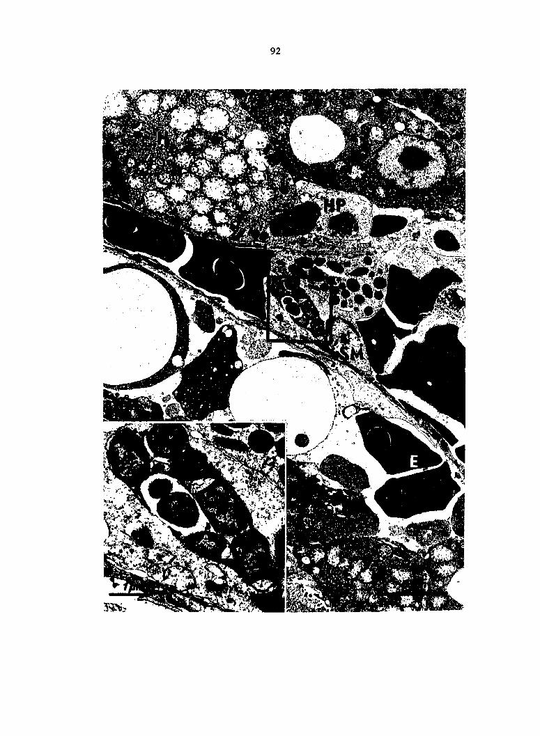
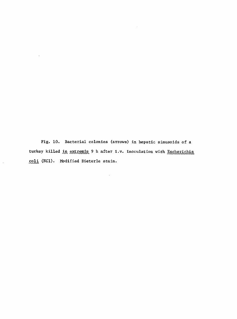
Fig. 10. Bacterial colonies (arrows) in hepatic sinusoids of a
turkey killed in extremis 9 h after l.v. inoculation with Escherichia
coli (ECl). Modified Dieterle stain.
94
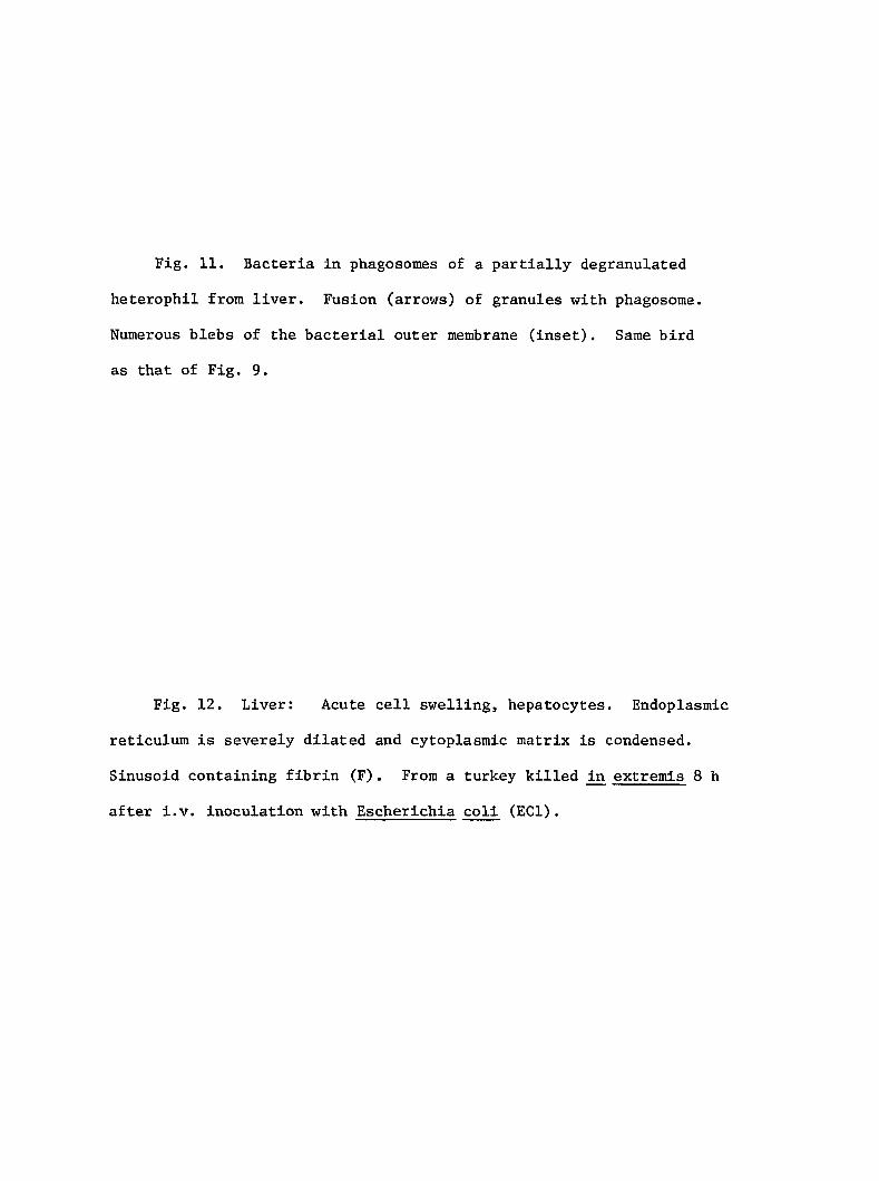
Fig. 11. Bacteria in phagosomes of a partially degranulated
heterophil from liver. Fusion (arrows) of granules with phagosome.
Numerous blebs of the bacterial outer membrane (inset). Same bird
as that of Fig. 9.
Fig. 12. Liver: Acute cell swelling, hepatocytes. Endoplasmic
reticulum is severely dilated and cytoplasmic matrix is condensed.
Sinusoid containing fibrin (F). From a turkey killed extremis 8 h
after i.v. inoculation with Escherichia coll (ECl).
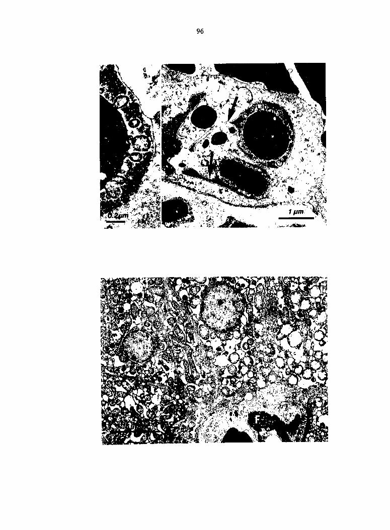
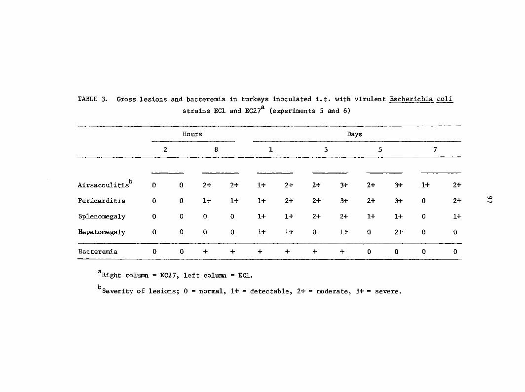
TABLE 3. Gross lesions and bacteremia in turkeys inoculated i.t. with virulent Escherichia coli
strains ECl and EC27^ (experiments 5 and 6)
Hours Days
Airsacculitis^ 0 0 2+ 2+ 1+ 2+ 2+ 3+ 2+ 3+ 1+ 2+
Pericarditis 0 0 1+ 1+ 1+ 2+ 2+ 3+ 2+ 3+ 0 2+
Splenomegaly 0 0 0 0 1+ 1+ 2+ 2+ 1+ 1+ 0 1+
Hepatomegaly 0 0 0 0 1+ 1+ 0 14- 0 2+ 0 0
Bacteremia 0 0 + + + + + + 0 0 0 0
^Right column = EC27, left column = ECl.
^Severity of lesions; 0 = normal, 1+ = detectable, 2+ = moderate, 3+ = severe.
98
The onset and duration of bacteremia in birds inoculated i.t. is
expressed in Table 4. Bacteremia associated with EC27 had a more rapid
onset, higher titer, and longer duration than that associated with ECl.
Two turkeys inoculated with EC27 died between 1 to 2 days p.i. Except
for the 2 birds which died, maximal bacterial titers were at 12 h p.i.
when the mean CFU of E. coli per ml of blood was 1,200 for EC27 and 400
for ECl. Blood bacterial titers were generally less than 100 CFU/ml at
other sample times, although the 2 birds which died had 14,000 and 5,000
CFU of E^. coll per ml of blood at 1 day p.i. The duration of bacteremia
correlated with the period of most severe visceral lesions seen in
experiments 5 and 6 (Table 3).
Control turkeys of each experiment had neither lesions or bacter
emia. Sera tested for agglutinating antibody against live coli
inocula had titers less than 1:4.
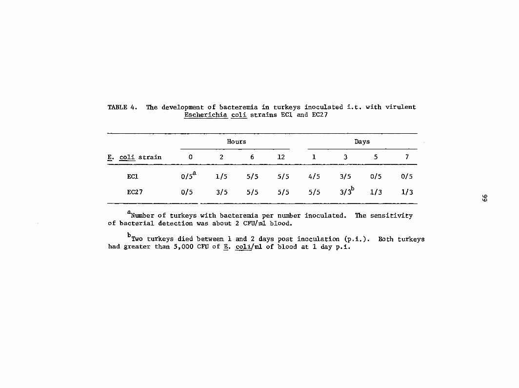
TABLE 4. The development of bacteremia in turkeys inoculated i.t. with virulent Escherichia coli strains ECl and EC27
Hours Days
E. coli strain 0 2 6 12 1 3 5 7
ECl 0/5* 1/5 5/5 5/5 4/5 3/5 0/5 0/5
EC27 0/5 3/5 5/5 5/5 5/5 3/3^ 1/3 1/3
^Number of turkeys with bacteremia per number inoculated. The sensitivity of bacterial detection was about 2 CFU/ml blood.
^Two turkeys died between 1 and 2 days post inoculation (p.i.). Both turkeys had greater than 5,000 CFU of jE. coli/ml of blood at 1 day p.i.
100
DISCUSSION
The spleen of most animals has a significant role in eliminating
infectious agents from cirulating blood. Blood-bome coli in turkeys
is phagocytized by reticular sheath macrophages and less so by phago
cytes In the red pulp (3). The splenic lesions associated with JE. coli
bacteremia in the present study are a reflection of the acute inflam
matory response focused on the reticular sheaths. Concentration of
fibrinopurulent exudates within and adjacent to reticular sheaths may be
a response to potent chemotactic substances generated during the inter
action of coli with plasma coiiq>onents (e.g. complement) and phago
cytes. Heterophils appear to emigrate to reticular sheaths subsequent
to more diffuse accumulation in the red pulp and adjacent lymphoid
tissue. Such a response would be expected to a chemotactic gradient
originating in reticular sheaths.
The more rapid onset of inflammation in spleen and liver of birds
injected with avirulent E^. coli (EC3) compared with virulent coli
(ECl) was also seen in a previous study (3). This difference may be due
to a greater capacity of avirulent JE. coll to cause generation of chemo
tactic substances. Gonococci isolated from patients with local infec
tion readily activate complement, whereas gonococci Isolated from
patients with disseminated gonococcal infection activate complement
slowly and are poor stimulators of chemotaxls (19). Inefficient activa
tion of the alternative complement pathway by coli isolated from
patients with bacteremia has been associated with the presence of K
capsular polysaccharide on the bacterial surface (10). Therefore,
101
the delayed inflammatory response to virulent coll in the present
study may have been due to inhibition of complement activation caused by
capsular antigens. The greater inflammatory response in livers against
EC3 compared with ECl may have been due to both greater localization of
EC3 in liver (3) and greater generation of chemotactic substances.
The increased size and staining intensity of lymphoid tissue adjacent
to reticular sheaths was probably due to blast transformation of lympho
cytes. A similar response of splenic lymphoid tissue has been reported
in turkeys infected with Mycoplasma meleagridis and E . coli (51). In
creased numbers of periarterial lymphoid follicles seen at 5 to 7 days
p.i. may represent proliferating clones of B lymphocytes. In chicks,
low levels of serum antibody are detectable within a week after inocula
tion with coli (40).
The pathogenesis of venous thrombosis in small hepatic veins of
birds killed extremis is speculative. In previous studies, rapid
formation of thrombocytic aggregates and thrombosis of small vessels in
lung, spleen, and liver occurred after i.v. injection of large numbers
of coli, particularly in the presence of specific antibody (2, 3).
The presence of thrombocytic thrombi, especially in small hepatic veins,
may be due to severe endotheial damage in collecting branches of hepatic
veins. Truscott et al. (84) attributed many vascular and parenchymal
lesions in spleen and liver of chickens to the action of a chick-lethal
toxin produced by some strains of E . coli. Also, small hepatic veins,
like centrolobular hepatocytes, would be more vulnerable to hypoxic
injury associated with severe septic shock. Scattered foci of hepatic
102
necrosis were likely the result of vascular occlusion since these areas
were free of both bacteria and granulocytes. Thrombosis of hepatic
veins in birds extremis may have impaired sinusoidal circulation to
the extent that severe sinusoidal congestion, bacterial colonization,
and hepatocellular necrosis were inevitable sequelae. Extensive bacterial
colonization of hepatic sinusoids, in contrast to splenic sinusoids,
indicates that the liver may be an important site of bacterial seeding
into the bloodstream in progressive coll bacteremia. The abundance
of fibrin within hepatic sinusoids and spleens of birds in extremis
indicates that systemic activation of the coagulation system may have
also impaired the microcirculation.
Lesions of spleen and liver were much less severe in turkeys inocu
lated i.t. compared with those inoculated i.v. Thus, severity of lesions
in spleen and liver appears to be a function of the duration and magni
tude of bacteremia and the rate of coll localization within tissues.
Low numbers of coli in the blood are apparently effectively managed
in most birds by the mononuclear phagocytes of spleen and liver without
the development of an intense granulocytic response. The death of 2
birds (Table 4), 1 to 2 days after i.t. inoculation with coll suggests
that the stress of repeated blood sampling and handling may have im
paired their capacity to control early coli bacteremia.
Although colisepticemia may occur secondary to the stress of
primary disease or environmental factors (72), whether such stress causes
dysfunction of the mononuclear phagocyte system is unknown. Since the
liver is of major importance for removal of JE. coli from blood (2, 3, 7),
103
impairment of hepatic circulation may predispose to progressive bac
teremia. Hypothetically, hepatic circulation may be reduced in shock or
dehydration resulting from primary disease.
104
GENERAL SUMMARY AND DISCUSSION
Bacteria that cause acute systemic disease generally do so because
of their capacity to resist ingestion by host phagocytes (67). Although
similar numbers of virulent and aviulent JE. coli localized in spleen,
greater numbers of avirulent coli were present in liver. Studies of
bacteremia in mammals (7) and the present study have shown that rapid
clearance of bacteria from blood is dependent upon avid phagocytosis by
hepatic macrophages. The greater importance of liver conçared with spleen
in clearance of blood-borne bacteria is probably due to the greater size
and blood flow of liver (7). In turkeys, retention of bacteria by splenic
macrophages is probably more inçortant for presenting bacterial antigens
to immunocoBçetent lymphocytes than for clearing large numbers of bacteria
from blood.
Avirulent bacteria and opsonized bacteria are rapidly cleared from
the blood of mammals by hepatic macrophages (7). In the present study,
the clearance from blood and localization of opsonized virulent JE. coli
was similar to that of avirulent JE. coli. Although serum agglutinin
titers of passively inanune turkeys were 1:8 or less, these turkeys were
protected against large numbers of virulent coll. In contrast, other
investigators have suggested that there is no correlation between 0 and K
serum agglutinins and protection against colisepticemia (17, 18). They
found that some experimental vaccines protected birds from challenge with
virulent JE. coli. but sera from only a small percentage of vaccinated
birds had agglutinin titers greater than 1:5 (17, 18). It appears
105
likely from the present studies that humoral antibody may be pro
tective even when the quantity is too little for vitro detection-
Localization of coli in reticular sheath macrophages is con
sistent with the findings of White and Gordon (92). They found that in
chickens blood-borne staphlococci and soluble protein antigens localize
in reticular sheaths whereas larger particles (Candida albicans) localize
exclusively in red pulp (92). In the present studies, virulent coli
localized predominantly in reticular sheaths but avirulent coll and
opsonized JE. coli localized both in reticular sheaths and in red pulp
sinusoids. Phagocytosis of large numbers of virulent coli by macro
phages of the reticular sheaths may be due to the very limited extra
cellular space of these macrophages compared with those of sinusoids in
splenic red pulp and liver. The close association among these macrophages
would enhance bacteria-phagocyte contacts and reduce the repulsive effects
of antiphagocytic components on the surface of virulent E. coll.
Aggregation of thrombocytes was a common finding in turkeys killed
in extremiR and in those inoculated i.v. with large numbers of coli,
particularly in the presence of antibody. Platelet aggregation may occur
in mammals inoculated i.v. with large numbers of bacteria (37). Throm
bosis of pulmonary capillaries in passively immune birds inoculated with
coli is probably irrelevant to natural coli bacteremia because
numbers of JE. coli are generally not great except in terminal stages of
disease. Thrombosis of small hepatic veins was seen in all birds killed
in extremis ; however, a cause-effect relationship is difficult to es
tablish between severe bacteremia and venous thrombosis. Thrombosis
106
of small hepatic veins may further impede sinusoidal circulation,
enhance bacterial colonization, and ensure a fatal conclusion to the
disease.
The lesions in spleen and liver reflected the magnitude of
bacteremia and sites of bacterial localization in these tissues. That
is, avirulent JE. coli bacteria are trapped largely In liver and cause a
transient hyperemia with multifocal accumulation of heterophils in
sinusoids. Virulent coli localize In splenic reticular sheaths and
incite an intense granulocytic response. Intratracheal inoculation is
associated with a persistent bacteremia of low magnitude and only mild
splenic and hepatic lesions in most birds.
Although these studies have contributed to our understanding of the
role of bacteremia in the pathogenesis of colisepticemia in poultry,
important questions remain. Why, in some birds, does coli bacteremia
become progressive and fatal? Are virulent coli bacteria opsonized
by "natural" antibody or components of complement? Where do predis
posing factors operate in the pathogenesis of colisepticemia to make
birds more susceptible to the disease? Is the capacity of the mono
nuclear phagocyte system of the spleen and liver to control bacteremia
impaired by primary disease and dehydration?
107
LITERATURE CITED
Arp, L. H. 1980. Consequences of active or passive immunization
of turkeys against Escherichia coli 078. Avian Dis. 24:808-815
Arp, L. H. 1981. Effect of passive immunization on phagocytosis of
blood-bome Escherichia coll in spleen and liver of turkeys.
Infect. Immun. (in press, IAI245).
Arp, L. H., and N. F. Cheville. 1981. Interaction of blood-bome
Escherichia coli with phagocytes of spleen and liver in turkeys
Am. J. Vet. Res. 42:650-657.
Arp, L. H., C. L. G. Graham, and N. F. Cheville. 1979. Conçarison
of clearance rates of virulent and avirulent Escherichia coli
in turkeys after aerosol exposure. Avian Dis. 23:386-391.
Arp, L. H., and A. E. Jensen. 1980. Filiation, hemagglutination,
motility, and generation time of Escherichia coli that are
virulent or avirulent for turkeys. Avian Dis. 24:153-161.
Benacerraf, B., and P. Miescher. 1960. Bacterial phagocytosis by
the reticuloendothelial system vivo under different immune
conditions. Ann. N. Y. Acad. Sci. 88:184-195.
Benacerraf, B., M. M. Sebestyen, and S. Schlossman. 1959. A quanti
32 tative study of the kinetics of blood clearance of P -labelled
Escherichia coli and staphylococci by the reticuloendothelial
system. J. Exp. Med. 110:27-48.
Bennett, I. L., Jr., and P. B. Beeson. 1954. Bacteremia: a con
sideration of some experimental and clinical aspects. Yale
J. Biol. Med. 26:241-261.
108
9. Black, A. P,, A. M. Crichlow, and J. R. Saunders. 1980. Bacteremia
during ultrasonic teeth cleaning and extraction in the dog.
J. Am. Anim. Hosp. Assoc. 16:611-616.
10. Bortolussi, R., P. Ferrieri, B. Bjorksten, and P. G. Quie. 1979.
Capsular K1 polysaccharide of Escherichia coli ; relationship
to virulence in newborn rats and resistance to phagocytosis.
Infect. Immun. 25:293-298.
11. Braude, A. I., H. Douglas, and J. Jones. 1969. Experimental pro
duction of lethal Escherichia coli bacteremia of pelvic origin.
J. Bacteriol. 98:979-991.
12. Brinton, C. C., Jr. 1965. The structure, function, synthesis, and
genetic control of bacterial pili and a molecular model for DNA
and RNA transport in gram-negative bacteria. Trans. NY Acad.
Sci. 27:1003-1054.
13. Cheville, N. F. 1976. Cell pathology. Iowa State University Press,
Ames, Iowa.
14. Cheville, N. F., and L. H. Arp. 1978. Comparative pathologic
findings of Escherichia coli infection in birds. J. Am. Vet.
Med. Assoc. 173:584-587.
15. Costerton, J. W., J. M. Ingram, and K. J. Cheng. 1974. Structure
and function of the cell envelope of grammegative bacteria.
Bacteriol. Rev. 38:87-110.
16. Davis, C. R. 1938. Colibacillosis in young chicks. J. Am. Vet.
Med. Assoc. 92:518-522.
109
17. Deb, J. R., and E. G. Harry. 1976. Laboratory trials with
inactivated vaccines against Escherichia coli (078 K80)
infection in fowls. Res. Vet. Sci. 20:131-138.
18. Deb, J. R., and E. G. Harry. 1978. Laboratory trials with
inactivated vaccines against Escherichia coli (02:K1) infection
in fowls. Res. Vet. Sci. 24:308-313.
19. Densen, P., L. MacKeen, and R. A. Clark. 1979. Gonococci causing
uncomplicated gonorrhea or disseminated gonococcal infection
differ in stimulation of neutrophil chemotaxis (abstract no. 4).
In Proceedings of the 11th international congress of chemo
therapy and the 19th interscience conference on antimicrobial
agents and chemotherapy. American Society for Microbiology,
Washington, D. C.
20. Dijk, W. C. van, H. A. Verbrugh, M. E. van der Toi, R. Peters,
P. K. Peterson, P. G. Quie, and J. Verhoef. 1980. Inter
actions of phagocytic and bacterial cells in patients with
bacteremia caused by gram-negative rods. J. Infect. Dis.
141:441-448.
21. Fabricant, J., and P. P. Levine. 1962. Experimental production of
complicated chronic respiratory disease infection ("air sac
disease"). Avian Dis. 6:13-23.
22. Firth, G. A. 1977. The normal lymphatic system of the domestic
fowl. Vet. Bull. 47:167-179.
110
23. Furth, R. van, and Z. A. Cohn, J. G. Hirsch, J. H. Humphrey,
W. G. Spector, and H. L. Langevoort. 1972. The mononuclear
phagocyte system: a new classification of macrophages,
monocytes, and their precursor cells. Bull. W. H. 0. 46:845-852.
24. Gilbert, D. N., J. A. Barnett, and J. P. Sanford. 1973. Escherichia
coli bacteremia in the squirrel monkey. I. Effect of cobra
venom factor treatment. J. Clin. Invest. 52:406-413.
25. Glantz, P. J., S. Narotsky, G. Bubash. 1962. Escherichia coli
serotypes isolated from salpingitis and chronic respiratory
disease of poultry. Avian Dis. 6:322-328.
26. Griffin, F. M., Jr., J. A. Griffin, J. E. Leider, and S. C.
Silverstein. 1975. Studies on the mechanism of phagocytosis.
I. Requirements for circumferential attachment of particle-
bound ligands to specific receptors on the macrophage plasma
membrane. J. Exp. Med. 142:1263-1282.
27. Gross, W. B. 1956. Escherichia coli as a complicating factor in
chronic respiratory disease of chickens and infectious
sinusitis of turkeys. Poult. Sci. 35:765-771.
28. Gross, W. B. 1958. Symposium on chronic respiratory diseases of
poultry II. The role of Escherichia coli in the cause of
chronic respiratory disease and certain other respiratory
diseases. Am. J. Vet. Res. 19:448-452.
29. Gross, W. B. 1961. The development of "air sac disease."
Avian Dis. 5:431-439.
Ill
30. Gross, W. B. 1962. Blood cultures, blood counts and temperature
records in an experimentally produced "air sac disease" and
uncomplicated Escherichia coli infection of chickens. Poult.
Sci. 41:691-700.
31. Gross, W. B. 1978. Colibacillosis, p. 321-330. In M. S. Hofstad
(ed.), Diseases of poultry, 7th ed. Iowa State University
Press, Ames, Iowa.
32. Gross, W. B., and Siegel, P. B. 1959. Coliform peritonitis of
chickens. Avian Dis. 3:370-373.
33. Harry, E. G. 1957. The effect of embryonic and chick mortality of
yolk contamination with bacteria from the hen. Vet. Rec.
69:1433-1439.
34. Hjarre, A., and G. Wramby. 1945. Undersokningar over en med
specifika granulom forlopande honssjukdom orsaked av mukoida
Kolibacterier (Koli-granulom). Skan Veterinaertidskr.
35:449-507.
35. Hodges, R. D. 1974. The histology of the fowl. Academic Press,
New York, New York.
36. Hoffmann-Fezer, G., H. Rodt, D. GOtze, and S. Thierfelder. 1977.
Anatomical distribution of T and B lymphocytes identified by
immunohistochemistry in the chicken spleen. Int. Arch.
Allergy Appl. Immunol. 55:86-95.
112
37. Horn, R. G., M. G. Koenig, J. S. Goodman, and R. D. Collins. 1969.
Phagocytosis of Staphylococcus aureus by hepatic reticulo
endothelial cells. An ultrastructural study. Lab. Invest.
21:406-414.
38. Howard, C. J., and A. A. Glynn. 1971. The virulence for mice of
strains of Escherichia coli related to the effects of K antigens
on their resistance to phagocytosis and killing by complement.
Immunology 20:767-777.
39. Howard, J. G. , and A. C. Wardlaw. 1958. The opsonic effect of
normal serum on the uptake of bacteria by the reticuloendo
thelial system. Immunology 1:338-352.
40. Kendler, J., and E. G. Harry. 1967. Systemic Escherichia coli
infection as a physiological stress in chickens. Res. Vet.
Sci. 8:212-218.
41. Klein, E. 1889. Uber eine akute Infektiose Krankeit des schottlschen
Moorhuhnes (Lagopus scotrius). Zentralbl. Bakteriol. Orig. 6:36.
42. Kreger, B. E., D. E. Craven, P. C. Carling, and W. R. McCabe. 1980.
Gram-negative bacteremia III. Reassessment of etiology, epi
demiology and ecology in 612 patients. Am. J. Med. 68:332-355.
43. Lignieres, M. J. 1894. Septicemic a coli-bacille chez la poule.
R. Soc. Biol. 46:135-137.
44. Lopez-Alvarez, J., and C. L. Gyles. 1980. Occurrence of the Vir
plasmid among animal and human strains of invasive Escherichia
coll. Am. J. Vet. Res. 41:769-774.
113
45. McCabe, W. R. 1970. Endotoxin and bacteremia due to gram-negative
organisms. N. Engl. J. Med. 283-1342-1343.
46. McCabe, W. R. 1973. Serum complement levels in bacteremia due to
gram-negative organisms. N. Engl. J. Med. 288:21-23.
47. McCabe, W. R., P. C. Carling, and S. Bruins. 1975. The relation of
K-antigen to virulence of Escherichia coli. J. Infect. Dis.
131:6-10.
48. Medearis, D. N., B. M. Camitta, and E. C. Heath. 1968. Cell wall
composition and virulence in Escherichia coli. J. Exp. Med.
128:399-414.
49. Medway, W., and M. R. Kare. 1959. Thiocyanate space in growing
domestic fowl. Am. J. Physiol. 196:873-875.
50. Moon, H. W., R. E. Isaacson, and J. Pohlenz. 1979. Mechanisms of
association of enteropathogenic Escherichia coli with intestinal
epithelium. Am- J. Clin. Nutr. 32:119-127.
51. Moorhead, P. D., and Y. M. Saif. 1970. Mycoplasma meleagridis and
Escherichia coli infections in germfree and specific-pathogen-
free turkey poults: pathologic manifestations. Am. J. Vet. Res.
31:1645-1653.
52. Nairn, M. E. 1973. Bacterial osteomyelitis and synovitis of the
turkey. Avian Dis. 17:504-517.
53. Ofek, I., E. H. Beachey, and A. L. Bisno. 1974. Resistance of
Neisseria gonorrhoeae to phagocytosis: relationship to
colonial morphology and surface pili. J. Infect. Dis.
129:310-316.
114
54. 0rskov, F. 1978. Virulence factors of the bacterial cell
surface. J. Infect. Dis. 137:630-633.
55. Ottow, J. C. G. 1975. Ecology, physiology, and genetics of
fimbriae and pili. Annu. Rev. Microbiol. 29:79-108.
56. Palmer, C. C., and H. R. Baker. 1923. Studies on infectious
enteritis of poultry caused by Bacterium coli communis.
J. Am. Vet. Med. Assoc. 63:85.
57. Piercy, D. W. T., and B. West. 1976. Experimental Escherichia
coli infection in broiler chickens: course of the disease
induced by inoculation via the air sac route. J. Comp. Pathol.
86:203-210.
58. Punsalang, A. P., Jr., and W. D. Sawyer. 1973. Role of pili in the
virulence of Neisseria gonorrhoeae. Infect. Immun. 8:255-263.
59. Purton, M. D. 1969. Structure and ultrastructure of the liver in
the domestic fowl. Callus gallus. J. Zool., Lond. 159:273-282.
60. Robinson, J. P., S. S. Schuffman, and S. H. W. Sell. 1972. Electron
microscopic studies of complexes of Haemophilus influenzae,
type b, with specific antibodies. Immunology 23:101-104.
61. Rogers, D. E. 1960. Host mechanisms which act to remove bacteria
from the blood stream. Bacteriol. Rev. 24:50-66.
62. Rogers, D. E., and M. A. Melly. 1957. Studies on bacteriemia.
III. The blood stream clearance of Escherichia coli in rabbits.
J. Exp. Med. 105:113-124.
115
63. Ruggiero, G., A. Andreana, R. Utili, and D. Galante. 1980. Enhanced
phagocytosis and bactericidal activity of hepatic reticuloendo
thelial system during endotoxin tolerance. Infect. Immun.
27:798-803.
64. Saif, Y. M. , P. D. Moorhead, and E. H. Bohl. 1970. Mycoplasma
meleagridis and Escherichia coli infections in germfree and
specific-pathogen-free turkey poults: production of complicated
airsacculitis. Am. J. Vet. Res. 31:1637-1643.
65. Siccardi, F. J. 1975. Considerations concerning the host-parasite
relationship of Escherichia coli in poultry. Am. J. Vet. Res.
36:572.
66. Silverblatt, F. J., J. S. Dreyer, and S. Schauer. 1979. Effect of
pili on susceptibility of Escherichia coli to phagocytosis.
Infect. Immun. 24:218-223.
67. Smith, H. 1976. Survival of vegetative bacteria in animals,
p. 299-326. ^ T. R. Gray and J. R. Postgate (ed.). The survival
of vegetative microbes. Cambridge University Press, New York,
New York.
68. Smith, H. 1977. Microbial surfaces in relation to pathogenicity.
Bacteriol. Rev. 41:475-500.
69. Smith, H. W. 1974. A search for transmissible pathogenic characters
in invasive strains of Escherichia coli: the discovery of a
plasmid-controlled toxin and a plasmid-controlled lethal
character closely associated, or identical, with colicine V.
J. Gen. Microbiol. 83:95-111.
116
70. Smith, H. W. 1978. Transmissible pathogenic characteristics of
invasive strains of Escherichia coli. J. Am. Vet. Med. Assoc.
173:601-607.
71. Smith, H. W., and M. B. Muggins. 1976. Further observations on the
association of the colicine V plasmid of Escherichia coli with
pathogenicity and with survival in the alimentary tract.
J. Gen. Microbiol. 92:335-350.
72. Sojka, W. J. 1965. Escherichia coll in domestic animals and poultry.
Commonwealth Agricultural Bureaux, England.
73. Sojka, W. J., and R. B. A. Cranaghan. 1961. Escherichia coli
infection in poultry. Res. Vet. Sci. 2:340-352.
74. Spiegelberg, H. L., P. A. Miescher, and B. Benacerraf. 1963.
Studies on the role of complement in the immune clearance of
Escherichia coli and rat erythrocytes by the reticuloendothelial
system in mice. J. Immunol. 90:751-759.
75. Stendahl, 0., and B. Normann. 1979. Surface characteristics of
Escherichia coli strains in relation to development of
bacteremia. Acta Pathol. Microbiol. Scand., Sect. B. 87:93-96.
76. Stendahl, 0., B. Normann, and L. Edebo. 1979. Influence of 0 and
K antigens on the surface properties of Escherichia coli in
relation to phagocytosis. Acta Pathol. Microbiol. Scand.,
Sect. B, 87:85-91.
117
77. Stevens, P., C. L. Chu, and L. S. Young. 1980. K-1 antigen content
and the presence of an additional sialic acid-containing antigen
among bacteremic K-1 Escherichia coli: correlation with sus
ceptibility to opsonophagocytosis. Infect. Immun. 29:1055-1061.
78. Stiffel, C., D. Mouton, and G. Biozzi. 1970. Kinetics of the
phagocytic function of reticuloendothelial macrophages in vivo,
p. 335-381. j[n R. van Furth (ed.). Mononuclear phagocytes.
Blackwell Scientific Publications, Oxford.
79. Stossel, T. P. 1976. The mechanism of phagocytosis. J.
Reticuloendothel. Soc. 19:237-245.
80. Svanborg Eden, C., and H. A. Hansson. 1978. Escherichia coli pili
as possible mediators of attachment to human urinary tract
epithelial cells. Infect. Immun. 21:229-237.
81. Sweeny, P. R., and H. C. Carlson. 1968. Electron microscopy and
histochemical demonstration of lysosomal structures in chicken
thrombocytes. Avian Dis. 12:636-644.
82. Tee, G. L., and G. K. Scott. 1980. Analysis of outer membrane
components of Escherichia coli ML308 225 and of a serum-
resistant mutant. Infect. Immun. 28:387-392.
83. Truscott, R. B. 1973. Studies on the chick-lethal toxin of
Escherichia coli. Can. J. Comp. Med. 37;375-381.
84. Truscott, R. B., J. Lopez-Alvarez, and J. R. Pettit. 1974. Studies
of Escherichia coli infection in chickens. Can. J. Comp. Med.
38:160-167.
118
85. Trylich, C., J. Howell, and D. W. MacDonald. 1977. Avian
coligranuloma: case histories. Can. Vet. J. 18:38-40.
86. Twisselmann, N. M. 1939. An acute infectious disease of pullets
apparently caused by Escherichia coli communis. J. Am. Vet.
Med. Assoc. 94:235-236.
87. Van Orden, A. E., and P. W. Greer. 1977. Modification of the
Dieterle spirochete stain. J. Histotech. 1:51-53.
88. Wardlaw, A. C., and J. G. Howard. 1959. A comparative survey of
the phagocytosis of different species of bacteria by Kupffer
cells. Perfusion studies with isolated rat liver. Br. J.
Exp. Pathol. 40:113-117.
89. Wasserman, B., V. J. Yates, and D. E. Fry. 1954. On so-called
air-sac infection. Poult. Sci. 33:622-623.
90. Weinstein, R. J., and L. S. Young. 1976. Neutrophil function in
gram-negative rod bacteremia. The interaction between phago
cytic cells, infecting organisms, and humoral factors. J. Clin.
Invest. 58:190-199.
91. Weller, P. F., A. L. Smith, and D. H. Smith. 1978. Role of immunity
in the clearance of bacteremia due to Haemophilus influenzae.
J. Infect. Dis. 138:427-436.
92. White, R. G., and J. Gordon. 1970. Macrophage reception and
recognition mechanisms in the chicken spleen, p. 510-523. In_
R. van Furth (ed.). Mononuclear phagocytes. Blackwell Scientific
Publications, Oxford.
119
, W. B. , Jr. 1960. Phagocytosis, with particular reference
to encapsulated bacteria. Bacteriol. Rev. 24:41-49.
120
ACKNOWLEDGMENTS
I would first like to thank my wife, Rosemary and my sons,
Chris and Tim for their love and understanding during my graduate
training; to them I dedicate this dissertation.
My special «thanks go to my co-major professors, Drs. Norman F.
Cheville and John P. Kluge. Dr. Cheville*s supervision of my research
and his wise counsel in the preparation of manuscripts are appreciated.
I am indebted to Dr. Kluge for guiding me through my graduate program.
I also thank the other members of my graduate committee, Drs. D. L.
Graham, M. S. Hofstad, and R. A. Packer for their time and advice
in my behalf.
The help of Dr. G. D. Booth in the interpretation of statistical
data is appreciated.
Without the technical assistance of my dedicated co-workers
this research would have been impossible or pure drudgery at best.
I am grateful to Karen Schlueter (electron microscopy), Loren Elliott
(histology), John Flickinger (special stains), Allen Jensen (bacteriol
ogy) , Wayne Romp and Tom Glasson (photography), Gene Hedberg
(illustrations), Diane Finch (typing of manuscripts), Leah Oppedal
(typing of dissertation), and Janice Eifling and Carol Moeller
(library services).
Lastly, I thank Dr. Phillip 0'Berry, Director of the National
Animal Disease Center, who has afforded me the time and facilities
for this research.