Embed Size (px)
Citation preview

Clinical StudyThe Prevalence of Tonsilloliths and Other Soft TissueCalcifications in Patients Attending Oral and MaxillofacialRadiology Clinic of the University of Iowa
Babatunde Olamide Bamgbose,1 Axel Ruprecht,2 John Hellstein,2
Sherry Timmons,2 and Fang Qian3
1 Department of Oral and Diagnostic Sciences, Faculty of Dentistry, Bayero University, Kano, Nigeria2 Department of Oral Pathology, Radiology, and Medicine, College of Dentistry and Dental Clinics, The University of IA,Iowa City, IA, USA
3Department of Preventive and Community Dentistry, College of Dentistry and Dental Clinics, The University of Iowa,Iowa City, IA, USA
Correspondence should be addressed to Babatunde Olamide Bamgbose; [email protected]
Received 24 October 2013; Accepted 1 December 2013; Published 22 January 2014
Academic Editors: S.-C. Choi and G. Kulkarni
Copyright © 2014 Babatunde Olamide Bamgbose et al. This is an open access article distributed under the Creative CommonsAttribution License, which permits unrestricted use, distribution, and reproduction in any medium, provided the original work isproperly cited.
Objective. The purpose of this study was to determine the prevalence of tonsiliths in patients attending the oral and maxillofacialradiology clinic of The University of Iowa and to determine if there is any correlation between the presence of tonsiliths and thepresence of stones in other body tissues, ducts, or organs. Study Design. This was a two-part study. The first part was a prevalencestudy whereas the second was a matched pair case-control study. The matched pair case-control study commenced after theprevalence study was concluded. No new or unusual radiographs were made in this study. The study only reviewed radiographsthat were made for clinical purposes. Results. A total of 1524 pantomographs were reviewed and 124 subjects (53 males and 71females) aged 9 years and 2 months to 87 years (mean age 52.6 years) were included for data analysis. Thirty-eight subjects hadsingle tonsiliths whereas 86 subjects had multiple tonsiliths. The prevalence of tonsiliths in the study population was 8.14%. Atotal of 20 subjects were included in the second part of the study, comprising 10 each for matched pair case-control groups. Theobservations did not indicate any correlation between the presence of tonsiliths and the presence of stones in other body tissues,ducts, or organs. Conclusion. The prevalence of tonsiliths in our study population was 8.14%. The observations in our study do notsupport any correlations between tonsiliths and calcifications in other body tissues, organs, or ducts.
1. Introduction
1.1. Tonsiliths. Tonsiliths, also known as tonsilloliths andtonsillar concretions or simply called liths, are stones thatarise from calcium being deposited on desquamated cells andbacterial growth in the tonsillar or adenoidal crypts and occurin patients with or without a history of inflammatory disor-ders of either the tonsils or adenoids [1, 2]. Tonsiliths maybe associated with symptoms, including nonspecific chronicsore throat, irritable cough, dysphagia, otalgia, chronic hali-tosis, a foreign body-like sensation, or foul taste [1–7].Patients with tonsiliths may also be asymptomatic, with the
liths discovered incidentally on pantomographic or lateralcephalometric radiographs [7–9]. Superimposition of hardand soft tissue structures on such radiographic images iscommon, creating a diagnostic challenge. This necessitatesthe consideration of several interpretations of radiopacityin the mandibular molar-ramus region including sialolith,tonsilith, phlebolith, calcified lymph node, carotid arteryarteriosclerosis, stylohyoid ligament ossification, and dys-trophic calcification in acne scars [3]. These entities can bedifferentiated by the radiographic features and locations.
On clinical examination, a superficial tonsilith may beseen as a white or yellowish hard mass within the tonsillar
Hindawi Publishing CorporationISRN DentistryVolume 2014, Article ID 839635, 9 pageshttp://dx.doi.org/10.1155/2014/839635

2 ISRN Dentistry
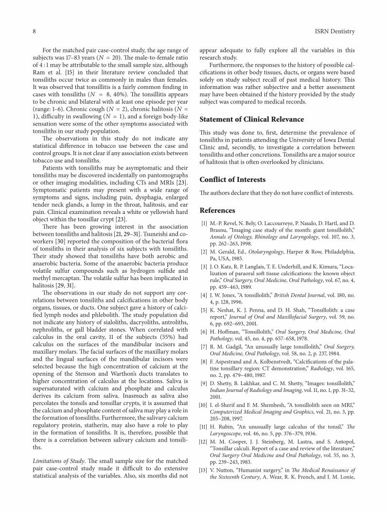
Figure 1: Clinical photograph of a patient with small, superficialtonsilith in the left tonsillar crypt (arrow).
Figure 2: Multiple, well-defined calcifications in the right tonsil(circle) in a 79-year-old male.
crypt [10]. The tonsilith may also have a deeper location andpresent as an enlarged or calcified mass within the tonsil [10].Superficial tonsiliths often flake off periodically, especiallywhen the patient gargles vigorously (Figure 1). Tonsiliths canbe multiple and may vary in size from small to very large[4–10], the largest tonsilith to date, measuring 14.5 cm, wasreported by Rubin [11] in 1936.
Treatment is usually removal of the tonsilith by curettage.Larger concretions may require local excision under topicalor local infiltration anesthesia. If there is evidence of chronictonsillitis, tonsillectomy offers definitive therapy [5, 10].
1.2. Composition of Tonsiliths. Tonsiliths are composed ofphosphate and/or carbonate salts of calcium. These arearranged in a structure similar to that of bone crystals ofhydroxyapatite Ca
5[OH | (PO
4)3].The hydroxyl ion (OH−) in
the hydroxyapatite can be replaced by fluoride, carbonate, orchloride. The hydroxyapatite crystal has a specific gravity of3.08 and is 5 on the Mohs hardness scale [10, 12]. A proteinmatrix has also been demonstrated as part of the compositionof tonsilith [12–14].
1.3. Radiology of Tonsiliths. Although a pantomograph is areliable and standard modality for interpreting the presenceof tonsiliths, superimposition of a lesion involving one sideof the jaw may create a pseudotonsilith or ghost image onthe contralateral side which could lead to a misinterpretationof bilateral lesions [15]. A ghost image is formed when theobject is located between the X-ray source and the centerof rotation of the cassette [16–19]. On the pantomograph,
Figure 3: Multiple, well-defined calcifications in the right tonsil(circle) in a 54-year-old male.
Figure 4:Multiple, well-defined, bilateral calcifications in the angle-ramus region of the mandible (circle) in a 57-year-old male. Noteghost images on the right (arrow).
tonsiliths commonly appear as multiple, small, and ill-defined radiopacities, (Figures 2, 3, 4, and 5).
Other Stones. Stones or liths can occur in various organsand ducts in the body, including the gallbladder (cholelith),kidneys (nephrolith), and lower urinary tract (urolith). Lithsare also seen in the nasolacrimal duct (dacryolith), nasal cav-ity (rhinolith), maxillary antrum (antrolith), lymph nodes,liver (intrahepatic lith), testes (testicular microlith), intestine(fecalith), and the semicircular canal (canalolith) [17, 20–22].Other liths include dental calculus, ocularlith, pancreatolith,sialolith, and phlebolith.
2. Materials and Methods
Approval for both parts of the study was granted by theUniversity of Iowa Institutional Review Board. This studycomplied with the Helsinki Declaration as regards humansubjects.This was a two-part study.The first part was a preva-lence study while the second was a matched pair case-controlstudy.The matched pair case-control study commenced afterthe prevalence study was concluded. No new or unusualradiographs weremade in this study.The study only reviewedradiographs that were made for clinical purposes.
2.1. Part 1: Prevalence Study. The intended population forthe prevalence study comprised all the patients who attendedthe oral and maxillofacial radiology clinic of the Collegeof Dentistry of The University of Iowa over a four-yearperiod, between January 2000 andDecember 2003.The studypopulation included both subjects with andwithout tonsilithsfound on pantomographs. The age range was three to ninetyyears. A total of 1524 subjects were recruited.

ISRN Dentistry 3
Figure 5: Multiple, well-defined, and bilateral calcifications in theangle-ramus region of the mandible (circle and arrow) in a 67-year-old female.
Only subjects with diagnostic quality pantomographswere included because these radiographs could potentiallyshow calcifications in the tonsils over the angle and ramus ofthe mandible.
In order to determine prevalence, subjects with andwithout tonsiliths demonstrable on the pantomographs wereincluded in the study. Both analog and digital pantomographswere included.
Individuals below the age of three or whose pantomo-graphs were of poor diagnostic quality or with a history oftonsillectomy were excluded.
Tonsiliths measuring 2mm or less were excluded becausethis dimension did not give a high confidence level ofinterpretation.
The radiographs were viewed in the interpretation roomin subdued lighting over a clean and bright illuminator orcomputer screen. Amagnifying glass or digital magnificationwas used to assist in viewing the concretions on the radio-graphs.
2.1.1. Ascertainment of Tonsiliths. An interpretation of ton-siliths was made when radiopaque masses not deemed tobe part of the stylohyoid complex, sialoliths, calcified lymphnodes, phleboliths, or changes in the bone pattern were seenover or near the angle and ramus of themandible.The concre-tions needed to have a radiopacity similar to, but demarcatedfrom, the overlying bone. Unilateral and bilateral concretionswere recorded. Ghost images were identified to prevent errorsof interpretation. The size, site, and number of concretionswere recorded. Unique identifiers (chart numbers) of thesubjects were recorded.
The principal investigator was calibrated against multipleobservers such that a capital value of agreement was estab-lished. This was done in order to establish criteria for inter-preting tonsiliths.The exercise was repeated halfway throughthe study to prevent a drift from established standards.
A standardized chart abstraction formwas used to extractrelevant information from the dental records of the subjects.The information extracted included demographics such asage and sex and radiographic appearance of tonsiliths.
The lack of previous data or studies made it difficult toestimate the sample size. Therefore, a convenience sampleinclusive of all the subjects who attended the oral and max-illofacial radiology clinic from January 2000 to December2003 was used.
2.1.2. Statistical Analysis. Descriptive statistics were com-puted, and frequency tables were generated. Bivariate anal-yses were performed to determine if there was a statisti-cally significant difference between two groups of patients(with single tonsiliths versus with multiple tonsiliths) fordemographic factors. Chi-square test, Fisher’s exact test, andnonparametric Wilcoxon rank-sum test were conducted forthe data analysis.
All tests had a 0.05 level of statistical significance. SAS forWindows (version 9.1, SAS Institute Inc., Cary, NC, USA) wasused for the data analysis.
2.2. Part 2: Matched Pair Case-Control. The second part,the matched pair case-control study, included the rollingenrolment of subjects referred to the department over a six-month period (October 2007 to March 2008). The controlgroup comprised subjects without the radiographic evidenceof tonsiliths on pantomographs, whose medical history mayor may not reveal presence of calcifications or stones inother tissues and organs, including kidneys and salivaryglands.The case group comprised subjects with radiographicevidence of tonsiliths on pantomographs, who may or maynot have history of calcifications or stones in other tissues ororgans, including the kidneys and salivary glands. For eachsubject with tonsiliths, that was enrolled into the case group,another potential subject without tonsiliths was recruited tothe control group.The subjects recruited to the control groupwere matched for age (±2 years) and sex with the subjectsin the case group. The subjects for both case and controlgroups were enrolled from the patients attending the oral andmaxillofacial radiology clinic.
The involvement of subjects consisted of one clinical visitof approximately 30 minutes. No additional clinic follow-ups were necessary. There were no foreseeable risks fromparticipating in the study.
2.2.1. Selection Criteria
Inclusion Criteria
(1) Age range: 3–90 years.(2) Only subjects without tonsiliths on pantomographs
were included in the control group.(3) Only subjects with tonsiliths on pantomographs were
included in the case group.(4) Diagnostic quality pantomograph.
The features of a diagnostic quality pantomographinclude
(a) both rami of the mandible visible on the radiograph;(b) proper positioning of the patient in the unit when the
radiograph was made;(c) areas of the tonsils included in the image;(d) the overall density of the radiograph acceptable for
interpretation;(e) no artifacts over the rami of the mandible to preclude
the visualization of tonsiliths.

4 ISRN Dentistry
Exclusion Criteria
(1) Subjects below the age of three years.(2) Subjects with poor diagnostic quality pantomo-
graphs.(3) Subjects who declined to give informed consent.(4) Subjects with a history of tonsillectomy.
2.2.2. Statistical Analysis. Descriptive statistics were com-puted and frequency tables were generated. Bivariate analyseswere performed to determine whether there was a statis-tically significant difference between the case and controlgroups. Chi-square test, Fisher’s exact test, and nonparamet-ric Wilcoxon rank-sum test were used for data analysis.
All tests have a 0.05 level of statistical significance. SASfor Windows (version 9.1, SAS Institute Inc., Cary, NC, USA)was used for the statistical analysis.
To our knowledge, no similar studies have been con-ducted before and, therefore, no analogous information iscurrently available for determination of sample size of case-control groups. This initial study, which was completed inapproximately 6 months, therefore included all the subjectswith a matched pair for case-control study.
The results of the prevalence study that was earlierconducted could not be used to calculate the sample size forthe matched, pair case-control study because the data did notinclude correlations between tonsiliths and other stones inbody organs, tissues, and ducts.
3. Results
3.1. Part 1: Prevalence Study
3.1.1. Descriptive Statistical Results. A total of 1524 pan-tomographs were reviewed in the oral and maxillofacialradiology clinic of The University of Iowa. 205 subjects withpossible tonsiliths were identified. Of the 205 subjects, 124had tonsiliths over 2mm in size with a high confidence level.The 81 subjects eliminated possibly had tonsiliths, but the2mm dimension did not give a high confidence level ofinterpretation.
Therefore, 124 subjects (53 males and 71 females) aged9.2 to 87 years (mean age 52.6 years) were included for thedata analysis. The male-to-female ratio was 0.75 : 1.00. Thirtyeight subjects had single tonsiliths whereas 86 subjects hadmultiple tonsiliths. The prevalence of tonsiliths in the studypopulation was 8.14%.
Table 1 displays the frequency distribution analyses forsome categorical variables, andTable 2 reports the descriptivestatistics for some quantitative variables.
3.1.2. Comparison of Two Groups of Subjects (with SingleTonsilith versus Multiple Tonsiliths). Based on chi-squaretest or Fisher’s exact test, the data revealed that there wasa significant difference between the two groups for theunilateral right side of the jaw (𝑃 = 0.0242) and bilateralside of the jaw (𝑃 < 0.0001). The results indicated that singletonsiliths were more likely to be on the right side of the jaw
Table 1: Frequency distribution of sex, imaging modalities, radio-graphic appearance, location, side of jaw, ghost images, and size oftonsiliths.
Frequency (𝑁) Percent (%)Sex
Male 53 42.74Female 71 57.26Total 124 100.00
Imaging modalitiesPlain film 124 100.00Digital images None 0.00Total 124 100.00
Radiographic appearance of tonsilithsSingle, welldefined 35 28.23Single, illdefined 3 2.42Multiple, welldefined 78 62.90Multiple, ill-defined 8 6.45Total 124 100.00
Location of tonsilithsUpper 1/3 of mandibular ramus 0 0.00Middle 1/3 of mandibular ramus 8 6.45Lower 1/3 of mandibular ramus 116 93.55Total 124 100.00
Side of jawUnilateral left 41 33.06Unilateral right 50 40.32Bilateral 33 26.62Total 124 100.00
Ghost imagesPresent 8 6.45Not present 116 93.55Total 124 100.00
Size of tonsiliths (mm)3 < 4 69 55.654 < 6 24 19.356 < 7 17 13.717 < 8 10 8.068 < 9 1 0.819 < 10 1 0.8110 < 11 1 0.8111 1 0.81Total 124 100.00
(55.3%). On the other hand, multiple tonsiliths were morelikely to present on both sides of the jaws (37.2%).
Based on the nonparametricWilcoxon rank-sum test, thedata showed that there was no significant difference in meanage and mean size of tonsiliths between the two groups ofsubjects (with single tonsilith versus multiple tonsiliths) (𝑃 =0.4318 and 0.0551, resp.).
Based on chi-square test or Fisher’s exact test, the datarevealed that there was no significant difference between thetwo groups for sex (𝑃 = 0.6248), location over the ramus

ISRN Dentistry 5
Table 2: Mean of age of subjects and size of tonsiliths for both single and multiple tonsiliths.
Variable Mean Standard deviation Minimum MaximumSubjects with both single and multiple tonsiliths (𝑁 = 124)
Age (year) 52.65 20.64 9.20 87.00Size of tonsiliths (mm) 3.90 1.38 3.00 11.00
Subjects with single tonsiliths (𝑁 = 38)Age (year) 50.32 21.67 9.20 84.00Size of tonsiliths (mm) 4.18 1.54 3.00 11.00
Subjects with multiple tonsiliths (𝑁 = 86)Age (year) 53.68 20.21 13.20 87.00Size of tonsiliths (mm) 3.78 1.29 3.00 10.00
Table 3: Frequency distribution of history of tonsilitis for thematched pair case-control study.
Variable Frequency (𝑁) Percent (%)Subjects in case group (𝑁 = 10)
Previous history of tonsilitis 6 60No previous history oftonsillitis 4 40
Subjects in control group (𝑁 = 10)Previous history of tonsilitis 2 20No previous history oftonsillitis 8 80
of the mandible (𝑃 = 0.6998), unilateral left presentation(𝑃 = 0.1549), and presence of ghost images (𝑃 = 0.6998).
3.1.3. Comparisons of Locations for Tonsiliths. No significantresults were found (𝑃 > 0.05 for all instances).
3.2. Results of Matched Pair Case-Control Study onthe Prevalence of Tonsiliths
3.2.1. Descriptive Statistical Results. A total of 20 subjectswereincluded in this study, comprising 10 each for the matchedpair case and control groups. The age range was 17–83 years,with a mean age of 42.95 years and standard deviation of22.09. The sex ratio was 4 : 1 for both the case and the controlgroups.
A total of 8 subjects (40%) provided a history of recentepisodes of tonsillitis. But 2 out of the 8 subjects (10%) did notprovide a record of the date of the last episode of tonsillitis.Out of the 6 subjects with a record of the last date of tonsillitis,4 (67%) were in the case group while 2 (33%) were in thecontrol group (Table 3). The number of episodes of tonsillitisper year and the side(s) affected are depicted in Tables 4 and5.
The subjects gave varied responses to questions about thesymptoms of tonsiliths.The symptoms included the history ofchronic sore throat, chronic cough, difficulty in swallowing,pain in the ear, chronic halitosis, foreign body-like sensationin the throat, and bad/altered taste. The observations arepresented in Table 6.
Table 4: Frequency distribution of unilateral and bilateral tonsilitisfor the matched pair case-control study.
Side affected Frequency (𝑁) Percent (%)Subjects in case group (𝑁 = 10)
Unilateral 1 10Bilateral 5 50Not applicable 4 40
Subjects in control group (𝑁 = 10)Unilateral None 0Bilateral 2 20Not applicable 8 80
Two subjects gave a history of tonsillectomy for themanagement of tonsillitis.These subjects were excluded fromthe data. Two other subjects (10%) gave a history of antibioticregimen for the management of tonsillitis. There was norecord of curettage in the observations.
The medical history of stones in other body tissues,organs, and ducts was sought by asking questions aboutsialoliths, phleboliths, arteriosclerosis, calcified lymph nodes,dacryoliths, antroliths, rhinoliths, nasopharyngeal stones,nephroliths, and gall bladder stones. One subject each had ahistory of phlebolith and calcified lymph nodes, respectively.There was no other history of body tissue concretions.
A total of 5 (25%) gave a history of cigarette smoking. Outof the sample, only three still smoke regularly.The date of lastdental cleaning at a dental facility and calculus distribution onthe lingual and facial surfaces of the mandibular incisors andmaxillary molars were recorded for each subject. The date oflast professional cleaning from a dentist ranged from less than1 week to 10 years. The observations are presented in Table 7.
A total of 13 (65%) pantomographs were analog, whereas7 (35%) were digital. A total of 18 (90%) pantomographs weremade on the OC 100-3-1-2 machine, whereas the remaining 2(10%) were made on the OP 100-3-1-2 machine.
The radiographic appearance of tonsilithswas categorizedas single, well-defined (𝑛 = 3, 15%) andmultiple, well defined(𝑛 = 7, 35%). The location of tonsiliths, side of the jaw,and ghost images were also recorded. The observations arepresented in Table 8.

6 ISRN Dentistry
Table 5: Frequency distribution of the number of episodes oftonsilitis per year for the matched pair case-control study.
Number of episodesof tonsillitis per year Frequency (𝑁) Percent (%)
Subjects in case group (𝑁 = 10)1 4 402 1 106 1 10Not applicable 4 40
Subjects in control group (𝑁 = 10)1 2 20Not applicable 8 80
Table 6: Frequency distribution of symptoms of tonsilitis (𝑁 = 20).
Variable FrequencyCase (𝑁 = 10) Control (𝑁 = 10)
Sore throat 2 0Chronic cough 2 0Difficulty swallowing 1 0Pain in ear 1 0Chronic halitosis 1 0Foreign body-like feel 2 0Altered taste None NonePrevious history of tonsilith None None
Table 7: Frequency distribution of dental calculus.
Calculus Case(𝑁 = 10)
Control(𝑁 = 10)
Lingual surfaces of mandibularincisors 1 1
Facial surfaces of maxillary molars 0 2Lingual surfaces of the mandibularincisors and facial surfaces of themaxillary molars
5 2
No calculus 4 5
The tonsils were examined clinically. The observationsshowed that all the tonsiliths (𝑛 = 10, 50%) were located deepin the tonsillar crypt and were not visible to the naked eye.
The largest tonsilith in the radiographic observation was6mm, whereas the smallest was 3mm (Table 8).
The mean values of observations for the case and controlgroups are presented separately on Tables 9 and 10.
3.2.2. Statistical Results on Comparison of Case Group versusControl Group for Selected Variables in the Study. Based onthe nonparametricWilcoxon rank-sum test, the data revealedthat there was a significant difference between case andcontrol as the date of a last professional cleaning was receivedfrom a dental facility (𝑃 = 0.0105). The results indicated thatthe average weeks for last professional cleaning at the dentalfacility for subjects in the case group (mean = 226.88 weeks)
Table 8: Frequency of radiographic appearance, location, side ofjaw, ghost images, and size of tonsiliths.
Case(𝑁 = 10)
Control(𝑁 = 10)
Radiographic appearanceSingle, welldefined 3 0Multiple, welldefined 7 0Nil tonsiliths (control group) 0 10
Location of tonsilith(s)Lower third of mandibular ramus 10 0Nil tonsiliths (control group) 0 10
Side of the jaw with tonsilithUnilateral left 1 0Unilateral right 6 0Bilateral 3 0Nil tonsiliths (control group) 0 10
Ghost imagesPresent 2 0Not present 8 0Nil tonsiliths (control group) 0 10
Size of tonsilith seen on radiographs(mm)
3 2 04 3 05 3 06 2 0Nil (control group) 0 10
were significantly greater than those observed in the controlgroup (mean = 43.5 weeks).
Based on Fisher’s exact test, the data revealed that therewas no significant difference between case and control groupsfor tonsillitis, side(s) affected, sore throat, chronic cough,difficulty swallowing, pain in ear, chronic halitosis, foreignbody feel, antibiotics, phlebolith, calcified lymph node, useof tobacco products, and dental calculus (𝑃 > 0.05 for eachinstance).
Based on the nonparametric Wilcoxon rank-sum test,there was no significant difference between case and controlgroups for the date of last episode of tonsillitis (𝑃 = 0.1588),number of episodes of tonsillitis per year (𝑃 = 0.8308), whenthe subject quit tobacco use (𝑃 = 0.3711), average number ofcigarettes smoked per day (𝑃 = 0.7671), and age distributionof patients (𝑃 = 0.9396).
4. Discussion and Conclusion
This study was designed as a two-part study to determinethe prevalence of tonsiliths and the relationship betweentonsiliths and concretions in other body organs, tissues, orducts in the patients attending the oral and maxillofacialradiology clinic of The University of Iowa. In the first part ofthe study, a total of 1524 charts, representing pantomographs

ISRN Dentistry 7
Table 9: Mean values of observations for the case group.
Variable Subjects (𝑁) Mean Standard deviation Minimum Maximum MedianLast tonsillitis (weeks ago) 4 3.75 3.10 1.00 8.00 3Episodes of tonsillitis per year 5 2.00 2.35 0.00 6.00 1.00When quit smoking (weeks ago) 3 312.00 187.49 156.00 520.00 260.00Num. of cigarettes per day 3 7.33 10.97 1.00 20.00 1.00Last dental cleaning (weeks ago) 8 226.88 162.92 43.00 520.00 234.00Size of tonsiliths (mm) 10 4.50 1.08 3.00 6.00 4.50Age of subject (years) 10 42.70 22.39 17.00 82.00 39.00
Table 10: Mean values of observations for the control group.
Variables Subjects (𝑁) Mean Standard deviation Minimum Maximum MedianLast tonsillitis (weeks) 2 30.00 31.11 8.00 52.00 30.00Episodes of tonsillitis per year 2 1.00 0.00 1.00 1.00 1.00When quit smoking (weeks) 1 104.00 0.00 104.00 104.00 104.00Number of cigarettes per day 2 6 1.41 5.00 7.00 6.00Last clean (weeks) 10 43.50 46.22 2.00 156.00 52.00Size of tonsiliths (mm) 0 0.00 0.00 0.00 0.00 0.00Age of subjects (yr) 10 43.20 22.99 17.00 83.00 39.00
made between January 2000 and December 2003, werereviewed. One hundred and twenty-four (8.14%) cases oftonsiliths, measuring above 2mm on a linear scale wereincluded in the final analysis. Eighty-one cases with possibletonsiliths, measuring 2mm and below, were removed fromthe data because this dimension does not give a highconfidence level of interpretation. Therefore, the prevalenceof tonsiliths in patients attending the oral and maxillofacialradiology clinic of The University of Iowa was observed tobe 8.14%. This figure is similar to what was reported byCooper and his coworkers in 1983 [12]. The minimum sizeof tonsilith in the Cooper study was 5mm. An importantaspect of our study is that we established a cut-off pointof 2mm for the interpretation of tonsiliths based on ourreliability of interpretation and high degree of consensus ofthis measurement amongst the researchers in this study. Thisis not a common feature in all the previous studies reviewed.
The earliest known description of concretions in theoropharynx is thought to be recorded by Lang in 1560[12, 23]. Although many views have been expressed, theexact etiology and pathogenesis are unknown [24]. It hasbeen shown that the calcifications develop within a mass ofdesquamated epithelium, serum, food debris, and bacterialcolonies. Recurrent tonsillar inflammation may promote thedevelopment of tonsillar concretions [25].
Out of the 124 cases of tonsiliths in our study, 53 weremales and 71 were females. The male-to-female ratio was0.75 : 1.00 and there is no sex predilection. The age range ofsubjects was 9.2–87 years (mean 52.6 years). The average sizeof tonsilith was 4mm (range: 3–11mm). The incidence oflarge tonsiliths is low.They are seenmainly in elderly patients[24, 26–28]. On the other hand, small tonsiliths are frequentlyreported [1, 4, 7, 10–12, 18, 24, 26–28]. A few of the subjectsin our study mentioned that they often see fragments of
calcific materials in their sputum, especially during episodesof tonsillitis. This may account for the low prevalence oflarge tonsiliths, especially in healthy individuals. Accordingto the literature review by Cooper and his co-workers [12],the average age of patients at the time of medical interventionis 46.4 years (41.6 years for males, 50.7 years for females). Areview of the English literature showed that the mean age ofoccurrence was 46.2 years (age range: 16–77 years) [1, 4, 7, 10–12, 18, 20, 24, 26–28].
The radiographic appearance of tonsiliths in this studywas predominantly multiple and well defined (𝑁 = 78,62.90%). The single, well-defined tonsilith in a similar loca-tion constituted 28.23% (𝑁 = 35) of the sample. Tonsiliths areusually single and unilateral, although they can be multipleand bilateral [26].Theymay grow to large sizes andmay occurin palatine or lingual tonsils [24].
The majority of the cases were located in the lower one-third of the mandibular ramus (𝑁 = 116, 93.55%). Thisis not unusual, inasmuch as the tonsils are found in thelateral aspect of the oro pharyngeal wall corresponding tothe angle of the mandible bilaterally [14, 15]. There was ahigher percentage of tonsiliths (𝑁 = 50, 40.32%) in theright tonsillar crypt than in the left (𝑁 = 41, 33.06%). Asimilar distribution was observed in the smaller sample forthe matched pair case-control study. It is also not uncommonto have bilateral tonsiliths (𝑁 = 33, 26.61%). According to theliterature review by Ram and his co-workers [15], tonsilithsoccur more often in the right tonsil than in the left one.
The mean age of subjects with multiple and well-definedconcretions was slightly higher (53.68 years) when comparedto the mean age of subjects with single, well-defined con-cretions (50.32 years). A similar pattern was observed withthe average size of tonsiliths, which is 4.18mm for singleconcretions and 3.78mm for multiple concretions.
8 ISRN Dentistry
For the matched pair case-control study, the age range ofsubjects was 17–83 years (𝑁 = 20). The male-to-female ratioof 4 : 1 may be attributable to the small sample size, althoughRam et al. [15] in their literature review concluded thattonsiliths occur twice as commonly in males than females.It was observed that tonsillitis is a fairly common finding incases with tonsiliths (𝑁 = 8, 40%). The tonsillitis appearsto be chronic and bilateral with at least one episode per year(range: 1–6). Chronic cough (𝑁 = 2), chronic halitosis (𝑁 =1), difficulty in swallowing (𝑁 = 1), and a foreign body-likesensation were some of the other symptoms associated withtonsiliths in our study population.
The observations in this study do not indicate anystatistical difference in tobacco use between the case andcontrol groups. It is not clear if any association exists betweentobacco use and tonsiliths.
Patients with tonsiliths may be asymptomatic and theirtonsiliths may be discovered incidentally on pantomographsor other imaging modalities, including CTs and MRIs [23].Symptomatic patients may present with a wide range ofsymptoms and signs, including pain, dysphagia, enlargedtender neck glands, a lump in the throat, halitosis, and earpain. Clinical examination reveals a white or yellowish hardobject within the tonsillar crypt [23].
There has been growing interest in the associationbetween tonsiliths and halitosis [21, 29–31]. Tsuneishi and co-workers [30] reported the composition of the bacterial floraof tonsiliths in their analysis of six subjects with tonsiliths.Their study showed that tonsiliths have both aerobic andanaerobic bacteria. Some of the anaerobic bacteria producevolatile sulfur compounds such as hydrogen sulfide andmethyl mercaptan. The volatile sulfur has been implicated inhalitosis [29, 31].
The observations in our study do not support any cor-relations between tonsiliths and calcifications in other bodyorgans, tissues, or ducts. One subject gave a history of calci-fied lymph nodes and phlebolith. The study population didnot indicate any history of sialoliths, dacryoliths, antroliths,nephroliths, or gall bladder stones. When correlated withcalculus in the oral cavity, 11 of the subjects (55%) hadcalculus on the surfaces of the mandibular incisors andmaxillary molars. The facial surfaces of the maxillary molarsand the lingual surfaces of the mandibular incisors wereselected because the high concentration of calcium at theopening of the Stenson and Warthon’s ducts translates tohigher concentration of calculus at the locations. Saliva issupersaturated with calcium and phosphate and calculusderives its calcium from saliva. Inasmuch as saliva alsopercolates the tonsils and tonsillar crypts, it is assumed thatthe calcium and phosphate content of salivamay play a role inthe formation of tonsiliths. Furthermore, the salivary calciumregulatory protein, statherin, may also have a role to playin the formation of tonsiliths. It is, therefore, possible thatthere is a correlation between salivary calcium and tonsili-ths.
Limitations of Study. The small sample size for the matchedpair case-control study made it difficult to do extensivestatistical analysis of the variables. Also, six months did not
appear adequate to fully explore all the variables in thisresearch study.
Furthermore, the responses to the history of possible cal-cifications in other body tissues, ducts, or organs were basedsolely on study subject recall of past medical history. Thisinformation was rather subjective and a better assessmentmay have been obtained if the history provided by the studysubject was compared to medical records.
Statement of Clinical Relevance
This study was done to, first, determine the prevalence oftonsiliths in patients attending the University of Iowa DentalClinic and, secondly, to investigate a correlation betweentonsiliths and other concretions. Tonsiliths are amajor sourceof halitosis that is often overlooked by clinicians.
Conflict of Interests
Theauthors declare that they do not have conflict of interests.
References
[1] M.-P. Revel, N. Bely, O. Laccourreye, P. Naudo, D. Hartl, and D.Brasnu, “Imaging case study of the month: giant tonsillolith,”Annals of Otology, Rhinology and Laryngology, vol. 107, no. 3,pp. 262–263, 1998.
[2] M. Gerald, Ed., Otolaryngology, Harper & Row, Philadelphia,Pa, USA, 1985.
[3] J. O. Katz, R. P. Langlais, T. E. Underhill, and K. Kimura, “Loca-lization of paraoral soft tissue calcifications: the known objectrule,”Oral Surgery, Oral Medicine, Oral Pathology, vol. 67, no. 4,pp. 459–463, 1989.
[4] J. W. Jones, “A tonsillolith,” British Dental Journal, vol. 180, no.4, p. 128, 1996.
[5] K. Neshat, K. J. Penna, and D. H. Shah, “Tonsillolith: a casereport,” Journal of Oral and Maxillofacial Surgery, vol. 59, no.6, pp. 692–693, 2001.
[6] H. Hoffman, “Tonsillolith,” Oral Surgery, Oral Medicine, OralPathology, vol. 45, no. 4, pp. 657–658, 1978.
[7] R. M. Gadgil, “An unusually large tonsillolith,” Oral Surgery,Oral Medicine, Oral Pathology, vol. 58, no. 2, p. 237, 1984.
[8] F. Aspestrand and A. Kolbenstvedt, “Calcifications of the pala-tine tonsillary region: CT demonstration,” Radiology, vol. 165,no. 2, pp. 479–480, 1987.
[9] D. Shetty, B. Lakhkar, and C. M. Shetty, “Images: tonsillolith,”Indian Journal of Radiology and Imaging, vol. 11, no. 1, pp. 31–32,2001.
[10] I. el-Sherif and F. M. Shembesh, “A tonsillolith seen on MRI,”Computerized Medical Imaging and Graphics, vol. 21, no. 3, pp.205–208, 1997.
[11] H. Rubin, “An unusually large calculus of the tonsil,” TheLaryngoscope, vol. 46, no. 5, pp. 376–379, 1936.
[12] M. M. Cooper, J. J. Steinberg, M. Lastra, and S. Antopol,“Tonsillar calculi. Report of a case and review of the literature,”Oral Surgery Oral Medicine and Oral Pathology, vol. 55, no. 3,pp. 239–243, 1983.
[13] V. Nutton, “Humanist surgery,” in The Medical Renaissance ofthe Sixteenth Century, A. Wear, R. K. French, and I. M. Lonie,

ISRN Dentistry 9
Eds., p. 91, CambridgeUniversity Press, Cambridge,Mass, USA,1985.
[14] B. Z. Pilch, “Thenasopharynx andWaldeyer’s ring,” inHead andNeck Surgical Pathology, B. Z. Pilch, Ed., pp. 157–194, LippincottWilliams and Wilkins, Philadelphia, Pa, USA, 2001.
[15] S. Ram, C. H. Siar, S. M. Ismail, and N. Prepageran, “Pseudobilateral tonsilloliths: a case report and review of the literature,”Oral Surgery, OralMedicine, Oral Pathology, Oral Radiology andEndodontology, vol. 98, no. 1, pp. 110–114, 2004.
[16] W. D. McDavid, R. P. Langlais, U. Welander, and C. R. Morris,“Real, double, and ghost images in rotational panoramic radio-graphy,”Dentomaxillofacial Radiology, vol. 12, no. 2, pp. 122–128,1983.
[17] S. T. Deahl II and A. Ruprecht, “Radiographic interpretation oftonsiliths,” The Iowa Dental Journal, vol. 77, no. 3, pp. 19–20,1991.
[18] B. Sezer, Z. Tugsel, and C. Bilgen, “An unusual tonsillolith,”Oral Surgery, OralMedicine, Oral Pathology, Oral Radiology, andEndodontics, vol. 95, no. 4, pp. 471–473, 2003.
[19] M. Giudice, M. G. Cristofaro, M. G. Fava, and A. Giudice, “Anunusual tonsillolithiasis in a patient with chronic obstructivesialoadenitis,” Dentomaxillofacial Radiology, vol. 34, no. 4, pp.247–250, 2005.
[20] B. Gapany-Gapanavicius, “Peritonsillar abscess caused by alarge tonsillolith,” Ear, Nose and Throat Journal, vol. 55, no. 11,pp. 343–345, 1976.
[21] B.W. Neville, D. D. Damm, C.M. Allen, and J. E. Bouquet, Eds.,Oral and Maxillofacial Pathology, W.B. Saunders, Philadelphia,Pa, USA, 2nd edition, 2002.
[22] D. J. Kok, “Clinical implications of physicochemistry of stoneformation,” Endocrinology and Metabolism Clinics of NorthAmerica, vol. 31, no. 4, pp. 855–867, 2002.
[23] C. W. Pruet and D. A. Duplan, “Tonsil concretions and tonsil-loliths,” Otolaryngologic Clinics of North America, vol. 20, no. 2,pp. 305–309, 1987.
[24] L. H. Hiranandani, “A giant tonsillolith,” Journal of Laryngologyand Otology, vol. 81, no. 7, pp. 819–822, 1967.
[25] M. A. Jaber, S. R. Porter, M. S. Gilthorpe, R. Bedi, and C. Scully,“Risk factors for oral epithelial dysplasia—the role of smokingand alcohol,” Oral Oncology, vol. 35, no. 2, pp. 151–156, 1999.
[26] U.M. Hadi andM. S. Samara, “Giant tonsillolith,” Ear, Nose andThroat Journal, vol. 64, no. 10, pp. 507–508, 1985.
[27] D. von Arx and R. J. Carr, “Displaced tooth mimicing atonsillolith,” Journal of Laryngology and Otology, vol. 102, no.7, pp. 652–653, 1988.
[28] H. Hoffman, “Tonsillolith,” Oral Surgery, Oral Medicine, OralPathology, vol. 45, no. 4, pp. 657–658, 1978.
[29] J. Tonzetich, “Direct gas chromatographic analysis of sulphurcompounds in mouth air in man,” Archives of Oral Biology, vol.16, no. 6, pp. 587–597, 1971.
[30] M. Tsuneishi, T. Yamamoto, S. Kokeguchi, N. Tamaki, K. Fukui,and T. Watanabe, “Composition of the bacterial flora in tonsil-loliths,” Microbes and Infection, vol. 8, no. 9-10, pp. 2384–2389,2006.
[31] T. Ansai and T. Takehara, “Tonsillolith as a halitosis-inducingfactor,” British Dental Journal, vol. 198, no. 5, pp. 263–264, 2005.