Embed Size (px)
Citation preview
238 Conelia Tudorache et al Posttraumatic Occlusion of the Vertebral Artery
The Posttraumatic Occlusion of the Vertebral Artery Case presentation
Cornelia Tudorache1, Adriana Moldovanu1,2,3, Anda Esanu1, E. Moldovanu1,2,3, Carmen Gherman1,3, C. Ichim1, C. Popescu4, M. Dabija4, Danisia Haba1,2
1Department of Radiology, Emergency Hospital “Prof. Dr. N. Oblu”, Iasi 2“Gr.T. Popa” University of Medicine and Pharmacy, Faculty of Dentistry, General and Dental Radiology Discipline 3MRI Departament “Supermeditest” Iasi 4Neurosurgery, Emergency Hospital “Prof. Dr. N. Oblu”, Iasi
Abstract We investigate radiographic features of
vertebral artery injury/occlusion associated with nonpenetrating cervical spine trauma and to demonstrate the importance of the CTA in high-risk cases. With the popularization of CTA (Computed Tomography Angiography) and MRA (Magnetic Resonance Angiography), vertebral artery injury has been a common complication of cervical spine trauma.
The occlusion of the vertebral artery secondary to non-penetrating trauma of the cervical spine (fractures and/dislocations) can be found in approximately 20% of the cases. Vertebral artery occlusion was rarely symptomatic because of sufficient collateral blood supply through not only contralateral vertebral artery but also the circle of Willis.
Keywords: occlusion of the vertebral artery, computed tomography angiography.
Case presentation We are presenting the case of a 61-year-
old patient, who has suffered multiple trauma as result of a fall from a great height.
The patient was tetraplegic. He had radio-imagistic investigations done: X-ray,
IRM exam for the cervical spine and cervical angio-CT scan (cervical angio-CT scan, with sections of 1 mm, pitch 1,5 mm, reconstructions 0,8 mm, followed by MPR and MIP processing).
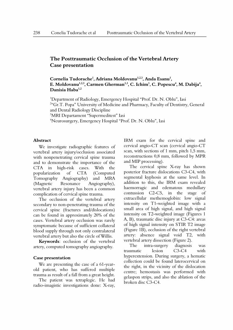
The cervical spine X-ray has shown posterior fracture dislocations C3-C4, with segmental kyphosis at the same level. In addition to this, the IRM exam revealed haemorragic and edematous medullary contussion C2-C5, in the stage of extracellular methemoglobin: low signal intensity on T1-weighted image with a small area of high signal, and high signal intensity on T2-weighted image (Figures 1 A, B), traumatic disc injury at C3-C4: areas of high signal intensity on STIR T2 image (Figure 1B), occlusion of the right vertebral artery: absence signal void T2, with vertebral artery dissection (Figure 2).
The intra-surgery diagnosis was traumatic lesion C3-C4 with hyperextension. During surgery, a hematic collection could be found laterocervical on the right, in the vicinity of the dislocation centre; hemostasis was performed with gelaspon strips, and also the ablation of the broken disc C3-C4.
Romanian Neurosurgery (2010) XVII 2: 238 – 245 239
1A
1B
Figure 1 MRI exam – sagital sections: A STIR T2-weighted axial image,
B T1-weighted axial image: - posterior fracture dislocations C3-C4, with
anterior angulation at the same level - edematous and hemorrhagic medullary contussion C2-C5, in the stage of extracellular methemoglobin (low signal intensity on T1-weighted image with a
small area of high signal, and high signal intensity on T2-weighted image)
- traumaic disc injury at C3-C4 (areas of high signal intensity on STIR T2 image)
2A
2B
Figure 2 MRI of the cervical spine T2-weighted image - axial and sagital sections: high signal
intensity of the right vertebral artery, occlusion of the lumen with vertebral artery dissection
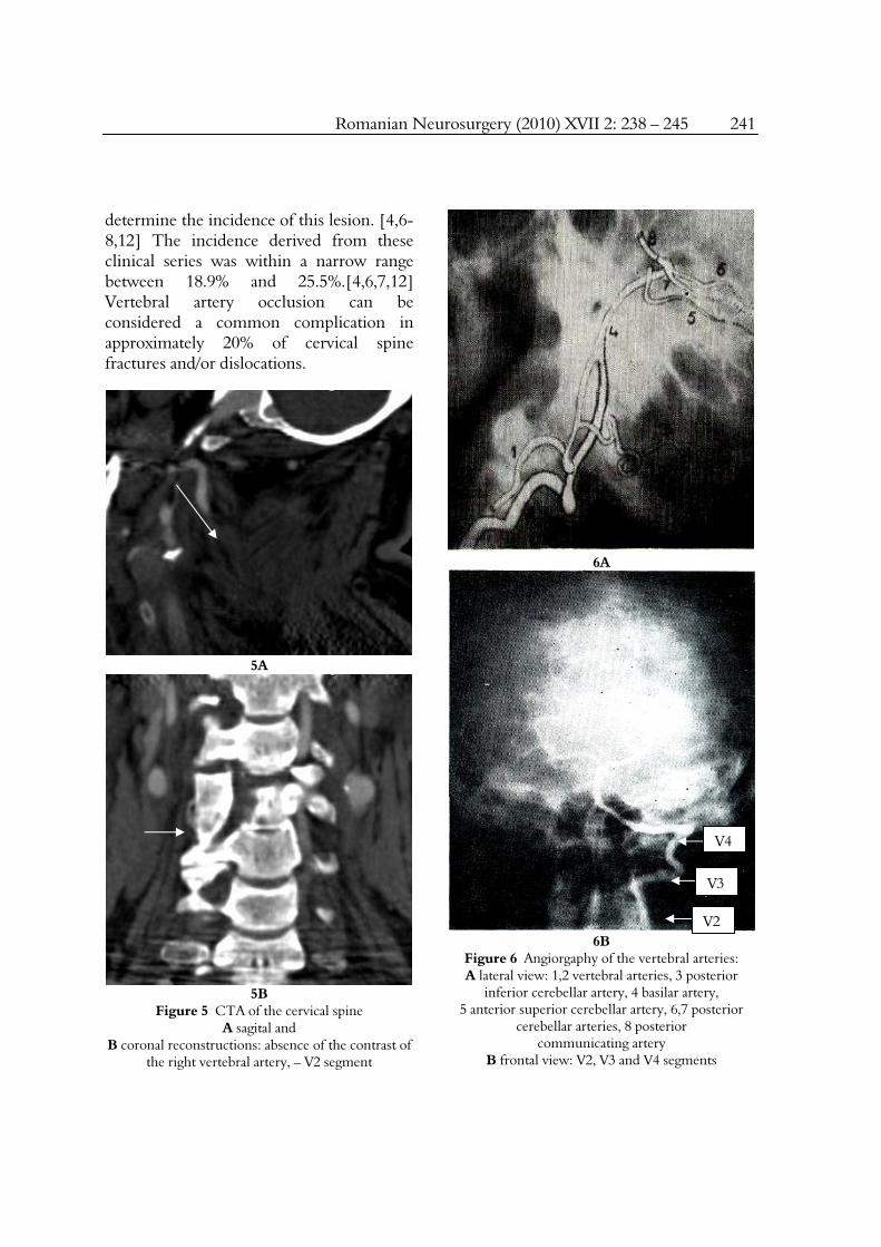
Postoperative, an angio-CT cervical
exam was performed, which highlighted the absence of the contrast of the right vertebral artery, having the inferior limit at the level of a section plan, which passes through the superior end-plate of the vertebral body C5, and the superior one – at the level of a
240 Conelia Tudorache et al Posttraumatic Occlusion of the Vertebral Artery
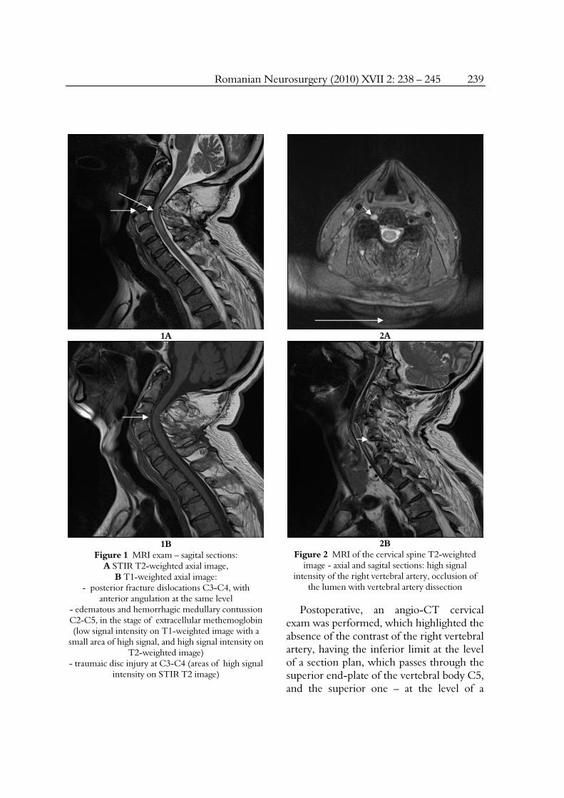
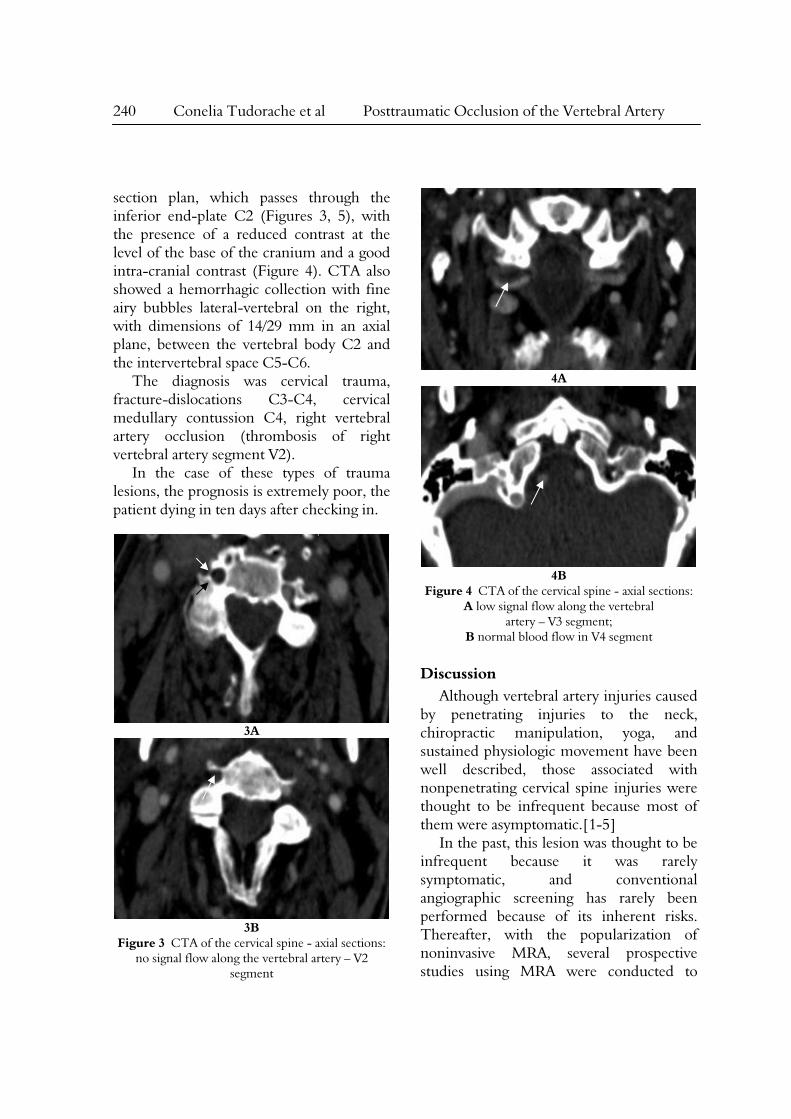
section plan, which passes through the inferior end-plate C2 (Figures 3, 5), with the presence of a reduced contrast at the level of the base of the cranium and a good intra-cranial contrast (Figure 4). CTA also showed a hemorrhagic collection with fine airy bubbles lateral-vertebral on the right, with dimensions of 14/29 mm in an axial plane, between the vertebral body C2 and the intervertebral space C5-C6.
The diagnosis was cervical trauma, fracture-dislocations C3-C4, cervical medullary contussion C4, right vertebral artery occlusion (thrombosis of right vertebral artery segment V2).
In the case of these types of trauma lesions, the prognosis is extremely poor, the patient dying in ten days after checking in.
3A
3B
Figure 3 CTA of the cervical spine - axial sections: no signal flow along the vertebral artery – V2
segment
4A
4B
Figure 4 CTA of the cervical spine - axial sections: A low signal flow along the vertebral
artery – V3 segment; B normal blood flow in V4 segment
Discussion Although vertebral artery injuries caused
by penetrating injuries to the neck, chiropractic manipulation, yoga, and sustained physiologic movement have been well described, those associated with nonpenetrating cervical spine injuries were thought to be infrequent because most of them were asymptomatic.[1-5]
In the past, this lesion was thought to be infrequent because it was rarely symptomatic, and conventional angiographic screening has rarely been performed because of its inherent risks. Thereafter, with the popularization of noninvasive MRA, several prospective studies using MRA were conducted to
Romanian Neurosurgery (2010) XVII 2: 238 – 245 241
determine the incidence of this lesion. [4,6-8,12] The incidence derived from these clinical series was within a narrow range between 18.9% and 25.5%.[4,6,7,12] Vertebral artery occlusion can be considered a common complication in approximately 20% of cervical spine fractures and/or dislocations.
5A
5B
Figure 5 CTA of the cervical spine A sagital and
B coronal reconstructions: absence of the contrast of the right vertebral artery, – V2 segment
6A
6B
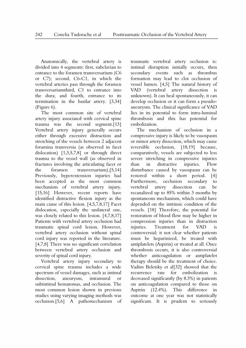
Figure 6 Angiorgaphy of the vertebral arteries: A lateral view: 1,2 vertebral arteries, 3 posterior
inferior cerebellar artery, 4 basilar artery, 5 anterior superior cerebellar artery, 6,7 posterior
cerebellar arteries, 8 posterior communicating artery
B frontal view: V2, V3 and V4 segments
V4
V3
V2
242 Conelia Tudorache et al Posttraumatic Occlusion of the Vertebral Artery
Anatomically, the vertebral artery is divided into 4 segments: first, subclavian to entrance to the foramen transversarium (C6 or C7); second, C6-C1, in which the vertebral arteries pass through the foramen transversariumthird, C1 to entrance into the dura; and fourth, entrance to its termination in the basilar artery. [3,34] (Figure 6).
The most common site of vertebral artery injury associated with cervical spine trauma was the second segment.[13] Vertebral artery injury generally occurs either through excessive distraction and stretching of the vessels between 2 adjacent foramina transversia (as observed in facet dislocation) [1,3,5,7,8] or through direct trauma to the vessel wall (as observed in fractures involving the articulating facet or the foramen transversarium).[5,14] Previously, hyperextension injuries had been accepted as the most common mechanism of vertebral artery injury. [15,16] However, recent reports have identified distractive flexion injury as the main cause of this lesion. [4,5,7,8,17] Facet dislocation, especially the unilateral one, was closely related to this lesion. [4,7,8,17] Patients with vertebral artery occlusion had traumatic spinal cord lesion. However, vertebral artery occlusion without spinal cord injury was reported in the literature. [4,7,8] There was no significant correlation between vertebral artery occlusion and severity of spinal cord injury.
Vertebral artery injury secondary to cervical spine trauma includes a wide spectrum of vessel damages, such as intimal dissection, aneurysm, intramural or subintimal hematomas, and occlusion. The most common lesion shown in previous studies using varying imaging methods was occlusion.[5,6] A pathomechanism of
traumatic vertebral artery occlusion is: intimal disruption initially occurs, then secondary events such as thrombus formation may lead to clot occlusion of vessel lumen. [4,5] The natural history of VAD (vertebral artery dissection is unknown). It can heal spontaneously, it can develop occlusion or it can form a pseudo-aneurysm. The clinical significance of VAD lies in its potential to form intra-luminal thrombosis and this has potential for embolization.
The mechanism of occlusion in a compressive injury is likely to be vasospasm or minor artery dissection, which may cause reversible occlusion, [18,19] because, comparatively, vessels are subjected to less severe stretching in compressive injuries than in distractive injuries. Flow disturbance caused by vasospasm can be restored within a short period. [4] Furthermore, occlusion secondary to vertebral artery dissection can be recanalized up to 85% within 3 months by spontaneous mechanism, which could have depended on the intrinsic condition of the vessels. [18] Therefore, the potential for restoration of blood flow may be higher in compression injuries than in distraction injuries. Treatment for VAD is controversial; it not clear whether patients must be heparinized, be treated with antiplatelets (Aspirin) or treated at all. Once thrombosis occurs, it is also controversial whether anticoagulation or antiplatelet therapy should be the treatment of choice. Vadim Beletsky et al[32] showed that the recurrence rate for embolization is decreased significantly (by 8.3%) in patients on anticoagulation compared to those on Aspirin (12.4%). This difference in outcome at one year was not statistically significant. It is prudent to seriously
Romanian Neurosurgery (2010) XVII 2: 238 – 245 243
consider prophylactic treatment (unless contra-indications exist) because the prognosis for brainstem ischaemia is very poor.
One of the most controversial issues in traumatic cerebro-vascular trauma is what is the best modality for investigating blunt cerebro-vascular injury. The gold standard is Digital Subtraction Angiography (DSA). The problem with DSA is that it is an invasive procedure. Other modalities available are multi-detector CT-Angiography (CTA), MRI, and MRI-Angiography .
MRA is quite accurate in the detection of near or total occlusion of the extracranial neck vessels. [6, 20, 21] On the other hand, slow blood flow in the small vessels on MRA can be confused with occlusion as a result of insufficient resolution. [5,22] Although conventional angiography is a much more invasive modality, it is superior to MRA in the detection of nonoccluded intimal disruption, which occasionally causes distal embolization. However, because occlusion is the most common vertebral artery injury, the majority can be successfully detected by noninvasive MRA. [5,6] . Alexander L. Eastman et al. [33] in a large series of 162 patients demonstrated that CTA is a very good screening tool for blunt cervical injury. They demonstrated that the overall sensitivity, specificity, positive predictive value, negative predictive value, and accuracy of CTA for the diagnosis of blunt cerebro-vascular injury were 97.7%, 100%, 100%, 99.3%, and 99.37, respectively.
Symptoms of vertebrobasilar ischemia include headaches, dizziness, vertigo, tinnitus, unsteady gait, dysarthrias, diplopia, visual field defect, blurry vision, ptosis, drowsiness, syncope, altered consciousness,
nystagmus, and dysphagia.[4-8,17] A low frequency of vertebrobasilar ischemia in patients with cervical spine trauma has been shown in many published reports. However, it is also apparent that vertebrobasilar ischemia can have devastating consequences (mortality rate 75% to 86%).[23] Unilateral occlusion of the vertebral artery rarely results in a neurologic deficit because of sufficient collateral blood supply through the contralateral vertebral artery. [4-6, 13, 17] Whereas, approximately 15% of patients have hypoplasia of one vertebral artery, which emphasizes the fact that there may not always be sufficient collateral arterial supply in a patient with unilateral occlusion. [14,17]
There are other potential sources of collateral circulation to the vertebrobasilar territory, such as the posterior communicating arteries, which are the important elements of the circle of Willis, [28] the posterior inferior cerebellar arteries, [29] distal branches of the thyrocervical and costocervical trunks, the occipital artery, interspinal branches, and muscular branches. [13, 30, 31] The circle of Willis plays an important role in the collateral pathway between the anterior and posterior circulation of the brain.[28] In patients with a complete circle of Willis, sufficient collateral circulation is present even when bilateral vertebral artery occlusion occurs.[10,28] A possible explanation for this phenomenon is the existence of sufficient collateral blood flow from anterior circulation via the posterior communicating artery and/or early restoration of blood flow in the occluded artery.
244 Conelia Tudorache et al Posttraumatic Occlusion of the Vertebral Artery
Conclusion Based on the literature and on this case
report, we make the following recommendation:
- CTA (Computed Tomography Angiography) has the advantage of minimally invasive speed of acquisition that is vital in the acute setting; CTA is a very good screening tool for blunt cervical injury;
-vertebral artery injury must be excluded in high-risk cases, CTA or MRA are increasingly used to help make the diagnosis;
-the practical disadvantage of CTA is the confounding effect of vessel wall calcifications on image interpretation;
-prophylactic treatment must be seriously considered unless there are contra-indications: Aspirin 650 mg orally twice a day for three months.
References 1.Deen HG, McGirr SJ. Vertebral artery injury associated with cervical spine fracture. Spine 1992;17:230–4. 2.Miyachi S, Okamura K, Watanabe M, et al. Cerebellar stroke due to vertebral artery occlusion after cervical spine trauma. Two case reports. Spine 1994;19:83–8. 3.Parent AD, Harkey HL, Touchstone DA, et al. Lateral cervical spine dislocation and vertebral artery injury. Neurosurgery 1992; 31:501–9. 4.Vaccaro AR, Klein GR, Flanders AE, et al. Long-term evaluation of vertebral artery injuries following cervical spine trauma using magnetic resonance angiography. Spine 1998;23:789–94. 5.Willis BK, Greiner F, Orrison WW, et al. The incidence of vertebral artery injury after midcervical spine fracture or subluxation. Neurosurgery 1994; 34:435–42. 6.Friedman D, Flanders A, Thomas C, et al. Vertebral artery injury after acute cervical spine trauma: Rate of occurrence as detected by MR angiography and assessment of clinical consequences. AJR Am J Roentgenol 1995; 164: 443–7.
7.Giacobetti FB, Vaccaro AR, Bos-Giacobetti MA, et al. Vertebral artery occlusion associated with cervical spine trauma. A prospective analysis. Spine 1997; 22:188–92. 8.Veras LM, Pedraza-Gutierrez S, Castellanos J, et al. Vertebral artery occlusion after acute cervical spine trauma. Spine 2000; 25:1171–7. 9.Allen BL, Ferguson RL, Lehmann TR. A mechanistic classification of closed, indirect fractures and dislocations of the lower cervical spine. Spine 1982; 7: 1–27. 10.Hartkamp MJ, Grond JVD, Everdingen KJV, et al. Circle of Willis collateral flow investigated by magnetic resonance angiography. Stroke 1999; 30: 2671–8. 11.Hoksbergen AWJ, Legemate DA, Csiba L, et al. Absent collateral function of the Circle of Willis as risk factor for ischemic stroke. Cerebrovasc Dis 2003; 16:191–8. 12.Parbhoo AH, Govender S, Corr P. Vertebral artery injury in cervical spine trauma. Injury 2001; 32:565–8. 13.Prabhu V, Kizer J, Patil A, et al. Vertebrobasilar thrombosis associated with nonpenetrating cervical spine trauma. J Trauma 1996; 40:130–7. 14.Weller SJ, Rossitch E, Malek AM. Detection of vertebral artery injury after cervical spine trauma using magnetic resonance angiography. J Trauma 1999; 46:660–6. 15.Simeone FA, Goldberg HI. Thrombosis of the vertebral artery from hyperextension injury to the neck. J Neurosurg 1968; 29:540–4. 16.Six EG, Stringer WL, Cowley AR, et al. Posttraumatic bilateral vertebral artery occlusion: Case report. J Neurosurg 1981; 54:814–7. 17.Louw JA, Mafoyane NA, Small B, et al. Occlusion of the vertebral artery in cervical spine dislocation. J Bone Joint Surg 1990; 72-B: 679–81. 18.Caso V, Paciaroni M, Corea F, et al. Recanalization of cervical artery dissection: Influencing factors and role in neurological outcome. Cerebrovasc Dis 2004; 17:93–7. 19.Pelkonen O, Tikkakoski T, Leinonen S, et al. Extracranial internal carotid and vertebral artery dissections: Angiographic spectrum, course and prognosis. Neuroradiology 2003; 45:71–7. 20.Bowen BC, Quencer RM, Margosian P, et al. MR angiography of occlusive disease of the arteries in the head and neck: Current concepts. AJR Am J Roentgenol 1994; 162:9–18. 21.Heiserman JE, Drayer BP, Fram EK, et al. Carotid artery stenosis: Clinical efficacy of two-dimensional time-of-flight MR angiography. Radiology 1992; 182:761–8. 22.Rodriguez M, Tyberghien A, Matge G. Asymptomatic vertebral artery injury after acute cervical spine trauma. Acta Neurochir (Wien) 2001; 143:939–45.
Romanian Neurosurgery (2010) XVII 2: 238 – 245 245
23.Becker KJ, Monsein LH, Ulatowski J, et al. Intraarterial thrombolysis in vertebrobasilar occlusion. AJNR Am J Neuroradiol 1996; 17:255–62. 24.Brandt T, Kummer RV, Mueller-Kueppers M, et al. Thrombolytic therapy of acute basilar artery occlusion. Stroke 1996; 27:875–81. 25.Cross III DT, Moran CJ, Akins PT, et al. Relationship between clot location and outcome after basilar artery thrombolysis. AJNR Am J Neuroradiol 1997; 18:1221–8. 26.Schellinger PD, Schwab S, Krieger D, et al. Masking vertebral artery dissection by severe trauma of the cervical spine. Spine 2001; 26:314–9. 27.Wirbel R, Pistorius G, Braun C, et al. Bilateral vertebral artery lesion after dislocating cervical spine trauma. A case report. Spine 1996; 21:1375–80. 28.Wentz KU, Roether J, Schwartz A, et al. Intracranial vertebrobasilar system: MR angiography. Radiology 1994; 190:105–10.
29.Golueke P, Sclafani S, Phillips T. Vertebral artery injury–Diagnosis and management. J Trauma 1987; 27:856–65. 30.Faillace WJ, Okawara SH. Pontomedullary infarction from sustained cervical spine hyperflexion. Surg Neurol 1986; 26:282–6. 31.Jabre A. Subintimal dissection of the vertebral artery in subluxation of the cervical spine. Neurosurgery 1991; 29:912–5. 32.Beletsky Vadim, Nadareishvili Zurab, Lynch John, Shuaib Ashfaq, Woolfenden Andrew, Norris JohnW. Cervical Arterial Dissection: Time for a Therapeutic Trial? Stroke. 2003; 34:2856–2860. doi: 10.1161/01.STR.0000098649.39767.BC. 33.Eastman Alexander L, Chason David P, Perez Carlos L, McAnulty Amy L, Minei Joseph P. Computed Tomographic Angiography for the Diagnosis of Blunt Cervical Vascular Injury: Is It Ready forPrimetime? J Trauma. 2006; 60:925–929. 34.Aldescu C., Neuroradiodiagnostic, 1982, 98, 99.