Embed Size (px)
Citation preview
Dr Mohammed Alfarsi Page 1 9 December 2013
Principles of Occlusion
Overview:
The occlusion is a very large, yet easy to manage once properly understood, topic. Thus,
no one handout is enough to fully understand occlusion. This handout is aimed to get
the KKU dental students level 8 (course SDS-414) a brief summery of what has been
given in the lecture about the principles of occlusion. Of course, reading other
resources, namely lecture slides and reference books, is essential to fully understand the
principles of occlusion.
Introduction:
To properly maintain or correct the patients’ occlusion, the practitioner must have a
full understanding of the principles of occlusion along with the necessary knowledge
and skills to analyze the occlusion and diagnose the occlusion-related problems.
Following this, the occlusal correction and/or rehabilitation can be safely carried out to
achieve the optimal occlusion.
Two main points have to be stressed from start. The first one is the patient ability to
adapt to changes in their occlusion within certain limits. Thus, working within the
patient’s adaptation limits is more important than which concept of occlusion should
you follow to rehabilitate the occlusion. This can be done through the use of
provisional restorations (e.g. crowns) to test the patient’s adaptability to the newly
introduced changes to make sure we have not crossed the “red line”.
The second important point is the association between malocclusion and
tempromandibular joint (TMJ) problems, such as the tempromandibular joint
dysfunction syndrome (TMD). Although logically it may seem that the occlusion can
cause TMD, there are no studies that have proved such association.
Maxillo-mandibular relationship:
These describe the different position of the mandible/condyle in relation to the
maxilla/base of the skull.
Dr Mohammed Alfarsi Page 2 9 December 2013
o Centric relation (CR):
There are over 25 different definitions of CR. The one most convincing anatomically is
the following.
Max-Mand relation where; 1- condyles are in their most superior/anterior unstrained
position, 2- Mand is most retruded, 3- Mand can do hinge movement, and 4- Mand can
do lateral movements 5- irrespective of teeth position (NO teeth contacts).
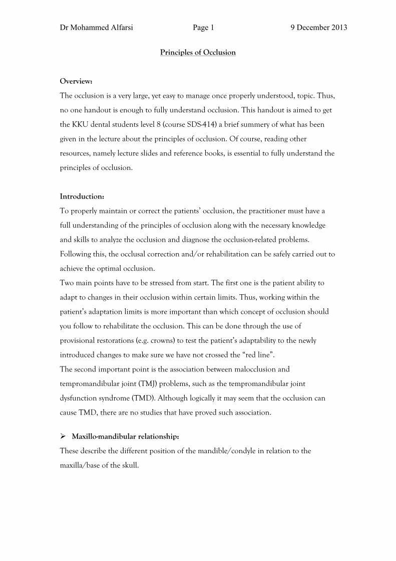
o Centric occlusion (CO):
This position describes the first teeth contact (not intercuspation, most often posterior
teeth) in CR. This term used to describe maximum intercuspation, so expect some
confusion when discussing occlusion with other practitioners.
o Maximum intercuspation (MI):
This is simply the position where the maximum interdigitation of maxillary and
mandibular teeth occurs.
In CO, condyles are in CR and only some teeth started to touch (mostly posterior teeth). This position is usually around 1.5mm distal to MI, and rarely does it coincide with MI. CO MI
The way the muscle of mastication pulls the
mandible up & forward makes it more anatomically convincing that the condyles
in CR are in their most superior/anterior position
CR
Dr Mohammed Alfarsi Page 3 9 December 2013
o Habitual relation (HR):
This is where the patient is used to occlude. Most often coincide with CR (muscular-
driven), but can be influenced by teeth. Thus to record it, we disclude the teeth and get
the muscle to drive the mandible.
Centric contacts:
These describe the contact between maxillary palatal cusps’ tips and mandibular teeth
in MI. There are 4 scenarios for such contacts.
o Cusp to cusp slope: This puts too much horizontal force on the posterior teeth and
thus SHOULD BE AVOIDED.
o Cusp to cusp tip: This rarely happens, and when it does the cusps’ tips would wear
off rapidly. Of course if we can, we should avoid it (like when making crowns).
o Cusp to marginal ridge: Here the maxillary cusps’ tips occlude with the mandibular
teeth marginal ridges, which is acceptable.
o Cusp to fossae: Here the maxillary cusps’ tips occlude with the mandibular teeth
fossae, with is the most common and preferred centric contacts.
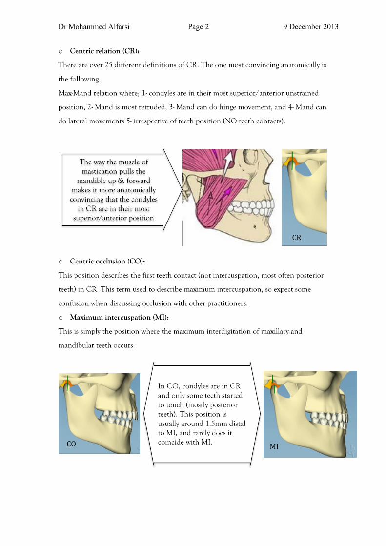
Eccentric contacts:
These describe the contact between maxillary palatal cusps’
tips and mandibular teeth during the mandibular movement.
o Protrusive:
Where the mandible moves antero-posterioly. Initially, up to
2mm, posterior teeth can contact, as follows, but once the
mandibular incisors start sliding over the maxillary incisors’
palatal surfaces the posterior teeth disengage and no longer contact (here any contact
between posterior teeth is considered an interference).
• Point centric:
This is where the mandible and maxilla are locked together in one position.
• Long centric:
This is where the mandibular fossae are elongated to allow a slide between CR and MI,
usually 0.5-1.5mm.
Dr Mohammed Alfarsi Page 4 9 December 2013
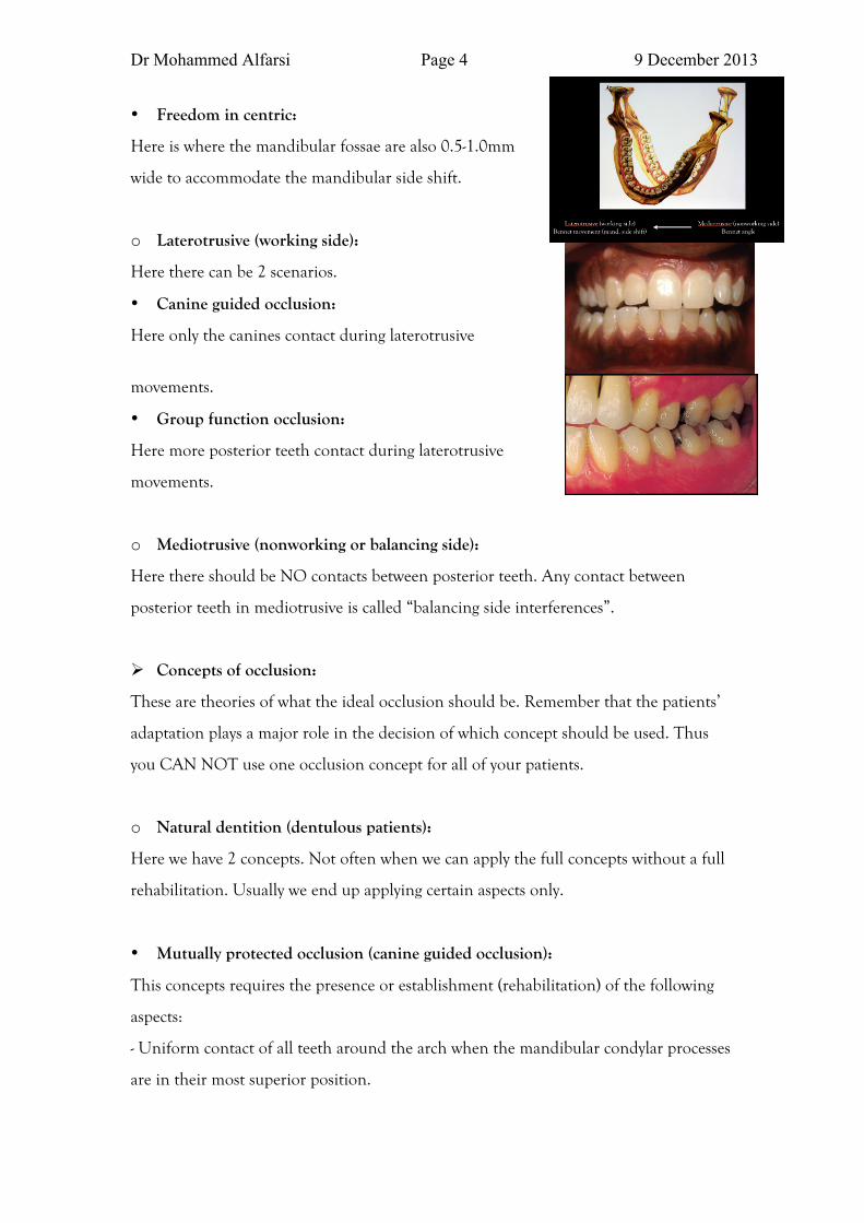
• Freedom in centric:
Here is where the mandibular fossae are also 0.5-1.0mm
wide to accommodate the mandibular side shift.
o Laterotrusive (working side):
Here there can be 2 scenarios.
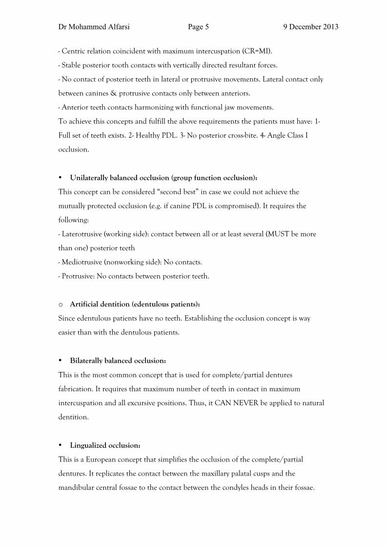
• Canine guided occlusion:
Here only the canines contact during laterotrusive
movements.
• Group function occlusion:
Here more posterior teeth contact during laterotrusive
movements.
o Mediotrusive (nonworking or balancing side):
Here there should be NO contacts between posterior teeth. Any contact between
posterior teeth in mediotrusive is called “balancing side interferences”.
Concepts of occlusion:
These are theories of what the ideal occlusion should be. Remember that the patients’
adaptation plays a major role in the decision of which concept should be used. Thus
you CAN NOT use one occlusion concept for all of your patients.
o Natural dentition (dentulous patients):
Here we have 2 concepts. Not often when we can apply the full concepts without a full
rehabilitation. Usually we end up applying certain aspects only.
• Mutually protected occlusion (canine guided occlusion):
This concepts requires the presence or establishment (rehabilitation) of the following
aspects:
- Uniform contact of all teeth around the arch when the mandibular condylar processes
are in their most superior position.
Dr Mohammed Alfarsi Page 5 9 December 2013
- Centric relation coincident with maximum intercuspation (CR=MI).
- Stable posterior tooth contacts with vertically directed resultant forces.
- No contact of posterior teeth in lateral or protrusive movements. Lateral contact only
between canines & protrusive contacts only between anteriors.
- Anterior teeth contacts harmonizing with functional jaw movements.
To achieve this concepts and fulfill the above requirements the patients must have: 1-
Full set of teeth exists. 2- Healthy PDL. 3- No posterior cross-bite. 4- Angle Class I
occlusion.
• Unilaterally balanced occlusion (group function occlusion):
This concept can be considered “second best” in case we could not achieve the
mutually protected occlusion (e.g. if canine PDL is compromised). It requires the
following:
- Laterotrusive (working side): contact between all or at least several (MUST be more
than one) posterior teeth
- Mediotrusive (nonworking side): No contacts.
- Protrusive: No contacts between posterior teeth.
o Artificial dentition (edentulous patients):
Since edentulous patients have no teeth. Establishing the occlusion concept is way
easier than with the dentulous patients.
• Bilaterally balanced occlusion:
This is the most common concept that is used for complete/partial dentures
fabrication. It requires that maximum number of teeth in contact in maximum
intercuspation and all excursive positions. Thus, it CAN NEVER be applied to natural
dentition.
• Lingualized occlusion:
This is a European concept that simplifies the occlusion of the complete/partial
dentures. It replicates the contact between the maxillary palatal cusps and the
mandibular central fossae to the contact between the condyles heads in their fossae.
Dr Mohammed Alfarsi Page 6 9 December 2013
Thus, the only contacts are between maxillary molars palatal cusps and mandibular
molar CENTRAL fossae in ALL maxillo-mandibular positions. However, it needs a
specific articular, which is why its use is limited.
Pathologic occlusion:
It is the occlusal relationship that is capable of producing pathologic changes in the
stomatognathic system. Also known as “traumatic occlusion”.
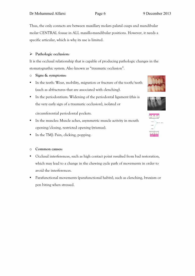
o Signs & symptoms:
• In the teeth: Wear, mobility, migration or fracture of the tooth/teeth
(such as abfractures that are associated with clenching).
• In the periodontium: Widening of the periodontal ligament (this is
the very early sign of a traumatic occlusion), isolated or
circumferential periodontal pockets.
• In the muscles: Muscle aches, asymmetric muscle activity in mouth
opening/closing, restricted opening (trismus).
• In the TMJ: Pain, clicking, popping.
o Common causes:
• Occlusal interferences, such as high contact point resulted from bad restoration,
which may lead to a change in the chewing cycle path of movements in order to
avoid the interferences.
• Parafunctional movements (parafunctional habits), such as clenching, bruxism or
pen biting when stressed.
Dr Mohammed Alfarsi Page 7 9 December 2013
Clinical Step-By-Step Guideline for (Basic) Occlusion Practice
Before start drilling: PLEASE!!
• Check the centric contacts points with an articulating paper to see where are those
points in relation to the cavity you WILL drill. There MUST be at least ONE
centric contact point on natural tooth structure, NOT on the margin. If there is not
(or will be not after drilling), then you SHOULD NOT make a class I/II but rather
you should do an onlay or a crown.
• Check the eccentric contacts areas visually or with articulating paper to see if the
occlusion is canine guided or group functioned and to check for any occlusal
interferences, like contacts in balancing side. You do not have to remove all the
interferences, but DO NOT make new ones with the restoration you are doing. Of
course if the tooth you are working on had an interference, you can remove it as
part of restoring the proper function.
After placing the restoration:
• Check the centric and eccentric again and make sure you either left them as they
were or improved them (by removing an interference from ONLY the tooth you
restored)