Embed Size (px)
Citation preview
ORIGINAL ARTICLE
Alteration of the Murine Gut Microbiota During Infection withthe Parasitic Helminth Heligmosomoides polygyrus
Seth T. Walk, PhD,* Arthur M. Blum,† Sarah Ang-Sheng Ewing,* Joel V. Weinstock, MD,†
and Vincent B. Young, MD, PhD*,‡
Background: In a murine model of inflammatory bowel disease
(IBD), treatment of colitis in IL-10 gene-deficient mice with the
parasitic helminth Heligmosomoides polygyrus ameliorates colonic
inflammation. The cellular and molecular mechanisms driving this
therapeutic host response are being studied vigorously. One pro-
posed mechanism is that H. polygyrus infection favors the out-
growth or suppression of certain bacteria, which in turn help
modulate host immunity.
Methods: To quantify the effect of H. polygyrus infection on
the composition of the gastrointestinal (GI) tract microbiota, we
conducted two independent microbial ecology analyses of C57BL/
6 mice. We obtained and analyzed 3,353 bacterial 16S rRNA
encoding gene sequences from the ileum and cecum of infected
and uninfected mice as well as incective H. polygyrus larvae at
the outset of the second experiment and adult worms taken
directly from the mouse duodenum at the end of the second
experiment.
Results: We found that a significant shift in the abundance and
relative distribution of bacterial species in the ileum of mice is
associated with H. polygyrus infection. Members of the bacterial
family Lactobacillaceae significantly increased in abundance in
the ileum of infected mice reproducibly in two independent
experiments despite having different microbiotas present at the
outset of each experiment.
Conclusions: These data support the concept that helminth
infection shifts the composition of intestinal bacteria. The clinical
consequences of these shifts in intestinal flora are yet to be
explored.
(Inflamm Bowel Dis 2010;16:1841–1849)
Key Words: inflammatory bowel diseases, microbial ecology,Heligmosomoides polygyrus, microbiota, microbiome
T he etiology of inflammatory bowel disease (IBD) is
complex and influenced by genetic, microbiologic, and
environmental factors. Lifestyle changes accompanying the
modernization of developing countries have been epidemio-
logically linked to an increase in IBD incidence.1–6 These
observations have led to the IBD hygiene hypothesis,
which states that raising children in extremely hygienic
environments negatively affects immune development and
predisposes them to immunological diseases later in life.6
Empirical support for the IBD hygiene hypothesis
has been developed in mouse and rat models.7–9 These
studies revealed that intestinal parasites and parasite prod-
ucts modulate host immunity and decrease IBD-like
inflammation.6,10 Likewise, Trichuris suis (pig whipworm)
treatment was effective in decreasing symptoms of IBD in
human clinical trials.11,12
It is not entirely understood how helminthic parasites
modulate the host mucosal immune response and alter sus-
ceptibility to immunological diseases.10,13 One unexplored
hypothesis is that helminth infection changes the abun-
dance and/or distribution of gastrointestinal (GI) tract bac-
teria (the microbiota), resulting in an overall decrease in
the proinflammatory cytokine milieu associated with the
chronically inflamed state.6 To begin to address this
hypothesis, we studied the effect of helminthic infection on
the composition of the GI tract microbiota of their host.
We found that the murine roundworm, Heligmoso-moides polygyrus, changes the microbiota of the GI tract of
otherwise healthy wildtype mice. We used clone library
analysis of the bacterial 16S rRNA encoding gene (16S) to
Received for publication February 12, 2010; Accepted February 22,
2010.
From the *Department of Internal Medicine, Division of Infectious
Diseases, University of Michigan Health System, Ann Arbor, Michigan,†Department of Internal Medicine, Division of Gastroenterology and
Hepatology, Tufts New England Medical Center, Boston, Massachusetts,‡Department of Microbiology and Immunology, University of Michigan,
Ann Arbor, Michigan.
Reprints: Vincent B. Young, MD, PhD, 4618 Med Sci II, 1150 W.
Medical Center Dr., Ann Arbor, MI 48109 (e-mail: [email protected])
Supported by US National Institutes of Health: DK38327 (to J.V.W.),
DK058755 (to J.V.W.), DK070875 (to V.B.Y.), and a Ruth L. Kirschstein
National Research Service Award T32 HL07749-15 (to S.T.W.).
Additional support: Broad Foundation (to J.V.W.), Schneider family (to
J.V.W.), Friedman family (to J.V.W.), Gilman family (to J.V.W.),
Michigan Institute for Clinical and Health Research Postdoctoral
Translational Scholars Program (to S.T.W.), Crohn’s and Colitis
Foundation of America Student Research Fellowship (to S.A.E.), Crohn’s
and Colitis Foundation of America Senior Investigator Award (to V.B.Y.).
CopyrightVC 2010 Crohn’s & Colitis Foundation of America, Inc.
DOI 10.1002/ibd.21299
Published online 18 May 2010 in Wiley Online Library
(wileyonlinelibrary.com).
Inflamm Bowel Dis � Volume 16, Number 11, November 2010 1841
quantify changes that occur in the microbiota associated
with the distal small intestine (ileum) and the tip of the
cecum. In two independent experiments, we found that the
ileal microbiota from H. polygyrus-infected mice was sig-
nificantly different from uninfected controls. Also, the total
bacterial load was greater in infected mice versus controls,
and the dominant bacteria associated with the ileum of
treated mice were also associated with adult worms that
had been removed at the time of necropsy. These data sup-
port the hypothesis that parasites alter the abundance and
relative distribution of GI tract bacteria and provide the
basis for future experiments that investigate the overall im-
portance that this alteration has on host immunity.
MATERIALS AND METHODS
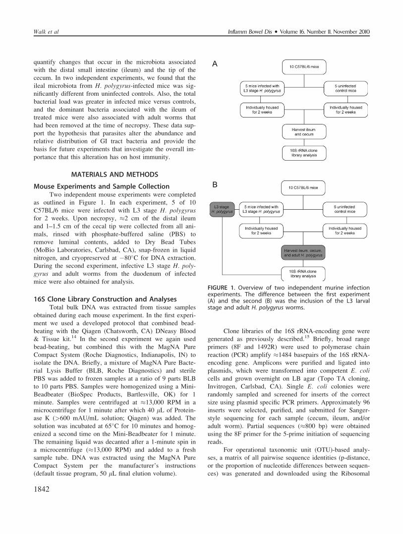
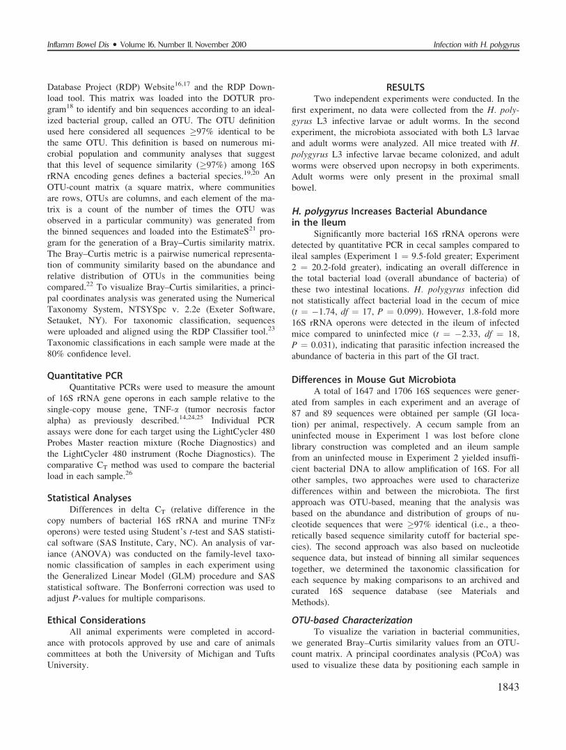
Mouse Experiments and Sample CollectionTwo independent mouse experiments were completed
as outlined in Figure 1. In each experiment, 5 of 10
C57BL/6 mice were infected with L3 stage H. polygyrusfor 2 weeks. Upon necropsy, �2 cm of the distal ileum
and 1–1.5 cm of the cecal tip were collected from all ani-
mals, rinsed with phosphate-buffered saline (PBS) to
remove luminal contents, added to Dry Bead Tubes
(MoBio Laboratories, Carlsbad, CA), snap-frozen in liquid
nitrogen, and cryopreserved at �80�C for DNA extraction.
During the second experiment, infective L3 stage H. poly-gyrus and adult worms from the duodenum of infected
mice were also obtained for analysis.
16S Clone Library Construction and AnalysesTotal bulk DNA was extracted from tissue samples
obtained during each mouse experiment. In the first experi-
ment we used a developed protocol that combined bead-
beating with the Qiagen (Chatsworth, CA) DNeasy Blood
& Tissue kit.14 In the second experiment we again used
bead-beating, but combined this with the MagNA Pure
Compact System (Roche Diagnostics, Indianapolis, IN) to
isolate the DNA. Briefly, a mixture of MagNA Pure Bacte-
rial Lysis Buffer (BLB, Roche Diagnostics) and sterile
PBS was added to frozen samples at a ratio of 9 parts BLB
to 10 parts PBS. Samples were homogenized using a Mini-
Beadbeater (BioSpec Products, Bartlesville, OK) for 1
minute. Samples were centrifuged at �13,000 RPM in a
microcentrifuge for 1 minute after which 40 lL of Protein-
ase K (>600 mAU/mL solution; Qiagen) was added. The
solution was incubated at 65�C for 10 minutes and homog-
enized a second time on the Mini-Beadbeater for 1 minute.
The remaining liquid was decanted after a 1-minute spin in
a microcentrifuge (�13,000 RPM) and added to a fresh
sample tube. DNA was extracted using the MagNA Pure
Compact System per the manufacturer’s instructions
(default tissue program, 50 lL final elution volume).
Clone libraries of the 16S rRNA-encoding gene were
generated as previously described.15 Briefly, broad range
primers (8F and 1492R) were used to polymerase chain
reaction (PCR) amplify �1484 basepairs of the 16S rRNA-
encoding gene. Amplicons were purified and ligated into
plasmids, which were transformed into competent E. colicells and grown overnight on LB agar (Topo TA cloning,
Invitrogen, Carlsbad, CA). Single E. coli colonies were
randomly sampled and screened for inserts of the correct
size using plasmid specific PCR primers. Approximately 96
inserts were selected, purified, and submitted for Sanger-
style sequencing for each sample (cecum, ileum, and/or
adult worm). Partial sequences (�800 bp) were obtained
using the 8F primer for the 5-prime initiation of sequencing
reads.
For operational taxonomic unit (OTU)-based analy-
ses, a matrix of all pairwise sequence identities (p-distance,
or the proportion of nucleotide differences between sequen-
ces) was generated and downloaded using the Ribosomal
FIGURE 1. Overview of two independent murine infectionexperiments. The difference between the first experiment(A) and the second (B) was the inclusion of the L3 larvalstage and adult H. polygyrus worms.
Inflamm Bowel Dis � Volume 16, Number 11, November 2010Walk et al
1842
Database Project (RDP) Website16,17 and the RDP Down-
load tool. This matrix was loaded into the DOTUR pro-
gram18 to identify and bin sequences according to an ideal-
ized bacterial group, called an OTU. The OTU definition
used here considered all sequences �97% identical to be
the same OTU. This definition is based on numerous mi-
crobial population and community analyses that suggest
that this level of sequence similarity (�97%) among 16S
rRNA encoding genes defines a bacterial species.19,20 An
OTU-count matrix (a square matrix, where communities
are rows, OTUs are columns, and each element of the ma-
trix is a count of the number of times the OTU was
observed in a particular community) was generated from
the binned sequences and loaded into the EstimateS21 pro-
gram for the generation of a Bray–Curtis similarity matrix.
The Bray–Curtis metric is a pairwise numerical representa-
tion of community similarity based on the abundance and
relative distribution of OTUs in the communities being
compared.22 To visualize Bray–Curtis similarities, a princi-
pal coordinates analysis was generated using the Numerical
Taxonomy System, NTSYSpc v. 2.2e (Exeter Software,
Setauket, NY). For taxonomic classification, sequences
were uploaded and aligned using the RDP Classifier tool.23
Taxonomic classifications in each sample were made at the
80% confidence level.
Quantitative PCRQuantitative PCRs were used to measure the amount
of 16S rRNA gene operons in each sample relative to the
single-copy mouse gene, TNF-a (tumor necrosis factor
alpha) as previously described.14,24,25 Individual PCR
assays were done for each target using the LightCycler 480
Probes Master reaction mixture (Roche Diagnostics) and
the LightCycler 480 instrument (Roche Diagnostics). The
comparative CT method was used to compare the bacterial
load in each sample.26
Statistical AnalysesDifferences in delta CT (relative difference in the
copy numbers of bacterial 16S rRNA and murine TNFaoperons) were tested using Student’s t-test and SAS statisti-
cal software (SAS Institute, Cary, NC). An analysis of var-
iance (ANOVA) was conducted on the family-level taxo-
nomic classification of samples in each experiment using
the Generalized Linear Model (GLM) procedure and SAS
statistical software. The Bonferroni correction was used to
adjust P-values for multiple comparisons.
Ethical ConsiderationsAll animal experiments were completed in accord-
ance with protocols approved by use and care of animals
committees at both the University of Michigan and Tufts
University.
RESULTSTwo independent experiments were conducted. In the
first experiment, no data were collected from the H. poly-gyrus L3 infective larvae or adult worms. In the second
experiment, the microbiota associated with both L3 larvae
and adult worms were analyzed. All mice treated with H.polygyrus L3 infective larvae became colonized, and adult
worms were observed upon necropsy in both experiments.
Adult worms were only present in the proximal small
bowel.
H. polygyrus Increases Bacterial Abundancein the Ileum
Significantly more bacterial 16S rRNA operons were
detected by quantitative PCR in cecal samples compared to
ileal samples (Experiment 1 ¼ 9.5-fold greater; Experiment
2 ¼ 20.2-fold greater), indicating an overall difference in
the total bacterial load (overall abundance of bacteria) of
these two intestinal locations. H. polygyrus infection did
not statistically affect bacterial load in the cecum of mice
(t ¼ �1.74, df ¼ 17, P ¼ 0.099). However, 1.8-fold more
16S rRNA operons were detected in the ileum of infected
mice compared to uninfected mice (t ¼ �2.33, df ¼ 18,
P ¼ 0.031), indicating that parasitic infection increased the
abundance of bacteria in this part of the GI tract.
Differences in Mouse Gut MicrobiotaA total of 1647 and 1706 16S sequences were gener-
ated from samples in each experiment and an average of
87 and 89 sequences were obtained per sample (GI loca-
tion) per animal, respectively. A cecum sample from an
uninfected mouse in Experiment 1 was lost before clone
library construction was completed and an ileum sample
from an uninfected mouse in Experiment 2 yielded insuffi-
cient bacterial DNA to allow amplification of 16S. For all
other samples, two approaches were used to characterize
differences within and between the microbiota. The first
approach was OTU-based, meaning that the analysis was
based on the abundance and distribution of groups of nu-
cleotide sequences that were �97% identical (i.e., a theo-
retically based sequence similarity cutoff for bacterial spe-
cies). The second approach was also based on nucleotide
sequence data, but instead of binning all similar sequences
together, we determined the taxonomic classification for
each sequence by making comparisons to an archived and
curated 16S sequence database (see Materials and
Methods).
OTU-based CharacterizationTo visualize the variation in bacterial communities,
we generated Bray–Curtis similarity values from an OTU-
count matrix. A principal coordinates analysis (PCoA) was
used to visualize these data by positioning each sample in
Inflamm Bowel Dis � Volume 16, Number 11, November 2010 Infection with H. polygyrus
1843
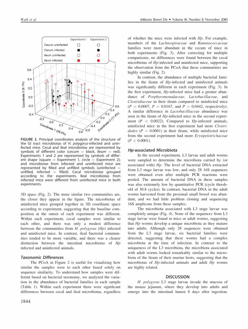
3D space (Fig. 2). The more similar two communities are,
the closer they appear in the figure. The microbiotas of
uninfected mice grouped together in 3D coordinate space
according to experiment, suggesting that the baseline com-
position at the outset of each experiment was different.
Within each experiment, cecal samples were similar to
each other, and there was only a modest difference
between the communities from H. polygyrus (Hp) infectedand uninfected mice. In contrast, ileal bacterial commun-
ities tended to be more variable, and there was a clearer
distinction between the intestinal microbiotas of Hpinfected and uninfected animals.
Taxonomic DifferencesThe PCoA in Figure 2 is useful for visualizing how
similar the samples were to each other based solely on
sequence similarity. To understand how samples were dif-
ferent based on bacterial taxonomy, we analyzed the varia-
tion in the abundance of bacterial families in each sample
(Table 1). Within each experiment there were significant
differences between cecal and ileal microbiotas, regardless
of whether the mice were infected with Hp. For example,
members of the Lachnospiraceae and Ruminococcaceaefamilies were more abundant in the cecum of mice in
both experiments (Fig. 3). After correcting for multiple
comparisons, no differences were found between the cecal
microbiotas of Hp-infected and uninfected mice, supporting
the observation from the PCoA that these communities are
highly similar (Fig. 2).
In contrast, the abundance of multiple bacterial fami-
lies in the ileum of Hp-infected and uninfected animals
was significantly different in each experiment (Fig. 3). In
the first experiment, Hp-infected mice had a greater abun-
dance of Porphyromonadaceae, Lactobacillaceae, and
Clostridiaceae in their ileum compared to uninfected mice
(P ¼ 0.0007, P ¼ 0.0167, and P ¼ 0.0442, respectively).
A similar difference in Lactobacilliaceae abundance was
seen in the ileum of Hp-infected mice in the second experi-
ment (P < 0.0022). Compared to Hp-infected animals,
uninfected mice in the first experiment had more Clostri-diales (P ¼ 0.0001) in their ileum, while uninfected mice
from the second experiment had more Erysipelotrichaceae(P < 0.0001).
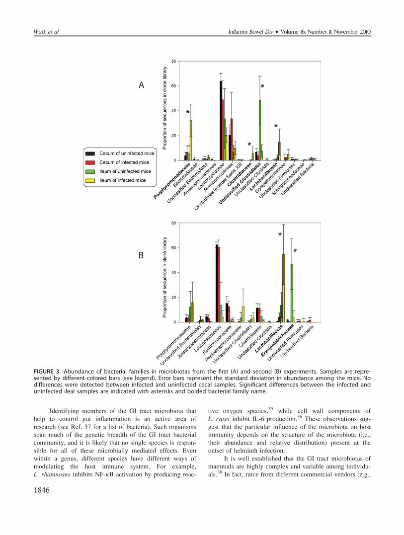
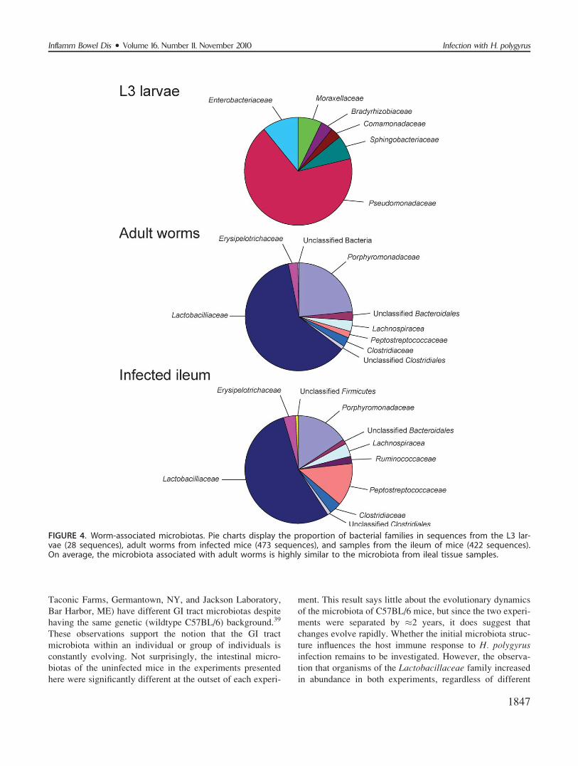
Hp-associated MicrobiotaIn the second experiment, L3 larvae and adult worms
were sampled to determine the microbiota carried by (or
associated with) Hp. The level of bacterial DNA extracted
from L3 stage larvae was low, and only 28 16S sequences
were obtained even after multiple PCR reactions were
pooled. The amount of bacterial DNA in these samples
was also extremely low by quantitative PCR (cycle thresh-
old of 30.8 cycles). In contrast, bacterial DNA in the adult
worms harvested from the proximal small bowel was abun-
dant, and we had little problem cloning and sequencing
16S amplicons from these samples.
The microbiota associated with L3 stage larvae was
completely unique (Fig. 4). None of the sequences from L3
stage larvae were found in mice or adult worms, suggesting
that Hp worms develop a unique microbiota as they mature
into adults. Although only 28 sequences were obtained
from the L3 stage larvae, six bacterial families were
detected, suggesting that these worms had a complex
microbiota at the time of infection. In contrast to the
uniqueness of the L3 microbiota, the microbiota associated
with adult worms looked remarkably similar to the micro-
biota of the ileum of their murine hosts, suggesting that the
microbiotas of Hp-infected animals and adult Hp worms
are highly related.
DISCUSSIONH. polygyrus L3 stage larvae invade the mucosa of
the mouse jejunum, where they develop into adults and
emerge into the intestinal lumen 8 days after ingestion.
FIGURE 2. Principal coordinates analysis of the structure ofthe GI tract microbiotas of H. polygyrus-infected and unin-fected mice. Cecal and ileal microbiotas are represented bysymbols of different color (cecum ¼ black, ileum ¼ red);Experiments 1 and 2 are represented by symbols of differ-ent shape (square ¼ Experiment 1, circle ¼ Experiment 2);and microbiotas from infected and uninfected mice arerepresented by filled and unfilled symbols (uninfected ¼unfilled, infected ¼ filled). Cecal microbiotas groupedaccording to the experiments. Ileal microbiotas frominfected mice were different from uninfected mice in bothexperiments.
Inflamm Bowel Dis � Volume 16, Number 11, November 2010Walk et al
1844
Infection induces a polarizing T-helper cell type 2 (Th2)
host immune response characterized by the production of
IL-4 and IL-13, as well as activation of the STAT-6 path-
way.27 These events activate mast cells, induce the alterna-
tively activated phenotype in tissue-resident macrophages,
increase mucus secretion by goblet cells, and enhance intes-
tinal smooth muscle contraction.28,29 During a primary H.polygyrus infection, the host Th2 response does not result in
rapid clearance of the adult worms; rather, it has a signifi-
cant deleterious effect on H. polygyrus fecundity, as meas-
ured by the number of eggs shed per gram of feces.28,30
H. polygyrus infection has a profound influence on a
number of immunoregulatory signals that decrease intesti-
nal inflammation. H. polygyrus infection increases the pro-
duction of the global transcription factor, FoxP3, in T cells
of the lamina propria, and these cells decrease intestinal
inflammation upon transfer to mice with established coli-
tis.31 In the terminal ileum of mice, H. polygyrus induces
mucosal T cells to express Toll-like receptor 4 (TLR4).32
TLR4 signaling by bacterial lipopolysaccharide (LPS) nor-
mally stimulates cells to produce proinflammatory cyto-
kines, like TNFa and IL-12. In contrast to what happens
normally, mucosal T cells from H. polygyrus-infected mice
do not produce TNFa or IL-12, but instead produce the
regulatory cytokine TGFb.32 In the absence of this TGFbsignaling, there is insufficient control of interferon gamma
(IFN-c) production by IL-10 and colitis is maintained.33
To our knowledge, it has never been examined if H.polygyrus infection alters the composition of the GI tract
microbiota. A number of observations from experiments
with germfree mice suggest that H. polygyrus needs GI
tract bacteria to establish a robust infection.34 Compared to
mice with a conventional microbiota, total worm burden
was lower in germfree mice 13 days postinoculation. More-
over, germfree mice rapidly cleared these worms between
days 13–30 postinoculation, while worm burden remained
high in conventional mice. Also, a robust intestinal muco-
sal eosinophilia developed in germfree mice, which was
absent in conventional mice, suggesting that the host
immune response to H. polygyrus differs in the absence of
intestinal bacteria. Interestingly, mortality in mice given a
‘‘heavy’’ infection (1600 L3 infective larvae) was only
observed in conventional mice. These observations suggest
that the GI microbiota is necessary for the normal host
response to H. polygyrus.Infection of mice with H. polygyrus resulted in an
increased abundance of various bacteria, suggesting that
some species find conditions in the ileum of H. polygyrus-infected mice favorable. This observation is what one
would expect from a microbial-helminth mutualism, where
both bacteria and worm benefit from symbiosis. Interest-
ingly, members of the Lactobacillaceae family increased in
both experiments. The Lactobacillaceae family is com-
posed of the genera Lactobacillus, Paralactobacillus,Pediococcus, and other bacteria that are not yet classified
by microbial taxonomists. Most of the known Lactobacilla-ceae species belong to the genus Lactobacillus, which is a
diverse group of Gram-positive, facultative anaerobic spe-
cies commonly known as lactic acid bacteria because most
convert sugars into lactic acid. Certain Lactobacillus spe-
cies, like L. rhamnosus35 and L. casei,36 decrease intestinal
inflammation in murine models of IBD, and it is tempting
to speculate that Lactobacillus species play a role in the
antiinflammatory/immune modulatory effects of H. polygy-rus infection. More studies are needed to elucidate the role
of this potentially important mutualism.
TABLE 1. Statistically Significant Differences in the Abundance of Bacterial Families for Each Mouse Experiment
Bacterial Family F-value df Pr > F Significant Difference
Experiment 1
Porphyromonadaceae 13.1 3 0.0002 Abundant in ileum of infected mice
Lachnospiraceae 6.71 3 0.0043 Abundant in cecum of mice
Ruminococcaceae 5.31 3 0.0108 Abundant in cecum of mice
Clostridiaceae 4.94 3 0.0139 Abundant in ileum of infected mice
Unclassified Clostridiales 19.94 3 <0.0001 Abundant in ileum of uninfected mice
Lactobacillaceae 7.66 3 0.0025 Abundant in ileum of infected mice
Experiment 2
Anaeroplasmataceae 7.37 3 0.0029 Abundant in cecum of mice
Lachnospiraceae 51.99 3 <0.0001 Abundant in cecum of mice
Ruminococcaceae 16.25 3 <0.0001 Abundant in cecum of mice
Unclassified Clostridiales 11.74 3 0.0003 Abundant in ileum of mice
Lactobacillaceae 17.26 3 <0.0001 Abundant in ileum of infected mice
Erysipelotrichaceae 23.67 3 <0.0001 Abundant in ileum of uninfected mice
Inflamm Bowel Dis � Volume 16, Number 11, November 2010 Infection with H. polygyrus
1845
Identifying members of the GI tract microbiota that
help to control gut inflammation is an active area of
research (see Ref. 37 for a list of bacteria). Such organisms
span much of the genetic breadth of the GI tract bacterial
community, and it is likely that no single species is respon-
sible for all of these microbially mediated effects. Even
within a genus, different species have different ways of
modulating the host immune system. For example,
L. rhamnosus inhibits NF-jB activation by producing reac-
tive oxygen species,35 while cell wall components of
L. casei inhibit IL-6 production.36 These observations sug-
gest that the particular influence of the microbiota on host
immunity depends on the structure of the microbiota (i.e.,
their abundance and relative distribution) present at the
outset of helminth infection.
It is well established that the GI tract microbiotas of
mammals are highly complex and variable among individu-
als.38 In fact, mice from different commercial vendors (e.g.,
FIGURE 3. Abundance of bacterial families in microbiotas from the first (A) and second (B) experiments. Samples are repre-sented by different-colored bars (see legend). Error bars represent the standard deviation in abundance among the mice. Nodifferences were detected between infected and uninfected cecal samples. Significant differences between the infected anduninfected ileal samples are indicated with asterisks and bolded bacterial family name.
Inflamm Bowel Dis � Volume 16, Number 11, November 2010Walk et al
1846
Taconic Farms, Germantown, NY, and Jackson Laboratory,
Bar Harbor, ME) have different GI tract microbiotas despite
having the same genetic (wildtype C57BL/6) background.39
These observations support the notion that the GI tract
microbiota within an individual or group of individuals is
constantly evolving. Not surprisingly, the intestinal micro-
biotas of the uninfected mice in the experiments presented
here were significantly different at the outset of each experi-
ment. This result says little about the evolutionary dynamics
of the microbiota of C57BL/6 mice, but since the two experi-
ments were separated by �2 years, it does suggest that
changes evolve rapidly. Whether the initial microbiota struc-
ture influences the host immune response to H. polygyrusinfection remains to be investigated. However, the observa-
tion that organisms of the Lactobacillaceae family increased
in abundance in both experiments, regardless of different
FIGURE 4. Worm-associated microbiotas. Pie charts display the proportion of bacterial families in sequences from the L3 lar-vae (28 sequences), adult worms from infected mice (473 sequences), and samples from the ileum of mice (422 sequences).On average, the microbiota associated with adult worms is highly similar to the microbiota from ileal tissue samples.
Inflamm Bowel Dis � Volume 16, Number 11, November 2010 Infection with H. polygyrus
1847
initial microbiotas, suggests that there may be compositional
similarities among H. polygyrus infections that ultimately
influence host immunomodulation.
Relevance of This Study to Helminth Treatmentof IBD
Normally, Th1/Th17 driven inflammation character-
ized by the production of proinflammatory cytokines
(IL-12, IL-23, IL-17, and IFN-c) is held in check by the
production of immunoregulatory cytokines and transcrip-
tion factors (e.g., TGF-b, IL-10, and FoxP3). In patients
with IBD, homeostatic regulatory signals are impaired,
and a chronic inflammation develops that is perpetuated
by antigens from the commensal GI tract microbiota.40
A number of therapies commonly used in IBD like aza-
thioprine, anti-TNF monoclonal antibodies, and cortico-
steroids suppress this inflammatory cascade.41 Some stud-
ies suggest that helminths may prove useful for the
treatment of both Crohn’s disease and ulcerative coli-
tis.11,12 The data presented here demonstrate that H. pol-ygyrus infection is associated with a significant shift in
the abundance and relative distribution of GI tract bacte-
ria. The relatively large effect size of the shift among
ileal-associated microbiotas compared to cecal-associated
microbiotas suggests that either helminths directly influ-
enced the microbiota or that the host immune response
to infection mediated the observed alterations. The
dynamic interactions among H. polygyrus, the host mu-
cosal immune response, and the intestinal microbiota
require more study.
REFERENCES1. Asakura H, Suzuki K, Kitahora T, et al. Is there a link between
food and intestinal microbes and the occurrence of Crohn’s dis-ease and ulcerative colitis? J Gastroenterol Hepatol. 2008;23:1794–1801.
2. Bernstein CN, Shanahan F. Disorders of a modern lifestyle: reconcil-ing the epidemiology of inflammatory bowel diseases. Gut. 2008;57:1185–1191.
3. Frangos CC.Inflammatory bowel disease: reviewing an old studyunder a new perspective. Gut. 2007;56:1638–1639.
4. Goh K, Xiao SD. Inflammatory bowel disease: a survey of the epide-miology in Asia. J Dig Dis. 2009;10:1–6.
5. Shanahan F, Bernstein CN. The evolving epidemiology of inflamma-tory bowel disease. Curr Opin Gastroenterol. 2009;25:301–305.
6. Weinstock JV, Elliott DE. Helminths and the IBD hygiene hypothesis.Inflamm Bowel Dis. 2009;15:128–133.
7. Elliott DE, Li J, Blum A, et al. Exposure to schistosome eggs protectsmice from TNBS-induced colitis. Am J Physiol Gastrointest LiverPhysiol. 2003;284:G385–391.
8. Moreels TG, Nieuwendijk RJ, De Man JG, et al. Concurrent infectionwith Schistosoma mansoni attenuates inflammation induced changes incolonic morphology, cytokine levels, and smooth muscle contractilityof trinitrobenzene sulphonic acid induced colitis in rats. Gut. 2004;53:99–107.
9. Ruyssers NE, De Winter BY, De Man JG, et al. Therapeutic potentialof helminth soluble proteins in TNBS-induced colitis in mice. InflammBowel Dis. 2009;15:491–500.
10. Weinstock JV, Summers RW, Elliott DE. Role of helminths in regu-lating mucosal inflammation. Springer Semin Immunopathol. 2005;27:249–271.
11. Summers RW, Elliott DE, Urban JF Jr, et al. Trichuris suis therapy inCrohn’s disease. Gut. 2005;54:87–90.
12. Summers RW, Elliott DE, Urban JF Jr, et al. Trichuris suis therapyfor active ulcerative colitis: a randomized controlled trial. Gastroen-terology. 2005;128:825–832.
13. Jackson JA, Friberg IM, Little S, et al. Review series on helminths,immune modulation and the hygiene hypothesis: immunity againsthelminths and immunological phenomena in modern human popula-tions: coevolutionary legacies? Immunology. 2009;126:18–27.
14. Antonopoulos DA, Huse SM, Morrison HG, et al. Reproducible com-munity dynamics of the gastrointestinal microbiota following antibi-otic perturbation. Infect Immun. 2009;77:2367–2375.
15. Young VB, Schmidt TM.Antibiotic-associated diarrhea accompaniedby large-scale alterations in the composition of the fecal microbiota.J Clin Microbiol. 2004;42:1203–1206.
16. Cole JR, Chai B, Farris RJ, et al. The ribosomal database project(RDP-II): introducing myRDP space and quality controlled publicdata. Nucleic Acids Res. 2007;35:D169–172.
17. Cole JR, Wang Q, Cardenas E, et al. The Ribosomal Database Project:improved alignments and new tools for rRNA analysis. Nucleic AcidsRes. 2009;37:D141–145.
18. Schloss PD, Handelsman J. Introducing DOTUR, a computer programfor defining operational taxonomic units and estimating species rich-ness. Appl Environ Microbiol. 2005;71:1501–1506.
19. Konstantinidis KT, Ramette A, Tiedje JM. The bacterial species defi-nition in the genomic era. Philos Trans R Soc Lond B Biol Sci. 2006;361:1929–1940.
20. Konstantinidis KT, Tiedje JM. Genomic insights that advance the spe-cies definition for prokaryotes. Proc Natl Acad Sci U S A. 2005;102:2567–2572.
21. Colwell RK. Estimate S: statistical estimation of species richness andshared species from samples. 2006. Available at: Persistent URL pur-l.oclc.org/estimates
22. Beals EW. Bray-Curtis ordination: an effective strategy for analysis ofmultivariate ecological data. In: MacFadyen A, Ford ED, editors.Advances in ecological research. Orlando, FL: Academic Press;1984:1–56.
23. Wang Q, Garrity GM, Tiedje JM, et al. Naive Bayesian classifier forrapid assignment of rRNA sequences into the new bacterial taxonomy.Appl Environ Microbiol. 2007;73:5261–5267.
24. Nadkarni MA, Martin FE, Jacques NA, et al. Determination of bacte-rial load by real-time PCR using a broad-range (universal) probe andprimers set. Microbiology. 2002;148:257–266.
25. Nitsche A, Becker M, Junghahn I, et al. Quantification of human cellsin NOD/SCID mice by duplex real-time polymerase-chain reaction.Haematologica. 2001;86:693–699.
26. Schmittgen TD, Livak KJ. Analyzing real-time PCR data by the com-parative C(T) method. Nat Protoc. 2008;3:1101–1108.
27. Chen CC, Louie S, McCormick B, et al. Concurrent infection with anintestinal helminth parasite impairs host resistance to enteric Citro-bacter rodentium and enhances Citrobacter-induced colitis in mice.Infect Immun. 2005;73:5468–5481.
28. Hashimoto K, Uchikawa R, Tegoshi T, et al. Immunity-mediated regu-lation of fecundity in the nematode Heligmosomoides polygyrus —the potential role of mast cells. Parasitology. 2009:1–7.
29. Weng M, Huntley D, Huang IF, et al. Alternatively activated macro-phages in intestinal helminth infection: effects on concurrent bacterialcolitis. J Immunol. 2007;179:4721–4731.
30. Urban JF Jr, Katona IM, Finkelman FD. Heligmosomoides polygyrus:CD4þ but not CD8þ T cells regulate the IgE response and protectiveimmunity in mice. Exp Parasitol. 1991;73:500–511.
31. Elliott DE, Setiawan T, Metwali A, et al. Heligmosomoides polygyrusinhibits established colitis in IL-10-deficient mice. Eur J Immunol.2004;34:2690–2698.
32. Ince MN, Elliott DE, Setiawan T, et al. Heligmosomoides polygyrusinduces TLR4 on murine mucosal T cells that produce TGFbeta afterlipopolysaccharide stimulation. J Immunol. 2006;176:726–729.
Inflamm Bowel Dis � Volume 16, Number 11, November 2010Walk et al
1848
33. Ince MN, Elliott DE, Setiawan T, et al. Role of T cell TGF-beta sig-naling in intestinal cytokine responses and helminthic immune modu-lation. Eur J Immunol. 2009;39:1870–1878.
34. Wescott RB. Experimental Nematospiroides dubius infection ingermfree and conventional mice. Exp Parasitol. 1968;22:245–249.
35. Lin PW, Myers LE, Ray L, et al. Lactobacillus rhamnosus blocksinflammatory signaling in vivo via reactive oxygen species generation.Free Radic Biol Med. 2009;47:1205–1211.
36. Matsumoto S, Hara T, Nagaoka M, et al. A component of polysaccha-ride peptidoglycan complex on Lactobacillus induced an improvementof murine model of inflammatory bowel disease and colitis-associatedcancer. Immunology. 2009;128:e170–180.
37. Round JL, O’Connell RM, Mazmanian SK. Coordination of tolero-genic immune responses by the commensal microbiota. J Autoimmun.2010;34:J220–225.
38. Ley RE, Hamady M, Lozupone C, et al. Evolution of mammals andtheir gut microbes. Science. 2008;320:1647–1651.
39. Ivanov, II, Frutos Rde L, Manel N, et al. Specific microbiota directthe differentiation of IL-17-producing T-helper cells in the mucosa ofthe small intestine. Cell Host Microbe. 2008;4:337–349.
40. Balfour Sartor R. Bacteria in Crohn’s disease: mechanisms of inflam-mation and therapeutic implications. J Clin Gastroenterol. 2007;41(suppl 1):S37–43.
41. Afif W, Loftus EV Jr. Safety profile of IBD therapeutics: infectiousrisks. Gastroenterol Clin North Am. 2009;38:691–709.
Inflamm Bowel Dis � Volume 16, Number 11, November 2010 Infection with H. polygyrus
1849

![HELMINTH PARASITES IN MAMMALSparasite.org.au/para-site/text/helminth-checklist.pdf · HELMINTH PARASITES IN MAMMALS ... Subclass: EUTHERIA [placental mammals] ... NEM:Asc Ascaris](https://img.pdfslide.us/doc/110x75/5ad4fa137f8b9a5d058c90e9/helminth-parasites-in-parasites-in-mammals-subclass-eutheria-placental-mammals.jpg)