Embed Size (px)
Citation preview

1
Published November 1, 2012
The Non‐Accidental Trauma Evaluation: How to get it RIGHT
Elizabeth Weinstein, MD Assistant Professor of Clinical Emergency Medicine and Pediatrics Indiana University School of Medicine Indianapolis, IN
Non‐accidental trauma (NAT) is one of the most depressing issues in Emergency Medicine. But it is also a
diagnosis that is, perhaps, best understood in the same context as other diagnoses that Emergency
Physicians (EPs) commonly make in the Emergency Department. For example, EPs routinely diagnose
acute coronary syndrome (ACS). It is on the differential for every chest pain patient that walks through
the doors. Countless hours of continuing education and residency training are spent on identifying
current of injury patterns on EKG and clinical features that make ACS more or less likely. The stakes,
after all, of missing this life threatening disease are high. As a result, miss rates for ACS in the US have
been estimated to be as low as 2%. 1,2 By contrast, it is estimated that up to 30% of ED cases of non‐
accidental trauma are missed. Unfortunately the consequences of missing this diagnosis may be
catastrophic as up to 10% of children with missed NAT diagnosis may go on to suffer lethal injuries. 3,4
NAT is tragically common. In 2010 there were 754,000 identified cases of abuse and neglect and 1,560
deaths in the U.S. 5 Yet, children who are abused rarely present with a clear history. Often they present
with either non‐specific symptoms related to their inflicted injuries or with a routine pediatric illness but
with evidence of inflicted injuries on exam. These subtle presentations make identification of children
who have been victims of inflicted injury exceptionally challenging. In a review of 44 cases of pediatric
deaths from abusive trauma, 19 % of these children were evaluated by a physician in the month
preceding their deaths.6 The overwhelming majority of these evaluations occurred in an Emergency
Department. Chief complaints at the time of the evaluations included facial bruising, fussiness, vomiting
and poor feeding. 6 At the times of their deaths, many of these children had evidence of old injuries,
suggesting that they could have been diagnosed earlier and that we may have been able to save their
lives. Given the high stakes it is imperative that EPs are not only fluent in the evaluation of children
when abuse is suspected, but also that they are skillful in identifying subtle patterns that should raise
clinical suspicion for abusive injuries. EPs often feel comfortable in the evaluation of obvious
presentations of abusive injury such as the child with extensive bruising and intracranial injury, but may
struggle with more subtle presentations. Consider the following case:

2
A 4‐month‐old child presents for vomiting and diarrhea. The family reports that the infant has taken no
formula but has taken 12 ounces of an oral rehydration solution. He has had 4 wet diapers. On exam the
infant is awake and drinking from a bottle. He is mildly tachycardic, but has good capillary refill and
normal skin turgor. The abdomen is soft, non‐tender and non‐distended. A bruise is noticed on the back
of the infant’s left hand and right thigh. The family is unsure how the bruises got there.
What is the appropriate evaluation for this child? Do these bruises require evaluation? If so, what
studies are warranted?
Part I: Patterns You Need to Recognize
Bruising
Just as the EP is trained to recognize patterns of cardiac ischemia on the EKG, the EP must also be facile
in the recognition of physical exam findings or injury patterns that are suggestive of inflicted injury.
Bruises are the most important finding to recognize. They are the most common sign of maltreatment
and are often the first indication that maltreatment is occurring. Notably, bruising is missed as an
indication of abuse in up to 44% of fatal or severe cases of child abuse. 7,8 Bruising, however, is common
in childhood and distinguishing bruising that is worrisome from bruising that is not worrisome can be
challenging. Multiple studies examining bruising patterns in children are instructive to the EP and can
help the EP determine which bruising patterns should incite a more substantial evaluation.
Sugar et al., conducted a prospective cross‐sectional survey of children, under 36 months of age,
presenting to their pediatrician’s office for well child care. 9 All children were evaluated for bruising; and
presence and location of all bruises were recorded. A total of 973 children were included in the study.
Only 203 children had any bruising. In children under 6 months of age, only 0.6% had bruising anywhere
on their bodies. Further, only 2.2% of non‐mobile children of any age had bruising. Once children began
cruising and walking, bruising incidence increased substantially, however the majority of bruising was
noted on the anterior shins, knees and forehead. There were no bruises noted to the hands or the feet,
and bruising to the trunk was rare. This landmark study resulted in the clinical pearl “Those who don’t
cruise, rarely bruise.”
A systematic review of 23 articles evaluating bruising patterns in children echoes this finding, reporting
that less than 1% of non‐independently mobile infants have any bruises. Further they identified the
following bruising locations and patterns as concerning: bruising away from bony prominences, bruises
to the face and neck, bruises to trunk and buttocks, patterned and clustered bruises.10
A recently derived, but not yet prospectively validated, decision rule to identify concerning bruises
shows promise. The rule, called the TEN‐4 rule had a sensitivity of 97% and a specificity of 84% in the
derivation study. The TEN‐4 rule works as follows: any bruising in a suspicious body region in a child
under 4 years of age merits further investigation. Suspicious body regions are defined by the TEN part of
the decision rule, where T stands for torso (includes chest, abdomen, back, buttocks, genitourinary
region and hip), E stands for ears and N stands for neck. In addition, any bruising in any child under 4
months of age merits further investigation. 8 Why age 4 months? This rule was derived from a

3
retrospective case‐control study comparing bruising characteristics of children 0‐ 48 months of age
admitted to the PICU with accidental injuries to those with inflicted injuries. Detailed analyses revealed
these were the age and bruising characteristics that offered the best sensitvity and specificity.
Bruising should be documented clearly in the medical chart with location, size and pattern (if present) all
described in detail. When possible, photo‐documentation can be helpful. Photos should be taken with a
ruler or measuring tape in the photograph, as these scale details may be forensically important. No
attempt should be made to date bruises. Several studies have demonstrated physicians cannot
accurately date bruises, including one study in which the dating accuracy of physicians was less than
50%.11 Dating estimations impede the legal community’s efforts to correctly identify and prosecute the
perpetrator.
Case 1 epilogue and conclusion: Skeletal survey and non‐contrast head CT reveal small bilateral subdural
hematomas and multiple rib fractures. Bruising is an important sign of inflicted injury; and it is often
the only external sign that abuse is ongoing. Any bruising in a non‐mobile infant or bruising in unusual
areas such as those identified by the TEN‐4 rule, regardless of the reason for the child’s visit to the ED,
should be treated as suspicious and prompt further evaluation for inflicted injury.
Another presentation that may challenge the EP is the child who arrives for evaluation of a fracture.
Consider the following case:
Case 2: Mom and dad bring in their 8‐month old for swelling to the right thigh and refusal to bear
weight. Dad reports falling on the stairs earlier that day while holding the child. The exam is remarkable
for a fussy infant with notable swelling to the right thigh. There are no bruises noted on the child’s skin.
Careful exam of the head and oropharynx including frenula is normal. The remainder of the infant’s exam
is normal. X‐ray reveals a transverse fracture of the femur.
What is the appropriate evaluation for this child?
Fractures
Fractures are the most common presentation of child abuse after bruising. Abusive etiology is initially
missed in approximately 20% of cases.12 Importantly, when these injuries are missed the risk of
subsequent ongoing abuse is 31‐50%, with an estimated 10% risk of lethal injury. 13 Though there are
some fracture patterns that are virtually pathognomonic for abuse, most fractures require more
thorough investigation. The EP must consider the following features: type of fracture, fracture location,
history offered, presence of associated injuries, developmental stage of the child, and whether or not
there has been any delay in seeking care. Unfortunately, with rare exceptions, no fracture can
independently differentiate abusive from inflicted injury. 14 However, there are some types of fractures
and features that are more suggestive and suspicious for abusive etiology.
The majority of abusive fractures occur in children under 3 years of age, with most of these occurring in
children less than 18 months of age.13 14 In general the younger the child, the more likely it is that the
injury was inflicted. In addition, presence of more than one fracture, or presence of significant additional

injury or b
etiology. 1
Certain fr
known as
fractures
the result
Image 1: C
bruising in the14
actures are in
a corner frac
occur at the j
t of forceful p
Classic Metap
e absence of
ndependently
cture or bucke
juncture of th
ulling, yankin
physeal Lesion
a compelling
y predictive o
et handle frac
he metaphysi
ng or shaking
n
trauma histo
of abusive etio
cture is virtua
s and epiphys
of a child (Im
ory, should ra
ology. The cla
ally pathogno
sis of long bo
mage 1).
aise suspicion
assic metaphy
omonic for ab
ones. It is beli
for an abusiv
yseal lesion, a
buse.15 These
eved to occu
4
ve
also
r as

In childre
Image 2: M
Fractures
populatio
found to b
children. 1
however a
must inclu
specific de
en under 1 ye
Multiple Rib F
in general, in
on are estimat
be abusive inj13 Distinguish
and requires
ude very spec
etails surroun
ar of age, rib
Fractures
n children und
ted to be abu
juries in 80%
hing inflicted
a thorough, d
cific question
nding the fall
fractures (Im
der a year of
usive in 40‐80
of non‐ambu
injury from a
detailed histo
ing. For exam
(e.g. What po
mage 2) are al
age, should in
% of cases.15
ulatory childre
accidental inju
ory and physic
mple in the ca
osition did th
so usually ca
ncite concern
Femur fract
en and in up
ury in these p
cal examinati
se above it is
he child land i
used by inflic
n. Long bone
ures specifica
to 1/3rd of am
populations ca
ion. This mea
s critical to as
n? How did t
cted trauma. 1
fractures in t
ally have been
mbulatory
an be difficult
ans that the h
k about the
he child fall?
5
16
his
n
t
istory
What

6
position did you find the child in?)17 In a study by Pierce et al, caretakers provided graphic and detailed
information about accidental falls and in almost all cases could at least give a good description of the
position in which the child was found or landed. 18 In abusive cases, by contrast, the caretaker was more
likely to provide detailed information about the stairs and physical environment with little information
provided about the child and the fall. Type of fracture is important to the clinical evaluation, however it
is important that the EP remember that transverse, spiral and comminuted fractures may occur
accidently. Similarly, buckle fractures may occur from inflicted trauma. The most critical information is in
the details. Does the history offered match the child’s developmental abilities? Is the history consistent
– or does it keep changing? Does the mechanism described explain the fracture observed?
Distinguishing abusive from accidental injury also requires a thorough physical examination to look for
other signs of injury such as bruising. In children with accidental falls there was rarely bruising in more
than one region. 18 Further, in stair falls it is highly unlikely for the child to have more than one
significant injury.
Case 2 epilogue and conclusions: A thorough history revealed that the fall occurred in a public venue that
was witnessed by 5 other adults, including a police officer who called 911 after witnessing the fall. This
was confirmed directly with the police officer. Fractures are common pediatric injuries. It can be
challenging for the EP to determine which fractures are concerning for abusive injury. Considering the
following questions can help in this process: Is this an unusual or highly suspicious fracture such as a
classic metaphyseal lesion or a rib fracture? Is the child less than a year of age and/or non‐
ambulatory? Is the history offered implausible or vague? Is there more than one fracture or other
associated injuries? Are there concerning bruising patterns? Yes to any of these questions should
prompt further investigation for abusive injury.
Once the EP identifies injuries or patterns that are concerning for abuse, it is critical that the EP conduct
a thorough and appropriate evaluation including evaluation for occult injury. Consider the following
case:
Case 3: A 3 month‐old is brought to an Emergency Department for evaluation after her twin sister has
had multiple rib fractures identified concerning for abusive injury. The child looks clinically well, she is
appropriately interactive and is meeting milestones. She is feeding well. Her exam is completely normal,
there is no bruising or external signs of trauma. She looks marvelous.
What is the appropriate evaluation for this child?
Part II: Occult Injuries
Siblings
Bruising or fracture patterns, as already reviewed, often trigger evaluation for non‐accidental trauma.
Another important population that requires a full evaluation for inflicted injury is siblings of children
with known inflicted injury. 4 There is good evidence that if one child is being abused in a household that
another child may also be being victimized. This evidence is even more compelling in the setting of
twins. 19 I
evaluation
children m
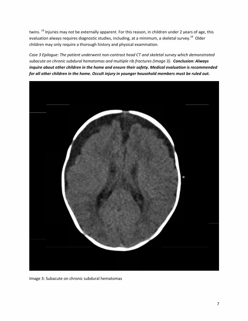
Case 3 Ep
subacute
inquire ab
for all oth
Image 3: S
njuries may n
n always requ
may only requ
ilogue: The p
on chronic su
bout other ch
her children in
Subacute on c
not be externa
uires diagnost
uire a thoroug
atient underw
ubdural hema
hildren in the
n the home. O
chronic subd
ally apparent
tic studies, in
gh history and
went non‐con
atomas and m
home and en
Occult injury
ural hematom
t. For this reas
ncluding, at a
d physical exa
ntrast head CT
multiple rib fra
nsure their sa
in younger h
mas
son, in childr
minimum, a s
amination.
T and skeleta
actures (Imag
afety. Medica
household me
en under 2 ye
skeletal surve
al survey whic
ge 3). Conclus
al evaluation
embers must
ears of age, t
ey.19 Older
ch demonstra
sion: Always
n is recommen
be ruled out.
7
his
ted
nded
.

8
Abusive Head Trauma
EPs must frequently weigh the risks and benefits of radiation exposure. This is especially true in children.
Faced with a well appearing infant, the EP is often reticent to order CT imaging. However, in the child
less than a year of age, with evaluation concerning for abuse, non‐contrast Head CT imaging is
imperative. Abusive head trauma (AHT) is the leading cause of death from inflicted injury in children.
Though incidence is difficult to determine20, it has been estimated to occur in approximately 1/3300
children under 12 months of age. 21 Unfortunately many children have no external evidence of trauma.
Furthermore, infants with significant intracranial injury are often asymptomatic or may present with
non‐specific complaints such as vomiting or increased fussiness. In a study looking at 173 cases of
children with AHT, the diagnosis was initially missed for 31%. Mean time to correct diagnosis in these
children was 7 days, but approximately 27% were reinjured between the initial evaluation and the
correct diagnosis, and 40% had medical complications related to the diagnostic delay. Initial
misdiagnosis included the following: acute gastroenteritis, rule‐out sepsis, increasing head size, apnea,
and bruising of unknown origin.3 Another study investigated the occurrence of occult head injury in
children with other injuries suspicious for inflicted trauma. 22 These were neurologically asymptomatic
children without external signs of head injury who underwent non‐contrast head CT as part of their
evaluation for other injuries concerning for abuse. 29% of these children had evidence of abusive head
trauma on CT. For these reasons ‐ the dire consequences of missed head injury and the high incidence of
occult injury in abused children ‐ aggressive evaluation is warranted when physical abuse is strongly
suspected. Evaluation should include non‐contrast head CT in children less than 12 months of age with
suspicious injuries or injury patterns. 23 In children 12 months and older, non‐contrast head CT should be
considered if there is any external evidence of injury about the head or neck or if there are clinical
symptoms such as change in mental status, that are suggestive of intracranial injury.23
Abusive Abdominal Trauma
In contrast to AHT, abusive abdominal trauma (AAT) occurs far less frequently. But like AHT, abusive
abdominal trauma often presents with occult or non‐specific complaints. Abusive abdominal trauma is
the second leading cause of traumatic death from abusive injuries.24 Its actual prevalence has been is
difficult to determine, however, estimates based on several studies put prevalence at 1‐4%. 25, 24 Coupled
with this low frequency however, is a high mortality rate – as high as 50% in some studies.24
Unfortunately, like AHT, children with AAT often have occult injuries or present with non‐specific
symptoms, such as vomiting or abdominal pain. Bruising, for example, may be absent in as many as 80%
of cases.26 In fact, in one study, over 40% of children with lethal injuries had no external abdominal wall
contusions. Routine screening Abdominal CT is not recommended because AAT is, overall, a low
frequency event. However, it is a high stakes injury. The current recommendations for evaluation for
occult AAT in children presenting with other findings or complaints concerning for abuse is to obtain
screening labs, inclusive of liver function tests. 27,28 Many experts recommend that this screening should
take place for all young children with findings that are concerning for abusive injury.23,25,27,28 Contrast
Abdominal CT imaging is indicated for AST or ALT greater than 80 IU. In one study, this cut‐off yielded a
sensitivity of 77% and a specificity of 82%. 28 Abdominal CT imaging is also indicated if the clinical exam

9
is suspicious for intra‐abdominal injury, such as bruising, regardless of the lab values. Screening with
ultrasound is not recommended due to poor sensitivity overall. 25,26
Part 3: The Medicolegal Issues
All EPs strive to provide optimal care to their patients, and to identify and respond to potentially life
threatening illnesses. This includes child abuse. Unfortunately the medicolegal risks associated with
missing these diagnoses are real. Practitioners have been sued for missed diagnoses of child abuse. EPs
must be equipped with the best information to care for evaluate these children.
Part 4: The Standard of Care When You Suspect Abuse
Just as aspirin is considered standard of care treatment for acute myocardial infarction and a full septic
evaluation is considered standard of care for the febrile neonate, there IS a standard of care for the
evaluation of the child with suspected abusive injuries. Consider the following case:
Case 4: A 4‐month old infant presents to the emergency department with complaint of cough and fever.
On exam, she is alert and interactive, sitting on mom’s lap and drinking from a bottle. She has a fever to
101.7° F. She is mildly tachypneic and mildly tachycardic. HEENT exam is notable for mild rhinorrhea.
Right sided crackles and tacyhpnea are noted on auscultation. Her respiratory exam is otherwise normal
without use of accessory muscles, nasal flaring or grunting respirations. Her abdomen is soft, non‐tender
and non‐distended. She is well hydrated with brisk capillary refill. Skin exam is notable for a 5mm bruise
to the left lateral chest wall. The family has no explanation for the bruise.
What is the appropriate evaluation for this child?
The standards that have been developed for the evaluation of suspected non‐accidental trauma exist
because of the high incidence of significant occult injuries in generally well appearing children. In
addition, even clinically insignificant injuries are forensically critical to ensuring the safety of children.
Identifying such injuries may be the difference between sending a child back to an unsafe home
environment and the introduction of family services and removal to safety. As with many pediatric
illnesses and injuries, standard evaluation varies by patient age. Here is how it works:
Skeletal Surveys: Any child under 2 years of age undergoing evaluation for suspected abuse
requires a skeletal survey. 17, 4 Skeletal surveys are rarely indicated or helpful in children
older than 24 months of age. It is important to realize that a skeletal survey is a well‐defined
20 view radiograph series. 19 It is NOT a babygram. Anything less than this series is
insufficient to detect the sometimes subtle, but always forensically important fracture.29
Non‐Contrast Head CT: This imaging is strongly encouraged for children under 12 months of
age when there is suspicion for abuse. Recall that many children in this age range may have
significant intracranial injuries in the absence of any focal neurologic findings or external
evidence of head or neck trauma. Children 13 months of age and older should have brain
imaging as clinically indicated. 22 23

10
Retinal Exam: A formal exam by ophthalmology is required in any infant or young child with
identified intracranial injury.4 Note that some child abuse specialists may also request this
exam in the setting of an infant with highly suspicious injuries regardless of findings on Head
CT.
IV Contrast Abdominal CT: An abdominal CT with IV contrast is indicated in children with
external signs or clinical symptoms or exam suspicious for intra‐abdominal injury.4 CT is also
indicated for children undergoing laboratory screening evaluation for occult abdominal
trauma if either the serum AST or the ALT are greater than 80 IU.25
Screening Liver Function Tests: This testing is recommended by many experts in the
evaluation of children with suspected inflicted injuries.25 Inclusion of other trauma labs,
such as lipase, CBC and basic metabolic panel in this evaluation has been suggested,
however, data is limited to support sensitivity and specificity of these studies.25 23
Evaluation for suspected abuse can be time consuming and, in some instances, technically beyond the
scope and resources of an institution. In these cases, it is quite reasonable for the EP to transfer the
child to a tertiary center for further evaluation. In addition, in many states, consultation with a child
abuse specialist may be available through the regional Children’s Hospital. This resource can be
instrumental to providing comprehensive care for at risk children.
Case 4 epilogue: The unusual bruising patterns in a non‐ambulatory infant prompted further evaluation.
Skeletal survey revealed multiple fractures including multiple bilateral rib fractures, skull fracture, right
humeral shaft fracture, left both‐bone forearm fracture, left tibial shaft fracture and bilateral tibial
corner fractures. Chest x‐ray revealed the rib fractures as noted and right basilar airspace disease. Non‐
contrast Head CT demonstrated the skull fracture but no intracranial injuries. Complete blood count and
comprehensive metabolic panel demonstrated anemia with a hemoglobin of 9 g/dL and elevated
transaminases with AST of 844 IU and ALT of 1177 IU. An abdominal CT with IV contrast demonstrated a
grade III liver laceration. It further defined the right basilar airspace disease as a hemothorax.
Conclusion
Unfortunately, child abuse is common and it is commonly missed. Children will present to Emergency
Departments with routine pediatric complaints, but may, on exam, have evidence of bruising or injury
that should raise the EPs suspicion for inflicted injury. On other occasions children may present with
traumatic injury that should incite concern for a non‐accidental event. The EP must be as vigilant about
identifying child maltreatment as he or she is in identifying ACS. The EP must consider these diagnoses
and must be fluent in the appropriate evaluation and management. A child’s life may depend on it.

11
1. Pope JH, Aufderheide TP, Ruthazer R, et al. Missed diagnoses of acute cardiac ischemia in the emergency department. The New England journal of medicine. Apr 20 2000;342(16):1163‐1170.
2. Schull MJ, Vermeulen MJ, Stukel TA. The risk of missed diagnosis of acute myocardial infarction associated with emergency department volume. Annals of emergency medicine. Dec 2006;48(6):647‐655.
3. Jenny C, Hymel KP, Ritzen A, Reinert SE, Hay TC. Analysis of missed cases of abusive head trauma. JAMA : the journal of the American Medical Association. 1999;281(7):621‐626.
4. Kellogg ND, American Academy of Pediatrics Committee on Child A, Neglect. Evaluation of suspected child physical abuse. Pediatrics. 2007;119(6):1232‐1241.
5. Gateway CWI. Child Maltreatment 2010: Summary of key findings. In: Health and Human Services CsB, ed. Washington, D.C.2012.
6. King WK, Kiesel EL, Simon HK. Child abuse fatalities: are we missing opportunities for intervention? Pediatric emergency care. 2006;22(4):211‐214.
7. Pierce MC, Smith S, Kaczor K. Bruising in infants: those with a bruise may be abused. Pediatric emergency care. 2009;25(12):845‐847.
8. Pierce MC, Kaczor K, Aldridge S, O'Flynn J, Lorenz DJ. Bruising characteristics discriminating physical child abuse from accidental trauma. Pediatrics. 2010;125(1):67‐74.
9. Sugar NF, Taylor JA, Feldman KW. Bruises in infants and toddlers: those who don't cruise rarely bruise. Puget Sound Pediatric Research Network. Archives of pediatrics & adolescent medicine. 1999;153(4):399‐403.
10. Maguire S, Mann MK, Sibert J, Kemp A. Are there patterns of bruising in childhood which are diagnostic or suggestive of abuse? A systematic review. Archives of disease in childhood. 2005;90(2):182‐186.
11. Bariciak ED, Plint AC, Gaboury I, Bennett S. Dating of bruises in children: an assessment of physician accuracy. Pediatrics. 2003;112(4):804‐807.
12. Pierce MCKKL, D.; Richter, K.; Starling, S.;. A Practical Guide to Differentiating Abusive From Accidental Fractures: An Injury Plausibility Approach. Clinical Pediatric Emergency Medicine. 2012;13(3):166‐177.
13. Baldwin K, Pandya NK, Wolfgruber H, Drummond DS, Hosalkar HS. Femur fractures in the pediatric population: abuse or accidental trauma? Clinical orthopaedics and related research. 2011;469(3):798‐804.
14. Kemp AM, Dunstan F, Harrison S, et al. Patterns of skeletal fractures in child abuse: systematic review. BMJ (Clinical research ed ). 2008;337:a1518.
15. Pierce MC, Bertocci GE, Vogeley E, Moreland MS. Evaluating long bone fractures in children: a biomechanical approach with illustrative cases. Child abuse & neglect. 2004;28(5):505‐524.
16. Bulloch B, Schubert CJ, Brophy PD, Johnson N, Reed MH, Shapiro RA. Cause and clinical characteristics of rib fractures in infants. Pediatrics. 2000;105(4):E48.
17. Pierce MCB, Gina. Fractures Resulting From Inflicted Trauma: Assessing Injury and History Compatibility. Clinical Pediatric Emergency Medicine. 2006;7(3):6.
18. Pierce MC, Bertocci GE, Janosky JE, et al. Femur fractures resulting from stair falls among children: an injury plausibility model. Pediatrics. 2005;115(6):1712‐1722.
19. Section on R, American Academy of P, Di Pietro MA, et al. Diagnostic imaging of child abuse. Pediatrics. 2009;123(5):1430‐1435.
20. Ford CRC, C; Sirotnak, a. . Pearls and Pitfalls for the Pediatric Emergency Medical Provider in the Evaluation of Abusive Head Trauma. Clinical Pediatric Emergency Medicine. 2012;13(3):178‐187.
21. Bechtel K. Inflicted Traumatic Brain Injury: Making the Diagnosis in the Emergency Department. Clinical Pediatric Emergency Medicine. 2006;7(3):5.

12
22. Laskey AL, Holsti M, Runyan DK, Socolar RRS. Occult head trauma in young suspected victims of physical abuse. The Journal of pediatrics. 2004;144(6):719‐722.
23. Pierce MC. Appendix 2: Physical Child Abuse Workup for Children 4 Years of Age and Younger in the Emergency Department Setting. Clinical Pediatric Emergency Medicine. 2006;7(3):1.
24. Wood J, Rubin DM, Nance ML, Christian CW. Distinguishing inflicted versus accidental abdominal injuries in young children. The Journal of trauma. 2005;59(5):1203‐1208.
25. Lindberg DM. Abusive Abdominal Trauma ‐ An Update for the Pediatric Emergency Medicine Physician. Clinical Pediatric Emergency Medicine. 2012 13(3):187‐193.
26. Herr S, M Fallat. Abusive Abdominal and Thoracic Trauma. Clinical Pediatric Emergency Medicine. 2006;7(3):4.
27. Lane WG, Dubowitz H, Langenberg P. Screening for occult abdominal trauma in children with suspected physical abuse. Pediatrics. 2009;124(6):1595‐1602.
28. Lindberg D, Makoroff K, Harper N, et al. Utility of hepatic transaminases to recognize abuse in children. Pediatrics. 2009;124(2):509‐516.
29. Pierce MC. Appendix 3: The Ten Deadly Sins (or Misconceptions) of an Emergency Department Physician. Clinical Pediatric Emergency Medicine. 2006;7(3):2.


![UvA-DARE (Digital Academic Repository) Forensic pediatric ... · SG, van Rijn RR [2009] Forensic aspects of pediatric fractures: differentiating accidental trauma from child Figure](https://img.pdfslide.us/doc/110x75/5f64707ea63aae7dc739600d/uva-dare-digital-academic-repository-forensic-pediatric-sg-van-rijn-rr-2009.jpg)