Embed Size (px)
Citation preview

1
Chapter 42
Osteogenesis Imperfecta andNon-Accidental Trauma
Copyright © 2014 Elsevier Inc. All rights reserved.

2Copyright © 2014 Elsevier Inc. All rights reserved.
FIGURE 42.1 (a) Healing bilateral posterior rib fractures in a 9-month-old infant. (b) Healing LT lateral rib fractures found in a 44-day-old infant with severe intracranial injury (see Figure 42.5 ).

3Copyright © 2014 Elsevier Inc. All rights reserved.
FIGURE 42.2 (a) “Bucket-handle” appearance of CML of the RT proximal tibia in a 3-month-old infant with intracranial hemorrhage. (b) “Corner appearance” of CML LT distal femur and proximal tibia in a 56-day-old with “leg injury.”
4Copyright © 2014 Elsevier Inc. All rights reserved.
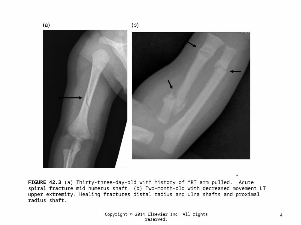
FIGURE 42.3 (a) Thirty-three-day-old with history of “RT arm pulled.” Acute spiral fracture mid humerus shaft. (b) Two-month-old with decreased movement LT upper extremity. Healing fractures distal radius and ulna shafts and proximal radius shaft.

5Copyright © 2014 Elsevier Inc. All rights reserved.
FIGURE 42.4 Four-month-old with acute LT acromion fracture found on skeletal survey done because of RT leg swelling.

6Copyright © 2014 Elsevier Inc. All rights reserved.
FIGURE 42.5 Forty-four-day-old S/P fall from couch. (a) RT subdural hemorrhage with mass effect causing RT to LT shift and subfalcean herniation. Diffuse bilateral cerebral edema. (b), (c) Diastatic LT parietal fracture with herniation of brain through the defect. (d) Same patient as in (a–c). Abdomen could not be assessed due to neurological status. Contrast-enhanced CT abdomen demonstrates grade 4 liver laceration. This figure is reproduced in color in the color plate section.