Embed Size (px)
Citation preview

Multiple Identities of V. parahaemolyticus 51
The Multiple Identities of Vibrio parahaemolyticus
*For correspondence. Email [email protected];Tel. (319) 335-9721; Fax. (319) 335-7679.
J. Molec. Microbiol. Biotechnol. (1999) 1(1): 51-57.
© 1999 Horizon Scientific Press
Linda McCarter*
Microbiology Department, University of Iowa,Iowa City, Iowa 52242, USA
Abstract
Vibrio parahaemolyticus is a ubiquitous marinebacterium and human pathogen. The organismpossesses multiple cell types appropriate for life underdifferent circumstances. The swimmer cell, with asingle polar flagellum, is adapted to life in liquidenvironments. The polar flagellum is powered by thesodium motive force and can propel the bacterium atfast speeds. The swarmer cell, propelled by manyproton-powered lateral flagella, can move throughhighly viscous environments, colonize surfaces, andform multicellular communities which sometimesdisplay highly periodic architecture. Signals thatinduce differentiation to the surface-adapted cell typeare both physical and chemical in nature. Surface-induced gene expression may aid survival, whetherattached to inanimate surfaces or in a host organism.Genetic rearrangements create additional phenotypicversatility, which is manifested as variable opaque andtranslucent colony morphotypes. Discovery that aLuxR homolog controls the opaque cell type implicatesintercellular signaling as an additional survivalstrategy. The alternating identities of V.parahaemolyticus may play important roles inattachment and detachment, how bacterial populationsadapt to growth on surfaces, form structuredcommunities, and develop biofilms.
Introduction
Occupying a variety of niches, Vibrio parahaemolyticus isa common bacterium in marine and estuarineenvironments. It can exist planktonically or attached tosubmerged, inert and animate surfaces, includingsuspended particulate matter, zooplankton, fish andshellfish (Kaneko and Colwell, 1973, 1975). This organismis recognized as a major, worldwide cause ofgastroenteritis, particularly in areas of the world whereseafood consumption is high such as Southeast Asia(Joseph et al., 1982). It is an emerging pathogen in NorthAmerica. In 1997, a large outbreak of V. parahaemolyticusfood poisoning, attributed to raw oyster consumption,occurred along the Pacific coast (CDC, 1998). Thus, V.parahaemolyticus seems suited to multiple lifestyles: aplanktonic, free-swimming state and a sessile existencewithin a microbial community attached, for example, toshellfish in a commensal relationship, to the bottoms ofboats or other surfaces in the ocean (biofouling), or in ahost organism (pathogenesis). What are the survival
strategies that allow this bacterium to adapt to life in diluteliquid environments and to life on surfaces or in biofilms?
The Swimmer Cell
The free-living form of V. parahaemolyticus, the swimmercell (Figure 1), is well-suited for locomotion in liquidenvironments. The rod-shaped bacterium is efficientlypropelled by a single polar flagellum. Energy to power theflagellum is derived from the sodium motive force (Atsumiet al., 1992). Possession of sodium energetics has anadvantage in the marine environment because the pH ofseawater is approximately 8.0 (Kogure, 1998). Sodium-driven flagellar motors are remarkably fast. In liquid mediumwith 300mM NaCl, the swimming speed of the bacteriumis approximately 60µm per sec. Sodium-powered rotationof the polar flagellum of the closely related bacterium Vibrioalginolyticus, which swims at an equivalent speed in liquid,has been clocked using laser-darkfield microscopy at ratesas fast as 1,700 r.p.s. (Magariyama et al., 1994).
The polar organelle is a complex flagellum. There aresix polar flagellin genes, organized in two loci (McCarter,1995; GenBank Accession U12816 and U12817).Moreover, the polar flagellum is sheathed by what appearsto be an extension of the cell outer membrane (Allen andBaumann, 1971). The mechanism of how a sheathedflagellum rotates has not been elucidated. Potentially, theflagellar filament could rotate within the sheath, or thesheath and filament could rotate as a unit (Fuerst, 1980).The flagellum plays a key role in initial adsorption of bacteriato surfaces. A plethora of studies with a variety of bacteriahave shown that motility is important for adhesion as wellas pathogenesis (O’Toole and Kolter, 1998; Otteman andMiller, 1997; Pratt and Kolter, 1998). As a propulsiveorganelle, it brings the bacteria into close proximity withsurfaces and perhaps aids in overcoming negativeelectrostatic interactions. In fact, studies have shown thatthe faster the swimming, the greater the adhesion to glass(Kogure et al., 1998). Flagellar sheaths allow specificinteraction between a bacterium and a surface (Sjobladand Doetsch, 1982; Sjoblad et al., 1983). Certainly, it seemsclear the sheath extends the surface area of the bacterium.Cell-surface components, including potential adhesins, maybe differentially distributed or available on the flagellumand cell body. The flagellum in Figure 1 is studded withcolloidal gold particles, after immunogold-labeling withrabbit antiserum prepared against whole cells. In contrast,there is little gold-labeling of the cell body, suggesting thatantigens are presented differently by the cell body and thesheathed flagellum.
The Swarmer Cell
Growth on surfaces or in viscous environments inducesdifferentiation to the swarmer cell. Septation ceases andthe cell elongates, usually up to 30 µm in length. Inductionof a second motility system, the lateral system, leads toelaboration of numerous peritrichously arranged flagella.The swarmer cell is adapted for movement on surfaces or
JMMB Symposium

• MALDI-TOF Mass Spectrometry in Microbiology
Edited by: M Kostrzewa, S Schubert (2016) www.caister.com/malditof
• Aspergillus and Penicillium in the Post-genomic Era
Edited by: RP Vries, IB Gelber, MR Andersen (2016) www.caister.com/aspergillus2
• The Bacteriocins: Current Knowledge and Future Prospects
Edited by: RL Dorit, SM Roy, MA Riley (2016) www.caister.com/bacteriocins
• Omics in Plant Disease Resistance
Edited by: V Bhadauria (2016) www.caister.com/opdr
• Acidophiles: Life in Extremely Acidic Environments
Edited by: R Quatrini, DB Johnson (2016) www.caister.com/acidophiles
• Climate Change and Microbial Ecology: Current Research and Future Trends
Edited by: J Marxsen (2016) www.caister.com/climate
• Biofilms in Bioremediation: Current Research and Emerging Technologies
Edited by: G Lear (2016) www.caister.com/biorem
• Microalgae: Current Research and Applications
Edited by: MN Tsaloglou (2016) www.caister.com/microalgae
• Gas Plasma Sterilization in Microbiology: Theory, Applications, Pitfalls and New Perspectives
Edited by: H Shintani, A Sakudo (2016) www.caister.com/gasplasma
• Virus Evolution: Current Research and Future Directions
Edited by: SC Weaver, M Denison, M Roossinck, et al. (2016) www.caister.com/virusevol
• Arboviruses: Molecular Biology, Evolution and Control
Edited by: N Vasilakis, DJ Gubler (2016) www.caister.com/arbo
• Shigella: Molecular and Cellular Biology
Edited by: WD Picking, WL Picking (2016) www.caister.com/shigella
• Aquatic Biofilms: Ecology, Water Quality and Wastewater Treatment
Edited by: AM Romaní, H Guasch, MD Balaguer (2016) www.caister.com/aquaticbiofilms
• Alphaviruses: Current Biology
Edited by: S Mahalingam, L Herrero, B Herring (2016) www.caister.com/alpha
• Thermophilic Microorganisms
Edited by: F Li (2015) www.caister.com/thermophile
• Flow Cytometry in Microbiology: Technology and Applications
Edited by: MG Wilkinson (2015) www.caister.com/flow
• Probiotics and Prebiotics: Current Research and Future Trends
Edited by: K Venema, AP Carmo (2015) www.caister.com/probiotics
• Epigenetics: Current Research and Emerging Trends
Edited by: BP Chadwick (2015) www.caister.com/epigenetics2015
• Corynebacterium glutamicum: From Systems Biology to Biotechnological Applications
Edited by: A Burkovski (2015) www.caister.com/cory2
• Advanced Vaccine Research Methods for the Decade of Vaccines
Edited by: F Bagnoli, R Rappuoli (2015) www.caister.com/vaccines
• Antifungals: From Genomics to Resistance and the Development of Novel Agents
Edited by: AT Coste, P Vandeputte (2015) www.caister.com/antifungals
• Bacteria-Plant Interactions: Advanced Research and Future Trends
Edited by: J Murillo, BA Vinatzer, RW Jackson, et al. (2015) www.caister.com/bacteria-plant
• Aeromonas
Edited by: J Graf (2015) www.caister.com/aeromonas
• Antibiotics: Current Innovations and Future Trends
Edited by: S Sánchez, AL Demain (2015) www.caister.com/antibiotics
• Leishmania: Current Biology and Control
Edited by: S Adak, R Datta (2015) www.caister.com/leish2
• Acanthamoeba: Biology and Pathogenesis (2nd edition)
Author: NA Khan (2015) www.caister.com/acanthamoeba2
• Microarrays: Current Technology, Innovations and Applications
Edited by: Z He (2014) www.caister.com/microarrays2
• Metagenomics of the Microbial Nitrogen Cycle: Theory, Methods and Applications
Edited by: D Marco (2014) www.caister.com/n2
Caister Academic Press is a leading academic publisher of advanced texts in microbiology, molecular biology and medical research. Full details of all our publications at caister.com
Further Reading
Order from caister.com/order

52 McCarter
through highly viscous environments. The polar flagellumperforms poorly in medium of high viscosity while lateralflagella efficiently propel the bacterium in highly viscousenvironments. The speed of laterally flagellated cells isunaffected, remaining constant at about 25µm per second,on addition of the long branched-chain polymerpolyvinylpyrrolidone (10% PVP-360) which increasesviscosity to 10 centipoise (cP). In comparison, polarlypropelled swimming motility decreases from 60 to less than15 µm per second under this condition (Atsumi et al., 1992).It has been shown for V. alginolyticus that lateral flagellacan work at viscosities as high as 100 cP (Atsumi et al.,1996).
Lateral flagella are not sheathed, and the filament ispolymerized from a single flagellin subunit (McCarter andWright, 1993). The flagella are very sensitive to mechanical
shearing (Allen and Baumann, 1971). So many flagellaare produced per cell and sloughed, or broken off, thatdetached flagellar aggregates are readily visualized in thelight microscope as giant, coiled bundles (Belas et al., 1986;Ulitzur, 1974). These flagella are entirely distinct from thepolar flagellum. Genetic experiments suggest that thereare no shared structural components among the two motilitysystems. Mutants unable to swarm retain swimming motilityand vice versa (McCarter et al., 1988). Flagellar motilitysystems are typically encoded by at least 40 genes;therefore, two very large, distinct gene sets encoding themotility systems exist. When grown on a surface, theorganism assembles two distinct types of flagella. Whatthis means in terms of signals for flagellar export andassembly will be interesting to elucidate. Not only do thelateral and polar flagella differ at the structural level, butalso are powered by different energy sources. Although ineach case, energy is derived from the electrochemicaltransmembrane potential, the coupling ions differ. Lateralflagella are powered by the flow of protons through themotor in contrast to the sodium-driven polar motors (Atsumiet al., 1992).
When inoculated on solidified medium, differentiationto the swarmer cell allows the bacterium to swarm, or move,over surfaces or through viscous environments, leading tocolonization of surfaces (Figure 2). The movement is veryvigorous. The percent agar over which swarming will occuris high (up to 2.0%), and the rate of radial expansion israpid, for example, on Heart Infusion agar at 30oC swarmingwill progress at a rate of 5mm/hour. A number of bacteriaare known to swarm. Although some show mixedflagellation like V. parahaemolyticus with distinct polar andperitrichous organelles (e.g., V. alginolyticus,Rhodospirillum centenum, and Azospirillum brasilense;Allen and Baumann, 1971; Jiang et al., 1998; Moens etal., 1996), others possess single, peritrichous flagellarsystems. In general the extent of hyperflagellation of theswarm cell correlates with successfulness of swarming. V.parahaemolyticus and Proteus mirabilis are superiorswarmers and can have hundreds of flagella per cell. Incomparison, Serratia species, Escherichia coli, andSalmonella typhimurium swarm under a more restrictedset of conditions with agar concentrations at 0.5 – 0.8 %(Harshey, 1994; Harshey and Matsuyama, 1994).Movement for some of these swarming organisms hasbeen shown to also require production of extracellularmolecules capable of altering surface tension. Surfactant-like lipopeptides are produced by S. marcesans and S.liquefaciens (Matsuyama et al., 1992; Lindum et al., 1998).Rapid spreading of P. mirabilis requires production ofextracellular capsular polysaccharide (Gygi et al., 1995).Extracellular agents acting as swarming facilitators havenot been demonstrated for V. parahaemolyticus. It may bethat the copious production of fragile flagella substitutes toreduce frictional drag.
The concentric ring patterns or terraces, which arecharacteristic of the periodic swarming of Proteus species(Rauprich et al., 1996), develop as a result of alternatingcycles of active swarming and consolidation. During theconsolidation period, swarmer cells divide to produce shortcells, which eventually differentiate to the swarmer cellsthat initiate a new wave of swarming. Although thephenomenon has been studied extensively in P. mirabilis,this type of behavior is not restricted to Proteus species.
Figure 1. Electron Micrograph of a Swimmer CellElectron micrograph of a swimmer cell reacted with rabbit antiserumprepared against whole cells and 15nm protein A-bound colloidal goldparticles. Bar indicates approximately 1 µm.
Figure 2. Swarming Colony on Rich Medium after Overnight Incubationat 30 °C.

Multiple Identities of V. parahaemolyticus 53
Under certain conditions (specifically, swarm plates non-optimally conducive to swarming such as those made withLuria broth rather than Heart Infusion broth), certain V.parahaemolyticus strains produce the highly developed,periodic architecture shown in Figure 2. Detection of patternformation may require a special balance between the ratesof reproduction and movement of the cells as well as theparticular surface properties of the substratum (i.e., surfacetension).
Physical Signaling: The Polar Flagellum as TactileSensor
In addition to its role as a propulsive organelle and itspotential for aiding attachment, the polar flagellum acts asa sensor. Polar flagellar function is coupled to expressionof the swarmer cell gene system. The polar flagellum isproduced constitutively, irrespective of liquid or surface-associated growth. All conditions that slow down polarflagellar rotation, lead to swarmer cell induction. Suchconditions include increasing viscosity or using antibodiesto inhibit flagellar function (Belas et al., 1986, McCarter etal., 1988). Physical conditions that impede flagellar rotationseem to act as a signal. Polar flagellar function can beperturbed in other ways. Genetic interference with polarfunction affects swarmer cell gene expression (McCarteret al., 1988). All swimming-defective, transposon-generatedmutants constitutively express swarmer cell genes whengrown in liquid (Figure 3). The motor itself can be sloweddown by using the sodium-channel-blocking drug phenamil(Kawagishi et al., 1996). Just as increasing viscosity leadsto swarmer cell differentiation, increasing concentrationsof phenamil lead to decreasing swimming speed andconcomitant induction of swarmer cell gene expression.Thus, the polar flagellum seems to act as amechanosensor: interference with flagellar rotation signalsswarmer cell differentiation. How signaling is transducedto program gene expression is not known. Current workinvolves dissecting the architecture of the sodium-drivenmotor of V. parahaemolyticus (McCarter, 1994a, 1994b;Jaques et al., 1999).
Cell Division and the Long Cell Phenotype
One of the initial events after transfer to a surface isinhibition of cell division, and as a consequence, swarmercells are characteristically very long. Differentiation istransient. Since, swarmer cells must grow, i.e., divide, forthe swarm colony to expand, the cell must have amechanism to escape from the block to cell division. Thus,the inhibition of cell division during the swarm cell cyclemust be carefully regulated for prolonged repression ofcell division would be a terminal event.
Strains in one class of swarming constitutive mutantspossess defects in lonS, a gene that codes for a homologof the E. coli Lon protease (Stewart et al., 1997). E. colilon mutants were originally isolated as a class of UV-sensitive mutants (Gottesman, 1996). The role of Lon inE. coli is multifunctional, e.g., it targets the degradation ofa transcriptional regulatory protein controlling productionof extracellular polysaccharide (cps) as well as the celldivision inhibitor SulA. SulA is induced by UV exposure aspart of the SOS DNA repair response. Wild-type cells formlong filaments after UV exposure, but with time SulA is
degraded by Lon and the filaments resolve. Mutants withdefects in lon cannot recover from elongation.
The V. parahaemolyticus counterpart closelyresembles E. coli lon and substitutes functionally, bycomplementing E. coli mutants to restore UV resistanceand cps regulation. In addition, lonS mutants in V.parahaemolyticus are more UV sensitive than the wild-typestrain. It is attractive to hypothesize the existence ofswarmer-cell specific cell division inhibitor. LonS could actas a policeman to keep in check the important cell divisionand regulatory proteins that mediate surface sensing.Potential regulatory targets for LonS could includetranscriptional activators controlling the lateral flagellargene system or extracellular polysaccharide.
Chemical Signaling
Differentiation to the swarmer cell requires considerablecommitment in terms of cellular economy, including energyexpenditure and gene expression. As a result, thedevelopmental switch seems tightly controlled and multipleenvironmental stimuli are essential for cueing development.One cue that has been determined is iron starvation.Starvation signals seem an appropriate cue because theavailability of nutrients or the diffusion of nutrients may belimiting for cells in a community attached to a substratum.Development to the swarmer requires iron limitation andperturbation of flagellar function (McCarter and Silverman,1989). Nutrient deprivation and surface signaling mayensure detection of the specific conditions for whichswarming is appropriate, e.g., a dense community of sessilecells.
Although others kinds of signals may also be important,V. parahaemolyticus can be induced to swarm on minimalmedium (Figure 4; McCarter, 1998). This has not beenshown to be the case for other bacteria where amino acidsare implicated as swarming signals. Glutamine has beenreported as essential to induce swarming of P. mirabilis onminimal medium (Allison et al., 1993). Casamino acidsupplementation is necessary for S. liquefaciens (Eberl etal., 1996b); however, failure to swarm on minimal mediumcould also represent a growth rate barrier and not the lack
Figure 3. Electron Micrograph of a Constitutive Swarmer CellGrown in liquid and negatively-stained with 0.5 % phosphotungstic acid.Bar indicates approximately 3 µm.
54 McCarter
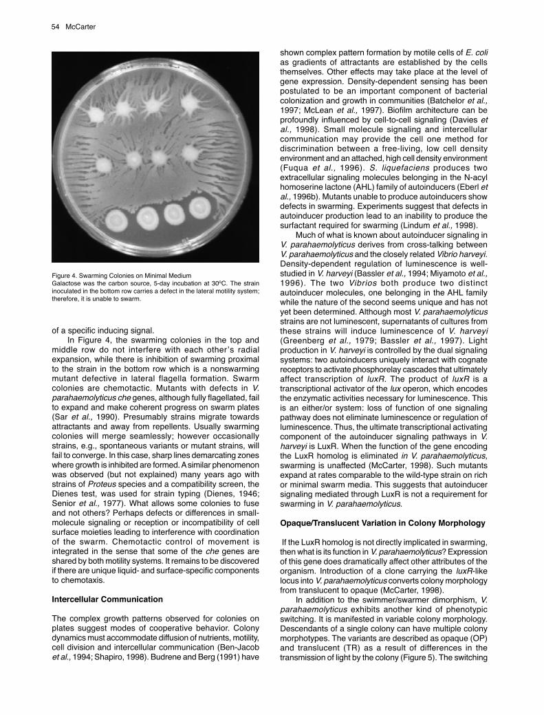
of a specific inducing signal.In Figure 4, the swarming colonies in the top and
middle row do not interfere with each other’s radialexpansion, while there is inhibition of swarming proximalto the strain in the bottom row which is a nonswarmingmutant defective in lateral flagella formation. Swarmcolonies are chemotactic. Mutants with defects in V.parahaemolyticus che genes, although fully flagellated, failto expand and make coherent progress on swarm plates(Sar et al., 1990). Presumably strains migrate towardsattractants and away from repellents. Usually swarmingcolonies will merge seamlessly; however occasionallystrains, e.g., spontaneous variants or mutant strains, willfail to converge. In this case, sharp lines demarcating zoneswhere growth is inhibited are formed. A similar phenomenonwas observed (but not explained) many years ago withstrains of Proteus species and a compatibility screen, theDienes test, was used for strain typing (Dienes, 1946;Senior et al., 1977). What allows some colonies to fuseand not others? Perhaps defects or differences in small-molecule signaling or reception or incompatibility of cellsurface moieties leading to interference with coordinationof the swarm. Chemotactic control of movement isintegrated in the sense that some of the che genes areshared by both motility systems. It remains to be discoveredif there are unique liquid- and surface-specific componentsto chemotaxis.
Intercellular Communication
The complex growth patterns observed for colonies onplates suggest modes of cooperative behavior. Colonydynamics must accommodate diffusion of nutrients, motility,cell division and intercellular communication (Ben-Jacobet al., 1994; Shapiro, 1998). Budrene and Berg (1991) have
shown complex pattern formation by motile cells of E. colias gradients of attractants are established by the cellsthemselves. Other effects may take place at the level ofgene expression. Density-dependent sensing has beenpostulated to be an important component of bacterialcolonization and growth in communities (Batchelor et al.,1997; McLean et al., 1997). Biofilm architecture can beprofoundly influenced by cell-to-cell signaling (Davies etal., 1998). Small molecule signaling and intercellularcommunication may provide the cell one method fordiscrimination between a free-living, low cell densityenvironment and an attached, high cell density environment(Fuqua et al., 1996). S. liquefaciens produces twoextracellular signaling molecules belonging in the N-acylhomoserine lactone (AHL) family of autoinducers (Eberl etal., 1996b). Mutants unable to produce autoinducers showdefects in swarming. Experiments suggest that defects inautoinducer production lead to an inability to produce thesurfactant required for swarming (Lindum et al., 1998).
Much of what is known about autoinducer signaling inV. parahaemolyticus derives from cross-talking betweenV. parahaemolyticus and the closely related Vibrio harveyi.Density-dependent regulation of luminescence is well-studied in V. harveyi (Bassler et al., 1994; Miyamoto et al.,1996). The two Vibrios both produce two distinctautoinducer molecules, one belonging in the AHL familywhile the nature of the second seems unique and has notyet been determined. Although most V. parahaemolyticusstrains are not luminescent, supernatants of cultures fromthese strains will induce luminescence of V. harveyi(Greenberg et al., 1979; Bassler et al., 1997). Lightproduction in V. harveyi is controlled by the dual signalingsystems: two autoinducers uniquely interact with cognatereceptors to activate phosphorelay cascades that ultimatelyaffect transcription of luxR. The product of luxR is atranscriptional activator of the lux operon, which encodesthe enzymatic activities necessary for luminescence. Thisis an either/or system: loss of function of one signalingpathway does not eliminate luminescence or regulation ofluminescence. Thus, the ultimate transcriptional activatingcomponent of the autoinducer signaling pathways in V.harveyi is LuxR. When the function of the gene encodingthe LuxR homolog is eliminated in V. parahaemolyticus,swarming is unaffected (McCarter, 1998). Such mutantsexpand at rates comparable to the wild-type strain on richor minimal swarm media. This suggests that autoinducersignaling mediated through LuxR is not a requirement forswarming in V. parahaemolyticus.
Opaque/Translucent Variation in Colony Morphology
If the LuxR homolog is not directly implicated in swarming,then what is its function in V. parahaemolyticus? Expressionof this gene does dramatically affect other attributes of theorganism. Introduction of a clone carrying the luxR-likelocus into V. parahaemolyticus converts colony morphologyfrom translucent to opaque (McCarter, 1998).
In addition to the swimmer/swarmer dimorphism, V.parahaemolyticus exhibits another kind of phenotypicswitching. It is manifested in variable colony morphology.Descendants of a single colony can have multiple colonymorphotypes. The variants are described as opaque (OP)and translucent (TR) as a result of differences in thetransmission of light by the colony (Figure 5). The switching
Figure 4. Swarming Colonies on Minimal MediumGalactose was the carbon source, 5-day incubation at 30oC. The straininoculated in the bottom row carries a defect in the lateral motility system;therefore, it is unable to swarm.

Multiple Identities of V. parahaemolyticus 55
event is slow enough so that it is possible to obtainessentially uniform populations with less than 1 alternateform per 1000 cells. Properties of OP and TR are distinctand multiple traits are affected. For example, OP cellsaggregate in certain kinds of liquid media, possess a thick,ruthenium-red staining capsular material, display a differentarray and distribution of outer membrane proteins, andswarm very poorly compared to TR cells. It is postulatedthat differences in cell surface characteristics lead todifferential cell packing within the colony, which determinesthe opaque/translucent properties.
Evidence suggests that the LuxR homolog is a globalregulator controlling opacity in V. parahaemolyticus, andso the gene has been named opaR. Expression of opaRcorrelates with opacity. The gene is transcribed in OP butnot TR cells. When the coding sequence for opaR is placedunder control of the pTac promoter, opacity becomesinducible by isopropyl-ß-D thiogalactopyranoside.Furthermore, disruption of the gene by transposon insertion
converts an OP strain to TR. Due to the extremely highhomology of the gene and its promoter region with the V.harveyi locus, it seems likely that opaR expression will beresponsive to autoinducers; however, this remains to bedetermined as does the nature of the signaling inputs, whichare also unknown in V. harveyi.
DNA rearrangements occur in the opaR locus and thisdetermines one basis for variation in colony morphology.Physical alterations in the DNA preclude transcription ofopaR in the TR state. Whether the recombinational switchitself is controlled, i.e. responsive to environmental signals,or is a result of spontaneous genetic rearrangement is notknown. So, there is potential in the system for multiple levelsat which this variable phenotype can be determined. OP/TR switching alternates between expression competentand incompetent states. Transcription, in the competentstate, may be regulated by environmental and orintercellular signals, and the switching event itself may bespontaneous or responsive to specific cues.
Multiple Identities of V. parahaemolyticus
In order to survive in changing environments, bacteriapossess enormous adaptive capabilities that allow themto modulate their behavior and program gene expressionin response to environmental and intercellular cues. Figure6 illustrates potential roles for the multiple identities of V.parahaemolyticus and is consistent with many observationson bacterial survival in the real world (Costerton et al.,1995). Motility and chemotaxis have obvious advantagesfor the lifestyle of the free-living swimmer cell. Moreover,swimming may allow a bacterium to find and closelyapproach a surface or viscous layer. Initial contact withsurfaces may be facilitated by specific adhesins on thecell body or the sheathed flagellum. In liquids and/or onsurfaces, cell types switch reversibly between OP and TR.Switching may occur randomly, so that a subset of thepopulation is preadapted, or it may be responsive to specificenvironmental conditions. The OP and TR forms, havingdifferent cell surface characteristics, may adherepreferentially to different surfaces or selectivelyautoaggregate and thus facilitate detachment. Once thecell makes initial contact with the surface, performance of
Figure 5. Opaque (OP) and Translucent (TR) Colonies are Shown on Rightand Left, Respectively
SURFACE
TRTR OP
& biofilm formationcolony development
OP
OP
LIQUIDSWIMMERS
SWARMERTR
Figure 6. Multiple Identities of V. parahaemolyticusSwimmer, Swarmer, Opaque (OP) and Translucent (TR) cell types allow adaptation and survival under different circumstances, for example planktonicsurvival in liquid versus growth in viscous environments, on surfaces, or in biofilms. OP and TR forms interconvert and may preferentially attach, or detach,to different surfaces (indicated by thick versus thin arrows). Immobilization of polar flagellar rotation (indicated by “X”) signals a surface and leads to inductionof surface-induced genes, including specific adhesins, potential virulence factors and the lateral motility system. Swarmer cell differentiation, colony developmentand biofilm formation is also influenced by chemical and cell-to-cell signaling.

56 McCarter
the polar flagellum is impeded. This constitutes surfacerecognition, and the cell synthesizes new moleculesappropriate for life on surfaces. What are these molecules?Some may aid adherence or allow protection. For otherbacteria known to swarm, virulence factors are clearlyproduced in response to growth on surfaces (Mobley andBelas, 1995). Chemical signals, such as iron starvation,are required in addition to mechano-inactivation of flagellarrotation, to induce the swarmer cell developmentalprogram. Differentiation to the swarmer cell allowsmovement over surfaces and through viscousenvironments. Cells recognize each other, and movementis coordinated and social in nature, resulting in complexmulticellular behavior and growth in organizedcommunities.
References
Allen, R., and Baumann, P. 1971. Structure and arrangement of flagella inspecies of the genus Beneckea and Photobacterium fischeri. J. Bacteriol.107: 295-302.
Allison, C., Lai, H.C., Gygi, D., and Hughes, C. 1993. Cell differentiation ofProteus mirabilis is initiated by glutamine, a specific chemoattractant forswarming cells. Mol. Microbiol. 8: 53-60.
Atsumi, T., McCarter, L., and Imae, Y. 1992. Polar and lateral flagellar motorsof marine Vibrio are driven by different ion membrane forces. Nature.355: 182-184.
Atsumi, T., Maekawa, Y.,Yamada, T., Kawagishi, I., Imae, Y., and Homma,M. 1996. Effect of viscosity on swimming by the lateral and polar flagellaof Vibrio alginolyticus. J. Bacteriol. 178: 5024-5026.
Bassler, B.L., Greenberg, E.P., and Stevens, A.M. 1997 Cross-speciesinduction of luminescence in the quorum-sensing bacterium Vibrio harveyi.J. Bacteriol. 179: 4043-4045.
Bassler, B.L., Wright, M., and Silverman, M.R. 1994. Multiple signallingsystems controlling expression of luminescence in Vibrio harveyi:sequence and function of genes encoding a second sensory pathway.Mol. Microbiol. 13: 273-286.
Batchelor, S.E., Cooper, M., Chhabra, S.R., Glover, L.A., Stewart, G.S.A.B.,Williams, P., and Prossner, J.I. 1997. Cell density-regulated recovery ofstarved biofilm populations of ammonia-oxidizing bacteria. Appl. Environ.Microbiol. 63: 2281-2286.
Belas, R., Simon, M., and Silverman, M. 1986. Regulation of lateral flagellagene transcription in Vibrio parahaemolyticus. J. Bacteriol. 167: 210-218.
Ben-Jacob, E., Schochet, O., Tenenbaum, A., Cohen, I., Czirok, A., andVicsek, T. 1994. Generic modelling of cooperative growth patterns inbacterial colonies. Nature. 368: 46-49.
Budrene, E.O., and Berg, H.C. 1991. Complex patterns formed by motilecells of Escherichia coli. Nature. 349: 630-633.
CDC. 1998. Outbreak of Vibrio parahaemolyticus infections associated witheating raw oysters-Pacific Northwest, 1997. MMWR 47: 457-462.
Costerton, J.W., Lewandowski, Z., Caldwell, D.E., Korber, D.R., and Lappin-Scott, H.M. 1995. Microbial biofilms. Annu. Rev. Microbiol. 49: 711-745.
Davies, D.G., Parsek, M.R., Pearson, J.P., Iglewski, B.H., Costerton, J.W.,and Greenberg, E.P. 1998 The involvement of cell-to-cell signals in thedevelopment of a bacterial biofilm. Science. 280: 295-8.
Dienes, L. 1946. Reproductive processes in Proteus cultures. Proc. Societyfor Experimental Biol. Med. 63: 265-270.
Eberl, L., Christiansen, G., Molin, S., and Givskov, M. 1996. Differentiationof Serratia liquefaciens into swarm cells is controlled by the expressionof the flhD master operon. J. Bacteriol. 178: 554-559.
Eberl, L., Winson, M.K., Sternberg, C., Stewart, G.S.A.B., Christiansen,G., Chhabra, S.R., Bycroft, B., Williams, P., Molin, S., and Givskov, M.1996b. Involvement of N-acyl-L-homoserine lactone autoinducers incontrolling the multicellular behavior of Serratia liquefaciens. Mol.Microbiol. 20: 127-136.
Fuerst, J.A. 1980. Bacterial sheathed flagella and the rotary motor modelfor the mechanism of bacterial motility. J. Theor. Biol. 84: 761-774.
Fuqua, C., Winans, S.C., and Greenberg, E.P. 1996. Census and consensusin bacterial ecosystems: The LuxR-LuxI family of quorum sensingtranscriptional regulators. Annu. Rev. Microbiol. 50: 727-751.
Furness, R.B., Fraser, G.M., Hay, N.A., and Hughes, C. 1997. Negativefeedback from a Proteus class II flagellum export defect to the flhDCmaster operon controlling cell division and flagellum assembly. J. Bacteriol.179: 5585-5588.
Givskov, M., Ostling, J., Eberl, L., Lindum P.W., Christiensen, G.Christiensen, A.B., Molin, S., and Kjelleberg, S. 1998. Two separateregulatory systems participate in control of swarming motility of Serratialiquefaciens MG1. J. Bacteriol. 180: 742-745.
Gottesman. S. 1996. Proteases and their targets in Escherichia coli. Annu.Rev. Genet. 30: 465-506.
Greenberg, E.P., Hastings J.W., and Ulitzur, S. 1979. Induction of luciferasesynthesis in Beneckea harveyi by other marine bacteria. Arch. Microbiol.120: 87-91.
Gygi, D., Rahman, M.M., Lai, H.-C., Carlson, R., Guard-Petter, J., andHughes, C. 1995. A cell-surface polysaccharide that facilitates rapidpopulation migration by differentiated swarm cells of Proteus mirabilis.Mol. Microbiol. 17: 1167-1175.
Harshey, R.M. 1994. Bees aren’t the only ones: swarming in Gram-negativebacteria. Mol. Microbiol. 13: 389-394.
Harshey, R.M., and Matsuyama, T. 1994. Dimorphic transition in Escherichiacoli and Salmonella typhimurium: Surface-induced differentiation intohyperflagellate swarmer cells. Proc. Natl. Acad. Sci. USA 91: 8631-8635.
Jaques, S., Kim, Y.K., and McCarter, L.L. 1999. Mutations conferringresistance to phenamil and amiloride, inhibitors of sodium-driven motilityof Vibrio parahaemolyticus. Proc. Natl. Acad. Sci. USA. 96: 5740-5745.
Jiang, Z.Y, Rushing, B.G., Bai, Y., Gest, H., and Bauer, C.E. 1998. Isolationof Rhodospirillum centenum mutants defective in phototactic colonymotility by transposon mutagenesis. J. Bacteriol. 180: 1248-1255.
Joseph, S.W., Colwell, R.R., and Kaper, J.B. 1982. Vibrio parahaemolyticusand related halophilic Vibrios. Crit. Rev. Microbiol. 10: 77-124.
Kaneko, T., and Colwell, R.R. 1973. Ecology of V. parahaemolyticus inChesapeake Bay. J. Bacteriol. 113: 24-32.
Kaneko, T., and Colwell, R.R. 1975. Adsorption of V. parahaemolyticusonto chitin and copepods. Appl. Microbiol. 29: 269-274.
Kawagishi, I., Imagawa, M., Imae, Y., McCarter, L., and Homma, M. 1996.The sodium-driven polar flagellar motor of marine Vibrio as themechanosensor that regulates lateral flagellar gene expression. MolMicrobiol. 20: 693-699.
Kogure, K. 1998. Bioenergetics of marine bacteria. Curr. Opin. Biotechnol.9: 278-82.
Kogure, K., Ikemoto, E., and Morisaki, H. 1998. Attachment of Vibrioalginolyticus to glass surfaces is dependent on swimming speed. J.Bacteriol. 180: 932-937.
Lindum, P.W, Anthoni, U., Christophersen, C., Eberl, L., Molin, S., andGivskov, M. 1998. N-Acyl-L-homoserine lactone autoinducers controlproduction of an extracellular lipopeptide biosurfactant required forswarming motility of Serratia liquefaciens MG1. J. Bacteriol. 180: 6384-6388.
Magariyama, Y., Sugiyama, S., Muramoto, K., Maekawa, Y., Kawagishi, I.,Imae, Y., and Kudo, S. 1994. Very fast flagellar rotation. Nature. 371:752.
Matsuyama, T., Kaneda, K., Nakagawa, Y., Isa, K., Hara-Hotta, H., andYano, I. 1992. A novel extracellular cyclic lipopeptide which promotesflagellum-dependent and -independent spreading growth of Serratiamarcescens. J. Bacteriol. 174: 1769-1776.
McCarter, L. 1994a. MotY, a component of the sodium-type flagellar motor.J. Bacteriol. 176: 4219-4225.
McCarter, L. 1994b. MotX, a channel component of the sodium-type flagellarmotor. J. Bacteriol. 176: 5988-5998.
McCarter, L. 1995. Genetic and molecular characterization of the polarflagellum of Vibrio parahaemolyticus. J. Bacteriol. 177: 1595-1609.
McCarter, L.L. 1998. OpaR, a homolog of Vibrio harveyi luxR, controlsopacity of Vibrio parahaemolyticus. J. Bacteriol. 180: 3166-3173.
McCarter, L., Hilmen, M., and Silverman, M. 1988. Flagellar dynamometercontrols swarmer cell differentiation of V. parahaemolyticus. Cell. 54: 345-351.
McCarter, L., and Silverman, M. 1989. Iron regulation of swarmer celldifferentiation of Vibrio parahaemolyticus. J. Bacteriol. 171: 731-736.
McCarter, L.L., and Wright, M.E. 1993. Identification of genes encodingcomponents of the swarmer cell flagellar motor and propeller and a sigmafactor controlling differentiation of Vibrio parahaemolyticus. J. Bacteriol.175: 3361-3371.
McLean, R.J.C., Whitely, M., Stickler, D.J., and Fuqua, W.C. 1997. Evidenceof autoinducer activity in naturally occurring biofilms. FEMS Microbiol.Lett. 154: 259-263.
Miyamoto, C.M, Chatterjee, J., Swartzman, E., Szittner, R., and Meighen,E.A. 1996. The role of the lux autoinducer in regulating luminescence inVibrio harveyi: control of luxR expression. Mol. Microbiol. 19: 767-775.
Mobley, H.L., and Belas,R. 1995. Swarming and pathogenicity of Proteusmirabilis in the urinary tract. Trends Microbiol. 3: 280-284.
Moens, S., Schloter, M., and Vanderleyden, J. 1996. Expression of thestructural gene, laf1, encoding the flagellin of the lateral flagella in

Multiple Identities of V. parahaemolyticus 57
Azospirillum brasilense Sp7. J. Bacteriol. 178: 5017-5019.Ottemann, K.M., and Miller, J.F. 1997. Roles for motility in bacterial-host
interactions. Mol. Microbiol. 24: 1109-1117.O’Toole, G.A., and Kolter, R. 1998. Flagellar and twitching motility are
necessary for Pseudomonas aeruginosa biofilm development. Mol.Microbiol. 30: 295-304.
Pratt, L.A., and Kolter, R. 1998. Genetic analysis of Escherichia coli biofilmformation: roles of flagella, motility, chemotaxis and type I pili. Mol.Microbiol. 30: 285-293.
Rauprich, O., Matsushita, M., Weijer, C.J., Siegert, F., Esipov, S.E., andShapiro, J.A. 1996. Periodic phenomena in Proteus mirabilis swarm colonydevelopment. J. Bacteriol. 178: 6525-6538.
Sar, N., McCarter, L., Simon, M., and Silverman, M. 1990. Chemotacticcontrol of the two flagellar systems of Vibrio parahaemolyticus. J. Bacteriol.172: 334-41.
Shapiro, J.A. 1998. Thinking about bacterial populations as multicellularorganisms. Annu. Rev. Microbiol. 52: 81-104.
Senior, B.W. 1977. The Dienes Phenomenon: Identification of thedeterminants of compatibility. J. Gen Microbiol. 102: 235-244.
Sjoblad, R.D., and Doetsch, R.N. 1982. Adsorption of polarly flagellatedbacteria to surfaces. Curr. Microbiol. 7: 191-194.
Sjoblad, R.D., Emala, C.W., and Doetsch, R.N. 1983. Bacterial flagellarsheaths: structures in search of a function. Cell Motility. 3: 93-103.
Stewart, B.J., Enos-Berlage, J., and McCarter, L.L. 1997. The lonS gene ofVibrio parahaemolyticus regulates swarmer cell differentiation. J. Bacteriol.179: 107-114.
Ulitzur, S. 1974. Induction of swarming in V. parahaemolyticus. Arch.Microbiol. 101: 357-363.

58 McCarter