Embed Size (px)
Citation preview
www.elsevier.com/locate/ydbio
Developmental Biology 265 (2004) 127–139
The minibrain kinase homolog, mbk-2, is required for spindle
positioning and asymmetric cell division in early
C. elegans embryos
Ka Ming Pang,a Takao Ishidate,a Kuniaki Nakamura,a Masaki Shirayama,a Chris Trzepacz,a
Charlotte M. Schubert,b James R. Priess,b,c and Craig C. Melloa,c,*
aDepartment of Molecular Medicine, University of Massachusetts Medical School, Worcester, MA 01605, USAbDepartment of Basic Science, Fred Hutchinson Cancer Research Center, Seattle, WA 98109, USA
cHoward Hughes Medical Institute, Chevy Chase, MD 20815, USA
Received for publication 5 August 2003, revised 24 September 2003, accepted 24 September 2003
Abstract
In the newly fertilized Caenorhabditis elegans zygote, cytoplasmic determinants become localized asymmetrically along the anterior–
posterior (A–P) axis of the embryo. The mitotic apparatus then orients so as to cleave the embryo into anterior and posterior blastomeres that
differ in both size and developmental potential. Here we describe a role for MBK-2, a member of the Dyrk family of protein kinases, in
asymmetric cell division in C. elegans. In mbk-2 mutants, the initial mitotic spindle is misplaced and cytoplasmic factors, including the
germline-specific protein PIE-1, are mislocalized. Our findings support a model in which MBK-2 down-regulates the katanin-related protein
MEI-1 to control spindle positioning and acts through distinct, as yet unknown factors, to control the localization of cytoplasmic
determinants. These findings in conjunction with work from Schizosaccharomyces pombe indicate a possible conserved role for Dyrk family
kinases in the regulation of spindle placement during cell division.
D 2003 Elsevier Inc. All rights reserved.
Keywords: mbk-2; pie-1; P-granules; mei-1; Asymmetric division; Spindle orientation
Introduction proteins become localized at the posterior cortex of the egg
The Caenorhabditis elegans zygote undergoes a series of
asymmetric divisions that exhibit well-defined polarity,
timing, and outcome. This precisely orchestrated process
depends on maternally expressed gene products that become
localized along the anterior–posterior (A–P) axis of the
embryo after fertilization. The PAR proteins PAR-1, PAR-2,
PAR-3, PAR-6, and the atypical protein kinase C (PKC-3)
protein compose conserved polarity regulators that act at an
upstream step in this pathway (Kemphues, 2000; Pellettieri
and Seydoux, 2002; Rose and Kemphues, 1998; Tabuse et
al., 1998). Shortly after fertilization, the PAR-1 and PAR-2
0012-1606/$ - see front matter D 2003 Elsevier Inc. All rights reserved.
doi:10.1016/j.ydbio.2003.09.024
* Corresponding author. Howard Hughes Medical Institute and
Program in Molecular Medicine, University of Massachusetts Medical
School, 373 Plantation Street, Worcester, MA 01605. Fax: +1-508-856-
2950.
E-mail address: [email protected] (C.C. Mello).
while a complex consisting of PAR-3, PAR-6, and PKC-3
becomes localized at the anterior cortex. In response to the
PAR proteins, MEX-5 and MEX-6, two highly homologous
zinc finger proteins, localize to the anterior cytoplasm where
they function via unknown mechanisms to regulate the A–P
distribution of cytoplasmic and membrane-associated deter-
minants such as the germline determinant PIE-1(Schubert et
al., 2000).
Spindle positioning downstream of the PAR proteins
requires a non-receptor-mediated heterotrimeric G-protein
signaling cascade that appears to regulate the differences
in force generation between the anterior and the posterior
spindle poles, possibly through cortically anchored astral
microtubules (Colombo et al., 2003; Gotta and Ahringer,
2001; Gotta et al., 2003; Grill et al., 2001; Srinivasan et
al., 2003; Tsou et al., 2002). Additional genes that
function in spindle placement appear to regulate the length
of astral microtubules and thus ensure proper contact
between the microtubules and the cortical signals that

Fig. 1. MBK-2 is required for spindle positioning. DIC images showing sequences of early cell division in wild-type (A–E) and mbk-2(ne992) (F–J):
pronuclear meeting (A and F); prometaphase (B and G); late anaphase (C and H); two-cell stage (D and I); anaphase of second division (E and J). Two-cell
stage mbk-2 embryos exhibiting daughter blastomeres of wild-type size (I), excessive asymmetric (K), and equal size with anterior cytoplast (L). Four-cell stage
mbk-2 embryo with liner array of daughter cells (M). mbk-2 embryos undergoing tetrapolar cell division (N). Terminal mbk-2 embryo with cytokinesis defects
(O). Arrowheads in C and H indicate cleavage furrows. Arrows in E and J indicate the orientation of the spindles in the second mitotic division. All embryos in
this and following figures are approximately 50 Am long and oriented with anterior to the left and posterior to the right.
K. Ming Pang et al. / Developmental Biology 265 (2004) 127–139128
direct spindle rotation and positioning. These latter genes
include mel-26 and rfl-1. MEL-26 is a conserved but
poorly understood protein that contains one BTB and
one MATH domain, which have been implicated in
protein–protein interactions (Dow and Mains, 1998; Tsu-
kuba and Bond, 1998; Zollman et al., 1994). RFL-1 is a
member of the Uba3 family of E1-activating enzymes that
interacts with the ubiquitin-like protein Nedd8. Genetic
studies suggest that MEL-26 and RFL-1, along with
additional components of the Nedd8 pathway, function
to remove MEI-1, a homolog of the microtubule-severing
protein katanin, after completion of meiosis (Dow and
Mains, 1998; Kurz et al., 2002). Failure to remove MEI-1
after meiosis results in fragmented microtubules and
failure of the astral microtubules to contact the cortex
during the first mitotic division. The genetic removal of

Fig. 2. The mbk-2 locus. (A) Gene structure of mbk-2. Boxes represent exons. Filled boxes represent the predicted kinase domain. Asterisks represent stop
codons. The last exon contains no coding sequence but only a TGA stop codon. Dotted bars represent regions used to make dsRNA for RNAi experiments. (B)
Sequence alignment of the kinase domain of the Dyrk family proteins: MBK-2 of C. elegans; Dyrk1A, 2, and 3 of human; Pom1p of S. pombe; Minibrain
(Mnb) of D. melanogaster; Yak1p of S. cerevisiea; YakA of D. discoidium. The lesions in the three alleles of mbk-2 are indicated.
K. Ming Pang et al. / Developmental Biology 265 (2004) 127–139 129
mei-1 activity can suppress the mitotic defects of both
mel-26 and rfl-1 mutants.
Here we describe identification and characterization of
three mutant alleles of mbk-2, which encodes a conserved
member of the dual specificity and Yak1-regulated kinase
(Dyrk) family of protein kinases, with homologs in yeast,
insects, and vertebrates. All three mutant alleles, as well
as RNAi targeting mbk-2, cause penetrant defects in the
positioning of the first mitotic spindle and in the locali-
zation of germline-specific factors including the P-gran-
ules and the PIE-1 protein. The PAR-1, PAR-2, and PAR-3
proteins are properly localized in mbk-2 mutant embryos,
suggesting that MBK-2 functions downstream or indepen-
dently of the PAR proteins to transduce polarity signals.
Our genetic studies suggest that mbk-2 is required along
with MEL-26 and RFL-1 for the down-regulation of the
microtubule-severing protein MEI-1/katanin and suggest
that mbk-2 regulates P-granule and PIE-1 protein locali-

Fig. 3. MBK-2 negatively regulates MEI-1/katanin to stabilize microtubules. Indirect immunostaining of microtubules (green), MEI-1 (red), and DNA (blue) in
wild-type (A and D), mbk-2(ne992) (B and E), and mbk-2(ne992); mei-1(RNAi) double mutants (C). Blue spots at the anterior of the embryos represent the
polar bodies. Note the highly disorganized and fragmented microtubules in mbk-2 mutant embryos (compare B to A). In wild-type embryos, MEI-1 is localized
to the polar bodies only (arrows in D and E); but in mbk-2 embryos, MEI-1 is also localized to the microtubules around the centrosomes (arrowheads in E). In
the mei-1 mutant embryo (C), meiotic daughter pronuclei fails to condense and therefore exhibit a larger DNA signal. All embryos are in the first mitotic
anaphase.
K. Ming Pang et al. / Developmental Biology 265 (2004) 127–139130
zation through a distinct mechanism. Our studies along
with previous work on the Pom1p kinase from Schizo-
saccharomyces pombe (Bahler and Nurse, 2001; Bahler
and Pringle, 1998) demonstrate that MBK-2 is a con-
served regulator of spindle positioning and suggest that
it has multiple functions in transducing the polarity sig-
nals that direct asymmetric division in early C. elegans
embryos.
Materials and methods
Strains and alleles
The Bristol strain N2 was used as the standard wild-
type strain. ne992 was isolated in the Hawaiian
(CB4856) background. zu124 and zu281 were isolated
in the N2 background. In addition, the following strains
and genetic reagents were used: mei-1(b284); mel-
26(ct61); MT464 [unc-5(e53)IV; dpy-11(e224)V; lon-
2(e678)X]; MT465 [dpy-5(e61)I; bli-2(e768)II; unc-
32(e189)III]; AZ235 (unc-119(ed3)III; ruIs48[unc-119(+)
pie-1DtubulinDGFP fusion]); KK866 (itIs153, pie-
1Dpar-2DgfpDpie-1); dpy-4(e1166); dpy-20(e1282);
unc-5(e53); unc-30(e191). C. elegans culture and genet-
ics were as described (Brenner, 1974). Strain expressing
PIE-1DGFP was constructed by rescuing a pie-1(zu127)
strain using a transgene made in YAC by homologous
recombination as described (Rocheleau et al., 1999).
Isolation of the mbk-2 and mutant characterization
mbk-2(zu124) was isolated in a screen for strict ma-
ternal effect lethal mutants. mbk-2(ne992) was isolated in
an F2 screen for temperature-sensitive embryonic lethal
mutants. P0 lin-11 Hawiian (CB4856) animals were

Fig. 4. MBK-2 is required for P-granule and PIE-1 localization. P-granules (green) and DNA (blue) staining in one-cell (A, G, and M) and four-cell (B, H, and
N) embryos of wild-type (A and B), mbk-2(ne992) (G and H), and mbk-2(ne992); mei-1(RNAi) double mutants (M and N) are shown. Time sequences showing
PIE-1DGFP in wild-type (C to F),mbk-2(RNAi) (I to L), andmbk-2(RNAi);mei-1(RNAi) (O to R) are shown. Comparable stages are shown: early anaphase (C, I,
and O); late anaphase (D, J, and P); two-cell stage (E, K, and Q); and four-cell stage (F, L, and R). Arrows in panel C, I, and O mark the boundary of PIE-1DGFP
localization in the one-cell embryos. Arrowheads in J mark anterior ectopic cleavage furrows. The anterior most cytoplasm in panel J is separated completely from
the two daughter cells and is marked by arrowheads in panel K. All four daughter cells in panel L contain PIE-1DGFP signal; however, one daughter of the
posterior cell is located slightly below the focal plane.
K. Ming Pang et al. / Developmental Biology 265 (2004) 127–139 131

K. Ming Pang et al. / Developmental Biology 265 (2004) 127–139132
mutagenized by N-nitroso-N-ethylurea (ENU) or ethyl-
methanesulfonate (EMS). After growth at permissive
temperature (15jC), the F2 L4 stage animals were shifted
to 25jC for 27 h then downshifted to 15jC for 18 more
h. The population is then briefly hypochlorited to kill all
hatched larvae and adults so that dead embryos made at
25jC and viable embryos made after the shift down
remain trapped together in the mothers uterus. The
carcasses are then floated in 50% sucrose solution where
adults filled with inviable or unhatched eggs remain
floating in a band at the top. Candidate TS mutants were
then identified by looking for viable L1 larvae bracketing
a collection of dead eggs. Candidate mutants were
retested for TS embryonic lethal and phenotypes ana-
lyzed. mbk-2(zu281) was isolated in a similar temperature-
sensitive maternal effect lethal screen in N2 background.
ne992 was first located to individual chromosome by
crossing to MT464 and MT465. Then three-factor map-
ping using first unc-5 and dpy-4 then dpy-20 and unc-30
were carried out to located ne992 to about 7.6 map unit
on chromosome IV. SNP mapping further located ne992
to between SNPs on K10D11(position 1231) and
F49E11(position 21006). mbk-2 was identified by RNAi
of candidate genes in the regions and confirmed by DNA
sequencing.
Microinjection and molecular biology
RNAi was performed as described (Fire et al., 1998;
Rocheleau et al., 1997). dsRNAs were made in vitro
(Megascript T7 kit, Ambion) using PCR products con-
taining T7 promoters in both primers as templates.
Regions of the following genes were amplified from
genomic DNA or cDNA: mbk-2 (short form), amino
acids 276-456; mbk-2 (long form), amino acids 44-186;
mei-1, full-length (yk786a08); mei-2, full-length
(yk314c6).
Microscopy
In time-lapse video microscopy experiments, young
adult animals were cut open in M9 solution and embryos
were mounted on 2% agarose pads in M9 solution for
recording. Time-lapse DIC images were collected by a
Hamamatsu Ocre-ER digital camera mounted on a Zeiss
Axioplan 2 under the control of Openlab 3.0 software.
Stacks of images were edited by NIH Image 1.6 program
(developed at the U.S. National Institutes of Health and
available on the Internet http://rsb.info.nih.gov/nih-image/).
In immunostaining experiments, antibodies against the
following proteins were used as described: PGL-1 (Kawa-
saki et al., 1998), MEI-1/2 (Clark-Maguire and Mains,
1994), MEX-5 (Lin et al., 1995), PAR-1 (Guo and
Kemphues, 1995), and PAR-3 (Etemad-Moghadam et al.,
1995). Microtubules were stained with a monoclonal
antitubulin antibody at 1:200 (YOL1/34, Accurate Chem-
ical and Scientific Corp.), and DNA was stained with
DAPI (4,6-diamidine-2-phenylindole-dihydrochoride,
Roche Applied Science). Fluorescence images were col-
lected using the same camera, microscope, and software
as described above. Images collected were merged and
enhanced by Photoshop 6 (Adobe System Inc.). Images of
embryos expressing PIE-1DGFP and PAR-2DGFP were
collected by Leica TCS SP2 confocal microscope system.
In temperature upshift experiments, animals were cut open
and embryos were put on pads preincubated at 15jC in a
temperature-controlled room. The stages and early cell
division patterns of the embryos were monitored by
digital camera. These embryos were then put in wet
chamber preincubated at 25jC and transferred to an
adjacent temperature-controlled room set at 25jC for
recording. Downshift experiments were performed in
reverse order.
Results
mbk-2 is required for spindle positioning and cell fate
determination in early C. elegans embryos
To identify genes required for cell-fate determination and
cell polarity, we have conducted several large-scale screens
for embryonic lethal mutants. Alleles of mbk-2 were iden-
tified in two different genetic screens. One allele was
identified in a screen for strict maternal-effect lethal mutants
(Mello et al., 1992), while two alleles were identified in
screens for temperature sensitive embryonic lethal mutants
(see Materials and methods). All three alleles cause similar
defects in which tissues differentiate properly but embryos
arrest with abnormal and variable numbers of certain cell
types. For example, 9% (n = 368) of mbk-2(ne992ts)-
arrested embryos exhibited a total lack of endoderm while
11% of the arrested embryos produce nearly twice the
normal complement of intestinal cells.
The terminal-arrest phenotype of the mbk-2 mutant
embryos was similar in some respects to the phenotypes
caused by mutants that disrupt early cell division patterns.
We therefore used time-lapse video microscopy to analyze
early cell divisions in mbk-2 mutants. In wild-type embryos,
the maternal pronucleus migrates to the posterior where it
meets the paternal pronucleus. The nuclei then migrate to
the center of the embryo, the centrosomes rotate 90j so that
the mitotic spindle becomes aligned along the longitudinal
axis (anterior–posterior axis). During the first cell division
in wild-type embryos, the mitotic apparatus exhibits a slight
posterior displacement such that the chromosomes align
during metaphase at approximately 55% F 5.3% (n = 13)
of the length of the embryo. During anaphase, the posterior
centrosome undergoes a rocking motion and the spindle is
further displaced towards the posterior pole, ultimately
resulting in a larger anterior and a smaller posterior cell
(Figs. 1A–D). In the second division, the anterior AB cell

K. Ming Pang et al. / Developmental Biology 265 (2004) 127–139 133
divides transversely while the posterior P1 cell divides
longitudinally (Fig. 1E). In mbk-2 mutants, the maternal
pronucleus migrates to the posterior and meets the paternal
pronucleus, as observed in wild-type embryos (Fig. 1F).
However, the pronuclei do not migrate to the center of the
embryo and the centrosomes do not rotate (Fig. 1G). During
metaphase of the first cell-cycle, the zygotic nucleus is
positioned at 71% F 5.4% (n = 23) of embryo length, and
in 100% of the embryos, the mitotic spindle aligns trans-
versely to the anterior–posterior (A–P) axis (Fig. 1H).
During cytokinesis, multiple cleavage furrows form: one
circumferential furrow forms at the anterior of the embryo; a
second furrow forms at the posterior end of the embryo,
perpendicular to the spindle (arrowheads in Fig. 1H). Both
furrows ingress and in most embryos the posterior furrow
ultimately intersects with the anterior furrow to cleave the
embryo into two cells that can vary widely in size, from
approximately wild type (68%, n = 25; Fig. 1I) to exces-
sively asymmetric (8%; Fig. 1K). In 12% of the embryos
analyzed, the anterior furrow cleaves the embryo to produce
an anterior cytoplast and a posterior cell, which also
completes cytokinesis to produce two small posterior
daughter cells (Fig. 1L).
In the second cell division, the majority of the embryos
analyzed (64%) produced daughter cells that divided trans-
versely to the A–P axis (Fig. 1J). While in 24% of the
embryos, the spindles of both daughter cells aligned along
the A–P axis to produce a linear array of cells at the four-
cell stage (Fig. 1M). The remaining 12% of embryos
analyzed failed to divide during the first division and
underwent a tetrapolar division to generate four cells during
the second division (Fig. 1N). The average time from
pronuclear fusion to the end of first and second mitotic
divisions was similar to that observed in wild-type embryos
(data not shown). However, at the second division, daughter
blastomeres divided synchronously in most mutant embryos
(94%, n = 18) instead of asynchronously as in wild type
(100%, n = 10). Both the terminal phenotype and the cell
division phenotypes described above were similar for all
three mbk-2 mutant strains. However, a higher percentage of
the RNAi embryos (60%, n = 20) failed to complete
cytokinesis, resulting in large multinucleated cells (Fig. 1O).
mbk-2 functions primarily during the first division
To determine when mbk-2 activity is required during
embryogenesis, we performed a series of temperature-shift
experiment on early embryos. The mbk-2(ne992ts) mutant
embryos were acutely sensitive to temperature shift, exhib-
iting a change from the wild-type division pattern to the
mbk-2 division pattern within 1 min after upshift. All of the
mbk-2(ne992ts) embryos that were upshifted during the one-
cell stage exhibited a spectrum of terminal arrest phenotypes
similar to those observed in embryos cultured continuously
at nonpermissive temperature (data not shown). In contrast,
when cultured at permissive temperature until the two-cell
stage and then shifted up to nonpermissive temperature,
approximately 50% (n = 30) of the embryos survived to
hatching. The proportion of viable embryos increased to
nearly 100% when embryos were shifted up to nonpermis-
sive temperature after the eight-cell stage (n = 24). Down-
shift experiments resulted in a reciprocal outcome; all
embryos (n = 94) downshifted after the first division
arrested development, indicating that mbk-2 activity is
required during the first mitotic division and that its con-
tinued activity may contribute, albeit less critically, at later
stages.
mbk-2 encodes a putative kinase of the Dyrk family
To identify the mbk-2 gene, we used visible markers to
map the gene to a small genetic interval on chromosome IV.
Single nucleotide polymorphisms (SNPs) were then used to
map the gene to cosmid F49E11 (see Materials and meth-
ods). RNAi of candidate genes within this region revealed
that F49E11.1 exhibits an RNAi phenotype very similar to
that of the mbk-2 mutants. DNA sequencing confirmed that
each allele of mbk-2 contains a single point mutation in the
predicted open reading frame of F49E11.1 (Fig. 2). Se-
quencing of cDNA clones confirmed the structures of the
F49E11.1a, b, and c isoforms predicted by the genome
consortium. However, RNAi targeting the predicted long
isoforms failed to induce a phenotype, suggesting that this
predicted longer isoform is either not produced or is not
essential for mbk-2 function in the embryo (data not shown).
We also detected several alternative isoforms not predicted
by Wormbase that result from alternative splicing at the 3Vend of the gene (Fig. 2A). F49E11.1 corresponds to a
previously identified gene minibrain kinase 2 (mbk-2), a
homolog of the human gene Dyrk and the S. pombe gene
pom1 (Raich et al., 2003). All three of our mutant alleles
contained lesions in the putative kinase domain, suggesting
that the kinase activity may be important for MBK-2
function.
Microtubule organization is defective in mbk-2 embryos
The spindle placement and orientation defects of mbk-2
mutant embryos are similar to those observed previously for
several genes required for microtubule stability, including
mel-26 and rfl-1, and are also similar to the defects caused
by treating embryos with the microtubule destabilizing drug
nocodozole (Dow and Mains, 1998; Kurz et al., 2002).
Therefore, we asked whether mbk-2 embryos have micro-
tubule defects. We found that the microtubules in one cell
mbk-2(ne992) mutant (n = 21; Fig. 3B) or mbk-2(RNAi)
embryos (n = 20, data not shown) appeared shorter and less
well organized, with fewer microtubules extending to the
cortex when compared to wild-type embryos (Fig. 3A).
There were also numerous apparent fragments of micro-
tubules observed throughout the embryos, especially in the
anterior region of the embryo (Fig. 3B).

Fig. 5. Localization of PAR-1, PAR-2, and PAR-3 in mbk-2 mutant embryos. Immunostaining of PAR-1 (red in A and F) and PAR-3 (red in B and G) in one-
cell stage wild-type (A and B) and mbk-2(ne992) mutants (F and G). Microtubules (green) and DNA (blue) are also shown. PAR-2DGFP localization in wild-
type (C to E) and mbk-2(RNAi) (H to J) live embryos of the following stages: metaphase (C and H); late anaphase (D and I); and two-cell stage (E and J).
Arrows in panel D and I mark the cleavage furrow that bisects the spindles. Arrowheads in panel I mark the anterior ectopic cleavage furrow that coincides with
the boundary of PAR-2DGFP localization.
K. Ming Pang et al. / Developmental Biology 265 (2004) 127–139134
This defect in the organization of microtubules could
reflect either a failure to promote microtubule growth or
increased turnover or severing of microtubules. Previous
studies have shown that the inappropriate activity of MEI-1,
which is related to the microtubule-severing protein katanin,
underlies the microtubule defects observed in the mutant
mel-26 and rfl-1 (Dow and Mains, 1998; Kurz et al., 2002).
In wild-type animals, MEI-1 associates with the meiotic
spindles and is required for the completion of meiosis.
However, MEI-1 disappears shortly after fertilization in
the wild-type zygote, possibly due to protein degradation.
To ask if MEI-1 protein persists inappropriately after fertil-
ization in mbk-2 mutants, we used a MEI-1-specific anti-
body to stain one-cell stage mbk-2 mutant embryos. We
found that, in both mbk-2(ne992ts) (n = 22) and mbk-
2(RNAi) (n = 25) embryos, MEI-1 protein persists at high
levels around the centrosomes (Fig. 3E and data not shown).
These results suggest that MBK-2 functions to down-regu-
late MEI-1 protein levels before the onset of mitosis. To ask
if the mbk-2 phenotype is caused by the ectopic accumula-
tion of MEI-1, we constructed double mutants. In several
different double mutant combinations, including mbk-
2(ne992ts);mei-1(RNAi), mbk-2(RNAi);mei-1(RNAi), and
mbk-2(RNAi);mei-1(b284), 100% of the embryos from each
double mutant combination exhibited proper spindle place-
ment and orientation during the first two mitotic divisions
(Table 1). Moreover, the organization of the microtubule
network was restored in the double mutant (Fig. 3C). Since

K. Ming Pang et al. / Developmental Biology 265 (2004) 127–139 135
MEI-1 requires MEI-2 for proper localization (Srayko et al.,
2000), we also examined the spindle orientation in mbk-
2(ne992ts);mei-2(RNAi) mutant embryos. We found that a
lower percentage of mbk-2(ne992ts);mei-2(RNAi) embryos
exhibited proper spindle placement and division pattern
(Table 1), consistent with a less crucial role for MEI-2.
Taken together, these findings suggest that MBK-2 is
required to ensure low levels of MEI-1 protein before the
onset of the first mitotic division.
mbk-2 is required for proper localization of P-granules and
the PIE-1 protein
The cell-fate defects associated with mbk-2 mutants
could stem from the mislocalization of cytoplasmic deter-
minants during the early divisions. We therefore asked
whether P-granules and the PIE-1 protein, which are nor-
mally restricted to the posterior before the first division
(Figs. 4A and C), are mislocalized in mbk-2 mutants. We
used the K76 monoclonal antibody to follow P-granule
localization and a rescuing PIE-1DGFP fusion protein to
follow PIE-1 protein localization. All of the mutant embryos
examined (n = 30) exhibited abnormally high levels of P-
granules and PIE-1DGFP protein in the anterior cytoplasm
at the one-cell stage (Figs. 4G and I).
Using the PIE-1DGFP marker, we were able to view the
dynamics of PIE-1 mislocalization. After fertilization, PIE-
1DGFP began to localize to the posterior of the embryo
coincident with the migration of the maternal pronucleus.
However, the extent of posterior localization was less than
that of the wild type. For wild-type embryos, PIE-1DGFP
was localized to the posterior-most 40% of the one-cell
embryo (n = 7), while in mbk-2(RNAi) embryos PIE-
1DGFP was excluded only from the anterior-most 30% to
40% of the embryo and was diffusely localized throughout
the remaining region (n = 9) (Fig. 4, compare G and I with
A and C). During cytokinesis, PIE-1DGFP was divided by
ingression of the posterior cleavage furrow and became
localized to both daughter cells (Figs. 4K and L). The
cytoplasm anterior to the ectopic cleavage furrow exhibited
very low levels of PIE-1DGFP suggesting that a mechanism
required for PIE-1 localization remains intact in the anterior-
most region of the embryo (Figs. 4J and K).
Since mislocalization of both the P-granules and PIE-1
protein was apparent before the first division, we reasoned
that although the misalignment of the first mitotic spindle
Table 1
Suppression of mbk-2 spindle orientation defects by mei-1
Percentage with normal
pattern of first two
mitotic divisions
n
mbk-2(ne992); mei-1(RNAi) 100 (23/23)
mbk-2(zu124); mei-1(RNAi) 100 (14/14)
mei-1(b284); mbk-2(RNAi) 100 (8/8)
mbk-2(ne992); mei-2(RNAi) 67 (6/9)
clearly contributes to mislocalization of these factors, it was
unlikely to be entirely responsible. We therefore examined
localization of PIE-1 and P-granules in double mutants with
mei-1 in which spindle alignment is restored to the wild-
type orientation. We found that although mei-1 mutants fully
restore proper spindle placement and orientation as de-
scribed above, they do not restore proper P-granule or
PIE-1 localization caused by mbk-2 (n = 9; Figs. 4M, N,
P–R). In contrast, although mel-26 and rfl-1 both exhibit
mei-1 suppressible spindle placement defects that are very
similar to those described here for mbk-2 (Dow and Mains,
1998; Kurz et al., 2002), we did not observe mislocalization
of P-granules or of PIE-1DGFP protein in one-cell embryos
from these mutant backgrounds. Furthermore, in double
mutants with mei-1, we found that mel-26 and rfl-1 mutant
embryos not only exhibited proper spindle positioning but
also exhibited proper localization of both P-granules (n = 30
and 25, respectively) and of PIE-1 protein (n = 20 and 22,
respectively). Taken together, these findings suggest that
MBK-2 differs from MEL-26 and RFL-1 in that it is
required to regulate not only MEI-1 and hence spindle
placement, but is also required to regulate the localization
of cytoplasmic factors in the early embryo.
MBK-2 is not required for the initial anterior-posterior
asymmetries
The MEX-5 protein localizes to the anterior of the one-
cell embryo and is required for the posterior localization of
both P-granules and PIE-1(Schubert et al., 2000). Conceiv-
ably, MBK-2 could act through MEX-5 or could be required
for the expression or localization of MEX-5. We therefore
asked if MEX-5 protein is expressed and properly localized
in mbk-2 mutants. We found that MEX-5 was localized to
the anterior cytoplasm of one-cell mbk-2 mutant embryos
with a pattern identical to wild-type embryos (data not
shown). Although MEX-5 was mislocalized in the two- or
four-cell mbk-2 embryos, this mislocalization was fully
suppressed by mei-1(RNAi), suggesting that the defects in
MEX-5 localization are a secondary consequence of the
improper cleavage planes in mbk-2 mutant embryos.
We next asked if the polarity markers PAR-1, PAR-2, and
PAR-3 are properly localized in mbk-2 mutants. In wild-type
embryos, PAR-1 and PAR-2 localize to the posterior cortex
in the one-cell embryo and are localized to the cortex of the
posterior daughter cell in two-cell embryos (Figs. 5A, C,
and E). Conversely, PAR-3 localizes to the anterior cortex in
one-cell embryos and to the cortex of the anterior cell in
two-cell embryos (Fig. 5B and not shown). We found that
during the one-cell stage in mbk-2 mutant embryos, the
localization of PAR-1 (n = 20), PAR-2 (n = 8), and PAR-3
(n = 23) was similar to that observed in wild-type embryos
(Figs. 5F–H). However, these proteins became mislocalized
during the two-cell stage (Fig. 5J and data not shown). To
follow the dynamics of PAR-2 localization, we used a PAR-
2DGFP (Wallenfang and Seydoux, 2000) to monitor cell

K. Ming Pang et al. / Developmental Biology 265 (2004) 127–139136
divisions in mbk-2(RNAi) embryos. We found that PAR-
2DGFP was localized properly to the posterior of the embryo
before the onset of the first mitotic anaphase (n = 8; Fig. 5H).
However, during anaphase, PAR-2DGFP began to extend
toward the anterior to the point where the ectopic cleavage
furrows were observed (Figs. 5I and 1H). Also, the posterior
furrow started to ingress and bisected the cortex marked by
PAR-2DGFP. As a result, PAR-2DGFP localized to both the
anterior and the posterior daughter cells (Fig. 5J). Since
PAR-2 is required to restrict the localization of PAR-3 to the
anterior cortex and to mark the area for PAR-1 localization, it
is likely that PAR-1 and PAR-3 mislocalization at the two-
cell stage both result from the improper spindle and cleavage
furrow placement.
At anaphase, the boundary of PAR-2DGFP localization
was displaced anteriorly relative to its wild-type position in
the mbk-2 embryos (compare Figs. 5D and I). We wondered
if this displacement was caused indirectly by misplacement
of the spindle. To address this possibility, we monitored
localization of PAR-2DGFP in mbk-2(ne992ts); mei-
1(RNAi) (n = 8) or mbk-2(RNAi);mei-1(RNAi) (n = 6)
double mutant embryos. We found that in the double
mutants, the PAR-2DGFP localization pattern was indistin-
guishable from that of wild-type (data not shown), suggest-
ing that the anterior displacement of PAR-2 was caused
indirectly by the misplaced microtubule network.
Discussion
Here we have described the genetic identification and
characterization of three mutant alleles of the C. elegans
mbk-2 gene. MBK-2 is a member of the Dyrk family of
protein kinases, with homologs in yeast, insects, and verte-
brates. These homologs include the mammalian Dyrks,
minibrain of Drosophila, Pom1p of S. pombe, Yak1p of
Saccharomyces cerevisiae, and YakA of Dictyostelium
(Bahler and Pringle, 1998; Garrett and Broach, 1989;
Kentrup et al., 1996; Souza et al., 1998; Tejedor et al.,
1995). In Drosophila, Minibrain (Mnb) is involved in
postembryonic neurogenesis, regulating the size of optic
lobes and brain hemispheres (Tejedor et al., 1995). The
mammalian ortholog of Mnb, Dyrk1A, is required for
proper neurodevelopment in mouse (Altafaj et al., 2001;
Fotaki et al., 2002). In humans, overexpression of Dyrk1A,
which resides on chromosome 21, may be linked to the
neurological defects associated with Down syndrome
(Smith et al., 1997).
In unicellular eukaryotes, Dyrk family members have
been implicated in cell cycle control and transition from
growth to development (Bahler and Nurse, 2001; Garrett et
al., 1991; Souza et al., 1998). For example, in S. pombe, the
kinase activity of Pom1p is cell-cycle regulated and pom1
mutants exhibit defects in the placement of the mitotic
spindle, cleavage furrow, and septum (Bahler and Nurse,
2001; Bahler and Pringle, 1998). In C. elegans, there are
three members of this family, mbk-1, mbk-2, and hpk-1
(Raich et al., 2003). The overexpression of MBK-1 causes
defects in chemotaxis. However, no obvious defects in
neuronal function were observed in mbk-1 and hpk-1 RNAi
studies (Raich et al., 2003). Although a null maternal effect
lethal allele of mbk-2 was generated in this previous study,
the lethal phenotype was not analyzed (Raich et al., 2003).
Our characterization of three mbk-2 alleles and of mbk-
2(RNAi) phenotypes in C. elegans implicate MBK-2 in the
proper positioning of the first mitotic spindle and in the
localization of cytoplasmic determinants.
MBK-2 and asymmetric division
Asymmetric cell division is a fundamental mechanism
for generating different cell types in metazoan development.
In addition to the differential localization of cytoplasmic and
cortical determinants along one axis of the cell, asymmetric
cell division requires that the mitotic apparatus align with
the polarized axis, such that cell division generates daugh-
ters that inherit distinct developmental determinants. Here
we have identified the gene, mbk-2, which is required for
proper asymmetric division of the fertilized C. elegans
embryo. We have shown that MBK-2 activity is required
for at least two distinct processes: First, MBK-2 is required
for the proper positioning and orientation of the initial
mitotic spindle; and second, MBK-2 is required to ensure
the proper asymmetric distribution of the P-granules and of
the PIE-1 protein in daughter cells.
In S. pombe, the MBK-2 homolog, Pom1p, is required
for positioning the mitotic spindle to ensure symmetric cell
division. Thus, the mutant phenotypes of pom1 and mbk-2
both include mispositioning of the mitotic spindle. Howev-
er, the underlying cytoskeletal defects appear to be nearly
opposite in these two organisms. In mbk-2 mutants, the
microtubules are short and appear fragmented. In contrast,
in pom1 mutants the microtubules are long and curved.
Furthermore, when pom1 is overexpressed in S. pombe, the
microtubules appeared shorter and aberrant (Bahler and
Pringle, 1998). From these studies, it is clear that both
MBK-2 and Pom1p regulate microtubule stability. However,
the sign of regulation appears to be opposite, while it is
possible that these phenotypic effects are purely coinciden-
tal and do not reflect a conserved role for these proteins in
regulating spindle positioning. It is also possible that these
kinases share a conserved target required for the regulation
of microtubule stability but have acquired divergent affects
on the activity of the target protein or protein complex. We
have shown that the fragmented microtubules observed in
mbk-2 mutants result from the persistent expression of a
microtubule-severing, katanin homolog, MEI-1. Thus, it
will be interesting to learn if the microtubule stability
defects associated with pom-1 might also result from mis-
regulation of a katanin.
Although the direct targets for MBK-2 are not yet
known, our genetic studies suggest several potential targets.
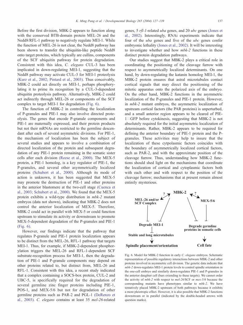
Fig. 6. Model for MBK-2 function in early C. elegans embryos. Schematic
representation of possible regulatory interactions between MBK-2 and other
proteins involved in asymmetric cell division. The genetic data indicate that
mbk-2 down-regulates MEI-1 protein levels to control spindle orientation in
the one-cell embryo and similarly down-regulates PIE-1 and P-granules in
the anterior daughter cell (bars extending to these targets). We cannot order
the activity of mbk-2 with respect to mel-26/SCF or mex-5/6 because the
corresponding mutants have phenotypes similar to mbk-2. We have
tentatively placed MBK-2 upstream of both pathways because it exhibits
a more pleiotropic effect. However, it is also possible that MBK-2 functions
downstream or in parallel (indicated by the double-headed arrows with
question marks).
K. Ming Pang et al. / Developmental Biology 265 (2004) 127–139 137
Before the first division, MBK-2 appears to function along
with the conserved BTB-domain protein MEL-26 and the
Nedd8/RFL-1 pathway to negatively regulate MEI-1. While
the function of MEL-26 is not clear, the Nedd8 pathway has
been shown to transfer the ubiquitin-like peptide Nedd8
onto target proteins, which typically are cullins, components
of the SCF ubiquitin pathway for protein degradation.
Consistent with this idea, C. elegans CUL-3 has been
implicated in down-regulating MEI-1, suggesting that the
Nedd8 pathway may activate CUL-3 for MEI-1 proteolysis
(Kurz et al., 2002; Pintard et al., 2003). Thus conceivably,
MBK-2 could act directly on MEI-1, perhaps phosphory-
lating it to prime its recognition by a CUL-3-dependent
ubiquitin proteolysis pathway. Alternatively, MBK-2 could
act indirectly through MEL-26 or components of the SCF
complex to target MEI-1 for degradation (Fig. 6).
The function of MBK-2 in controlling the localization
of P-granules and PIE-1 may also involve directed prote-
olysis. The genes that encode P-granule components and
PIE-1 are maternally expressed, and their protein products
but not their mRNAs are restricted to the germline descen-
dant after each of several asymmetric divisions. For PIE-1,
the mechanism of localization has been the subject of
several studies and appears to involve a combination of
directed localization of the protein and subsequent degra-
dation of any PIE-1 protein remaining in the somatic sister
cells after each division (Reese et al., 2000). The MEX-5
protein, a PIE-1 homolog, is a key regulator of PIE-1, the
P-granules, and several other asymmetrically localized
proteins (Schubert et al., 2000). Although its mode of
action is unknown, it has been suggested that MEX-5
may promote the destruction of PIE-1 and other proteins
in the anterior blastomere at the two-cell stage (Cuenca et
al., 2003; Schubert et al., 2000). We found that the MEX-5
protein exhibits a wild-type distribution in mbk-2 mutant
embryos (data not shown), indicating that MBK-2 does not
control the anterior localization of MEX-5. Therefore,
MBK-2 could act in parallel with MEX-5 or could function
upstream to stimulate its activity or downstream to promote
MEX-5-dependent degradation of the P-granules and PIE-1
(Fig. 6).
However, our findings indicate that the pathway that
regulates P-granule and PIE-1 protein localization appears
to be distinct from the MEL-26, RFL-1 pathway that targets
MEI-1. Thus, for example, if MBK-2-dependent phosphor-
ylation triggers the MEL-26 and RFL-1-dependent SCF
substrate-recognition process for MEI-1, then the degrada-
tion of PIE-1 and P-granule components may depend on
other proteins related to, but distinct from, MEL-26 and
RFL-1. Consistent with this idea, a recent study indicated
that a complex containing a SOCS-box protein, CUL-2 and
UBC-5, is specifically required for the degradation of
several germline zinc finger proteins including PIE-1,
POS-1, and MEX-5/6 but not for degradation of other
germline proteins such as PAR-2 and PGL-1 (DeRenzo et
al., 2003). C. elegans contains at least 35 mel-26-related
genes, 5 rfl-1-related uba genes, and 20 ubc genes (Jones et
al., 2002). Interestingly, RNAi experiments indicate that
four of the uba genes and five of the ubc genes confer
embryonic lethality (Jones et al., 2002). It will be interesting
to investigate whether and how mbk-2 functions in these
distinct protein degradation pathways.
Our studies suggest that MBK-2 plays a critical role in
coordinating the positioning of the cleavage furrow with
respect to asymmetrically localized determinants. On one
hand, by down-regulating the katanin homolog MEI-1, the
MBK-2 protein ensures that astral microtubules contact
cortical signals that may direct the positioning of the
mitotic apparatus onto the polarized axis of the embryo.
On the other hand, MBK-2 functions in the asymmetric
localization of the P-granules and PIE-1 protein. However,
in mbk-2 mutant embryos, the asymmetric localization of
upstream cortical factors (the PAR proteins) is unperturbed,
and a small anterior region appears to be cleared of PIE-
1DGFP before cytokinesis, suggesting that MBK-2 is not
absolutely required for the initial asymmetric localization of
determinants. Rather, MBK-2 appears to be required for
defining the anterior boundary of PIE-1 protein and the P-
granules. These activities may help to insure that the
localization of these cytpolasmic factors coincides with
the boundary of asymmetrically localized cortical factors,
such as PAR-2, and with the approximate position of the
cleavage furrow. Thus, understanding how MBK-2 func-
tions should shed light on the mechanisms that coordinate
the localization of cortical and cytoplasmic factors both
with each other and with respect to the position of the
cleavage furrow; mechanisms that at present remain almost
entirely mysterious.

K. Ming Pang et al. / Developmental Biology 265 (2004) 127–139138
In summary, the findings described here indicate that
MBK-2 functions in both spindle positioning and in the
localization of cytoplasmic determinants in early C. ele-
gans embryos. These findings suggest that Dyrk family
kinases may be conserved regulators of microtubule sta-
bility and spindle placement and provide the first evidence
linking this family of kinases to asymmetric cell division.
Understanding the biochemical and cell-biological mecha-
nisms underlying MBK-2 action may provide insights into
how other Dyrk family kinases function in cell division,
nervous system development, and possibly in Down
syndrome.
Acknowledgments
The authors would like to thank Tae-Ho Shin and
members of the laboratory for helpful discussion and
comments on the manuscript. In addition, we thank Paul
Mains, Ken Kemphues, and Susan Strome for providing
antibodies; Alan Coulson and Yoji Kohara for providing
YAC and cDNA clones; and the C. elegans Genetics Center
(funded by the NIH National Center for Research Support)
for providing strains. Research support was provided in
part by an NIH grant (HD36247) to C.C.M. C.C.M is a
Howard Hughes Medical Institute assistant investigator.
K.M.P. was supported by NIH postdoctoral fellowship
(GM20795) and The Charles A. King Trust and The
Campbell and Hall Charity Fund (Boston, MA).
References
Altafaj, X., Dierssen, M., Baamonde, C., Marti, E., Visa, J., Guimera, J.,
Oset, M., Gonzalez, J.R., Florez, J., Fillat, C., Estivill, X., 2001. Neuro-
developmental delay, motor abnormalities and cognitive deficits in
transgenic mice overexpressing Dyrk1A (minibrain), a murine model
of Down’s syndrome. Hum. Mol. Genet. 10, 1915–1923.
Bahler, J., Nurse, P., 2001. Fission yeast Pom1p kinase activity is cell cycle
regulated and essential for cellular symmetry during growth and divi-
sion. EMBO J. 20, 1064–1073.
Bahler, J., Pringle, J.R., 1998. Pom1p, a fission yeast protein kinase that
provides positional information for both polarized growth and cytoki-
nesis. Genes Dev. 12, 1356–1370.
Brenner, S., 1974. The genetics of Caenorhabditis elegans. Genetics 77,
71–94.
Clark-Maguire, S., Mains, P.E., 1994. Localization of the mei-1 gene
product of Caenorhaditis elegans, a meiotic-specific spindle component.
J. Cell Biol. 126, 199–209.
Colombo, K., Grill, S.W., Kimple, R.J., Willard, F.S., Siderovski, D.P.,
Gonczy, P., 2003. Translation of polarity cues into asymmetric spin-
dle positioning in Caenorhabditis elegans embryos. Science 300,
1957–1961.
Cuenca, A.A., Schetter, A., Aceto, D., Kemphues, K., Seydoux, G., 2003.
Polarization of the C. elegans zygote proceeds via distinct establishment
and maintenance phases. Development 130, 1255–1265.
Dow, M.R., Mains, P.E., 1998. Genetic and molecular characterization of
the Caenorhabditis elegans gene, mel-26, a postmeiotic negative regu-
lator of mei-1, a meiotic-specific spindle component. Genetics 150,
119–128.
DeRenzo, C., Reese, K.J., Seydoux, G., 2003. Exclusion of germ plasm
proteins from somatic lineages by cullin-dependent degradation. Nature
424, 685–689.
Etemad-Moghadam, B., Guo, S., Kemphues, K.J., 1995. Asymmetrically
distributed PAR-3 protein contributes to cell polarity and spindle align-
ment in early C. elegans embryos. Cell 83, 743–752.
Fire, A., Xu, S., Montgomery, M.K., Kostas, S.A., Driver, S.E., Mello,
C.C., 1998. Potent and specific genetic interference by double-stranded
RNA in Caenorhabditis elegans. Nature 391, 806–811.
Fotaki, V., Dierssen, M., Alcantara, S., Martinez, S., Marti, E., Casas, C.,
Visa, J., Soriano, E., Estivill, X., Arbones, M.L., 2002. Dyrk1A hap-
loinsufficiency affects viability and causes developmental delay and
abnormal brain morphology in mice. Mol. Cell. Biol. 22, 6636–6647.
Garrett, S., Broach, J., 1989. Loss of Ras activity in Saccharomyces cer-
evisiae is suppressed by disruptions of a new kinase gene, YAKI, whose
product may act downstream of the cAMP-dependent protein kinase.
Genes Dev. 3, 1336–1348.
Garrett, S., Menold, M.M., Broach, J.R., 1991. The Saccharomyces cere-
visiae YAK1 gene encodes a protein kinase that is induced by arrest
early in the cell cycle. Mol. Cell. Biol. 11, 4045–4052.
Gotta, M., Ahringer, J., 2001. Distinct roles for Galpha and Gbetagamma in
regulating spindle position and orientation in Caenorhabditis elegans
embryos. Nat. Cell Biol. 3, 297–300.
Gotta, M., Dong, Y., Peterson, Y.K., Lanier, S.M., Ahringer, J., 2003.
Asymmetrically distributed C. elegans homologs of AGS3/PINS con-
trol spindle position in the early embryo. Curr. Biol. 13, 1029–1037.
Grill, S.W., Gonczy, P., Stelzer, E.H., Hyman, A.A., 2001. Polarity controls
forces governing asymmetric spindle positioning in the Caenorhabditis
elegans embryo. Nature 409, 630–633.
Guo, S., Kemphues, K.J., 1995. par-1, a gene required for establishing
polarity in C. elegans embryos, encodes a putative Ser/Thr kinase that
is asymmetrically distributed. Cell 81, 611–620.
Jones, D., Crowe, E., Stevens, T.A., Candido, E.P., 2002. Functional and
phylogenetic analysis of the ubiquitylation system in Caenorhabditis
elegans: ubiquitin-conjugating enzymes, ubiquitin-activating enzymes,
and ubiquitin-like proteins. Genome Biol. 3, 2.1–2.15 (Research).
Kawasaki, I., Shim, Y.H., Kirchner, J., Kaminker, J., Wood, W.B., Strome,
S., 1998. PGL-1, a predicted RNA-binding component of germ gran-
ules, is essential for fertility in C. elegans. Cell 94, 635–645.
Kemphues, K., 2000. PARsing embryonic polarity. Cell 101, 345–348.
Kentrup, H., Becker, W., Heukelbach, J., Wilmes, A., Schurmann, A., Hup-
pertz, C., Kainulainen, H., Joost, H.G., 1996. Dyrk, a dual specificity
protein kinase with unique structural features whose activity is depend-
ent on tyrosine residues between subdomains VII and VIII. J. Biol.
Chem. 271, 3488–3495.
Kurz, T., Pintard, L., Willis, J.H., Hamill, D.R., Gonczy, P., Peter, M.,
Bowerman, B., 2002. Cytoskeletal regulation by the Nedd8 ubiquitin-
like protein modification pathway. Science 295, 1294–1298.
Lin, R., Thompson, S., Priess, J.R., 1995. pop-1 encodes an HMG box
protein required for the specification of a mesoderm precursor in early
C. elegans embryos. Cell 83, 599–609.
Mello, C.C., Draper, B.W., Krause, M., Weintraub, H., Priess, J.R., 1992.
The pie-1 and mex-1 genes and maternal control of blastomere identity
in early C. elegans embryos. Cell 70, 163–176.
Pellettieri, J., Seydoux, G., 2002. Anterior–posterior polarity in C. elegans
and Drosophila—PARallels and differences. Science 298, 1946–1950.
Pintard, L., Kurz, T., Glaser, S., Willis, J.H., Peter, M., Bowerman, B.,
2003. Neddylation and deneddylation of CUL-3 is required to target
MEI-1/Katanin for degradation at the meiosis-to-mitosis transition in
C. elegans. Curr. Biol. 13, 911–921.
Raich, W.B., Moorman, C., Lacefield, C.O., Lehrer, J., Bartsch, D., Plas-
terk, R.H., Kandel, E.R., Hobert, O., 2003. Characterization of Caeno-
rhabditis elegans homologs of the down syndrome candidate gene
DYRK1A. Genetics 163, 571–580.
Reese, K.J., Dunn, M.A., Waddle, J.A., Seydoux, G., 2000. PIE-1 local-
ization to the germ lineage depends on two distinct mechanisms that act
on separate domains in the PIE-1 protein. Mol. Cell 6, 445–455.

K. Ming Pang et al. / Developmental Biology 265 (2004) 127–139 139
Rocheleau, C.E., Downs, W.D., Lin, R., Wittmann, C., Bei, Y., Cha, Y.H.,
Ali, M., Priess, J.R., Mello, C.C., 1997. Wnt signaling and an APC-
related gene specify endoderm in early C. elegans embryos. Cell 90,
707–716.
Rocheleau, C.E., Yasuda, J., Shin, T.H., Lin, R., Sawa, H., Okano, H.,
Priess, J.R., Davis, R.J., Mello, C.C., 1999. WRM-1 activates the
LIT-1 protein kinase to transduce anterior/posterior polarity signals in
C. elegans. Cell 97, 717–726.
Rose, L.S., Kemphues, K.J., 1998. Early patterning of the C. elegans
embryo. Annu. Rev. Genet. 32, 521–545.
Schubert, C.M., Lin, R., de Vries, C.J., Plasterk, R.H., Priess, J.R., 2000.
MEX-5 and MEX-6 function to establish soma/germline asymmetry in
early C. elegans embryos. Mol. Cell 5, 671–682.
Smith, D.J., Stevens, M.E., Sudanagunta, S.P., Bronson, R.T., Makhinson,
M., Watabe, A.M., O’Dell, T.J., Fung, J., Weier, H.U., Cheng, J.F.,
Rubin, E.M., 1997. Functional screening of 2Mb of human chromosome
21q22.2 in transgenic mice implicates minibrain in learning defects
associated with Down syndrome. Nat. Genet. 16, 28–36.
Souza, G.M., Lu, S., Kuspa, A., 1998. YakA, a protein kinase required for
the transition from growth to development in Dictyostelium. Develop-
ment 125, 2291–2302.
Srayko, M., Buster, D.W., Bazirgan, O.A., McNally, F.J., Mains, P.E., 2000.
MEI-1/MEI-2 katanin-like microtubule severing activity is required for
Caenorhabditis elegans meiosis. Genes Dev. 14, 1072–1084.
Srinivasan, D.G., Fisk, R.M., Xu, H., Van Den Heuvel, S., 2003. A com-
plex of LIN-5 and GPR proteins regulates G protein signaling and
spindle function in C. elegans. Genes Dev. 17, 1225–1239.
Tabuse, Y., Izumi, Y., Piano, F., Kemphues, K.J., Miwa, J., Ohno, S.,
1998. Atypical protein kinase C cooperates with PAR-3 to establish
embryonic polarity in Caenorhabditis elegans. Development 125,
3607–3614.
Tejedor, F., Zhu, X.R., Kaltenbach, E., Ackermann, A., Baumann, A.,
Canal, I., Heisenberg, M., Fischbach, K.F., Pongs, O., 1995. Minibrain:
a new protein kinase family involved in postembryonic neurogenesis in
Drosophila. Neuron 14, 287–301.
Tsou, M.F., Hayashi, A., DeBella, L.R., McGrath, G., Rose, L.S., 2002.
LET-99 determines spindle position and is asymmetrically enriched in
response to PAR polarity cues in C. elegans embryos. Development
129, 4469–4481.
Tsukuba, T., Bond, J.S., 1998. Role of the COOH-terminal domains of
meprin A in folding, secretion, and activity of the metalloendopepti-
dase. J. Biol. Chem. 273, 35260–35267.
Wallenfang, M.R., Seydoux, G., 2000. Polarization of the anterior-poste-
rior axis of C. elegans is a microtubule-directed process. Nature 408,
89–92.
Zollman, S., Godt, D., Prive, G.G., Couderc, J.L., Laski, F.A., 1994. The
BTB domain, found primarily in zinc finger proteins, defines an evo-
lutionarily conserved family that includes several developmentally
regulated genes in Drosophila. Proc. Natl. Acad. Sci. U. S. A. 91,
10717–10721.