Embed Size (px)
Citation preview
The juxtamembrane region of TrkA kinase is critical for inhibitor selectivity
Noritaka Furuya1,2, Takaki Momose1, Kenji Katsuno1, Nobuhiko Fushimi1, [email protected]
Hideyuki Muranaka1, Chiaki Handa1, Tomonaga Ozawa1, Takayoshi Kinoshita2
1 Kissei Pharmaceutical, 4365-1, Kashiwabara, Hotaka, Azumino City, Nagano Pref. 399-8304, Japan
2 Graduate School of Science, Osaka Prefecture University, 1-1 Gakuen-cho, Naka-ku, Sakai, Osaka 599-8531, Japan
Keywords: TrkA kinase, Juxtamembrane, Crystal structure, Selective allosteric inhibitor Although numerous crystal structures for protein kinases have been reported, many include only
the kinase domain but not the juxtamembrane (JM) region, a critical activity-controlling segment of receptor tyrosine kinases (RTKs). In this study, we determined the X-ray crystal structure of the tropomyosin receptor kinase (Trk) A selective inhibitor A1 complexed with the TrkA kinase domain and the JM region. This structure revealed that the unique inhibitor-binding pocket created by a novel JM configuration yields significant potency and high selectivity against TrkB and TrkC. Moreover, we validated the importance of the JM region for the potency of A1 using in vitro assays. The introduction of moieties that interact with the JM region will be one of the most effective strategies for producing highly selective RTK inhibitors. [1] Furuya, N.; Momose, T.; Katsuno, K.; Fushimi, N.; Muranaka, H.; Handa, C.; Ozawa, T.;
Kinoshita, T., The juxtamembrane region of TrkA kinase is critical for inhibitor selectivity. Bioorg Med Chem Lett 2017, 27 (5), 1233-1236.
[2] Benito-Gutiérrez, E.; Garcia-Fernàndez, J.; Comella, J. X., Origin and evolution of the Trk family of neurotrophic receptors. Mol Cell Neurosci 2006, 31 (2), 179-92.
[3] Bertrand, T.; Kothe, M.; Liu, J.; Dupuy, A.; Rak, A.; Berne, P. F.; Davis, S.; Gladysheva, T.; Valtre, C.; Crenne, J. Y.; Mathieu, M., The crystal structures of TrkA and TrkB suggest key regions for achieving selective inhibition. J Mol Biol 2012, 423 (3), 439-53.
[4] Chang, D. S.; Hsu, E.; Hottinger, D. G.; Cohen, S. P., Anti-nerve growth factor in pain management: current evidence. J Pain Res 2016, 9, 373-83.
[5] Wang, T.; Lamb, M. L.; Scott, D. A.; Wang, H.; Block, M. H.; Lyne, P. D.; Lee, J. W.; Davies, A. M.; Zhang, H. J.; Zhu, Y.; Gu, F.; Han, Y.; Wang, B.; Mohr, P. J.; Kaus, R. J.; Josey, J. A.; Hoffmann, E.; Thress, K.; Macintyre, T.; Omer, C. A.; Yu, D., Identification of 4-aminopyrazolylpyrimidines as potent inhibitors of Trk kinases. J Med Chem 2008, 51 (15), 4672-84.
Calculation of intramolecular Reaction of Hydroxymethyl
Rhodamine Derivatives for Development of Fluorescent
Probes Based on Computational Chemistry
Ryo Tachibana1 Mako Kamiya
2,4
[email protected] [email protected]
Satoshi Suzuki3 Keiji Morokuma
3
[email protected] [email protected]
Yasuteru Urano1,2,5
1
Graduate School of Pharmaceutical Sciences, The University of Tokyo, 7-3-1 Hongo,
Bunkyo-ku, Tokyo 113-0033, Japan 2
Graduate School of Medicine, The University of Tokyo, 7-3-1 Hongo, Bunkyo-ku, Tokyo
113-0033, Japan 3
Fukui Institute for Fundamental Chemistry, Kyoto University, Takano-Nishihiraki-cho 34-4,
Sakyou-ku, Kyoto 606-8103, Japan 4
JST PRESTO 5
AMED CREST
Keywords: Free Energy Calculation, Intramolecular Reaction, Acid-base Balance, Transition State
Calculation, First-Shell Hydration
HMR (Hydroxymethyl Rhodamine) derivatives can exist in two forms (open, closed) by
intramolecular spirocyclization. The proportion of two forms at equilibrium state changes
according to pH. Various fluorescence probes have been developed using pKcycl(pH value where
the concentration of open form and that of closed form are the same ) as an indicator[1][2]
. In this
research, we analyzed this intramolecular reaction computationally and found a powerful method
to predict pKcycl of HMR derivatives directly, without synthesizing any reference compounds .
We focused on closed-to-open reaction at acidic condition, and performed free energy
calculation of local minima of HMR derivatives, considering first -shell hydration and proton
transfer. Next, we searched each transition states which corresponded to calculated local minimum,
and evaluated their contribution ratio to the reaction. As a result, we found energetically favorable
chemical pathway, which dominated almost all of close-to-open reaction. We made a formula for
calculating pKcycl, based on equilibrium model of HMR derivatives including acid -base balance of
their amino group and hydroxymethyl group. By calculating free energy gap throughout
above-mentioned reaction path, we succeeded in reproducing pKcycl of measured HMR derivatives
and predicting proportion of unknown derivatives.
Moreover, we succeeded in developing new red fluorescence peptidase probes by applying pKcycl
prediction, which shows the possibility of accurate and efficient molecular design using
calculation with careful analysis of solvent behavior including first-shell hydration.
[1] Sakabe, M.; Asanuma, D.; Kamiya, M.; Iwatate, R. J.; Hanaoka, K.; Terai, T.; Nagano, T.;
Urano, Y. Journal of the American Chemical Society 2013, 135, 409.
[2] Uno, S. N.; Kamiya, M.; Yoshihara, T.; Sugawara, K.; Okabe, K.; Tarhan, M. C.; Fujita, H.;
Funatsu, T.; Okada, Y.; Tobita, S.; Urano, Y. Nature chemistry 2014, 6, 681.
Evaluation of log P of endocrine disruptors
using DFT methods
Masao Fujisawa1 Mamu Tabe1 [email protected] [email protected]
Hirohito Ikeda2 Takayoshi Kimura3 [email protected] [email protected]
1 Department of Biotechnological Science, Kindai University, 930 Nishimitani, Kinokawa-City,
Wakayama, Tokyo 649-6493, Japan 2 Department of Pharmaceutical Science, Fukuoka University, 8 -19-1 Nanakuma Fukuoka
814-0180, Japan 3 Department of Chemistry, Kindai University, Kowakae, Higashi-osaka 577-8502, Japan
Keywords: DFT-D, Vibrational frequencies, log P
Solvation phenomena play a significant role in chemical reactions and biomolecular recognition;
however, it can be very difficult to determine the related thermodynamic quantities. For example,
to calculate the solvation Gibbs energy, one must know temperature dependence of the
vaporization enthalpy or the vapor pressure. Moreover, the dispersion energy is an important
contributor to the solvation energy of a solute. In an attempt to predict solvation Gibbs energy,
theoretical methods including explicit or continuum solvent models have been developed and
applied [1-3]. In an explicit solvent model, there have been an attempt of hydration Gibbs
energies computed using the molecular dynamics simulation and the energy -representation theory
of solvation [4]. In this study, the solvation Gibbs energies for several endocrine disruptors were
calculated using dispersion-corrected density functional theory (DFT-D) method, which were then
compared with experimental thermodynamic data. To decide the lowest energy structure, conformation search was performed. The obtained lowest energy conformer was the initial
structure of the DFT-D calculations in gas phase. Vibrational frequencies were calculated for
these optimized geometries at the same level of theory in gas phase. The solute ge ometries were
optimized in water using DFT-D functions. Similarly, vibrational frequencies were calculated for
these optimized geometries at the same level of theory in water. Gibbs energies of solutes were
determined by vibrational frequencies in gas phase and in water. Partition coefficients (log P) were
determined using the differences in Gibbs energy between two solvents (water, n-octanol). These
predicted values well reproduced the experimental log P.
Conformational regularity of the condensed tannins investigated by free-energy calculation
Reina Takahashi1 Takuma Todoroki1
[email protected] [email protected]
Hiroshi Fujii1,2 Hidefumi Makabe1 [email protected] [email protected]
Koji Umezawa1,2
1 Faculty of agriculture, Shinshu University, 8304 Minami-minowa, Kami-ina, Nagano, 399-4598, Japan
2 Department of Interdisciplinary Genome Sciences and Cell Metabolism, Institute for Biomedical Sciences, Interdisciplinary Cluster for Cutting Edge Research, Shinshu University, Minami-minowa, Kami-ina, Nagano, 399-4598, Japan
Keywords: Natural product, Flavan-3-ol, Proanthocyanidin
Three-dimensional (3D) molecular structure is responsible for biological function. Biopolymers can adopt some stable conformations. The condensed tannins are known as the polymer of polyphenol in natural products. However, most of their conformations are still elusive. Then, we have investigated the stable conformations of the condensed tannins by free-energy calculation to figure out the conformational regularity.
Free-energy calculation was done for the natural polymer, the condensed tannins. The unit of the condensed tannin is flavan-3-ol such as catechin derivatives. The two adjacent units are connected by a single covalent bond; inter-flavan bond. The conformational freedom around the inter-flavan bond designates the relative direction between the upper and the lower units. In the free-energy calculation, we conducted the umbrella-sampling molecular dynamics simulations with the reaction coordinate of the dihedral angle around the inter-flavan bond. Herein, we chose the unit of catechin (cat), epicatechin (epi), gallocatechin (gc) and epigallocatechin (egc). For dimers of tannins, all permutation of two units were calculated (e.g. cat-cat, cat-epi, epi-cat, cat-gc, gc-cat, etc; total of 16 dimers). For trimers of tannins, total of 4 trimers (cat-cat-cat, epi-epi-epi, gc-gc-gc and egc-egc-egc) were investigated.
The results showed that the free-energy landscape (FEL) of the dimer depended on the upper unit. The FELs of the dimers with the upper unit of cat and gc had one minima while those of epi and egc did two minimal basins. It may be explained by hydrogen-bond pattern between upper and lower units. The cat unit differs from the epi one mainly at the point of chirality of connection to the lower unit. The hydrogen bonds between the upper and lower units can be formed according to the upper-unit conformation, which can contribute the structural stability. For the trimers, the FELs of cat and gc showed two low regions whereas those of epi and egc did four, which might indicate that the conformational stability of polymer depends on the adjacent units. In our presentation, the detail of conformation will be displayed. Furthermore, we will show the recent results for cat- and epi-polymers up to 7-mer.
Functional profiling of asymmetrically-organized human
CCT/TRiC chaperonin
Kazutaka Araki1 Atsushi Suenaga
1, 2
[email protected] [email protected]
Tohru Natsume1, 3
Kazuhiko Fukui1
[email protected] [email protected]
1 Molecular Profiling Research Center for Drug Discovery, National Institute of Advanced
Industrial Science and Technology, Tokyo 135-0064, Japan 2 Department of Biosciences, College of Humanities and Sciences, Nihon University, 3-25-40
Sakurajosui Setagaya-Ku, Tokyo 156-8550, Japan 3 Robotic Biology Institute, Inc., Tokyo 135-0064, Japan
Keywords: Chaperonin, CCT, Cysteine, Protein modeling, Electrostatic calculation
Molecular organization of the eukaryote chaperonin known as CCT/TRiC complex was recently
clarified. Eight distinct subunits are uniquely organized, providing a favorable folding cavity for
specific client proteins such as tubulin and actin. Because of its heterogeneous subunit
composition, CCT complex has polarized inner faces, which may underlie an essential part of its
chaperonin function. In this study, we structurally characterized the closed and open states of CCT
complex, using molecular dynamics analyses. Our results showed that the inte r-subunit interaction
energies were asymmetrically distributed and were remodeled during conformational changes of
CCT complex. In addition, exploration of redox related characteristics indicated changes in inner
surface properties, including electrostatic potential, pKa and exposure of inner cysteine thiol
groups, between the closed and open states. Cysteine activation events were experimentally
verified by interaction analyses, using tubulin as a model substrate. Our data highlighted the
importance of dynamics-based structural profiling of asymmetrically oriented chaperonin function.
[1] Araki, K., Suenaga, A., Kusano, H., Tanaka, R., Hatta, T., Natsume, T., and Fukui, K.,
Functional profiling of asymmetrically-organized human CCT/TRiC chaperonin, Biochemical
and biophysical research communications , 481:232-238, 2016.
[2] Araki, K., Kusano, H., Sasaki, N., Tanaka, R., Hatta, T., Fukui, K., and Natsume, T ., Redox
Sensitivities of Global Cellular Cysteine Residues under Reductive and Oxidative Stress,
Journal of proteome research , 15:2548-2559, 2016.
[3] Araki, K., Ushioda, R., Kusano, H., Tanaka, R., Hatta, T., Fukui, K., Nagata, K., and Natsume,
T., A crosslinker-based identification of redox relay targets, Analytical biochemistry, 520:22-26,
2017.
Flexible Docking using Replica-Exchange Molecular Dynamics Simulation
Suyong Re1 Hiraku Oshima1
[email protected] [email protected]
Motoshi Kamiya2 Yuji Sugita1,2, 3 [email protected] [email protected]
1 RIKEN Quantitative Biology Center (QBiC), Integrated Innovation Building 7F, 6-7-1 Minatojima-minamimachi, Chuo-ku, Kobe, Hyogo 650-0047, Japan
2 RIKEN Advanced Institute for Computational Science (AICS), Integrated Innovation Building 7F, 6-7-1 Minatojima-minamimachi, Chuo-ku, Kobe, Hyogo 650-0047, Japan
3 RIKEN Theoretical Molecular Science Laboratory and iTHES, 2-1 Hirosawa Wako-shi, Saitama 351-0198, Japan.
Keywords: Molecular dynamics simulations, enhanced sampling, protein-ligand binding Accurate prediction of ligand binding structure and energetics remains a challenge. Accounting for both ligand binding and protein structure changes, such as binding site reorganization, is still very difficult. Two-dimensional replica-exchange approach1 is promising in that it enhances the binding events to predict the binding structure with high statistical accuracy. Here, we extend the method by introducing the generalized replica-exchange with solute tempering (gREST)2. The protein flexibility is incorporated by scaling the temperature of a wide solute region, involving the ligand and the active site residues, using gREST. We applied the method to study the ligand binding of Src kinase. We show that the native binding structure is correctly predicted. Importantly, the consideration of protein flexibility significantly improves the docking efficiency compared to the existing methods. We also stress that the method enhances the binding events to give high statistical accuracy, while long-time simulation could sample the event once per several microseconds. This approach, followed by binding free energy calculation, allows us to accurately predict protein-ligand binding affinity. [1] Kokubo, H. et al. “Two-dimensional replica-exchange method for predicting protein-ligand
binding structures.” J. Comput. Chem. (2013) 34:2601-2614. [2] Kamiya, M. et al. in preparation.
FMO calculations on specific interactions between
vitamin-D receptor and its ligands
Ryosuke Takeda1, Ittetsu Kobayashi1, Kanako Shimamura1, Hiromi Ishimura1,
Ryushi Kadoya1, Kentaro Kawai2, Atsushi Kittaka3, Midori Takimoto-Kamimura4,
Noriyuki Kurita1 [email protected]
1 Department of Computer Science and Engineering, Toyohashi University of Technology,
Tempaku-cho, Toyohashi, Aichi, 441-8580, Japan
2 Drug Research Center, Kaken Pharmaceutical Co. Ltd.,
14, Shinomiya, Minamigawara-cho, Yamashina-ku, Kyoto, 607-8042, Japan
3 Faculty of Pharmaceutical Sciences, Teikyo University, 2-11-1 Kaga, Itabashi, Tokyo 173-8605, Japan
4 Teijin Institute for Bio-Medical Research, Teijin Pharma Ltd.,
4-3-2 Asahigaoka, Hino, Tokyo, 191-8512, Japan
Keywords: Molecular simulation, Fragment molecular orbital , Vitamin D receptor, Inhibitor
Vitamin D3 is hydroxylated at both the 25 and the 1α sites in a liver and a kidney, resulting in the
active form of vitamin D3 metabolites 1α,25-dihydroxyvitamin D3 (1α,25(OH)2D3), which plays
important roles in the regulation of calcium and phosphorus metabolism as well as in the bone
formation. The physiological actions caused by 1α,25(OH) 2D3 are triggered by its specific
interaction with vitamin D receptor (VDR)[1]. The previous study [2] confirmed that almost all
cell tissues in a living organism have VDR and that the binding of 1α,25(OH)2D3 to the VDR is
deeply related to the pathogenesis of the immunological diseases such as cancer as well as the
response anomaly to hormone. For elucidating the biological effect of VDR, it is indispensable to
determine the structures of VDR bound by vitamin D derivatives. Therefore, various types of
1α,25(OH)2D3 derivatives were synthesized [3] and the structures of their complexes with human
VDR were determined to clarify that the derivatives form hydrogen bonds to Arg274 residue of
VDR and have high binding affinity for VDR. However, it is not elucidate the reason why a slight
difference in structure of the derivatives causes a large difference in binding affinity between VDR
and the derivatives. This fact might be a bottleneck for proposing novel derivatives as a potent
modulator or inhibitor to VDR.
In the present study, we employed two types of 1α,25(OH)2D3 derivatives, whose structures are
almost the same but their effect on VDR activity is significantly different, and their specific
interactions with VDR were investigated at an electronic level, using ab initio molecular
simulations based on fragment molecular orbital (FMO) method [4]. Based on the results of FMO
calculations, we elucidated which parts of the derivatives and which residues of VDR are
important for the specific binding between VDR and the derivatives [5].
[1] Darwish H., DeLuca H. F., “Vitamin D-regulated gene expression,” Crit. Rev. Eukaryot. Gene Expr.,
3: 89−116, 1993.
[2] Walters M. R., “Newly identified actions of the vitamin D endocrine system,” Endocr. Rev., 13:
719−764, 1992.
[3] Matsuo M., Hasegawa A., Takano M., Saito H., Kakuda S., Takagi K., Ochiai E., Horie K.,
Takimoto-Kamimura M., Takenouchi K., Sawada D., Kittaka A., “Design and synthesis of
2α-(tetrazolylethyl)-1α,25-dihydroxyvitamin D3 as a high affinity ligand for vitamin D receptor,” J.
of Steroid Biochem. & Mole. Biol., 144: 201−203, 2014.
[4] Kitaura K., Ikeo E., Asada T., Nakano T., Uebayashi M., “Fragment molecular orbital method: an
approximate computational method for large molecules,” Chem. Phys. Lett., 313: 701−706, 1999.
[5] Takeda R., Kobayashi I., Shimamura K., Ishimura H., Kadoya R., Kawai K., Kittaka A.,
Takimoto-Kamimura M., Kurita N., “Specific interactions between vitamin-D receptor and its
ligands: ab initio molecular orbital calculations in water”, J. of Steroid Biochem. & Mole. Biol.,
171: 75−79, 2017.
Repetition of binding and unbinding processes between
protein and ligand by supervised molecular dynamics
Takashi Mitsui1 [email protected]
1 Bio-IT R&D Office, Research& Development Division, Healthcare Systems Unit, Fujitsu
Limited, 1-17-25 Shin-Kamata, Ota-ku, Tokyo 226-8504, Japan
Keywords: Molecular dynamics, Ligand binding
The mechanism of the binding/unbinding processes between protein and ligand is a crucial issue
in in-silico drug design. In recent published papers, unbiased long-time molecular dynamics (MD)
simulations reproduced experimentally observed protein-ligand complex structures. However,
ligand binding to its receptor in correct pose is rare event, therefore it requires microsecond
time-scale MD simulation.
Supervised molecular dynamics (SuMD) [1][2] is tabu-like
computational algorithm developed to follow protein-ligand
approaching processes within a relatively short time-scale
compared to traditional MD simulation. In this study, we
extended the original protocol to an unbinding process to
enable repetition of multiple binding/unbinding events.
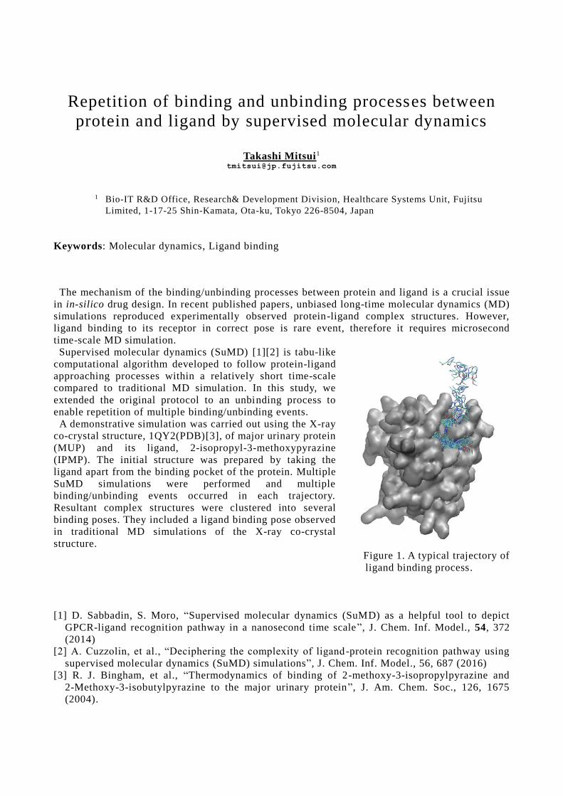
A demonstrative simulation was carried out using the X-ray
co-crystal structure, 1QY2(PDB)[3], of major urinary protein
(MUP) and its ligand, 2-isopropyl-3-methoxypyrazine
(IPMP). The initial structure was prepared by taking the
ligand apart from the binding pocket of the protein. Multiple
SuMD simulations were performed and multiple
binding/unbinding events occurred in each trajectory.
Resultant complex structures were clustered into several
binding poses. They included a ligand binding pose observed
in traditional MD simulations of the X-ray co-crystal
structure.
Figure 1. A typical trajectory of
ligand binding process.
[1] D. Sabbadin, S. Moro, “Supervised molecular dynamics (SuMD) as a helpful tool to depict
GPCR-ligand recognition pathway in a nanosecond time scale”, J. Chem. Inf. Model., 54, 372
(2014)
[2] A. Cuzzolin, et al., “Deciphering the complexity of ligand -protein recognition pathway using
supervised molecular dynamics (SuMD) simulations”, J. Chem. Inf. Model., 56, 687 (2016)
[3] R. J. Bingham, et al., “Thermodynamics of binding of 2-methoxy-3-isopropylpyrazine and
2-Methoxy-3-isobutylpyrazine to the major urinary protein”, J. Am. Chem. Soc., 126, 1675
(2004).
In silico protein design for functional modification of photoactivated adenylate cyclase
Mayu Tanaka1 Toru Ekimoto1 Mio Ohki1 Tsutomu Yamane1
Sam-Yong Park1 Mitsunori Ikeguchi1
[email protected] [email protected]
1 Graduate School of Medical Life Science, Yokohama City University, 1-7-29 Suehiro-cho, Tsurumi-ku, Yokohama 230-0045, Japan
Keywords: Photoactivated adenylate cyclase, Mutation analysis, Protein design Photoactivated adenylate cyclase (PAC) is a photoreceptor protein that produces the second messenger cyclic-AMP (cAMP) upon light illumination. PAC consists of the blue light using flavin (BLUF) and the adenylate cyclase (AC) domains. Recently, the crystal structure of Oscillatoria acuminate photoactivated adenylate cyclase (OaPAC) has been solved [1]. Optogenetics is a rapidly growing field in which light is used to control biological systems. OaPAC is expected as an optgenetic tool to produce cAMP upon blue-light illumination. In addition to cAMP, the control of the cGMP level upon light illumination would be useful to investigate the effects of the cGMP on biological systems. Therefore, in this study, we attempt in-silico protein design to modify OaPAC to produce cGMP instead of cGMP. In the crystal structure of OaPAC, ATP or ATP analog was not bound to the ATP binding site. Therefore, in this study, the crystal structure of CyaC, a homolog of OaPAC, was used to design the nucleotide-binding site, because ApCpp, an ATP analog, was bound to the crystal structure of CyaC. First, ApCpp was replaced to GTP in the crystal structure of CyaC. Then, amino acid in the binding site were comprehensively mutated, and the changes in GTP affinity upon mutations were calculated. Also, the changes in stability of the protein upon mutations were calculated. Mutants predicted to have high affinity of GTP forms hydrogen bonds to GTP and not to ATP. Interactions with both two GTPs of the CyaC dimer were found to be important to raise affinity to GTP. [1] Ohki, M. et al., Structural insight into photoactivation of an adenylate cyclase from a photosynthetic cyanobacterium, PNAS, 113:6659-6664, 2016
Validation of Epigenetic Therapeutic Target Proteins
for Homogenous Assay Performance
Masato Yonezawa1, Mary Anne Jelinek1, Melissa Ritland1, Jake Dabrowski1, Wei Gong1,
Lingchun Kong2 and Fei Lan2
1 Active Motif Inc., Carlsbad CA, USA 2 Active Motif China, Shanghai, China
Email: [email protected]
Keywords: Protein-small molecule interaction, Enzymatic reaction
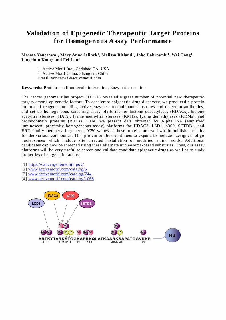
The cancer genome atlas project (TCGA) revealed a great number of potential new therapeutic
targets among epigenetic factors. To accelerate epigenetic drug discovery, we produced a protein
toolbox of reagents including active enzymes, recombinant substrates and detection antibodies,
and set up homogeneous screening assay platforms for histone deacetylases (HDACs), histone
acetyltransferases (HATs), lysine methyltransferases (KMTs), lysine demethylases (KDMs), and
bromodomain proteins (BRDs). Here, we present data obtained by AlphaLISA (amplified
luminescent proximity homogeneous assay) platforms for HDAC3, LSD1, p300, SETDB1, and
BRD family members. In general, IC50 values of these proteins are well within published results
for the various compounds. This protein toolbox continues to expand to include “designer” oligo
nucleosomes which include site directed installation of modified amino a cids. Additional
candidates can now be screened using these alternate nucleosome -based substrates. Thus, our assay
platforms will be very useful to screen and validate candidate epigenetic drugs as well as to study
properties of epigenetic factors.
[1] https://cancergenome.nih.gov/
[2] www.activemotif.com/catalog/5
[3] www.activemotif.com/catalog/744
[4] www.activemotif.com/catalog/1068
Comprehensive database analysis of protein kinase structures
Kei Moritsugu Yoshihiko Nishino [email protected] [email protected]
Akinori Kidera
Graduate School of Medical Life Science, Yokohama City University, 1-7-29 Suehirocho, Tsurumi-ku, Yokohama 230-0045, Japan
Keywords: Protein-small molecule interaction, Molecular dynamics method, Protein kinase,
KLIFS database, Structural bioinformatics
Protein kinases are ATP phosphotransferases targeting at Ser/Thr/Tyr residues of specific substrate proteins and then play an important role for signaling in cells. While some kinase proteins involved in diseases have been extensively studied as drug-discovery targets, few of studies have been done to understand complete structural dynamics for overall protein kinases. A protein kinase database, KLIFS [1], collects a variety of human/mouse protein kinases with various kinase families and species, kinase activities, bound (drug) molecules, site-directed mutations, and so on, which however takes interest on local atom interactions between the proteins and ligands. In the present study, by use of the KLIFS database, we have attempted comprehensive analyses of protein kinase collective motions in relation to the way of local protein-drug interactions.
Data collection for this study was carried out from 849 human tyrosine kinases in the KLIFS database by excluding structures with a number of lacking atoms or with large similarities, leading to 150 representative kinase structures. Motion Tree [2,3] calculated using 207 C atoms after multiple alignment of the 150 structures demonstrated a significance of the domain motion between kinase N-lobe and C-lobe, as well as the motions of both activation loop and C-helix which have often been discussed in previous studies. Structural classifications were performed by projecting of each protein kinase onto the three motions and by assigning the information on kinase activities (DFG-in/C-helix-in, etc) and kinase species, which were then analyzed in relation to the atom interactions with bound molecules such as ATP-analog and drugs, and in combination with the structural dynamics data calculated from comprehensive molecular dynamics simulations. These findings yielded a general picture of how protein kinases are inactivated due to what kinds of drugs on which binding pockets, which will be useful for designing new drug molecules for specific protein kinases. [1] A. J. Kooistra et al., “KLIFS: a structural kinase-ligand interaction database”, Nucleic Acids
Res 44: D365-D371 (2016). [2] K Moritsugu, R Koike, K Yamada, H Kato, A Kidera, "Motion Tree Delineates Hierarchical
Structure of Protein Dynamics Observed in Molecular Dynamics Simulation", PLoS ONE 10: e0131583 (2015).
[3] R Koike, M Ota, A Kidera, "Hierarchical description and extensive classification of protein structural changes by Motion Tree", Journal of Molecular Biology 426: 752-762 (2014).
Finite-size effect on the charging free energy in the
alchemical perturbation and ``warp drive’’ method
Toru Ekimoto Tsutomu Yamane [email protected] [email protected]
Mitsunori Ikeguchi [email protected]
Graduate School of Medical Life Science, Yokohama City University, 1 -7-29 Suehiro-cho,
Tsurumi-ku, Yokohama 230-0045, Japan
Keywords: Binding free energy, Free energy perturbation, Charging free energy, Finite-size effect
With increase of computational power, calculations of the binding free energy by exact methods,
such as the alchemical free energy perturbation (alchemical-FEP), routinely become possible with
all-atom molecular dynamics (MD) simulations. In the alchemical-FEP, the bound ligand is
thermodynamically eliminated from the binding site and is emerged to a bulk region. The
alchemical-FEP can treat buried ligands in deep pocket, however, accuracy for highly charged
ligands is poor. One of the reasons is due to the finite-size effect on the charging free energy (CFE)
in periodic systems. The CFE is the free energy change for tuning on the electrostatic interaction
of the solute with the solvent. The finite-size effect refers to the cell-size dependence of the CFE at
different cell sizes. [1] When the CFE is left uncorrected, a comparison of the CFE among
different charged states is erroneous as shown in the examinations for ions and proteins [1,2]. The
CFE is a key component of the binding free energy, therefore, a correction scheme is necessary to
obtain the CFE at the limit of large cell size.
In this study, we examine the finite-size effect on the CFE in the alchemical-FEP with
systematically varied cell sizes of the MD unit cell , assess the performance of the correction
scheme formulated by Hummer et al. [1], and introduce an alternative perturbation, termed “warp
drive method” [3], providing the CFE at the limit of large cell size without any corrections. The
phosphotyrosine peptide (-5e) bound to the Src homology 2 domain (+1e) is employed as a test
complex, and the CFE is calculated by thermodynamic integration method. We show that the
finite-size effect arises at ~92 kcal/mol in the annihilation process and ~180 kcal/mol in the
emerging process. The self-energy correction essentially corrects them within ~2 kcal/mol and ~3
kcal/mol, respectively, and the additional correction from the solvation effect reduces the remains
to ~2 kcal/mol and ~1 kcal/mol, respectively. This shows that the corrections scheme is absolutely
necessary to obtain the CFE at the limit of large cell size, however, its performance at the small
cell sizes is less effective in this examination. We also examine the warp drive method in which
both processes in the alchemical-FEP is simultaneously executed using a solution system
consisting of a protein-ligand complex and a distantly-positioned unbound-ligand in one MD unit
cell. The eliminated partial charges of the bound ligand simultaneously emerge on the other ligand
in bulk, and therefore, the total charge of the system does not change at all intermediate sates. The
CFE by this method does not show the finite-size effect even at small cell sizes, and the value
without any corrections is in good agreement with the summation of the corrected CFEs calculated
by the alchemical-FEP.
[1] Hummer, G., Pratt, L.R., Garcia, A.E. , Journal of Physical Chemistry , 100: 1206-1215, 1996.
[2] Ekimoto, T., Matubayasi, N., and Ikeguchi, M., Journal of Chemical Theory and Computation ,
11:215-223, 2015.
[3] Ekimoto, T., Yamane, T., and Ikeguchi, M., in preparation.
Molecular dynamics and ab initio FMO calculations for
amyloid-β nonamer
Shogo Tomioka1, Haruki Sogawa
1, Hiromi Ishimura
1, Akisumi Okamoto
1,
Sergiy Shulga2, Pavel Karpov
2, Yaroslav Blume
2, Noriyuki Kurita
1
1 Department of Computer Science and Engineering, Toyohashi University of Technology,
Tempaku-cho, Toyohashi, Aichi, 441-8580, Japan
2 Institute for Food Biotechnology and Genomics, National Academy of Sciences of Ukraine,
2a. Osypovskogo Str., Kyiv-123, 04123, Ukraine
Keywords: Molecular simulation, fragment molecular orbital, molecular dynamics, Alzheimer’s
disease, amyloid-beta, aggregate
The accumulation of amyloid-β (Aβ) oligomers and fibrils in a brain has been recognized to be a
major cause of the onset of Alzheimer’s disease (AD). It is thus expected that the inhibition of Aβ
aggregations can prevent the onset of AD, and many kinds of agents with strong binding affinity to
Aβ have been developed. In the design of these potent inhibitors against the Aβ aggregation, it is
necessary to make clear the structures of the Aβ oligomers and fibrils as well as the mechanism of
the aggregation. Recently, Lu et al.[1] revealed structural models for the in vivo Aβ nonamer,
based on the solid-state nuclear magnetic resonance analyses for the Aβ fibrils derived from the
brains of two different AD patients. Each of these Aβ nonamer models has a single and
patient-specific structure possessing three-fold symmetry with respect to the axis of fibril growth .
However, it is not elucidated why such high symmetry structure of Aβ fibrils can be stabilized.
Molecular simulations such as molecular mechanics (MM) and molecular dynamics (MD) o nes
for Aβ fibrils are efficient for investigating their stable structures and the mechanism of
aggregation at an atomic level. Kahler et al.[2] conducted a systematic computational study based
on all-atom classical MD simulations for many types of fibrillary Aβ oligomers and concluded that
the pairs of Aβ protofilaments are important as a seed for forming Aβ fibrils. However, they
considered only Aβ(9-42) fragment, missing the residues of N-terminal region, which were found
to be important for Aβ aggregation in the previous experiment.
In our previous studies [3,4], replica-exchange MD (RE-MD) simulations were conducted in
water for monomer and dimer models of Aβ(1-42) peptides, in order to search widely for their
stable conformations in water. In addition, we carried out ab initio fragment molecular orbital
(FMO) calculations for the conformations obtained by the RE-MD simulations and determined the
stable conformations of the solvated Aβ(1-42) monomer and dimer, with considering water
molecules explicitly. We furthermore investigated the specific interactions between Aβ peptides in
the Aβ hexamers at an electronic level, using ab initio FMO method [5]. Our ab initio simulations
elucidated the importance of structural water molecules for the stabilization of Aβ fibrils.
In the present study, to clarify the reason why the Aβ nonamer with three -fold symmetry
obtained by Lu et al.[1] is stable, we investigated the change in structure of the Aβ nonamer by use
of classical MD simulations in water. In addition, ab initio FMO calculations were carried out for
some snapshots obtained by the MD simulations. The results revealed that the interactions between
the Aβ peptides of stacked Aβ pairs make resultant contribution in stability of the Aβ nonamer. [1] Lu J.X., Qiang W., Yau W.M., Schwieters C.D., Meredith S.C., Tycko R., Cell 154 (2013) 1257–1268.
[2] Kahler A., Sticht H., Horn A.H.C., Plos One 8 (2013) e70521.
[3] Yano A., Okamoto A., Nomura K., Higai S., Kurita N., Chem. Phys. Lett. 595/596 (2014) 242–249.
[4] Okamoto A, Nomura K., Yano A., Higai S., Kondo T., Kamba S., Kurita N., Chem. Phys. Lett. 577
(2013) 131–137.
[5] Ishimura H., Tomioka S., Kadoya R., Simamura K., Okamoto A., Shulga S., Kurita, N.,
Chem. Phys. Lett. 672 (2017) 13–20.
Specific interactions between mycobacterial FtsZ and
curcumin derivatives: molecular docking and ab initio
molecular simulations
Mitsuki Fujimori1, Haruki Sogawa
1, Shintaro Ota
1, Pavel Karpov
2, Sergiy Shulga
2,
Yaroslav Blume2, Noriyuki Kurita
1
1 Department of Computer Science and Engineering, Toyohashi University of Technology,
1-1 Hibarigaoka, Tenpaku-cho, Toyohashi, Aichi, 411-8580, Japan
2 Institute for Food Biotechnology and Genomics, National Academy of Scienc es of Ukraine,
2a. Osypovskogo Str., Kyiv-123, 04123, Ukraine
Keywords: Curcumin; Inhibitor; FtsZ; Tuberculosis; Cell division; Fragment molecular orbital;
Protein-ligand docking
Tuberculosis (TB) is one of the most widespread infectious diseases caused by the bacillus
Mycobacterium tuberculosis (Mtb). In the treatment of TB, many kinds of drugs such as isoniazid,
rifampicin, pyrazinamide and ethambutol have been administered. However, there is a considerable
potential for Mtb to have resistance against these drugs. In particular, the Mtb having resistance
against multiple drugs is called multidrug-resistant TB (MDR-TB), and the number of MDR-TBs is
increasing rapidly. It is thus necessary to develop new anti-TB drugs targeting the most
conservative proteins, which cannot be mutated easily [1]. As a candidate of anti-TB drugs, new
compounds were proposed, which suppress the growth of Mtb by inhibiting the division of Mtb cell
[2]. These drugs are targeted to cytoskeletal protein FtsZ (filamenting temperature-sensitive
mutant Z), which plays an essential role in the cell division mechanism [3]. The inhibitor against
FtsZ function is expected to suppress the Mtb cell growth.
As a novel inhibitor against Mtb FtsZ, we here considered curcumin derivatives. Curcumin is a
natural product and contained in the root of Curcumae Rhizoma, while other curcuminoids such as
demethoxycurcumin and bisdemethoxycurcumin are also included in the root [4]. These curcumin
derivatives have been widely used [4] as conventional drugs for treating many diseases. It was
found that curcumin suppresses bacterial cell proliferation by inhibiting the FtsZ function [ 5].
However, since there are some ligand-binding pockets in Mtb FtsZ, the binding site of curcumin on
FtsZ and the specific interactions between curcumin and FtsZ are not elucidated yet.
In the present study, we investigated the specific interactions between Mtb FtsZ and some
curcumin derivatives, using ab initio molecular simulations based on protein-ligand docking,
classical molecular mechanics optimization and ab initio fragment molecular orbital (FMO)
calculation. Based on the FMO results, we attempted to reveal which curcumin derivative can bind
more strongly to FtsZ. In addition, we elucidated which parts of FtsZ and curcumin derivative are
important for the specific interactions between them. The result will be useful for proposing novel
anti-TB drugs based on curcumin derivatives.
[1] H. Tomioka, Kekkaku, 85: 815–822, 2010.
[2] P.A.J de Boer, et al., Cell, 56: 641–649, 1989.
[3] M. Osawa, et al., Cell, 56: 641–649, 1989.
[4] F. Kiuchi, et al., Chem. Pham. Bull, 41: 1640–1643, 1993.
[5] D. Rai, et al, Biochem. J., 410: 147–155, 2008.
QCLObot: an automation engine of canonical MO calculation in proteins
Toshiyuki Hirano1 Fumitoshi Sato1
[email protected] [email protected]
1 Institute of Industrial Science, the University of Tokyo 4-6-1 Komaba, Meguro-ku, Tokyo 153-8505, Japan
Keywords: canonical molecular orbital calculation, electronic structure calculation, proteins
The initial guess of the SCF calculation is one of the most important factors to successfully achieve canonical molecular orbital (CMO) calculation. In order to get a precise initial guess for large systems, the QCLO method [1, 2] has demonstrated some great performances [3-5]. The QCLO is a kind of localized orbital, which is not only localized in a certain region of the molecule but also canonical in that region. Gathering of the subsystem QCLOs, we can obtain the precise initial guess of the super-system. Considering the protein structure, such as secondary structure and ion pairs, we can get more precise initial guess which is almost equal to the SCF convergence [2]. However, the procedure of computing and gathering QCLOs is so complicated and difficult.
We have developed an automation program, QCLObot [6]. The QCLObot is inspired by an open-source automation provisioning program, Ansible [7]. Both of the applications are written by the Python, and the format of input file is based on the YAML human-readable serialization data format [8]. The QCLObot playbook is also idempotent, in order to prevent redundant calculations and unexpected side-effects on the QCLO method. The QCLObot has also used template engine based on Jinja2 [9], so using variables and dynamic expression are enabled in the playbook.
The fragments, which are components of the computing molecule named as the frame molecule, are encouraged to define by the hierarchic structure of the YAML format. The corresponding QCLOs of the fragments are automatically formed the basis for the YAML playbook. Therefore, not only contiguous amino-acid residues, but also spatially-designated chemical components can be treated as a QCLO fragment. Heteroatoms, such as coenzyme and prosthetic group, can be also easily computed based on the QCLO method by using the QCLObot program.
We have performed some CMO calculations of proteins by using the QCLObot program. Recently, the simple geometrical optimization method (steepest descent and conjugate gradient method) based on the QCLO method has been implemented. Computational efficiency strategy by using the combination of corresponding orbitals in the previous optimization step is under consideration. And, in order to provide an easy way to make a calculation model, an elementary modeling system for proteins has been developed. In this presentation, we report these new features and some simulation results using the QCLObot application. [1] H. Kashiwagi, H. Iwai, K. Tokieda, M. Era, T. Sumita, T. Yoshihiro, F. Sato, Mol. Phys., 101:81-
86, 2003. [2] N. Nishino-Uemura, T. Hirano, F. Sato, J. Chem. Phys., 127:184106, 2007. [3] K. Chiba, T. Hirano, F. Sato, M. Okamoto, Quant. Chem., 113: 2345-2354, 2013. [4] Y. Yagi, Y. Naoshima, Trans. J. Soc. Sim. Tech., 4:127-136, 2012. [5] T. Inaba, N. Tsunekawa, T. Hirano, T. Yoshihiro, H. Kashiwagi, F. Sato, Chem. Phys. Lett.,
434:331-335, 2007. [6] https://github.com/ProteinDF/QCLObot [7] https://www.ansible.com [8] http://yaml.org [9] http://jinja.pocoo.org
FMO calculations on specific interactions between
androgen receptor and non-steroid agents
Ittetsu Kobayashi1, Ryosuke Takeda1, Rie Suzuki1, Kanako Shimamura1, Hiromi Ishimura1,
Ryushi Kadoya1, Kentaro Kawai2, Midori Takimoto-Kamimura3,
Noriyuki Kurita1 [email protected]
1 Department of Computer Science and Engineering, Toyohashi University of Technology,
Tempaku-cho, Toyohashi, Aichi, 441-8580, Japan 2 Drug Research Center, Kaken Pharmaceutical Co. Ltd.,
14, Shinomiya, Minamigawara-cho, Yamashina-ku, Kyoto, 607-8042, Japan 3 Teijin Institute for Bio-Medical Research, Teijin Pharma Ltd.,
4-3-2 Asahigaoka, Hino, Tokyo, 191-8512, Japan
Keywords: Molecular simulation, Fragment molecular orbital, Androgen receptor,
Non-steroid agent
Androgen receptor (AR) is a family of nuclear receptor proteins and a ligand-activated
transcription factor [1]. The bindings of ligand as well as coactivator to AR are considered to be a
trigger for the onset of the progression mechanism of prostate cancer. Accordingly, many types of
antagonists against AR have been developed as promising agents for treating prostate cancers.
Among these agents, bicalutamide [2] with a non-steroid skeleton compound has been widely used.
However, since some mutant-type ARs with strong drug-resistance have appeared, it is essential to
develop novel agents alternative to bicalutamide.
In the present study, we investigated the specific interactions between AR and several types of
non-steroid agents at an electronic level, using ab initio molecular simulations. We first obtained
the stable structures of AR+ligand complexes in water by classical molecular mechanics (MM)
calculations, and the electronic properties of the stable structures were investigated by the ab initio
fragment molecular orbital (FMO) method [3]. The interaction energies between AR and each of
the ligands evaluated by FMO were confirmed to explain the trend of the observed binding affinity
between AR and the ligands. It was highlighted that Asn705 and Arg752 of AR contribute main ly
to the binding between AR and the ligands.
In addition, we proposed some novel agents promising as potent ligands against AR and
investigated the binding properties between AR and these agents [4]. The ab initio molecular
simulations indicate that some of our proposed agents can bind more strongly with AR than the
existing ligands and be a potent ligand against AR.
[1] S. Grosdidier, L.R. Carbó, V. Buzón, G. Brooke, P. Nguyen, J.D. Baxter, C. Bevan, P. Webb, E.
Estébanez-Perpiná, J. Fernández-Recio, Allosteric conversation in the androgen receptor
ligand-binding domain surfaces, Mol. Endocrinol., 26:1078–1090, 2012.
[2] C.E. Bohl, Z. Wu, J. Chen, M.L. Mohler, J. Yang, D.J. Hwang, S. Mustafa, D.D. Miller, C.E.
Bell, J.T. Daltona, Effect of B-ring substitution pattern on binding mode of propionamide
selective androgen receptor modulators, Bioorg. Med. Chem. Lett., 18:5567–5570, 2008.
[3] K. Kitaura, E. Ikeo, T. Asada, T. Nakano, M. Uebayashi, Fragment molecular orbital method:
an approximate computational method for large molecules, Chem. Phys. Lett., 313:701–706,
1999.
[4] I. Kobayashi, R. Takeda, R. Suzuki, K. Shimamura, H. Ishimura, R. Kadoya, K. Kawai, M.
Takimoto-Kamimura, N. Kurita, Specific interactions between androgen receptor and its
ligand: ab initio molecular orbital calculations in water, J. of Mole. Graph. & Model.,
75:383-389, 2017.
Binding properties between curcumin and malarial
tubulin: molecular-docking and ab initio FMO
calculations
Shintaro Ota1, Mitsuki Fujimori
1, Sergiy Shulga
2, Noriyuki Kurita
1
1
Department of Computer Science and Engineering, Toyohashi University of Technology,
Tempaku-cho, Toyohashi, Aichi, 441-8580, Japan 2
Institute for Food Biotechnology and Genomics, National Academy of Sciences of Ukraine,
2a. Osypovskogo str., Kyiv-123, 04123, Ukraine
Keywords: Fragment molecular orbital, Protein and ligand docking, in silico drug design
Malaria is caused by the protozoa of the genus Plasmodium. Because of its prevalence, virulence,
and drug resistance, it is the most serious and widespread parasitic disease. In spite of decades of
intense researches, malaria remains a lethal disease in the world. Therefore it is urgently required
to develop inexpensive and effective anti-malarial drugs.
Curcumin, a polyphenolic organic molecule derived from turmeric, has been used widely as a
traditional medicine [1]. It was found that curcumin is effective against malaria parasites, and the
possibility that curcumin can be used a novel therapeutic agent against malaria was recognized [2].
Curcumin is also known to have inhibitory effect on mammalian tubulin, a protein that is
essential for cell division and cellular trafficking. It was found that curcumin can bind strongly to
tubulin in vitro to inhibit polymer assembly and induce tubulin dimer aggregation, resulting in
depolymerization of mitotic microtubules during cell division [3].
Recent immunofluorescence as well as molecular docking studies for the human cancer cell lines
suggested that curcumin might bind at the interface of alpha-beta tubulin heterodimer leading to
altered microtubule morphology [4]. This binding site was confirmed to be similar to that of the
microtubule destabilizing drug colchicine. Therefore, curcumin is expected to have a similar effect
on tubulin as the existing drug colchicine. However, it is not elucidated why curcumin binds to the
colchicine site and not to the other ligand-binding sites of tubulin.
In the present study, we searched widely the binding site of curcumin and its derivatives on the
alpha- and beta-tubulin monomers, using ab initio molecular simulations based on fragment
molecular orbital (FMO) method. In addition, the same simulations were conducted for the existing
drugs (colchicine and vinblastine), and the results for these different types of drugs were compared.
The results obtained by our simulations will contribute to the development of new therapeutic
agents for malaria.
[1] R. K. Maheshwari, A. K. Singh, J. Gaddipati, R. C. Srimal, Multiple biological activities of
curcumin: A short review, Life Sciences, 78:2081-2087, 2006.
[2] R. C. Reddy, P. G. Vatsala, V. G. Keshamouni, G. Padmanaban, P. N. Rangarajan, Curcumin for
malaria therapy, Biochemical and Biophysical Research Communications , 326:472-474, 2005.
[3] K. K. Gupta, S. S. Bharne, K. Rathinasamy, N. R. Naik, D. Panda, Dietary antioxidant
curcumin inhibits microtubule assembly through tubulin binding, The FEBS Journal, 273:
5320-5332, 2006.
[4] R. Chakrabarti, P. S. Rawat, B. M. Cooke, R. L. Copeel, S. Patankar, Cellular Effects of
Curcumin on Plasmodium falciparum Include Disruption of Microtubules, PLOS ONE, 8(3),
2013.
A soft-core potential suitable to a special purpose computerfor molecular dynamics
Teruhisa S. Komatsu 1 Makoto Taiji1
[email protected] [email protected]
1 Research and Development Project of Special Purpose Supercomputers for Drug Design,Quantitative Biology Center, RIKEN,
2F QBiC Building B, 6-2-4 Furuedai, Suita Osaka 565-0874, Japan
Keywords: molecular dynamics simulation, soft-core potential, special purpose computer
We are developing a special purpose computer for classical molecular dynamics, MDGRAPE-4A, aimproved version of MDGRAPE-4 [1], which aimes a long time molecular dynamics simulation with thespeed of 10 microseconds per day. One of the main application of its ability to perform a long timesimulation is expected to accelerate a drug design process. The system is designed to perform simplemolecular dynamics simulation faster as possible. Although the basic software interface is developed basedon GROMACS [2], most of the molecular dynamics code for our special purpose computer should berewritten from a scratch and thus only a limited number of the specific schemes available in GROMACSwill be implemented from the viewpoints of computational speed and software developmental cost.
Soft-core potential is a common scheme to treat particle creation/annihilation process often used indouble-decoupling/alchemical binding free energy calculations [3]. The full calculation of these schemesrequire massively parallel computation for many lambda states and is not considered as the taskexpected/suitable for MDGRAPE-4A. But long time simulation for a lambda state may have a merit. Weare planing to implement (without loss of computational speed) soft-core potential in a limited way, mainlyusable for double-decoupling scheme. The soft-core potential form implemented in MDGRAPE-4A ischosen as a suitable form to special purpose computer, more concretely, the force exerted by the newsoft-core potential inside the softening region (r<rc) is simply proportional to the particle distance r,
f r∝
which is different from that [4] used in GROMACS and also from recently proposed one [5]. In order toconfirm that our new soft-core potential form performs well, we simulate simple test systems and comparethe results.
[1] I. Ohmura, G. Morimoto, Y. Ohno, A. Hasegawa, M. Taiji, MDGRAPE-4: a special-purpose computersystem for molecular dynamics simulations, Phil. Trans. R. Soc. A 372 (2014) 20130387.
[2] M. J. Abraham, T. Murtola, R. Schulz, S. Páll, J. C. Smith, B. Hess, E. Lindahl, GROMACS: Highperformance molecular simulations through multi-level parallelism from laptops to supercomputers.SoftwareX 1 (2015) 19-25.
[3] Y. Deng, B. Roux, Calculation of standard binding free energies: aromatic molecules in the T4Lysozyme L99A mutant. JCTC 2 (2006) 1255-1273.
H. Fujitani, Y. Tanida, A. Matsuura, Massively parallel computation of absolute binding free energy withwell-equibrated states, Phys. Rev. E79 (2009) 021914.
[4] T. C. Beutler, A. E. Mark,R. C. van Schaik, P. R. Gerber, W. F. van Gunsteren, Avoiding singularitiesand numerical instabilities in free energy calculations based on molecular simulations, Chem. Phys. Lett.222 (1994) 529-539.
[5] V. Gapsys, D. Seeliger, B. L. de Groot, New soft-core potential function for molecular dynamics basedalchemical free energy calculations, J.C.T.C. 8 (2012) 2373-2382.
Free-energyanalysisoftheureaandalkylurea
effectsonthestructureofprotein
Yu Yamamori Nobuyuki Matubayasi [email protected] [email protected]
DivisionofchemicalEngineering,GraduateSchoolofEngineeringScience,Osaka
University,Toyonaka,Osaka560-8531,Japan Keywords: protein denaturation, cosolvent effect, free-energy calculation, molecular dynamics
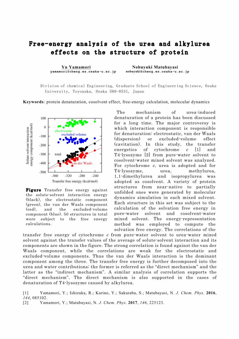
The mechanism of urea-induced denaturation of a protein has been discussed for a long time. The major controversy is which interaction component is responsible for denaturation: electrostatic, van der Waals (dispersion) or excluded-volume effect (cavitation). In this study, the transfer energetics of cytochrome c [1] and T4-lysozyme [2] from pure-water solvent to cosolvent-water mixed solvent was analyzed. For cytochrome c, urea is adopted and for T4-lysozyme, urea, methylurea, 1,1-dimethylurea and isopropylurea was adopted as cosolvent. A variety of protein structures from near-native to partially unfolded ones were generated by molecular dynamics simulation in each mixed solvent. Each structure in this set was subject to the calculation of the solvation free energy in pure-water solvent and cosolvent-water mixed solvent. The energy-representation method was employed to compute the solvation free energy. The correlations of the
transfer free energy of cytochrome c from pure-water solvent to urea-water mixed solvent against the transfer values of the average of solute-solvent interaction and its components are shown in the figure. The strong correlation is found against the van der Waals component, while the correlations are weak for the electrostatic and excluded-volume components. Thus the van der Waals interaction is the dominant component among the three. The transfer free energy is further decomposed into the urea and water contributions; the former is referred as the “direct mechanism” and the latter as the “indirect mechanism”. A similar analysis of correlation supports the “direct mechanism”. The direct mechanism is also supported in the cases of denaturation of T4-lysozyme caused by alkylurea. [1] Yamamori, Y.; Ishizuka, R.; Karino, Y.; Sakuraba, S.; Matubayasi, N. J. Chem. Phys. 2016, 144, 085102. [2] Yamamori, Y.; Matubayasi, N. J. Chem. Phys. 2017, 146, 225123.
-500
-400
-300
-200
-100
0
100
-360 -320 -280 -240
So
lute
-so
lven
t in
tera
ctio
n
en
erg
y c
om
po
nen
t (k
cal/
mo
l)
Transfer free energy (kcal/mol)
van der Waals
total
excluded volumeelectrostatic
F i g u r e Transfer free energy against the solute-solvent interaction energy (black), the electrostatic component (green), the van der Waals component (red), and the excluded-volume component (blue). 50 structures in total were subject to the free energy calculations.
Efficient calculation of electrostatic potential of
biomolecule based on fragment molecular orbital method
Takeshi Ishikawa1
1 Department of Molecular Microbiology and Immunology, Graduate School of Biomedical Science,
Nagasaki University, 1-12-4 Sakamoto, Nagasaki, 852-8523, Japan
Keywords: electrostatic potential, fragment molecular orbital method, point-charge approximation
Accurate calculation of electrostatic potential (ESP) is one of the important issues in the
computational chemistry and biology. For example, ESP at the molecular surface plays a key role in the
protein-protein or protein-ligand interaction, and is also responsible for the catalytic activity of many
enzymes. In this study, efficient quantum chemical calculations of the ESP were performed based on
fragment molecular orbital (FMO) method [1]. In FMO method, ESP at a position rm is defined as the
following equation:
(1)
where I and J are indices of the fragments, and α is index of the atom. The φI is a direct contribution
from the monomer, and ΔφIJ is a two-body correction from the dimer. They are calculated as
(2)
where μ and ν are indices of the basis functions. The matrix DI is the density matrix of the monomer,
ΔDIJ is the difference density matrix [2], and uμν is a one-electron integral like a nuclear attraction
integral. The numerical errors of the ESP associated with FMO scheme were examined at HF, MP2, and
RI-MP2[3,4] levels of theory. As a result, the FMO errors in ESP were significantly smaller than the
amplitude of the electron correlation effect, indicating that the FMO method provides sufficiently
accurate electrostatic properties for chemical and biological researches.
Additionally, an attempt to reduce the computational effort was proposed by combining the FMO
scheme and a point-charge approximation. When the distance between a position rm and monomer or
dimer is sufficiently large, the monomer contribution and two-body correction in equations (2) are
evaluated using a point-charge approximation as the following equations:
(3)
where qαI and Δqα
IJ are a monomer contribution and a two-body correction to the atomic point-charge
of the electron, respectively. The error of this approximation was examined using two proteins, prion
protein and human immunodeficiency type 1 protease. Finally, as an illustrative example, ESP at the
molecular surface of these proteins at MP2 level of theory were performed. In this study, PAICS
program package[5] was used for all the calculations.
[1] K. Kitaura, et al, Chem. Phys. Lett., 313 (1999) 701
[2] T. Nakano, et al, Chem. Phys. Lett., 351 (2002) 475
[3] T. Ishikawa and K. Kuwata, Chem. Phys. Lett., 474 (2009) 195
[4] T. Ishikawa and K. Kuwata, J. Phys. Chem. Lett., 3 (2012) 375
[5] T. Ishikawa, et al, J. Comput. Chem., 30 (2009) 2594
Mechanism analysis of estrogen receptor beta selective
agonist by molecular dynamics and fragment molecular
orbital method
Kensuke Misawa1 Tadahiro Ozawa
1
[email protected] [email protected]
Yoshiya Sugai1
Taketoshi Fujimori1
[email protected] [email protected]
Takatsugu Hirokawa2,3
1
Biological Science Laboratories, Kao Corporation, 2606 Akabane, Ichikai -machi, Haga-gun,
Tochigi 321-3497, Japan 2
Molecular Profiling Research Center for Drug Discovery (molprof), National Institute of
Advanced Industrial Science and Technology (AIST), 2 -4-7 Aomi, Koto-ku, Tokyo 135-0064,
Japan 3
Division of Biomedical Science, Faculty of Medicine, University of Tsukuba, 1-1-1 Tennodai,
Tsukuba, Ibaraki 305-8575, Japan
Keywords: Estrogen receptor, Ligand docking, Molecular dynamics, Fragment molecular orbital
Estrogen receptors (ERs) are nuclear transcription factors that play an important role in the
regulation of various physiological processes in humans and located in the various tissues such as
breast, bone and skin[1]-[2]
. There are two major subtypes of ERs: estrogen receptor alpha (ERα)
and estrogen receptor beta (ERβ) [1]
. Estradiol binds both the ERs equally, but this often leads to an
increased risk of breast and endometrial cancers because of ERα agonism[2]-[3]
. Therefore, many
ERβ selective agonists have been developed so far [4]
.
We focused on ERβ selective agonist meso-2,3-bis(4-hydroxyphenyl)succinonitrile[5]
and
analysed the mechanism of ERβ selectivity by the combination of induced-fit docking, molecular
dynamics and fragment molecular orbital. The details will be argued on the day.
[1] Paterni, I., Granchi, C., Katzenellenbogen, J. A., and Minutolo, F., Estrogen receptors alpha
(ERα) and beta (ERβ): Subtype-selective ligands and clinical potential, Steroids, 90:13-29,
2014.
[2] Eyster, K. M., The Estrogen Receptors: Methods and Protocols, Humana Press, 2016.
[3] Jordan V. C., The secrets of selective estrogen receptor modulation: cell -specific coregulation,
Cancer Cell, 1:215-217, 2002.
[4] Mohler, M. L. et al., Estrogen receptor β selective nonsteroidal estrogens: seeking cl inical
indications, Expert Opinion on Therapeutic Patents, 20:507-534, 2010.
[5] Meyers, M. J. et al., Estrogen Receptor-β Potency-Selective Ligands: Structure-Activity
Relationship Studies of Diarylpropionitriles and Their Acetylene and Polar Analogues , J. Med.
Chem., 44:4230-4251, 2001.
Study of BACE1/2 selectivity mechanism for highly
selective inhibitor using metadynamics
Naoya Asada1 Kazunari Hattori
1
[email protected] [email protected]
Ken-ichi Kusakabe1
1
Shionogi Pharmaceutical Research Center, SHIONOGI & CO., LTD., 3-1-1,
Futaba-cho, Toyonaka-shi, Osaka 561-0825, Japan
Keywords: Selectivity, Molecular Dynamics, Metadynamics, Mutation analysis
BACE1 (beta-site APP cleaving enzyme 1) is one of the aspartic proteases and protein targets for
Alzheimer’s disease. Selectivity against BACE2 is desired for BACE1 inhibitor to avoid potential
side effects. Highly selective inhibitor of BACE1 was reported [1], however the mechanism of
selectivity has been unknown. We performed metadynamics simulation using Desmond [2] for the
elucidation of the selectivity mechanism of this compound.
It was noted that a shift of flap loop region, which is a part of the binding pocket, to a more open
orientation was occurred when the selective inhibitor bound [1] and comparison of X-ray crystal
structures between BACE1 and BACE2 revealed that the flap orientation was different between
them. Based on this information, we hypothesized that flap motion got involved in the selectivity.
To validate this hypothesis, metadynamics simulation was performed with the setting of collective
variables as the distance between catalytic region and the flap for the calculation of Free Energy
Surface (FES). As a result, it was observed that FES was different between two proteins. In
BACE1, free energy kept stable from close to open orientation of the flap, on the other hand, free
energy became high when the flap was open in BACE2. This result showed that BACE2 was more
unstable than BACE1 when the selective inhibitor bound with the flap opening and this could be
the main factor for BACE1/2 selectivity. We also performed protein mutation analysis in virtual
model for identifying amino acid residues responsible for the difference of FES. On the flap region,
four residues are different between BACE1 and BACE2 (BACE1/BACE2: Y68/T84, P70/K86,
K75/S91, E77/T93). We made four BACE2 models which mutated one of the four residues to that
correspond to BACE1 and FES was calculated using metadynamics simulation for each model.
Finally, we found that FES of two mutation models (T84Y and S91K) showed similar results with
those of BACE1, which indicated that these two residues might contribute to the difference of the
flap motion.
[1] Michael S. Malamas, Keith Barnes, Matthew Johnson, Yu Hui, Ping Zhou, Jim Turner, Yun Huc,
Erik Wagner, Kristi Fan, Rajiv Chopra, Andrea Olland, Jonathan Bard, Men elas Pangalos, Peter
Reinhart, Albert J. Robichaud, Bioorg. Med. Chem. 2010, 18, 630–639.
[2] Desmond Molecular Dynamics System, D. E. Shaw Research, New York, NY, 2016.
Maestro-Desmond Interoperability Tools, Schrödinger, New York, NY, 2016.
Comparing two molecular dynamics simulation trajectories in terms of residue-residue interaction
Chie Motono Takatsugu Hirokawa [email protected] [email protected]
Molecular Profiling Research Center for Drug Discovery (molprof), National Institute of Advanced Industrial Science and Technology (AIST), 2-4-7 Aomi, Koto-ku, Tokyo, 135-0064, Japan
Keywords: molecular dynamics simulation, Protein, Mutation, residue-residue interaction. Molecular Dynamics (MD) simulations provide hi-resolution information on biomolecular dynamics. It has been used to elucidate the effects of mutations to a protein molecule, or binding/dissociation of chemicals to biomolecules on the motion and function of the molecules at a atomic level.
It is non-trivial a task to compare the two MD trajectories with different conditions (ex. with or without mutations, binding other molecules) and to detect the fundamental differences. As initial and intuitive analyses, time evolutions of root mean square deviations (RMSD), root mean square fluctuations (RMSF) of each position, and average structures of each trajectory are often compared. To observe statistical differences in molecular motions, Principal Component Analysis (PCA) is used.1 It was proposed to use a more sophisticated technique, Partial Least Square for Discriminant Analysis (PLS-DA) for comparison of two MD trajectories.2 These statistical methods suffer from the anisotropy of the molecular structures. Linear Discriminant Analysis with ITERative procedure (LDA-ITER) is an excellent method which maximizes the ratio of the between-ensemble fluctuation compared to the within-ensemble fluctuation to diminish the anisotropy effect.3
In this study, we propose a new approach to compare the fluctuations of residue-residue interactions of two MD trajectories. Identification of the positions where the dynamics of residue-residue interactions differs between two trajectories leads to the determination of the cause of different molecular motions or functions. This method indexes the residues with which a residue interacts and the durability of the interactions within a trajectory, and then compare the index with that in another trajectory to give a similarity. We applied the procedure to compare the trajectories of a wild-type protein and its mutant, and identify the most affected positions. This method is versatile and applicable to any different conditions like mutations, binding of other molecules, or unfolding, etc. [1] Kitao, A. and Go,N., Investigating protein dynamics in collective coordinate space, Current
Opinion in Structural Biology, 9:164-169, 1999. [2] Peters, J. H. and de Groot, B. L., Investigating protein dynamics in collective coordinate space,
PLoS Comput. Biol., 8: e1002704, 2012. [3] Sakuraba, S. and Kono, H., Spotting the difference in molecular dynamics simulations of
biomolecules, J. Chem. Phys., 145074:116, 2016.
An integrated workflow for accurately predicting standard
free energy of binding
Yoshiaki Tanida [email protected]
Azuma Matsuura [email protected]
Fujitsu Laboratories Ltd., Atsugi, Kanagawa 243-0197, Japan
Keywords: standard free energy, alchemical free energy calculation, metadynamics, cluster
analysis, reweighting
To realize computational “de novo” drug design (e.g. w/o structural information in
ligand-receptor complex), we propose a new strategy of binding affinity evaluation and binding
structure search in combination.
(i) Well-tempered metadynamics is used for exploring the available binding structures of new
drug-like molecule to a target in a suitable few collective variables space.
(ii) Some “pose” of the ligand providing local free energy minima is obtained by cluster
analysis and reweighting.
(iii) “Local” standard free energies can be estimated by alchemical free energy calculations;
the standard free energy ligand binding is obtained.
We also introduce the application of this procedure in some typical case. We believe that our
workflow described here will become a promising way in rational drug discovery.
An efficient approach for finding fragment-binding
conformations
Hiroyuki Sato1 Yoshiaki Tanida1 [email protected] [email protected]
Azuma Matsuura1 [email protected]
1 Fujitsu Laboratories Ltd., 10-1 Morinosato-Wakamiya, Atsugi 243-0197, Japan
Keywords: Protein pocket, Fragment molecule, Binding conformation, Non-bonding potential
We present a novel and efficient approach for finding various fragment-binding conformations to
a protein using molecular dynamics (MD) simulation. In general, a long-time MD simulation is
required to get possible binding variations in the conventional approaches[1]. Our approach adopts
randomly exchangeable protein replicas with high concentration of fragment molecules whose
non-bonding potential functions are different from each other. We applied this approach to human
coagulation factor Xa[2] with 4 M concentration of 6-chloro-1-benzothiophene-2-ol, and found
that fragment molecules bound to protein pocket more than 60 times during a 10-ns MD simulation.
This indicates that our approach provides a sufficient number of fragment-binding conformations
through a short-time MD simulation.
[1] Guvench, O., et al., PLoS Comput. Biol. 2009, 5, e1000435.
[2] Maignan, S., et al., J. Med. Chem. 2003, 46, 685-690.
Computational Study on Structure-Activity Relationship in FAS Inhibitors Based on Three-Dimensional Electronic
Similarity
Takafumi Inoue1 Toshihiro Ideo1 [email protected] [email protected]
Manabu Sugimoto1,2,3
1 Graduate School of Science and Technology, Kumamoto University, 2-39-1 Kurokami, Chuo-ku, Kumamoto 860-8555, Japan
2 Faculty of Advanced Science and Technology, Kumamoto University, 2-39-1 Kurokami,
Chuo-ku, Kumamoto 860-8555, Japan 3 Research Center for Advanced Science and Technology (RCAST), University of Tokyo, 4-6-1
Komaba, Meguro-ku, Tokyo, Japan Keywords: Structure-activity relationship, FAS Inhibitor, Electronic similarity The fatty acid synthase (FAS) inhibitor is expected to work as an anti-cancer drug. Several candidates as the FAS inhibitor have experimentally suggested by Wang et al. [1] and Li et al. [2-3]. In order to investigate the structure-activity relationship of these molecules, we are developing a computational method evaluating three-dimensional topology similarity where the topology is obtained by using electronic-structure calculations. We call this similarity “three-dimensional electronic-shape similarity (3D-ESS)”. Herein we focus on electro-static potential (ESP) as an example of 3D-ESS. In the presentation, we will introduce a program for scoring ESP similarity among the compounds. We will discuss correlation between the similarity score and IC50 for the FAS inhibitors through evaluation of 3D-ESS. The combined use of 3D-ESS with energy-based electronic- similarity (EB-ES) will also be discussed. [1] Wang, X., Song, K. S., Guo, Q. X., and Tian, W. X., The galloyl moiety of green tea catechins
is the critical structural feature to inhibit fatty acid synthase, Biochem. Pharma., 66: 2039-2047, 2003.
[2] Li , B. H. , and Tian, W. X. , Inhibitory effects of flavonoids animal fatty acid synthase, J. Biochem., 135: 85-91, 2004.
[3] Li, B. H., Ma, X. F., Wang, Y., and Tian, W. X., Structure-activity relationship of polyphenols that inhibit fatty acid synthase, J. Biochem., 138: 679-685, 2005.
Analysis of Ras/Raf-RBD Complex by Molecular Dynamics Simulation
Shota Matsunaga Shigenori Tanaka [email protected] [email protected]
Department of Computational Science, Graduate School of System Informatics, Kobe University, 1-1, Rokkodai, Nada-ku, Kobe, Hyogo
657-8501, Japan
Keywords: Ras/Raf-RBD complex, cancer, molecular dynamics (MD) method, GTPase-activating proteins (GAPs)
The Ras/Raf/MEK/ERK signal transduction pathway plays an important role in controlling cell proliferation and differentiation[1].
Abnormal activation of Ras or Raf genes has frequently been reported in human cancer[2]. In this study, to elucidate the mechanism
of the Ras/Raf interaction, we investigate the interconversion between two conformational states, State 1 and State 2[3], of the
Ras/Raf-RBD complex using the molecular dynamics (MD) method. In particular, we analyze the two switch regions (Switch I
and Switch II) of both GTP-bound form and GDP-bound form. To accelerate the conformational change of GDP-bound form, the
GDP molecule was given the thermal energy generated when the GTP molecule was hydrolyzed by GTPase-activating proteins
(GAPs) [4].
As a result of calculating dT (the distance between the oxygen atom of Thr35 of Ras and either the γ-phosphorus atom of GTP or
the β-phosphorus atom of GDP) and dY (the oxygen atom of Tyr64 of Ras and either the γ-phosphorus atom of GTP or the β-
phosphorus atom of GDP) [5], it was found that Switch II is repeatedly opened and closed because of the allosteric property of binding
in the standard MD simulation. On the other hand, we found that the thermal energy from hydrolysis of GTP is important for the
Switch I to be opened sufficiently. From the results of the simulations with and without the added energy, we succeeded to obtain
the comprehensive pictures of the signal transduction in the Ras/Raf-RBD complex.
References
[1] S. K. Fetics, H. Guterres, B. M. Kearney, G. Buhrman, B. Ma, R. Nussinov, C. Mattos, Allosteric Effects of the oncogenic
RasQ61L mutant on Raf-RBD, Struct. Des., 23: 3, 2015.
[2] L. Santarpia, S. L. Lippman, A. K. EI-Naggar, Targeting the mitogen-activated protein kinase Ras-Raf signaling pathway in
cancer therapy, NIH Pub., 16: 1, 2012
[3] S. Matsumoto, N. Miyano, S. Baba, J. Liao, T. Kawamura, C. Tsuda, A. Takeda, M. Yamamoto, T. Kumasaka, T. Kataoka, F.
Shima, Molecular mechanism for conformational dynamics of Ras·GTP elucidated from In-Situ structural transition in crystal, Sci.
Rep., 6: 25931, 2016.
[4] K. Ogata, J. Shen, S. Sugawa, S. Nakamura, MD simulation study of Ras/Raf dissociation and the resonating structure of
deactivated Ras, Chem. Soc., 85: 12, 2012.
[5] S. Matsunaga, Y. Hano, Y. Saito, K. Fujimoto, T. Kumasaka, S. Matsumoto, T. Kataoka, F. Shima, S. Tanaka, Structural
transition of solvated H-Ras/GTP revealed by molecular dynamics simulation and local network entropy, J. Mol. Graph. Model.,
2017, in press.
Energy correlation of protein at equilibrium fluctuation and its connection with ligand binding
Hikaru Iba1 Yuu Ymamori1
[email protected] [email protected]
Nobuyuki Matubayasi1 [email protected]
1 Division of Chemical Engineering, Graduate School of Engineering Science, Osaka University, Toyonaka, Osaka 560-8531, Japan
Keywords: Molecular dynamics simulation, Ligand binding, Energy correlation
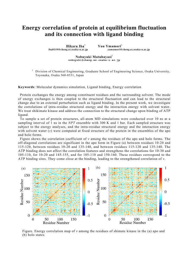
Protein exchanges the energy among constituent residues and the surrounding solvent. The mode of energy exchanges is then coupled to the structural fluctuation and can lead to the structural change due to an external perturbation such as ligand binding. In the present work, we investigate the correlations of intra-residue structural energy and the interaction energy with solvent water. We treat shikimate kinase and address the connection to the structural change upon binding of ATP ligand.
To sample a set of protein structures, all-atom MD simulations were conducted over 10 ns at a sampling interval of 1 ns in the NPT ensemble with 300 K and 1 bar. Each sampled structure was subject to the energy analyses, and the intra-residue structural energy and the interaction energy with solvent water (v) were computed at fixed structure of the protein in the ensembles of the apo and holo forms.
Figure shows the correlation coefficient of v among the residues of the apo and holo forms. The off-diagonal correlations are significant in the apo form in Figure (a) between residues 10-20 and 115-120, between residues 10-20 and 135-140, and between residues 115-120 and 135-140. The ATP binding does not affect the correlation features and strengthens the correlations for 10-30 and 105-110, for 10-20 and 145-155, and for 105-110 and 150-160. These residues correspond to the ATP binding sites. They come close at the binding, leading to the strengthened correlation of v.
(a)
Figure. Energy correlation map of v among the residues of shimate kinase in the (a) apo and (b) holo states.
(b)
In silico binding affinity analysis for
phosphodiesterase-10A inhibitors.
Chisa Yuasa1 Toru Ekimoto1 [email protected] [email protected]
Tsutomu Yamane1 Mitsunori Ikeguchi1
[email protected] [email protected]
1 Graduate School of Medical Life Science, Yokohama City University, 1-7-29
Suehiro-cho, Tsurumi-ku, Yokohama-shi, Kanagawa, Japan
Keywords: Ligand docking, Binding free energy calculation, Molecular Dynamics simulation
Phosphodiesterase (PDE) is an enzyme that hydrolyzes cyclic-AMP (c-AMP) and cyclic-GMP (c-GMP), which play important roles in signal transduction. PDE10A specifically expresses in brains, and PDE10A is considered as a drug target of schizophrenia. In order to understand how the drug activity relates to interactions manner between PDE10A and ligand, we performed docking simulations and calculated the binding free energies between PDE10A and 24 ligands using 4 approaches: the Glide docking score, MM-GBSA, FEP+ and MP-CAFEE method. The calculated binding free energies were compared with experimental IC 50. Although no correlation between the Glide docking score and experimental IC 50 was observed, the binding free energies calculated by MM-GBSA and FEP+ were correlated with IC50. In addition, we researched the docking-pose dependence of the binding free energy calculations. We examined effects of water molecules in the binding site on the docking poses. Inclusion of water molecules improved the correlation between the calculated binding free energy and IC50. FEP+ showed the best performance among the methods considered here. From these examinations, we found two important points for the high correlation: precise docking poses of ligands and account of hydration water molecules in binding free energy calculations. [1] J. Kunitomo, M. Yoshikawa,
M. Fushimi,
A. Kawada, J F. Quinn, H Oki,
H. Kokubo,
M.
Kondo, K. Nakashima,
N. Kamiguchi,
K. Suzuki,
H. Kimura,
and T. Taniguchi,
Discovery of 1‐[2-Fluoro-4-(1H‐pyrazol-1-yl)phenyl]-5-methoxy-3-(1- phenyl‐1H‐pyrazol-5-yl)pyridazin-4(1H)‐one (TAK-063), a Highly Potent, Selective, and Orally Active Phosphodiesterase 10A (PDE10A) Inhibitor . J. Med. Chem. 2014, 57, 9627−9643 [2] L. Wang, B. J. Berne and A. Friesner On achieving high accuracy and reliability in the calculation of relative protein–ligand binding affinities. Proc. Natl. Acad. Sci. USA 2012, 109, 1937-1942 [3] E. Harder, W. Damm, J. Maple, C. Wu, M. Reboul, J. Y. Xiang, L. Wang, D. Lu pyan, M. K. Dahlgren, J. L. Knight, J. W. Kaus, D. S. Cerutti, G. Krilov, W. L. Jorgensen, R. Abel, and R. A. Friesner, OPLS3: A Force Field Providing Broad Coverage of Drug-like Small Molecules and Proteins. J. Chem Theory Comput. 2016, 12, 281-296
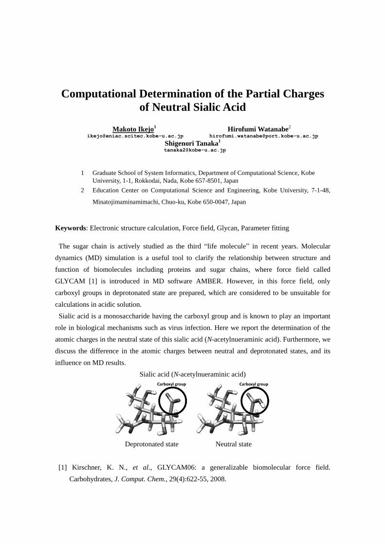
Computational Determination of the Partial Charges
of Neutral Sialic Acid
Makoto Ikejo1 Hirofumi Watanabe
2
[email protected] [email protected]
Shigenori Tanaka1
1 Graduate School of System Informatics, Department of Computational Science, Kobe
University, 1-1, Rokkodai, Nada, Kobe 657-8501, Japan
2 Education Center on Computational Science and Engineering, Kobe University, 7-1-48,
Minatojimaminamimachi, Chuo-ku, Kobe 650-0047, Japan
Keywords: Electronic structure calculation, Force field, Glycan, Parameter fitting
The sugar chain is actively studied as the third “life molecule” in recent years. Molecular
dynamics (MD) simulation is a useful tool to clarify the relationship between structure and
function of biomolecules including proteins and sugar chains, where force field called
GLYCAM [1] is introduced in MD software AMBER. However, in this force field, only
carboxyl groups in deprotonated state are prepared, which are considered to be unsuitable for
calculations in acidic solution.
Sialic acid is a monosaccharide having the carboxyl group and is known to play an important
role in biological mechanisms such as virus infection. Here we report the determination of the
atomic charges in the neutral state of this sialic acid (N-acetylnueraminic acid). Furthermore, we
discuss the difference in the atomic charges between neutral and deprotonated states, and its
influence on MD results.
Sialic acid (N-acetylnueraminic acid)
Deprotonated state Neutral state
[1] Kirschner, K. N., et al., GLYCAM06: a generalizable biomolecular force field.
Carbohydrates, J. Comput. Chem., 29(4):622-55, 2008.
Time-dependent Variance-covariance Matrix Analysis of
Molecular Dynamics for Protein-ligand Interaction
Masanori Yamanaka1 [email protected]
1 Department of Physics, College of Science and Technology, Nihon University, 1-8
Kanda-Surugadai, Chiyoda-ku, Tokyo 101-8308, Japan
Keywords: Molecular dynamics, Protein ligand interaction, Variance-covariance matrix, Random
matrix, Cluster analysis
We study the protein-ligand interaction using the characteristic scale of coupling formation [1]
developed in random matrix theory. The eigensystems of the timescale -dependent
variance-covariance matrix, which are obtained from the time series data of the atomic coordinates
of a protein produced by the all-atom molecular dynamics of the solvent, are analyzed. [2] As an
example, we present a result for a protein, N-terminal PDZ domain of ZASP in complex with
myotilin C-terminal peptide, PDBID:4YDP, which has a ligand of peptide with three amino acids.
We find that there are at least four different time scales involved in the coupling formation of
correlated sectors of atoms and an oscillation of the eigenvalue as a function of the sampling
interval of the variance-covariance matrices. These time scales and the oscillation behavior coexist
simultaneously. From the study of the eigenvector components, the weights of the ligand atoms are
dominant in a small time scale. We further compare the results with those without ligand and with
the dataset which we artificially displace the coordinates of the ligand.
[1] For example see a review article, J. Kwapień and S. Drożdż, Phys. Rep. 515, 115 (2012).
[2] M. Yamanaka, J. Phys. Soc. Jpn. 84, 063801 (2015).
In silico protein-protein interaction analysis of axon
guidance molecule semaphorin and receptor plexin
Erena Shimoji1 Tsutomu Yamane1 [email protected] [email protected]
Toru Ekimoto1 Nogi Terukazu1 [email protected] [email protected]
Mitsunori Ikeguchi1 [email protected]
1 Graduate School of Medical Life Science, Yokohama City University, 1-7-29 Suehiro-cho,
Tsurumi-ku, Yokohama, Kanagawa 230-0045, Japan
Keywords: Semaphorin, Guidance molecule, Protein-protein interaction
Semaphorin has been known as an axon guidance molecule, secreted around cells.
Plexin is a receptor of semaphorin, and their interactions are guidance cues for axons of
neurons during development of the nervous system. Because the protein-protein
interactions (PPI) of semaphorin and plexin have wide interaction surfaces, it is not
trivial to understand the determinants of PPI. The surface of protein is too shallow and
wide to bind small compound, so it is difficult to discover drug for PPI target. It is
important to understand the mechanism to control PPI for drug discovery. Semaphorin
and plexin include “Sema domain” with high sequence similarity, and they form PPI and
interact through the sema domain. Therefore, we analyzed the PPI using in silico
approaches to recognize the feature of specific interactions. More specifically, we used
program Rosetta to calculate the binding free energy and to perform in-silico alanine
scanning [1,2], and identified the “hotspots” which are significant amino acids in
binding. Then, the details of the hotspot interactions were analyzed to understand the
determinants of PPI.
[1] T. Kortemme and D. Baker, A simple physical model for binding energy hot spots in
protein–protein complexes. Proc. Natl. Acad. Sci. 99, 14116-14121, 2002.
[2] T. Kortemme, D. E. Kim, and D. Baker, Computational Alanine Scanning of Protein-Protein
Interfaces. Sci. STKE 10, 2004.
[3] B. J. C. Janssen, R. A. Robinson, F. Perez-Brangul , C. H. Bell, K. J. Mitchell, C. Siebold & E.
Y. Jones, Structural basis of semaphorin–plexin signaling. Nature 467 1118-1122, 2010.
Characterization of TCR-pMHC interaction at
residue-level based on FMO and PIEDA Hiromichi Tsurui1 Yuko Tsuchiya2
[email protected] [email protected]
Yoshiki Namiuchi3 Hiroshi Wako4
[email protected] [email protected]
1 Department of Pathology, Juntendo University School of Medicine, Bunkyo-ku, Tokyo, Japan 2 Institute for Protein Research, Osaka University, Suita, Osaka, Japan 3 RIKEN Quantitative Biology Center, Suita, Osaka, Japan 4 School of Social Sciences, Waseda University, Shinjuku-ku, Tokyo, Japan
Keywords: TCR-pMHC, FMO, PIEDA, CH-, hydrogen bond,
Interaction between T cell receptor (TCR) and peptide-MHC (pMHC) is essential for determining T cell fate and immunological response. However, determining actual binding profile of TCR by experiment requires vast amount of labor and by computer simulation is still a challenge. To investigate what interactions determine binding profile of TCR, we examined TCR-pMHC complexes having common pMHC and different TCRs with variety of binding profile, namely narrow (PDBID: 3c5z, 3rdt, 4p5t), modestly (3c6l), and highly cross-reactive (3c60). TCRs with specific profile formed more hydrogen bonds (about 10) between CDRs to presented peptide than that with cross-reactive profile (about 6 to 7), suggesting the contribution of hydrogen bond to the specific interaction. CH-p interaction seemed between CDR3 aromatic residues and MHC helix in both complexes containing specific and cross-reactive TCRs. Both hydrogen and CH-p bonds are principally determined geometrically and actual interaction-energies are obscure. We examined those bonds with FMO calculation at MP2/6-31G* level and PIEDA.
A QM/MM study on binding affinity computation of tankyrase 2-ligand system
Yoshinori Hirano1 Takao Otsuka1 [email protected] [email protected]
Noriaki Okimoto1 Makoto Taiji1 [email protected] [email protected]
1 QBiC, RIKEN, QBiC Building B, 6-2-4 Furuedai, Suita, Osaka 565-0874, Japan Keywords: Binding affinity computation, QM/MM method
Accurate binding affinity computation of protein-ligand system is of significant importance for the drug discovery. There are various computational methods to predict the protein-ligand binding affinity. One of the most popular approach for protein-ligand binding affinity is the free energy calculation based on molecular dynamics (MD) simulation, such as the linear interaction energy method, the molecular mechanics/Poisson-Boltzmann and surface area (MM-PB/SA) method, the alchemical free energy calculation, and so on. Most of these methods use classical molecular mechanics (MM) force fields.
Protein-ligand interactions consist of various non-bonded interactions such as π-stacking, charge transfer, polarization effects, and dispersion interaction, as well as the usual and/or weak hydrogen bonds. Therefore, it is difficult to treat these interactions by the classical MM force fields. Quantum mechanical (QM) calculations can consider these non-bonded interactions. We have expected to improve the accuracy of protein-ligand binding affinity compared with that of the MM force fields. In our previous study, we focused on the evaluation of binding affinity through a full QM calculation and proposed the accurate and efficient computational scheme by using multilayer fragment molecular orbital (MFMO) method1.
In this study, we explore a computational-cost effective scheme to calculate protein-ligand binding affinity by using QM/MM method. One of the key points is how the size of the QM region influences the performance of binding affinity prediction. We will report the binding affinities of tankyrase 2-ligand systems using QM/MM method2-4 with some different QM regions.
[1] Otsuka, T.; Okimoto, N.; Taiji, M., J. Comput. Chem., 2015, 36, 2209-2218. [2] Schrödinger Release 2017-2: QSite, Schrödinger, LLC, New York, NY, 2017. [3] Murphy, R. B.; Philipp, D. M.; Friesner, R. A., "A mixed quantum mechanics/molecular mechanics (QM/MM) method for large-scale modeling of chemistry in protein environments," J. Comp. Chem., 2000, 21, 1442-1457 [4] Philipp, D. M.; Friesner, R. A., "Mixed ab initio QM/MM modeling using frozen orbitals and tests with alanine dipeptide and tetrapeptide," J. Comput. Chem., 1999, 20, 1468-1494
The distinct dynamic mechanisms of peptide recognition by specific and cross-reactive T-cell receptors
Yuko Tsuchiya1 Yoshiki Namiuchi2 [email protected] [email protected]
Hiroshi Wako3 Hiromichi Tsurui4
[email protected] [email protected]
1 Institute for Protein Research, Osaka University, 3-2 Yamadaoka, Suita, Osaka 565-0871, Japan 2 RIKEN Quantitative Biology Center, 6-2-3 Furuedai, Suita, Osaka 565-0874, Japan 3 School of Social Sciences, Waseda University, 1-6-1 Nishi-Waseda, Shinjuku-ku, Tokyo
169-8050, Japan 4 Department of Pathology, Juntendo University School of Medicine, 2-1-1 Hongo, Bunkyo-ku,
Tokyo 113-8421, Japan Keywords: Cross-reactive TCR recognition, binding affinity-interface area relationship, Molecular dynamics (MD) simulation, Fragment molecular orbital (FMO) method Some T-cell receptors (TCRs) recognize an antigen peptide presented by Major Histocompatibility Complex (pMHC) specifically, whereas others recognize it cross-reactively. We focused on five TCRs in complex with the same class II MHC-peptide, three and two of which recognize the peptide specifically and cross-reactively, respectively. These complexes have similar binding affinities, whereas the interface areas between TCR and pMHC differ significantly. In this study, we investigated static and dynamic structural features of the TCR-pMHC complex and of TCR in free state, and the relationship between binding affinity and interface area. Our dynamics study revealed differences in the recognition mechanisms of TCRs, i.e., differences in interaction types and balances of enthalpy gain and entropy loss upon binding.
Interaction Analysis between Beta-Secretase and its Inhibitors
by Fragment Orbital Method
Norihito KAWASHITA1 Yuji HASHIMOTO2 Hirotomo MORIWAKI2 [email protected] [email protected] [email protected]
Yu-Shi TIAN2 Tatsuya TAKAGI2 [email protected] [email protected]
1 Faculty of Science and Engineering, Kindai University, 3-4-1 Kowakae, Higashiosaka City,
Osaka 577-8502, Japan 2 Graduate School of Pharmaceutical Sciences, Osaka University, 1-6 Yamadaoka, Suita, Osaka
565-0871, Japan
Keywords: Beta-Secretase, Fragment molecular orbital (FMO) method, Inter-fragment interaction
energy (IFIE)
Beta-Secretase (BACE) 1 is an aspartyl protease, it’s active site is made up of two aspartate
residues: Asp32 and Asp228. The β-hairpin loop over the active site, known as the “flap”, is also
an important feature to BACE1. While the active site remains inactive, the flap stays in its open
conformation. However, the flap is stabilized while closed over its subst rate or some inhibitor [1].
Currently, aminoquinolines, aminoisoquinolines, and aminopyrimidone with cyclic-amidine
structure were reported as potent fragment hits binding to BACE1 [2]. Since then, several studies
on similar inhibitors with cyclic-amidine structure have been reported.
In this study, we performed FMO calculation to examine the molecular interactions between
BACE1 and its ligands with cyclic-amidine structure for identified the key information for
screening of more potent inhibitors.
Cocrystal structures of ligands and BACE1 were obtained from PDB. MOE [3] was used for
structure preparation. MMFF94x force field was used for energy minimizations. FMO calculations
were carried out in ABINIT-MP (Ver. 6.0+) [4] with MP2/6-31G*.
From the results, we confirmed the correlation between IC50 and IFIE-SUM (IFIE between
ligand and receptor). The coefficient of determination R2 was 0.728, suggesting a good correlation.
Next, Asp28 and 228’s IFIE notably contributed to the stabilization of protein -ligand interaction.
And we detected tendency that ligands without aromaticity contributed to st abilization more than
ligands with aromaticity. Therefore, when we design BACE1 inhibitors with cyclic-amidine
structure, it is better that we choose structure without aromaticity.
In this study, we performed FMO method to examine the molecular int eractions between
BACE1 and its ligands. Not only good correlation between IC 50 and IFIE-SUM but also interaction
tendency in BACE1-ligand complex were obtained. Hereafter, we will execute further calculations
and more detailed IFIE analyses. Eventually, we aim at contribution to design novel inhibitors.
This research was done in activities of the FMO drug design consortium (FMODD). The results
were obtained using the K computer (project ID: hp150160, hp160103, and hp170183). PIEDA
calculation was done by using MIZUHO/BioStation software package.
[1] Lin et al., Science, 290: 150-153, 2008.
[2] Congreve et al., J. Med. Chem. 50: 1124-1132, 2007.
[3] Chemical Computing Group, MOE. 2015.
[4] Nakano et al., Chem. Phys. Lett. 351: 475-480, 2002.
In silico modeling of PAX8–PPARγ fusion protein with
unknown three-dimensional structure in follicular thyroid
adenoma and carcinoma
Kaori Sakaguchi1 Yoshio Okiyama2 [email protected] [email protected]
Shigenori Tanaka1 [email protected]
1 Department of Computational Science, Graduate School of System Informatics, Kobe
University, 1-1, Rokkodai, Nada, Kobe, Hyogo 657-8501, Japan 2 National Institute of Health Sciences, 1-18-1 Kamiyoga, Setagaya-ku, Tokyo 158-8501, Japan
Keywords: Follicular neoplasm, fusion protein, protein modeling, Molecular dynamics simulation
Follicular neoplasms including follicular thyroid adenoma (FTA) and follicular thyroid carcinoma
(FTC) have no molecular-targeted agents. Specifically in these tumor cells, a t(2,3) (q13,p25)
chromosomal translocation results in production of paired-box gene 8 (PAX8)-peroxisome
proliferator-activated receptor γ gene (PPARγ) fusion protein (PPFP). While the three-dimensional
(3D) structure of PPFP and its functions are not reported so far, a transgenic mouse model shows
that pioglitazone, an effective agonist of PPARγ, makes PPFP behave as PPARγ-like transcription
factor; it turns tumor cells into adipocytes and deprives their malignant characters [1]. Based on
this finding, it is possible to obtain more effective anticancer agents for FTA and FTC if we can
design compounds specifically bound to PPFP.
We here propose a 3D structure of PPFP for establishment of targeted therapies in FTA and FTC.
We constructed amino acid sequence of PPFP that was composed of PAX8 (Met1-Ala396) and
PPARγ (Met1-Tyr505) based on the currently proposed schematic diagram of PPFP [2]. In PDB,
there are X-ray crystal structure of PAX6 paired domain with DNA (PDB ID: 6PAX) having high
homology with PAX8 and the intact PPARγ on DNA (PDB ID: 3DZY). Using them, we assumed a
fusion construct containing both PAX8 and PPARγ on DNA, and predicted the structure of PAX8
and the fusing region by I-TASSER [3] online servers. After some modification and relaxation, the
final 3D structures were obtained in this study. To validate the predicted models, we analyzed their
physicochemical characteristics, Ramachandran plot distributions and dynamical stabilities with
molecular dynamics simulation.
[1] B. Xu et al., J. Biol. Chem. 291, 19274-19286 (2016).
[2] P. Raman et al., Nat. Rev. Endocrinol. 10, 616-623 (2014).
[3] J. Yang et al., Nat. Methods. 12, 7-8 (2015).
Computational characterization of influenza A virus hemagglutinin recognition sites by a cross-neutralizing
monoclonal antibody
Manabu Igarashi1,2 Masakazu Sekijima3 Nobuaki Yasuo3 [email protected] [email protected] [email protected]
Takashi Abe4 Teruaki Watabe5 Rashid Manzoor1
[email protected] [email protected] [email protected]
Takatsugu Hirokawa6 Ayato Takada1,2 [email protected] [email protected]
1 Research Center for Zoonosis Control, Hokkaido University, Sapporo, Hokkaido, Japan 2 Global Station for Zoonosis Control, Global Institution for Collaborative Research and
Education (GI-CoRE), Hokkaido University, Sapporo, Hokkaido, Japan 3 Global Scientific Information and Computing Center, Tokyo Institute of Technology,
Meguro-ku, Tokyo, Japan 4 Department of Information Engineering, Niigata University, Niigata, Japan 5 Center of Medical Information Science, Kochi Medical School, Kochi University, Kochi, Japan 6 Molecular Profiling Research Center for Drug Discovery (molprof), National Institute of
Advanced Industrial Science and Technology (AIST), Koto-ku, Tokyo, Japan Keywords: Molecular dynamics, Influenza virus, Antibody, Protein modeling, Binding free energy Background The hemagglutinin (HA) of influenza A viruses is classified into 16 subtypes (H1-H16). It is generally known that HA-specific antibodies have little cross-neutralizing activity against multiple HA subtypes. Recently, however, several broadly neutralizing antibodies were reported and have attracted attention due to their potential application to therapeutics and vaccine design. We have previously reported a cross-reactive antibody, designated S139/1, which neutralizes H1, H2, H3, H13, and H16 subtypes and its crystal structure in complex with the HA of the A/Victoria/3/1975 (H3N2) strain. However, detailed structural basis of its cross-neutralizing activity still remain to be elucidated. In this study, we characterized the S139/1 recognition sites on different HAs using computational structural biology methods. Materials and Methods The HAs from eight strains (subtypes H1, H2, H3, H6, H9, and H13) were analyzed. The structure models of the HA-S139/1 complex were constructed by homology modeling. Using the structures as starting points, we performed molecular dynamics (MD) simulations, and then calculated binding free energies (ΔG) between S139/1 and each HA. Results and Discussions The ΔG values of the strains neutralized by S139/1 were lower than the other strains tested. We next investigated the contribution of individual residues on each HA to the interaction with S139/1 and found that amino acids at positions 98, 136, 153, 156, 158, 159, 193, 194, 196, and 226 (H3 numbering) on HA strongly contributed to S139/1 binding as for the strains neutralized by S139/1. Additionally, we found that, in the strains neutralized by S139/1, K156 formed a salt bridge with the Ig heavy chain E50. Indeed, amino acid substitutions at adjacent residues were experimentally observed in the mutant viruses escaping from neutralization by S139/1. Thus, our computational methods identified the amino acid residues critical for the cross-neutralizing activity of S139/1.
Characteristics of Biomolecule Dynamics under the
Crowding Environment of Cytoplasm Discovered by
Massive All-atom simulation and Big-data analysis
Isseki Yu1,2 Po-hung.wang2 [email protected] [email protected]
Michael Feig5 Yuji Sugita1,2,3,4 [email protected] [email protected]
1iTHES Research Group, RIKEN, Saitama, Japan 2Theoretical Molecular Science Laboratory, RIKEN, Saitama, Japan 3Laboratory for Biomolecular Function Simulation, RIKEN Quantitative Biology Center, Kobe, Japan 4Computational Biophysics Research Team, RIKEN Advanced Institute for Computational Sci ence, Kobe,
Japan 5Department of Biochemistry and Molecular Biology, Michigan State University, East Lansing, United
States
Keywords: Cellular environment, Molecular crowding, Protein diffusion, Metabolites
Inside of a cell is highly crowded with a large number of macromolecules together with solvents
and metabolites. How variable specific/non-specific interactions within dense cellular
environments may affect the structure and dynamics, and ultimately function is one of the most
fundamental questions in life science. We constructed full atomistic model of the cytoplasm of
bacteria and various levels of protein crowding models. Using these model, we performed massive
all-atom molecular dynamics (MD) simulation with the highly parallelized MD simulator
GENESIS on K-computer. Influence of crowding and non-specific interaction between
macromolecules on the translational/rotational /collective motion of proteins are analyzed.
Anomalous diffusion and the localization of metabolites, such as ATP and amino acids, on the
macromolecule surface are also investigated. These work is an important step towards physically
realistic in silico whole-cell models that connect molecular with cellular biology.
[1] Michael Feig, Isseki Yu, Po-Hung Wang, Grzegorz Nawrocki, Yuji Sugita, Crowding in Cellular
Environments at an Atomistic Level from Computer Simulations , Journal of Physical chemistry B (in
press)
[2] Po-hung Wang, Isseki Yu, Michael Feig, Yuji Sugita , Influence of Protein Crowder Size on
Hydration Structure and Dynamics in Macromolecular Crowding, Chemical Physics Letters 671, 63-70,
(2017)
[3] Isseki Yu, Takaharu Mori, Tadashi Ando, Ryuhei Harada, Jaewoon Jung, Michael Feig, Yuji
Sugita, Biomolecular interactions modulate macromolecular structure and dynamics in atomistic model
of a bacterial cytoplasm, eLife 2016;5:e19274
[4] Michael Feig, Ryuhei Harada, Takaharu Mori, Isseki Yu, Koichi Takahashi, Yuji Sugita , Complete
atomistic model of a bacterial cytoplasm for integratingphysics, biochemistry, and systems bio logy
Journal of Molecular Graphics and Modelling , 58, 1–9, (2015)
Molecular Dynamics Simulations of HIV Tat proteins and
Amyloid-β peptides
Kazumi Omata1 Satoru G. Itoh2 [email protected] [email protected]
Hisashi Okumura2,3 [email protected]
1 Department of Data Science, National Center for Global Health and Medicine, 1-21-1 Toyama,
Shinjuku, Tokyo 162-0052, Japan 2 Institute for Molecular Science, 38 Nishigonaka, Myodaiji, Okazaki, Aichi 444-8585, Japan 3 The Graduate University for Advanced Studies, 38 Nishigonaka, Myodaiji, Okazaki, Aichi
444-8585, Japan
Keywords: HIV Tat protein, Amyloid-β, Ligand docking, Molecular dynamics, neurocognitive
dysfunction
To identify binding sites of an HIV transactivator of transcription (Tat) protein and an amyloid-β
(Aβ) peptide, the present study has investigated their docking, using molecular dynamics (MD)
simulations.
Recently, the experiments by Hategan et al. suggested that HIV Tat binds to the exterior surfaces of
Aβ fibrils, increasing β-sheet formation and lateral aggregation into thick multifibrillar structures
[1]. As a result, the fibers increase their rigidity and mechanical resistance. They also showed that
the aggregate of Tat-Aβ complexes synergistically induced neurotoxicity both in vitro and in
animal models. Apart from such a study, Okumura and Itoh examined the structure of Aβ fibrils
by all-atom MD simulations, and demonstrated that structural fluctuations at the fibril ends play
an important role for the fibril formation [2]. Based on the knowledge about the structure of Aβ
fibrils derived from their MD simulations, the present study has attempted to find which parts of
Aβ peptides are tightly associated with HIV Tat proteins, using MD simulations.
The all-atom MD simulations were carried out using the Generalized -Ensemble Molecular
Biophysics (GEMB) program [3]. The AMBER99SB force field was used for the HIV Tat and Aβ,
and the TIP3P rigid-body model was used for the water molecules. The electrostatic potential was
calculated by the particle mesh Ewald method. Temperature was controlled at 298 K with the
Nosé-Hoover thermostat, and pressure was controlled at 0.1 MPa with the Andersen barostat. The
symplectic quaternion scheme was employed for the rigid-body water molecules.
The results of our MD simulations could be used for the inhibition of the Tat-Aβ complex
formation and the development of drugs for neurocognitive dysfunction of HIV-infected
individuals.
[1] Hategan, A., et al., HIV Tat protein and amyloid-β peptide form multifibrillar structures that cause
neurotoxicity. Nature Structural & Molecular Biology 24, 379–386, 2017.
[2] Okumura, H. and Itoh, S. G., Structural and fluctuational difference between two ends of
Aβamyloid fibril: MD simulations predict only one end has open conformations . Scientific
Reports 6, Article number:38422, 2016.
[3] Okumura, H., Temperature and pressure denaturat ion of chignolin: Folding and unfolding
simulation by multibaric-multithermal molecular dynamics method. Proteins, 80: 2397-2416,
2012.
An index to select appropriate homologous sequences for functional region
prediction of a protein
Shoichiro Kato1 Hiroyuki Toh2 [email protected] [email protected]
Wataru Nemoto1
1 Division of Life Science and Engineering, Graduate School of Science and Engineering,
Graduate School of Tokyo Denki University (TDU), JAPAN. 2 Department of Biomedical Chemis try, School of Science and Technology, Kwansei
Gakuin Univers i t y, JAPAN.
Keywords: sequence selection, phylogenetic tree, conservation score,
The conserved amino acid residues among homologous amino acid sequences are ofte n closely
located each other at and near the functional regions in the tertiary structures. Based on the
observations, several methods have been developed to predict functional regions, in which both
homologous amino acid sequences and tertiary structures are used for the analysis. We have also
proposed a method with the same idea [1]. Given a target protein for the prediction, homologous
sequences are collected under the condition that the sequence identity to the target protein is less
than or equal to a given threshold value. Then, a multiple sequence alignment is made for the
target protein and the collected sequences, and the conservation score is calculated at each
alignment site. Next, the conservation score of each alignment site is mapped on the cor responding
residue of a tertiary structure of one of the homologous proteins or the target protein. Finally,
clusters of conserved residues are detected as functional regions in the tertiary structure.
In this strategy, a set of homologous sequences to calculate conservation score is required to
contain the sequences with their functional regions at the corresponding sites on their structures. If
a set of homologous sequences includes the sequences with their functional regions at distinct
sites on their structures, it would be difficult to accurately evaluate residue conservation for this
strategy. Hence, we introduced a new index based on the spatial autocorrelation to evaluate the
spatial clustering of the conserved residues [1]. By changing the threshold value for the sequence
collection from 90 % to 10 % with an interval of 5 %, different sets of the homologous sequences
are generated, to each of which the new index is calculated. We selected a set of the homologous
sequences with the maximum value of the index as the most appropriate set for the prediction because the set is appropriate to evaluate the spatial clustering of the conserved residues . Our
method can not only predict the functional regions, but also determine the best set of the
homologous sequences for the prediction. Our method successfully improved the performance of functional region pred ictions [1]. As
suggested in our manuscript, however, the collection of the homologous sequences based on the
sequence identity to the target protein is too rough for such an approach, and the phylogenetic
relationship among the collected sequences should be taken into account to generate a set of the
homologous sequences for the prediction. In this work, hence, a set of homologous sequences was
constructed as a cluster corresponding to a subtree in the phylogenetic tree of the target and its
homologs. The performance of the prediction was evaluated by shifting the subtree from the
closely related node to the distantly related node. The experimentally identified catalytic sites
obtained in Catalytic Site Atlas was used for the performance evaluation. We will explain details
of the procedure of our method and discuss advantages and disadvantages of the new approach.
[1] Nemoto, W. & Toh, H. BMC Struct. Biol. 12, 11, 2012
Influence of Molecular Force Field on Performance of
Protein Ligand Docking
Daisuke Takaya1 Kikuko Kamisaka1 Shunpei Nagase1
[email protected] [email protected] [email protected]
Teruki Honma1 [email protected]
1 RIKEN Center for Life Science Technologies, 1-7-22 Suehiro-cho, Tsurumi, Yokohama
230-0045, Japan.
Keywords: SBDD, Protein-Ligand docking, Benchmark
Structure Based Drug Design (SBDD) is one of the most powerful approach when a target protein is
known and its 3D-coordinates are experimentally determined by X-ray crystal structure analysis or can
be constructed based on its template by using homology modeling. Since protein-ligand complex
structures are not always available in drug design, protein-ligand docking programs (e.g. AutoDock
Vina1, FRED2, Glide3, and so on) are used for ligand binding mode prediction. Moreover, the docking
programs are also useful for predicting its affinity as a docking score to the target protein, searching
putative inhibitors from compound database and discriminating between active and inactive
compounds. Docking programs are widely used for in silico screening by not only computational
chemists but also biological researchers.4,5
Generally, docking programs provide some parameters and conditions which influence the output such
as predicted binding modes and docking score of ligands. Therefore, it affects the prediction
performance of docking programs. For example, preprocess of the receptor structure such as addition
of hydrogen atoms, assignment of atom types of molecular force field, coefficients of interaction terms
of the score function, exploring methods for molecular conformation and position. Additionally, their
combination should be considered.
There are several approaches to handle the docking conditions. Firstly, the docking condition is
performed under default setting. Secondly, if there is data of known inhibitors for the target protein,
cross docking and test docking are performed for optimizing the conditions for the target. Finally, the
conditions are determined by visual inspection of the prediction results and experience of
computational researchers. It was thought that it would be useful for selecting docking setting if the
influence of the parameters and conditions were known in advance. In this study, we examined the
extent to which docking conditions affect the prediction performance by using benchmark sets. The
results will give guidance on whether the condition should be changed or not from the default setting.
[1] Trott, O. et al., AutoDock Vina: Improving the Speed and Accuracy of Docking with a New Scoring
Function, Efficient Optimization, and Multithreading J. Comput. Chem. 31:455-461, 2010
[2] McGann, M. FRED Pose Prediction and Virtual Screening Accuracy J. Chem. Inf. Model.,
51(3):578-596, 2011
[3] Friesner, R. A. et al., Glide: A New Approach for Rapid, Accurate Docking and Scoring. 1. Method
and Assessment of Docking Accuracy J. Med. Chem. 2004, 47, 1739-1749
[4] Takaya, D., et al., A new method for induced fit docking (GENIUS) and its application to virtual
screening of novel HCV NS3-4A protease inhibitors, Bioorg. Med. Chem. 19:6892–6905, 2011
[5] Tajiri, H., et al., Targeting Ras-Driven Cancer Cell Survival and Invasion through Selective
Inhibition of DOCK1,Cell Reports 19(5):969-980, 2017
Prediction of cancer-associated hotspot mutations that
affect GPCR oligomerization Wataru Nemoto1,2 Vachiranee Limviphuvadh3 Sebastian Maurer-Stroh3
[email protected] [email protected] [email protected]
Yoshihiro Yamanishi4 Motoaki Yamanoi2 Hiroyuki Toh5
[email protected] [email protected] [email protected]
1 Division of Life Science and Engineering, School of Science and Engineering, Tokyo Denki University, ishizaka, Hatoyama-machi, Saitama 350-0311, Japan
2 Division of Life Science and Engineering, Graduate School of Science and Engineering, Graduate School of Tokyo Denki University, ishizaka, Hatoyama-machi, Saitama 350-0311, Japan
3 Bioinformatics Institute (BII), Agency for Science, Technology and Research (A*STAR), 30 Biopolis Street, #07-01 Matrix, 138671, Singapore
4 Medical Institute of Bioregulation, Kyushu University, Fukuoka, 812-8581, Japan
5 Department of Biomedical Chemistry, School of Science and Technology, Kwansei Gakuin University, 2-1 Gakuen, Sanda-shi, Hyogo, 669-1337, Japan.
Keywords: membrane proteins, protein–protein interaction, interaction partner prediction, support vector machine, drug design; At present, approximately 30% of marketed pharmaceutical medicines target G Protein-Coupled Receptors (GPCRs). Numerous studies have reported that GPCRs function not only as monomers but also as homo- or hetero- dimers or higher-order molecular complexes (oligomers). Many GPCRs exert a wide variety of functions by forming specific combinations of GPCR subtypes. Several hetero GPCR hetero dimers are reportedly associated with diseases. Therefore, GPCR oligomerization is recognized as an important event in various biological phenomena, and many researchers are investigating this subject. Hence, we developed a method to predict interacting pairs for GPCR oligomerization by integrating the structure and sequence information (GGIP). The area under the Receiver Operating Characteristic curve was 0.938. GGIP is the only prediction method for interacting pairs among GPCRs. Our prediction server is available at http://protein.b.dendai.ac.jp/GGIP/. A recent study reported that somatic mutations in GPCR-encoding genes are frequently found in various types of cancer. Hotspot mutations are defined as recurrent amino acid changes occurring in coding sequences among hotspot mutations. It has been revealed that many GPCRs have hotspot mutations in the same types of cancer tissues although most of the hotspot mutations have not been characterized yet. In addition, their effects on the cancer pathways remain unknown. Some of the hotspot mutations may be related to cancers through modifying GPCR oligomerization since they are considered to be present on the surface of transmembrane helices. In this work, we examined the predicted interacting pairs including the GPCRs with hotspot mutations. We will discuss the characteristics of these mutations and introduce several examples. [1] Nemoto, W. et al., Proteins. 12, 11, 2012
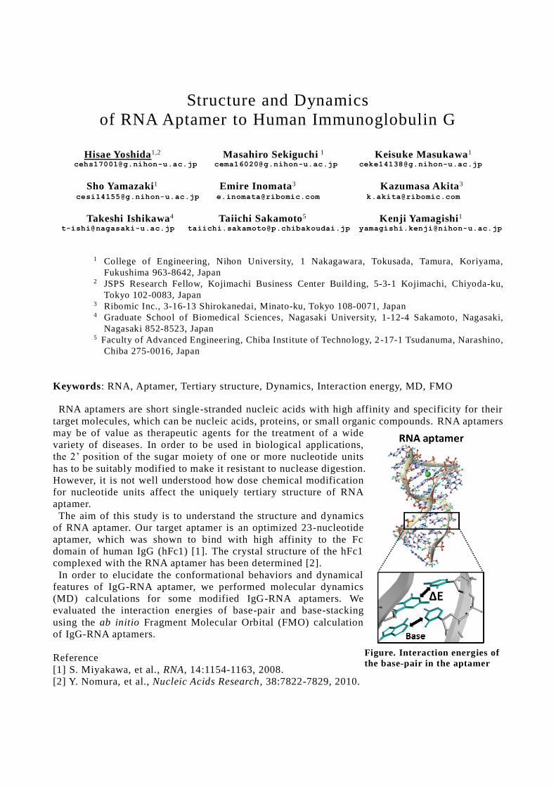
Structure and Dynamics
of RNA Aptamer to Human Immunoglobulin G
Hisae Yoshida1,2 Masahiro Sekiguchi 1 Keisuke Masukawa1 [email protected] [email protected] [email protected]
Sho Yamazaki1 Emire Inomata3 Kazumasa Akita3 [email protected] [email protected] [email protected]
Takeshi Ishikawa4 Taiichi Sakamoto5 Kenji Yamagishi1 [email protected] [email protected] [email protected]
1 College of Engineering, Nihon University, 1 Nakagawara, Tokusada, Tamura, Koriyama,
Fukushima 963-8642, Japan 2 JSPS Research Fellow, Kojimachi Business Center Build ing, 5-3-1 Kojimachi, Chiyoda-ku,
Tokyo 102-0083, Japan 3 Ribomic Inc., 3-16-13 Shirokanedai, Minato-ku, Tokyo 108-0071, Japan 4 Graduate School of Biomedical Sciences, Nagasaki University, 1-12-4 Sakamoto, Nagasaki,
Nagasaki 852-8523, Japan 5 Faculty of Advanced Engineering, Chiba Institute of Technology, 2 -17-1 Tsudanuma, Narashino,
Chiba 275-0016, Japan
Keywords: RNA, Aptamer, Tertiary structure, Dynamics, Interaction energy, MD, FMO
RNA aptamers are short single-stranded nucleic acids with high affinity and specificity for their
target molecules, which can be nucleic acids, proteins, or small organic compounds. RNA aptamers
may be of value as therapeutic agents for the treatment of a wide
variety of diseases. In order to be used in biological applications,
the 2’ position of the sugar moiety of one or more nucleotide units
has to be suitably modified to make it resistant to nuclease digestion.
However, it is not well understood how dose chemical modification
for nucleotide units affect the uniquely tertiary structure of RNA
aptamer.
The aim of this study is to understand the structure and dynamics
of RNA aptamer. Our target aptamer is an optimized 23-nucleotide
aptamer, which was shown to bind with high affinity to the Fc
domain of human IgG (hFc1) [1]. The crystal structure of the hFc1
complexed with the RNA aptamer has been determined [2].
In order to elucidate the conformational behaviors and dynamical
features of IgG-RNA aptamer, we performed molecular dynamics
(MD) calculations for some modified IgG-RNA aptamers. We
evaluated the interaction energies of base-pair and base-stacking
using the ab initio Fragment Molecular Orbital (FMO) calculation
of IgG-RNA aptamers.
Reference
[1] S. Miyakawa, et al., RNA, 14:1154-1163, 2008.
[2] Y. Nomura, et al., Nucleic Acids Research, 38:7822-7829, 2010.
Figure. Interaction energies of
the base-pair in the aptamer
WhichP450: Predicting Which Cytochrome P450 Isoforms
are involved in the Metabolism of a Xenobiotic
Peter Hunt1 Jonathan Tyzack
1 Sumie Tajima
2
[email protected] [email protected] [email protected]
Marina Takahashi2 Matthew Segall
1
[email protected] [email protected]
1
Optibrium Ltd., 7221 Cambridge Research Park, Beach Drive, Cambridge, CB25 9TL, UK 2
Hulinks Inc., 5-14 Nihonbashi Hakozaki-cho, Chuo-ku, Tokyo 103-0015, Japan
Keywords: Cytochrome P450 metabolism, isoform specificity, QSAR models, metabolite profile
Predicting which cytochrome P450 isoforms are involved in the metabolism of a molecule is
important in assessing its metabolic fate in vivo. The various P450 isoforms have active sites of
different shapes, sizes and characters [1], favouring different binding pharmacophores which can
lead to metabolism at different sites within the molecule. Therefore, predicting the P450 isoforms
likely to be involved in metabolism is a useful precursor to predicting the metabolites that are
likely to be formed and considering which isoform-specific predictive models should be applied.
'Which P450' models also have application in assessing the risk of drug -drug interactions and the
impact of genetic polymorphisms. A molecule reliant on a single isoform for metabolic clearance is
at an increased risk of exhibiting drug-drug interactions or a greater impact of genetic
polymorphisms on its pharmacokinetics.
Here we present a QSAR model to predict which P450 isoforms, out of a list of the 7 leading
CYP450 isoforms, are likely to be involved in the metabolism of novel molecules. This model was
generated using a multi-class categorical Random Forest method using the Auto-Modeller module
in StarDrop [2] and has a 75% success rate with a Top-1 criterion and a 90% success rate for a
Top-2 criterion and showed significantly improved performance over random estimates.
We furthermore illustrate how 'which P450' models can be combined with isoform -specific
models for prediction of site of metabolism to predict the metabolite profile due to P450
metabolism of a xenobiotic.
[1] M.B.G. Cruciani et al., Molecular Interaction Fields, Wiley-VCH, 12:273-290, 2006.
[2] StarDrop. www.optibrium.com/stardrop
Escape from ‘availability bias’ in compound design
Andras Stracz1 Akos Tarcsay1
Gabor Imre1 Ivan Solt1 Daisuke Miyazaki2 [email protected]
1 ChemAxon Ltd. 1031 Budapest. Zahony u. 7, Hungary 2 Patcore,Inc. Sapia Tower 26F, 1-7-12 Marunouchi, Chiyoda-ku, Tokyo 100-0005, Japan
Keywords: Compound Design Support, Fast Structure Search Small molecule design is an information demanding activity, since all relevant knowledge is to be accessible within a single space and requires synchronized application of computational models to assist decision making on synthesis candidates. Our study aims to evaluate a software platform coping with this complexity (Marvin Live). The tool provides central management of innovative ideas and a framework that helps triage these. Decision support is based on predicted properties that span phys-chem descriptors, combined metrics like CNS MPO score, 3D overlay and modelling results conducted with KNIME on the one hand, and cross-checking with available knowledge collected from a variety of sources to do rapid freedom to operate analysis and SAR by catalog: by ultra-fast searching of patent and compound catalog databases, respectively on the other. This hypothetic study shows the potential evolution of a compound idea from the reference compound to a synthesis candidate through an example discovery project of 5-hydroxytryptamine receptor 6 (HTR6) ligands. [1] Wager, Travis T, et al. ACS Chem. Neurosci., 2010, 1 (6), pp 435-449 [2] Papadatos, George, et al. Nucleic Acids Res. 2016 Jan 4; 44(Database issue): 01220-01228.
Database downloaded 01 May 2017 [3] https://nodejs.org/ Accessed 04 June 2017 [4] https://www.chemaxon.com/products/madfast/ Accessed 04 June 2017 [5] Shivanyuk, A. N., et al. Chemistry Today 2007,25,58-59. Database downloaded 01 May 2017 [6] Tolmachev, Andrey, et al. ACS Combinatorial Science 2016, 18.10,616-624. [7]https://docs.chemaxon.com/display/docs/Chemical+Hashed+Fingerprint Accessed 04 June 2017 [8]https://www.chemaxon.com/products/calculator-plugins/molecular-modelling/ Accessed 04
June 2017
Virtual Screening of New Inhibitors from Indonesian Herbal Compounds against Mycobacterium tuberculosis
CYP121
Vivitri Dewi Prasasty1 Sandra Cindana2 [email protected] [email protected]
Fransiskus Xaverius Ivan2
1 Faculty of Biotechnology, Atma Jaya Catholic University of Indonesia, Jakarta, 12930, Indonesia
2 Faculty of Life Sciences, Surya University, Tangerang, Banten, 15810, Indonesia Keywords: M. tuberculosis CYP121, molecular docking, molecular dynamics, kaempferol Tuberculosis (TB) is a serious threat with the emergence of resistant bacterial variants, namely multidrug-resistant tuberculosis (MDR-TB) and extremely drug-resistant tuberculosis (XDR-TB). This study aims to find new inhibitors for M. tuberculosis CYP121 from Indonesian medicinal plants, especially Rhoeo spathacea and Pluchea indica. A virtual structure-based screening was performed on 16 molecules through molecular docking using AutoDock Vina and subsequent molecular dynamics simulations were performed using YASARA. The two best compounds, i.e. KAE (kaempferol) and KAE3 (kaempferol derivative) were selected based on their lowest binding energies (-9.1 kcal/mol and -10.5 kcal/mol, respectively) and their adherence to the Lipinski rule. Results from 50 ns molecular dynamics simulation suggest that KAE and KAE3 have an inhibitory mechanism againsts M. tuberculosis CYP121 that is different from GGJ (control inhibitor). Based on the RMSF scores comparison, GGJ alters the overall dynamics of the receptor characterized by the changes in the flexibility of amino acid residues far away from the receptor binding side. In contrast, the binding of KAE and KAE3 changes the flexibility of amino acids around the binding site of the receptor. [1] Collins, D., Hafidz, F. & Suraratdecha, C., The Economic Burden of Tuberculosis in Indonesia, Cambridge: TB CARE I - Management Sciences for Health, 2013. [2] Radji, M., Kurniati, M. and Kiranasari, A., Comparative antimycobacterial activity of some
Indonesian medicinal plants against multi-drug resistant Mycobacterium tuberculosis. Journal of Applied Pharmaceutical Science, 05(01): 019-022, 2015.
[3] McLean, K. J. and Munro, A. W., Structural biology and biochemistry of cytochrome P450 systems in Mycobacterium tuberculosis. Drug Metabolism Review, 40: 427-446, 2008.
[4] Sliwoski, G., Kothiwale, S., Meiler, J. and Lowe, E. W., Computational methods in drug discovery. Pharmacological Reviews, 66(1): 334-395, 2014.
A new rotation-translation invariant molecular descriptor forLigand-Based Virtual Screening and beyond
Francois Berenger1 Yoshihiro Yamanishi1,2
[email protected] [email protected]
1 System Cohort Division, Medical Institute of Bioregulation, Kyushu University3-1-1 Maidashi, Higashi-ku, Fukuoka 812-8582, Japan
2 PRESTO, Japan Science and Technology Agency, Kawaguchi, Saitama 332-0012, Japan
Keywords: Ligand-Based Virtual Screening, molecular similarity, chemical fingerprints, autocorrelation,3D.
Various methods exist to compare ligands: fingerprints like MACCS, ECFP4 or PubChem and specializedsoftware like ROCS[1], EON, ElectroShape[2], ShaEP[3] and Blaze[4]. There are also various methods tocompare protein binding sites like sup-CK[5], SuMo[6], Shaper[7], SiteAlign[7] and FuzCav[7]. While fingerprints are only working in 2D, binding site comparison methods are complex and incorporateseveral parameters. Interestingly, both kind of methods perform the same task of comparing two sets ofatoms. In this study, we propose a novel method, scarce in parameters and working in 3D, that can do both ligandand binding site similarity search, modulo re-parametrization of the method towards the intended use case(to maximize performance). The proposed method will be able to directly find ligands given a binding site;without docking or prior knowledge of ligands for this binding site. Similarly, the opposite task of finding abinding site given a ligand is theoretically possible. We will show the usefulness of our proposed method bycomparing its performance with popular 2D chemical fingerprints. Our method considers ligands in 3D andis rotation-translation invariant. Ligands do not need to be optimally superposed prior to being compared.Another innovative point of our method is that it can work in any or a combination of "chemical spaces" likeshape, electrostatic, hydrophobic and hydrogen bonding.
[1] Rush, T. S., Grant, J. A., Mosyak, L., & Nicholls, A. (2005). A shape-based 3-D scaffold hopping methodand its application to a bacterial protein− protein interaction. JMedChem, 48(5), 1489-1495.
[2] Armstrong, M. S., Morris, G. M., Finn, P. W., Sharma, R., Moretti, L., Cooper, R. I., & Richards, W. G.(2010). ElectroShape: fast molecular similarity calculations incorporating shape, chirality andelectrostatics. JCAMD, 24(9), 789-801.
[3] Vainio, M. J., Puranen, J. S., & Johnson, M. S. (2009). ShaEP: molecular overlay based on shape andelectrostatic potential.
[4] Cheeseright, T., Mackey, M., Rose, S., & Vinter, A. (2006). Molecular field extrema as descriptors ofbiological activity: definition and validation. JCIM, 46(2), 665-676.
[5] Hoffmann, B., Zaslavskiy, M., Vert, J. P., & Stoven, V. (2010). A new protein binding pocket similaritymeasure based on comparison of clouds of atoms in 3D: application to ligand prediction. BMCbioinformatics, 11(1), 99.
[6] Jambon, M., Andrieu, O., Combet, C., Deléage, G., Delfaud, F., & Geourjon, C. (2005). The SuMoserver: 3D search for protein functional sites. Bioinformatics, 21(20), 3929-3930.
[7] Kellenberger, E., Schalon, C., & Rognan, D. (2008). How to measure the similarity between proteinligand-binding sites? CCADD, 4(3), 209.
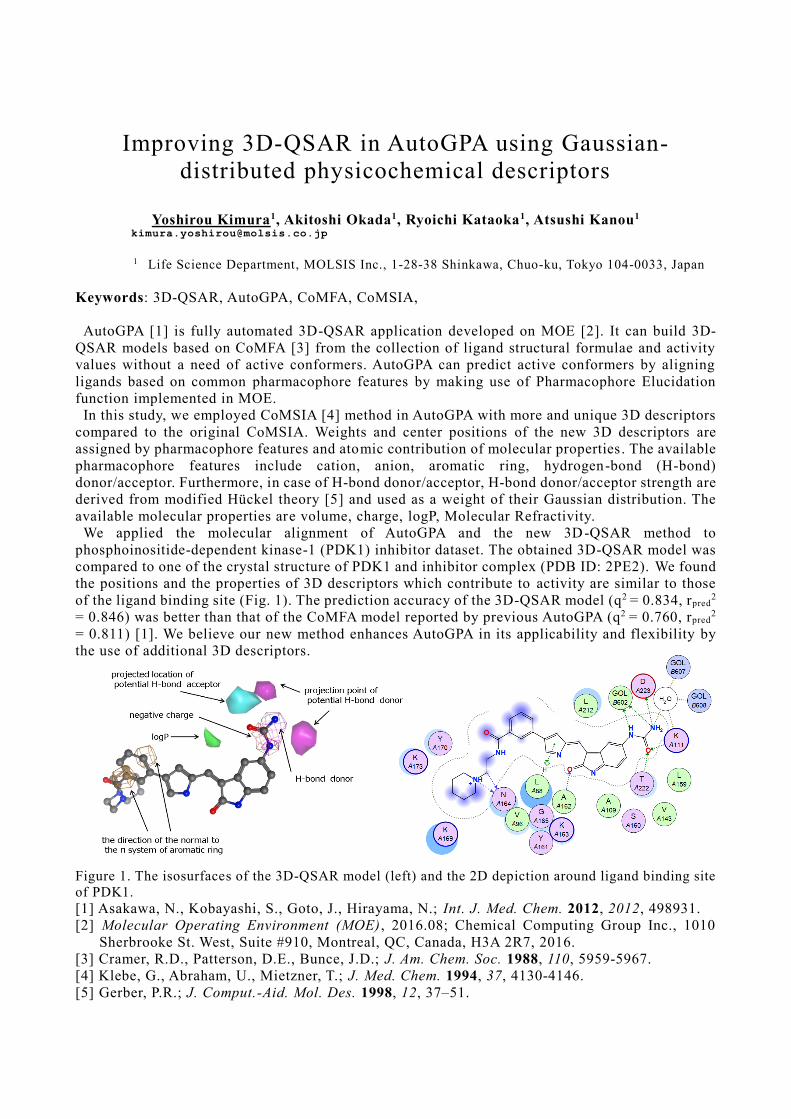
Improving 3D-QSAR in AutoGPA using Gaussian-
distributed physicochemical descriptors
Yoshirou Kimura1, Akitoshi Okada1, Ryoichi Kataoka1, Atsushi Kanou1 [email protected]
1 Life Science Department, MOLSIS Inc., 1-28-38 Shinkawa, Chuo-ku, Tokyo 104-0033, Japan
Keywords: 3D-QSAR, AutoGPA, CoMFA, CoMSIA,
AutoGPA [1] is fully automated 3D-QSAR application developed on MOE [2]. It can build 3D-
QSAR models based on CoMFA [3] from the collection of ligand structural formulae and activity
values without a need of active conformers. AutoGPA can predict active conformers by aligning
ligands based on common pharmacophore features by making use of Pharmacophore Elucidation
function implemented in MOE.
In this study, we employed CoMSIA [4] method in AutoGPA with more and unique 3D descriptors
compared to the original CoMSIA. Weights and center positions of the new 3D descriptors are
assigned by pharmacophore features and atomic contribution of molecular properties. The available
pharmacophore features include cation, anion, aromatic ring, hydrogen-bond (H-bond)
donor/acceptor. Furthermore, in case of H-bond donor/acceptor, H-bond donor/acceptor strength are
derived from modified Hückel theory [5] and used as a weight of their Gaussian distribution. The
available molecular properties are volume, charge, logP, Molecular Refractivity.
We applied the molecular alignment of AutoGPA and the new 3D-QSAR method to
phosphoinositide-dependent kinase-1 (PDK1) inhibitor dataset. The obtained 3D-QSAR model was
compared to one of the crystal structure of PDK1 and inhibitor complex (PDB ID: 2PE2). We found
the positions and the properties of 3D descriptors which contribute to activity are similar to those
of the ligand binding site (Fig. 1). The prediction accuracy of the 3D-QSAR model (q2 = 0.834, rpred2
= 0.846) was better than that of the CoMFA model reported by previous AutoGPA (q2 = 0.760, rpred2
= 0.811) [1]. We believe our new method enhances AutoGPA in its applicability and flexibility by
the use of additional 3D descriptors.
Figure 1. The isosurfaces of the 3D-QSAR model (left) and the 2D depiction around ligand binding site
of PDK1.
[1] Asakawa, N., Kobayashi, S., Goto, J., Hirayama, N.; Int. J. Med. Chem. 2012, 2012, 498931.
[2] Molecular Operating Environment (MOE) , 2016.08; Chemical Computing Group Inc., 1010
Sherbrooke St. West, Suite #910, Montreal, QC, Canada, H3A 2R7, 2016.
[3] Cramer, R.D., Patterson, D.E., Bunce, J.D.; J. Am. Chem. Soc. 1988, 110, 5959-5967.
[4] Klebe, G., Abraham, U., Mietzner, T.; J. Med. Chem. 1994, 37, 4130-4146.
[5] Gerber, P.R.; J. Comput.-Aid. Mol. Des. 1998, 12, 37–51.

In silico search for proanthocyanidins binding proteins
Takuma Todoroki1 Reina Takahashi1 [email protected] [email protected]
Hiroshi Fujii1,2 Hidefumi Makabe1
[email protected] [email protected]
Koji Umezawa1,2 [email protected]
1 Faculty of agriculture, Shinshu University, 8304 Minami-minowa, Kami-ina, Nagano, 399-4598, Japan
2 Department of Interdisciplinary Genome Sciences and Cell Metabolism, Institute for Biomedical Sciences, Interdisciplinary Cluster for Cutting Edge Research, Shinshu University, Minami-minowa, Kami-ina, Nagano, 399-4598, Japan
Keywords: Natural products, Ligand docking, Binding-pocket search
There are a lot of natural products good for health via a specific biological function while, in most cases, the receptors for the molecules derived from the natural products are still unknown. Proanthocyanidins belong to the functional natural products of polyphenols involved in grape, apple and so on, which exhibit various functions such as suppression of cell proliferation. The receptors for proanthocyanidins have not been identified widely.
The proanthocyanidin is the polymer conjugating the units of catechin (cat), epicatechin (epi) and their derivatives of gallocatechin (gc) and epigallocatechin (egc). Therefore, the chemical structure of proanthocyanidin has a lot of variety depending on combination of the species of units and the number of polymerization. For instance, the dimer of egc (egc-egc) is called prodelphinidin B2 (PDB2). Interestingly, the PDB2 exhibits a biological activity although the dimer of gc (gc-gc) does not. Thus, the chemical variety is crucial for the biological activity. It should be related to a receptor for proanthocyanidins. However, the receptor proteins that proanthocyanidins bind are not found comprehensively. Then, we have explored the proanthocyanidin-binding proteins from Protein Data Bank.
For the purpose of searching the proanthocyanidin-binding proteins from the database, we took the two steps: local-similarity search and ligand-docking simulation. First, in the local-similarity search, we chose the known cat-binding and epi-binding protein structures to construct binding-pocket templates for cat and epi. The binding-pocket template is composed of the coordinates of protein atoms in the vicinity of cat or epi. We compared the binding-pocket templates with the all structures deposited in Protein Data Bank. We collected the proteins that have the similar sites with the binding-pocket templates. In the next step, we conducted the ligand-docking simulation for the collected proteins with proanthocyanidins. The proanthocyanidin-binding candidates were ranked in order of the docking scores. Then, we could search proteins related to biological functions including cell proliferation. These proteins might be future drug targets as lead compounds of proanthocyanidins.
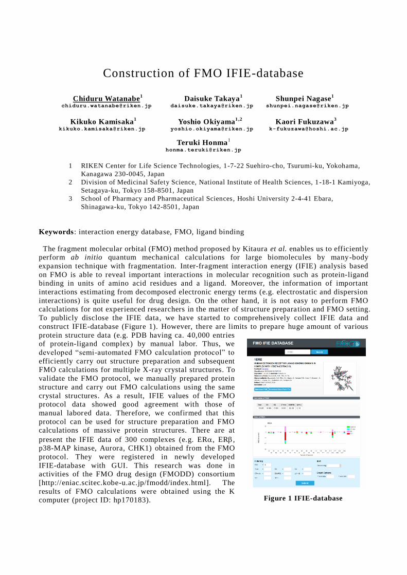
Construction of FMO IFIE-database
Chiduru Watanabe1 Daisuke Takaya
1 Shunpei Nagase
1
[email protected] [email protected] [email protected]
Kikuko Kamisaka1 Yoshio Okiyama
1 ,2 Kaori Fukuzawa
3
[email protected] [email protected] [email protected]
Teruki Honma1
1 RIKEN Center for Life Science Technologies, 1-7-22 Suehiro-cho, Tsurumi-ku, Yokohama,
Kanagawa 230-0045, Japan
2 Division of Medicinal Safety Science, National Institute of Health Sciences, 1-18-1 Kamiyoga,
Setagaya-ku, Tokyo 158-8501, Japan
3 School of Pharmacy and Pharmaceutical Sciences, Hoshi University 2-4-41 Ebara,
Shinagawa-ku, Tokyo 142-8501, Japan
Keywords: interaction energy database, FMO, ligand binding
The fragment molecular orbital (FMO) method proposed by Kitaura et al. enables us to efficiently perform ab initio quantum mechanical calculations for large biomolecules by many-body
expansion technique with fragmentation. Inter-fragment interaction energy (IFIE) analysis based
on FMO is able to reveal important interactions in molecular recognition such as protein-ligand binding in units of amino acid residues and a ligand. Moreover, the information of important
interactions estimating from decomposed electronic energy terms (e.g. electrostatic and dispersion
interactions) is quite useful for drug design. On the other hand, it is not easy to perform FMO calculations for not experienced researchers in the matter of structure preparation and FMO setting.
To publicly disclose the IFIE data, we have started to comprehensively collect IFIE data and
construct IFIE-database (Figure 1). However, there are limits to prepare huge amount of various
protein structure data (e.g. PDB having ca. 40,000 entries of protein-ligand complex) by manual labor. Thus, we
developed “semi-automated FMO calculation protocol” to
efficiently carry out structure preparation and subsequent FMO calculations for multiple X-ray crystal structures. To
validate the FMO protocol, we manually prepared protein
structure and carry out FMO calculations using the same
crystal structures. As a result, IFIE values of the FMO protocol data showed good agreement with those of
manual labored data. Therefore, we confirmed that this
protocol can be used for structure preparation and FMO calculations of massive protein structures. There are at
present the IFIE data of 300 complexes (e.g. ER, ER, p38-MAP kinase, Aurora, CHK1) obtained from the FMO
protocol. They were registered in newly developed
IFIE-database with GUI. This research was done in activities of the FMO drug design (FMODD) consortium
[http://eniac.scitec.kobe-u.ac.jp/fmodd/index.html]. The
results of FMO calculations were obtained using the K computer (project ID: hp170183).
Figure 1 IFIE-database
The Role of Water Molecules in Protein-Ligand Binding:
Fragment Molecular Orbital Calculations on the
Complexes of Renin with its Inhibitors
Yoichiro Yagi1 Takatomo Kimura
2
[email protected] [email protected]
Chiduru Watanabe3 Yoshio Okiyama
4
[email protected] [email protected]
Shigenori Tanaka5 Teruki Honma
3 Kaori Fukuzawa
6
[email protected] [email protected] [email protected]
1
Department of Electrical and Electronic Engineering, Okayama University of Science,
1-1 Ridai-cho, Kita-ku, Okayama 700-0005, Japan 2
Konan Chemical Industry Co., Ltd., 5-12 Nakagawa-cho, Takatsuki, Osaka 569-0066, Japan 3
RIKEN Center for Life Science Technologies, 1-7-22 Suehiro-cho, Tsurumi-ku, Yokohama,
Kanagawa 230-0045, Japan 4
National Institute of Health Sciences, 1-18-1 Kamiyoga, Setagaya-ku, Tokyo 158-8501, Japan 5
Department of Computational Science, Graduate School of System Informatics,
Kobe University, 1-1 Rokkodai, Nada-ku, Kobe, Hyogo 657-8501, Japan 6
School of Pharmacy and Pharmaceutical Sciences, Hoshi University, 2-4-41 Ebara,
Shinagawa-Ku, Tokyo 142-8501, Japan
Keywords: Fragment Molecular Orbital (FMO) Calculation, Renin, Inhibitor, Water molecules,
Interaction energy, Binding energy, Pair interaction energy decomposition analysis (PIEDA)
We have carried out the fragment molecular orbital (FMO) calculations on the 22 different
renin-inhibitor complexes at FMO2-MP2/6-31G* level to clarify a relationship between the
calculated binding energy for the renin-inhibitor complexes and the activity value on 50%
inhibitory concentration (IC50) of the inhibitors. The PDB structures of complexes with TIP3P
model waters around 8Å of complex were optimized by an energy minimization using AMBER.
After energy minimization, TIP3P model waters were removed and FMO calculations using
MIZUHO/BioStation program were performed on the K computer [1]. FMO computations
indicated that the binding energy can be correlated to IC50. In the FMO calculation that contains
crystal waters within 5Å of ligand, the binding energies showed a good correlation with IC50 for
each inhibitor [2]. The aim of this study is to elucidate a role of water molecules in binding of
renin and inhibitor. By analyzing of the interactions between ligand and water molecules and
between water molecules and amino acid residues in protein, we found that ligand interacts with
protein through the water molecules. This result suggests that the water molecules may support the
binding of ligand in active site of protein. In addition, we performed pair interaction energy
decomposition analysis (PIEDA) calculation to examine the energy components.
This research was done in activities of the FMO drug design consortium (FMODD). The results
were obtained using the K computer (project ID: hp170183).
[1] Tanaka, S., Mochizuki, Y., Komeji, Y., Okiyama, Y., and Fukuzawa, K., Electron -correlated
fragment-molecular-orbital calculations for biomolecular and nano systems, Physical Chemistry
Chemical Physics, 16:10310-10344, 2014.
[2] Yagi, Y., Kimura, T., Watanabe, C., Okiyama, Y., Tanaka, S., Honma, T., and Fukuzawa, K.,
Fragment Molecular Orbital Calculations on the Interaction and Binding Energies of Human
Protease Renin and its Inhibitors, Chem-Bio Informatics Society (CBI) Annual Meeting 2016 ,
P1-08, 2016.
Using multiple molecular fingerprints for improvement of drug activity predictions
Yusuke Matsuyama1,3 Takashi Ishida2,3 [email protected] [email protected]
1 Department of Computer Science, Tokyo institute of technology, Meguro, Tokyo, Japan
2 School of Computing Department of Computer Science, Tokyo institute of technology, Meguro, Tokyo, Japan
3 Education Academy of Computational Life Sciences, Tokyo Institute of Technology, Meguro, Tokyo, Japan
Keywords: Virtual screening; Machine learning; Molecular fingerprint; Stacking; Ensemble learning Drug activity prediction based on machine learning method is useful for ligand-based virtual screening. And it is drawing attention as a key to keep down drug discovery costs which are said to be 300 billion[1] and still increasing now. When using machine learning technique, the information of molecule have to be converted into a
vector with fixed length. For many years, huge number of molecular fingerprints are proposed, however, it is known that the
best fingerprint is different for the target[2], and it is difficult to select the most suitable fingerprint.
To solve this problem, we propose a new technique to use multiple fingerprint by emsemble learning for drug activity prediction.
As a result of performance evaluation, the proposed method greatly increased the predicted performance compared to the prediction model using single molecular fingerprint.
In addition, we compared the feature and prediction performance of major molecular fingerprints, and analyzed the reason why our ensemble method works well.
[1] Mullard, A.: New drugs cost US$2.6 billion to develop. Nature reviews. Drug discovery 13(12), 877 (2014). doi:10.1038/nrd4507 [2] Riniker, S., Landrum, G.A.: Open-source platform to benchmark fingerprints for ligand-based
virtual screening. Journal of Cheminformatics 5(5) (2013). doi:10.1186/1758-2946-5-26
K4: An in silico Drug Design Suite combined with HPCs
Biao Ma1,2 Yuta Isaka1,2 [email protected] [email protected]
Mitsugu Araki3,4 Hiroaki Iwata3 [email protected] [email protected]
Yasushi Okuno2,3,4 [email protected]
1 Research and Development Group for In Silico Drug Discovery, Pro-Cluster Kobe, Foundation
for Biomedical Research and Innovation (FBRI) , 1-6-5, Minatojima-Minamimachi Chuo-ku,
Kobe 650-0047, Japan 2 Simulation-Driven Drug Discovery Group, IBRI laboratory, Foundation for Biomedical
Research and Innovation (FBRI), 1-6-5, Minatojima-Minamimachi Chuo-ku, Kobe 650-0047,
Japan 3 Graduate School of Medicine, Kyoto University, 53 Kawahara-cho, Shogoin Sakyo-ku, Kyoto
606-8507, Japan 4 RIKEN Advanced Institute for Computational Science, 7 -1-26 Minatojima-Minamimachi,
Chuo-ku, Kobe, Hyogo 650-0047, Japan
Keywords: SBDD, Molecular dynamics simulation, Docking simulation, Virtual screening,
Binding free energy calculation, MP-CAFEE, HPCs, K-computer, Graphical user interface, GUI
In drug discovery process, computational methods to efficiently explore and optimize drug candidates
are strongly required. Since virtual screening from huge amount of chemical compounds (> 10 60) and
accurate prediction of the protein-drug binding affinity need a large computational cost, we attempt to
overcome this problem by utilizing high-performance computers (HPCs) such as K-computer, Osaka
Univ VCC, and so on. However, in silico drug discovery on HPCs requires complicated operation. To
solve this problem, we have developed an in silico drug design suite, K4(K-Force), which has a friendly
graphical user interface (GUI). The medicinal chemists can easily utilize computational power of
K-computer and other HPCs.
In K4, we constructed a single platform that integrates the following main functions: 1) protein
structure sampling using temperature replica-exchange molecular dynamics (T-REMD), 2) docking
simulation (DS) or virtual screening (VS) based on rDock[1], 3) Ab initio calculation of compound
partial charges based on GAMESS, 4) the protein-compound free energy calculation based on
molecular dynamics (MD) simulation using MP-CAFEE[2,3] or MM-PB/GBSA method.
We will provide more methods for rational design of drug molecules in next version, such as
protein-ligand inter-fragment interaction energy calculation(IFIE) by use of fragment molecular orbital
(FMO) method and ensemble docking, which enables to dock a chemical library against multiple
receptor conformations. If you are interested into our applications suite, please visit KOBE-CITY and
FBRI booth.
[1] Ruiz-Carmona, S., Alvarez-Garcia, D., Foloppe, N., Garmendia-Doval, A. B., Juhos, S.,
Schmidtke, P., Barril, X., Hubbard, R. E., and Morley, S. D. rDock: A Fast, Ver satile and Open
Source Program for Docking Ligands to Proteins and Nucleic Acids. PLoS Comput. Biol. (Prlic,
A., Ed.) 10, e1003571, 2014.
[2] Fujitani, H., Tanida, Y., and Matsuura, A. Massively parallel computation of absolute binding
free energy with well-equilibrated states. Phys. Rev. E 79, 21914, 2009.
[3] Araki, M., Kamiya, N., Sato, M., Nakatsui, M., Hirokawa, T., and Okuno, Y. The Effect of
Conformational Flexibility on Binding Free Energy Estimation between Kinases and Their
Inhibitors. J. Chem. Inf. Model. 56, 2445–2456, 2016.
ReCGen: Development of a novel approach to generate virtual compounds
Chisato Kanai1 Edmund Taylo1 [email protected] [email protected]
1 Kyoto Constella Technologies Co., Ltd., fourth floor Kyozome Kaikan, 481 Tourouyama-cho, Nakagyo-ku, Kyoto City, 604-8225, Japan
Keywords: de novo design, ECFP, RECAP, synthetic accessibility It is widely accepted that one of the important elemental technologies of de novo
design [1] is the creation of novel structures. To date, various methods have been proposed which are roughly classified into the following three types: atom-based method, fragment-based method, and chemical reaction (synthetic route)-based method. The most common type is fragment-based structure generation method which is represented by those using RECAP [2], BRICS [3] and MMPA (Matched molecular pair analysis) [4]. RECAP, in particular, is a type of retrosynthetic analysis method focusing on chemical reactions frequently used in drug discovery. The structures generated by this method are relatively easy to synthesize, so the method is considered good for de novo design. We have recently developed a new fragment-based method that utilizes the concept
of ECFP [5], a type of fingerprint, in the generation of new fragments that are subsequently used as building blocks in the assembly of new structures. In our method, the size of ECFP fragments can be set arbitrarily by specifying the diameter of the graph, so a wide variety of fragments can be obtained as compared with the RECAP method. Therefore, compared to RECAP, which is subject to rough structural transformation, relatively fine structure transformation is possible when using our method. Furthermore, synthetic accessibility (SA) [6] of structures generated by our method is as favorable as that generated using the RECAP method. Because of those features, we refer to it as “Refined Compound Generator (ReCGen)”. In this poster we present a comparison between the principle of our method and the
features of RECAP method. [1] Schneider G. and Fechner U., Nature Reviews Drug Discovery 4:649–663, 2005. [2] Lewell, X.Q., Judd, D.B., Watson, S.P. and Hann, M.M., J. Chem. Inf. Comput. Sci. 38:511-522,
1998. [3] Degen J, Wegscheid-Gerlach C, Zaliani A and Rarey M., ChemMedChem, 3:1503-1507, 2008. [4] Kenny P. W., Sadowski J., “Structure Modification in Chemical Databases”. In
Chemoinformatics in Drug Discovery. Oprea, T. I., Ed. Wiley-VCH: Weinheim, Germany. pp. 271−285, 2004.
[5] David R. and Mathew H., J. Chem. Inf. Model. 50:742-754, 2010. [6] Fukunishi Y., Kurosawa T., Mikami Y. and Nakamura H., J. Chem. Inf. Model. 54:3259-3267,
2014.
Quick and precise homology modeling method of GPCRs
Mika Nabeno1 Takatsugu Hirokawa
2
[email protected] [email protected]
1 Discovery Technology Laboratories, Sohyaku. Innovative Research Division, Mitsubishi
Tanabe Pharma Corporation, 1000 Kamoshida-cho, Aoba-ku, Yokohama, Kanagawa, 227-0033,
Japan 2 Molecular Profiling Research Center for Drug Discovery, National Institute of Advanced
Industrial Science and Technology, 2-4-7 Aomi, Koto-ku, Tokyo, 135-0064, Japan
Division of Biomedical Science, Faculty of Medicine, University of Tsukuba, 1-1-1 Tennodai,
Tsukuba, Ibaraki, 305-8575, Japan
Keywords: GPCR, Homology modeling, Extracellular loop II, Ligand docking, Molecular
dynamics simulation
G protein-coupled receptors (GPCR) are major drug discovery targets, and the reports on the
X-ray crystal structures of GPCRs, which are rapidly increasing in recent years, are very useful for
GPCR-targeted drug design. However, because X-ray crystallography of GPCRs is still
challenging, we cannot acquire the structures complexed with compounds at the time when they
can be utilized in drug design effectively. Therefore, the needs of homology modeling are high at
the drug discovery field. A number of modeling methods of GPCRs have been studied to date1.
Particularly in recent years, there is a tendency to perform a large amount of calculation to build a
highly accurate model2. However, in the drug discovery research, we often have multiple
compounds to be modeled, and the predicted models need to be revised according to SAR of
compounds. In addition to that, when a new X-ray crystal structure more suitable for a template is
revealed, the homology models have to be built from scratch. For the reasons shown above, it is
desirable that the modeling method be not only highly accurate but also simple.
In this study, we propose the quick and precise homology modeling method in the case of the
compound binding to the orthosteric site of GPCR. The site is covered with the extracellular loop
II (ECL2), which affects binding of the compound seriously. However, because of its diversity
among GPCRs, its modeling is generally difficult. Here, a multi -template method was adopted
which selected different templates for transmembrane(TM) region and ECL2. We found out that
the accuracy of modeling is improved by selecting one templat e for the TM region with high
sequence homology and another template for the ECL2 with almost the same number of amino acid
residues. In drug design, although the prediction of the docking pose of the compound is quite vital,
it is the most difficult step in GPCR modeling. Then, we built an easy method to narrow down the
possible docking poses after searching them in a wide range. It was also confirmed that the
accuracy of the models was remarkably improved by performing molecular dynamics simulation
for 50 ns on finishing of the obtained models. In our validation, the RMSDs of the bound ligands
predicted by the above method were less than 2 Å from those of the X-ray crystal structures.
Although further verification is necessary, this method can be promising for drug design.
[1] Koehler Leman, J., Ulmschneider, M. B., Miller, and W., Gray, J.J., Computational modeling of
membrane proteins, Proteins, 83(1):1-24, 2015.
[2] Rodríguez, D., Ranganathan, A., Miller, W., and Carlsson, J., Strategies for Improved Modeling
of GPCR-Drug Complexes: Blind Predictions of Serotonin Receptors Bound to Ergotamine ,
Journal of Chemical Information and Modeling , 54:2004-2021, 2014.
KampoDB: An integrated platform for mode-of-action
analysis and repositioning of natural medicines
Ryusuke Sawada1 Michio Iwata1 [email protected] [email protected]
Masahiro Umezaki2 Yoshihiko Usui2 [email protected]
Takaki Kubono3 Makoto Kadowaki3 [email protected] [email protected]
Yoshihiro Yamanishi1,4 [email protected]
1 Division of System Cohort, Multi -scale Research Center for Medical Science, Medical Institute
of Bioregulation, Kyushu University, 3-1-1 Maidashi, Higashi-ku, Fukuoka, Fukuoka 812-8582,
Japan. 2 Division of Chemo-Bioinformatics, Institute of Natural Medicine, University of Toyama,
Toyama 930-0194, Japan. 3 Division of Gastrointestinal Pathophysiology, Institute of Natural Medicine, University of
Toyama, Toyama 930-0194, Japan. 4 PRESTO, Japan Science and Technology Agency, Kawaguchi, Saitama 332 -0012, Japan.
Keywords: Kampo medicines, functional analysis, target prediction, Kampo repositioning
Natural medicines (i.e., herbal medicines, traditional formulas) are useful for treatment of
multifactorial and chronic diseases. Here, we present KampoDB
(http://wakanmoview.inm.u-toyama.ac.jp/kampo/), a novel database of natural medicines, which
provides various useful scientific resources on Japanese traditional formulas Kampo medicines,
constituent herbal drugs, constituent compounds, and target proteins of these constituent
compounds. Potential target proteins of these constituent compounds were predicted by docking
simulations and machine learning methods based on large-scale omics data (e.g., genome,
proteome, metabolome, interactome). The current version of KampoDB contains 42 Kampo
medicines, 54 crude drugs, 1230 constituent compounds, 460 known target proteins, and 1369
potential target proteins, and has functional annotations for biological pathways and molecular
functions. KampoDB is useful for mode-of-action analysis of natural medicines and prediction of
new indications for a wide range of diseases.
Integrated multiomics analysis for dengue hermorrhogic fever
Takayuki Amemiya1 M. Michael Gromiha1,2 [email protected] [email protected]
Katsuhisa Horimoto1 Kazuhiko Fukui1 [email protected] [email protected]
1 Molecular Profiling Research Center for Drug Discovery (molprof), National Institute of
Advanced Industrial Science and Technology (AIST), Tokyo 135-0064, Japan 2 Department of Biotechnology, Indian Institute of Technology Madras, Tamilnadu 600 036,
India Keywords: Multi-omics analysis, Drug repositioning, Dengue hermorrhagic fever
Due to recent advances in the development of various kinds of high throughput technology in life sciences, it is possible to collect and analyze multiple "omics" data. Omics analysis as integrative system biology has been studied for drug discovery, biomarker and assessment of their toxicity and efficacy. Here, a multi-omics approach is applied for dengue hermorrhagic fever (DHF) characterized by vasculopathy, which results in sudden plasma leakage that reduces the blood volume and may result in hypovolemic shock, known as dengue shock syndrome [1]. However, there are no approved drug, DHF is classified as a neglected tropical disease by WHO.
In our analysis, three layers of experimental data such as transcriptomic, proteomic and interactomic data are considered. In each layer, we obtained the signature genes and proteins to find the drug candidates and the significant biological pathway [2]-[4]. The effective drug candidates are identified by using a drug repositioning method [5] to inverse drug-disease relationship in patients and healthy control gene expression profile and using interactomic relationships between signature proteins and chemical compounds. We will present the integrated multi-omics analysis for DHF, which leads to identify and narrow down drug candidates based on expression profiles and molecular interaction. [1] D. J. Gubler, Dengue and dengue hemorrhagic fever., Clin. Microbiol. Rev., 11:480–96, 1998. [2] J. Lamb, The Connectivity Map: Using Gene-Expression Signatures to Connect Small
Molecules, Genes, and Disease,,Science, 313:1929–1935, 2006. [3] A. Subramanian et al., Gene set enrichment analysis: a knowledge-based approach for
interpreting genome-wide expression profiles., Proc. Natl. Acad. Sci. U. S. A., 102:15545–15550, 2005.
[4] D. Szklarczyk et al., STITCH 5: Augmenting protein-chemical interaction networks with tissue and affinity data, Nucleic Acids Res., 44:D380–D384, 2016.
[5] Lamb, J., et al., The Connectivity Map: using gene-expression signatures to connect small molecules, genes, and diseases, Science, 313: 1929-1935, 2006.
Development of Structure-Free Design and Analysis for Compound Libraries Based on Chemical and Bioactive
Diversity
Yugo Shimizu1 Kazuyoshi Ikeda1 [email protected] [email protected]
Masanori Osawa1
1 Keio University, Faculty of Pharmacy, Division of Physics for Life Functions, 1-5-30 Shibakoen, Minato-ku, Tokyo 105-8512, Japan
Keywords: Library design, Structure-blinded analysis, Bioactivity space
High through screening is a starting point for drug discovery in many pharmaceutical companies. To expand collaborative drug discovery between industry and academia, the Drug-discovery Innovation and Screening Consortium (DISC) of over 20 pharmaceutical companies in Japan has been established [1]. DISC generated a library that consists of compounds collected from the attending companies, but diversity of compound structures in the library is unknown due to the intellectual property, and therefore, it is necessary to analyze the compound library without compound structure. For this purpose, we started a project, “Structure-Free Compound-Library Diversity Analysis”, supported by Japan Agency for Medical Research and Development to develop a computational system for diversity analysis of large-scale structure-blinded compound library.
In consideration of security, we adopted a secure cloud-based web system for collecting physicochemical descriptors and fingerprints of the compounds from companies, and Pipeline Pilot was used as a platform for manipulating and calculating large-scale compound data. An automated protocol for converting 2D/3D molecular descriptors of physicochemical property including principal moment of inertia and lead/drug-likeness score was developed for subsequent analysis of compound library diversity. The developed protocol can compare chemical spaces of small-molecule libraries from approved drugs, candidates, commercial compounds, and patent compounds from ChEMBL database [2]. We also developed a method for analyzing coverage of bioactive space. We extracted compounds in the ChEMBL database with bioactivity information for over 1,400 targets. Models for each target were created by several machine learning (ML) methods such as naïve Bayes, random forest, support vector machine, and deep neural networks using the descriptors and fingerprints. The prediction performance of each ML method was tested by cross-validation and the best one was used for predicting the bioactivity of compounds from large-scale libraries to evaluate the distribution of diversity in bioactive space. [1] http://www.amed.go.jp/program/list/06/03/05.html [2] Bento, A. P., Gaulton, A., Hersey, A., Bellis, L. J., Chambers, J., Davies, M., Krüger, F. A.,
Light, Y., Mak, L., McGlinchey, S., Nowotka, M., Papadatos, G., Santos, R., and Overington, J. P., The ChEMBL bioactivity database: an update, Nucleic Acids Research, 42(Database issue):D1083-1090, 2014.
This work is conducted as a part of “Construction of Drug Discovery Informatics System” supported by Japan Agency for Medical Research and Development (AMED).
Developing novel inhibitors of Heme-copper oxygen reductases
Yuya Nishida1 Yasunori Shintani1 [email protected] [email protected]
Hitomi Yuki2 Teruki Honma2, 3
[email protected] [email protected]
Seiji Takashima1 [email protected]
1 Medical Biochemistry, Osaka University Graduate School of Medicine,
2-2 Yamadaoka, Suita-shi, Osaka, 565-0871, Japan 2 RIKEN Center for Life Science Technologies,
1-7-22 Suehiro-cho, Tsurumi-ku, Yokohama, Kanagawa, 230-0045, Japan 3 Tokyo Institute of Technology, School of Computing, Department of Computer Science,
4259 Nagatsuta-cho, Midori-ku, Yokohama, 226-8503, Japan Keywords: Heme-copper oxygen reductase, cytochrome c oxidase, allosteric inhibitor, ligand-based virtual screening
Heme-copper oxygen reductase (HCO) is a family of transmembrane proteins that work as a
member of the respiratory electron transport chain. HCO receives electrons from cytochrome c or ubiquinol and then reduces oxygen to water, which couples with proton transfer across membrane. The generated transmembrane difference in proton concentration is a major part of proton-motive force, which is a driving force of ATP synthase. These reactions are crucial in most organisms, therefore the composing proteins are considered as drug targets. Here we describe inhibitors of two HCOs: mitochondrial cytochrome c oxidase (mtCcO) and bacterial ubiquinol oxidase (UqO). Recently, we identified novel allosteric inhibitors of mtCcO, and successfully determined crystal structures of mtCcO in complex with two inhibitors. The structures showed that both inhibitors bound to the same position of mtCcO, which is far from the ligand-binding site. This suggests that mtCcO activity is inhibited by a novel allosteric mechanism. In addition, as comparing mtCcO with bacterial UqO, the inhibitor binding site in mtCcO has some similarity to the homologous position in bacterial UqO, though mtCcO is much larger complex than bacterial UqO. These led us to hypothesize that the novel allosteric site has a potential to be a novel antibiotic target. To elucidate the inhibitory mechanism by approach of the structure-activity relationship and to develop a novel inhibitor targeting bacterial UqO, we performed ligand developments with the inhibitors we found. By using of the ligand-based virtual screening system LAILAPS in commercially available approximately 70,000,000 compounds, 270 compounds were selected and subsequently, we assessed these inhibitory activities against mtCcO and bacterial UqO. As results with mtCcO, 17 compounds had IC50 less than approximately 10 uM, and the most improved compound had IC50 of 0.1 uM, which is one-order better than that of the query inhibitor. Moreover, the result of the assay with bacterial UqO from Escherichia coli showed that 13 compounds had IC50 less than approximately 100 uM, and above all, the most effective compound had IC50 less than 10 uM. In conclusion, we succeeded to obtain various novel inhibitors of both mtCcO and bacterial UqO, which can be subsequently applied to the structure-activity relationship analysis and developing a novel antibiotic.
The Contribution of Entropy in Drug-Protein Binding
Takeshi Tanaka Ken Ikeda [email protected] [email protected]
Hirotsugu Komatsu Takao Matsuzaki [email protected] [email protected]
Interprotein Corporation, SIC-2-205, 5-4-30 Nishihashimoto, Midori-ku, Sagamihara, Kanagawa, 252-0131, Japan
Keywords: in Silico Screening, Protein-Protein Interaction, Small Molecule Inhibitor, Entropy, Drug Design
The entropy change of drug-protein binding can now be observed by Isothermal Titration Calorimetry (ITC). The
reported values of the entropy change of drug-protein binding range up to 10 kcal/mol, which consists significant portion of
free energy change. The entropy effect for drug-protein binding can be positive or negative depending on the nature of
binding reaction. In either case, drug-protein binding can not be predicted correctly without considering entropy change.
This seems to be the main reason why conventional docking methods fail in most cases. We have developed a system
which consider entropy contribution and concentrates compounds with high total energy into higher rank. It has been
successfully applied for 20 PPI targets and 10 enzyme targets. The details about Runx1-CBFβ are shown.
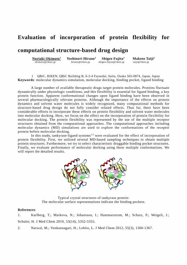
Evaluation of incorporation of protein flexibility for
computational structure-based drug design
Noriaki Okimoto1 Yoshinori Hirano1 Shigeo Fujita1 Makoto Taiji1 [email protected] [email protected] [email protected] [email protected]
1 QBiC, RIKEN, QBiC Building B, 6-2-4 Furuedai, Suita, Osaka 565-0874, Japan, Japan Keywords: molecular dynamics simulation, molecular docking, binding pocket, ligand binding
A large number of available therapeutic drugs target protein molecules. Proteins fluctuate dynamically under physiologic conditions, and this flexibility is essential for ligand binding, a key protein function. Apparent conformational changes upon ligand binding have been observed in several pharmacologically relevant proteins. Although the importance of the effects on protein dynamics and solvent water molecules is widely recognized, many computational methods for structure-based drug design do not fully consider related effects. Thus far, there have been considerable efforts to incorporate these effects on protein flexibility and solvent water molecules into molecular docking. Here, we focus on the effect on the incorporation of protein flexibility for molecular docking. The protein flexibility was represented by the use of the multiple receptor structures obtained from the computational approaches. The computational approaches including molecular dynamics (MD) simulations are used to explore the conformations of the receptor protein before molecular docking.
In this study, tankyrase-ligand systems1,2 were evaluated for the effect of incorporation of protein flexibility. First, we utilized several MD-based sampling techniques to obtain multiple protein structures. Furthermore, we try to select characteristic druggable binding pocket structures. Finally, we evaluate performance of molecular docking using these multiple conformations. We will report the detailed results.
Typical crystal structures of tankyrase protein:
The molecular surface representations indicate the binding pockets.
References 1. Karlberg, T.; Markova, N.; Johansson, I.; Hammarstrom, M.; Schutz, P.; Weigelt, J.;
Schuler, H. J Med Chem 2010, 53(14), 5352-5355.
2. Narwal, M.; Venkannagari, H.; Lehtio, L. J Med Chem 2012, 55(3), 1360-1367.
Computer simulation-based prediction of drug-induced
arrhythmia by evaluating repolarization reserve
Shingo Murakami Taichiro Tomida Yoshinori Mikami [email protected] [email protected] [email protected]
Masanori Ito Satomi Adachi-Akahane [email protected] [email protected]
Department of Physiology, School of Medicine, Toho University, 5 -21-16
Omori-Nishi, Ota-ku, Tokyo 143-8540, Japan
Keywords: Drug induced arrhythmia. Action potential simulation, Repolarization reserve
Some of drugs under development and in clinical use turned out to induce arrhythmia and were
withdrawn from the market. The drug-induced arrhythmia is mainly accounted for by prolongation
of action potential duration (APD) due to a block of one type of repolarizing K+ currents in cardiac
myocyte called IKr. Therefore, time-consuming and costly in vitro hERG screenings are conducted
for all candidate compounds. However, it turned out that in vitro hERG screening also excludes
potentially useful drug candidates that may not induce arrhythmia. The risk of drug-induced
arrhythmia differed among various IKr blockers because normal repolarization of membrane
potential in cardiac myocytes is determined by multiple and redundant mechanisms. The
compensation of the repolarizing component by other currents is called repolarization reserve,
which has been used only for a qualitative explanation to account for different risks among drugs.
To assess the risk of drug-induced arrhythmia, we redefined the repolarization reserve and
examined if the quantified repolarization reserve can be used to predict the APD prolongation
caused by various drugs. In the present study, we developed a method for quantification of
repolarization reserve. We redefined repolarization reserve as a source of repolarizing currents that
are activated during prolonged depolarization. By using human ventricular myocyte si mulation
models, we searched a novel quantification method based on this definition. We calculated a new
quantitative index of repolarization reserve by simulating action potential (AP) clamp with
prolonged AP waveforms and the human ventricular myocyte models.
We verified the effectiveness of this method by using the O’Hara and Rudy model of human
ventricular myocytes. The calculated index quantitatively accounted for prolongation of APD
under various conditions in cardiac myocytes. We also confirmed that the quantified repola rization
reserve could be used to predict the prolongation of APD by different drugs. The proposed index of
repolarization reserve is expected to contribute to further understanding of APD prolongati on and
predicting the risk of drug-induced arrhythmia.
Ligand binding site analysis with protein flexibility for fragment to lead optimization
Masa-aki Ito 1 Takatsugu Hirokawa 2, 3 [email protected] [email protected]
1 Pharmaceuticals Research Laboratory, UBE INDUSTRIES,LTD., 1978-5,kogushi,Ube,Yamaguchi 755-8633,Japan
2 Molecular Profiling Research Center for Drug Discovery, National Institute of Advanced Industrial Science and Technology, 2-4-7 Aomi, Koto-ku, Tokyo 135-0064, Japan
3 Division of Biomedical Science, Faculty of Medicine, University of Tsukuba, 1-1-1 Tennodai, Tsukuba-shi, Ibaraki 305-8575, Japan
Keywords: Molecular dynamics, Drug design, Fragment to lead optimization
With the development of increasing number of protein structure determination, pharmacological and drug discovery research based on the structural biology data have been accelerated. However, some of experimental structural data may be inconvenient to use for drug discovery due to the constraints on crystallization conditions or undesirable biological state. In silico analysis including molecular dynamics (MD) simulation can help to bridge the gap between original state of experimental structural data and drug discovery oriented structural models. In case of ligand biding analysis, a variety of pocket detection algorithms are available for the analysis of static protein structures. However, since proteins are dynamic entities. Static structures represent a serious limitation to account for the intrinsic plasticity of the binding pocket including pre-existing equilibrium, induced fit model, cryptic and allosteric sites. Characterization of cavities taking into account protein conformational ensembles should be valuable.
In this work, we developed workflow of ligand binding site analysis using MD simulation and chemoinformatics: (1) generating conformational ensemble of the target protein using MD simulation with 100ns time step based on the input structure of apo-form and fragment-bound form, (2) alpha spheres are detected on different pre-aligned conformation of protein and (3) analysis of the density of surrounding alpha spheres and frequency for fragment to lead optimization. For example, we applied this workflow to evaluate the fragment to lead expansion by fragment growing on DPP4 protein. Two type of MD simulation were performed, apo-form and fragment-bound form. As the result of MD simulation based on input structure of apo-form, fragment and lead were overlapped with frequency map at about 40 and 20%, respectively. We found the additional two grid spaces around fragment at frequency map at 20% and lead functional group by fragment growing occupied one of them. These results tell us if ligand design by fragment free approach, three types of lead shape will be acceptable. These findings should prove useful in lead optimization from fragment by our approach.
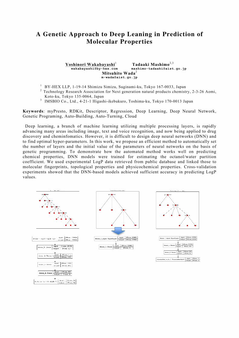
A Genetic Approach to Deep Leaning in Prediction of Molecular Properties
Yoshinori Wakabayashi1 Tadaaki Mashimo2,3
[email protected] [email protected] Mitsuhito Wada2 [email protected]
1 BY-HEX LLP, 1-19-14 Shimizu Simizu, Suginami-ku, Tokyo 167-0033, Japan 2 Technology Research Association for Next generation natural products chemistry, 2-3-26 Aomi,
Koto-ku, Tokyo 135-0064, Japan 3 IMSBIO Co., Ltd., 4-21-1 Higashi-ikebukuro, Toshima-ku, Tokyo 170-0013 Japan
Keywords: myPresto, RDKit, Descriptor, Regression, Deep Learning, Deep Neural Network, Genetic Programing, Auto-Building, Auto-Turning, Cloud Deep learning, a branch of machine learning utilizing multiple processing layers, is rapidly advancing many areas including image, text and voice recognition, and now being applied to drug discovery and cheminfomatics. However, it is difficult to design deep neural networks (DNN) and to find optimal hyper-parameters. In this work, we propose an efficient method to automatically set the number of layers and the initial value of the parameters of neural networks on the basis of genetic programming. To demonstrate how the automated method works well on predicting chemical properties, DNN models were trained for estimating the octanol/water partition coefficient. We used experimental LogP data retrieved from public database and linked those to molecular fingerprints, topological properties and physicochemical properties. Cross-validation experiments showed that the DNN-based models achieved sufficient accuracy in predicting LogP values.
Seed Generation by Integrating Computer-aided Drug
Design with Complementary Experimental Methods
Taiji Oashi1,2 Michihiko Suzuki2
[email protected] [email protected]
Osamu Saku3 Yuki Takayama
2
[email protected] [email protected]
Yuichi Takahashi2 Hikaru Miyagi1
[email protected] [email protected]
Kuniyuki Kishikawa2 Fumikazu Shinohara4
[email protected] [email protected]
Jun-ichi Saito5,6
1
Research Core Function Laboratories, Research Functions Unit, R&D Division, Kyowa Hakko
Kirin Co., Ltd 3-6-6 Asahi-machi, Machida-shi, Tokyo, 194-8533, Japan 2
Chemical Research Laboratories, Research Functions Unit, R&D Division, Kyowa Hakko Kirin
Co., Ltd 1188 Shimotogari, Nagaizumi-cho, Sunto-gun, Shizuoka, 411-8731, Japan 3
Environment and Safety Section, Ube Plant, Production Division, Kyowa Hakko Kirin Co., Ltd
2547-3 Fujimagari, Ube-shi, Yamaguchi, 755-8501, Japan 4
Innovative Technology Laboratories, Research Functions Unit, R&D Division, Kyowa Hakko
Kirin Co., Ltd 3-6-6 Asahi-machi, Machida-shi, Tokyo, 194-8533, Japan 5
R&D Planning Department, R&D Division, Kyowa Hakko Kirin Co., Ltd 1-9-2 Otemachi,
Chiyoda-ku, Tokyo 100-0004, Japan 6
Open Innovation Department, R&D Division, Kyowa Hakko Kirin Co., Ltd 1-9-2 Otemachi,
Chiyoda-ku, Tokyo 100-0004, Japan
Keywords: Structure-based drug design, 3D-database screening, Druggability evaluation, Oral
bioavailability prediction,
Structure-based drug design (SBDD) has been used to obtain the compounds that could fit the
target site of interest, modulating the disease-associated biological process. Here we will present
how a combination of computational and experimental methods was utilized in the early seed
generation process by two examples; (1) identification of novel small-molecule inhibitor for a
specific target potentially involved in cancer-related disease via 3D-database screening and in vitro
enzyme inhibition assay, (2) discovery of potency-enhancing non-natural nucleotide for siRNA by
using 3D-database screening, NMR-based binding assay, X-ray crystallography, and in vitro siRNA
knockdown experiment. For further improving the seed generation process, we will also discuss
key elements in the computational approach, including identification and druggability evaluation
of putative ligand-binding site [1]
and in silico screening as well as prediction of potential
bioavailability and druglikeness for compounds selected from database screen prior to the final
selection for experimental assay [2].
[1] Zhong, S., and MacKerell, A.D. Binding response: a descriptor for selecting ligand binding site
on protein surfaces. Journal of Chemical Information and Modeling, 47:2303-2315, 2007.
[2] Oashi, T., Ringer, A.L., Raman, E.P., and Mackerell, A.D. Automated selection of compounds
with physicochemical properties to maximize bioavailability and druglikeness. Journal of
Chemical Information and Modeling, 51:148-158, 2011.
A Graph Genome Browser
Toshiyuki Yokoyama Yoshitaka Sakamoto Masahide Seki [email protected] [email protected] [email protected]
Yutaka Suzuki Masahiro Kasahara [email protected] [email protected]
Department of Computational Biology and Medical Sciences, Graduate School of Frontier Sciences, The University of Tokyo, 5-1-5 Kashiwanoha, Kashiwa City, Chiba 277-8583, Japan
Keywords: NGS, Graph Genome, Structural Variations, Visualization, Genome Browser Background Continuous reduction in sequencing cost and continuous improvement in sequencing throughput realized population-scale sequence data, which underscores the importance of the development of graph genome analysis for handling a large number of genomes with variations of different scales. Since most part of the genomes is shared between individuals, we need to focus on differences in order to minimize the computation time. Single-nucleotide variants or small insertions and deletions (indels) called from those data can be handled more accurately by a graph genome. On top of that, long-read sequencing enables us to more accurately identify large-scale structural variants (SVs) and fusion genes, which include duplications or indels as loops or branching and merging against a reference genome. Graph genomes have been considered promising especially in human variation analysis. Although graph genome analysis is inextricably linked to the visualization of graph genomes, means to visualize graph genomes are yet to be developed. Existing genome browsers can visualize linear genomes but not graph genomes. Existing tools for handling graph genomes at the human genome scale cannot visualize genomes interactively and intuitively. Results We propose a graph genome browser for visualizing genome constructed by combining mathematical graphs with biological annotations. We define a sequence graph as a bidirected graph composed of a set of multiple DNA sequences as nodes and the corresponding end-to-end connections as edges. With these representations, we aim to visualize cancer genomes including SVs or fusion genes through comparison with the reference genome using sequencing data including long reads. Conclusions We implement a novel graph genome browser in order to provide an infrastructure for graph genome analysis. Our graph genome browser enables genome researchers to interactively explore biologically meaningful features observed as structures on a graph.
Fragmentation prediction using neutral loss-fragment pairs
Hiroyuki Yamamoto1 Hiroshi Tsugawa2
[email protected] [email protected]
1
Human Metabolome Technologies, Inc., 246-2 Mizukami Kakuganji, Tsuruoka, Yamagata
997-0052, Japan 2
RIKEN Center for Sustainable Resource Science, 1-7-22 Suehiro-cho, Tsurumi-ku, Yokohama,
Kanagawa 230-0045, Japan
Keywords: MS/MS Fragmentation prediction, Machine learning, Metabolomics
In metabolomics, unknown peaks that chemical structures have not been determined are
sometimes selected as metabolomic biomarker candidates in disease associated biomarker
discovery. Recently, some chemoinformatics approaches using MS/MS spectra with the objective
with structural elucidation of unknown peaks have been reported [1, 2]. In this study, we aimed to
predict fragmentation by using neutral loss-fragment pairs.
For 438 metabolites, we cut of all combination of chemical bonds in two step computations. The
disconnected structure pairs were compared with neutral loss and fragment pairs registered in
MassBank database [3], and matched pairs were assigned as positive samples and unmatched pairs
as negative samples. For 424 metabolites, PubChem fingerprint for the disconnected structures was
computed. A fragment feature vector was generated by the similar way as generation of the feature
vector from the compound pair in the prediction of the enzymatic reaction by Kotera et al . [4]. The
fragment feature vector was set as explanatory variable and matched or unmatched information as
a binary response variable to make a prediction model of fragmentation. Regularized logistic
regression approaches of ridge logistic regression, elastic net and lasso were performed. A regularized
parameter was optimized by cross-validation, and the prediction accuracy was computed for
independent test set. A maximum value of area under the curve of the receiver observation curve for the
test set was 0.7839 by using logistic regression with lasso. We plan to examine to improve prediction
accuracy by using machine learning approaches such as deep learning in the future.
[1] Tsugawa, H. et al., Hydrogen rearrangement rules: computational MS/MS fragmentation and
structure elucidation using MS-FINDER software, Analytical Chemistry, 88(16):7946-58 (2016).
[2] Duhrkop, K. et al., Searching molecular structure databases with tandem mass spectra using
CSI:FingerID, PNAS, 112(41):12580-5 (2015).
[3] Horai, H. et al., MassBank: a public repository for sharing mass spectral data for life sciences,
Journal of Mass Spectrometry, 45(7):703-14 (2010).
[4] Kotera, M. et al., Supervised de novo reconstruction of metabolic pathways from metabolome -scale
compound sets, Bioinformatics, 29(13):i135-44 (2013).
Target Genes and Pathways in Drug Development for Rare Diseases in Clinical Trials
Ryuichi Sakate1 Akiko Fukagawa1 Masashi Suzuki2
[email protected] [email protected] [email protected]
Masami Morita2 Akifumi Matsuyama1
[email protected] [email protected]
1 National Institutes of Biomedical Innovation, Health and Nutrition, 7-6-8 Saito-Asagi, Ibaraki-shi, Osaka 567-0085, Japan
2 Office of Pharmaceutical Industry Research, Japan Pharmaceutical Manufacturers Association, Nihonbashi Life Science Bldg., 2-3-11 Nihonbashi-honcho, Chuo-ku, Tokyo 103-0023, Japan
Keywords: Rare disease, Drug development, Clinical trial
Unmet medical needs in drug development for rare diseases are growing worldwide. In order to contribute to the drug development and related studies, trends in clinical trials for 306 rare diseases (intractable diseases) designated by the Ministry of Health, Labour and Welfare Japan had been analyzed [1-4]. In this study, further analyses were conducted to clarify the relation between tested drugs and their target genes and pathways. As a result, it was found that, for 122 rare diseases, 287 genes in 109 pathways (KEGG) were targeted by 338 drugs (DrugBank). Information of how drugs, genes and targets were shared by diseases was obtained. This information is expected to facilitate drug repositioning and studies of pathogenic mechanism. [1] Suzuki, M., Sakate, R., and Fukagawa, A., [Status of implementation of clinical trials on
intractable diseases designated in Japan], The Office of Pharmaceutical Industry Research News, 48:23-27. 2016
[2] Suzuki, M., Sakate, R., and Fukagawa, A., [Implementers of clinical trials for the designated intractable diseases], The Office of Pharmaceutical Industry Research News, 49:29-34. 2016
[3] Suzuki, M., Sakate, R., and Fukagawa, A., [Connecting information of the designated intractable diseases to databases], The Office of Pharmaceutical Industry Research News, 49:35-46. 2016
[4] Suzuki, M., Sakate, R., and Fukagawa, A., [Launched drugs used for clinical trials of the designated intractable diseases], The Office of Pharmaceutical Industry Research News, 50:39-43. 2017
Prediction of natural biosynthetic pathway by reverse synthetic analysis
Tsubasa Matsumoto Kohei Amano [email protected] [email protected]
Miyabi Hishinuma Masaaki Kotera
[email protected] [email protected]
School of Life Science and Technology Department of Life Science and Technology, Tokyo Institute of Technology, 2-12-1, Oookayama, Meguro-ku, Tokyo, 152-8550, Japan
Keywords: Reverse synthetic analysis, Machine learning The relationship between natural products and enzymes that synthesize them and their genes has been extensively studied in the fields of synthetic biology, metabolic engineering, phytochemistry and the like. However, the number of natural products whose metabolic pathways are unknown, and the number of enzyme genes whose function is unknown are both increasing. So it is desired to develop a method for exhaustively connecting these two types of information. Most of the informatics methods on the metabolic pathway are gene expression level and metabolite quantity for known metabolic pathways such as glycolysis system and Krebs cycle. On the other hand, Kotera and colleagues have established the methods such as predicting an enzyme gene (or enzyme protein) that catalyzes a specified reaction[1][2] or selecting a combination that can be a substrate / product pair of an enzyme reaction from tens of thousands of compounds[3][4]. In this study, we develop a chemical informatics method for predicting the biosynthetic pathway of various natural products using large scale computer system and extracting biosynthetic genes. By integrating reverse synthetic analysis and machine learning, we propose a framework to predict chemical transformations that make up the unknown metabolic pathway and support the identification of enzyme genes and enzyme proteins involved in it. [1] Tabei Y, Yamanishi Y, Kotera M. Simultaneous prediction of enzyme orthologs from chemical transformation patterns for de novo metabolic pathway reconstruction. Bioinformatics, 32, i278-i287, 2016. [2] Moriya Y, Yamada T, Okuda S, Nakagawa Z, Kotera M, Tokimatsu T, Kanehisa M, Goto S. Identification of Enzyme Genes Using Chemical Structure Alignments of Substrate-Product Pairs. J Chem Inf Model. 56(3):510-516, 2016. [3] Yamanishi Y, Tabei Y, Kotera M. Metabolome-scale de novo pathway reconstruction using regioisomer-sensitive graph alignments. Bioinformatics. 31(12):i161-i170, 2015. [4] Kotera M, Tabei Y, Yamanishi Y, Tokimatsu T, Goto S. Supervised de novo reconstruction of metabolic pathways from metabolome-scale compound sets. Bioinformatics, 29(13), i135-i144, 2013.
Construction of a Transcriptome-based Toxicity Prediction Model of Adjuvants
Natsuko Kishishita1,2,4 Yoshinobu Igarashi3
[email protected] [email protected]
Hiroshi Yamada3 Ken Ishii1,2,4 [email protected] [email protected]
1-3 1Laboratory of Mockup Vaccine, 2Laboratory of Adjuvant Innovation, and 3Laboratory of Toxicogenomics Informatics, National Institutes of Biomedical Innovation, Health and Nutrition, Ibaraki, Osaka, 567-0085, Japan
4 Laboratory of Vaccine Science, Immunology Frontier Research Center, World Premier International Research Center, Osaka University, Suita, Osaka 565-0871, Japan
Keywords: Adjuvant, Toxicity, Transcriptome, Biomarker
Research and development of adjuvants is very active today to improve the efficacy of vaccines against various diseases such as cancers and chronic disorders. Many adjuvants are ligands for pattern-recognition receptors that are important for innate immunity, and they skew immune responses in a certain way to induce Th1- or Th2-type response, for instance. Many adjuvants are developed over the world by pharmaceutical and venture companies as well as academia, and some of them are in clinical trials. And yet, it is not easy to evaluate the safety and efficacy of adjuvants accurately in preclinical studies or clinical trials to avoid adverse reactions in humans.
We have constructed the Adjuvant Database (http://adjuvantdb.nibiohn.go.jp/) to evaluate safety and efficacy of adjuvants with the grant from the Ministry of Health Labour and Welfare and AMED. Toxicity tests with rats were performed to obtain gene expression profiles of major tissues such as liver and kidney, and dataset were analyzed collectively with a large toxicogenomics database developed by our institute (http://toxico.nibiohn.go.jp/). We set drugs and adjuvants with known toxicity as “positive controls” to construct a toxicity prediction model based on gene expression data of each tissue. In addition, we analyzed several blood factors including conventional tissue-damage biomarkers (e.g., AST and ALT) and microRNAs to search for new biomarkers. As a result, a toxicity prediction model determined reactogenic adjuvants as “toxic” and those adjuvants that are clinically-approved or under clinical trials as “safe”. In fact, one of the “toxic” adjuvant was known for liver toxicity, greatly affected a set of genes involved in cell death and increased multiple tissue-damage biomarkers in the periphery blood. Meanwhile, “safe” adjuvants did not show toxicity-related features as expected. We further plan to optimize a toxicity prediction model to generalize molecular signatures of toxicity and aim to identify such toxicity biomarkers that are applicable to humans.
Prediction of Biosynthetic Basic Parts of Compounds with Complicated Structures
Kohei Amano1 Tsubasa Matsumoto1 [email protected] [email protected] Miyabi Hishinuma1 Masaaki Kotera1 [email protected] [email protected]
1 School of Life Science and Technology, Tokyo Institute of Technology, M6-2, 2-12-1 Ookayama, Meguro-ku, Tokyo, 152-8550, Japan
Keywords: Biosynthetic pathway analysis, Genetic Algorithm Many pharmaceutical compounds such as antibiotics are derived from natural products. Identification of their metabolic pathways is required not only for metabolic engineering but also for synthetic biology. Natural product chemists estimate reaction pathways or identify enzyme proteins on the basis of their own knowledge and experiences. Most of the informatics methods on the metabolic pathway deal with known metabolic pathways such as glycolysis system and Krebs cycle, and do not focus on identifying previously unknown metabolism. On the other hand, Kotera and colleagues published the series of methods such as predicting an enzyme gene or an enzyme protein that catalyzes a specified reaction[1][2] or selecting a combination that can be a substrate / product pair of an enzyme reaction from tens of thousands of compounds[3][4]. We focused on the fact that living things make a lot of natural products from a limited number of basic parts. This study provides the method of predicting the biosynthetic basic parts from natural products that contain complicated structures. We propose a new approach that we clarify the basic metabolite in its metabolic pathway in advance to avoid combinatorial explosion. The database of compounds which can be candidates of the biosynthetic basic parts is prepared. These compounds are supposed to be detectable by metabolome analysis. We virtually cut any chemical bonds contained in the interesting compound, and decompose it into partial structures. We make several evaluation functions with some molecular fingerprints and molecular similarities, and decide which bonds are to be cut by comparing with the database. It is tested by genetic algorithm to adopt compounds which metabolic pathways are known. We believe our method can support the elucidation of biosynthetic pathway. [1] Tabei Y, Yamanishi Y, Kotera M. Simultaneous prediction of enzyme orthologs from chemical transformation patterns for de novo metabolic pathway reconstruction. Bioinformatics, 32, i278-i287, 2016. [2] Moriya Y, Yamada T, Okuda S, Nakagawa Z, Kotera M, Tokimatsu T, Kanehisa M, Goto S. Identification of Enzyme Genes Using Chemical Structure Alignments of Substrate-Product Pairs. J Chem Inf Model. 56(3):510-516, 2016. [3] Yamanishi Y, Tabei Y, Kotera M. Metabolome-scale de novo pathway reconstruction using regioisomer-sensitive graph alignments. Bioinformatics. 31(12):i161-i170, 2015. [4] Kotera M, Tabei Y, Yamanishi Y, Tokimatsu T, Goto S. Supervised de novo reconstruction of metabolic pathways from metabolome-scale compound sets. Bioinformatics, 29(13), i135-i144, 2013.
A system level investigation to unravel mechanisms and
potential direct targets of the osteogenic small molecule
TH
Mochammad Ichsan1 Kosuke Kanke2 Hironori Hojo1,3 [email protected] [email protected] [email protected]
Ung-il Chung1,3 Shinsuke Ohba1,3
[email protected] [email protected]
1 Department of Bioengineering, University of Tokyo, Tokyo 113-8656, Japan 2 Department of Sensory and Motor System Medicine, University of Tokyo, Tokyo 113 -0033,
Japan 3 Center for Disease Biology and Integrative Medicine, University of Tokyo, Tokyo 113 -8655,
Japan
Keywords: Kinase inhibition, Microarray, Molecular docking, Osteoblast differentiation, Reverse
docking
Direct reprogramming of human somatic cells such as dermal fibroblast s (DFs) into several cell
types without introduction of transcription factors or genetic manipulation has drawn much
attention. Although TH, an osteogenic helioxanthin-derivative, has been shown to act as a robust
small molecule inducing differentiation of several cell types into osteoblasts [1,2,3,4], detailed
mechanisms and direct targets of TH remain unclear. In the present study, we aimed to unravel
them in order to develop novel strategies to chemically reprogram DFs into osteoblasts.
Microarray analysis revealed 1,166 differentially expressed genes during an osteoblast induction
phase in TH-mediated stepwise differentiation of mouse embryonic stem cells (mESCs) into
osteoblasts. We also confirmed the increased expression of osteoblast differentiation-related genes,
such as Col1a1, Ibsp, Runx2 and Sp7, in the dataset. Furthermore, we identified focal adhesion,
ECM-receptor interaction, and PI3K-Akt signaling pathways as the most enriched pathways
modulated by TH. Bioactivity prediction showed that TH potentially acted as a kinase inhibitor
with the highest bioactivity score compared to other 22 kinase inhibitors obtained from
Osteogenics DB, an osteoblast-related molecule database which we newly developed. We then
identified 145 potential targets of TH by pharmacophore- and structure-based reverse docking and
62 kinases, which were potentially inhibited during the osteoblast induction phase, by a kinase
enrichment analysis. By intersecting all identified potential targets of TH with 344 kinases which
are targeted by osteogenic kinase inhibitors in Osteogenics DB, we identified 42 proteins as the
most potential direct targets of TH that fulfilled all criteria used in this study. Lastly, we performed
molecular docking analyses to predict binding affinity and binding specificity of TH against the
most potential direct targets. Taken together, these findings suggest that TH achieves its osteogenic
activity at least partially via modulating the focal adhesion, ECM-receptor interaction, and
PI3K-Akt signaling pathways and potentially acts as a kinase inhibitor against several kinases.
[1] Ohba S et al. Biochem Biophys Res Commun. 357(4):854-60, 2007
[2] Nakajima K et al. Biochem Biophys Res Commun. 395(4):502-8, 2010
[3] Kanke K et al. Stem Cell Reports. 2(6):751–60, 2014
[4] Zujur D et al. Sci Adv. 3(5):e1602875, 2017
Large-scale diseasome analysis of gene expression signatures toward understanding disease–disease
associations and drug discovery Michio Iwata1 Ryusuke Sawada1 Yasuo Tabei2 [email protected] [email protected] [email protected]
Yoshihiro Yamanishi1,3
1 Division of System Cohort, Medical Institute of Bioregulation, Kyushu University, 3-1-1 Maidashi, Higashi-ku, Fukuoka, Fukuoka 812-8582, Japan
2 RIKEN Center for Advanced Intelligence Project, 2-1 Hirosawa, Wako, Saitama 351-0198, Japan
3 PRESTO, Japan Science and Technology Agency, Kawaguchi, Saitama 332-0012, Japan Keywords: diseasome, patients, gene expression, drug discovery, machine learning
Diseases are caused by dysfunction of the human biological system consisting of genes, proteins, and pathways. The pathogenesis is basically considered disease-specific, but characteristic molecular features are often similar among patients of different diseases, suggesting the commonality of the underlying molecular mechanisms. Most drugs are small compounds that modulate the activity of biological systems for the treatment of diseases. Thus, the knowledge on molecular correlations among diseases can be useful for drug discovery. Diseases are characterized by impaired expression of genes, and drugs modulate the activity of gene expression system. Thus, the use of transcriptome data for diseases and those for drug candidate compounds is a rational approach toward drug discovery. However, the integration of disease-specific and drug-induced transcriptome data has been an open question.
In this study we performed a large-scale diseasome analysis of gene expression signatures for 79 diseases (e.g., adrenoleukodystrophy, leukemia, Alzheimer’s disease, asthma, atopic dermatitis, breast cancer, cystic fibrosis, dengue and inflammatory bowel disease). We extracted characteristic gene expression patterns that explain the specificity of each disease and commonality between different diseases. We also proposed a novel machine learning method for drug discovery from the integration of disease-specific and drug-induced transcriptome data using multi-layered diseasome similarities computed from various molecular features (e.g., gene expressions, diagnostic markers, disease-causing genes, disordered pathways, and environmental factors), and demonstrated the usefulness of the proposed method in terms of accuracy and applicability. The diseasome analysis is expected to be useful for understanding disease-disease associations and for drug discovery.
Prediction of novel mitochondrial proteins having
disease-associated mutations toward discovery of drug
target
Kenichiro Imai1 Yoshinori Fukasawa2 [email protected] [email protected]
Kentaro Tomii2 Paul Horton2 [email protected] [email protected]
1 Molecular Profiling Research Center for Drug Discovery, National Institute of Advanced
Industrial Science and Technology (AIST), 2-4-7 Aomi, Koto-ku, Tokyo 135-0064, Japan 2 Artificial Intelligence Research Center, National Institute of Advanced Industrial Science and
Technology (AIST), 2-4-7 Aomi, Koto-ku, Tokyo 135-0064, Japan
Keywords: Mitochondria, Disease-associated mutations, Proteome, Transcriptome
Mitochondria provide numerous essential functions for cells and their dysfunction leads to a
variety of diseases. Thus, obtaining a complete mitochondrial proteome should be a crucial step
toward understanding the roles of mitochondria in the diseases. Many mitochondrial proteins have
been identified experimentally but a complete list is not yet available. To fill this gap, methods to
computationally predict mitochondrial proteins from amino acid sequence have been developed
and are widely used, but unfortunately, their accuracy is far from perfect. We developed MitoFates
[1], an improved prediction method for cleavable N-terminal mitochondrial targeting signals
(presequences) and their cleavage sites. MitoFates is a Support Vector Machine-based predictor
and considers positively charged amphiphilicity, presequence motifs, position weight matrices
modeling the presequence cleavage sites and classical features such as amino acid composition and
physico-chemical properties. On independent test data, MitoFates attains better performance than
existing predictors in both detection of presequences and in predicting their cleavage sites . In this
study, we used MitoFates to look for novel mitochondrial proteins involved in human diseases. We
obtained 83583 curated disease-associated variants from 6388 human diseases, which are collected
form UniProt, ClinVar [2], and the GWAS Catalog [3] using DisGeNET [4]. Then we analyzed
transcripts possessing the disease-associated variants using MitoFates and found more than 100
transcripts cording novel mitochondrial protein candidates involved in human diseases. We will
report the functional annotation of the candidates. Also we will discuss interpretation of relevance
between the candidates, the mutations and the diseases. The candidate list would be helpful in
elucidating the role of mitochondria in health and disease.
[1] Fukasawa et al., MitoFates: improved prediction of mitochondrial targeting sequences and their
cleavage sites, Mol Cell Proteomics, 14(4):1113-26, 2015
[2] Landrum M.J. et al., ClinVar: public archive of interpretations of clinically relevant variants.
Nucleic Acids Res., 44:D862–D868, 2016.
[3] Welter D. et al., I The NHGRI GWAS Catalog, a curated resource of SNP-trait associations,
Nucleic Acids Res., 42:D1001–D1006, 2014
[4] Piñero J., et al., DisGeNET: a comprehensive platform integrating information on human
disease-associated genes and variants, Nucleic Acids Res., 45(D1):D833-D839, 2017
Image-based phenotypic profiling associated with EGF receptor trafficking and signal transductions reveals
novel mode of action of drugs
Kenji Tanabe [email protected]
Medical Research Institute, Tokyo Women’s Medical University, 8-1 Kawada-cho, Shinjuku-ku, Tokyo 162-8666, Japan
Keywords: High contents screening, Mode of action of drugs, EGF receptor, small-molecule inhibitors Small-molecule compounds, including kinase inhibitors, are widely used as anti-cancer drugs. However, as almost all small-molecule compounds have multiple targets, it may cause an unexpected side effect and contribute to high attrition rate in clinical trials. Therefore, target deconvolution of drug candidates is a critical theme for drug development. Although various approaches are used for the target deconvolution, revealing a functionally unrelated target molecule, such as non-kinase targets for kinase inhibitors, is difficult. Recently, we established an image-based compound profiling, and found an unexpected non-kinase target (microtubules) for a most uni-specific kinase inhibitor (Tanabe, Sci. Rep. 2016). This study applied the image-based compound profiling for the target deconvolution of an inhibitor library, which consists of ~400 clinical drugs and various inhibitors. A549 cells, which express GFP-EGF receptor, were exposed to inhibitors, stimulated by EGF and visualized several major signaling molecules. Cells were photographed using automated fluorescent microscopy, and analyzed to extract ~500 image features to quantitatively evaluate their phenotypes. These extracted image features were used to classify inhibitors using unsupervised machine learning. As result, many clusters with unexpected combination of inhibitor were observed. For example, sodium channel inhibitor or transferase inhibitor were classified with some PI3K-Akt inhibitors. Interestingly, biochemical assay showed that they share target molecules associated with PI3K-Akt pathway. Similar observations were obtained in other clusters including cytoskeleton inhibitors. This result indicates that this image-based compound profiling is a powerful tool to reveal mode of action of drugs. [1] Tanabe, K., Image-based compound profiling reveals a dual inhibitor of tyrosine kinase and
microtubule polymerization, Scientific Reports, 6:25095, 2016.
Analysis of “uncallable” regions in the human reference genome associated with gene annotation and segmental
duplication
Shu Saikawa1 Masahiro Kasahara2 [email protected] [email protected]
1 Department of Computational Biology and Medical Sciences, Graduate School of Frontier Sciences, The University of Tokyo, Kashiwa 277-8568, Japan.
Keywords: Next generation sequencing, Disease omics analysis
Genome wide association study (GWAS) is one of the most important applications of
sequencing technology and contributed to the detection of gene regions associated with diseases in
many researches[1][2]. However, most of those projects did not result in the detection of highly
correlated SNV and the effectiveness of GWAS study is questioned by researchers [3].
We expect that GWAS study so far does not achieve adequate results because there are some
problems in bioinformatics analysis processes and software. Particularly, we focused on the
problems in mapping procedures. Read mapping software often used in genome sequencing
research such as BWA [4] and Bowtie [5] discard reads or select a certain position randomly from
multiple positions (in this case, map quality becomes zero) when they deal with multi-mapping
reads. In both cases multi-mapping reads are not used in subsequent analysis.
The probability that certain number of reads are uniquely mapped in each genome position
can be modeled by poisson distribution. Therefore, in this research we calculated the probability
that enough number of reads for SV detection are mapped in each genome position using poisson
distribution. Then we focused on the regions where this probability is low (which we call
“uncallable region”) and associated with gene annotation and segmental duplication.
[1] Okada, Y., et al., Genetic of rheumatoid arthritis contributes to biology and drug discovery, Nature
506:376-381, 2013.
[2] Imamura, M., et al., Genome-wide association studies in the Japanese population identify seven novel
loci for type 2 diabetes, Nature Communications. 7:10531, 2016.
[3] Gallaway, E., New concerns raised over value of genome-wide disease studies, Nature, 546:463, 2017.
[4] Li, H. & Durbin, R., Fast and accurate short read alignment with Burrows-Wheeler transform,
Bioinformatics, 25:1754-1760, 2009.
[5] Langmead, B. , Trapnel l , C . , Pop, M. , Salzberg , S .L. , Ul t rafas t and memory-eff ic ient a l ignment
of shor t DNA sequences to the human genome, Genome Biology , 10:R25, 2009.
Toward the next step in G protein-coupled receptor
research: a knowledge-driven analysis for the next targets
in drug discovery
Koji Nagata1 Yukie Katayama1 Tomomi Sato2 [email protected] [email protected] [email protected]
Yeondae Kwon1 Takeshi Kawabata3
[email protected] [email protected]
1 Department of Applied Biological Chemistry, Graduate School of Agricultural and Life
Sciences, The University of Tokyo, 1-1-1 Yayoi, Bunkyo-ku, Tokyo 113-8657, Japan 2 Structural Biology Research Center, Photon Factory, Institute of Materials Structure Science,
High Energy Accelerator Research Organization (KEK), 1-1 Oho, Tsukuba, Ibaraki 305-0801, Japan
3 Institute for Protein Research, Osaka University, 3-2 Yamadaoka, Suita, Osaka 565-0871, Japan Keywords: DDSS, OMIM, Orphan GPCR, VaProS More than 800 G protein-coupled receptor (GPCR) genes have been discovered in the human genome. Towards the next step in GPCR research, we performed a knowledge-driven analysis of orphan class-A GPCRs that may serve as novel targets in drug discovery [1]. We examined the relationship between 61 orphan class-A GPCR genes and diseases using the OMIM database [2] and the DDSS tool [3]. The OMIM database contains data on disease-related variants of the genes. Particularly, the variants of GPR101, GPR161, and GPR88 are related to the respective diseases: growth hormone-secreting pituitary adenoma 2; pituitary stalk interruption syndrome (interpretations conflicting); and childhood-onset chorea with psychomotor retardation. On the other hand, the DDSS tool suggests that 49 out of the 61 orphan receptor genes are related to diseases, judging from their co-occurrences in abstracts of biomedical literature. Notably, GPR50 and GPR3 are related to as many as 25 and 24 disease-associated keywords, respectively. The aforementioned five orphan GPCRs were characterized genetically, structurally and functionally using the structural life science data cloud VaProS [4], so as to evaluate their potential as next targets in drug discovery. [1] Nagata, K., Katayama, Y., Sato, T., Kwon, Y., and Kawabata, T., Toward the next step in G
protein-coupled receptor research: a knowledge-driven analysis for the next potential targets in drug discovery, J. Struct. Funct. Genomics, 17, 111-133, 2017.
[2] Amberger, J. S., Bocchini, C. A., Schiettecatte, F., Scott, A. F., and Hamosh, A., OMIM.org: Online Mendelian Inheritance in Man (OMIM), an online catalog of human genes and genetic disorders, Nucleic Acids Res., 43, D789-798, 2015.
[3] Kwon, Y., Shimizu, S., Sugawara, H., and Miyazaki, S., A novel evaluation measure for identifying drug targets from the biomedical literature, IPSJ Transactions on Bioinformatics, 7, 16-23, 2014.
[4] Gojobori, T., Ikeo, K., Katayama, Y., Kawabata, T., Kinjo, A. R., Kinoshita, K., Kwon, Y., Migita, O., Mizutani, H., Muraoka, M., Nagata, K., Omori, S., Sugawara, H., Yamada, D., and Yura, K., VaProS: a database-integration approach for protein/genome information retrieval, J. Struct. Funct. Genomics, 17, 69-81, 2017.
Impact of read trimming on Illumina
paired-end-sequencing samples in the microbiome analysis
using Qiime
Attayeb Mohsen Jonguk Park Yi-An Chen [email protected] [email protected] [email protected]
Hitoshi Kawashima Kenji Mizuguchi [email protected] [email protected]
1 National institutes of Biomedical Innovation, Health, and Nutrition, 7-6-8, Saito-Asagi, Ibaraki
City, Osaka 567-0085, Japan
Keywords: Quality trimming, Next generation sequencing, Microbiome data analysis.
Paired-end sequences are generated by sequencing the target DNA from its both sides producing
partially overlapped two pair-end reads for the target DNA, then both reads are merged to produce
a longer final read. The quality of the result ing final read is affected by the base-specific quality
for the original reads. The Illumina paired-end sequencing technology yielded a good quality
product, and is the most commonly used technology nowadays. However, base-specific quality
scores reduce substantially with machine cycles, leading to low-quality bases as sequencing
approaches to the end of the read, which worsens the merging process and the subsequent analysis
steps in Qiime1.
Trimming2 is used to remove the parts with low quality, and helps improve the outcome of the
merging step and overall analysis. In this poster, we examined the importance of the trimming
quality threshold using different datasets, and showed the effect of this threshold on the number of
reads preserved after trimming, the number of picked operational taxonomic units (OTUs) and the
diversity measures.
[1] Caporaso J. G., et al., QIIME allows analysis of high-throughput community sequencing data.
Nature Methods 7(5): 335-336, 2010.
[2] Bushnell B., BBTools available at: (http://sourceforge.net/projects/bbtools/).
Has Proximity in Human Genome been Conserved through 2-Round Whole Genome Duplication ?
Hyogo Kajiyama1 Hiroyuki Toh1
[email protected] [email protected]
1 Department of Biomedical Chemistry, School of Science and Technology, Kwansei Gakuin University, 2-1 Gakuen, Sanda, Hyogo, 669-1337, Japan
Keywords: Two-Round Whole Genome Duplication, Hi-C Recent development of the NGS technology has enabled us to analyze various biological phenomena. One of the applications of the NGS is “Hi-C”, which can detect spatially adjacent chromosomal regions in human genome. Then, a model for the structural conformation of the human genome is built based on the Hi-C data [1, 2]. On the other hand, it is suggested that the vertebrates have undergone the two-round whole genome duplication (2RWGD). Associated with the events, four copies of paralogs were generated in the vertebrate genomes, although the numbers may have changed due to the lineage specific deletions and/or duplications after the events. In this research, we examined whether the adjacency of chromosomal regions has been conserved after the 2RWGD or not. At first, we obtained a data from NCBI GEO [3], which includes proximity scores between chromosomal regions generated from a Hi-C experiment [1]. In the data, each chromosome is divided into non-overlapped regions or bins of 1Mbp in length. We also obtained a whole set of pairs, gene name and protein ID, included in the human genome (hg19) from UCSC Genome Browser [4]. In addition, the information about the chromosomal locations of human genes was obtained through Galaxy [5]. Then, these data were integrated to generate a table, in which each row corresponds to a gene and consists of three elements, gene name, chromosomal location and protein ID of the gene. Next, we obtained the amino acid sequence data encoded by human genes from Uniprot [6]. The sequences thus obtained were sorted so that the proteins encoded in the same bin constitute one set, to make the sequence comparison described below easier. Thridly, we obtained a list of the paralogous genes associated with 2RWGD from OHNOLOGS [7,8]. Then, the bins including the paralogs were identified. The gene products encoded by the bins spatially adjacent to the different bins containing the paralogs were compared by making the alignments for all possible pairs between the adjacent bins. If paralogs are observed among the adjacent bins, it suggests the conservation of chromosomal adjacency after the 2RWGD. The conservation of the chromosomal contingency will be discussed based on the analysis. [1] Lieberman-Aiden, E. et al. (2009). Comprehensive mapping of long-range interactions reveals folding principles of the human genome. Science, 326(5950), 289-293. [2] Li, C. et al. (2014). The 3DGD: a database of genome 3D structure. Bioinformatics, 30.11: 1640-1642. [3] http://www.ncbi.nlm.nih.gov/geo/ [4] https://genome.ucsc.edu/ [5] https://usegalaxy.org/ [6] http://www.uniprot.org/ [7] Singh, P. P., Arora, J., & Isambert, H. (2015). Identification of ohnolog genes originating from whole genome duplication in early vertebrates, based on synteny comparison across multiple genomes. PLoS Comput Biol, 11(7), e1004394. [8] http://ohnologs.curie.fr/
Development of a pharmacokinetics prediction system using multiscale integrated modeling:
1. A curated database combining public and newly acquired experimental data
Hitoshi Kawashima1 Reiko Watanabe1 Tsuyoshi Esaki1 [email protected] [email protected] [email protected]
Rikiya Ohashi1,2 Daisuke Satoh3 Kazuyoshi Ikeda3
[email protected] [email protected] [email protected]
Chioko Nagao1 Yayoi Natsume-Kitatani1 Kenji Mizuguchi1 [email protected] [email protected] [email protected]
1 Laboratory of Bioinformatics, National Institutes of Biomedical Innovation, Health and
Nutrition, 7-6-8, Saito Asagi, Ibaraki-shi, Osaka 567-0085, Japan 2 Discovery Technology Laboratories, Mitsubishi Tanabe Pharma Corporation, 2-2-50, Kawagishi,
Toda-shi, Saitama 335-8505, Japan 3 Software Division, Level Five Co., Ltd., 2-25, Kanda Suda-cho, Chiyoda-ku, Tokyo 101-0041,
Japan Keywords: Pharmacokinetics, Database, Prediction models
To address pharmacokinetic and toxicological issues in drug development, once the main source of late attrition of drug candidates, many pharmaceutical companies have now implemented early ADME or early toxicological studies. However, such approaches are difficult to emulate in the academic drug discovery environment. Therefore, we began an initiative “Development of a Drug Discovery Informatics System”, with support from the Japan Agency for Medical Research and Development (AMED) in collaboration with several other research groups. The main aim of this initiative is to develop accurate prediction systems for ADME and tox properties primarily targeting academic scientists. Our group’s focus is to develop a pharmacokinetics database and prediction models.
Any good prediction system depends on high-volume, high-quality training datasets. We collected pharmacokinetic and physicochemical parameters from the public bioactivity database, ChEMBL [1]. However, since ChEMBL compiles data obtained in different experimental conditions, we developed a curation workflow to select the data measured in compatible conditions and to reformat the results as appropriate for our prediction system.
In addition to the public data, we have been acquiring both in vitro and in vivo experimental data using consistent protocols. The in vitro experiments include physicochemical parameters such as solubility and distribution coefficient, and pharmacokinetic parameters such as metabolic stability, protein binding in plasma, protein binding in brain homogenate, and blood-to-plasma concentration ratio. The in vivo data include the drug concentrations in plasma and tissues after oral or intravenous administration of the drug and pharmacokinetic parameters calculated therefrom.
We stored all the data in a PostgreSQL database and developed a web application to view and analyze the database content. We plan to integrate this system with the cardiotoxicity and hepatotoxicity databases developed within the AMED initiative described above. [1] https://www.ebi.ac.uk/chembl/ This work was conducted as part of “Development of a Drug Discovery Informatics System” supported by Japan Agency for Medical Research and Development (AMED).
Development of a pharmacokinetics prediction system
using multiscale integrated modeling:
2. Classification of aqueous solubility measured
using the dried-DMSO method
Tsuyoshi Esaki1 Reiko Watanabe1 Hitoshi Kawashima1 [email protected] [email protected] [email protected]
Yayoi Natsume-Kitatani1 Chioko Nagao1 Rikiya Ohashi1,2 [email protected] [email protected] [email protected]
Kenji Mizuguchi1
1 Laboratory of Bioinformatics, National Institutes of Biomedical Innovation, Health and
Nutrition, 7-6-8, Saito Asagi, Ibaraki-shi, Osaka 567-0085, Japan 2 Discovery Technology Laboratories, Mitsubishi Tanabe Pharma Corporation, 2-2-50, Kawagishi,
Toda-shi, Saitama 335-8505, Japan Keywords: Aqueous solubility, Dried-DMSO, Machine learning
It has been demonstrated that high-throughput in vitro measurements for the evaluation of
physicochemical, pharmacokinetic, and safety parameters can save time and cost in drug
development and ultimately improve the success rate. Solubility is an essential physicochemical
property having great influences on drug absorption. The oral administration of compounds with
low solubility can not only have low effects, but also be affected by the individual variation.
Therefore, in silico methods to estimate the solubility of compounds are needed and both free and
proprietary software programs have been developed. However, solubility is one of the properties
that have been reported to be difficult to predict with high accuracy [1]. In the academic drug
discovery environment, it is unfeasible to collect a large volume of new experimental data and
although a considerable amount of data are available from public databases, they came from a
variety of different experimental conditions. Thus, in this study, we compiled publicly available
solubility data that were measured using a single procedure, the dried-DMSO method, and
constructed computational models to predict solubility and to classify target compounds from
chemical structures alone. The dried-DMSO method is a high-throughput experimental method
widely used in pharmaceutical companies. All the experimental data were collected from the public
bioactivity database, ChEMBL (ver.23) [2], often by checking the original literature and excluding
the data measured using other (or unclear) methods. The collected compounds data were separated
into two groups, training and test sets. Different combinations of chemical descriptors and machine
learning algorithms were examined on the training set and the constructed models were evaluated
on the test set, resulting in an accurate system for predicting compound solubility measured using
the dried-DMSO method.
[1] Hughes L.D., et al., Why are some properties more difficult to predict than others? A study of
QSAR models of solubility, melting point, and LogP, J. Chem. Inf. Model., 48:220-232, 2008.
[2] https://www.ebi.ac.uk/chembl/ This work was conducted as part of “Development of a Drug Discovery Informatics System”
supported by Japan Agency for Medical Research and Development (AMED).
Development of a pharmacokinetics prediction system using multiscale integrated modeling:
3. Prediction of human plasma protein binding from chemical structures
Reiko Watanabe1 Tsuyoshi Esaki1 Hitoshi Kawashima1
[email protected] [email protected] [email protected]
Yayoi Natsume-Kitatani1 Chioko Nagao1 Rikiya Ohashi1,2 [email protected] [email protected] [email protected]
Kenji Mizuguchi1
1 Laboratory of Bioinformatics, National Institutes of Biomedical Innovation, Health and
Nutrition, 7-6-8, Saito Asagi, Ibaraki-shi, Osaka 567-0085, Japan 2 Discovery Technology Laboratories, Mitsubishi Tanabe Pharma Corporation, 2-2-50, Kawagishi,
Toda-shi, Saitama 335-8505, Japan
Keywords: Plasma protein binding, Protein unbound, Machine learning
The fraction of chemicals unbound to plasma proteins is an important determinant of the efficacy of a drug in pharmacokinetic and pharmacodynamic studies, since the free (unbound) form is available for pharmacological interactions. The unbound fraction affects the membrane permeability, metabolism and glomerular filtration rate, and consequentially it influences the volume of distribution in different tissues and the total clearance. In addition, unbound drugs exert pharmacological and toxicological effects. In the early stages of drug discovery, determining the fraction of drug unbound to plasma proteins in humans and in animal models provides an understanding of the ADME properties to assist candidate selection. Furthermore, it is important to predict the fraction of unbound drug in the brain for assessing brain drug delivery. Accordingly, the in silico prediction of unbound drug in plasma is perceived as an effective tool to mitigate the risk of late-stage attrition during drug development and to optimize further screening [1]. Human plasma contains several binding proteins such as human serum albumin (HSA), α-1 acid glycoprotein (AGP), lipoproteins and globulins. Consequently, only a few studies have reported in silico modeling to predict the binding ratio in intact plasma [2], although previous studies have reported the prediction of protein binding to simple substances such as HSA or AGP.
The aim of this study is to build high precision models to predict the fraction unbound in the intact human plasma. We employed three machine learning techniques, using topological descriptors to represent molecular structures, to generate models of human plasma protein binding based on the large data set of experimental values; these models were subsequently used to estimate the unbound fraction. For this purpose, multiple prediction models were created; a regression model to predict the protein binding in human plasma, a classification model to predict the fraction unbound in human plasma and different regression models based on the compound polarity such as acidic, basic and neutral. [1] Lambrinidis G., et al., In vitro, in silico and integrated strategies for the estimation of plasma
protein binding. A review, Adv. Drug Deliv. Rev., 86:27-45, 2015. [2] Votano J.R., et al., QSAR modeling of human serum protein binding with several modeling
techniques utilizing structure-information representation, J. Med. Chem., 49:7169-7181, 2006. This work was conducted as part of “Development of a Drug Discovery Informatics System” supported by Japan Agency for Medical Research and Development (AMED).
Development of a pharmacokinetics prediction system
using multiscale integrated modeling:
4. Development of a program for predicting the sites of
metabolism by CYP1A2 using a two dimensional template
Junya Ohori1 Hiromi Koga1 [email protected] [email protected]
Mayumi Matsushita1 Kenji Mizuguchi2
[email protected] [email protected]
1 Life Innovation Department, Healthcare Solutions Unit, Fujitsu Kyushu Systems Limited, 5-13
Higashihie 1-chome, Hakata-ku, Fukuoka 812-0007, Japan 2 Laboratory of Bioinformatics, National Institutes of Biomedical Innovation, Health and
Nutrition, 7-6-8 Saito Asagi, Ibaraki-shi, Osaka 567-0085, Japan
Keywords: In Silico, Drug Metabolism, Regioselectivity, Cytochrome P450
In drug discovery research it is important to predict whether metabolic enzymes metabolize drugs
and what metabolites would they produce. In silico methods for predicting various drug
metabolisms, mainly by cytochrome P450s, have been studied, but their very limited accuracy is
the main reason why they still remain only for supplementary use at the early stages of drug
discovery. In order to predict the safety of drug candidates as early as possible and to accelerate
the development of drugs, a prediction system that can predict metabolism with high accuracy is
desired.
In this study, we developed a program to predict the metabolic site of CYP1A2 based on the
method [1] of superimposing compounds to be predicted on a template representing the relevant
binding and metabolic sites of the enzyme. The template was prepared by overlaying several
known substrates of the metabolic enzyme and assigning additional physicochemical constraints to
it. The program developed to predict regioselectivity of the enzyme only requires the structural
formula of the query compound and does not require any complicated computational chemistry
methods. Here we report the prediction results of the program which were validated and compared
with the actual experiment results collected from literature.
[1] Yamazoe, Y. et al., Prediction of regioselectivity and preferred order of metabolisms on
CYP1A2-mediated reactions. Part 1. Focusing on polycyclic arenes and the related chemicals.
Drug Metabolism and Pharmacokinetics. 31(5):363-384, 2016.
This work was conducted as part of “Development of a Drug Discovery Informatics System”
supported by Japan Agency for Medical Research and Development (AMED).
Development of a pharmacokinetics prediction system using multiscale integrated modeling:
5. Prediction of sites of drug metabolism by cytochrome P450 by molecular simulation
Hiroaki Saito1, Taku Mizukami2, Yoshinori Hirano1,
[email protected], [email protected], [email protected],
Takao Otsuka1, Noriaki Okimoto1, Makoto Taiji1 totsuka.riken.jp, [email protected], [email protected]
1 Laboratory of computational molecular design computational biology, computational biology
research core, RIKEN Quantitative Biology Center (QBiC) 2 Department of Materials Sciences, Japan Institute of Science and Technology (JAIST)
Keywords: Molecular dynamics, Molecular docking, Site of metabolism, Cytochrome P450 (CYP) Cytochrome P450s (CYPs), a superfamily of haem-containing enzymes, are the major enzymes involved in drug metabolism. In humans, it has been estimated that CYPs metabolize approximately 75% of all marketed drugs, 95% of which are metabolized by CYP3A4, 2D6, 2C9 and 1A2 [1]. In the drug metabolism by the CYPs, identification of sites of metabolism (SOMs) on molecules and the structure of their metabolites can be decisive for the design of molecules with favorable metabolic properties. However, experimental techniques to determine SOMs and structures of metabolites are still highly resource-demanding and challenging [2]. Thus, developing fast and accurate computational methods to predict the SOMs/products of compounds metabolized by the CYPs is one of the important tasks for the optimization of ADME and toxicity properties. In this study, we present a new computational method (score function) to predict SOMs of compounds metabolized by CYP1A2. The new score function is composed of accessibility and reactivity scores. The accessibility scores for the sites (atoms) of compounds are estimated by ensemble docking simulation, while the activation energies of atoms stored in the SMARTCyp program[3] are adopted as the reactivity scores in the method. We carried out a molecular dynamics (MD) simulation of apo type of CYP1A2 for 10 micro second to sample the receptor (pocket) structures for the ensemble docking simulation. In the molecular docking, DOCK program with Grid score, which are optimized for CYP system [4], was used. We prepared 42 test set compounds metabolized by the CYP1A2 and 100,000 receptor snapshots (which correspond to the 10 micro second MD simulation) for the ensemble docking simulation. The top three ranked atoms by the new score function are defined as possible SOMs for each compound in this study. We found the success rate of the predicted SOMs was 94 %, showing better result than those by the ensemble docking (80%) and SMARTCyp program (88%) alone. [1] Wienkers LC, heath TG., Nature Rev. Drug. Discov. 4 (2005), 825-833. [2] Johannes Kirchmair et al., J. Chem. Inf. Model. 52 (2012), 617−648. [3] P. Rydberg et al., ACS Med. Chem. Lett., 1 (2010), 96-100 [4] Kiumars Shahrokh, Anita Orendt, Garold S. Yost, Thomas E. Cheatham, III, J. Comp. Chem., 33 (2012), 119-33. This work is conducted as a part of “Construction of Drug Discovery Informatics System” supported by Japan Agency for Medical Research and Development (AMED).
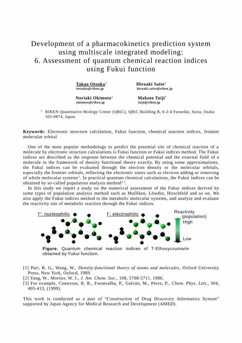
Reactivity (population)
High
Low
Figure. Quantum chemical reaction indices of 7-Ethoxycoumarin
obtained by Fukui function.
f+: nucleophilic f-: electrophilic
Development of a pharmacokinetics prediction system
using multiscale integrated modeling:
6. Assessment of quantum chemical reaction indices
using Fukui function
Takao Otsuka1 Hiroaki Saito1 [email protected] [email protected]
Noriaki Okimoto1 Makoto Taiji1 [email protected] [email protected]
1 RIKEN Quantitative Biology Center (QBiC), QBiC Building B, 6-2-4 Furuedai, Suita, Osaka
565-0874, Japan
Keywords: Electronic structure calculation, Fukui function, chemical reaction indices, frontier
molecular orbital
One of the most popular methodology to predict the potential site of chemical reaction of a
molecule by electronic structure calculations is Fukui function or Fukui indices method. The Fukui
indices are described as the response between the chemical potential and the external field of a
molecule in the framework of density functional theory exactly. By using some approximations,
the Fukui indices can be evaluated through the electron density or the molecular orbitals,
especially the frontier orbitals, reflecting the electronic states such as electron adding or removing
of whole molecular systems1. In practical quantum chemical calculations, the Fukui indices can be
obtained by so-called population analysis method2,3.
In this study we report a study on the numerical assessment of the Fukui indices derived by
some types of population analysis method such as Mulliken, Löwdin, Hirschfeld and so on. We
also apply the Fukui indices method to the metabolic molecular systems, and analyze and evaluate
the reactivity site of metabolic reaction through the Fukui indices.
[1] Parr, R. G., Wang, W., Density-functional theory of atoms and molecules , Oxford University
Press, New York, Oxford, 1989.
[2] Yang, W., Mortier, W. J., J. Am. Chem. Soc., 108, 5708-5711, 1986.
[3] For example, Contreras, R. R., Fuentealba, P., Galván, M., Pérez, P., Chem. Phys. Lett., 304,
405-413, (1999).
This work is conducted as a part of “Construction of Drug Discovery Informatics System”
supported by Japan Agency for Medical Research and Development (AMED).
Development of a pharmacokinetics prediction system using
multiscale integrated modeling:
7. Enhancement of the custom SoC LSI functionalities to
improve the system performance
Gentaro Morimoto Yousuke Ohno Hao Zhang [email protected] [email protected] [email protected]
Itta Ohmura Keigo Nishida Teruhisa Komatsu Yohei M. Koyama [email protected] [email protected] [email protected] [email protected]
Aki Hasegawa Hiroshi Koyama Makoto Taiji [email protected] [email protected] [email protected]
Research and Development Project of Special Purpose Supercomputers for Drug Design,
RIKEN Quantitative Biology Center, 6-2-4 Furuedai, Suita, Osaka 565-0874, Japan
Keywords: Molecular dynamics simulations, Ligand binding, Protein modeling, Drug design
We have been developing a series of special purpose computers for molecular dynamics
simulations. The current system is the MDGRAPE-4[1]. The whole system consists of 512 custom
System on Chip(SoC) LSI chips in 3D torus network. The target performance is to make it possible to simulate typical protein-ligand complex surrounded by water for 10 microseconds per day,
which is two orders of magnitude faster than commercially available systems. One of the
applications of long-term MD simulations of proteins is to predict the site of metabolism.
The MDGRAPE-4 hardware development completed in 2014, and the stabilization of operation and software development process succeeded for two years. However, the final performance does
not reach the expected one. In the process of the development, we realized that the classical MD
simulation of the typical system size has already reached the strong scaling limit of parallel computation and not only accelerating the computationally demanding part but also more
specialized hardware for the type of computation is required to improve the performance.
To achieve the target performance, we have started the development of the improved system that is named MDGRAPE-4A. The system architecture is basically common with the MDGRAPE-4.
The enhancement of the functionalities are as follows:
1) support of management and migration of atoms by the memory hardware
2) more sophisticated treatment of exclusion and reduced non-bonded interaction pairs in the special purpose pipelines
3) implementation of SIMD functions and special instructions required in the MD simulations in
the general-purpose cores 4) reduction of the latency and more flexible routing patterns in the network interface
5) support of a long-range electrostatic interaction calculation met hod that is suitable for the
parallel systems with torus network topology Currently the development process is under the design and the verification of the revised LSI.
The new system hardware will be completed in 2018. In this presentation, we would like to show
the ideas to improve the performance, progress and simulation results of the new LSI design.
[1] Ohmura, I., et al., MDGRAPE-4: a special-purpose computer system for molecular dynamics
simulations, Phil. Trans. R. Soc. A, 372:20130387, 2014.
Some of this work is conducted as a part of “Construction of Drug Discovery Informatics System”
supported by Japan Agency for Medical Research and Development (AMED).

Library Design With ChEMBL and Virtual Fragments Based On Electrostatic and Shape Bioisosterism Approach
Kazuyoshi Ikeda 1 Tomoki Yonezawa 2
[email protected] [email protected]
1 Keio University, Faculty of Pharmacy, Division of Physics for Life Functions, 1-5-30 Shibakoen, Minato-ku, Tokyo 105-8512, Japan
2 Division of Drug Discovery, Software Division, Level Five Co., Ltd., 2-25, Kanda Suda-cho, Chiyoda-ku, Tokyo 101-0041, Japan
Keywords: Field-based ligand design, Scaffold hopping, Tyrosine-protein kinase inhibitor, WaterSwap Computational approaches play an important role for efficient ligand design. Our group is analyzing a compound library of a diverse set of compounds as well as developing a core/focused library design method by utilizing information from drug discovery data-resources such as drugs/compounds/bioactivities and the targets. In this study, we have developed a protocol to generate a focused library using a molecular-field ligand-based method for a target. The field-based method, which is an improved method of the conventional pharmacophore model, can directly express molecule interactions of ligand to a protein target including electrostatic and hydrophobic interactions. The method can be also applied for scaffold/fragment hopping of a given reference-molecule to make the bioisosteric compound dataset [1]. Using the Pipeline Pilot [2], an automatic protocol has been developed to extract frequent rings and fragments from the ChEMBL database [3] and split based on the frequency of occurrence of the fragments in the databases. We also added the fragment data from historical/recent approved-drugs in the DrugBank [4] and a novel ring information from the VEHICLe (virtual exploratory heterocyclic library) to expand the chemical space. This protocol can filter drug-like/lead-like compounds with pharmaceutically desirable physicochemical properties. Tyrosine-protein kinase ABL/BTK inhibitor was selected as a test-set in this study and expanded their known drugs to the diverse set of the bioisosteric compounds with the similar electrostatic patterns. Additionally, the protein structure information was used for analyzing the residue-level ligand-protein interaction energetics in the active-site and identifying the substitution point of ligand for scaffold hopping using the WaterSwap method which is recently developed by Woods et al. [implemented in the software 1.]. In this presentation, we will explain and discuss the automatic protocol of generating a forced library and the applications to the known drug targets. [1] http://www.cresset-group.com/products [2] http://accelrys.co.jp/products/collaborative-science/biovia-pipeline-pilot [3] Bento, A. P., Gaulton, A., Hersey, A., Bellis, L. J., Chambers, J., Davies, M., Krüger, F. A.,
Light, Y., Mak, L., McGlinchey, S., Nowotka, M., Papadatos, G., Santos, R., and Overington, J. P., The ChEMBL bioactivity database: an update, Nucleic Acids Research, 42(Database issue):D1083-1090, 2014.
[4] Law V, Knox C, Djoumbou Y, Jewison T, Guo AC, Liu Y, Maciejewski A, Arndt D, Wilson M, Neveu V, Tang A, Gabriel G, Ly C, Adamjee S, Dame ZT, Han B, Zhou Y, Wishart DS. DrugBank 4.0: shedding new light on drug metabolism. Nucleic Acids Res. 2014 Jan 1;42(1):D1091-7.
[5] Rapid decomposition and visualisation of protein-ligand binding free energies by residue and by water. [6] Woods CJ, Malaisree M, Michel J, Long B, McIntosh-Smith S, Mulholland AJ. Faraday Discuss. 2014;169:477-99.
Development of an informatics system for predicting
cardiotoxicity: 1. Exploring binding modes of hERG
inhibitors based on docking simulation and
the validation with site-directed mutagenesis data
Hitomi Yuki Tomohiro Sato [email protected] [email protected]
Keiji Ogura Teruki Honma
[email protected] [email protected]
RIKEN Center for Life Science Technologies, 1-7-22, Suehiro-cho, Tsurumi-ku,
Yokohama, Kanagawa 230-0045, Japan
Keywords: hERG, Ligand docking, Pharmacophore, Site-directed mutagenesis
ADMET (absorption, distribution, metabolism, excretion and toxicity) studies are indispensable
to lead efficacious and safe drug candidates. Since the 2000s, toxicity has become a major par t of the cause of development failure,
1 among which the cardiotoxicity accounts for about 40% of the
total toxicity.2 hERG (human ether-à-go-go-related potassium channel) is a member of potassium
ion channels and involved in cardiac repolarization. Drug-induced hERG blocking causes long QT syndrome and it is a major cause of cardiotoxicity. Therefore, drug design for avoiding the hERG
inhibition is quite important through the all stages of drug discovery. Although 3D models of
hERG would provide worthy compound design ideas for reducing hERG inhibition, there was no
experimentally validated 3D model for several decades. This year, using emerging cryo -electron microscopy technologies, the molecular structure of hERG has been determined.
3
We performed MD simulation of the hERG cryo-electron microscopy structure and subsequent
docking simulations of known representative hERG inhibitors such as dofetilide to identify the essential pharmacophores for the hERG inhibition. hERG is a channel and shows high mobility
like cytochrome P450. It makes difficult to determine the appropriate binding mode from diverse
docking poses by only docking scores. Thus, we used the site -directed mutagenesis (SDM) results
for the hERG pore to select and validate the binding mode. SDM reveals which residues are associated with the inhibition of hERG for each compound. We would like to discuss the analysis
of these docking results and the pharmacophores of the hERG inhibition.
This work was done as a part of “Construction of Drug Discovery Informatics System” supported by Japan Agency for Medical Research and Development (AMED).
[1] Kola, I., and Landis, J. Can the pharmaceutical industry reduce attrition rates? Nat. Rev. Drug
Discov. 3:711–716, 2004.
[2] Laverty, H., Benson, C., Cartwright, E., Cross, M., Garland, C., Hammond, T., Holloway, C., McMahon, N., Milligan, J., Park, B., Pirmohamed, M., Pollard, C., Radford, J., Roome, N.,
Sager, P., Singh, S., Suter, T., Suter, W., Trafford, A., Volders, P., Wallis, R., Weaver, R., York,
M., and Valentin, J. How can we improve our understanding of cardiovascular safety liabilities
to develop safer medicines? Br. J. Pharmacol. 163:675–693, 2011 [3] Wang, W., and MacKinnon, R. Cryo-EM Structure of the Open Human Ether-à-go-go-Related
K+ Channel hERG. Cell 169:422–430.e10, 2017
Development of an informatics system for predicting
cardiotoxicity: 2. Update of integrated database for hERG
blocking small molecules considering deviation of assay
results.
Tomohiro Sato Hitomi Yuki [email protected] [email protected]
Keiji Ogura Teruki Honma [email protected] [email protected]
Center for Life Science Technologies, RIKEN, 1-7-22 Suehiro-cho, Tsurumi-ku, Yokohama
City, Kanagawa, 230-0045, Japan
Keywords: hERG, human ether-a-go-go, QT prolongation, database
The inhibition of hERG potassium channel is closely related to the prolonged QT interval [1], and to assess the risk could greatly contribute to the development of safer therapeutic compounds. To
utilizing the recent increase of information about hERG inhibitors in public databases, integrated
database for hERG blocking compounds was built and reported in CBI annual meeting 2016. Here, we updated the database using latest versions of ChEMBL, PubChem, GOSTAR, and hERG
Central [2], and refined the merging procedure to accommodate to compounds for which
contradictory assay results were found across multiple databases. Assessment of structural
diversity using Murcko frameworks revealed that the integrated database contains more than 2 times as many scaffold for hERG inhibitors as any of the existing databases, and covering 18.2%
of all chemical space occupied by whole compounds in ChEMBL (438,551 frameworks). (Table 1)
The database could provide most comprehensive information about hERG inhibitors and be useful to predict the inhibitory activity and design safer compounds for drug discovery.
This work was done as a part of “Construction of Drug Discovery Informatics System” supported
by Japan Agency for Medical Research and Development (AMED).
Table 1. Number of hERG inhibitors and inactive compounds in each database.
Database Class Compounds Murcko frameworks
ChEMBL (v22) Inhibitors 4,793 2,474
Inactives 5,275 3,012
GOSTAR Inhibitors 3,260 1,727
Inactives 3,509 1,692
NCGC Inhibitors 232 173
Inactives 1,234 504
hERGCentral Inhibitors 4,321 2,708
Inactives 274,536 73,419
Integrated DB Inhibitors 9,890 5,516
Inactives 281,329 76,420
[1] S. Picard, S. Goineau, P. Guillaume, J. Henry, J. -L. Hanouz, R. Rouet, Supplemental studies for
cardiovascular risk assessment in safety pharmacology: a critical overview, Cardiovasc. Toxicol.11,
285–307, 2011
[2] Du F., Yu H., Zou B., Babcock J., Long S., Li M. hERGCentral: a large database to store, retrieve,
and analyze compound-human Ether-à-go-go related gene channel interactions to facilitate cardiotoxicity assessment in drug development, Assay Drug Dev Technol. 9(6), 580-588, 2011
Development of an informatics system for predicting
cardiotoxicity: 3. hERG prediction model based on
integrated database combined with in silico molecular
design for generation of safer compounds
Keiji Ogura Tomohiro Sato
[email protected] [email protected]
Hitomi Yuki Teruki Honma
[email protected] [email protected]
RIKEN Center for Life Science Technologies, 1-7-22 Suehiro-cho, Tsurumi-ku, Yokohama,
Kanagawa, 230-0045, Japan
Keywords: hERG, machine learning, in silico molecular design
Inhibition of the human ether a-go-go related gene product (hERG) has been identified as a major contributor to QT interval prolongation. It can cause critical cardiovascular side effects and was
accounted for the withdrawal of several drugs from the market. Therefore, blockade of hERG has
been extensively investigated since the early 2000s , and in silico models for hERG prediction have been continuously reported in recent years [1,2]. Despite the increasing number of hERG-related
information in publicly available database [3], most of the previous researches were performed
based on insufficient number or imbalanced dataset, resulting in limited the predictability and/or
scope of models. In addition to the prediction of hERG blocking activity, suggestion of structural conversion for safer compounds to hERG is a quite useful tool for drug discovery process.
In this study, in silico model to predict hERG blockade was built using integrated dataset
consisting of hERG information from multiple databases, and combined with fragment replacement algorithm to suggest structural conversion for safer compounds. This system enables us to not only
predict hERG inhibition but also suggest synthetic candidates with drug-likeness to release the
hERG issue. The validation using the test set (87,361 compounds) revealed the superior prediction
performance of the constructed hERG prediction model (accuracy: 0.984, kappa: 0.732, ROC-AUC: 0.962) compared to commercially available prediction models (accuracy: 0.573-0.905,
kappa: 0.0622-0.291, ROC-AUC: 0.827-0.890). Then, to demonstrate the efficiency of the
combined system to suggest structural conversion, the system was applied to our in house HCK inhibitor dataset to assess whether the system can generate and predict hERG inactive compounds
from a compound which inhibit hERG.
This work was done as a part of “Construction of Drug Discovery Informatics System” supported
by Japan Agency for Medical Research and Development (AMED).
[1] Braga, R. C., Alves, V. M., Silva, M. F.B., Muratov, E., Fourches, D., Lião, L. M., Tropsha, A., and Andrade, C. H., Pred-hERG: A Novel web-Accessible Computational Tool for Predicting
Cardiac Toxicity, Mol. Inf. 10:698-701, 2015.
[2] Villoutreix, B, O., Taboureau, O., Computational investigations of hERG channel blockers:
New insights and current predictive models , Adv Drug Deliv Rev. 86:72-82, 2015. [3] Wang, S., Li, Y., Xu, L., Li, D, and Hou, Y., Recent Developments in Computational Prediction
of hERG Blockage, Curr. Top. Med. Chem. 13:1317-1326, 2013.
Development of an informatics system for predicting drug-induced liver injury:
1. An ontological approach for structuring knowledge of drug-induced liver toxicity
Yuki Yamagata Yoshinobu Igarashi
[email protected] � � [email protected]
Noriyuki Nakatsu � Hiroshi Yamada� � � [email protected] [email protected]
Toxicogenomics, Informatics Project, National Institutes of Biomedical Innovation, Health and Nutrition, 7-6-8 Asagi Saito Ibaraki-City Osaka 567-0085, Japan
Keywords: Ontology, Drug induced liver injury, Hepatotoxicity, Toxic course, Toxic process One of the major issues in drug development is the safety assessment of toxicity. The liver toxicity has been recognized as a primary cause of withdrawal of drug development. In the poster, we discuss the course of the drug-induced liver toxicity. First, we introduce our visualization model of hepatotoxic course map, which shows causal relationships of cellular processes from early cellular responses to toxicological end points. Next, we propose our ontological model to structuralize the knowledge of liver toxicity. Based on upper ontology such as YAMATO [1] and BFO [2], we classified concepts involved in hepatotoxic courses as follows:
l Occurrent: toxic process (e.g., unfolded protein refolding), toxic course (e.g., ER stress) l Continuant: molecular entity, structure (e.g., organ, cell, organelle), organism l Dependent entity: role (e.g., transcription factor), function
In the course of realization of toxicity, toxic processes are diverse with multiple granularities. Therefore, in our model, a process can be decomposed into sub-processes represented by “hasPart” relations. For example, Unfolded protein response (UPR) hasPart three sub-processes, i.e., translation attenuation, unfolded protein refolding and unfolded protein degradation. Furthermore, sub-processes can also be decomposed for instance, “unfolded protein refolding” hasPart “Perk-Bip dissociation” at the molecular level. Based on role theory [3], we also define the toxic process by specifying the roles assigned to participants, focusing on feature genes, e.g., UPR process hasParticipant Bip/Grp78, and Bip/Grp78 hasRole “chaperone.” This work provides high-quality data with manual curation from articles/textbooks and covers granularities of toxic processes from molecule to organ level. Hence, it will contribute to understanding knowledge about liver toxicity including bridging a gap between in vivo and in vitro experiments. Acknowledgment This research is supported by the Drug Discovery Support Promotion Project from Japan Agency for Medical Research and Development, AMED. [1] Mizoguchi, R., YAMATO: Yet Another More Advanced Top-level Ontology, Proc. of the Sixth
Australasian Ontology Workshop, 1-16, 2010. [2] Smith, B., and Spear, A. D. Building Ontologies with Basic Formal Ontology, MIT Press, 2015. [3] Mizoguchi, R., et al., Applied Ontology 2 (2): 159-179, 2007.
Development of an informatics system for predicting drug-induced liver injury:
2. Drug-induced liver toxicity prediction system using transcriptomics data of human primary hepatocyte
Yoshinobu Igarashi Yuki Yamagata
[email protected] [email protected]
Noriyuki Nakatsu Hiroshi Yamada [email protected] [email protected]
Toxicogenomics Informatics Project, National Institutes of Biomedical Innovation, Health and Nutrition, 7-6-8 Asagi Saito Ibaraki-City Osaka 567-0085, Japan
Keywords: DILI, Toxicogenomics, Transcriptomics, Human primary hepatocyte Drug-induced liver injury (DILI) is a primary safety issue in drug development. The DILI often causes failures in clinical trials as well as withdrawals from the market. To decrease the failures, it is important to characterize and predict potential mechanism of liver toxicity of a drug candidate compound in advance. We are developing the in silico integrated analysis platform for DILI. This platform consists of two components. One component is the database of newly obtained toxicogenomics data of DILI compounds which are defined in FDA/Liver Toxicity Knowledge Base [1]. We also assembled other public transcriptomics data like NIH/LINCS [2] to complement the database. Another component is the DILI prediction system using multiple approaches. One of the approaches is the DILI prediction using transcriptomics data of human primary hepatocyte. We utilize transcriptomics data of human primary hepatocyte because the animal testing on rodents has limitations to predict unexpected DILI in human due to the species gap. Each DILI prediction result is shown up with a statistical probability and with current predicted location in each hepatotoxicity course map which is constructed based on the ontological model. This DILI prediction system will assist to prioritize compounds to select further toxicology testing. Acknowledgment This research is supported by the Drug Discovery Support Promotion Project from Japan Agency for Medical Research and Development, AMED. [1] Chen M, Vijay V, Shi Q, Liu Z, Fang H, Tong W. FDA-approved drug labeling for the study of
drug-induced liver injury. Drug Discov Today. 2011 16(15-16):697-703 [2] http://www.biorxiv.org/content/early/2017/05/10/136168
Development of an informatics system for predicting drug-induced liver injury: 3. Bridging for drug-induced hepatotoxicity using in vivo and in vitro gene expression data
Yang Zhao1 Kazuhiko Fukui1 [email protected] [email protected]
Katsuhisa Horimoto1
1 Molecular Profiling Research Center for Drug Discovery, National Institute for Advanced
Industrial Science and Technology, 2-4-7Aomi, Koto-ku, Tokyo 135-0064 Japan
Keywords: statistical analysis, gene expression, Toxicogenomics, hepatotoxicity
The Japanese Toxicogenomics Project (TGP) [1] measured multiple toxic compounds in liver and have provided a large amount of gene expression data for rat in vivo/vitro and human in vitro. Transcriptomic understanding of in vivo/in vitro toxic mechanisms and phenotypic changes when drugs are exposed is important for risk management in drug discovery. In particular, bridging in vivo and in vitro data is a key topic to interpret potential hepatotoxicity of drug candidates [2].
In this work, we developed toxBridge – a statistical interpretation of building relationship between in vivo and in vitro of TGP data. Based on the rat in vivo and human in vitro gene expression data, fold change and data normalization were taken into account to analyze the significance (p-value) of gene signatures and pathways for about 160 medical compounds at four dose levels and time points, where the compounds are selected from toxicological model compounds known to possess hepatotoxicity/nephrotoxicity. These results are compiled the toxBridge, which provides 1) the significant signatures and pathways depending on dose and time series for each compound and 2) bridging rat in vivo and rat in vitro by using basic statistical analyses at pathway level. Indeed, at the pathway level, target compounds are likely to take feasible bridging capabilities between rat in vivo/vitro and human in vitro whose pathway the Jaccard index is up to 0.4. The results of combined probability and rank correlation analyses demonstrate that particular compounds with high (combined probability or rank correlation) values are capable to bridge rat in vivo/vitro and human in vitro.
We further proposed toxRank – rank analyses of drugs using rat in vivo/vitro and human in vitro gene expression data. Given a signature of query drug, toxRank computes the similarity with reference to expression profiles which are our own developed rank matrix data, and predicts the hepatotoxicity of the query drug.
The interpretation we give of the toxBridge and toxRank make an interesting link with TGP gene expression data that measure how many medical compounds are translatable from rat in vivo/vitro to human in vitro. Both of these two studies are expected to be useful for prediction of drug-induced hepatotoxicity.
Acknowledgment
This research is supported by the Drug Discovery Support Promotion Project from Japan Agency for Medical Research and Development, AMED.
[1] Igarashi, Y., Nakatsu, N., Yamashita, T., Ono, A., Ohno, Y., Urushidani, T. and Yamada, H., Open TG-GATEs: a large-scale toxicogenomics database, Nucleic Acids Research, 43:D921-D927, 2015.
[2] Hardt, C., Beber, M.E., Rasche, A., Kamburov, A. and Hebels, D.G., ToxDB: pathway-level interpretation of drug-treatment data, The journal of biological databases and curation, 2016.
Development of an informatics system for predicting drug-induced liver injury:
4. QSAR prediction system for drug-induced liver toxicities using adverse drug event database
Yoshihiro Uesawa
Department of Clinical Pharmaceutics, Meiji Pharmaceutical University 2-522-1 Noshio, Kiyose, Tokyo 204-8588, JAPAN
Keywords: DILI, nuclear receptor, stress response pathway, JADER, FAERS Adverse drug events and toxicities are one of the most important factors in the dropout of new drugs during their development period. Currently, in the early stages of drug development, such as lead optimization step for a large number of chemicals, the in silico approach is expected to offer powerful tools for discriminating among compounds that are toxic and have adverse effects. In this presentation, the development of the QSAR prediction model for a wide range of compounds that possibly result in drug-induced liver toxicities, one of the most frequent toxic events of drugs, was mentioned. In our strategy for developing the QSAR prediction model, activities related to the adverse outcome pathway (AOP) [1], such as nuclear receptors and stress response pathways stored in Tox21-AOP database, were first targeted. Then, the predicted activities and molecular descriptors were applied to develop the prediction model for hepatic disorders in adverse drug event databases such as PMDA-JADER [2] and FDA-FAERS [3]. Acknowledgment This research is supported by the Drug Discovery Support Promotion Project from Japan Agency for Medical Research and Development, AMED. [1] Ankley GT., Bennett RS., Erickson RJ., Hoff DJ., Hornung MW., Johnson RD., Mount DR.,
Nichols JW., Russom CL., Schmieder PK., Serrrano JA., Tietge JE., Villeneuve DL., Environ. Toxicol. Chem., 29, 730-741 (2010).
[2] Pharmaceuticals and Medical Devices Agency (http://www.pmda.go.jp/safety/info-services/drugs/adr-info/suspected-adr/0005.html) [3] Questions and Answers on FDA's Adverse Event Reporting System (FAERS) (https://www.fda.gov/drugs/guidancecomplianceregulatoryinformation/surveillance/adversedrugef
fects/)

Enantiomeric Molecular Recognition of Barbitals in
Propofol Binding Site of GABAA Receptor
Tomoyoshi Seto1, Ken Koyano2 [email protected]
1Department of Anesthesiology, Shiga University of Medical Science Hospital, 2Faculty of Medicine,
Shiga University of Medical Science, Seta-Tsukinowa-cho, Otsu 520-2192, Japan
Keywords: Barbital, Enantiomer, Propofol, Molecular Recognition , GABAA Receptor
Stereoisomers of isoflurane, barbitals, etomidate and neuro steroid have been reported to have
difference in anesthetic potency, the minimum alveolar concentration (MAC) [1].
GABAA receptor (GABAAR) is ligand-gated Cl- channel, which exits post-synaptic membrane of
inhibitory neurons of central nerve system, has important role in loss of consciousness in
anesthesia [2]. In 2014, 3D structure of GABAAR has resolved [3]. Propofol binding site of
GABAAR has recently specified [4].
We address the questions as to how enantiomeric anesthetics bind to anesthetic target site, and
why there are differences between the enantiomers in their mode of binding. To clarify the
mechanisms of enantiomeric molecular recognition and binding to GABAAR, we investigated the
binding mode and energy of amobarbital (amobar), pentobarbital (pentobar) and isobarbital
(isobar) using docking simulation.
Method: 4COF ( Protein Data Bank ) was used as GABAAR structure [3]. Amobar, (R)-,
(S)-pentobarbital (pentobar) and (R)-, (S)-isobarbital (isobar) bindings were studied. ASEDock
(2016.01.07) ( Ryoka system, Japan ) [5] were used for the dockings and energy calculations on
MOE2016.0802 [6]. Ligand’s chirality was kept in flexible ligand docking. Amber10:ETH force
field was used. MOE-ligand Interactions was used to find interactions. Results and Discussions: (1) amobar bound to (a) M2-M2’ site ( propofol high affinity binding
site [4] ), (b) agonist binding site, (c) extracellular domain inter-subunit site and (d) M2-M3-M1'
site ( propofol low affinity binding site [4] ) of GABAAR. (2) NH of barbital ring of amobar
hydrogen-bonded to Gln224 of M2-M2’ site in the same as propofol binding. (3) (R)-, (S)-pentobar
and isobar bound to M2-M2’ site. N-H of barbital ring hydrogen-bonded to Glu270 of M2-M2’ site.
(4) Of pentobar’s enantiomer methyl group adjacent to chiral carbon directed to opposite way each
other. Steric hindrance of this methyl group determines enantiomer’s fitting. (5) Barbital ring
binding is presumed to be the major binding interaction of either enantiomer. The principal binding
force from the ring is modulated by methyl group hindrance. The steric hindrance produced -2.9
kcal mol-1 molecular discrimination of binding energy.
Conclusions: Amobar, pentobar and isobar bound to propofol high affinity site [4]. The
molecular discrimination of enantiomer turns out to be from modulation of major binding force by
methyl-group hindrance of enantiomer.
[1] Burke, D. and Henderson, D.J., Chirality: a blueprint for the future, Brit J Anaesth 88:563-576,
2002.
[2] Garcia, P.S., Kolesky, S.E. and Jenkins, A., General Anesthetic Actions on GABAA Receptors,
Curr Neuropharm, 8:2-9, 2010.
[3] Miller, P.S. and Radu Aricescu, A., Crystal Structure of a human GABAA receptor, Nature,
512:270-275, 2014.
[4] Franks, N.P., Structural Comparison of Ligand-gated Ion Channels in Open, Closed, and
Desensitized States Identify a Novel Propofol-binding Site on Mammalian-Aminobutyric Acid
Type A Receptors, Anesthesiology, 122:787-794, 2015.
[5] Goto, A., Kataoka R., et al., ASEDock-docking based on alpha spheres and excluded volumes, J
Chem Inf Model, 48:583-590, 2008. [6] Molecular Operating Environment (MOE) 2016.08.02, Chemical Computing Group ULC, 2016.
Genome-wide analysis of DNA methylation and gene
expression patterns in human iPS cell-derived hepatocytes
Seiichi Ishida1 Su-Ryang Kim
1 Shinichiro Horiuchi
1 Yukie Kuroda
1
Shoko Uchida1 Jane Synnergren
2 Yasunari Kanda
1
1
Division of Pharmacology, National Institute of Health Sciences,
1-18-1 Kamiyoga, Setagaya-ku Tokyo 1588501, Japan 2
Systems Biology Research Center, School of Bioscience, University of Skövde
Högskolevägen, Box 408, 541 28 Skövde, Sweden
Keywords: iPSC, Hepatocyte, differentiation, Gene expression, Genomic DNA Methylation
[Purpose] Human hepatocytes are frequently used for in vitro drug metabolism and toxicity tests
for drug discovery and development. However, since supply of human hepatocytes is limited,
construction of an alternative test system is awaited. Human induced pluripotent stem cells
(hiPSCs) are useful because of its proliferative properties and differentiation capabilities into
various cell types, so they are expected as a source of hepatocytes for in vitro testing.
Comprehensive gene expression analysis and genomic DNA methylation analysis were performed
on commercially available hiPSC-derived hepatocytes, and the methylation status of genomic DNA
of hiPSC-derived hepatocytes from three vendors was compared with that of human primary
hepatocyte cells.
[Materials and Methods] Three commercially available hiPSC-derived hepatocytes were seeded
and cultured according to the recommended protocol. For one of hiPSC -derived hepatocytes
(Vendor A), the changes of gene expression and genomic DNA methylation status during
differentiation from hiPSCs to hepatocytes were analyzed. Human primary hepatocytes derived
from three donors were used as controls. DNA and RNA were prepared from each cell culture.
Genome-wide gene expressions were measured by GeneChip Human Gene 1.0 ST Array
(Affymetrix, Santa Clara, CA, USA), and Genome DNA methylations were measured by
HumanMethylation450 BeadChip (Illumina, San Diego, CA, USA), according to the
manufacturer’s protocol . GeneSpring GX 12.0 (Agilent) was used for data analysis.
[Results and Discussion] Genes whose expression increased and methylation rate decreased during
differentiation of hiPSCs to hepatocytes were selected and 3,280 genes were obtained.
Hepatocytes-specific genes such as metabolic enzymes, transporters and nuclear receptors were
contained abundantly among them, while genes which show specific expression in other tissues
were also found. Comparison of the methylation status of hiPSC-derived hepatocytes from three
vendors and primary hepatocytes showed similar pattern, whereas the genes whose methylation
status was different were also observed.
[Conclusion] The results suggest that the analysis of methylation status of genomic DNA is useful
as an indicator of functional evaluation for hiPSC-derived hepatocytes.
Integrated analysis for drug toxicities in human
using multi-label classification.
Koki Matsuoka1 A. AMMAR GHAIBEH
2 Shiro Omura
1
[email protected] [email protected] [email protected]
Youichi Sato1 Hiroki Moriguchi
2 Aiko Yamauchi1
[email protected] [email protected] [email protected]
1
Department of Pharmaceutical Information Science, Graduate School of Biomedical Sciences, Tokushima
University 1-78-1, Sho-machi, Tokushima 770-8505, Japan 2
Department of Medical Informatics, Graduate School of Biomedical Sciences, Tokushima University
3-18-15, Kuramoto-cho, Tokushima 770-8503, Japan
Keywords: In silico prediction, multi-label classification, Decision tree, Random forest, SVM,
carcinogenicity, teratogenicity, nephrotoxicity, hepatotoxicity
Introduction: In recent years, many studies on drug toxicity prediction using in silico methods have been
reported. Many of them focus on a single toxicity such as carcinogenicity. However, the developed models
may not be practical because drugs often have multiple toxicities. Therefore, we developed models that can
predict carcinogenicity, teratogenicity, nephrotoxicity and hepatotoxicity of drugs in humans at once, using
Support Vector Machine (SVM), decision tree (gini) and random forest.
Methods: We collected 139 drug data that included information regarding carcinogenicity, teratogenicity,
nephrotoxicity and hepatotoxicity in human from International Agency for Research on Cancer (IARC),
Drugs in Pregnancy and Lactation 8th and Japanese prescription drug information database. The in
silico models were built on the dataset of drugs with 43 descriptors. We evaluated the model performance
with Hamming score, Hamming loss and Accuracy using holdout validation ( train : test = 9 : 1) in Python3.
Results and discussion: We used ‘One vs All’ approach to deal with the multi-label classification problem.
In addition, the methods with classification algorisms corresponding to multi-labels were used in the present
study. The best performances of SVM, decision tree and random forest were shown in Table.1.
Table.1 The performance based on hamming score, hamming loss and accuracy.
Classification hamming score hamming loss accuracy
SVM (rbf) 0.81 0.11 0.64
Decision tree (gini) 0.69 0.16 0.43
Random forest 0.67 0.20 0.29
Conclusion: The model developed was shown to be useful for not only predicting the toxicity of new
chemical entities in advance but also enabling safer pharmacotherapy.
In silico models for predicting repeated dose toxicity
using machine learning
Tatsuya Ochibe Kaori Ambe [email protected] [email protected]
Masahiro Tohkin [email protected]
Graduate School of Pharmaceutical Sciences, Nagoya City University,
3-1 Tanabe-dori, Mizuho-ku, Nagoya 467-8603, Japan
Keywords: Applicability domain, Hepatocellular hypertrophy, Machine learning method
It is expected to develop the alternative method for animal experiments to reduce the number of
experimental animals and improve efficiency of the risk assessment of environmental chemical
substances. Machine learning in silico evaluation method for the repeated dose toxicity (RDT) is
considered to be more useful because RDT has complex and unknown mechanisms. In this study,
using machine learning methods, we tried building in silico prediction models of
chemical-induced hepatocellular hypertrophy which is often observed in RDT studies based on
molecular descriptors [1]. Our original data sets which used to build the models were created
from two in vivo toxicological databases. The first was the risk assessment reports of pesticides,
food additives, and veterinary medicine products that were published by Food Safety
Commission of Japan [2], and the second was the Hazard Evaluation Support System Integrated
Platform (HESS) [3] by National Institute of Technology and Evaluation. Then, we developed
prediction models by support vector machine and random forest using descriptors calculated by
dragon 6 [4]. Because the prediction results of chemicals inside applicability domain (AD) are
high reliability [5], we defined AD by the data distance [6]. For both our models, prediction
accuracy of the test set inside the AD was above 74% and AUC was about 0.8. These results
suggested that our in silico models for hepatocellular hypertrophy are reliable and promising
method for RDT prediction.
[1] A. P. Hall., et al., Liver Hypertrophy: A Review of Adaptive (Adverse and Non-adverse)
Changes, Toxicologic Pathology, 40: 971-994, 2012.
[2] http://www.fsc.go.jp/english/index.html
[3] http://www.nite.go.jp/en/chem/qsar/hess-e.html
[4] http://talete.mi.it/index.htm
[5] OECD, ENV/JM/MONO, 2, 2007.
[6] I. V. Tetko., et al., Critical Assessment of QSAR Models of Environmental Toxicity against
Tetrahymena pyriformis: Focusing on Applicability Domain and Ove rfitting by Variable
Selection, J. Chem. Inf. Model. 48 (9): 1733-1746, 2008.
A prediction model for photolytic half-lives of chemicals
Yumi MATSUYAMA1 Norihito KAWASHITA1, 2 [email protected] [email protected]
Yu-Shi TIAN1 Kousuke OKAMOTO3 Tatsuya TAKAGI1
[email protected] [email protected] [email protected]
1 Graduate School of Pharmaceutical Science, Osaka University, 1-6 Yamadaoka, Suita, Osaka
562-0875, Japan 2 Faculty of Sciences and Engineering, Kindai University, 3-4-1 Kowakae, Higashiosaka, Osaka
577-8502, Japan 3 Faculty of Pharmaceutical Science, Hokuriku University, 1-1 Taiyogaoka. Kanazawa, 920-
1154, Japan
Keywords: Statistical analysis, Quantitative Structure-Property Relationships, Evaluation of
environmental pollution, Photolysis
It is an important issue to understand the physical properties and dynamics of chemicals in the
environment. However, experimental surveys on the environmental fates of all compounds which have
been produced is almost impossible since it takes huge amounts of cost and time. Hence, the QSPR
(Quantitative Structure- Property Relationship) predictions of environmental fates are expected as an
alternative approach. The QSPR models of photolysis have not been sufficiently developed yet and most
models were developed only for some analogous compounds [1]. The purpose of current study is to
construct a prediction model for the photolytic half -lives and to estimate some physicochemical
properties which are significantly effective for the prediction.
We used Random Forests [2], a commonly used machine learning algorithm for constructing the
model. Descriptors were calculated by MOE [3]. To remove the effects caused by the random split of
training and test sets, models were constructed multiple times. As a result, energy differences between
the HOMO and LUMO, charge-related descriptors, and some other descriptors were selected to be
significant. The average accuracies of the model were approximately 73.1% and 72.9% for training and
test sets, respectively. Some compounds were always correctly classified, while the others were
sometimes not. This result indicated that this model was almost perfect for predicting the photolytic
half-lives of the former compounds, whereas this was not suitable for the latter ones.
Therefore, we further classified the compounds into two groups (correctly predicted compounds and
the others) and constructed distinct models by the two groups. In the result, although some compounds
were classified almost incorrectly (the average accuracy was approximately 55.1% for test sets),
accuracy improved for other compounds (the average accuracy was approximately 81.2% for test sets). Some compounds which we could not classified previous model became predictable. This result
indicates that dividing dataset is effective to predict photolytic half -lives using the Random Forests
method.
[1] Nolte, T.M. and Ragas, A.M. A review of quantitative structure-property relationships for the
fate of ionizable organic chemicals in water matrices and identification of knowledge gaps.
Environmental science. Processes & impacts. 19:221–246, 2017
[2] Breiman, L. Random forests. Machine learning. 45:5–32, 2001
[3] Molecular Operating Environment (MOE) , 2013.08; Chemical Computing Group ULC, 1010
Sherbooke St. West, Suite #910, Montreal, QC, Canada, H3A 2R7, 2017.
Computational toxicology of cardiomyocyte gel image
Kei Kuramoto1 Hiroshi Matsumoto
1
[email protected] [email protected]
Tatsuya Takakuwa1 Shigehito Sagisaka
1
[email protected] [email protected]
Satoshi Tokuno1 Kaoru Uesugi
2
[email protected] [email protected]
Keisuke Morishima2
1
Technology and Innovation Center, Daikin Industries, Ltd.
1-1, Nishi-Hitotsuya, Settsu, Osaka 566-8585, Japan 2
Department of Mechanical engineering and Graduate School of Engineering, Osaka University
2-1, Yamada-oka, Suita, Osaka 565-0871, Japan
Keywords: computational toxicology, cardiomyocyte gel, image processing
Alternatives to animal experiments have been official policies all over the world and
international standards on the welfare of animals used experiments. In this study, we performed
image processing and machine learning with cuda GPGPU [1] and phase-based image processing
[2] to analyze the motion and dynamics of the correlation between toxic material and
cardiomyocyte gel [3]. The kinetics of myocardial cell gels has periodicity and as each moves
synchronously or asynchronously, we found that region division image processing was
necessary. And we also found that kinetic analysis could be performed with high accuracy by
using image matching by phase-only correlation method.
[1] http://www.nvidia.co.jp/object/cuda-parallel-computing-platform-jp.html
[2] Kuglin, C. D. and Hines, D.C., The phase correlation image alignment method, Proceedings
International Conference on Cybernetics and Society , pp. 163-165, 1975.
[3] Hoshino, T., Imagawa, and K., Morishima, K., Cardiomyocyte-Driven Wet Gel Robotics -
Chemical Modulation of Cardiac Network Pattern Generator-,The 2010 3rd IEEE RAS & EMBS
International Conference on Biomedical Robotics and Biomechatronics (BioRob 2010) , IEEE
Robotics and Automation Society & IEEE Engineering in Medicine and Biology Society,
547-552, 2010.
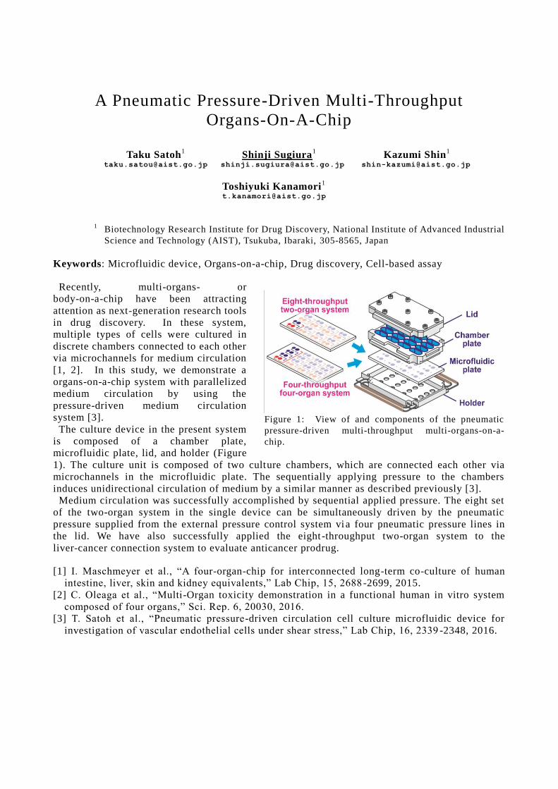
A Pneumatic Pressure-Driven Multi-Throughput
Organs-On-A-Chip
Taku Satoh1 Shinji Sugiura
1 Kazumi Shin
1
[email protected] [email protected] [email protected]
Toshiyuki Kanamori1
1
Biotechnology Research Institute for Drug Discovery, National Institute of Advanced Industrial
Science and Technology (AIST), Tsukuba, Ibaraki, 305-8565, Japan
Keywords: Microfluidic device, Organs-on-a-chip, Drug discovery, Cell-based assay
Recently, multi-organs- or
body-on-a-chip have been attracting
attention as next-generation research tools
in drug discovery. In these system,
multiple types of cells were cultured in
discrete chambers connected to each other
via microchannels for medium circulation
[1, 2]. In this study, we demonstrate a
organs-on-a-chip system with parallelized
medium circulation by using the
pressure-driven medium circulation
system [3].
The culture device in the present system
is composed of a chamber plate,
microfluidic plate, lid, and holder (Figure
1). The culture unit is composed of two culture chambers, which are connected each other via
microchannels in the microfluidic plate. The sequentially applying pressure to the chambers
induces unidirectional circulation of medium by a similar manner as described previously [3].
Medium circulation was successfully accomplished by sequential applied pressure. The eight set
of the two-organ system in the single device can be simultaneously driven by the pneumatic
pressure supplied from the external pressure control system vi a four pneumatic pressure lines in
the lid. We have also successfully applied the eight -throughput two-organ system to the
liver-cancer connection system to evaluate anticancer prodrug.
[1] I. Maschmeyer et al., “A four-organ-chip for interconnected long-term co-culture of human
intestine, liver, skin and kidney equivalents,” Lab Chip, 15, 2688 -2699, 2015.
[2] C. Oleaga et al., “Multi-Organ toxicity demonstration in a functional human in vitro system
composed of four organs,” Sci. Rep. 6, 20030, 2016.
[3] T. Satoh et al., “Pneumatic pressure-driven circulation cell culture microfluidic device for
investigation of vascular endothelial cells under shear stress,” Lab Chip, 16, 2339 -2348, 2016.
Figure 1: View of and components of the pneumatic
pressure-driven multi-throughput multi-organs-on-a-
chip.
On/off targets based toxicity prediction analysis and
Verification Kosuke Negishi1 Yasunori Okazaki1
[email protected] [email protected]
1 Life Science,Elsevier Japan K.K,1-9-15 Higashi-Azabu Minato-ku, Tokyo 106-0044 Japan
Keywords: Toxicology analysis, Medicinal chemistry, On/off target discovery, data integration SAR(Structure-activity relationship) is well known as one of major evaluation method for drug discovery and toxicity/biological activity prediction. On the other hand, protein"s" (on/off target"s") based analysis is not major yet. In this report, we tried to evaluate the feasibility of the protein"s" based analysis. At first we focused on drugs which have few side effects and chose phenylalanine derivative of oral hypoglycemic agent. This type of drug do not have clinical adverse event besides low blood sugar. We created on/off target"s" list from 2 kind of databases (affinity/bioassay type and Text mining type). By Using the proteins listed in these databases, we calculated candidates of toxicity phenotype. Moreover, we searched FDA/EMA documents (database) to verify the consistency of these predicted toxicities. [1] Hansch C., Smith N., Engle R., Wood H. Quantitative structure activity relationships of antineoplastic
drugs: Nitrosoureas and triazenoimidazoles Cancer chemotherapy reports 1972 56:4 (443-456)
[2] Miller EC, Miller JA. Searches for ultimate chemical carcinogens and their reactions with cellular
macromolecules. Cancer. 1981 May 15;47(10):2327-45.
[3]Hanefeld M, Bouter KP, Dickinson S, Guitard C. Rapid and short-acting mealtime insulin secretion with
nateglinide controls both prandial and mean glycemia. Diabetes Care. 2000 Feb;23(2):202-7.
[4] Kawamori R, Kaku K,Hanafusa T , Kashiwabara D, Kageyama S, and Hotta N Efficacy and safety of
repaglinide vs nateglinide for treatment of Japanese patients with type 2 diabetes mellitus J Diabetes
Investig. 2012 Jun 6; 3(3): 302–308.
Thermostability Prediction Model for Protein Single Point Mutations Using Machine Learning
Chie Imamura Keisuke Kawano [email protected] [email protected]
Satoshi Koide Yukihiro Tadokoro [email protected] [email protected]
Toyota Central R&D Labs. Inc., 41-1 Yokomichi, Ngakute, Aichi 480-1192, Japan
Keywords: Prediction of thermostability, Machine learning, SVM, Mutant protein, Protein structure
The thermostability of proteins has been improved by high-throughput experimental screenings from randomly mutated proteins so far. However, it is inefficient because it takes a lot of work and time to select a target. Therefore it is necessary to develop high-performance computational methods for predicting thermostability upon protein mutations.
In this study, we take an alternative approach to improve protein thermostability using machine learning. At first, we constructed 2,000 mutants which have single point mutation at various sites of two proteins. Then we obtained thermostability data by experimental evaluation. Also, we proposed some features based on relevant information about protein local structure around the mutation site.
Using the experimental data and those features as input data, we constructed a thermostability prediction model using support vector machine. Prediction accuracy of the model was evaluated by cross-validation. In the case which training and test data were selected from the same protein, the accuracy of predictions and the area under the curve (AUC) showed high values. On the other hand, when training and test data from different proteins were used, AUC values were decreased. Our results suggest that not only the features from local structure around the mutation site, but features from whole structure are important to improve prediction accuracy.
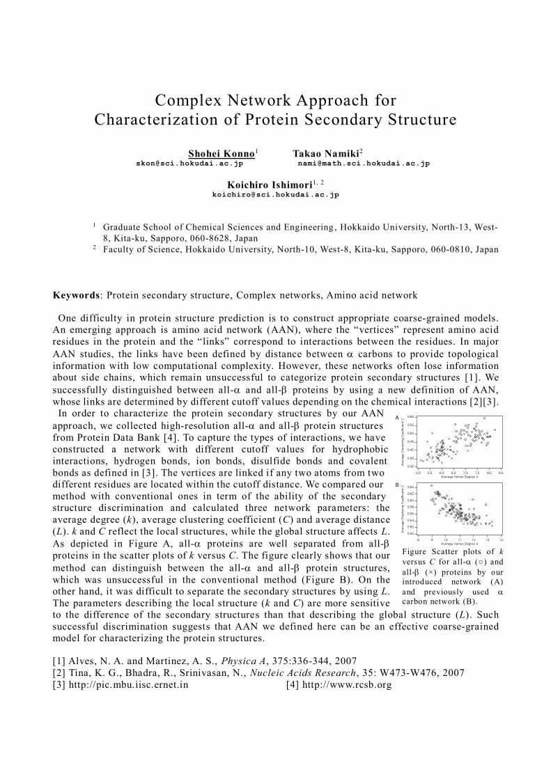
Complex Network Approach for
Characterization of Protein Secondary Structure
Shohei Konno1 Takao Namiki2 [email protected] [email protected]
Koichiro Ishimori1, 2 [email protected]
1 Graduate School of Chemical Sciences and Engineering , Hokkaido University, North-13, West-
8, Kita-ku, Sapporo, 060-8628, Japan 2 Faculty of Science, Hokkaido University, North-10, West-8, Kita-ku, Sapporo, 060-0810, Japan
Keywords: Protein secondary structure, Complex networks, Amino acid network
One difficulty in protein structure prediction is to construct appropriate coarse-grained models.
An emerging approach is amino acid network (AAN), where the “vertices” represent amino acid
residues in the protein and the “links” correspond to interactions between the residues. In major
AAN studies, the links have been defined by distance between carbons to provide topological
information with low computational complexity. However, these networks often lose information
about side chains, which remain unsuccessful to categorize protein secondary structures [1]. We
successfully distinguished between all- and all- proteins by using a new definition of AAN,
whose links are determined by different cutoff values depending on the chemical interactions [2][3].
In order to characterize the protein secondary structures by our AAN
approach, we collected high-resolution all- and all- protein structures
from Protein Data Bank [4]. To capture the types of interactions, we have
constructed a network with different cutoff values for hydrophobic
interactions, hydrogen bonds, ion bonds, disulfide bonds and covalent
bonds as defined in [3]. The vertices are linked if any two atoms from two
different residues are located within the cutoff distance. We compared our
method with conventional ones in term of the ability of the secondary
structure discrimination and calculated three network parameters: the
average degree (k), average clustering coefficient (C) and average distance
(L). k and C reflect the local structures, while the global structure affects L.
As depicted in Figure A, all- proteins are well separated from all-
proteins in the scatter plots of k versus C. The figure clearly shows that our
method can distinguish between the all- and all- protein structures,
which was unsuccessful in the conventional method (Figure B). On the
other hand, it was difficult to separate the secondary structures by using L.
The parameters describing the local structure (k and C) are more sensitive
to the difference of the secondary structures than that describing the global structure (L). Such
successful discrimination suggests that AAN we defined here can be an effective coarse-grained
model for characterizing the protein structures.
[1] Alves, N. A. and Martinez, A. S., Physica A, 375:336-344, 2007
[2] Tina, K. G., Bhadra, R., Srinivasan, N., Nucleic Acids Research, 35: W473-W476, 2007
[3] http://pic.mbu.iisc.ernet.in [4] http://www.rcsb.org
Figure Scatter plots of k
versus C for all- (○) and
all- (×) proteins by our
introduced network (A)
and previously used
carbon network (B).
Reconstruction of self-assembly structure of microtubules
Ryuzo Azuma Gregory Spence [email protected] [email protected]
Arif Pramudwiatmoko †Akihiko [email protected] [email protected]
Tokyo Institute of Technology, J3-25, 4259, Nagatsuta, Midori-ku, Yokohama, Kanagawa 226-8503, Japan
† Corresponding author
Keywords: Molecular robotics, artificial intelligence related technology
We have been developing a coarse-grained simulation method for analyses of super molecular structure formation processes. In this poster presentation, we demonstrate self-organized structures in terms of studies on mechanisms of microtubule structure formation. The halmark of our simulation is that it explicitly takes account of water molecules albeit it is based on a mathematical model employing purely stochastic rules for semi-microscopic molecular dynamics in a 3 dimensional space. Water molecules turned out to have versatile roles in governing electrostatic and hydrophobic interactions during the microtuble organization process.
In our microtuble simulation model, each tubulin monomer (α and β tubulins) is represented by a chain of coarse-grained subunits. Strength of associative interactions between those subunits are roughly double that of thermal fluctuations. For inter-monomer coupling, we assume ~0.2eV strength by taking account of electrostatics calculation in a previous study [1].
Using these simulation method and model, we have been successfully observing self-organized structure that associates well with microtuble triplet structure. Transient dynamics in those simulations reaches a plateau after ~20 milliseconds and thus the structure is stabilized. Diameter of each one in the triplet is close to that of 13 protofilament microtubles (23-4 nm.) These results support our view that coexistence of hydrophobic and electrostatic interaction between tubulin dimer drives self-organization of microtubules in presence of water molecules.
We have also been focusing on atomic-level tubulin α-β dimer structure responsible for interactions during microtubule assembly formation. In particular, surface charge and molecular dynamics are studied for scenarios where one of/both c-termini are detached from the surface, as proposed previously [2]. These features are going to be utilized to refine our coarse-grained simulation model.
This presentation is based on results obtained from a project commissioned by the New Energy and Industrial Technology Development Organization (NEDO.)
[1] Tuszynsky, J.A., Brown, A., Crawford, E.J., Carpenter, E.J., Molecular Dynamics Simulation of Tubulin Structure and Calculations of Electrostatic Properties of Microtubules, Mathematical and Computer Modeling, 41:1055-1070, 2005.
[2] Priel, A., Tuszynski, J.A., Woolf, N.J., Transitions in microtubule C-termini confirmations as a possible dendritic signaling phenomenon, Eur Biophys J, 35:40-52, 2005.
Fig. 3 Fig. 3
On Being Scientific
Tomokazu Konishi1 [email protected]
1 Graduate School of Bioresource Sciences, Akita Prefectural University, 010-0195, Japan
Keywords: falsifiability, falsification
Our researches often make a crowded lobby; for instance, I wonder how many procedures have
been introduced to normalize transcriptomics data. Most of them are totally out of use, because
they lack falsifiability, which is an idea introduced by Karl Popper as a touchstone for science (1).
As science has no rigid definition, distinguishing true science and others is controversial; however,
this idea has been widely accepted among scientists. Science hates a situation that observation
depends on analytical methodologies, as it is formed by integrating tons of observations reported
by many researchers. So, most scientists do not take ideas like “anything goes” (2, 3). As far as I
know, there has been found only one method that can analyze transcriptomics data fulfilling this
issue (4, 5). There would not be another solution, as it is compatible to thermodynamic property of
a living cell (6).
Another enthusiasm occurred to solve multiplicities of statistical tests (7). This claim
encompassed scientific community a mess, and many procedures were introduced. However, the
casted problem may not really exist; it could be understood as a stray bullet from war between
schools of statisticians (8).
Rushing into a wrong paradigm is waste of time, not only for analysts, but also for scientists .
How can we avoid such researchers’ bad habit? I believe we can learn from science. Presently, we
introduce a new method by comparing it to existing ones by focusing on properties defined by
ourselves. I rarely saw articles that defined problems clearly, and estimated the damages caused by
the problem. Also, I rarely saw articles that declare what was assumed, certainty of the
assumptions, and how they could be verified. Those should be clarified. I know it is easy to follow
someone’s idea and put some new decorations; however, this is not very scientific attitude.
Additionally, changing data to fulfil benefits of the analyst is prohibited in science, because it
could conflict to ethical rule; it would be recognized as falsification of data.
(1) Popper K. The logic of scientific discovery, Routledge; 1959.
(2) Feyerabend P. Against method: outline of an anarchist theory of knowledge , New Left Books;
1975.
(3) Sober E. Philosophy of Biology, Oxford University Press, 1993 .
(4) Konishi T, Konishi F, Takasaki S, Inoue K, Nakayama K, Konagaya A. Coincidence between
transcriptome analyses on different microarray platforms using a parametric framework. PLoS
ONE. 2008;3(10):e3555.
(5) Konishi T. Parametric analysis of RNA-seq expression data. Genes to Cells.
2016;21(6):639-47.
(6) Konishi T. A thermodynamic model of transcriptome formation. Nucleic Acids Res.
2005;33(20):6587-92.
(7) Dudoit S, Yang Y, Callow M, Speed T. Statistical methods for identifying differentially
expressed genes in replicated cDNA microarray experiments. Stat Sin. 2002;12:111–39.
(8) Konishi T. Microarray test results should not be compensated for multiplicity of gene contents.
BMC Systems Biology. 2011;5(Suppl 2):S6-S.
Using convolutional neural networks plus recurrent neural
network, automatic classification of human iPS
cell-derived cardiomyocytes extracellular potential
waveforms.
Tetsuo Kitamura, Mayumi Obo, [email protected] [email protected]
Tomoharu Osada, Hiroaki Inoue, [email protected] [email protected]
Yamamoto Yoshinori, Yasuyuki Oonishi, [email protected] [email protected]
Hideaki Hiratsuka [email protected]
Nonclinical Research Center, Drug Development Service Segment, LSI Medience Corp. 14 -1
Sunayama, Kamisu, Ibaraki, 314-0255, Japan
Keywords: convolutional neural networks, recurrent neural network, human iPS cell-derived
cardiomyocytes
[Introduction] Cardiac safety evaluation method which uses an extracellular potential of human
iPS stem cell-derived cardiomyocytes is proposed. The evaluated system is expected to predict
pro-arrhythmic potentials of compounds with high accuracy by detection of early after
depolarizations waveforms. This detection is done by human, so there is a problem in the labor and
precision. Automatic classification using convolutional neural networks plus recurrent neural
networks (CNN+RNN) are reported for movie classification technique. By the machine learning
with training data, CNN+RNN are possible to highly accurate classification. The purpose of this
study was to evaluate the accuracy of automatic classification with CNN+RNN for the waveforms
obtained in this evaluation system.
[Methods] Classification targets were waveforms of sequential beats from multi channels.
Framework of neural networks was TensorFlow. Supervised learning was used. Training data and
test data were selected from same experiments. These data were labeled with classification by
human.
[Results] With the test data, concordance rate of classification result by human and CNN+RNN
was 99.97%.
[Conclusion] CNN+RNN showed high accuracy in cardiomyocytes extracellular potential
waveforms classification.
Rational design of orthogonal gene transcription nano
device on DNA origami
Takeya Masubuchi 1, Masayuki Endo 2, Ryo Iizuka 3, Ayaka Iguchi 4, Dong Hyun Yoon 4,
Tetsushi Sekiguchi 5, Hao Qi 1,6, Ryosuke Iinuma 1, Yuya Miyazono 1, Shuichi Shoji 4,
Takashi Funatsu 3, Hiroshi Sugiyama 2,7, Yoshie Harada 2,8, Takuya Ueda 1
and Hisashi Tadakuma 1,2,8,
1 Graduate School of Frontier Science, The University of Tokyo, 2 Institute for Integrated Cell-Material
Sciences (WPI-iCeMS), Kyoto University, 3 Graduate School of Pharmaceutical Sciences, The University of
Tokyo, 4 Department of Nanoscience and Nanoengineering (ASE Graduate School), Waseda University, 5 Research Organization for Nano & Life Innovation, Waseda University, 6 Department of Chemical
Engineering and Technology, Tianjin University, 7 Department of Chemistry, Kyoto University, 8 Institute
for Protein Research, Osaka University
Keywords: Gene expression, Synthetic Biology, Molecular Robots
In synthetic biology, the design of gene expression requires devices that alter the output depending
on the situation. Inspired by RNA viruses that contain both enzymes and substrate genes, and
express specific genes through architectural modalities, we integrated an enzyme , T7 RNA
polymerase, and multiple target gene substrates onto a DNA origami -based nano-chip. This gene
nano-chip orthogonally transcribes its own genes, and using this system, we succeeded in a
rational design of gene expression by controlling the inter-molecular distances between the
enzyme and the substrate genes. Our approach of component integration on a nano -chips may
provide the basis for an integrated gene expression circuit.
[1] Tadakuma H, Masubuchi T, Ueda T. RNA Study Using DNA Nanotechnology. (review), Prog Mol Biol Transl Sci., 139:121-163 (2016).
Graph Neural Networks for Density Functional Theory
Masashi Tsubaki1
1 National Institute of Advanced Industrial Science and Technology (AIST) Artificial Intelligence Research Center, Machine Learning Research Team
Tokyo Waterfront BIO-IT Research Building, 2-4-7 Aomi, Koto-ku, Tokyo, 135-0064, Japan Keywords: Graph Neural Networks, End-to-End Learning, Density Functional Theory
In this poster, we propose the use of machine learning technique, graph neural networks (GNNs), for predicting various electronic ground-state properties molecules based on density functional (DFT) theory. GNN is a neural network, which takes a graph-structured data (i.e., molecular graph) as an input and outputs a low-dimensional real-valued vector representation (i.e., molecular embedding). In addition, our GNN uses an end-to-end learning technique to obtain the molecular embeddings, which does not require any molecular features (e.g., descriptors and fingerprints) and knowledge of chemistry and physics. The end-to-end GNN allows us to automatically learn all parameters including input features in the model. We experiment with thirteen electronic ground-state properties of 117k distinct organic molecules and show that our end-to-end GNN can be more accurate than hybrid DFT.
ChemiSpadon: an SNS-based platform for human-assisted chemical space exploration
Kazuki Z Yamamoto1 Ryuichi Kubo2 Hiroyuki Yamasaki3
[email protected] [email protected] [email protected]
Masahito Ohue4 Yusuke Yamada5 [email protected] [email protected]
1 Isotope Science Center, University of Tokyo, 2-11-16, Yayoi, Bunkyo-ku, Tokyo 113-0032, Japan 2 DeNA Life Science, Inc., Shibuya Hikarie, 2-21-1 Shibuya, Shibuya-ku, Tokyo 150-8510 Japan 3 Kitasato University School of Pharmacy, 5-9-1 Shirokane, Minato-ku, Tokyo 108-8641 Japan 4 School of Computing, Tokyo Institute of Technology, 2-12-1, Ookayama, Meguro-ku, Tokyo 152-8550,
Japan 5 Mishima.syk, Mishima-PostOffice-Kyokudome, Minami-Tamachi, Mishima-shi, Shizuoka 411-8799,
Japan
Keywords: Chemical space, Drug design, Human computation, SNS, Mastodon Exploration of undeveloped chemical space is essential for discovery of novel drugs. Conventionally, ‘druglikeness’ and ‘druggablity’ have been mainly considered for small molecules whose molecular weights are less than 500-600[1, 2]. However, to cope with novel drug targets previously considered ‘undruggable’ due to absence of druggable cavities, medium-sized molecules like natural products, macrocycles, peptidomimetics or others are needed[3]. Although medium-sized molecules constitute attractive chemical space, the size of such space is too large to explore comprehensively[4]. To facilitate exploration of such vast undeveloped chemical space, here we present “ChemiSpadon”, an SNS-based platform for human-assisted chemical space exploration. ChemiSpadon is based on the open-source SNS system called Mastodon[5] and equipped with chemical structure posting system, structure evaluation system, and structure generating bots. Taking advantage of human knowledge and instinct, we expect efficient exploration of undeveloped but feasible chemical space for drug discovery. Participants are very welcome. Please visit: www.chemical.space [1] Lipinski, C. A., Lombardo, F., Dominy, B. W., & Feeney, P. J., Experimental and computational
approaches to estimate solubility and permeability in drug discovery and development settings. Advanced drug delivery reviews, 23:3-25, 1997.
[2] Cheng, A. C., Coleman, R. G., Smyth, K. T., Cao, Q., Soulard, P., Caffrey, D. R., ... & Huang, E. S., Structure-based maximal affinity model predicts small-molecule druggability. Nature biotechnology, 25(1):71, 2007.
[3] Lipinski, C. A., Rule of five in 2015 and beyond: Target and ligand structural limitations, ligand chemistry structure and drug discovery project decisions, Advanced drug delivery reviews, 101:34-41, 2016.
[4] Ruddigkeit, L., Van Deursen, R., Blum, L. C., & Reymond, J. L., Enumeration of 166 billion organic small molecules in the chemical universe database GDB-17. Journal of chemical information and modeling, 52(11):2864-2875, 2012.
[5] Mastodon https://joinmastodon.org/