Embed Size (px)
Citation preview

RESEARCH PAPER
The fibrate gemfibrozil is aNO- and haem-independentactivator of soluble guanylylcyclase: in vitro studiesI G Sharina1, M Sobolevsky1, A Papakyriakou2, N Rukoyatkina3,4,G A Spyroulias5, S Gambaryan3,4 and E Martin1
1Department of Internal Medicine, Division of Cardiology, UT Health Science Center at Houston,
Medical School, Houston, TX, USA, 2National Center for Scientific Research, Department of
Physical Chemistry, Athens, Greece, 3Institute of Clinical Biochemistry and Pathobiochemistry,
University of Wuerzburg, Wuerzburg, Germany, 4Sechenov Institute of Evolutionary Physiology
and Biochemistry, Russian Academy of Sciences, Petersburg, Russia, and 5Department of
Pharmacy, University of Patras, Patras, Greece
CorrespondenceEmil Martin, Department ofInternal Medicine, Division ofCardiology, UT Health ScienceCenter at Houston, MedicalSchool, 1941 East Road, Houston,TX 77054, USA. E-mail:emil.martin@uth.tmc.edu----------------------------------------------------------------
Received27 August 2014Revised3 November 2014Accepted11 December 2014
BACKGROUND AND PURPOSEFibrates are a class of drugs widely used to treat dyslipidaemias. They regulate lipid metabolism and act as PPARα agonists.Clinical trials demonstrate that besides changes in lipid profiles, fibrates decrease the incidence of cardiovascular events, withgemfibrozil exhibiting the most pronounced benefit. This study aims to characterize the effect of gemfibrozil on the activityand function of soluble guanylyl cyclase (sGC), the key mediator of NO signalling.
EXPERIMENTAL APPROACHHigh-throughput screening of a drug library identified gemfibrozil as a direct sGC activator. Activation of sGC is unique togemfibrozil and is not shared by other fibrates.
KEY RESULTSGemfibrozil activated purified sGC, induced endothelium-independent relaxation of aortic rings and inhibited plateletaggregation. Gemfibrozil-dependent activation was absent when the sGC haem domain was deleted, but was significantlyenhanced when sGC haem was lacking or oxidized. Oxidation of sGC haem enhanced the vasoactive and anti-platelet effectsof gemfibrozil. Gemfibrozil competed with the haem-independent sGC activators ataciguat and cinaciguat. Computationalmodelling predicted that gemfibrozil occupies the space of the haem group and interacts with residues crucial for haemstabilization. This is consistent with structure-activity data which revealed an absolute requirement for gemfibrozil’s carboxylgroup.
CONCLUSIONS AND IMPLICATIONSThese data suggest that in addition to altered lipid and lipoprotein state, the cardiovascular preventive benefits of gemfibrozilmay derive from direct activation and protection of sGC function. A sGC-directed action may explain the more pronouncedcardiovascular benefit of gemfibrozil observed over other fibrates and some of the described side effects of gemfibrozil.
AbbreviationsBAY41-2272, (3-(4-amino-5-cyclopropylpyrimidine-2-yl)-1-(2-fluorobenzyl)-1H-pyrazolo[3,4-b]pyridine); DEA-NO,diethylammonium (Z)-1-(N,N-diethylamino)diazen-1-ium-1,2-diolate; ODQ, 1H-[1,2,4]oxadiazolo[4,3-a]quinoxalin-1-one; sGC, soluble GC
BJP British Journal ofPharmacology
DOI:10.1111/bph.13055www.brjpharmacol.org
2316 British Journal of Pharmacology (2015) 172 2316–2329 © 2014 The British Pharmacological Society

Introduction
Gemfibrozil is a member of the fibrate class of drugs that havebeen used for decades for the management of combineddyslipidaemia (Fruchart and Duriez, 2006). Fibrates wereintroduced in clinics in the early 1970s and are currentlywidely used for dyslipidaemic patients with diet-resistanthypertriglyceridaemia. Fibrates act as agonists for the tran-scription factor PPARα, which controls the expression ofgenes associated with lipid catabolism and metabolism oflipoproteins, resulting in stimulated cellular fatty aciduptakes, enhanced catabolism of triglyceride-rich particle andreduced secretion of very low-density lipoprotein (Staelset al., 1998). Fibrates cause a substantial decrease in the levelof plasma triglycerides, usually accompanied by a moderatedecrease of low-density lipoprotein cholesterol and anincrease in high-density lipoprotein cholesterol (Staels et al.,1998). Additional effects on endothelial function, inflamma-tion and vascular remodelling have also been reported(Chinetti-Gbaguidi et al., 2005). Meta-analyses of clinicaltrials demonstrate that fibrates improve lipid profile and havean overall reduction in cardiovascular and coronary events(Jun et al., 2010). While improved lipid profile is consistentlyobserved in all fibrate studies, the extent of cardiovascularand coronary benefits is drug-specific. Clinical trials of gem-fibrozil, such as the Helsinki Heart Study (Frick et al., 1987)and VA-HIT (Rubins et al., 1999), show the most pronouncedcardiovascular preventive benefits and a decreased incidenceof coronary events over other fibrates. We report here that attherapeutic concentrations gemfibrozil, but not otherfibrates, acts as an activator of soluble GC (sGC).
sGC synthesizes the secondary messenger cGMP inresponse to NO stimulation. In the cardiovascular system,sGC regulates BP, platelet function, vascular plasticity andangiogenesis (Murad, 2006). Diminished sGC activity andexpression exacerbate endothelial dysfunction, decrease vas-cular plasticity (Ruetten et al., 1999; Kloss et al., 2000) andcontribute to the development of a wide array of cardiovas-cular disorders, including hypertension, coronary arterydisease and atherosclerosis (Xu and Zou, 2009; Vita, 2011).Single nucleotide polymorphisms of human sGC loci arelinked to increased risk of hypertension and coronary arterydisease (Ehret et al., 2011; Lu et al., 2012). Familial studiesin humans demonstrate that diminished sGC expressioncorrelates with increased risk of myocardial infarctions
(Erdmann et al., 2013b), stenosis of carotid arteries oroesophageal achalasia (Herve et al., 2014).
Not surprisingly, sGC is an active target for pharmaco-logical intervention. Classical nitrovasodilators have beenused to manage angina and heart failure long before itbecame clear that they act as NO-generating compounds.The interest in sGC as a drug target was substantiallyrenewed because of the discovery of several NO-independent allosteric regulators of sGC. Some of theseregulators are being developed for clinical applications.Recently, riociguat, a sGC stimulator, which moderatelyactivates sGC, but significantly increases its affinity to NO(Stasch and Hobbs, 2009), has been approved as a newtherapy for pulmonary hypertension (Stasch and Evgenov,2013). Other compounds, such as ataciguat and cinaciguat,activate sGC by mimicking haem (Schmidt et al., 2009).Cinaciguat is considered for treatment of heart failure(Zamani and Greenberg, 2013), although some side effectsare yet to be resolved (Erdmann et al., 2013a). The effects ofboth sGC stimulators and haem-independent activators areenhanced by cobinamides, which act independently of sGChaem and its haem-binding region (Sharina et al., 2011a;Chrominski et al., 2013).
Multiple modes of sGC stimulation and the structuraldiversity of existing sGC regulators suggest that some of thedrugs currently used in clinical practice may inadvertentlytarget sGC. In this study, we report that gemfibrozil is ahaem-independent sGC activator with vasorelaxation andantiplatelet properties.
Methods
Recombinant human sGC enzymeFull-length sGC was purified from Sf9 cells as described pre-viously (Martin et al., 2001). To generate truncated sGC vari-ants, the open reading frames coding the residues 269–690 ofthe α subunit or the residues 200–619 of the β subunit werecloned into the transfer vector pBacPak9 (Clontech, Moun-tain View, CA, USA) to obtain the pBacPak-αΔ269 and thepBacPak-βΔ200 plasmids respectively. A hexahistidine tag wasalso inserted at the C-terminus of the αΔ269 variant by PCRmutagenesis. Using these plasmids and the linearized bacu-lovirus DNA (BaculoGold from Pharmingen, San Diego, CA,USA), the baculoviruses expressing the truncated αΔ269 or
Tables of Links
TARGETS
Nuclear hormone receptorsa Enzymesb
PPARα sGC
LIGANDS
ADP Ciprofibrate IBMX
Ataciguat Clofibrate Nitric oxide (NO)
Bezafibrate Fenofibrate ODQ
Cinaciguat Gemfibrozil Protoporphyrin IX
These Tables list key protein targets and ligands in this article which are hyperlinked to corresponding entries in http://www.guidetopharmacology.org, the common portal for data from the IUPHAR/BPS Guide to PHARMACOLOGY (Pawson et al., 2014) and arepermanently archived in the Concise Guide to PHARMACOLOGY 2013/14 (a,bAlexander et al., 2013a,b).
BJPGemfibrozil is an sGC activator
British Journal of Pharmacology (2015) 172 2316–2329 2317

βΔ200 sGC were generated according to the manufacturer’sprotocol. To generate the full-length or truncated sGC vari-ants, Sf9 cells were infected with the appropriate combina-tion of baculovirus and, after 72 h, sGC enzyme was partiallypurified from the lysates using the anion exchange chroma-tography on HR16/50 DEAE-FF Sepharose column (GEHealthcare Bio-Sciences, Pittsburgh, PA, USA).
High-throughput screening sGC assayThe libraries were screened in 96-well plates as describedpreviously (Sharina et al., 2011a). In brief, 1 μL of 10 mMpharmacophore was delivered robotically into 30 μL reactionsolution [40 mM triethanolamine (TEA), pH 7.4, 0.1 mMDTT, 0.2 mM MgCl2] containing 0.1 μg of purified humanrecombinant sGC and 10 mU of inorganic pyrophosphatase.Synthesis of cGMP was initiated after 5 min by adding 10 μLof 400 μM Mg2+-GTP. The reaction was stopped 15 min laterby 60 μL of 0.5 M HCl and the amount of generated phos-phate was determined colorimetrically at 630 nm 20 minafter adding 100 μL of developing reagent [1.05% (w v−1)ammonium molybdate, 1 M HCl, 0.034% (w v−1) malachitegreen and 0.05% Tween 20]. sGC activities in the absence ofadditives (basal), in the presence of BAY41-2272 (5 μM) or10 μM ODQ with 100 nM BAY58-2667 were used as positivecontrols. Positive samples were rescreened manually usingthe [α-32P]GTP→[α-32P]cGMP conversion assay. The pharma-cophores displaying OD >0.1 AU above the level of basalactivity were subjected to secondary screening.
Assay of sGC activity in vitroEnzymatic activity was assayed using [α-32P]GTP to [32P]cGMP conversion assay (Schultz, 1974). sGC, 0.5 μg, in25 mM TEA, pH 7.5, 1 mg·mL−1 BSA, 1 mM IBMX, 1 mM DTT,1 mM cGMP, 3 mM MgCl2, 0.05 mg·mL−1 creatine phospho-kinase and 5 mM creatine phosphate was incubated withindicated concentrations of agonist(s) (gemfibrozil, ataciguat,cinaciguat or BAY41-2272) for 10 min at room temperature.After transferring the sample to 37°C, the reaction was initi-ated by 1 mM GTP/[α-32P]GTP (∼100 000 cpm). To measureNO-induced sGC activity, 10 ng of sGC were used and NOdonor was supplied together with GTP. To deplete sGC ofhaem, purified sGC was incubated with 0.4% Tween 20 for20 min at room temperature before adding it into the reac-tion mixture. For the experiments with ferric sGC, theenzyme was incubated for 20 min with 10 μM ODQ before itwas used in the reaction. The reaction was stopped by zinccarbonate and processed following the protocol establishedpreviously (Schultz, 1974).
Aortic ring relaxationMale Sprague Dawley rats (20 animals total; 14–16 weeks old,300–350 g; Harlan Laboratories Inc, Indianapolis, IN, USA)were killed while under isoflurane anaesthesia and afterthoracotomy, the descending thoracic aorta was dissected,cut into 3–5 mm long segments and mounted on a four-channel wire Myograph 610 (DMT, Copenhagen, Denmark)under 1.5 g of passive tension. For endothelium-denudedvessels, the endothelium was removed by gently rubbing thevessel interior with a toothpick with a wet cotton swab.Removal of the endothelium was confirmed by the absence of
ACh relaxation in vascular strips pre-contracted with sub-maximal concentrations of phenylephrine (Phe). The ringswere equilibrated for 80 min in Krebs-Henseleit solutionpH 7.4, oxygenated with carbogen (95% O2, 5% CO2) with atleast three buffer changes every 20 min. All force measure-ments were recorded using Powerlab 400TM data acquisitionsystem and LabChart software (ADInstruments, ColoradoSprings, CO, USA). After equilibration the rings were pre-contracted with 60 mM K+ to determine the maximal con-tractile response, the buffer was then replaced and the ringswere titrated with 100–300 nM Phe to achieve submaximalcontraction of ∼4.5–5 g of tension. After stabilization, theagonists (gemfibrozil or gemfibrozil analogues) were addedcumulatively and changes in isometric tension wererecorded. In some experiments, the rings were pretreatedwith 10 μM ODQ or 0.1 μM BAY41-2272. All studies involv-ing animals are reported in accordance with the ARRIVEguidelines for reporting experiments involving animals (Kilk-enny et al., 2010; McGrath et al., 2010).
Preparation of washed human plateletsHuman platelets were prepared and used as previouslyreported (Gambaryan et al., 2010) with small modifications.Blood was collected into one-seventh volume of anticoagulantcitrate dextrose solution (12 mM citric acid, 15 mM sodiumcitrate, 25 mM dextrose and 2 μM EGTA, final concentra-tions). Platelet-rich plasma (PRP) was obtained by 5 min cen-trifugation at 330× g. To reduce leukocyte contamination, PRPwas diluted 1:1 with PBS and centrifuged at 240× g for 10 min.Subsequently, the supernatant was centrifuged for 10 min at430× g, then the platelets were washed once in citrate glucosesaline buffer (120 mM sodium chloride, 12.9 mM trisodiumcitrate, 30 mM D-glucose, pH 6.5) and resuspended in HEPESbuffer (150 mM sodium chloride, 5 mM potassium chloride,1 mM magnesium chloride, 10 mM D-glucose, 10 mM HEPES,pH 7.4). After 15 min rest in a 37°C water bath, washed plate-lets (WP) were used for experiments.
Western blot analysisOne hundred microlitres of WP (1 × 10⁸ mL−1) were incubatedat 37°C for 1 min with vehicle (resuspension HEPES buffer),10 or 100 μM NO donor DEA-NO or with 10 or 100 μMgemfibrozil for 5 min. In experiments with ODQ, plateletswere pre-incubated for 1 min with 10 μM ODQ before theaddition of the vehicle (HEPES buffer), DEA-NO or gemfibro-zil. At the end of the incubation, the reactions were stoppedby adding an equal volume of 2× Laemmli buffer (200 mMTris-HCl, pH 6.7, 20% glycerol, 4% SDS and 10%2-mercaptoethanol) and boiling at 97°C for 5 min. Plateletlysates were separated by SDS-PAGE, transferred to nitrocel-lulose membranes and the membranes were incubated withanti-Phospho-VASPSer239 (Nanotools, Teningen, Germany) oranti-β-actin (Cell Signaling, Frankfurt am Main, Germany)antibodies overnight at 4°C. To visualize the signal, goatanti-rabbit or anti-mouse IgG conjugated with HRP were usedas secondary antibodies, followed by ECL detection.
Platelet aggregationPlatelet aggregation was analysed in diluted PRP by recordingthe intensity of low angle light scattering using the method
BJP I G Sharina et al.
2318 British Journal of Pharmacology (2015) 172 2316–2329

described in detail previously (Mindukshev et al., 2011). Allexperiments were performed in 6 mL of modified HEPESbuffer (pH 7.4, osmolarity 302 mOsm, containing 140 mMNaCl, 10 mM HEPES, 10 mM NaHCO3, 2 mM KCl, 1 mMMgCl2, 2 mM CaCl2, 5.5 mM D-glucose). An appropriateamount of PRP was added to the buffer to achieve a density of10 000 platelets μL−1. After 2 min of establishing a constantbasal signal, platelets were pre-incubated with the indicatedconcentrations of gemfibrozil, SNP, BAY 41–2272 alone or incombination with ODQ (which was added 1 min before) for5 min. Aggregation was initiated by 0.1 μM ADP and thesignal was recorded for an additional 10 min. The mean lightscatter signal in control experiments without stimulation wasexpressed as 100% aggregation.
Molecular modelling of human β1 H-NOXcomplex with gemfibrozilHuman β1 H-NOX domain (hHNOX) was modelled based onthe crystal structure of the homologous NsHNOX proteinfrom Nostoc sp (PDB ID: 3L6J; Martin et al., 2010). Briefly, thecrystallographic coordinates were employed as a template forthe production of the hHNOX domain using the programmeModeller v9 (Fiser and Sali, 2003). The model with the lowesttarget function was selected among 50 models and its qualitywas assessed using the Structural Analysis and VerificationServer (see Supplemental Information for more details). Sub-sequently, the model of hHNOX was superimposed with thetemplate X-ray structure so as to transfer the ligand at itscrystallographic pose. To relax the resulting hHNOX complexwith cinaciguat from steric classes, the model was subjectedto energy minimization using AMBER v12 (Case et al., 2005).The ligand-free, energy minimized model of the hHNOXdomain was employed for docking of the inhibitors usingAutoDock v4 (The Scripps Research Institute, San Diego, CA,USA; Huey et al., 2007).
Statistical analysisNon-linear regression, calculation of EC50 and statisticalanalysis were performed using GraphPad Prism 5.1 (Graph-Pad Software, La Jolla, CA, USA). One-way ANOVA, followed byTurkey’s post hoc test was used for multiple comparisons.Two-way ANOVA followed by Bonferroni’s post hoc test wasused for comparison of the dose-response curves. P < 0.05 wasconsidered significant. Results are expressed as mean ± SEM.The EC50 values are expressed as means with 95% confidenceinterval shown in parentheses.
MaterialsIBMX, phosphocreatine, creatine kinase, GTP, pyrophos-phatase, ammonium molybdate, malachite green, Tween 20and gemfibrozil were obtained from Sigma-Aldrich (St Louis,MO, USA). Fibrates and gemfibrozil-like compounds used instructure-activity studies (Supporting Information Table S1)were also from Sigma-Aldrich. 2-(N,N-diethylamino)-diazenolate-2-oxide [diethylammonium salt] (DEA-NO),ataciguat, cinaciguat, BAY41-2272 and ODQ were obtainedfrom Enzo Life Sciences (Farmingdale, NY, USA).
Study approvalBlood was obtained from healthy volunteers according toinstitutional guidelines and the Declaration of Helsinki. The
studies with human platelets were performed at the Univer-sity of Würzburg according to the guidelines of the ethicscommittee of the University of Würzburg (Studies No. 67/92and 114/04). The studies of isolated rat aortic rings wereperformed in accordance to the guidelines of the AnimalWelfare Committee of the University of Texas Health ScienceCenter (protocol AWC 12–089).
Results
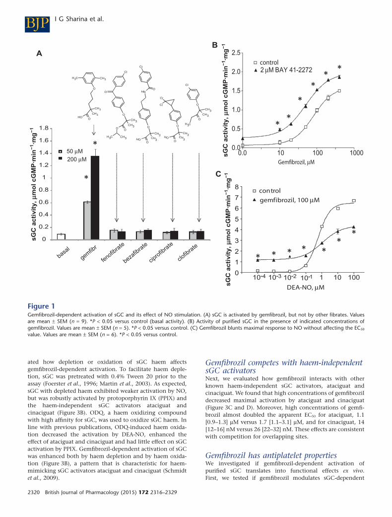
Identification of gemfibrozil as sGC activatorWe have previously developed a high-throughput in vitroscreening for selection of sGC activating compounds (Sharinaet al., 2011a). We have reported that screening of a smalllibrary containing 1120 ‘off-patent’ drugs (Prestwick Chemi-cal Library, ChemBridge Corporation, San Diego, CA, USA)and selected vitamins and pro-vitamins identified two sGC-activating compounds. The effect of vitamin B12 precursordicyanocobinamide on sGC activity was already reported(Sharina et al., 2011a). In addition, gemfibrozil (Figure 1A)was identified as a potential sGC activator. Because gemfibro-zil is a member the fibrate class of drugs, we tested all struc-turally related fibrates. We determined that none of them wascapable to activate sGC (Figure 1A). This indicates thatamong fibrates, sGC activation is a feature specific only togemfibrozil. Gemfibrozil-dependent activation of sGC wasobserved over a broad range of concentrations (Figure 1B),with an apparent EC50 of 94 [75–117] μM and a Hill slopecoefficient of 1.96. BAY41-2272, the structural precursor ofsGC stimulator riociguat, additively enhanced the effect ofgemfibrozil and decreased the EC50 to 48 [37–67] μM and theHill coefficient to 1.34. Such additive effect strongly suggestsnon-overlapping binding sites for BAY41-2272 and gemfibro-zil. Moreover, the EC50 for NO donor DEA-NO only margin-ally changed in the presence of gemfibrozil (Figure 1C),although the maximal NO activation was diminished. Theseanalyses indicate that gemfibrozil’s mechanism of action isdifferent from that of described sGC stimulators.
Gemfibrozil-dependent activation requires thehaem-binding domain of sGCTo further understand the mechanism of action of gemfibro-zil, we used the α1 splice isoform D, which lacks theN-terminal 239 residues of the α1 subunit (Sharina et al.,2008; 2011b; Martin et al., 2014) and/or truncated versions ofsGC lacking the N-terminal 200 residues of the β1 subunit(Figure 2A). Similar to wild-type sGC, all expressed truncatedsGC variants (Figure 2B) possessed cGMP-forming activityand were more active in the presence of Mn2+. These proper-ties argue for functional catalytic domains (Figure 2C) inthese truncated sGC. However, only sGC variants containingintact β1 HNOX domain were activated by gemfibrozil. Thesedata indicate that the haem-binding domain is necessary forgemfibrozil-dependent activation of sGC.
Gemfibrozil-dependent activation is enhancedby depletion and oxidation of sGC haemHeme plays a crucial role in the mechanism of action formost pharmacological sGC regulators. Therefore, we evalu-
BJPGemfibrozil is an sGC activator
British Journal of Pharmacology (2015) 172 2316–2329 2319
ated how depletion or oxidation of sGC haem affectsgemfibrozil-dependent activation. To facilitate haem deple-tion, sGC was pretreated with 0.4% Tween 20 prior to theassay (Foerster et al., 1996; Martin et al., 2003). As expected,sGC with depleted haem exhibited weaker activation by NO,but was robustly activated by protoporphyrin IX (PPIX) andthe haem-independent sGC activators ataciguat andcinaciguat (Figure 3B). ODQ, a haem oxidizing compoundwith high affinity for sGC, was used to oxidize sGC haem. Inline with previous publications, ODQ-induced haem oxida-tion decreased the activation by DEA-NO, enhanced theeffect of ataciguat and cinaciguat and had little effect on sGCactivation by PPIX. Gemfibrozil-dependent activation of sGCwas enhanced both by haem depletion and by haem oxida-tion (Figure 3B), a pattern that is characteristic for haem-mimicking sGC activators ataciguat and cinaciguat (Schmidtet al., 2009).
Gemfibrozil competes with haem-independentsGC activatorsNext, we evaluated how gemfibrozil interacts with otherknown haem-independent sGC activators, ataciguat andcinaciguat. We found that high concentrations of gemfibrozildecreased maximal activation by ataciguat and cinaciguat(Figure 3C and D). Moreover, high concentrations of gemfi-brozil almost doubled the apparent EC50 for ataciguat, 1.1[0.9–1.3] μM versus 1.7 [1.1–3.1] μM, and for cinaciguat, 14[12–16] nM versus 26 [22–32] nM. These effects are consistentwith competition for overlapping sites.
Gemfibrozil has antiplatelet propertiesWe investigated if gemfibrozil-dependent activation ofpurified sGC translates into functional effects ex vivo.First, we tested if gemfibrozil modulates sGC-dependent
A
O
CH3CH3
CH3
CH3OH
O
Cl
O
CH3
CH3
O
CH3
O
O
O
O
O
O
CH3
CH3
CH3
CH3
CH3
CH3O
O
O
Cl
Cl
Cl
Cl
HN
HOHOCH3 CH3
B
C
10-3 1 10 1000
1
2
3
4
5
6
7
8control
gemfibrozil, 100 μM
DEA-NO, μM
10-2 10-110-4
0
0.2
0.4
0.6
0.8
1
1.2
1.4
1.6
1.8
50 μM
basal
gemfib
r
fenofib
rate
bezafib
rate
cipro
fibra
te
clofib
rate
200 μM
sG
C a
cti
vit
y, mm
ol
cG
MP
·min
−1·m
g−
1
*
*
sG
C a
cti
vit
y, mm
ol
cG
MP
·min
−1·m
g−
1
* * * * **
*
0.0 10 100 10000.0
0.5
1.0
1.5
2.0
2.5
control2 μM BAY 41-2272
Gemfibrozil, μM
***
*
**
sG
C a
cti
vit
y, mm
ol c
GM
P·m
in−
1·m
g−
1
Figure 1Gemfibrozil-dependent activation of sGC and its effect of NO stimulation. (A) sGC is activated by gemfibrozil, but not by other fibrates. Valuesare mean ± SEM (n = 9). *P < 0.05 versus control (basal activity). (B) Activity of purified sGC in the presence of indicated concentrations ofgemfibrozil. Values are mean ± SEM (n = 5). *P < 0.05 versus control. (C) Gemfibrozil blunts maximal response to NO without affecting the EC50
value. Values are mean ± SEM (n = 6). *P < 0.05 versus control.
BJP I G Sharina et al.
2320 British Journal of Pharmacology (2015) 172 2316–2329

A
B
αβ αΔ269β
αΔ269β
Δ200
αβΔ200
αβ αΔ269β
αΔ269β
Δ200
αβΔ200
WB: anti-α anti-β
C
αΔ269βαβ0.00
0.01
0.02
0.03
0.04
0.05 controlgemfibrozil, 50 μM2 mM Mn2+
sGC
act
ivity
, mm
ol c
GM
P·m
in−
1·m
g−
1
αβΔ200 αΔ269β200
*
*
**
*
*
93725643
Figure 2Gemfibrozil targets the haem-binding domain. (A) Schematic representation of domain structure of full-length α1, β1 and truncated αΔ269,βΔ200 sGC variants. CAT, GC catalytic domains; CC, coil-coil elements and Per/Arnt/Sim –like (PAS) domain are involved in dimerization;haem-NO/oxygen binding domain of the β subunit (HNOXβ) binds haem; α NH2-dom- – amino-terminal domain of the α subunit. (B)Representative Western blotting of Sf9 lysates expressing α1β1, α1βΔ200, αΔ269β1 and αΔ260βΔ200. (C) Gemfibrozil activates only sGC variantswith intact β HNOX domain. cGMP-forming activity in lysates of Sf9cells expressing indicated sGC variants was tested without additives (control),in the presence of 50 μM gemfibrozil or 2 mM Mn2+. Values are mean ± SEM (n = 6) *P < 0.05.
BJPGemfibrozil is an sGC activator
British Journal of Pharmacology (2015) 172 2316–2329 2321

effects on platelets signalling and function. Phosphorylationof vasodilator-stimulated phosphoprotein VASP at Ser 239is a specific and sensitive marker for increased cGMP-dependent kinase activity. Therefore, we evaluated the levelof pVASPSer239 in isolated platelets treated with gemfibrozil. Asdemonstrated in Figure 4, the level of pVASPSer239 increased inresponse to gemfibrozil. Moreover, consistent with data onpurified sGC (Figure 3), platelets pretreated with ODQ exhib-ited a stronger response to gemfibrozil and a substantialincrease of pVASPSer239.
NO-dependent sGC activation inhibits platelet aggrega-tion. We evaluated if sGC activation by gemfibrozil has
similar effects on platelets. In agreement with VASP phospho-rylation data, gemfibrozil-treated platelets exhibited dimin-ished aggregation in response to 0.1 μM ADP. Moreover,platelets pretreated with ODQ displayed a more pronouncedantiplatelet effect of gemfibrozil (Figure 5).
Gemfibrozil is a vasoactive agent
Because activation of vascular sGC leads to relaxation ofblood vessels, we tested if gemfibrozil has any vasoactiveeffects. Gemfibrozil caused dose-dependent relaxation of rataortic rings pre-contracted with Phe (Figure 6A) with an esti-
ataciguat, mM0
0.0
2.5
5.0
7.5
10.0
12.5
control
gemfibrozil, 100 μM
gemfibrozil, 500 μM
10-1 110-2 10
B
C D
A
sG
C a
cti
vit
y, m
mo
l c
GM
P·m
in−
1·m
g−
1
* *
* *
**
**
****0.0
2.5
5.0
7.5
10.0
12.5
15.0
control
gemfibrozil, 500 μM
cinaciguat, mM110-2 10-110-3
sG
C a
cti
vit
y, m
mo
l c
GM
P·m
in−
1·m
g−
1
*
***
*****
**
*
* * *
O
O
O
N
OH
OH
OH
H3CH3C CH3
CH3
O
O
ON
S OO
NHO
Cl
NH
S
O O
S
Cl
Gemfibrozil
Cinaciguat Ataciguat
gemfibrozil, 100 μM
**
* * *
* * *
*
## # #
#
basal gemfibrozil PPIX cinaciguat ataciguat DEA-NO
sG
C a
cti
vit
y, mm
ol cG
MP
·min
−1·m
g−
1
0.0
2.0
4.0
5.0
10.0
control
0.4% Tween20
ODQ, 10 μM
Figure 3Gemfibrozil competes with haem-independent sGC activators. (A) Structures of gemfibrozil and haem-mimicking ataciguat and cinaciguat. (B)Effect of sGC haem depletion (0.4% Tween 20) or oxidation (10 μM ODQ) on sGC activation by 50 μM gemfibrozil, 25 μM protoporphyrin IX(PPIX), 1 μM cinaciguat, 10 μM ataciguat or 10 μM DEA-NO. Values are mean ± SEM from two independent experiments performed in triplicate.*P < 0.05 vs. control; #P < 0.05 vs. basal control. (C and D) Activity of human sGC treated with cinaciguat (C) or ataciguat (D) in the presenceof 0, 100 and 500 μM gemfibrozil. Values are mean ± SEM (n = 6). *P < 0.05 versus control.
BJP I G Sharina et al.
2322 British Journal of Pharmacology (2015) 172 2316–2329

Actin
P-VASP
Gemfibrozil, μM 10 100 10 -- --ODQ, μM -- -- 10 -- --
DEA-NO, μM -- -- ---- 100---- --10 10 --
10-- 10
Figure 4Gemfibrozil induces phosphorylation of VASP protein in platelets. One hundred microlitres of washed human platelets (1 × 10⁸ mL−1) werestimulated with the indicated concentrations of DEA-NO for 1 min, gemfibrozil for 5 min or pre-incubated for 1 min with ODQ and processed forWestern blot analysis with Phospho-VASPSer239 or β-actin (loading control) antibodies. Shown is one representative Western blot out of fourexperiments with similar results.
ADP
Gem 200mM
Gem 200mM +ODQ 5mM
% o
f a
gg
reg
ati
on
100
0
2 min
ADP
Gem 100mM
Gem 100mM +ODQ 5mM
100
0
% o
f a
gg
reg
ati
on
Control ODQ, 5 mMC
D
ADP
NO+ ODQ 5mM
100
0
% o
f a
gg
reg
ati
on
NO
Control
0
20
40
60
80
100
120
DEA
contr w 5 μM ODQ
*
*
**
*
**
*
100 μMcontrol -NO BAY41-2272 200 μMGemfibrozil
2 min
2 min
BA
% o
f a
gg
reg
ati
on
NS
#
#
#
#
Figure 5Gemfibrozil inhibits ADP-induced platelet aggregation. (A) Platelet aggregation induced by 0.1 μM ADP and light scatter signal was recorded asdescribed in the Methods section. PRP was pre-incubated for 5 min before stimulation with ADP with vehicle (control) or 1 μM NO donor, 2 μMBAY41-2272; 100 μM or 200 μM gemfibrozil. In all experiments, ODQ was added 1 min before gemfibrozil, NO donor or BAY 41–2271. The meanlight scatter signal in control experiments without stimulation is expressed as 100%. Data are expressed as a % of mean aggregation of plateletstreated with control vehicle. Each value represents the mean ± SEM (n = 5). *P < 0.05 versus untreated control; #P < 0.05. (B–D) Representativetraces depicting aggregation dynamics of control or ODQ-treated platelets in the presence of 1 μM NO (B), 100 μM (C) or 200 μM (D)gemfibrozil.
BJPGemfibrozil is an sGC activator
British Journal of Pharmacology (2015) 172 2316–2329 2323
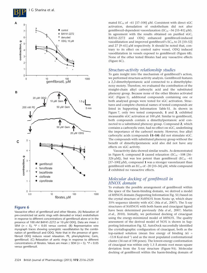
mated EC50 of ∼61 [37–100] μM. Consistent with direct sGCactivation, denudation of endothelium did not altergemfibrozil-dependent vasorelaxation (EC50 ∼56 [33–81] μM).In agreement with the results obtained on purified sGC,BAY41-2272 and ODQ enhanced gemfibrozil-inducedvasodilatation and improved gemfibrozil’s EC50 to 25 [10–52]and 27 [9–61] μM respectively. It should be noted that, con-trary to its effect on control naïve vessel, ODQ inducedvasodilatation in vessels exposed to gemfibrozil (Figure 6B).None of the other tested fibrates had any vasoactive effects(Figure 6C).
Structure-activity relationship studiesTo gain insight into the mechanism of gemfibrozil’s action,we performed structure-activity analysis. Gemfibrozil featuresa 2,2-dimethylpentanoic acid connected to a dimethylphe-noxy moiety. Therefore, we evaluated the contribution of thestraight-chain alkyl carboxylic acid and the substitutedphenoxy group. Because none of the other fibrates activatedsGC (Figure 1), additional compounds containing one orboth analysed groups were tested for sGC activation. Struc-tures and complete chemical names of tested compounds arelisted in Supporting Information Table S1. As shown inFigure 7, only two tested compounds, 1 and 3, exhibitedmeasurable sGC activation at 100 μM. Similar to gemfibrozil,both compounds contain a dimethylpentanoic acid con-nected to a substituted phenoxy group. Compound 2, whichcontains a carboxylic ester, had no effect on sGC, underliningthe importance of the carboxyl moiety. However, free alkylcarboxylic acids (compounds 11–14) did not stimulate sGC.The compounds with substituted phenoxy group without thebenefit of dimethylpentanoic acid also did not have anyeffects on sGC activity.
Vasoactivity data showed similar results. As demonstratedin Figure 8, compound 3 caused relaxation (EC50 ∼108 [36–326 μM]), but was less potent than gemfibrozil (EC50 ∼61[37–100] μM), compound 1 was a stronger vasorelaxant thangemfibrozil with an EC50 of ∼20 [10–36] μM, while compound2 exhibited no vasoactive effects.
Molecular docking of gemfibrozil inHNOX domainTo evaluate the possible arrangement of gemfibrozil withinthe space of the haem-binding domain, we derived a modelof hHNOX domain (Supporting Information Fig. S1) based onthe crystal structure of NsHNOX from Nostoc sp, which share35% sequence identity with sGC (Ma et al., 2007). The X-raystructures of NsHNOX with both haem and cinaciguat ligandhave been determined previously (Ma et al., 2007; Martinet al., 2010). Initially, we performed docking of cinaciguatusing the energy-minimized model of hHNOX. The qualityassessment of the derived model of NOX is shown in Sup-porting Information Fig. S2. AutoDock successfully identifiedthe crystallographic configuration of cinaciguat, both as thetop-ranked solution (mean free energy of binding ΔG =−13.8 Kcal·mol−1) and as the most populated conformationalcluster (34 out of 100 poses). The lowest-energy conformationof cinaciguat was within only 1.3 Å atomic root mean squaredeviation from the X-ray structure (Figure 9A). Moleculardocking of gemfibrozil within the haem-binding domain of
*
0 10 100 1000
0
1
2
3
4
5
6
7
8
control
BAY41-2272
denuded
ODQ, 10 μM
co
ntr
ac
tio
n, g
*
**
*
*
*
*
A
B
200 nM PE
20 μM ODQ
10 µM Gem
50 μM Gem
50 μM Gem
20 μM ODQ
1.5 g
gemfibrozil, μM
C
5 min
0.0 10 100 1000
0
1
2
3
4
5
gemfibrozil
fenofibrate
bezafibrate
clofibrate
ciprofibrate
fibrates, μM
co
ntr
ac
tio
n, g
*
*
* * *
Figure 6Vasoactive effect of gemfibrozil and other fibrates. (A) Relaxation ofpre-constricted rat aortic rings with denuded or intact endotheliumin response to different concentrations of gemfibrozil alone or in thepresence of 100 nM BAY41-2272 or 10 μM ODQ. Data are mean ±SEM (n = 5), *P < 0.05 versus control. (B) Representative wiremyograph traces showing synergistic vasodilatation by the combi-nation of gemfibrozil and ODQ. Note that in the presence of gem-fibrozil ODQ induces vessel relaxation. PE, phenylephrine; Gem,gemfibrozil. (C) Relaxation of aortic rings in response to differentconcentrations of fibrates. Values are mean ± SEM (n = 5). *P < 0.05versus gemfibrozil.
BJP I G Sharina et al.
2324 British Journal of Pharmacology (2015) 172 2316–2329

hHNOX model revealed two major conformational clusters(see Supplemental Information for more details). The top-ranked one (61% population) was the lowest-energy clusterwith mean free energy of binding ΔG = −8.0 Kcal·mol−1,whereas the second cluster (21% population) had a mean ΔG= −7.2 Kcal·mol−1. Interestingly, the two lowest-energy posesfrom two distinct clusters are non-overlapping. Therefore,two gemfibrozil molecules can both fit inside the haem-binding site of hHNOX (Figure 9B).
Discussion and conclusions
Gemfibrozil belongs to the fibrate class of lipid-modifyingagents, which function as PPARα agonists and are used totreat dyslipidaemias. In this report, we demonstrate that gem-fibrozil also directly activates sGC, a property that is notshared by other fibrates.
Soluble GC is strongly activated by NO binding to ferroushaem located in the HNOX domain of the β sGC subunit.Allosteric sGC stimulators, such as YC-1, BAY41-2272 or
riociguat, enhance sensitivity to NO (Stasch and Hobbs,2009). Although the binding site for these stimulators is notdetermined, it is clear that they are effective only with ferroussGC haem (Martin et al., 2001). On the contrary, haem-independent activators cinaciguat or ataciguat are morepotent when sGC haem is lost or oxidized (Schmidt et al.,2009). Crystallographic studies demonstrate that cinaciguatoccupies the same space as sGC haem (Martin et al., 2010).Despite the differences in structure and mechanisms ofaction, all these sGC regulators target, or at least require, thehaem-binding domain. To date, only cobinamides seems toregulate sGC through an allosteric mechanism that does notrequire haem domain (Sharina et al., 2011a).
Mapping studies performed in this report demonstratethat intact HNOX domain is required for activation of sGC bygemfibrozil. Gemfibrozil is not an NO generator as it does nothave NO moieties. Neither is gemfibrozil a sGC stimulator.We show that gemfibrozil and BAY41-2272 additively affectsGC activity, suggesting non-competing actions. Most impor-tantly, gemfibrozil does not sensitize sGC to low concentra-tions of NO, a feature characteristic to sGC stimulators. On
0
0.2
0.4
0.6
0.8
1
1.2
1.4
gemf 1 2 3 4 5 6 7 10 11 13 14
O
CH3
CH3O
O
CH3
O
CH3
CH3HO O
F
O
CH3CH3
CH3
CH3OH O
gemfibrozil Comp. 2 Comp. 3
O
OHOH
O
CH3
CH3
Comp. 6
O
HN
N
CH3
O
O
Cl
O
O
CH3
O
HN
CH3
O
F3C
OOO
Comp. 7 Comp. 10 Comp. 11 Comp. 13 Comp. 14
O
CH3
CH3HO O
CH3
Comp. 1
CH3
CH3
O
HN
NH
CH3
CH3
O
O
OHO
Comp. 4
CH3
CH3
Comp. 5
CH3
CH3
CH3
CH3
CH3
CH3
CH3
CH3
CH3
CH3CH3
CH3
CH3
OH OH OH
Comp: --
sG
C a
cti
vit
y, mm
ol cG
MP
·min
−1·m
g−
1
Figure 7Structure-activity studies. Activity of purified sGC was determined in the presence of 100 μM gemfibrozil or tested compounds containing thephenoxy or alkyl carboxyl moieties. Inset: structures of tested compounds. Data are expressed as fold activation over basal sGC activity (control)– 0.075 μmol·min−1·mg−1. Values are mean ± SEM (n = 6). *P < 0.05 versus control. Gemf, gemfibrozil; comp, compound.
BJPGemfibrozil is an sGC activator
British Journal of Pharmacology (2015) 172 2316–2329 2325

the contrary, gemfibrozil diminishes maximal NO activationof sGC. Moreover, gemfibrozil-dependent activation isenhanced when sGC haem is oxidized or absent. This sug-gests a mechanism of action similar to haem-mimicking sGCactivators. This conclusion is directly confirmed by competi-tion studies. It should be noted that although gemfibrozilcompetes both with cinaciguat and ataciguat, it is a lesspotent sGC regulator. However, excess gemfibrozil reducesthe extent of sGC activation by cinaciguat and ataciguat andincreases their EC50, behaving like a partial agonist. Together,these observations led us to conclude that gemfibrozil is anNO- and haem-independent sGC activator.
Structure-activity studies presented in this report werebased on the concept that gemfibrozil consists of a substi-tuted phenoxy group connected to a modified pentanoicacid. Testing sGC activation by a number of commerciallyavailable compounds that contain one or both of thesegroups provided insight into gemfibrozil’s moieties crucial forsGC activation. None of the compounds containing only thephenoxy moiety activated sGC. Similarly, none of the freestraight-chain alkyl carboxylic acids had any stimulatingeffects on sGC. Only molecules containing both moieties,such as compound 1 and 3, had any effect on sGC activity.Activation by compound 1 and 3 suggests that the phenoxymoiety tolerate some minor modification without detrimen-tal effects on sGC activation. In fact, compound 1, whichappears to have the same sGC activation potency as gemfi-brozil, is a better vasorelaxing agent, probably because ofbetter membrane permeability. Furthermore, compound 2,carrying an ethyl pentanoate ester instead of pentanoic acid,does not activate sGC. This clearly indicates that the intactcarboxyl group is crucial for gemfibrozil’s action. The require-ment for an intact –COOH is shared between gemfibrozil and
cinaciguat, which have some common structural features (redportion of cinaciguat in Figure 1A). The lack of sGC activa-tion by compound 5, which has a short alkyl chain, suggeststhat the chain length in the carboxylic acid is an importantstructural parameter that influences the potency of sGC acti-vation. Additional studies will have to be performed to deter-mine the optimal chain length.
Molecular modelling suggests that gemfibrozil may occupythe space reserved for the haem group, with two possible
0.0 0.5 1.0 1.5 2.0 2.5 3.0
0
1
2
3
4
5
gemfibrozil
compound 1
compound 2
compound 3
agonist, mM
co
ntr
ac
tio
n, g
* **
*
*
*
*
Figure 8Vasoactive effects of gemfibrozil-like compounds. Relaxation of pre-contracted rat aortic rings in response to different concentration ofgemfibrozil-like compounds. Values are mean ± SEM (n = 5). *P <0.05 versus gemfibrozil.
A
B
Figure 9Molecular modelling of gemfibrozil binding site to HNOX domain.Close-up view of the human HNOX haem-binding pocket withbound cinaciguat (A) or two distinct molecules of gemfibrozil (B).The ligands are shown with orange carbon atoms and the HNOXresidues in cyan. His105 and the hydrogen-bonding interacting resi-dues Tyr2, Tyr135, Ser137 and Arg139 are highlighted. The hydro-phobic side-chain atoms of the residues that interact with cinaciguatare shown in light cyan along with their van der Waals surfacerepresentation.
BJP I G Sharina et al.
2326 British Journal of Pharmacology (2015) 172 2316–2329
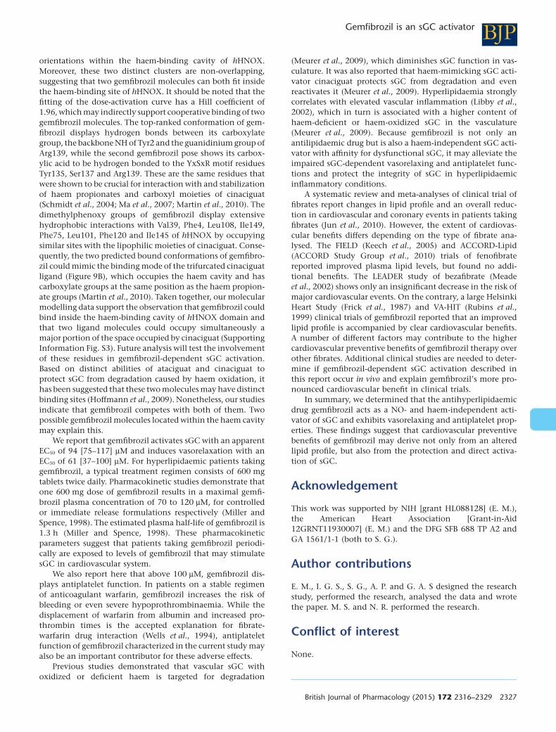
orientations within the haem-binding cavity of hHNOX.Moreover, these two distinct clusters are non-overlapping,suggesting that two gemfibrozil molecules can both fit insidethe haem-binding site of hHNOX. It should be noted that thefitting of the dose-activation curve has a Hill coefficient of1.96, which may indirectly support cooperative binding of twogemfibrozil molecules. The top-ranked conformation of gem-fibrozil displays hydrogen bonds between its carboxylategroup, the backbone NH of Tyr2 and the guanidinium group ofArg139, while the second gemfibrozil pose shows its carbox-ylic acid to be hydrogen bonded to the YxSxR motif residuesTyr135, Ser137 and Arg139. These are the same residues thatwere shown to be crucial for interaction with and stabilizationof haem propionates and carboxyl moieties of cinaciguat(Schmidt et al., 2004; Ma et al., 2007; Martin et al., 2010). Thedimethylphenoxy groups of gemfibrozil display extensivehydrophobic interactions with Val39, Phe4, Leu108, Ile149,Phe75, Leu101, Phe120 and Ile145 of hHNOX by occupyingsimilar sites with the lipophilic moieties of cinaciguat. Conse-quently, the two predicted bound conformations of gemfibro-zil could mimic the binding mode of the trifurcated cinaciguatligand (Figure 9B), which occupies the haem cavity and hascarboxylate groups at the same position as the haem propion-ate groups (Martin et al., 2010). Taken together, our molecularmodelling data support the observation that gemfibrozil couldbind inside the haem-binding cavity of hHNOX domain andthat two ligand molecules could occupy simultaneously amajor portion of the space occupied by cinaciguat (SupportingInformation Fig. S3). Future analysis will test the involvementof these residues in gemfibrozil-dependent sGC activation.Based on distinct abilities of ataciguat and cinaciguat toprotect sGC from degradation caused by haem oxidation, ithas been suggested that these two molecules may have distinctbinding sites (Hoffmann et al., 2009). Nonetheless, our studiesindicate that gemfibrozil competes with both of them. Twopossible gemfibrozil molecules located within the haem cavitymay explain this.
We report that gemfibrozil activates sGC with an apparentEC50 of 94 [75–117] μM and induces vasorelaxation with anEC50 of 61 [37–100] μM. For hyperlipidaemic patients takinggemfibrozil, a typical treatment regimen consists of 600 mgtablets twice daily. Pharmacokinetic studies demonstrate thatone 600 mg dose of gemfibrozil results in a maximal gemfi-brozil plasma concentration of 70 to 120 μM, for controlledor immediate release formulations respectively (Miller andSpence, 1998). The estimated plasma half-life of gemfibrozil is1.3 h (Miller and Spence, 1998). These pharmacokineticparameters suggest that patients taking gemfibrozil periodi-cally are exposed to levels of gemfibrozil that may stimulatesGC in cardiovascular system.
We also report here that above 100 μM, gemfibrozil dis-plays antiplatelet function. In patients on a stable regimenof anticoagulant warfarin, gemfibrozil increases the risk ofbleeding or even severe hypoprothrombinaemia. While thedisplacement of warfarin from albumin and increased pro-thrombin times is the accepted explanation for fibrate-warfarin drug interaction (Wells et al., 1994), antiplateletfunction of gemfibrozil characterized in the current study mayalso be an important contributor for these adverse effects.
Previous studies demonstrated that vascular sGC withoxidized or deficient haem is targeted for degradation
(Meurer et al., 2009), which diminishes sGC function in vas-culature. It was also reported that haem-mimicking sGC acti-vator cinaciguat protects sGC from degradation and evenreactivates it (Meurer et al., 2009). Hyperlipidaemia stronglycorrelates with elevated vascular inflammation (Libby et al.,2002), which in turn is associated with a higher content ofhaem-deficient or haem-oxidized sGC in the vasculature(Meurer et al., 2009). Because gemfibrozil is not only anantilipidaemic drug but is also a haem-independent sGC acti-vator with affinity for dysfunctional sGC, it may alleviate theimpaired sGC-dependent vasorelaxing and antiplatelet func-tions and protect the integrity of sGC in hyperlipidaemicinflammatory conditions.
A systematic review and meta-analyses of clinical trial offibrates report changes in lipid profile and an overall reduc-tion in cardiovascular and coronary events in patients takingfibrates (Jun et al., 2010). However, the extent of cardiovas-cular benefits differs depending on the type of fibrate ana-lysed. The FIELD (Keech et al., 2005) and ACCORD-Lipid(ACCORD Study Group et al., 2010) trials of fenofibratereported improved plasma lipid levels, but found no addi-tional benefits. The LEADER study of bezafibrate (Meadeet al., 2002) shows only an insignificant decrease in the risk ofmajor cardiovascular events. On the contrary, a large HelsinkiHeart Study (Frick et al., 1987) and VA-HIT (Rubins et al.,1999) clinical trials of gemfibrozil reported that an improvedlipid profile is accompanied by clear cardiovascular benefits.A number of different factors may contribute to the highercardiovascular preventive benefits of gemfibrozil therapy overother fibrates. Additional clinical studies are needed to deter-mine if gemfibrozil-dependent sGC activation described inthis report occur in vivo and explain gemfibrozil’s more pro-nounced cardiovascular benefit in clinical trials.
In summary, we determined that the antihyperlipidaemicdrug gemfibrozil acts as a NO- and haem-independent acti-vator of sGC and exhibits vasorelaxing and antiplatelet prop-erties. These findings suggest that cardiovascular preventivebenefits of gemfibrozil may derive not only from an alteredlipid profile, but also from the protection and direct activa-tion of sGC.
Acknowledgement
This work was supported by NIH [grant HL088128] (E. M.),the American Heart Association [Grant-in-Aid12GRNT11930007] (E. M.) and the DFG SFB 688 TP A2 andGA 1561/1-1 (both to S. G.).
Author contributions
E. M., I. G. S., S. G., A. P. and G. A. S designed the researchstudy, performed the research, analysed the data and wrotethe paper. M. S. and N. R. performed the research.
Conflict of interest
None.
BJPGemfibrozil is an sGC activator
British Journal of Pharmacology (2015) 172 2316–2329 2327

ReferencesACCORD Study Group, Ginsberg HN, Elam MB, Lovato LC, CrouseJR 3rd, Leiter LA et al. (2010). Effects of combination lipid therapyin type 2 diabetes mellitus. N Engl J Med 362: 1563–1574.
Alexander SPH, Benson HE, Faccenda E, Pawson AJ, Sharman JL,Spedding M et al. (2013a). The Concise Guide to PHARMACOLOGY2013/14: Nuclear hormone receptors. Br J Pharmacol 170:1652–1675.
Alexander SPH, Benson HE, Faccenda E, Pawson AJ, Sharman JL,Spedding M et al. (2013b). The Concise Guide to PHARMACOLOGY2013/14: Enzymes. Br J Pharmacol 170: 1797–1867.
Case DA, Cheatham TE 3rd, Darden T, Gohlke H, Luo R, Merz KMJr et al. (2005). The Amber biomolecular simulation programs.J Comput Chem 26: 1668–1688.
Chinetti-Gbaguidi G, Fruchart JC, Staels B (2005). Pleiotropic effectsof fibrates. Curr Atheroscler Rep 7: 396–401.
Chrominski M, Banach L, Karczewski M, ó Proinsias K, Sharina I,Gryko D et al. (2013). Synthesis and evaluation of bifunctional sGCregulators: optimization of a connecting linker. J Med Chem 56:7260–7277.
Ehret GB, Munroe PB, Rice KM, Bochud M, Johnson AD, ChasmanDI et al. (2011). Genetic variants in novel pathways influence bloodpressure and cardiovascular disease risk. Nature 478: 103–109.
Erdmann E, Semigran MJ, Nieminen MS, Gheorghiade M, AgrawalR, Mitrovic V et al. (2013a). Cinaciguat, a soluble guanylate cyclaseactivator, unloads the heart but also causes hypotension in acutedecompensated heart failure. Eur Heart J 34: 57–67.
Erdmann J, Stark K, Esslinger UB, Rumpf PM, Koesling D, de Wit Cet al. (2013b). Dysfunctional nitric oxide signalling increases risk ofmyocardial infarction. Nature 504 (7480): 432–436.
Fiser A, Sali A (2003). Modeller: generation and refinement ofhomology-based protein structure models. Methods Enzymol 374:461–491.
Foerster J, Harteneck C, Malkewitz J, Schultz G, Koesling D (1996).A functional heme-binding site of soluble guanylyl cyclase requiresintact N-termini of alpha 1 and beta 1 subunits. Eur J Biochem 240:380–386.
Frick MH, Elo O, Haapa K, Heinonen OP, Heinsalmi P, Helo P et al.(1987). Helsinki Heart Study: primary-prevention trial withgemfibrozil in middle-aged men with dyslipidemia. Safety oftreatment, changes in risk factors, and incidence of coronary heartdisease. N Engl J Med 317: 1237–1245.
Fruchart JC, Duriez P (2006). Mode of action of fibrates in theregulation of triglyceride and HDL-cholesterol metabolism. DrugsToday (Barc) 42: 39–64.
Gambaryan S, Kobsar A, Rukoyatkina N, Herterich S, Geiger J,Smolenski A et al. (2010). Thrombin and collagen induce a feedbackinhibitory signaling pathway in platelets involving dissociation ofthe catalytic subunit of protein kinase A from anNFkappaB-IkappaB complex. J Biol Chem 285: 18352–18363.
Herve D, Philippi A, Belbouab R, Zerah M, Chabrier S,Collardeau-Frachon S et al. (2014). Loss of alpha1beta1 solubleguanylate cyclase, the major nitric oxide receptor, leads tomoyamoya and achalasia. Am J Hum Genet 94: 385–394.
Hoffmann LS, Schmidt PM, Keim Y, Schaefer S, Schmidt HH, StaschJP (2009). Distinct molecular requirements for activation orstabilization of soluble guanylyl cyclase upon haemoxidation-induced degradation. Br J Pharmacol 157: 781–795.
Huey R, Morris GM, Olson AJ, Goodsell DS (2007). A semiempiricalfree energy force field with charge-based desolvation. J ComputChem 28: 1145–1152.
Jun M, Foote C, Lv J, Neal B, Patel A, Nicholls SJ et al. (2010).Effects of fibrates on cardiovascular outcomes: a systematic reviewand meta-analysis. Lancet 375: 1875–1884.
Keech A, Simes RJ, Barter P, Best J, Scott R, Taskinen MR et al.(2005). Effects of long-term fenofibrate therapy on cardiovascularevents in 9795 people with type 2 diabetes mellitus (the FIELDstudy): randomised controlled trial. Lancet 366: 1849–1861.
Kilkenny C, Browne W, Cuthill IC, Emerson M, Altman DG (2010).Animal research: Reporting in vivo experiments: the ARRIVEguidelines. Br J Pharmacol 160: 1577–1579.
Kloss S, Bouloumie A, Mulsch A (2000). Aging and chronichypertension decrease expression of rat aortic soluble guanylylcyclase. Hypertension 35 (1 Pt 1): 43–47.
Libby P, Ridker PM, Maseri A (2002). Inflammation andatherosclerosis. Circulation 105: 1135–1143.
Lu X, Wang L, Chen S, He L, Yang X, Shi Y et al. (2012).Genome-wide association study in Han Chinese identifies four newsusceptibility loci for coronary artery disease. Nat Genet 44:890–894.
Ma X, Sayed N, Beuve A, van den Akker F (2007). NO and COdifferentially activate soluble guanylyl cyclase via a hemepivot-bend mechanism. EMBO J 26: 578–588.
Martin E, Lee YC, Murad F (2001). YC-1 activation of humansoluble guanylyl cyclase has both heme-dependent andheme-independent components. Proc Natl Acad Sci U S A 98:12938–12942.
Martin E, Sharina I, Kots A, Murad F (2003). A constitutivelyactivated mutant of human soluble guanylyl cyclase (sGC):implication for the mechanism of sGC activation. Proc Natl AcadSci U S A 100: 9208–9213.
Martin E, Golunski E, Laing ST, Estrera A, Sharina IG (2014).Alternative splicing impairs sGC function in aortic aneurysm. Am JPhysiol Heart Circ Physiol 307 (11): H1565–H1575.
Martin F, Baskaran P, Ma X, Dunten PW, Schaefer M, Stasch JPet al. (2010). Structure of cinaciguat (BAY 58–2667) bound toNostoc H-NOX domain reveals insights into heme-mimeticactivation of the soluble guanylyl cyclase. J Biol Chem 285:22651–22657.
McGrath J, Drummond G, McLachlan E, Kilkenny C, Wainwright C(2010). Guidelines for reporting experiments involving animals: theARRIVE guidelines. Br J Pharmacol 160: 1573–1576.
Meade T, Zuhrie R, Cook C, Cooper J (2002). Bezafibrate in menwith lower extremity arterial disease: randomised controlled trial.BMJ 325: 1139.
Meurer S, Pioch S, Pabst T, Opitz N, Schmidt PM, Beckhaus T et al.(2009). Nitric oxide-independent vasodilator rescues heme-oxidizedsoluble guanylate cyclase from proteasomal degradation. Circ Res105: 33–41.
Miller DB, Spence JD (1998). Clinical pharmacokinetics of fibricacid derivatives (fibrates). Clin Pharmacokinet 34: 155–162.
Mindukshev I, Gambaryan S, Kehrer L, Schuetz C, Kobsar A,Rukoyatkina N et al. (2011). Low angle light scattering analysis: anovel quantitative method for functional characterization ofhuman and murine platelet receptors. Clin Chem Lab Med 50:1253–1262.
BJP I G Sharina et al.
2328 British Journal of Pharmacology (2015) 172 2316–2329

Murad F (2006). Shattuck Lecture. Nitric oxide and cyclic GMP incell signaling and drug development. N Engl J Med 355:2003–2011.
Pawson AJ, Sharman JL, Benson HE, Faccenda E, Alexander SP,Buneman OP et al.; NC-IUPHAR (2014). The IUPHAR/BPS Guide toPHARMACOLOGY: an expert-driven knowledgebase of drug targetsand their ligands. Nucl. Acids Res. 42 (Database Issue):D1098–D1106.
Rubins HB, Robins SJ, Collins D, Fye CL, Anderson JW, Elam MBet al. (1999). Gemfibrozil for the secondary prevention of coronaryheart disease in men with low levels of high-density lipoproteincholesterol. Veterans Affairs High-Density Lipoprotein CholesterolIntervention Trial Study Group. N Engl J Med 341: 410–418.
Ruetten H, Zabel U, Linz W, Schmidt HH (1999). Downregulationof soluble guanylyl cyclase in young and aging spontaneouslyhypertensive rats. Circ Res 85: 534–541.
Schmidt HH, Schmidt PM, Stasch JP (2009). NO- and haem-independent soluble guanylate cyclase activators. Handb ExpPharmacol 191: 309–339.
Schmidt PM, Schramm M, Schroder H, Wunder F, Stasch JP (2004).Identification of residues crucially involved in the binding of theheme moiety of soluble guanylate cyclase. J Biol Chem 279:3025–3032.
Schultz G (1974). General principles of assays for adenylate cyclaseand guanylate cyclase activity. Methods Enzymol 38: 115–125.
Sharina I, Sobolevsky M, Doursout MF, Gryko D, Martin E (2011a).Cobinamides are novel co-activators of NO receptor which targetsGC catalytic domain. J Pharmacol Exp Ther 340: 723–732.
Sharina IG, Jelen F, Bogatenkova EP, Thomas A, Martin E, Murad F(2008). Alpha1 soluble guanylyl cyclase (sGC) splice forms aspotential regulators of human sGC activity. J Biol Chem 283:15104–15113.
Sharina IG, Cote GJ, Martin E, Doursout MF, Murad F (2011b). RNAsplicing in regulation of nitric oxide receptor soluble guanylylcyclase. Nitric Oxide 25: 265–274.
Staels B, Dallongeville J, Auwerx J, Schoonjans K, Leitersdorf E,Fruchart JC (1998). Mechanism of action of fibrates on lipid andlipoprotein metabolism. Circulation 98: 2088–2093.
Stasch JP, Evgenov OV (2013). Soluble guanylate cyclase stimulatorsin pulmonary hypertension. Handb Exp Pharmacol 218: 279–313.
Stasch JP, Hobbs AJ (2009). NO-independent, haem-dependentsoluble guanylate cyclase stimulators. Handb Exp Pharmacol 191:277–308.
Vita JA (2011). Endothelial function. Circulation 124: e906–e912.
Wells PS, Holbrook AM, Crowther NR, Hirsh J (1994). Interactionsof warfarin with drugs and food. Ann Intern Med 121: 676–683.
Xu J, Zou MH (2009). Molecular insights and therapeutic targets fordiabetic endothelial dysfunction. Circulation 120: 1266–1286.
Zamani P, Greenberg BH (2013). Novel vasodilators in heart failure.Curr Heart Fail Rep 10: 1–11.
Supporting information
Additional Supporting Information may be found in theonline version of this article at the publisher’s web-site:
http://dx.doi.org/10.1111/bph.13055
Figure S1 (A) Homology model of hHNOX complex withcinaciguat (BAY58- 2667, orange sticks) showing His105 andthe YxSxR motif residues Tyr135, Ser137, Arg139 (cyansticks). (B) Superposition of the residues that comprise theheme-binding pocket of NsHNOX (orange sticks) andhHNOX (cyan sticks), highlighting only those that differ.Residue numbering is shown for NsHNOX/hHNOX respec-tively. Atom colours are blue for N, red for O and yellow forsulfur. (C) Alignment of human β1 H-NOX domain (Q02153)with the Nostoc sp H-NOX showing 34.6% sequence identity(63/182 residues highlighted in boldface letters).Figure S2 Quality assessment results for the model ofhHNOX (A) and the energy-minimized model of hHNOX–cinaciguat complex (B). The Ramachandran plots on the left-side panels and the assessment reports on the right-sidepanels were calculated using the Structural Analysis and Veri-fication Server version 3 (http://nihserver.mbi.ucla.edu/SAVES_3/). The overall quality of the energy-minimizedhHNOX model (B) is considerably higher with respect to theinitial hHNOX model (A), albeit the PROCHECK report (error#3) that identified two additional leucine residues in unfa-vorable side-chain conformations.Figure S3 Superposition of cinaciguat (cyan carbons) withthe two predicted gemfibrozil (orange carbons) poses thatcould mimic its crystallographic bound conformation. Thedesignated binding site #1 is occupied by the top-ranked poseof gemfibrozil and site #2 by the third conformational cluster(Supporting Information Table S2).Table S1 Compounds for structure-activity studies.Table S2 The top five conformational clusters from dockingof cinaciguat to the energy-minimized model of hHNOX.Table S3 Results of docking gemfibrozil to the energy-minimized model of hHNOX.
BJPGemfibrozil is an sGC activator
British Journal of Pharmacology (2015) 172 2316–2329 2329
![Effect of gemfibrozil levels of lipoprotein[a] in Type I1 ... · Lp[a] might be altered by ... of gemfibrozil on levels of lipoprotein[a] in Type I1 hyperlipo- ... ing gall bladder](https://img.pdfslide.us/doc/110x75/5c9a85b509d3f2e8608c0a51/effect-of-gemfibrozil-levels-of-lipoproteina-in-type-i1-lpa-might-be.jpg)


![The fibrate gemfibrozil is a NO- and haem independent ...lms.ndmctsgh.edu.tw/sysdata/70/5370/doc/0c48b6b292198045/...lipoprotein(a) [Lp(a)], LDL Low HDL • elevated VLDL, Lp(a); elevated](https://img.pdfslide.us/doc/110x75/6099bc5840e5043e3e6d9f91/the-fibrate-gemfibrozil-is-a-no-and-haem-independent-lms-lipoproteina.jpg)


![Effect of gemfibrozil levels of lipoprotein[a] in Type I1 subjects](https://img.pdfslide.us/doc/110x75/613d6085736caf36b75c9b75/effect-of-gemfibrozil-levels-of-lipoproteina-in-type-i1-subjects.jpg)