Embed Size (px)
Citation preview
DOCTORAL (PH.D.) THESIS
THE EFFECT OF PACAP (PITUITARY ADENYLATE
CYCLASE ACTIVATING POLYPEPTIDE) ON TOOTH
DEVELOPMENT IN ANIMAL MODEL
Balazs Sandor D.M.D
Tutors:
Andrea Tamas M.D., Ph.D., Habil., associate professor (Department of
Anatomy)
Akos Nagy D.M.D., Ph.D., Habil., associate professor (Department of
Dentistry, Oral and Maxillofacial Surgery)
Head of Doctoral Program: Valer Csernus, M.D., D.Sc., full professor
Head of Doctoral School: Laszlo Lenard M.D., D.Sc.,
emeritus professor
2017.

1
1. Introduction
1.1. PACAP (Pituitary adenylate cyclase activating polypeptide)
Pituitary adenylate cyclase activating polypeptide (PACAP) is a multifunctional
neuropeptide with widespread distribution. It was first isolated from ovine hypothalamic
extract on the basis of its ability to stimulate cAMP formation. PACAP is a member of the
vasoactive intestinal polypeptide (VIP)/secretin/growth hormone releasing
hormone/glucagon superfamily, with two known bioactive variants: PACAP-27 and
PACAP-38. PACAP has the most conserved amino acid sequence in the superfamily,
suggesting that it plays an important role in the regulation of basic physiologic functions.
Three receptors have been identified so far: PACAP-specific PAC1 receptor, and
PACAP/VIP indifferent VPAC1 and VPAC2 receptors. Alternative splicing of PAC1
receptor results in different ligand binding properties, exhibiting pleiotropic activities.
PACAP and PAC1 receptor expression in neuroepithelial cells appears at very early stage of
embryonic development. PACAP plays role in the regulation of various signaling cascades
in the neuronal cells affecting neurogenesis, neuronal protection, migration, differentiation
and the building of neuronal synaptic connections. It is most abundant in the central and
peripheral nervous system, nevertheless, the presence of PACAP and its receptors have been
shown in non-neuronal tissues, such as the respiratory, urogenital, cardiovascular system, in
the ear and in the dental pulp and periodontium.
PACAP plays a role in the regulation of various physiological functions, such as
thermoregulation, motor activity, nutrition and circadian rhythm. Besides its neurotrophic,
neuroprotective and general cytoprotective effect, anti-inflammatory and anti-apoptotic
effects are also known. The anti-inflammatory and anti-apoptotic effect could be the
background of its general cytoprotective effect in non-neuronal tissues.
The possible actions of endogenous PACAP can be studied in PACAP-deficient mice in
physiological and pathological conditions. There are no macroscopic differences between
wild-type, heterozygous and homozygous PACAP-deficient mice, but with more
sophisticated methods (immunohistochemistry, electron microscopy) and functional studies
significant alterations can be found. The lack of endogenous PACAP leads to biochemical,
behavioral, functional changes and neuronal developmental impairment. Compared to the
wild-type mice, PACAP deficient-mice show increased sensitivity against harmful stimuli,
2
such as after bilateral common carotid artery occlusion increased retinal damage could be
observed in the PACAP-deficient group.
1.2. Principles of tooth development
Teeth are derived from ectoderm and ectomesenchymal cells of the first pharyngeal arch.
Based upon its ectodermal/neural crest cell origin, we assumed that besides neuronal
development, the lack of PACAP may also have an effect on tooth development.
Tooth development in rodents is similar to that in human. The stages of tooth
development is a well conserved process in vertebrates. Four distinct stages can be
differentiated: development of the dental lamina, bud stage, cap stage and bell stage. In the
first part of our studies we examined the developing first and second molars of seven-day-
old mice. At this age these teeth are in the late bell stage (Figure 1).
Dental development is the result of a strictly regulated interaction between the oral
epithelium and the underlying ectomesenchyme. The conserved signaling pathways
regulating embryonic development are also crucial in tooth development. The initiation of
tooth development and tooth morphogenesis are regulated by the same factors, involved in
the development of other ectodermal tissues. More than 300 factors are involved in tooth
development. Recently, great progress has been made to identify various key transcription
factors and signaling molecules participating in epithelial–mesenchymal crosstalk, involving
different pleiotropic morphogens, such as bone morphogenetic proteins (BMPs), fibroblast
growth factors (FGFs), sonic-hedgehog (SHH) and Wnt proteins. Many studies have shown
interactions between PACAP and factors of tooth development. SHH is one of the major key
factors regulating ameloblast development and matrix secretion. Moreover, it is proven in
neuronal experimental models that downstream targets of PACAP receptors have a crosstalk
with SHH signaling pathways. Binding of SHH to its receptor (PTCH1) can activate Gli1
transcription factor subsequently, inducing several gene activation regulating proliferation
and extracellular matrix production of cells. Teeth develop from the oral ectodermal- and
cranial neural crest-derived mesenchymal cells
1.3. Composition of dental hard tissues
Mature enamel consist of 96% inorganic (mainly hydroxyapatite crystal-HAP), about
1% organic substance and about 3% water. During maturation its carbonate content is
gradually decreasing, which influences the physico-chemical properties of the enamel. The
3
organic matrix consists of amelogenin (90%) and non-amelogenin proteins (enamelin,
ameloblastin). These proteins play an important role in the regulation of crystal growth.
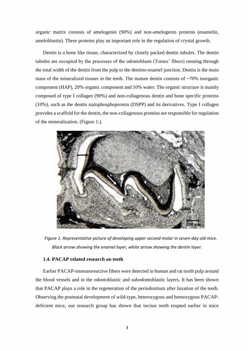
Dentin is a bone like tissue, characterized by closely packed dentin tubules. The dentin
tubules are occupied by the processes of the odontoblasts (Tomes’ fibers) running through
the total width of the dentin from the pulp to the dentino-enamel junction. Dentin is the main
mass of the mineralized tissues in the teeth. The mature dentin consists of ~70% inorganic
component (HAP), 20% organic component and 10% water. The organic structure is mainly
composed of type I collagen (90%) and non-collagenous dentin and bone specific proteins
(10%), such as the dentin sialophosphoprotein (DSPP) and its derivatives. Type I collagen
provides a scaffold for the dentin, the non-collagenous proteins are responsible for regulation
of the mineralization. (Figure 1.).
Figure 1. Representative picture of developing upper second molar in seven-day old mice.
Black arrow showing the enamel layer, white arrow showing the dentin layer.
1.4. PACAP related research on teeth
Earlier PACAP-immunoreactive fibers were detected in human and rat tooth pulp around
the blood vessels and in the odontoblastic and subodontoblastic layers. It has been shown
that PACAP plays a role in the regeneration of the periodontium after luxation of the teeth.
Observing the postnatal development of wild-type, heterozygous and homozygous PACAP-
deficient mice, our research group has shown that incisor teeth erupted earlier in mice

4
lacking PACAP. However, there is no data about the effect of endogenous PACAP on tooth
development.
5
2. Aims of our study
Our goal was to investigate the effect of PACAP deficiency in tooth development. In the
first part of our studies we compared the developing molar teeth in seven-day old
homozygous PACAP-deficient and wild-type mice. We performed morphometric, structural
comparison, using histological sections and Raman microscopy. It was supplemented by
immunohistochemical comparison to study the molecular background of tooth development.
In the second part we performed structural and morphometric analysis on the
continuously developing lower incisors of wild-type and homozygous PACAP-deficient
mice. Micro-CT was used for morphometric comparison and densitometry. Ground sections
from the lower incisors were analyzed with Raman microscopy for structural comparison.
3. Materials and methods
The examination was carried out on wild-type and homozygous PACAP-deficient mice
on CD1 strain. All procedures were performed in accordance with the ethical guidelines
approved by the University of Pecs (BA02/2000-15024/2011).
3.1. Structural and morphometric comparison of the developing molar teeth in
homozygous PACAP-deficient and wild-type mice
3.1.1. Preparation of frozen sections for morphometric analysis and Raman
microscopy
Heterozygous animals were bred and littermates were used to provide standard
circumstances for the development. Genotyping was performed with PCR reactions. For the
evaluation of the developing molars of the animals, we used wild-type (PACAP+/+, n=6
morphometric analysis, n=4 structural analysis) and homozygous PACAP-deficient
(PACAP−/−, n=6 morphometric analysis, n=4 structural analysis) mice. We made 10-μm
thick sagittal frozen sections from the skull of the animals.
3.1.2. Morphometric analysis
In the pre-eruptive developmental stage on the postnatal seventh day the first and second
molars are in the late bell stage of tooth development. The width of developing dentin and
enamel were measured on each cusp of molar teeth on native frozen sections of the skull.
Digital images were captured separately of each molar. The measurements were done on the
6
upper first, upper second, lower first and lower second molars. For statistical analysis
Student’s t-test was carried out.
3.1.3. Structural analysis with Raman microscopy on the molars of seven-day-
old mice
Raman analysis was performed on the same frozen sections as used for the morphometric
analyses. The measurements were performed in the Department of Mineralogy,
Geochemistry and Petrology, Faculty of Science and Informatics, University of Szeged.
Structural comparison of the dentin and enamel layers in wild-type and PACAP-deficient
mice was carried out using a Thermo Scientific DXR Raman Microscope. On the acquired
spectra of the enamel and the dentin HAP and protein peaks could be identified. After
spectral parameters were tested for normality two-tailed t-test was carried out. For the
evaluation of the inorganic components crystallinity index (CIRaman) was used, based on the
full width at half maximum (FWHM) values of PO43- peaks and the value of a reference
magmatic apatite (CIRaman=4.9/Γs). Higher FWHM values (lower CIRaman) refers to a higher
disordering in the HAP crystal structure. The ordering of the crystals is related to their
carbonate content, which can be evaluated as the ratio of carbonate (ν1 CO32- 1072±1 cm-1)
peaks and phosphate peaks (ν1 PO43- 960±1 cm-1). During the examination of the protein
structures, the following peaks were evaluated: amide I (ν CO, 1665±2 cm-1), amide III (δ
C-N, 1240±2 cm-1; δ N-H, 1272±6 cm-1 and δ CH2, 1346±3 cm-1), methyl (δ CH3) and
methylene (δ CH2). In practice, amide I and III bands in Raman spectroscopy can be used
for examining the secondary structures of proteins.
3.1.4. Sampling and tissue processing for immunohistochemistry
Immunohistochemistry was carried out in collaboration with the Department of
Anatomy, Histology and Embryology, Faculty of Medicine, University of Debrecen. From
the heads of seven-day-old mice (PACAP+/+, n=3, PACAP−/−, n=3) serial sections were
cut in sagittal plane at 5-μm thickness. For the investigation of the expression pattern of
SHH, PTCH1, Gli1 immunostaining was applied. Analysis of data was carried out on the
tooth germs of wild-type and homozygous PACAP-deficient mice. Photomicrographs were
taken, three researchers independently determined cell identity and localization for each
antibody used in each tooth germ studied.
7
3.2. Structural and morphometric comparison of lower incisors in adult (1-year-
old) homozygous PACAP-deficient and wild-type mice
Lack of factors involved in tooth development influence the development of the incisors
and the molars in a different manner. In this part of our study we carried out morphometric
(micro-CT) and structural (micro-CT and Raman microscopy) comparison in the incisors of
1-year-old PACAP-deficient (PACAP -/-; n=6) and wild-type mice (PACAP +/+; n=5).
3.2.1. Morphometric and densitometry measurements with micro-CT
For micro-CT scanning we prepared the mandibles of the animals according to the CT
manufacturer’s instruction. The samples were embedded and aligned in dental wax and were
scanned with the voxel size of 9 µm. For tissue mineral density (TMD) measurements,
phantom rods were used for calibration. Density is defined as the volumetric density of
calcium hydroxyapatite in terms of gram per cubic centimeter. We standardized the position
of the mandibles, and selected a volume ranging 900 μm distally from the alveolar crest (100
slices) on the lower incisor. We compared the volume of the dentin, the enamel, and the pulp
between wild-type and homozygous PACAP-deficient mice. We measured the total size of
the tooth in this 900-μm region (enamel volume+dentin volume+pulp chamber volume). To
compare the size of the pulp chamber pulp volume/dentin volume ratio was used for
correction of the data due to the size difference between the teeth. The density of the enamel,
the dentin, and the mandibular bone was also evaluated by selecting the different volumes
of interest in the analyzer software. For statistical analysis Student’s t test was used.
3.2.2. Structural analysis with Raman microscopy in the lower incisors of adult
(1-year-old) mice
Following the micro-CT analyses, standardized frontal ground sections were made from
the mandibles. Raman spectra were collected from 10-10 discrete points of the dentin and
the enamel. In the Raman spectrum of the enamel characteristic bands of hydroxyapatite
could be identified, but they were discarded due to the varied structural composition in width
and apico-incisal dimension of the layer. In the dentin HAP and protein band could be
observed. The inorganic hydroxyapatite band of the dentin was analyzed by comparing the
ratio of area under the carbonate and phosphate peaks (ν1 CO32-/ν1 PO4
3-).

8
4. Results
4.1. Comparison of the developing molar teeth in seven-day-old-mice
4.1.1. Morphometric analysis
No significant differences were found between the upper first molar teeth of wild-type
and PACAP-deficient mice. The comparison of the mesial cusps of the upper second molar
teeth revealed significantly thinner dentin (p<0.05) in PACAP-deficient mice compared to
wild-type animals. The same significant differences were found between the two groups in
the width of the dentin in the first (mesial) cusp of the lower first molar teeth (p<0.001) and
in the mesial and distal cusps of the lower second molar teeth (p<0.01).
4.1.2. Structural analysis with Raman microscopy in the molars of seven-day-
old mice
We found significant differences in the FWHM values of symmetric stretching vibration
of PO43− bands in the hydroxyapatite spectra of dentin between two groups. Majority of the
values were distributed in a broader range in the PACAP-deficient group (p<0.01).
Significant difference (p<0.05) was observed in the CIRaman, where the values of wild-type
mice were between 0.33 and 0.4, while the values of PACAP-deficient mice clustered in a
lower range between 0.25 and 0.3. We found difference in the relative carbonate content (ν1
CO32-/ν1 PO4
3-) between the two groups. Although the ratio in wild-type mice ranged
between 0.12 and 0.29, while a narrower range between 0.13 and 0.23, the data collected
from PACAP-deficient mice clustered at higher values. We did not find significant
differences in the dentin proteins.
We showed significant changes (p<0.05) in the enamel protein spectra of PACAP-
deficient mice compared to wild-type animals. The intensity ratio of amid III bands at
1240±2 and 1272±6 cm−1 of wild-type mice were distributed in a range between 0.47 and
1.3, while in PACAP-deficient group most of the values were clustered in a very short range
between 0.6 and 0.8. We did not find significant differences between other protein bands,
furthermore, no differences were observed in the hydroxyapatite spectra of the enamel.
4.1.3. Immunohistochemistry
Utilizing antigen retrieval procedure on samples, followed by immunohistochemistry,
we detected elevated SHH, PTCH1 and Gli1 expression levels in dental structures and
around the molar tooth germs in PACAP-deficient mice. SHH immunopositivity was more
9
pronounced in secretory ameloblast and stratum intermedium of PACAP-deficient mice
compared to wild-type samples. More intense PTCH1 expression was detected in enamel
secreting ameloblasts and in the odontoblastic processes in the PACAP-deficient group.
More intense intracellular Gli1 accumulation was observed in the PACAP-deficient group,
where the expression was restricted to the apical region of secretory ameloblasts.
4.2. Structural and morphometric comparison of lower incisors in adult (1-year-
old) homozygous PACAP-deficient and wild-type mice
4.2.1. Morphometric and densitometry measurements with micro-CT
The size of the incisors (pulp+dentin+enamel volume) was significantly smaller in the
PACAP-deficient mice (p<0.05). The ratio of pulp volume/dentin volume was significantly
smaller (p<0.05) in PACAP-deficient mice compared to wild-type animals. We compared
the density of the alveolar bone, the enamel, and the dentin of wild-type and PACAP-
deficient mice. We found significantly lower density in the dentin of the PACAP-deficient
mice with the average of 0.396±0.033 g/cm3 compared to wild-type mice with the average
value of 0.542±0.062 g/cm3 (p<0.05).
4.2.2. Structural analysis with Raman microscopy in the lower incisors of adult
(1-year-old) mice
Concerning the hydroxyapatite band of the dentin, we found significant (p<0.05)
difference between the ratio of the area under the carbonate and phosphate peaks (ν1 CO32−/ν1
PO43−). In wild-type mice the values are distributed between 0.1–0.27 with the average of
0.16, in the PACAP-deficient mice it ranges between 0.12–0.26, with the average value of
0.18. In the protein structure of the dentin, we found significant difference (p<0.03) in the
ratio of the area under the amide III 1240/1272 cm−1 peaks. The ratios are distributed in a
wider range in wild-type mice. In wild-type mice the values are distributed between 0.14–
1.57 with the average of 0.77. The ratio ranges between 0.28–1.24 in the PACAP-deficient
mice with the average value of 0.61 which is significantly lower compared to wild-type
animals. The results from the enamel layer were disregarded during structural examination
due to the reasons previously described.

10
5. Discussion
In our studies we found significant morphometric and structural differences between the
developing molars of seven-day-old mice and the continuously developing lower incisors of
1-year-old mice comparing wild-type and homozygous PACAP-deficient mice. In the seven-
day-old mice the thinner dentin layers in PACAP-deficient group suggest that there is a
developmental delay in this group, as it is more apparent in the second molars which are in
an earlier stage of tooth development. During dentinogenesis predentin (unmineralized
precursor of dentin) layer is formed, which is later mineralized by the deposition of apatite
crystals. An imbalance between the rate of mineralization and predentin deposition may lead
to pathological changes, such as the reduction of the dentin layer. We found significant
differences between wild-type and PACAP-deficient mice with morphometric
measurements carried out with micro-CT. The measurements revealed that the incisors were
significantly smaller and the size of the pulp chambers related to the dentin volume was also
significantly smaller in PACAP-deficient mice compared to wild-type animals. The smaller
tooth is also in accordance with results showing that PACAP-deficient mice have retarded
body growth, as shown by reduced weight gain. The narrowing of the pulp chamber is
physiologic with aging, but it may also occur as a defensive reaction to mechanical stimuli
or bacterial noxa to the dental pulp. Sometimes, it is associated with dentin developmental
disorders, as dentinogenesis imperfecta.
Structural examination with Raman microscopy revealed differences in the protein
spectra of the enamel and in the hydroxyapatite spectra of the dentin in the molars of wild-
type and PACAP-deficient seven-day-old mice. The FWHM values of phosphate bands
(ν1PO43−) in the dentin also showed significant difference between the PACAP-deficient and
wild-type mice. This band occurs at 959–960 cm−1 position in each specimen, characteristic
for biological apatite. The difference of the FWHM of ν1 PO43− bands can be explained by
the difference in short-range ordering of apatite crystals. We found significantly higher
FWHM values in PACAP-deficient mice, meaning higher disordering in hydroxyapatite
crystals. The disordering of the crystal structure is related to the size of the crystals and the
relative carbonate content of the hydroxyapatite. Crystallites of nanometer range show
higher FWHM values of ν1 PO43− band (peak broadening) while larger crystallites can result
in decreased FWHM value of ν1 PO43− band on Raman spectra. This short-range ordering
and small crystallite size can be due to substitution of PO43− by CO3
2− (B type substitution)
in the molecular structure. The ν1 CO32− vibration mode is typical in B-type carbonated

11
hydroxyapatite where CO32− can substitute PO4
3− within the phosphate lattice site. Ionic
substitutions and a minute crystallite size (i.e., nanocrystallinity) are not independent of each
other, and both impose some level of disorder. In order to determine B-type carbonate
substitution in apatite structure relative intensity ratio of ν1 PO43− and ν1 CO3
2− was
compared. The higher mean value in PACAP-deficient mice indicate slightly higher ionic
substitution of CO32− into PO4
3− site in the apatite structure which can be related to lower
crystallinity of dentin of PACAP-deficient mice. A well-crystallized apatite has a narrow
peak (lower FWHM value), while a crystal with a high carbonate content has a broader peak
(higher FWHM value). The results showed a less crystalline, more disordered bioapatite with
smaller crystallite size in the dentin layer of the molar teeth in PACAP-deficient mice.
We revealed a difference between the intensity ratios of amide III deformation bands,
which refers to a change in the secondary structures of the proteins. This difference can be
related to the ratio of random coil and ordered (α-helix; β-sheets; β-turn) conformations of
protein secondary structure. The enamel proteins of wild-type mice show a much higher
diversity in the ratio of random coil and ordered secondary structures compared with the
PACAP-deficient mice. The decreased diversity in the secondary structures of enamel
proteins in the PACAP-deficient group, might lead to alterations in the mineralization of the
enamel. Amelogenins comprise 90 % of the extracellular enamel matrix proteins, and they
play a critical role in controlling enamel mineralization. Amelogenin self-assembly is
influenced by the secondary structure of the protein and is essential for the oriented and
elongated growth of crystallites within enamel prisms and, therefore, for normal enamel
formation. Amelogenesis imperfecta, an enamel developmental disorder, is due to the
destabilization of the secondary structure in amelogenin.
With Raman microscopy in the incisors of 1-year-old mice structural analyses showed
significant difference in the hydroxyapatite and protein structure of the dentin. Similarly, to
the previously described, the higher mean value of carbonate/phosphate ratio in PACAP-
deficient mice indicates higher ionic substitution of PO43− by CO3
2− (B-type substitution) in
the apatite structure, referring to a higher disordering with smaller crystallite size. The higher
relative carbonate content of the dentin in the PACAP-deficient group correlates with our
previous findings in seven-day-old mice and may be in accordance with the results of TMD
measurement. With TMD measurements, the volumetric density of calcium hydroxyapatite
in the dentin of the PACAP-deficient mice was significantly lower. During mineral density
measurements, X-ray attenuation is assumed to be related to the calcium-hydroxyapatite
12
content of hard tissues (bone, dentin, and enamel). Although we found no data on the
relationship between carbonate/phosphate ratio and bone mineral density (BMD or TMD for
tooth), in osteoporosis, higher carbonate/phosphate ratio was also associated with lower
BMD.
Regarding the organic components of the dentin, the wider range 1240/1270 peaks in the
amide III band of wild-type mice refer to a higher structural diversity (more random coils)
in the secondary structure of the proteins. Similarly to the enamel proteins, the secondary
structures might have an effect on the mineralization of the dentin. Dentin proteins are
composed of type-I collagen (predominantly) and other proteins and proteoglycans termed
as non-collagenous proteins (NCPs). NCPs and especially SIBLINGs (small-integrin-
binding ligand, N-linked glycoproteins), which are a category of NCPs, play an important
role in the regulation of crystal growth and mineralization. Although information is lacking
about the exact mechanisms of dentin formation and mineralization, it has been shown that
there is a slight conformational change in the secondary structure of dentin matrix protein-1
(DMP1-a member of the SIBLING family) associated with binding to hydroxyapatite.
Random coil structure allows SIBLINGs to interact with minerals, collagen and cell
surfaces.
The structural differences observed in the dentin of seven-day-old and 1-year-old
PACAP-deficient mice (higher carbonate substitution, lower crystallinity, higher
disordering) can refer to a decreased resistance of the dentin, as smaller crystallite size gives
a higher specific surface available for acid attacks resulting in increased solubility. This
might also be the background for the narrowing of the pulp chamber in the incisors of the 1-
year-old mice.
The morphology, hard tissue formation and size are determined by a fine balance
between the signaling pathways involved in tooth development. The total mechanism is yet
unclear, and there are differences between the regulations of development of different tooth
types. Previously it was proven that in other tissues PACAP alters the expression of some of
the factors involved in tooth development. In most experiments, PACAP behaves as an
antagonist for bone morphogenetic protein-4 and SHH. Activation of protein kinase A
(PKA), regulated by PACAP receptors, can inhibit the transcriptional function of Gli1.
Consequently PACAP-induced signaling cascade is considered as a SHH signaling
suppressor in neuronal elements. In molar teeth of seven-day-old PACAP-deficient mice the
13
lower activation of the classical downstream signaling cascades of PAC1 receptor resulted
in an elevated SHH signaling expression. As SHH signaling pathway can regulate the
differentiation, growth and polarization of odontoblasts, ameloblasts and the dental cusp
morphology. The increased expression of the elements of SHH signaling pathway in the
PACAP-deficient mice, can be partly responsible for the altered mineralization and/or
protein secretion detected with Raman spectroscopy. Based on our present results the proper
balance of the various factors required for normal tooth development may become
inharmonic in the absence of PACAP.
Mesenchymal Wnt/β-catenin signaling inhibits anti-apoptotic effects of Fgf10 on stem
cells in the mesenchyme surrounding the cervical loop of the incisors. It has been shown that
altered expression of Fgf10 (Fgf10-deficient mice, delayed expression of mesenchymal
Fgf10) results in decreased incisor size. The ligand-independent intrinsic/basal activity of
PACAP-specific PAC1 receptor plays a key role in the activation and fine control of Wnt/β-
catenin signaling pathway through the dimerization of the PAC1 receptor. It has been
proposed that the binding of PACAP to the receptor may interrupt the dimerization of the
receptor, thus blocking the ligand-independent activity. So, we assume that in the ligand-
independent activity and consequently the Wnt/β-catenin signaling pathway may be
enhanced in PACAP-deficient mice.
Out of all the signaling pathways involved in the regulation of tooth development,
BMP’s have a great significance. The fine tuning of BMP activity is essential in the
morphoregulation of tooth development and the histogenesis and differentiation of
ameloblasts and odontoblasts BMP signaling pathways are regulated by PACAP; moreover,
the administration of PACAP to osteosarcoma cell line (UMR-106) increased expression of
BMPs and one of its major receptors BMPR1.
It is questionable what leads to the differences observed in the morphology and structure
of the teeth. It could be a direct result of PACAP deficency, or indirect result of the
physiological changes in absence of the polypeptide. Our research group has shown delayed
weight gain accompanied by the accelerated incisor eruption in PACAP-deficient mice,
compared to wild-type mice. Although this suggests the indirect background, the
immunohistochemistry in seven-day-old mice for SHH signaling pathway strongly suggests
a direct relation between tooth development and the lack of endogenous PACAP.

14
Our findings foremost shows that endogenous PACAP influences tooth development.
This is not surprising, as numerous data proves the role of PACAP during neural
development, and teeth are partially (dentin, cementum, pulp, periodontium) derived from
the neuroectoderm.
15
6. Summary
The following new results have been shown in tooth development during the comparison
of wild-type and homozygous PACAP-deficient mice:
1. With morphometric analysis the dentin development is delayed in the molar teeth of
seven-day-old mice compared to the wild-type littermates.
2. We found structural differences with Raman microscopy in the dentin and enamel of
molars in seven-day-old mice.
3. We found altered expression of the SHH signaling pathway in seven-day-old mice.
The expression of SHH/PTCH1/Gli1 was elevated in the PACAP-deficient group,
which in accordance with the literature suggest the antagonistic effect of PACAP on
the pathway.
4. With morphometric comparison of the incisors of 1-year-old mice, we found
significantly smaller incisors, relatively narrower pulp chamber, and lower density
in the dentin of the PACAP-deficient group.
5. With structural comparison of the incisors we found higher carbonate content, and
higher disordering of the crystals in the dentin of the PACAP-deficient group. There
is decreased diversity in the secondary structure of dentin proteins in the homozygous
PACAP-deficient mice.
Our further goals are to investigate the relationship of PACAP with other factors of tooth
development, to study the anti-apoptotic and anti-inflammatory effect of PACAP in the
dental pulp.
16
7. Publications
7.1. Publications related to the Ph.D. dissertation
Sandor B, Fintor K, Felszeghy S, Juhasz T, Reglodi D, Mark L, Kiss P, Jungling A, Fulop
BD, Nagy AD, Hashimoto H, Zakany R, Nagy A, Tamas A (2014) Structural and
morphometric comparison of the molar teeth in pre-eruptive developmental stage of
PACAP-deficient and wild-type mice. J Mol Neurosci. 54:331-341. (IF: 2.343)
Sandor B, Fintor K, Reglodi D, Fulop DB, Helyes Z, Szanto I, Nagy P, Hashimoto H, Tamas
A (2016) Structural and morphometric comparison of lower incisors in PACAP-deficient
and wild-type mice. J Mol Neurosci. 59:300-308. (IF: 2.229)
Reglodi D, Kiss P, Szabadfi K, Atlasz T, Gabriel R, Horvath G, Szakaly P, Sandor B, Lubics
A, Laszlo E, Farkas J, Matkovits A, Brubel R, Hashimoto H, Ferencz A, Vincze A, Helyes
Z, Welke L, Lakatos A, Tamas A. (2012) PACAP is an endogenous protective factor-insights
from PACAP-deficient mice. J Mol Neurosci. 48:482-492. (IF: 2.293)
7.2. Publications not related to the Ph.D. dissertation
Farkas J, Sandor B, Tamas A, Kiss P, Hashimoto H, Nagy AD, Fulop BD, Juhasz T,
Manavalan S, Reglodi D (2017) Early Neurobehavioral Development of Mice Lacking
Endogenous PACAP. J Mol Neurosci. 61:468-478. (IF:2.2292016)
Szanto I, Mark L, Bona A, Maasz G, Sandor B, Gelencser G, Turi Z, Gallyas F Jr (2012)
High-throughput screening of saliva for early detection of oral cancer: a pilot study. Technol
Cancer Res Treat. 11:181-188. (IF: 1.962)

17
9. Acknowledgements
I would like to express my gratitude and appreciation to my tutors, Andrea Tamas,
associate professor and Akos Nagy, associate professor, furthermore, I would like to thank
Dora Reglodi, full professor, the head of the MTA PACAP Research Group and the chair
of the Department of Anatomy, Medical School, University of Pecs, for their help and
support in my research and the preparation of my dissertation.
I am grateful to Krisztian Fintor, research fellow, Department of Mineralogy,
Geochemistry and Petrology, Faculty of Science and Informatics, University of Szeged; to
Tamas Juhasz, assistant professor and Szabolcs Felszeghy, associate professor, from the
Department of Anatomy, Histology and Embryology, Faculty of Medicine, University of
Debrecen, to Tamas Papp, resident doctor, from the Radiology Department, University of
Debrecen; to Zsuzsanna Helyes, full professor, Peter Nagy and Tamas Kiss from the
Department of Pharmacology and Pharmacotherapy, Medical School, University of Pecs,
and the Szentagothai Research Center; to Laszlo Mark, associate professor from the
Department of Biochemistry and Medical Chemistry, Medical School, University of Pecs;
to Dora Markovics, assistant professor from the Department of Dentistry, Oral and
Maxillofacial Surgery, Medical School, University of Pecs; to Eszter Hani, dentist; to my
coauthors, and to the staff of the Anatomy Department, Medical School, University of Pecs.
I would like to thank the staff of the division of Pediatric Dentistry, of Department of
Dentistry, Oral and Maxillofacial Surgery, Clinical Center, University of Pecs, for their
support and understanding.
Last but not least, I would like to thank my wife and children, Gergo and Panna for their
love, support and patience.
Support:
OTKA K104984, PD109644, NKFI K119759 TÁMOP 4.2.4.A/2-11-1-2012-0001
„NATIONAL EXCELLENCE PROGRAM OF THE MINISTRY OF HUMAN
CAPACITIES” TÁMOP 4.2.2.A-11/1/KONV-2012-0024; Arimura Foundation; Bolyai
Scholarship; MTA-PTE „Lendület Program”; Univerity of Debrecen Research Project
(RH/885/2013); GINOP-2.3.2-15-2016-00050 ‘‘PEPSYS”; UNKP-16-4-IV NATIONAL
EXCELLENCE PROGRAM OF THE MINISTRY OF HUMAN CAPACITIES; MTA TKI
14016 Program.