Embed Size (px)
Citation preview
INTERNATIONAL JOURNAL OF QUANTUM CHEMISTRY, VOL. XXXIX, 251-267 (1991)
The Discovery of the Chemical Evolution of Singlet Oxygen. Some Current Chemical, Photochemical,
and Biological Applications AHSAN ULLAH KHAN
Department of Chemistry, Harvard University, 12 Oxford Street, Cambridge, Massachusetts 02138
Abstract
From the speculations of G. N. Lewis in 1916, [J. Am. Chem. Soc. 38, 762 (1916)], the spectral predictions of R. S. Mulliken [Nature 122,505 (1928)], and the quantum mechanical treatment by E. Hiickel [Z. Phys. 50,423 (1930)], the three lowest states of molecular oxygen (%;, 'Agr and 'C;) remained spectroscopic elusives until Gerhard and Lisa Herzberg [G. Herzberg, Nature 133,759 (1934); L. Herzberg and G. Herzberg, Astrophys. J. 105,353 (1947); G. Herzberg and L. Herzberg, Astrophys. J. 108, 167 (1948)], precisely defined them by observed infared transitions. The ex- cited singlets remained of interest mainly to atmospheric spectroscopists and astrophysicists, and research on these species was scarce. The discovery in 1963 by A. U. Khan and M. Kasha [J. Chem. Phys. 39, 2105 (1963)l of the simple chemical production of singlet molecular oxygens by the aqueous reaction of hydrogen peroxide and hypochlorite released an explosion of research on the neglected species. Fifteen research symposia and research treatises confirm the broad range of chemical activities of singlet oxygen. The extension to biological systems is now proceeding, with surprising evidence of involvement of natural processes, including the action of singlet oxy- gen in white blood cell phagocytosis.
I. Introduction
What began as an exercise in the calibration of a spectrograph became one of the most active multidisciplinary research areas of recent time-the discovery of chemical generation of singlet oxygen. This article is written as a tribute to Pro- fessor Michael Kasha, a contribution to the International Journal of Quantum Chemistry that celebrates Kasha, the molecular spectroscopist. This article is written in an anecdotal and historical way to highlight the excitement and the enthusiasm that sprung from that serendipitous first spectroscopic plate back in 1963.
The discovery of the chemical generation of singlet oxygen [l] and my close scientific interaction with Professor Michael Kasha began almost at the same time, when in 1963, I joined him as a postdoctoral fellow. In his laboratory there is a three-prism Steinheil spectrograph to which he is particularly attached, it be- ing similar to the one he has used as a graduate student with G. N. Lewis when he discovered the organic triplet state [2]. On learning I had never used a photo- graphic spectrograph, Dr. Kasha immediately set me to learn how to use the Steinheil. Instead of using discharge tubes for calibrating the plates, he suggested
0 1991 John Wiley & Sons, Inc. CCC 0020-7608/91/030251-17$04.00

252 KHAN
that I calibrate against emission from the hydrogen peroxide-hypochlorite chemi- cal reaction using a hypersensitized 1N Kodak photographic plate. Dr. Kasha had earlier taken me to the dark room and, using a hypodermic syringe, had squirted hydrogen peroxide into Chlorox (NaOCl). Once our eyes were dark adapted, it was possible to see an occasional red flash from the reaction mixture. Howard Seliger, from the Johns Hopkins University, had used this faint red flash to generate and report a narrow spectrum at 6330 A, using his newly devel- oped, highly sensitive photometer [3].
I constructed a flow system and photographed the red chemiluminescence with the Steinheil. On developing the very first plate, I was amazed to find not one, but two strong bands. Thus, began the chemical era of singlet oxygen.
11. Chemical Production of Singlet Oxygen: The H202/0CI- Emission
Figure 1 is the red chemiluminescence spectrum of the simple aqueous chemi- cal reaction
H202 + OC1- 0 2 + C1- + H20
photographed with a Steinheil spectrograph at 20°C that provides the spectral evidence for the presence of electronically excited singlet oxygen in this reaction.
In Figure lc is the spectrum containing the two very strong bands, one at 6334 A (15,788 cm-'), already reported in the literature, and the newly discovered 7032 A (14,221 cm-'). The wavelength separation of 1567 cm-' between these
RED C H EM I LUM I NESCENCE SPECTRA
WAVELENGTH, A.U
Figure 1. Chemiluminescence emission from the reaction of aqueous hydrogen peroxide with sodium hypochlorite at 20°C. The spectral assignments are green chemiluminescence band-(a) ['Ag + ' X i ] (0,O): [('Ag)v=o + ( ' X ~ ) . = o ] + 2[32Ju=~, (b) 2['Ag] (1,O): [('Ag)v=l + ('Ag)v=o] + 2[3C,].=~; red chemiluminescence bands- (c) 2[lAg](0,O): [('Ag)"=o + ('Ag),=o] + 2[%,1.=0, 2['Ag](O,1): [('Ag)"=o + ('Ag)v=o] +
[ ( 2 , g ) v = o + (3S,)v=~l, (4 2['Agl ( 0 3 : [(lAg)v=o + ('Ag)o=ol + [('X,)U=o + (3Xg)v=zl; Far red chemiluminescence bund- [ 'Hi] (0,O): [ 'H~] .=O -+ [32J.=~. [Adapted from
A . U . Khan and M. Kasha, J. Am. Chem. SOC. 92,3293 (1970).]

SINGLET MOLECULAR OXYGEN 253
two emission bands correlated closely to the ground-state vibrational interval of the oxygen molecule, 1556 cm-' [4]. The chemical and isotopic studies of the H2O2/0Cl- reaction had shown that the 0-0 bond of H202 remained intact in the molecular oxygen generated in the reaction [5], and Khan and Kasha [l] identified the red chemiluminescence as arising from the excited singlet state of molecular oxygen. Since no unperturbed single oxygen molecule states corre- sponded to these two bands, two spectral assignments were considered: (1) Stauff and Schmidkunz's suggestion of a double molecule emission [6], 2['A,] -+
2[32g], and (2) a hydrated 0 2 molecule with solvent shifted '2; -+ 32; emis- sion. Soon afterward, the gas-phase electrical discharge studies by Arnold, Ogryzlo, and Witzke [7] in 1964 showed that the 6334 and 7032 A emission ob- served by Khan and Kasha were also present, very weakly, in the gas-phase spectrum. Also, Browne and Ogryzlo [S] showed that one of the weak IR bands of the chemiluminescence corresponded to the (0,O) '2; --* 32g transition that had been identified in the electric discharged gaseous oxygen by Kaplan [9]. These results made it likely that the 6334 and 7032 A bands of H202/OC1- reaction were simultaneous emission from pairs of oxygen molecules. Subse- quently, Khan and Kasha established a complete spectral correlation between the chemiluminescence bands observed for the peroxide-hypochlorite reaction in the aqueous phase at normal condition (20°C, 1 atmosphere) and the absorp- tion spectrum of gaseous oxygen at high pressure (20°C, 150 atmospheres) (see Fig. 2) [lo].
In Figure Id is shown the rotational resolved contours of the (0,O) '2; -+ 32; transition of oxygen molecule observed in the chemiluminescence reaction. The history of this spectrum is a rather interesting and an instructive story of the im- portance of a good question and the long gestation period it may take to see the obvious! The anecdote concerns Professor Robert Mulliken, Professor Kasha,
WAVELENGTH, A.U.
Figure 2. Absorption spectrum of compressed oxygen gas (150 atmospheres) at 20°C in 6.5 cm optical cell using Cary 15 Recording Spectrophotometer. The spec- tral assignments are (I) 2['8;] +- 2[38,] vibronic transitions: (O,O), (l,O), (2,0), (3,O); (11) ['Ag + 'St] +- 2[?2g] vibronic transitions: (O,O), (1,O); (111) 2['Ag] t 2[38;] vibronic transitions: (O,O), (l ,O), (2,O). [Adapted from A.U. Khan and M.
Kasha, J. Am. Chem. SOC. 92, 3293 (1970).]
254 KHAN
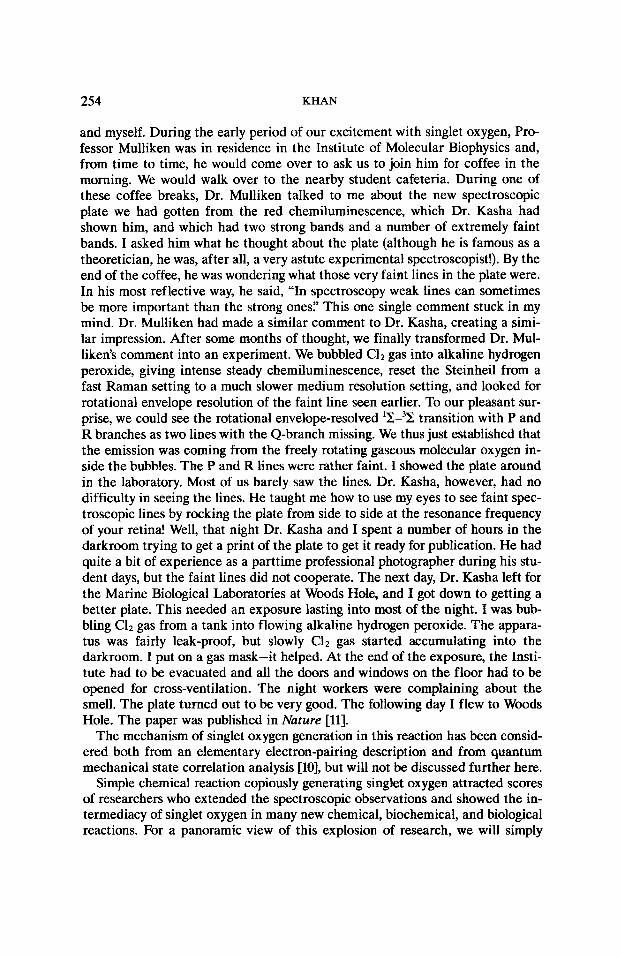
and myself. During the early period of our excitement with singlet oxygen, Pro- fessor Mulliken was in residence in the Institute of Molecular Biophysics and, from time to time, he would come over to ask us to join him for coffee in the morning. We would walk over to the nearby student cafeteria. During one of these coffee breaks, Dr. Mulliken talked to me about the new spectroscopic plate we had gotten from the red chemiluminescence, which Dr. Kasha had shown him, and which had two strong bands and a number of extremely faint bands. I asked him what he thought about the plate (although he is famous as a theoretician, he was, after all, a very astute experimental spectroscopist!). By the end of the coffee, he was wondering what those very faint lines in the plate were. In his most reflective way, he said, “In spectroscopy weak lines can sometimes be more important than the strong ones? This one single comment stuck in my mind. Dr. Mulliken had made a similar comment to Dr. Kasha, creating a simi- lar impression. After some months of thought, we finally transformed Dr. Mul- liken’s comment into an experiment. We bubbled C12 gas into alkaline hydrogen peroxide, giving intense steady chemiluminescence, reset the Steinheil from a fast Raman setting to a much slower medium resolution setting, and looked for rotational envelope resolution of the faint line seen earlier. To our pleasant sur- prise, we could see the rotational envelope-resolved ‘2-32 transition with P and R branches as two lines with the Q-branch missing. We thus just established that the emission was coming from the freely rotating gaseous molecular oxygen in- side the bubbles. The P and R lines were rather faint. I showed the plate around in the laboratory. Most of us barely saw the lines. Dr. Kasha, however, had no difficulty in seeing the lines. He taught me how to use my eyes to see faint spec- troscopic lines by rocking the plate from side to side at the resonance frequency of your retina! Well, that night Dr. Kasha and I spent a number of hours in the darkroom trying to get a print of the plate to get it ready for publication. He had quite a bit of experience as a parttime professional photographer during his stu- dent days, but the faint lines did not cooperate. The next day, Dr. Kasha left for the Marine Biological Laboratories at Woods Hole, and I got down to getting a better plate. This needed an exposure lasting into most of the night. I was bub- bling Clz gas from a tank into flowing alkaline hydrogen peroxide. The appara- tus was fairly leak-proof, but slowly C12 gas started accumulating into the darkroom. I put on a gas mask-it helped. At the end of the exposure, the Insti- tute had to be evacuated and all the doors and windows on the floor had to be opened for cross-ventilation. The night workers were complaining about the smell. The plate turned out to be very good. The following day I flew to Woods Hole. The paper was published in Nature [ll].
The mechanism of singlet oxygen generation in this reaction has been consid- ered both from an elementary electron-pairing description and from quantum mechanical state correlation analysis [lo], but will not be discussed further here.
Simple chemical reaction copiously generating singlet oxygen attracted scores of researchers who extended the spectroscopic observations and showed the in- termediacy of singlet oxygen in many new chemical, biochemical, and biological reactions. For a panoramic view of this explosion of research, we will simply

SINGLET MOLECULAR OXYGEN 255
0. R0ob.Z. Biol., 39, 524 (1900)
0 + DYE +02+LIGHT+OEAOCELL LlVlNQ CELL
A. Jodlbouar and H . v o n Toppeiner Oeut Arch. Kiln Med., 82,520(1905)
refer to the published conference proceedings and monographs [12]. In this ar- ticle, we will examine only the most critical results that provide the physical underpinning to singlet oxygen research.
M. Fritrsche Compt. Rend., 64, 1035 (1867)
111. Electronic Energy Transfer Generation of Singlet Oxygen in the Quenching of Organic Triplet States: Photooxidation Reactions,
Photodynamic Effects and Phototherapeutic Applications
Photooxidation and photodynamic reactions were among the first indicators that a light-dependent activation process exists for neutral oxygen molecules. Figure 3 is a summary of these early discoveries and mechanistic proposals. The most interesting early speculation was by Kautsky and deBruijn in 1931 [13], the Kautsky mechanism, that singlet oxygen was the reactive intermediate in photo- oxidation reactions generated in the quenching of excited sensitizer molecules.
PHOTODYNAMIC EFFECT PHOTO-OX! DATION REACTIONS I 0 i- DYE + LIGHT-OEAO CELL
LlVlNQ CELL
KAUTSKY SINGLET OXYGEN MECH.
too early ondconjecturol
M + h v - M* M * t 3 O 2 + M + ' 0 2
' 0 2 + s - so2
H. Kautsky and H.deBruijn Naturwiss., E,, 1043(1931)
GAFFRON critique cost doubt on
Koutsky conjecture
M*+O, - MO; SCHENCK MOz+ S- M +SO2
MOLOXIM MECHANISM
NO. 14, 143 (1953)
B 3
reconsidered ombivalently conjectured singlet oxygen and moloxide mechanisms
Figure 3. The early discoveries and mechanistic proposals of photodynamic effects and photooxidation reactions. [Adapted from A. U. Khan, Singlet molecular oxygen spectroscopy: chemical and photosensitized, SingZet 0 2 , A. A. Frimer, Ed. (CRC
Press, Boca Raton, FL, 1985), Vol. 1, Ch. 3.

256 KHAN
Kautsky's experiments were very ingenuous, but qualitative, and did not survive the criticisms of his contemporaries. The kinetic studies of Gaffron completely discredited the Kautsky proposal [14]. The Kautsky mechanism was soon buried in the literature, replaced by the moloxide mechanism of Schenck [15].
The trouble with all such mechanisms, as rightly attested by E. J. Bowen in a subsequent evaluation in 1953 [16], was the inability to distinguish between them experimentally with the available chemical and kinetic evidence. In 1963, with the discovery of chemical singlet oxygen, Khan and Kasha pointed out that the Kautsky proposal was open to a critical reevaluation [l]. In 1964, Foote and Wexler compared products of olefins with singlet oxygen generated in the peroxide/hypochlorite reaction to the products resulting from dye-sensitized photooxidation reaction and found them identical [17]. Corey and Taylor com- pared the photooxidation products of anthracenes with the products of an- thracenes on reaction with singlet oxygen generated in a gas discharge [18]. These studies established singlet oxygen to be the common reacting intermediate of sensitized photooxidation.
In 1967 Kawaoka, Khan, and Kearns theoretically examined, using quantum mechanical methods, the critical step of the Kautsky mechanism-the genera- tion of singlet oxygen in the quenching of organic triplet state [19]. Their results clearly predicted that the collisional quenching of organic triplet states by the triplet ground state of oxygen molecules leads to the generation of singlet oxy- gen-% :/or 'A,-with a rate corresponding to the diffusional approach of the two components. The exact rate of singlet oxygen generation is determined by the energy conservation and the spin statistics of the collision complex. Figure 4 is a schematic representation of the quenching of a triplet-state sensitizer by molecular oxygen. The calculated rates for the quenching of an organic triplet state at 72 kcal of energy generating various oxygen states are
ELECTRONIC ENERGY TRANSFER ENHANCED INTERSYSTEM TO OXYGEN MOLECULE CROSSING
'So - 3z; 'SfJ 3"; SENSITIZER 0 2 SENSITIZER 0 2 MOLECULE MOLECULE
Figure 4. Schematics of energy transfer processes in the quenching of the organic triplet state by molecular oxygen. [Adapted from K. Kawaoka, A . U . Khan, and
D. R. Kearns, J. Chem. Phys. 46, 1842, (1967); Bid. 47, 1883, (1967).]

SINGLET MOLECULAR OXYGEN 257
Energy Transfer Enhanced Intersystem Crossing
Direct spectroscopic observations have confirmed the electronic energy trans- fer generation of singlet 'A, oxygen both in the gas phase [20-221 and in solution [23,24]. Electronic energy transfer generation of '2; has been observed only in the gas phase [25,26].
Electronic energy transfer generation of singlet oxygen as a basis of sensitized photooxygenation reaction is a centrally important result that provides, for ex- ample, an understanding of the phototoxic reaction of the antibiotic tetracycline [27], phototherapy of psoriasis [28], and the exciting developments in photother- apy of cancer [29] based on the photosensitized generation of singlet oxygen. These are but some of the early results in an area that is bound to become more important as the "ozone hole" lets in more ultraviolet light and as more hydrocar- bons and other pollutants are released into the environment -generating more reactive singlet oxygen molecules.
IV. Ultrasensitive Near-infrared Solution Spectroscopy
As studies of singlet oxygen became widespread, chemical and biochemical re- actions were routinely explored for the participation of singlet oxygen based on chemical products, scavenger trappings, or other secondary evidence. But these techniques were not able to discriminate between '02,O 5, and OOH; and failed to distinguish between '2: and 'Ag oxygen. As a consequence, many controver- sies arose, particularly in aqueous and biochemical reactions. It was necessary to develop an ultrasensitive near-m emission spectrophotometer that would be able to spectroscopically and unambiguously detect singlet oxygen even when dis- solved in the H 2 0 solvent.
Khan and Kasha in 1979, using a thermoelectrically cooled lead sulfide detec- tor, developed such an ultrasensitive near-m emission spectrophotometer [24].
In Figure 5A is shown the 1268 nm emission of singlet oxygen observed in an aqueous solution obtained with the new spectrometer [24]. In Figure 6 is shown the spectra of dissolved oxygen molecules showing solvent-oxygen simultaneous transitions [30].
In Figure 7 is shown the latest version of the near-rR spectrometer in which the lead sulfide detector has been replaced with an ultrasensitive Ge-detector and with improved electronic and data processing peripherals.
V. Electron Transfer from Superoxide Anion Generating Singlet Oxygen: Water-induced Dismutation and Metal Cation Acceptors
Molecular oxygen is the ultimate electron acceptor in many biological reac- tions. Since superoxide anion, which possesses an additional electron compared to the neutral oxygen molecule, had been identified in enzymatic use of atmo-

258 KHAN
WAVELENGTH (nm)
Figure 5. Dye photosensitized emission of singlet oxygen at 1268 nm in liquid so- lution at room temperature. (A) Sensitizer, methylene blue; solvent, H a ; 0 2 satu- rated. (B) Sensitizer, hematoporphyrin; solvent, CC14; 0 2 saturated. (C) Sensitizer, 3,4-benzpyrene; solvent, CC14; O2 saturated. [Adapted from A.U. Khan and M.
Kasha, Proc. Natl. Acad. Sci. U.S.A. 76, 6049 (1979).]
spheric oxygen, it appeared that the transformation of superoxide anion to singlet oxygen could provide a general mechanism for the generation of singlet oxygen in dark biological reactions. Based on 0; sensitized chemiluminescence and iso- lation of a small amount of chemical product characteristic of singlet oxygen, I proposed in 1970 that singlet oxygen was generated from the superoxide anion in an electron transfer process [31].
-1 SOLVENT CCl, r
1.40 1.50 1.60
WAVELENGTH, p n
Figure 6. Photosensitized emission of dissolved molecular oxygen at room temper- ature. (A) Solvent, CC14; sensitizer, benzophenone; 0 2 saturated. Spectrum dis- plays: (0,O) 'Aa 4 $; at 1.28 pm, (0,l) 'A, - 2, at 1.58 pm; new band: CC14. . . 0 2
simultaneous transition at 1.42 pm. (B) Solvent, CDCl3; sensitizer, perfluoroben- zophenone; Oz saturated. new band: CDC13. . . 0 2 simultaneous transition at 1.42 Fm. [Adapted from P. Chou and A.U. Khan, Chem. Phys. Lett. 103, 281
(1984).]

SINGLET MOLECULAR OXYGEN
LIQUID NITROGEN
259
I CUT-OFF FILTER
ADC 403L
Ge-DETECTOR
PA R-I I 3 PREzAMP
v
LIGHT TIGHT BOX
REACTION CHAMBER
I I I "CHOPPER
TRIGGER
PAR 5207 STEPPIN LOCK-IN I
J AMPLIFIER
T
DRIVE
DATAMATE I PLOTTER
Figure 7. High-sensitivity 1 micron region luminescence spectrometer. [Adapted from A.U. Khan, Identifying singlet oxygen in chemical, photochemical, and en- zymic reactions, in Light Activated Pesticides, J. R. Heitz and K. R. Downum, Eds. ACS Symposium Series 339 (American Chemical Society, Washington, DC, 1987),
Ch. 4.
Transformation of superoxide anion to singlet oxygen became a topic of in- tense interest and controversy. Using the near-IR spectrophotometer, we have now characterized three routes for the conversion of superoxide anion to singlet oxygen:
1. Electron transfer from 0; to organic acceptors-one likely route to oxygen- enhanced radiation damage by uv, X-ray, and high-energy particles [32].
2. Water-induced dismutation of superoxide anion [33,34]-Figure 8 is the emission of singlet oxygen in the reaction of atmospheric moisture with KO2 suspension in Freon 113 containing 18-crown-6 ether at room temperature.
3. Electron transfer from 0; to bound metal cations [35,36]-Figure 9 is the spectrum of OZ('Ag) generated in the electron transfer reaction of 0; with Pb(acetate), in acetonitrile solution in a continuous flow cell.
Figure 10 is a summary of a series of kinetic emission data obtained by mixing a K02/18-crown-6 ether solution in acetonitrile to an acetonitrile solution of the acceptor molecule.
The metal cation acceptors generating singlet oxygen are characterized by the presence of d orforbitals with a bound ligand to the central atom. For example, for the 0; + Fe"' + O2 + Fe" reactions studied, only the reaction of O2 with

260 KHAN
WAVELENGTH, nrn
Figure 8. Singlet oxygen emission at 1268 nm from KOz/18-crown-6 ether/ Freon 113 bubbling moist air at room temperature. [Adapted from E. J. Corey, M.M. Mehrotra, and A.U. Khan, Biochem. Biophys. Res. Commun. 145, 842
(1987).]
WAVELENGTH, nm
Figure 9. Singlet oxygen emission at 1268 nm from the reaction of Pb(acetate)a (1 mmol/L) in acetonitrile with KOz solubilized with 18-crown-6 ether (1 mmol/L) in a continuous flow optical cell at room temperature. [Adapted from A.U. Khan,
J. Biolumin. Chemilumin. 4, 200 (1989).]

SINGLET MOLECULAR OXYGEN 261
T
J 10 0 i I 100 0 250
TIME (sec)
Figure 10. The 1268 nm kinetic emission data at room temperature from the reac- tion of KO2 solubilized with 18-crown-6 ether (1 mmol/L) in acetonitrile with an electron acceptor (approximately 1 mmol/L) in acetonitrile; 4 ml of each was mixed and continuously stirred. The electron acceptors are (A) bis-(cyclopentadienyl) tita- nium dichloride, (B) bis-(cyclopentadienyl) ferricineum hexafluorophosphate, (C) bis-(cyclopentadienyl) zirconium dichloride, (D) benzyltriethyl ammonium molybdenum tetrabromide, (E) ceric ammonium nitrate, (F) lead tetra-acetate.
[Adapted from A. U. Khan, Biolumin. Chemilumin. 4, 200 (1989).]
(C,)2FePF6 was observed to generate singlet oxygen. When FeC13 was used as the electron acceptor, no emission was seen. The distinction between these two ex- amples are consistent with the classification of outersphere- and innersphere- activated electron transfer ideas of Marcus [37].
Superoxide anion is generated many ways: via enzymes- of particular interest is the high concentration of 0 F inside phagosome of the polyhmorphonuclear leukocytes and the macrophages; via electron transfer to ground-state molecular oxygen, including electron transfer from electronically excited organic molecules; and via capture of solvated electrons in spurs of high-energy particles through aqueous solutions [38]. Singlet oxygen is, however, a much more reactive oxidant than is 0; toward many chemical and biological structures. Generation of singlet oxygen from superoxide electron transfer is a double-edged sword, 0; being both an efficient generator and efficient quencher of singlet oxygen [39]. Singlet oxygen is a more probable cause of accumulated oxidation damage.
VI. Biological Generation of Singlet Oxygen: Enzymes-White Blood Cells, Saliva, Milk, Fungus, and Plants
After the chemical discovery of singlet oxygen, many attempts were made to implicate singlet oxygen in biological and biochemical reactions. The main tools were the monitor of ultraweak visible chemiluminescence, chemical product, and chemical quencher techniques and deuterium kinetic effects. Although these techniques are nonspecific for detecting singlet oxygen, a number of valuable suggestions emerged. Krishnamurthy and Simpson [40] were the first to suggest the enzymatic generation of singlet oxygen in the fungus Aspergillus flavus.

262 KHAN
Based on the observation of ultraweak visible luminescence, Allen et al. [41] made the important suggestion that singlet oxygen might be produced by the phagocyting polymorphonuclear leukocytes (PMNL). These suggestions resulted in an extensive search for singlet oxygen in enzymic and biological processes, but no clear evidence emerged either in biology or in biochemistry.
Using the near-Irr spectrometer, Khan, Gebauer, and Hager published the first spectrum of singlet oxygen emission from an enzymic reaction, the chloroperoxi- dase/H202/C1- system, providing the incontrovertible evidence [42]. Kanofsky has also studied enzymic generation of singlet oxygen in this system and others with a kinetic apparatus based on a Ge-detector by monitoring 1270 nm emission using interference filters [43].
Microbicidal Enzymes: Myeloperoxidase: White Blood Cells (PMNL)
Probably the most significant enzymes of PMNL involved in the physiological defense against invading microbes is myeloperoxidase (MPO). MPO was origi- nally isolated by Agner and is estimated to constitute greater than 5% of the dry weight of the human PMNL [44]. The potent antimicrobial system, MPO/HzO2/ halide, is also toxic to a wide variety of organisms: bacteria, fungi, viruses, my- coplasma, chlymadia, protozoa, and multicellular organisms such as schistos- mula of Schistosoma mansoni. The peroxidase is also toxic to certain mammalian cells, e.g., spermatozoa, erythrocytes, platelets, and tumor cells, and inactivates certain soluble mediators, such as the chemotactic factor, C5a. The peroxidase can also transform prostaglandins, thus possibly playing a regulatory role in immune function by modulating inflammatory response [45].
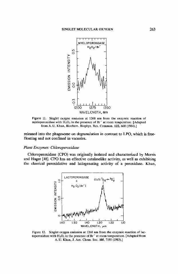
Figure 11 is the emission spectrum of singlet oxygen obtained in the MPO/ H202/Br - system. The mechanism of action of the MPO system is complex and the currently accepted mechanism is as follows:
H2Oz + C1- + H + % H 2 0 + HOCl - H + + OC1-
H 2 0 z + OC1- d Oz('A,) + HzO + C1-,
where the enzyme MPO generates the OC1- that leads to the classic singlet oxy- gen generating reaction of peroxide with hypochlorite. An estimate of the effi- ciency of singlet oxygen generation in this system is about 0.5, i.e., two H 2 0 2 molecules yield one molecule of 02('Ag) [46].
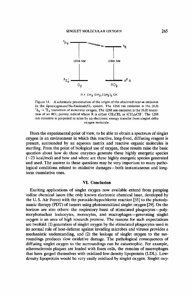
Lactoperoxidase: Milk and Saliva
Lactoperoxidase (LPO) is secreted into saliva by the human salivary glands and is also produced by the mammary glands and is found in high concentration in milk, particularly bovine milk. In Figure 12 is shown the emission spectrum of singlet oxygen obtained from the LPO/H20z/Br- system [47]. Our estimate of the efficiency of singlet oxygen generation is comparable to the efficiency of the MPO reaction, bearing out their similar antimicrobial action. MPO, how- ever, occurs inside the granules embedded in the membrane of the PMNL and is
SINGLET MOLECULAR OXYGEN 263
MYELOPEROXIDASE
2 *
?1,,,,I,,,,I 1200 1275 1350
WAVELENGTH, nm
Figure 11. Singlet oxygen emission at 1268 nm from the enzymic reaction of myeloperoxidase with HzOz in the presence of Br- at room temperature. [Adapted
from A.U. Khan, Biochem. Biophys. Res. Commun. 122,668 (1984).]
released into the phagosome on degranulation in contrast to LPO, which is free- floating and not confined in vacuoles.
Plant Enzymes: Chloroperoxidase
Chloroperoxidase (CPO) was originally isolated and characterized by Morris and Hager [48]. CPO has an effective catalaselike activity, as well as exhibiting the classical peroxidative and halogenating activity of a peroxidase. Khan,
LACTOPE ROX IDASE +
7 . I I I l 1 1 1 1 1
0 1.50 1.40 1.30 1.20 1.10 WAVELENGTH, p m
Figure 12. Singlet oxygen emission at 1268 nm from the enzymic reaction of lac- toperoxidase with HzOz in the presence of Br- at room temperature. [Adapted from
A.U. Khan, J. Am. Chem. SOC. 105, 7195 (1983).]

264 KHAN
Gebauer, and Hager examined the CPO/H202/C1- enzyme system for singlet oxygen and obtained a strong 1268 nm emission. This was the first reported spectrum of singlet oxygen generated in an enzymic system [42].
Lipooxygenase (LO)
Lipooxygenase catalyzes the oxidation of linoleic acid by molecular oxygen yielding 13-~-hydroperoxide [49]. The LO enzyme reaction is also a source of ultraweak visible chemiluminescence. Boveris et al., based on spectral data and kinetic response of the visible chemiluminescence to the quenchers and enzyme inhibitors, suggested that the emission was a product of an enzymatic side reac- tion involving free radicals [50]. They proposed a self-reaction of secondary per- oxy radicals to yield excited carbonyls and singlet oxygen through a Russell mechanism [51]. Kanofsky and Axelrod observed the 1268 nm singlet oxygen emission from soybean LO isozyme, mainly lipooxygenase-3, and also favor a Russell-like mechanism leading to singlet oxygen generation [52].
Figure 13 shows the emission we have observed from the LO/Na-linoleate/O’ system in Tris buffer, pH 9.2, at room temperature with slow oxygen bubbling. We observe the (0,O) ‘Ag + 31;g transition of 0’ at 1268 nm and another emis- sion band at 1288 nm [36,53]. We have tentatively identified the 1288 nm emis- sion band to the (0,O) transition of a peroxy radical, either CH3CH20; or (CH3)’CH0;, based on the results of Hunziker and Wendt on the absorption spectrum of peroxy radicals in the near-infrared [54].
This is the first example of an enzyme generating both singlet oxygen and a peroxy radical detected by luminescence spectroscopy. Figure 14 is a schematic proposing an energy transfer from O2(lAg) to the peroxy radical generating the excited (’E) state responsible for the 1288 nm emission. Since we can now simul- taneously monitor both the singlet oxygen emission and emission from the per- oxy radical, it may now be possible to examine the validity of the proposed Russell mechanism.
WAVELENGTH, nm
Figure 13. Near-rR chemiluminescence of singlet oxygen and peroxy radical at room temperature from reaction of lipooxygenase with sodium linoleate with slow bubbling of 0 2 at room temperature. [Adapted from A. U. Khan, Biolumin. Chemi-
lumin. 4, 200 (1989).]
SINGLET MOLECULAR OXYGEN 265
1268 NM 1288 NM
R 5 CH3 C H e r ( C H 3 ) 2 C H
Figure 14. A schematic presentation of the origin of the observed near-ra emission in the lipooxygenase/Na-linoleate/Oz system. The 1268 nm emission is the (0,O) 'Ag -+ 32, transition of molecular oxygen. The 1288 nm emission is the (0,O) transi- tion of an RO; peroxy radical where R is either CH3CH; or (CH&CH'. The 1288 nm emission is proposed to arise by an electronic energy transfer from singlet delta
oxygen molecule.
From the experimental point of view, to be able to obtain a spectrum of singlet oxygen in an environment in which this reactive, long-lived, diffusing reagent is present, surrounded by an aqueous matrix and reactive organic molecules is startling. From the point of biological use of oxygen, these results raise the basic question about how do these enzymes generate these highly energetic species (-23 kcal/mol) and how and where are these highly energetic species generated and used. The answer to these questions may be very important to many patho- logical conditions related to oxidative damages-both instantaneous and long- term cumulative ones.
VI. Conclusion
Exciting applications of singlet oxygen now available extend from pumping iodine chemical lasers (the only known electronic chemical laser, developed by the U. S. Air Force) with the peroxide-hypochlorite reaction [55] to the photody- namic therapy (PDT) of tumors using photosensitized singlet oxygen [29]. On the horizon are also others: the respiratory burst of stimulated phagocytes -poly- morphonuclear leukocytes, monocytes, and- macrophages-generating singlet oxygen is an area of high research promise. The reasons for such expectations are twofold: (1) generation of singlet oxygen by the stimulated phagocytes used in its normal role of host-defense against invading microbes and viruses provides a mechanistic understanding, and (2) the leakage of singlet oxygen to the sur- roundings produces slow oxidative damage. The pathological consequences of diffusing singlet oxygen to the surroundings can be catastrophic. For example, atherosclerosis plaques are loaded with foam cells, the remains of macrophages that have gorged themselves with oxidized low-density lipoprotein (LDL). Low- density lipoprotein would be very easily oxidized by singlet oxygen. Singlet oxy-

266 KHAN
gen can also react with the surrounding endothelial cells, causing oxidative damage to the cellular membrane resulting in lesions and stimulating further im- mune reaction. In cases of ischemic reperfusion, such as in bypass surgery, sud- den phagocytic stimulation could cause extensive vascular damage [56,57].
Once the underlying mechanism of singlet oxygen generation and toxicity are understood, a more competent medical defense can be formulated.
Acknowledgments
The author acknowledges the generous hospitality of Professor E. J. Corey and the financial support of the National Science Foundation and the Gillette Company.
Bibliography
[l] A.U. Khan and M. Kasha, J. Chem. Phys. 39, 2105 (1963). [2] G. N. Lewis and M. Kasha, J. Am. Chem. SOC. 66,2100 (1944). [3] H. H. Seliger, Anal. Biochem. 1, 60 (1960). [4] G. Herzberg, Molecular Spectra and Molecular Structure. I. Spectra of Diatomic Molecules, 2nd
[5] A. E. Cahill and H. Taube, J. Am. Chem. SOC. 74, 2312 (1952). [6] J. Stauff and H. Schmidkunz, Z. Phys. Chem. 35, 295 (1962). [7] S. J. Arnold, E. A. Ogryzlo, and H. Witzke, J. Chem. Phys. 40, 1769 (1964). [8] R. J. Browne and E.A. Ogryzlo, Proc. Chem. SOC. 117 (1964). [9] J. Kaplan, Nature 159, 673 (1947).
[lo] A.U. Khan and M. Kasha, J. Am. Chem. SOC. 92, 3293 (1970). [ll] A.U. Khan and M. Kasha, Nature 204, 241 (1964). [12] The following volumes are cited as examples of the exploding singlet oxygen research: (i)
R. F. Gould, Ed., Oxidation of Organic Compounds, Vol. 3, Ozone Chemistry, Photo and Sin- glet Oxygen and Biochemical Oxidations, Advances in Chemistry, Vol. 77 (American Chemical Society, Washington, DC, 1969). (ii) A.M. Trozzolo, Ed., International Conference on Sin- glet Molecular Oxygen and Its Role in Environmental Sciences, Ann. N.Y. Sci. 171, article 1 (1970). (iii) M. S. Chada, Ed., Singlet Molecular Oxygen (BhabI Atomic Research Centre, INSDOC Delhi, Bombay, India, 1976). (iv) A. P. Schaap, Ed., Singlet Molecular Oxygen, Benchmark Papers in Organic Chemistry (Dowden, Hutchinson and Ross, Stroudsburg, PA, 1976), Vol. 5. (v) A. Singh and A. Petkau, Guest Eds., Singlet Oxygen and Related Species in Chemistry and Biology, Photochem. Photobiol. 28 (4,5) (1978). (vi) B. Ranby and J.R. Rabek, Eds., Singlet Oxygen, Reactions with Organic Compounds and Polymers (Wiley New York, 1978). (vii) H. H. Wasserman and R.W. Murray, Eds., Singlet Oxygen (Academic Press, New York, 1979). (viii) A. A. Frimer, Ed., Singlet 0 2 , (CRC Press, Boca Raton, FL, 1985), Vols. I-IV.
ed. (Van Nostrand, New York, 1950).
[13] H. Kautsky and H. deBruijn, Naturwissenschaften 19, 1043 (1931). [14] H. Gaffron, Biochem. Z. 287, 130 (1936). [15] G. 0. Schenck, Naturwissenschaften 35, 28 (1948). [16] E. J. Bowen, Discuss. Faraday SOC. 14, 143 (1953). [17] C. S. Foote and S. Wexler, J. Am. Chem. SOC. 86, 3879 (1964). [18] E. J. Corey and W. C. Taylor, J. Am. Chem. SOC. 86, 3881 (1964). [19] K. Kawaoka, A.U. Khan, and D. R. Kearns, J. Chem. Phys. 46, 1842 (1967); Bid. 47, 1883
(1967). [20] D. R..Snelling, Chem. Phys. Lett. 2, 346 (1968).

SINGLET MOLECULAR OXYGEN 267
[21] D. R. Kearns, A.U. Khan, C. K. Duncan, and A. H. Maki, J. Am. Chem. SOC. 91, 1039
[22] E. Wasserman, V. J. Kuck, W. M. Deiavan, and W. A. Yaser, J. Am. Chem. SOC. 91, 1041
[23] A. A. Krasnovsky, Jr., Biofisika (in Russian) 21, 748 (1976). [24] A.U. Khan and M. Kasha, Proc. Natl. Acad. Sci. U. S. A. 76, 6049 (1979). [25] L. J. Andrews and E.W. Abrahmson, Chem. Phys. Lett. 10, 113 (1971). [26] C. K. Duncan and D. R. Kearns, J. Chem. Phys. 55,5822 (1971). [27] T. Hasan and A.U. Khan, Proc. Natl. Acad. Sci. U. S. A. 83, 4604 (1986). [28] T. Cech, M. A. Pathak, and R. K. Biswas, Biochim. Biophys. Acta 562, 342 (1979). [29] T. J. Dougherty, J. E. Kaufman, A. Goldfarb, K. Weishaupt, D. Boyle, and A. Mittleman,
[30] P. Chou and A.U. Khan, Chem. Phys. Lett. 103, 281 (1984). [31] A.U. Khan, Science 168, 476 (1970). [32] A.U. Khan, unpublished results. [33] A. U. Khan, J. Am. Chem. SOC. 103, 6516 (1981). [34] E. J. Corey, M. M. Mehrotra, and A.U. Khan, Biochem. Biophys. Res. Commun. 145, 842
[35] E. J. Corey, M. M. Mehrotra, and A.U. Khan, Science 236, 68 (1987). [36] A.U. Khan, J. Biolum. Chemilum. 4, 200 (1989). [37] R. A. Marcus, Annu. Rev. Phys. Chem. 13, 155 (1964). [38] See I. Fridovich, in Oxygen and Living Processes, D. L. Gilbert, Ed. (Springer-Verlag, New
[39] A.U. Khan, J. Am. Chem. SOC. 99, 370 (1977). [40] H. G. Krishnamurthy and F. J. Simpson, J. Biol. Chem. 245, 1467 (1970). [41] R. C. Allen, R. L. Stjernholm, and R. H. Steele, Biochem. Biophys. Res. Commun. 46, 679
[42] A. U. Khan, P. Gebauer, and L. P. Hager, Proc. Natl. Acad. Sci. U. S. A. 80, 5195 (1983). [43] J. R. Kanofsky, J. Biol. Chem. 259, 5996 (1984). [44] K. Agner, in Structure and Function of Oxidation-Reduction Enzymes, A. A. Keson and A.
[45] See S. J. Klebanoff, in Phagocytic Cells, J. I. Gallin and A. S. Fauci, Eds. (Raven Press, New
[46] A.U. Khan, Biochem. Biophys. Res. Commun. 122,668 (1984). [47] A.U. Khan, J. Am. Chem. SOC. 105, 7195 (1983). [48] D. R. Morris and L. P. Hager, J. Biol. Chem. 241, 1763 (1966). [49] A. L. Tappel, in The Enzymes, P. D. Boyer, H. Lardy and K. Myrback, Eds. (Academic Press,
[50] A. Boveris, E. Cadenas, and B. Chance, Photobiochem. Photobiophys. 1, 175 (1980). [Sl] G. A. Russell, J. Am. Chem. SOC. 79, 3871 (1957). [52] J. R. Kanofsky and B. Axeirod, J. Biol. Chem. 261, 1099 (1986). [53] A.U. Khan, in Light Activated Pesticides, J. R. Heitz and K. R. Downum, Eds., ACS Sympo-
[54] H. E. Hunziker and H. R. Wendt, J. Chem. Phys. 64,3488 (1976). [55] D. J. Bernard, W. E. McDermott, N. R. Pchelkin, and R. R. Bousek, Appl. Phys. Lett. 34,40
[56] D. Steinberg, S. Parthasarathy, T. E. Carew, J. C. Khoo, and J. L. Witztum, N. Engl. J. Med.
[57] M. S. Brown and J. L. Goldstein, Nature 343, 508 (1990).
(1969).
(1969).
Cancer Res. 38,2628 (1978).
(1987).
York, 1981), ch. 13.
(1972).
Ehrenberg, Eds. (Pergamon, Oxford, 1972), p. 329.
York, 1982), p. 111.
New York, 1963), Vol. 8, p. 275.
sium Series 339, American Chemical Society, Washington, D.C., 1987, Chapter 4.
(1979).
320, 915 (1989).
Received April 2, 1990 Accepted for publication May 14, 1990